Embed Size (px)
Citation preview

1
NEUROSURGERY FOR TEMPORAL LOBE EPILEPSY:
PSYCHIATRIC OUTCOME AND RELATIONSHIP TO
COGNITIVE FUNCTION
Rebecca Anne Pope, BSc(Hons)
Department of Clinical and Experimental Epilepsy
Institute of Neurology
University College London (UCL)
United Kingdom
Thesis submitted to University College London
for the Degree of Doctor of Philosophy, 2014

2
ABSTRACT
Temporal lobe epilepsy (TLE) is a chronic neurological disorder characterised by recurrent
seizures arising from temporal lobe structures. Medical treatment is effective for the majority
but for the remainder, seizure control remains difficult to achieve. Epilepsy surgery,
however, has proved an effective treatment. Following TLE surgery psychiatric symptoms
can develop for the first time (de novo), and pre-existing symptoms may worsen; having a
detrimental impact on patients’ quality of life. Yet, research data on psychiatric
complications following TLE surgery is limited, in sharp contrast to the continuing emphasis
on neuropsychological and neurological sequelae. The central aims of this thesis were to
increase our understanding of the psychiatric status of patients with intractable TLE pre- and
postoperatively, and to identify risk factors associated with poorer postoperative outcomes.
This thesis is divided into 2 main sections. Section 1 (Chapters 1-5) provide a literature
review that demonstrates pre- and postoperative psychopathology in TLE is common,
unrecognised, and under-treated. Emerging evidence suggests that pre-surgical psychiatric
morbidity is associated with more widespread cerebral pathology, but striking, is the lack of
attention to its relationship to cognitive variables. The central hypothesis formulated and
explored here is that TLE patients with less localised cerebral dysfunction, as supported by
electrophysiological, neuro-radiological and cognitive indicators will be at risk for
psychiatric disturbance preoperatively and have poorer outcomes following TLE surgery.
Section 2 consists of 5 interlinked studies incorporating retrospective and prospective
methodologies. In Study 1 (Chapter 7), the medical records of 280 TLE surgical cases were
reviewed, and more than a third presented with significant psychiatric morbidity within 4
years following surgery. Fifty-one patients (18%) developed de novo psychopathology, half
within 6 months of surgery and for the majority, persisted for more than 6 months. A

3
preoperative history of secondary generalised tonic-clonic seizures (SGTCS) was an
independent predictor of de novo psychopathology, but cognitive variables were not. Patients
with a history of SGTCS and those with a preoperative psychiatric diagnosis were
significantly less likely to remain seizure free.
Using voxel based morphometry (VBM), Study 2 (Chapter 8) explored the preoperative
neural correlates of de novo depression in a sub-group of patients (n=43) presented in Study
1. Grey matter (GM) reductions in the orbitofrontal cortices (OFC), ipsilateral cingulate
gyrus and ipsilateral thalamus were associated with the development of de novo depression
within 4 years postoperatively.
In Study 3 (Chapter 9), a sub-group of patients from Study 1, with a diagnosis of post-ictal
psychosis (TLE+PIP), were compared to age-matched TLE patients without any psychiatric
history (TLE-only; n=60), with respect to pre-surgical clinical and cognitive variables.
TLE+PIP patients were significantly less likely to have localised ictal epileptiform activity
and more likely to have a positive family psychiatric history than TLE controls. Other
clinical and cognitive variables did not distinguish between the groups. Patients with two or
more PIP episodes had significantly increased odds of developing de novo psychopathology
within 4 years of surgery, after controlling for comorbid pre-surgical psychiatric status and a
history of SGTCS. A history of PIP was not a predictor of seizure status or cognitive
outcome.
Study 4 (Chapter 11) investigated the relationship between executive function and concurrent
depression in TLE patients undergoing surgical evaluation. Depressed mood in TLE patients
was associated with clinical, cognitive and behavioural indicators of more diffuse cerebral
dysfunction.

4
Using multilevel modelling, Study 5 (Chapter 12) provides clinically relevant data
confirming that psychiatric disturbance is a significant complication following TLE surgery,
and is predicted by the presence of pre-surgical executive dysfunction.
The final chapter provides an overall summary of the findings, their implications,
methodological limitations and directions for future research. It is argued that these studies
have provided clinically relevant data that will aid the surgical decision-making process, and
hopefully guide and improve post-surgical care and support.

5
TABLE OF CONTENTS
ABSTRACT...............................................................................................................................2
LIST OF FIGURES..................................................................................................................18
LIST OF TABLES...................................................................................................................21
LIST OF ACRONYMS………………………………………………………………………24
DECLARATION OF OWN WORK........................................................................................29
ACKNOWLEDGMENTS........................................................................................................30
FUNDING SOURCES.............................................................................................................31
PUBLICATIONS ASSOCIATED WITH THIS THESIS.......................................................32
CONFERENCE TALKS RELATED TO THIS THESIS........................................................33
CONFERENCE POSTERS RELATED TO THIS THESIS....................................................34
SECTION 1: LITERATURE REVIEW
Chapter 1. EPILEPSY: a common and severe neurological disorder......................................36
1.1 Definitions..........................................................................................................................36
1.2 Epidemiology, morbidity & mortality................................................................................36
1.3 Epileptic seizures & epileptic syndromes..........................................................................37
1.3.1 1981 Classification of epileptic seizures.........................................................................38
6
1.3.2 1989 Classification of epileptic syndromes....................................................................39
1.4 Temporal Lobe Epilepsy....................................................................................................40
1.4.1 Mesial Temporal Lobe Epilepsy (mTLE).......................................................................40
1.4.2 Lateral (neocortical) Temporal Lobe Epilepsy...............................................................42
1.5 Management.......................................................................................................................43
1.5.1 Pharmacological therapy.................................................................................................43
1.5.2 Pharmaco-resistance & epilepsy surgery........................................................................46
1.5.3 Epilepsy surgery..............................................................................................................47
1.6 Pre-surgical evaluation.......................................................................................................49
1.6.1 Clinical history................................................................................................................49
1.6.2 Electrophysiological investigations.................................................................................51
1.6.3 Brain Imaging..................................................................................................................52
1.6.4 Neuropsychological evaluation.......................................................................................55
1.6 Psychiatric assessment.......................................................................................................56
7
1.7 Post-operative outcome......................................................................................................56
1.7.1 Seizure control.................................................................................................................56
1.7.2. Medical and Neurological..............................................................................................57
1.7.3. Neuropsychological........................................................................................................57
1.8 Conclusion..........................................................................................................................58
Chapter 2. PSYCHOPATHOLOGY IN TLE..........................................................................59
2.1 Temporal relationship with seizures...................................................................................59
2.2 Common, undertreated and multifactorial..........................................................................60
2.3 Psychopathology and TLE.................................................................................................62
2.4 Psychiatric morbidity in TLE: Surgical Impact.................................................................64
2.5 Systematic literature review...............................................................................................65
2.5 Depression..........................................................................................................................77
2.6 Anxiety...............................................................................................................................80
2.7 Interictal psychosis.............................................................................................................82

8
2.8 Postictal psychosis (PIP)....................................................................................................83
2.9 Methodological limitations................................................................................................85
2.10 Statistical analysis............................................................................................................88
Chapter 3. NEUROIMAGING CORRELATES OF PSYCHOPATHOLOGY IN TLE.........93
3.1 Neuroimaging in TLE........................................................................................................93
3.2 Systematic literature review...............................................................................................95
3.2.1 Depression.......................................................................................................................96
3.2.2 Anxiety............................................................................................................................99
3.2.3 Interictal Psychosis........................................................................................................101
3.2.4 Postictal Psychosis (PIP)...............................................................................................103
3.2.5 Conclusion.....................................................................................................................103
Chapter 4. PSYCHOPATHOLOGY & RELATIONSHIP TO COGNITIVE FUNCTION..107
4.1 Memory Function in TLE................................................................................................107
4.2 Cognitive Function in TLE..............................................................................................107

9
4.3 Executive function in TLE...............................................................................................110
4.4 Relationship between Psychopathology and Cognitive Function in TLE........................114
4.5 Relationship between Preoperative Psychiatric Status and Cognitive Outcome Following
TLE Surgery...........................................................................................................................117
4.6 Conclusion........................................................................................................................119
Chapter 5. HYPOTHESES AND APPROACH.....................................................................121
5.1 Hypotheses.......................................................................................................................123
5.2 Approach..........................................................................................................................124
SECTION 2: RETROSPECTIVE AND PROSPECTIVE STUDIES
Chapter 6. RETROSPECTIVE STUDY METHODOLOGY................................................126
6.1 Sample..............................................................................................................................126
6.2 Epilepsy............................................................................................................................129
6.3 Neuropsychiatric classification........................................................................................130
6.4 De novo neuropsychiatric cases.......................................................................................131
6.5 Neuropsychological tests..................................................................................................131
6.6 Aetiology..........................................................................................................................133
6.7 MRI & video-EEG data....................................................................................................133
6.8 Statistical Analyses: Retrospective and Prospective Studies...........................................134

10
6.8.1 Kolmogorov-Smirnov test.............................................................................................134
6.8.2 Mann-Whitney U test....................................................................................................134
6.8.3 Chi-squared test (X2).....................................................................................................135
6.8.4 Logistic Regression.......................................................................................................135
6.8.5 Spearman’s Correlation Coefficient..............................................................................137
Chapter 7. Study 1. Predictors of Psychiatric and Seizure Outcome following
Temporal Lobe Epilepsy Surgery
7.1 Study Aims.......................................................................................................................138
7.2 Methods............................................................................................................................138
Neuropsychological tests........................................................................................................138
7.3 Statistical Analyses..........................................................................................................141
Logistic Regression................................................................................................................141
7.4 Results..............................................................................................................................142
Psychiatric History.................................................................................................................142
Postoperative Psychopathology …………………………….................................................142
De Novo Psychopathology.....................................................................................................143
Predictors of De Novo Psychopathology...............................................................................144

11
Cognitive Outcome................................................................................................................151
Seizure Outcome....................................................................................................................151
Predictors of Seizure Freedom...............................................................................................151
7.5 Discussion........................................................................................................................160
Chapter 8. Study 2. Neural Correlates of De novo Depression Following Left Temporal
Lobe Epilepsy Surgery: A Voxel Based Morphometry Study of Pre-surgical Structural
MRI
8.1 Study Aims.......................................................................................................................168
8.2 Methods............................................................................................................................169
Study Sample..........................................................................................................................169
Magnetic Resonance Imaging................................................................................................169
Neuropsychological Measures...............................................................................................169
Memory Decline.....................................................................................................................173
8.3 Statistical Analyses..........................................................................................................173
8.4 Results..............................................................................................................................173
De Novo Depression..............................................................................................................173
Clinical Characteristics...........................................................................................................174
VBM Findings........................................................................................................................174
Seizure Outcome....................................................................................................................174

12
Cognitive Outcome................................................................................................................174
8.5 Discussion........................................................................................................................176
Chapter 9. Study 3. Postictal Psychosis in Temporal Lobe Epilepsy: Risk Factors &
Post-surgical Outcome?
9.1 Study Aims.......................................................................................................................181
9.2 Method.............................................................................................................................182
Study Sample..........................................................................................................................182
Neuropsychiatric Classification.............................................................................................182
Neuropathology......................................................................................................................183
Video-EEG Data....................................................................................................................183
Neuropsychological Measures...............................................................................................185
Memory Decline.....................................................................................................................186
9.3 Statistical Analyses..........................................................................................................186
9.4 Results..............................................................................................................................187
Clinical Characteristics...........................................................................................................187
Neuropsychology...................................................................................................................188
De Novo Psychopathology.....................................................................................................188
TLE+PIP Group.....................................................................................................................188
Predictors of De Novo Psychopathology: Group Analysis....................................................188

13
Recurrent Episodes of PIP and De Novo Psychopathology...................................................189
Histopathology.......................................................................................................................189
Cognitive Outcome................................................................................................................189
Seizure Outcome....................................................................................................................193
9.5 Discussion........................................................................................................................193
Chapter 10. PROSPECTIVE STUDIES
10.1 Introduction....................................................................................................................197
10.2 Sample............................................................................................................................197
10.3 Inclusion Criteria............................................................................................................198
10.4 Procedure........................................................................................................................198
10.5 Pre-surgical Clinical Measures.......................................................................................198
10.5.1 Clinical History...........................................................................................................198
10.5.2 MRI Scan.....................................................................................................................200
10.5.3 Prolonged Scalp Video-EEG.......................................................................................200
10.5.4 Neuropsychological Assessment.................................................................................201
1. Intellectual functioning..............................................................................................201
2. Semantic knowledge..................................................................................................201
3. Memory......................................................................................................................201
4. Executive functions....................................................................................................202
14
10.5.5 Neuropsychiatric Assessment.....................................................................................209
10.5.6 Psychiatric Rating Scales............................................................................................210
Becks Depression Inventory...................................................................................................210
Becks Anxiety Inventory.........................................................................................................210
Chapter 11. Study 4. Temporal Lobe Epilepsy & Depressed Mood: Correlations with
Cognitive and Clinical Factors
11.1 Study Aims.....................................................................................................................212
11.2 Methods..........................................................................................................................213
Study Sample..........................................................................................................................213
Procedure................................................................................................................................213
11.3 Statistical Analyses........................................................................................................213
11.4 Results............................................................................................................................214
Lifetime and Current Psychopathology..................................................................................214
Cognitive Performance...........................................................................................................217
Interaction: Depression and Seizure Focus............................................................................218
11.5 Discussion......................................................................................................................224
Chapter 12. Study 5. Profiling and Predicting Psychiatric Outcome Following TLE
surgery
12.1 Study Aims.....................................................................................................................230

15
12.2 Methods..........................................................................................................................231
Study Sample..........................................................................................................................231
Procedure................................................................................................................................231
12.3 Statistical Analyses........................................................................................................234
Multilevel Modeling (MLM).................................................................................................234
Logistic Regression................................................................................................................246
12.4 Results............................................................................................................................246
Preoperative Psychopathology: Lifetime and Current...........................................................246
De Novo Psychopathology.....................................................................................................250
Preoperative Mood Rating Scores..........................................................................................252
Temporal Profile of Psychiatric Rating Scales.......................................................................252
Predicting Change in Mood Rating Scales Following TLE Surgery.....................................253
Multivariable Model: Predicting Change in Depressive Symptoms Following TLE
Surgery...................................................................................................................................264
Seizure Outcome: Relation to Depressive Symptoms............................................................265
Cognitive Outcome: Relation to Depressive Symptoms........................................................265
Multivariable Analyses: BAI.................................................................................................267
Multivariable Model: Predicting Change in Anxiety Symptoms Following TLE
Surgery...................................................................................................................................271

16
Seizure Outcome: Relation to Anxiety Symptoms................................................................272
12.5 Discussion......................................................................................................................272
Chapter 13. Conclusions, limitations and future work
13.1 General Discussion.........................................................................................................280
Summary of the Main Findings..............................................................................................280
Preoperative Psychopathology...............................................................................................280
De novo Psychopathology......................................................................................................281
Psychopathology and Relationship to Cognitive Function....................................................282
13.2 Methodological Limitations...........................................................................................283
13.2.1 Clinical........................................................................................................................283
13.2.2 Statistical.....................................................................................................................288
13.3 Future Research Directions............................................................................................290
Appendices………………………………………………………………………………….293
Appendix 1. ...........................................................................................................................294
17
Appendix 2. ...........................................................................................................................345
Appendix 3. ...........................................................................................................................349
Appendix 4. ...........................................................................................................................351
Appendix 5. ...........................................................................................................................352
Appendix 6. ...........................................................................................................................353
Appendix 7. ...........................................................................................................................355
Appendix 8. ...........................................................................................................................356
Appendix 9….........................................................................................................................357
REFERENCES.......................................................................................................................363
18
LIST OF FIGURES
Chapter 2:
Figure 1. Timing of psychiatric disturbances in epilepsy.
Figure 2. Literature review search results.
Figure 3. An example of a 2-level hierarchical data structure, where level 1 is a repeated
measure (Beck Depression Inventory, (BDI) score) nested within a patient at level 2.
Chapter 3:
Figure 4. A schematic summary of the widespread abnormalities reported in neuroimaging
studies of mTLE patients.
Chapter 4:
Figure 5. Mean adjusted (age, gender, education) z scores for patients with TLE compared
with healthy control subjects.
Figure 6. A schematic summary of work reviewed.
Chapter 6:
Figure 7. Case selection process.
Figure 8. Sample attrition of the TLE patients (n=280) who underwent surgery between 1997
and 2007.
Chapter 7 (Study 1):
Figure 9a & b. Globally weak memory was based on a preoperative score of at least 1.5
standard deviations below the mean for both verbal and non-verbal memory.

19
Figure 9c. Histogram of point prevalence of patients diagnosed de novo psychopathologies
following TLE surgery.
Figure 10. Clustered boxplot depicting the odds of seizure freedom (ILAE=1) at 12 and for
the 48 months after TLE surgery, as a function of preoperative clinical characteristics.
Figure 11. The modifying effect of preoperative clinical factors on a patient’s chance of being
seizure free at 12 months and remaining seizure free during 48 months after TLE surgery.
Chapter 8 (Study 2):
Figure 12. Preoperative lifetime psychiatric history of the 12 left TLE patients excluded from
the VBM analysis.
Figure 13. Bilateral OFC, ipsilateral cingulate gyrus and thalamic atrophy in left-sided
TLE+HS patients who developed de novo depression compared to patients with no pre- or
postoperative psychopathology.
Chapter 9 (Study 3):
Figure 14. The modifying effect of preoperative PIP episodes on a patient’s chance of
developing de novo psychopathology within 4 years following TLE surgery.
Chapter 10:
Figure 15. Case selection process.
Figure 16a. CANTAB SWM display for the 3-box trial.
Figure 16b. Blue token ‘found’ in the 3-box trial of CANTAB SWM task.
Figure 16c. The blue token has been placed in the column on the right hand side of the screen
for 3-box problem of CANTAB SWM task.

20
Chapter 11 (Study 4):
Figure 17. Lifetime and current psychiatric diagnoses of TLE patients.
Figure 18. Positive correlation between preoperative depressive symptoms (BDI-FS) and
patient reported dysexecutive symptoms.
Figure 19. Positive correlation between preoperative depressive symptoms (BDI-FS) and
informant reported dysexecutive symptoms.
Chapter 12 (Study 5):
Figure 20. Sample attrition of the TLE patients who underwent surgery.
Figure 21. Lifetime and current psychiatric diagnoses of patients who underwent TLE
surgery.
Figure 22. Point prevalence of diagnosed de novo psychopathologies at 6- and 12 months
following TLE surgery.
Figure 23. Preoperative mood rating scores (BDI-FS & BAI) for the TLE surgical cohort.
Figure 24. Change in mean BDI-FS and BAI ratings following TLE surgery.
Figure 25. Empirical growth plots of mean BDI-FS rating for each patient following TLE
surgery.
Figure 26. Empirical growth plots of mean BAI rating for each patient following TLE
surgery.
Figure 27. ILAE rating for TLE patients following surgery.
21
LIST OF TABLES
Chapter 1:
Table 1. Antiepileptic drugs in present use.
Table 2. Antiepileptic drug options by seizure type.
Table 3. Surgical treatments for epilepsy.
Table 4. Terminology used in the description of focal epileptic activity.
Chapter 2:
Table 5. Factors related to the risk for psychiatric disorders.
Table 6. Psychiatric outcomes for TLE surgery.
Chapter 6:
Table 7. ILAE surgical classification scheme.
Chapter 7:
Table 8. Demographic and clinical characteristics of 280 patients who underwent TLE
surgery.
Table 9. Demographic and clinical characteristics of patients with and without a lifetime
psychiatric diagnosis who underwent TLE surgery.
Table 10. Cognitive characteristics of patients with (n=81) and without (n=199) a lifetime
psychiatric diagnosis who underwent TLE surgery.
Table 11. Point prevalence of diagnosed de novo psychopathology following TLE surgery.
Table 12. Characteristics of TLE patients who developed a de novo psychiatric diagnosis
versus those who did not, within four postoperative years.
Table 13. Uni- and multivariable logistic regression analyses examining whether preoperative
factors are predictive of patients who develop de novo psychopathology versus those who do
not within 4 years following TLE surgery.

22
Table 14. ILAE seizure outcome for patients who developed de novo psychopathology versus
those who did not at 4 yearly intervals following TLE surgery.
Table 15. Characteristics of TLE patients who remained seizure free (ILAE=1) versus those
who did not after TLE surgery at 12 month follow-up.
Table 16. Uni- and multivariable logistic regression analyses examining whether preoperative
factors are predictive of seizure freedom (ILAE=1) after TLE surgery at 12 month follow-up.
Table 17. Characteristics of TLE patients who remained seizure free (ILAE=1) versus those
who did not after TLE surgery during four years follow-up.
Table 18. Uni- and multivariable logistic regression analyses examining whether preoperative
factors are predictive of seizure freedom (ILAE=1) during four years after TLE surgery.
Chapter 8:
Table 19. Clinical and demographic characteristics of patients who developed de novo
depression compared to those who did not following TLE surgery.
Chapter 9:
Table 20. Demographic and clinical characteristics of TLE+PIP and TLE-only patients who
underwent TLE surgery.
Table 21. Preoperative cognitive performance of TLE+PIP and TLE-only patients.
Table 22. New cases of de novo psychopathologies for each group at specific time-intervals
following TLE surgery.
Chapter 11:
Table 23. Demographic and clinical characteristics of TLE patients with a current diagnosis
of depression versus those with no psychiatric history.
Table 24. Preoperative cognitive performance of TLE patients with a current diagnosis of
depression versus those with no psychiatric history.

23
Table 25a. Spearman’s Rho correlations between preoperative depressive symptoms (BDI-
FS), premorbid and assessed IQ discrepancy, and patient (DEX-S) and informant (DEX-I)
reported dysexecutive symptoms.
Table 25b. Spearman’s Rho correlations between patient and informant DEX ratings, and
preoperative neuropsychological measures of executive functioning.
Table 26. Preoperative cognitive performance of TLE patients with a current diagnosis of
depression versus those with no psychiatric history, as a function of seizure focus.
Chapter 12:
Table 27. Demographic and clinical characteristics of the 49 patients who underwent TLE
surgery.
Table 28. Cognitive characteristics of the 49 patients who underwent TLE surgery.
Table 29. Univariable logarithmic random intercept only models for BDI-FS, adjusting for
the main effect of time.
Table 30. Univariable logarithmic random intercept only models for BAI, adjusting for the
main effect of time.
Table 31. Correlation matrix of quantitative variables considered for multivariable BDI-FS
analysis.
Table 32. Multivariable random intercept only model for BDI-FS, adjusting for the main
effect of time.
Table 33. Change in neuropsychological status for patients with a current pre-surgical
depressive diagnosis compared to TLE patients with no psychiatric history.
Table 34. Correlation matrix of quantitative variables considered for multivariable BAI
analysis.
Table 35. Multivariable random intercept only model for BAI, adjusting for the main effect of
time.

24
LIST OF ACRONYMS
18FDG:
18F-deoxyglucose
ACC: Anterior Cingulate Cortex
AEDs: Antiepileptic Drugs
AMIPB: Adult Memory and Information Processing Battery
ATLR: Anterior temporal lobe resection
B: Beta coefficient
B0: Intercept
B1: Gradient/slope
BADS: Behavioural Assessment of the Dysexecutive Syndrome Test Battery
BAI: Beck Anxiety Inventory
BDI-FS: BDI-Fast Screen for medical patients
BDI: Beck Depression Inventory
BDI: Beck Depression Inventory
BMIPB: BIRT Memory and Information Processing Battery
BOLD: Blood-Oxygen-Level-Dependent
CANTAB: Cambridge Automated Test Battery
CBF: Cerebral Blood Flow
CBT: Cognitive Behavioural Therapy

25
CI: Confidence Interval
CIDI: Composite International Diagnostic Interview
CNS: Central Nervous System
CSF: Cerebrospinal Fluid
DARTEL: Diffeomorphic Anatomical Registration Through Exponentiated Lie Algebra
DEX: Dysexecutive Questionnaire
DNETs: Dysembryoplastic Neuroepithelial Tumours
DSM: Diagnostic Statistical Manual
DTI: Diffusion Tensor Imaging
EEG: Electroencephalogram
FA: Fractional Anisotropy
FCD: Focal Cortical Dysplasia
FDA: Food and Drug Administration
FET: Fisher Exact Test
FLAIR: Fluid Attenuated Inversion Recovery
FLE: Frontal Lobe Epilepsy
fMRI: Functional Magnetic Resonance Imaging
GLM: generalised linear model
GM: Grey Matter

26
GMV: Grey Matter Volume
HS: Hippocampal Sclerosis
IAP: Intra-carotid Amobarbital Procedure
ICC: Interclass Correlation
ICD: International Classification of Diseases
IDD: Interictal Dysphoric Disorder
IGC: Individual Growth Curve Modeling
IGT: Iowa Gambling Task
ILAE: International League Against Epilepsy
IP: Interictal Psychosis
IQ: Intellectual Quotient
K-S test: Kolmogorov-Smirnov test
LFB/CV: Luxol Fast Blue/Cresyl Violet Stain
LOG: Logarithmic
MD: Mean Diffusivity
MDD: Major Depressive Disorder
MEG: Magnetoencephalography
MLE: Maximum Likelihood Estimation
MLM: Multilevel Modeling

27
MRI: Magnetic Resonance Imaging
mTLE: mesial Temporal Lobe Epilepsy
MTR: Magnetization Transfer Ratio
NAA: Creatine/N-acetylaspartate Metabolite Ratio
NART: Nelson Adult Reading Test
NES: Non-epileptic Seizures
NICE: National Institute for Clinical Excellence
OFC: Orbito-Frontal Cortex
OR: Odds Ratio
PET: Positron Emission Tomography
PFC: Pre-Frontal Cortex
PIP: Postictal Psychosis
PIQ: Performance Intellectual Quotient
rANOVA: repeated-measures analysis of variance
ROI: Region-of-Interest
rs: Spearman’s Correlation Coefficient
Rs2: Coefficient of Determination
SCAN: Schedules for Clinical Assessment in Neuropsychiatry
SD: Standard Deviation

28
SEEG: Intracranial Electrode Implantation
SGTCS: Secondary Generalised Tonic-Clonic Seizures
SPECT: single photon emission computerised tomography
SSRIs: Serotonin Reuptake Inhibitors
SUDEP: Sudden Unexplained Death in Epilepsy Patients
SWM: Spatial Working Memory
TBI: Traumatic Brain Injury
TLE: Temporal Lobe Epilepsy
TMT (A/B): Trail Making Test
VBM: Voxel Based Morphometry
Video-EEG: Video electroencephalogram
VIQ: Verbal Intellectual Quotient
WAIS: Wechsler Adult Intelligence Scale
WCST: Wisconsin Card Sorting Task
WM: White Matter
X2:Chi-squared

29
DECLARATION OF OWN WORK
I, Rebecca Anne Pope (née Cleary) confirm that the work presented here is my own. All of
the work is original and where information has been derived from other sources, I confirm
that this has been indicated and referenced.
This thesis presents only scientific studies where I conducted all steps of the data analysis.
The interpretations of the results are my own and were formed following discussions at
supervision meetings. Drs Zoe Fox and Khadija Rantell, of the Education Unit at the
Institute of Neurology, corroborated statistical techniques and output. I collabrated with Dr
Maria Centeno who provided the data from the imaging analysis (Voxel Based
Morphometry) described in Chapter 8. This data had been collected previously by Dr
Dominique Flügel. Dr Thom, Consultant Neuropathologist, re-analysed the surgical
specimens of TLE patients described in Chapter 9. Dr Mary-Anne Wright (Clinical Electro-
physiologist, The National Hospital for Neurology and Neurosurgery) analysed and provided
the EEG data used in the prospective chapters.
Signed:
August 2014

30
ACKNOWLEDGMENTS
Firstly, I would like to thank the patients and their families who participated in this research -
without which this thesis would not have been possible. You are an inspiration.
I am deeply grateful to Dr Jacqueline Foong for giving me the opportunity to work on this
project. I still remember sitting at Marylebone station on my first day waiting for the
Gerrards Cross train, nervously reading ‘Epilepsy: the facts’. I have learned so much over
the last four years and I feel extremely privileged to have worked at The National Hospital
for Neurology and Neurosurgery and the Epilepsy Society; with research and clinical
colleagues that endeavour to better the care of patients with epilepsy.
I am also indebted to my primary supervisor, Dr Pamela Thompson. Thank you for having
confidence in me from the beginning, and encouraging my doctoral studies. You have been a
constant source of guidance and kindness; I will truly miss our times together.
Pa, thank you for endowing me with a ‘real education’! For your fatherly (and financial)
support over the last four years, and long before...
Finally, I dedicate this work to my beloved Emma. Thank you for putting up with my
absenteeism, particularly over the last year. You have and continue to be my cornerstone.
Thank you for your unparalleled love, support, patience and fun; you have always helped me
gain perspective and believed in me, even when I did not.
“Life is not about finding yourself; life is about creating yourself”
George Bernard Shaw (1856-1950)

31
FUNDING SOURCES
During this PhD thesis, I was supported by a Postgraduate Research Bursary from Epilepsy
Action awarded in 2011. I also acknowledge the financial support of the Henry Smith
Charity (http://www.henrysmithcharity.org.uk/index.html), which funded this project (grant
code G510).
The work was undertaken at University College London Hospital and University College
London who are supported by the National Institute for Health Research University College
London Hospitals Biomedical Research Centre.
The Epilepsy Society scanner was supported by the Wolfson Trust and Epilepsy Society.

32
PUBLICATIONS ASSOCIATED WITH THIS THESIS
Published
Stretton J*, Pope RA*, Winston GP, Sidhu MK, Symms M, Duncan JS, Koepp M,
Thompson PJ, Foong J. Temporal Lobe Epilepsy & Affective Disorders: The Role of the
Subgenual Anterior Cingulate Cortex. Journal of Neurology, Neurosurgery, and Psychiatry
Pope RA*, Centeno M*, Flügel D, Symms MR, Koepp M, Thompson PJ, Foong J. Neural
Correlates of De novo Depression Following Left Temporal Lobe Epilepsy Surgery: A Voxel
Based Morphometry Study of Pre-surgical Structural MRI. Epilepsy Res 2014;108:517-525
Cleary RA, Thompson PJ, Foong J. Postictal psychosis in temporal lobe epilepsy: Risk
factors and postsurgical outcome? Epilepsy Res 2013;106:264-272
Cleary RA, Thompson PJ, Foong J, Baxendale, S. Predicting and Preventing
Psychopathology following TLE Surgery. Epilepsy & Behav 2013;26:322-334
Cleary RA, Thompson PJ, Fox Z, Foong J. Predictors of Psychiatric & Seizure Outcome
Following Temporal Lobe Epilepsy Surgery. Epilepsia 2012;53:1705-1712
33
CONFERENCE TALKS RELATED TO THIS THESIS
Cleary RA, Stretton J, Winston G, Symms M, Sidhu M, Thompson PJ, Koepp M, Duncan
JS, Foong J. (2013). Temporal Lobe Epilepsy & Affective Disorders: The Role of the
Subgenual Prefrontal Cortex. British Neuropsychiatry Association National Conference,
London, UK; Alwyn Lishman Prize.
Cleary RA, Thompson PJ, Foong J. (2012). Postictal psychosis in temporal lobe epilepsy:
Risk factors and postsurgical outcome? 10th
European Congress on Epileptology, London,
UK; invited talk.
Cleary RA, Thompson PJ, Fox Z, Foong J. (2011). Preoperative Psychiatric Diagnosis in
TLE – Should Neurologists be Concerned? International League Against Epilepsy, UK
Branch, National Conference, York, UK; Platform Presentation Prize.
Cleary RA. (2010). Psychiatric Co-morbidities in Epilepsy – Neglected For Far Too Long?
International League Against Epilepsy, UK Branch, National Conference, Brighton, UK;
Gowers Health Professional Essay Award.
Cleary RA. (2010). Exploring the Psychological Effects of Temporal Lobe Epilepsy Surgery.
National Society for Epilepsy Research Associates Meeting, Chalfont St Peter,
Buckinghamshire, UK; invited talk.

34
CONFERENCE POSTERS RELATED TO THIS THESIS
Pope RA*, Centeno M*, Flügel D, Symms MR, Koepp M, Thompson PJ, Foong J. (2013).
Neural Correlates of De novo Depression Following Left Temporal Lobe Epilepsy Surgery:
A Voxel Based Morphometry Study of Pre-surgical Structural MRI. British Neuropsychiatry
Association National Conference, London, UK.
Cleary RA, Thompson PJ, Fox Z, Foong J. (2012). Pre- & Postoperative Psychiatric
Morbidity in Temporal Lobe Epilepsy Surgery. British Neuropsychiatry Association National
Conference, London, UK.
Cleary RA, Thompson PJ, Foong J. (2012). Postictal Psychosis in Temporal Lobe Epilepsy:
Risk Factors and Surgical Outcome. International Congress of the Royal College of
Psychiatrists, Liverpool, UK.
Cleary RA, Thompson PJ, Fox Z, Foong J. (2011). Preoperative Psychiatric Diagnosis in
TLE – Should Neurologists be Concerned? International League Against Epilepsy, UK
Branch, National Conference, York, UK.
Cleary RA, Thompson P, Foong J. (2010). Retrospective review of psychiatric disorders
following temporal lobe surgery. International League Against Epilepsy, UK Branch,
National Conference, Brighton, UK.

35
SECTION 1:
LITERATURE REVIEW

36
Chapter 1. EPILEPSY: a common and severe neurological
disorder
1.1 Definitions
According to the International League Against Epilepsy (ILAE), an epileptic seizure is “a
transient occurrence of signs and/or symptoms due to abnormal excessive or synchronous
neuronal activity in the brain” (Fisher et al., 2005, p. 471). The phenotype of each seizure is
determined by the point of origin and the degree of propagation of this pathological activity
(Elger & Schmidt, 2008).
Epilepsy is not one condition, but is a diverse family of disorders, having in common an
alteration in the brain that increases the likelihood of future seizures. The ILAE defines
epilepsy as a “disorder of the brain characterised by an enduring predisposition to generate
epileptic seizures and by the neurobiologic, cognitive, psychological, and social
consequences of this condition” (Fisher et al. 2005, p. 471).
1.2 Epidemiology, morbidity & mortality
Epilepsy is one of the most common and serious neurological disorders worldwide (Elger et
al., 2008). The incidence of epilepsy in the UK is approximately 51 per 100,000 people per
year (Joint Epilepsy Council, 2011). The incidence has a J-shaped curve as a function of age,
with a higher incidence for infants and the elderly (Forsgren et al., 2005). Childhood
incidence may be a result of abnormal brain development, metabolic disorders or perinatal
insults, whereas acquired brain lesions, neurodegenerative disorders, head trauma or alcohol

37
abuse may increase the risk of epilepsy in the elderly (Duncan et al., 2006). Approximately
600,000 people in the UK have a diagnosis of epilepsy, which is equivalent to a prevalence
rate of 9.7 per 1,000 people per year (Joint Epilepsy Council, 2011).
Stigma and prejudice demarcate epilepsy from most other neurological conditions (Jacoby,
2002). Epilepsy is associated with increased psychiatric morbidity, cognitive impairment,
scholastic difficulties, unemployment, lower rates of marriage, reduced leisure opportunities
and greater social isolation and family dysfunction than those without the condition (Baker,
2002). Furthermore, epilepsy patients have an increased risk of premature death that is two
times higher than the general population, especially in the first few years after diagnosis
(Lhatoo et al., 2001). Common causes of death include seizure-related death (e.g. status
epilepticus, Sudden Unexplained Death in Epilepsy Patients, SUDEP) and accidents (e.g.
drowning or burns) (Pati & Alexopoulos, 2010). Patients with epilepsy also have a higher
risk of suicide, particularly in the first two years after onset (Hersdorffer et al., 2012).
1.3 Epileptic seizures & epileptic syndromes
Once a diagnosis of epilepsy has been established, the next step is seizure classification, and
if possible, identification of the epilepsy syndrome (Brodie & French, 2000). The
classification of epileptic seizures and syndromes is continually evolving (Olafsson et al.,
2005) and is recognised as a “work in progress” (Engel, 2001, p. 796). For the last forty
years the ILAE has been engaged in formulating an accepted system of classification
(Gastaut, 1970; Commission, 1981; Commission, 1989; Berg et al., 2010). The main reason
for this preoccupation is that a universally employed classification scheme would facilitate

38
communication among clinicians, but also establish a taxonomic foundation for consistent
clinical research (Engel, 2001).
Until recently, the ILAE standardised classification and terminology for “epileptic seizures”
of 1981 and “epilepsies and epileptic syndromes” of 1989 have been the prevailing
framework for organising and differentiating the epilepsies (Panayiotopoulos, 2011).
However, a revision of these classifications has been mandated by recent technological and
scientific advances, particularly in neuroimaging and genetics (Berg et al., 2010). Since the
new scheme was only published in 2010, and has met with dissent (see Panayiotopoulos,
2011; Luders et al., 2012, for discussion), the terminology employed in this thesis is based on
the pre-existing nomenclature of the previous ILAE classifications that is still widely used.
1.3.1 1981 Classification of epileptic seizures
The 1981 classification scheme revised the 1970 proposal (Gastaut, 1970); and was based
purposely on the observation of characteristic signs and symptoms during the seizure (ictal
semiology), together with the associated EEG changes (Engel, 2001).
A seizure is first classified as partial/focal, whereby the clinical and EEG changes indicate
activation of a system of neurons limited to part of one cerebral hemisphere, or generalised
where clinical and EEG changes indicate initial involvement of both hemispheres. The
second level of classification is based on the clinical manifestations of a seizure (seizure
semiology): focal seizures are divided into simple partial seizures (awareness preserved)
and complex partial seizures (awareness altered or lost), and further categorised as (1)
motor, (2) somatosensory, (3) special-sensory (visual, auditory, olfactory, gustatory,

39
vertiginous), (4) autonomic or (5) psychic. A partial seizure may progress to a “secondary
generalised seizure”.
Generalised seizures can be (1) absence, (2) tonic-clonic, (3) myoclonic, (4) clonic, (5) tonic
or (6) atonic in type. For seizures that cannot be classified due to inadequate or incomplete
data (e.g. neonatal seizures) a third seizure category “unclassified epileptic seizures” was
included.
1.3.2 1989 Classification of epileptic syndromes
The revised classification of epilepsies and epileptic syndromes (1989) introduced two major
classes: the first separated epilepsies with generalised seizures (generalised epilepsy) from
epilepsies with partial or focal seizures (localisation-related, partial or focal epilepsies). The
second divided epilepsies of known aetiology (“symptomatic” or secondary epilepsies) from
those with no identified cause (“idiopathic”) or whereby a focal origin is suspected but the
cause is unknown (“cryptogenic”).
In 2001, the ILAE Task Force recognised that these dichotomies were overly simplistic and
often difficult to apply (Engel, 2006). Consequently, changes in terminology (e.g. focal
seizures and syndromes replaced the terms partial seizures and localisation-related
syndromes, respectively) and the introduction of a diagnostic scheme, across five hierarchical
axes ((1) ictal semiology, (2) seizure type, (3) syndrome, (4) aetiology and (5) impairment)
describing the available knowledge of the condition (Engel, 2001).

40
Focal epilepsies are further categorised according to the affected hemisphere (i.e. right/left)
and lobe (frontal/temporal/parietal/occipital). Sub-lobar classification associated with
specific ictal semiology and EEG abnormalities can be further detailed (e.g. medial and
lateral temporal lobe epilepsy).
1.4 Temporal Lobe Epilepsy
Temporal lobe epilepsy is the most common form of focal epilepsy in adults, accounting for
60% of cases (Shorvon, 2010). A number of sub-classifications exist regarding the
neuroanatomical origin of the seizures, with the distinction between mesial temporal and
lateral temporal seizure onsets being one of the most widely used.
1.4.1 Mesial Temporal Lobe Epilepsy (mTLE)
The commonest pathology underlying mesial temporal lobe epilepsy is hippocampal
sclerosis (Kim & Spencer, 2001). This condition is often associated with a history of febrile
seizures in infancy. Other aetiologies include dysembryoplastic neuroepithelioma (DNETs)
and other benign tumours, cavernous angiomas, glioma, malformations of cortical
development, or gliosis as a result of encephalitis or meningitis.
Seizures originating in the mesial temporal lobe may be simple or complex partial in form.
Secondary generalisation may occur. A simple partial seizure has a short duration, lasting for
a matter of seconds. A complex partial seizure evolves gradually, developing over minutes.

41
More than 90% of patients with mesial temporal lobe epilepsy (mTLE) report a visceral aura,
most commonly an epigastric sensation that has a rising character (Elger et al., 2008). Other
auras maybe characterised by an abnormal sense of taste, an aversive smell, déjà vu or a
dreamy sensation. Fear is the most reported affective symptom, although other complex
emotional symptoms may also occur. Autonomic symptoms include changes in skin colour,
blood pressure, heart rate and piloerection (Shorvon, 2010).
An aura can occur in isolation or it can be the initial manifestation of a complex partial
seizure. The latter is characterised by prominent behavioural changes, often a motionless
stare. Speech usually ceases or is severely disrupted if the seizure involves the language-
dominant temporal lobe (normally the left). If the seizure onset is in the non-language
dominant hemisphere, speech may be retained throughout the seizure, but is generally marked
by meaningless repetitive vocalisations (Shorvon, 2010).
Behavioural automatisms which demarcate a seizure focus as originating in mesial temporal
lobe structures are usually oroalimentary (e.g. lip-smacking, chewing, swallowing) or
gestural (e.g. fumbling, fidgeting, repetitive motor action, undressing, walking, running or
sexually directed actions), and are often prolonged (Shorvon, 2010). Limb automatisms are
usually ipsilateral to the epileptogenic focus, with contralateral dystonic posturing.
Following a temporal lobe complex partial seizure, confusion and headache are common.
Post-ictal nose-rubbing may occur and most frequently occurs ipsilateral to the epileptogenic
zone (Geyer et al., 1999).
EEG correlates of mTLE often show anterior and mid-temporal spikes. Further changes
include intermittent or persisting slow activity over the temporal lobes, which can be
unilateral or bilateral. With advances in magnetic resonance imaging (MRI), structural

42
abnormalities are often identified in mesial temporal brain structures (Shorvon, 2010).
1.4.2 Lateral (neocortical) Temporal Lobe Epilepsy
Lateral temporal lobe epilepsy is often associated with detectable underlying structural
pathology, the most common being a glioma, angioma, cavernoma, hamartoma, DNET,
neuronal migration defect and post-traumatic change. Unlike mTLE, this condition is not
associated with febrile convulsions (Shorvon, 2010).
Unsurprisingly, there is considerable overlap between the clinical and electrophysiological
features of mesial and lateral temporal lobe epilepsy, due presumably to the rapid spread of
discharges between these two neighbouring anatomical areas. However, subtle differences
between lateral and mesial temporal lobe epilepsies are discernible. For example, during a
lateral temporal lobe seizure auras include hallucinations that are often structured with visual,
auditory, gustatory or olfactory forms, which can be crude or elaborate in nature or with
illusions of size, shape, weight, distance or sound. Compared to mTLE, affective, visceral or
psychic auras are far less frequent. Moreover, lateral temporal lobe seizures typically involve
more motor activity; automatisms are unilateral and have more prominent motor
manifestations. However, from a post-ictal perspective, mesial and lateral temporal lobe
epilepsies are difficult to distinguish.
The electrophysiological pattern between seizures (interictal pattern) shows spikes over the
temporal region, maximal over the posterior or lateral temporal rather than inferomesial
electrodes. In contrast to mTLE, hippocampal volumes and T2 measures (fluid attenuated
inversion recovery; FLAIR) on MRI are usually normal (Shorvon, 2010).

43
1.5 Management
1.5.1 Pharmacological therapy
Antiepileptic drugs (AEDs) are the mainstay of epilepsy treatment (Duncan et al., 2006).
AEDs increase inhibition, decrease excitation or prevent the aberrant burst-firing of neurons.
They are a symptomatic treatment, suppressing the seizures, and have no influence on the
long-term natural course of the disease (epileptogenesis) (Duncan et al., 2006). At present,
over 20 AEDs have been licensed worldwide, each associated with adverse side-effects
(Table 1). The primary goals of pharmacological treatment are to achieve complete seizure
freedom, ideally without adverse side effects, to reduce morbidity and mortality and to
improve quality of life (Sisodiya & Sander, 2004).
Conventionally, AEDs are divided into new or old agents, depending upon whether they were
available before or after the 1990s. Despite vigorous debate, there is no evidence that new
drugs are more effective, although they may be better tolerated than old drugs (McCorry,
Chadwick & Marson, 2004; Duncan et al., 2006). A number of agents are used as first-line
treatment and are selected on the basis of their effectiveness for the seizure type (Table 2) or
epileptic syndrome. In addition, the tolerability, safety, ease of use, pharmacokinetics and
cost of AEDs are also considered before commencing treatment (Schmidt, 2009). This
patient-tailored approach to treatment is strongly recommended in existing NICE guidelines
(NICE, 2012).

44
Antiepileptic agent Usual starting dose in adults (mg) Recommended daily maintenance dose for adults (mg) Side effects
Acetazolamide (1952) 250 500-1000 Idiosyncratic rash; rarely Stevens-Johnson
syndrome and toxic epidermal necrolysis;
aplastic anaemia
Carbamazepine (1963) 100-200 400-1800 Idiosyncratic reactions; rarely Stevens-Johnson
syndrome; aplastic anaemia, hepatotoxicity
Clobazam (1986) 10 11232 Rarely idiosyncratic rash
Clonazepam (1975) 0.5 41426 Rarely idiosyncratic rash, thrombocytopenia
Diazepam (1965) 44105 N/A Respiratory depression
Ethosuximide (1953) 250 500-1500 Rarely idiosyncratic rash, Stevens-Johnston
syndrome, aplastic anaemia
Felbamate (1993) 400 1800-3600 Hepatic failure, aplastic anaemia
Gabapentin (1993) 300 1800-3600 Paradoxical increase in seizures
Lamotrigine (1991) 50 100-400 Idiosyncratic rashes, rarely Stevens-
(10 if taking Johnson syndrome, toxic epidermal
Valproate) necrolysis, liver failure, aplastic anaemia,
multi-organ failure
Levetiracetam (1991) 250 750-3000 Behavioural problems
Lorazepam (1972) 41366 N/A Respiratory depression
Phenobarbital (1912) 30 30-180 Idiosyncratic rash; rarely toxic epidermal
necrolysis; hepatotoxicity; osteomalacia;
Dupuytren's contracture
Phenytoin (1938) 200 200-400 Idiosyncratic rash; rarely pseudolymphoma;
peripheral neuropathy; Stevens-Johnson
syndrome; Dupuytren's contracture; hepatotoxicity;
Osteomalacia
Pregabalin (2004) 50 100-600 Weight gain; rarely increased seizures
Primidone (1952) 125 500-1500 Idiosyncratic rash; rarely agranulocytosis;
thrombocytopenia; lupus-like syndrome
Oxcarbazepine (1990) 150-300 900-2400 Idiosyncratic rash; hyponatraemia
Tiagabine (1996) 5 30-45 Increased seizures; non-convulsive status
Topiramate (1995) 25 75-200 Weight loss; kidney stones; impaired
Cognition
Valproic acid (1968) 200 400-2000 Teratogenicity; rarely acute pancreatitis;
hepatotoxicity; thrombocytpenia; encephalopathy;
polycystic ovarian syndrome
Vigabatrin (1989) 500 1000-2000 Visual fields defects, increased seizures
Zonisamide (1990) 50-100 200-600 Rash; rarely blood dyscrasias
Table 1. Antiepileptic drugs in present use (year of introduction). Adapted from Duncan et al. (2006)

45
Seizure Type First-line drugs Second-line drugs Other drugs that can be considered Drugs to be avoided
(may worsen seizures)
Generalised tonic-clonic Carbamazepine Clobazam Acetazolamide Tiagabine
Lamotrigine Levetiracetam Clonazepam Vigabatrin
Sodium Valproate Oxcarbazepine Phenobarbital
Topiramate Phenytoin
Primidone
Absence Ethosuximide Clobazam Carbamazepine
Lamotrigine Clonazepam Gabapentin
Sodium Valproate Topiramate Oxcarbazepine
Tiagabine
Vigabatrin
Myoclonic Sodium Valproate Clobazam Carbamazepine
Topiramate Clonazepam Gabapentin
Lamotrigine Oxcarbazepine
Levetiracetam Tiagabine
Piracetam Vigabatrin
Topiramate
Tonic Lamotrigine Clobazam Acetazolamide Carbamazepine
Sodium Valproate Clonazepam Phenobarbital Oxecarbazepine
Levetiracetam Phenytoin
Topiramate Primidone
Atonic Lamotrigine Clobazam Acetazolamide Carbamazepine
Sodium Valproate Clonazepam Phenobarbital Oxcarbazepine
Levetiracetam Primidone Phenytoin
Topiramate
Focal with/without Carbamazepine Clobazam Acetazolamide
secondary generalisation Lamotrigine Gabapentin Clonazepam
Oxcarbazepine Levetiracetam Phenobarbital
Sodium Valproate Phenytoin Primidone
Topiramate Tiagabine
Table 2. Antiepileptic drug options by seizure type. Adapted from NICE (2012)

46
Monotherapy is the treatment of choice, as it avoids drug-interactions and reduces
teratogenicity, long-term toxic effects, and provides a simpler regimen that may improve
compliance (Leppik, 2000). Despite the good response to monotherapy in a large proportion
of patients, up to 50% will be managed with combination therapy (Duncan et al., 2006). For
example, in a Scottish epilepsy unit database, 21% of 1,617 seizure free patients were taking
more than one drug, with 14% of those receiving three or more (Stephen & Brodie, 2002).
1.5.2 Pharmaco-resistance & epilepsy surgery
Despite the existence of numerous AED drugs, 30% of patients who develop epilepsy
continue to experience seizures (Duncan et al., 2006). Kwan and Brodie (2000) demonstrated
in a prospective study that 47% of patients with new-onset epilepsy became seizure-free on
the first AED, 32% on the second AED, and 9% on the third AED. Forth and subsequent
AEDs had at most a 5% chance of bringing seizure remission. Although there is no
uniformly accepted definition of pharmaco-resistance (Pati et al., 2010), those continuing to
have seizures after trying three different AEDs are recommended to be considered for
surgical treatment (Duncan, 2007). In the UK, there is a median interval between diagnosis
and surgical intervention of between 15 to 20 years (Duncan, 2007; Engel et al., 2012). This
referral delay is sub-optimal for a number of reasons. First, throughout this period patients
have an increased risk of mortality and severe disability due to unremitting seizure activity
(Langfitt & Wiebe, 2008). Second, the earlier TLE surgery is performed the better the long-
term surgical (seizure) outcome (Janszky et al., 2005). Finally, earlier surgical intervention
may also reduce the deleterious psychological, cognitive and psychosocial consequences of
pharmaco-resistant epilepsy (Hermann, Wyler & Somes, 1992).

47
1.5.3 Epilepsy surgery
For a subset of individuals with medically refractory epilepsy, neurosurgery represents the
optimal treatment option (Spencer & Huh, 2008). A range of surgical techniques have been
developed (see Table 3), which can be divided into two major categories: functional or
resective surgeries.
It is beyond the scope of this work to review the variant forms of functional epilepsy surgery
(for further details, see Spencer et al., 2008). The objective of functional surgery is to
palliate rather than cure the epilepsy. Consequently, such procedures rarely result in seizure
freedom (Elger et al., 2008).

48
Surgical Procedure Method Clinical Use
Functional:
Corpus callosotomy The corpus callosum is sectioned to prevent interhemispheric Atonic drop attacks
propagation of epileptic discharges and generalisation of seizure
activity
Multiple subpial transection Selective vertical incisions are made in grey matter of the Epileptogenic zone lies in eloquent
eloquent cortex at 4mm intervals. Theoretically, this procedure cortex (e.g. Motor cortex, speech area).
prevents the propagation of epileptic activity within the eloquent
cortex, without disturbing functional integrity
Vagal nerve stimulation Pulse generator is placed subcutaneously in the left praecordium For patients not suitable for resective
and connected to the left vagal nerve surgery
Resective:
Anterior temporal lobe resection Resection of a large amount of the temporal neocortex, along with Mesial hippocampal sclerosis
resection of the medial temporal structures
Selective Limited neocortical resection of the anterior temporal lobe Mesial hippocampal sclerosis
amygdalohippocampectomy that allows access to the hippocampus
Lesionectomy Resection of small epileptogenic lesions Cavernomas, focal cortical dysplasia,
indolent tumours (e.g. Dysembryo-
plastic neuroepithelial tumours)
Extratemporal resections Comprise of single and multi-lobar resections. Chronic invasive Diffuse epileptogenic zone (with no
EEG recording may be needed to determine the extent of apparent lesion) or lesion resides
resection outside of the temporal lobe
Functional hemispherectomy Resection of the temporal lobe and central cortex followed by Diffuse cortical dysplasia
disconnection of frontal and occipital neocortex from the Rasmussen's encephalitis
subcortical structures and corpus callosum Sturge-Weber syndrome
Table 3. Surgical treatments for epilepsy. Adapted from Duncan et al. (2007)

49
In contrast to palliative surgical treatments, the aim of resective surgery is to remove or
disconnect the brain region identified as responsible for generating seizures (the
epileptogenic zone) without creating a new unacceptable handicap; rendering a patient
seizure free (Duncan, 2007). In order to satisfy this aim, a thorough and extensive pre-
surgical evaluation is necessary.
1.6 Pre-surgical evaluation
Information from the investigations described below are utilised to formulate
recommendations for surgical intervention.
1.6.1 Clinical history
The cornerstone of the pre-surgical evaluation involves a thorough review of the patient’s
epilepsy history, including details regarding past seizure frequency and current seizure
semiology (Ryvlin & Rheims, 2008). Questions regarding birth history, febrile convulsions,
head injuries, central nervous system infections and a family history of epilepsy may identify
possible causes of the epilepsy (Morris, Najm & Kahane, 2008). A seizure description will
yield clues as to the location of the symptomatogenic zone (see Table 4), and in addition,
indicate whether there is evidence of multifocal or diffuse epileptogenicity.

50
Terminology Definition
Symptomatogenic
zone The cortical area that produces the ictal symptoms of the individual patient when it is activated by the epileptic discharge.
It is defined by history and video-EEG semiology.
Irritative zone The cortical area that generates interictal epileptiform activity.
It is estimated by scalp EEG, MEG or intracranial EEG.
Ictal onset zone The zone capable to generate spontaneous seizures. It is a subset of the irritative zone.
It can be estimated with the same tools as the irritative zone except that MEG rarely captures seizures due to recording
sessions generally limited to less than one hour.
Epileptogenic zone
Area of brain tissue that is necessary to generate the seizures and which needs to be surgically removed to obtain seizure
freedom.
It is estimated by a combination of all the above zones estimated during pre-surgical evaluation.
Epileptogenic lesion Structural brain abnormalities with the potential of generating interictal and ictal epileptic activity.
It is identified by neuroimaging or by post-operative histological examination.
Eloquent cortex Cortical region that is identified as crucial for neurological or cognitive functions (i.e. motor, sensory, visual, language cortex).
Table 4. Terminology used in the description of focal epileptic activity. Adapted from Rosenow et al. (2001)

51
1.6.2 Electrophysiological investigations
Video-EEG (VEEG) monitoring involves an inpatient stay with continuous EEG and
simultaneous video monitoring over several days. The aim is to record ictal (seizure related)
and interictal epileptic activity (from the irritative zone), and demonstrate the correlation
between the signs and symptoms of seizures and their corresponding EEG pattern (Olson,
2002). Consistent electro-clinical patterns, over a number of habitual seizures, are deemed
necessary to ensure that the seizure disorder is unifocal.
Ictal scalp recording also provides valuable information in lateralising the ictal onset zone
(Kilpatric et al., 2003). Scalp EEG surface electrodes are located at a relatively large distance
from the cortex and are separated from the brain by a series of barriers (scalp, bone,
meninges) that interfere significantly with the transmission of the electrical signals,
consequently the localisation of the ictal onset zone must be inferred with caution (Rosenow
et al., 2001; Olson, 2002).
Intracranial electrode implantation (SEEG/depth electrodes) is indicated when there is a
lack of a potentially epileptogenic structural lesion, multiple putative epileptogenic lesions
(zones), scalp EEG with multifocal or no interictal epileptiform discharges, indeterminate or
multifocal ictal onset zone(s), discordant non-invasive findings, or if the ictal onset zone is in
close proximity or overlaps with eloquent cortex (Siegel, 2004).
Magnetoencephalography (MEG) is a non-invasive neurophysiological imaging technique
that detects interictal epileptiform discharges and therefore aids in the identification of the
irritative zone (Carreno & Luders, 2001). The advantage of MEG over scalp EEG is that it
52
has greater resolution owing to the lack of distortion of the magnetic signal by the meninges
or skull. However, MEG also has notable limitations. Firstly, it has a small signal-to-noise
ratio due to the low amplitude of the magnetic signal generated by electrical brain activity.
Secondly, the static recording device limits the patient’s mobility, which restricts the
possibility of prolonged recordings (Carreno et al., 2001). Finally, the main shortcoming of
MEG remains its lack of availability in the majority of epilepsy surgery centres (Ryvlin et al.,
2008), such that its exact role and clinical potency in the evaluation of patients for epilepsy
surgery remains undetermined (Morris et al., 2008).
1.6.3 Brain imaging
Crucial to the pre-surgical evaluation is the acquisition of a high quality MRI brain scan
(Duncan, 2007). The principal pathologies identified are hippocampal sclerosis,
malformations of cortical development such as focal cortical dysplasia (FCD), cavernomas,
DNETs, low-grade tumours (gliomas), arteriovenous malformations and focal cerebral
damage (Ryvlin et al., 2008).
Rapid advances in MRI resolution (3 Tesla) and techniques (e.g. FLAIR) have resulted in
patients who were previously regarded as “MRI negative” being re-evaluated for surgical
intervention as more subtle abnormalities become more easily detected (Duncan, 2010). The
ILAE has published guidelines and technical recommendations to optimise MRI scanning for
detecting focal epileptogenic abnormalities (ILAE, 2005).
Quantitative MRI assessment of the hippocampus (volumetry) can be helpful in determining
uni- or bilateral hippocampal atrophy (Duncan, 2010). This can be achieved by visual

53
inspection and manual segmentation of the hippocampi by a trained radiologist or with
automated segmentation techniques (Bonilha et al., 2009). Volumetry is particularly
important in the pre-surgical work-up to ensure the contralateral hippocampus is intact.
Positron emission tomography (PET) is used to map regional cerebral glucose metabolism
using the radioactive tracer 18
F-deoxyglucose (18
FDG) (Duncan, 1997). In the interictal state,
the hallmark of an epileptogenic focus is an area of reduced glucose metabolism
(hypometabolism). In contrast, focal seizures are associated with an increase in regional
cerebral glucose metabolism (hypermetabolism) in the region of the epileptogenic focus
(Duncan, 1997). The role of 18
FDG-PET as a tool for localising an epileptogenic focus in the
pre-surgical workup is being replaced by newer MRI techniques. A recent meta-analysis has
shown that 18
FDG-PET failed to have any additional preoperative decision making value in
TLE patients with well localised ictal scalp EEG and concordant MRI (Willman et al., 2007).
However, in the 20-25% of patients with refractory focal seizures with normal or non-
definitive MRI scans, the findings of focal hypometabolism can provide useful data towards a
decision to conduct an invasive intracranial EEG recording (Duncan, 2009).
For the pre-surgical assessment, single photon emission computerised tomography
(SPECT) is employed to localise the ictal onset zone by administering a tracer during or
immediately following a seizure (ictal SPECT). An injection of 99m
Tc-HMPAO at the time of
a seizure results in an image of the distribution of CBF 1-2 minutes after tracer
administration, which is then stable for several hours, permitting the patient to be imaged
once the seizure has terminated (Duncan, 1997). During the ictal state, the general pattern is
of localised ictal increased CBF (hyperperfusion), with surrounding decreased CBF
(hypoperfusion), followed by accentuated hypoperfusion in the region of the focus, which
gradually returns to the interictal state (Duncan, 1997). Simultaneous video-EEG is essential

54
to determine the relationship between the onset of a seizure and the tracer delivery; without
this precaution, there is a risk of blurring the ictal and post-ictal data.
Functional MRI (fMRI) techniques detect changes in the blood oxygenation and flow that
occur in response to neural activity (Duncan, 1997). During neuronal activity, there is an
increase cerebral blood flow to the active area(s) of the cerebrum, resulting in higher
concentrations of oxyhaemoglobin relative to deoxyhaemoglobin (Baxendale, 2002). The
subtle blood-oxygen-level-dependent (BOLD) differences in magnetic state result in a larger
BOLD signal from the active region. In order to infer whether the fMRI-derived activation is
related to a particular mental function, it is necessary to compare the activation during two
experimental conditions that are as similar as possible except for the mental function or
process of interest (Desmond et al., 1995).
The main use of fMRI in pre-surgical evaluations is the delineation of brain substrates for
specific functions, such as the motor cortex as well as receptive and expressive language
areas, and the identification of their anatomical relation to areas of planned neurosurgical
resection (Duncan, 1997).
A number of studies have demonstrated that lateralising hemispheric language dominance is
achievable using fMRI (Desmond et al., 1995; Binder et al., 1996; Hertz-Pannier et al.,
1997). Good concordance rates have been recorded with the Intra-carotid Amobarbital
Procedure (IAP, see below).

55
1.6.4 Neuropsychological evaluation
Neuropsychological testing has become an integral part of the pre-surgical evaluation at
most surgical centres (Helmstaedter, 2004). Assessments involve the testing of a broad range
of cognitive domains with an emphasis on memory functions in TLE candidates. A
neuropsychological assessment provides a means of establishing a patient’s functional deficit
zone and, in doing so, often provides supportive confirmatory information regarding the
location of the epileptogenic zone. For example, impairment of verbal memory suggests
involvement of the language-dominant hemisphere, while global memory deficits may be an
indicator of bilateral temporal lobe pathology, and therefore a marker of poor outcome
following focal resection (Malmgren et al., 2008). Increasingly, the assessment findings are
used to estimate the risk of postoperative cognitive decline; knowledge that may prove
crucial for patients deciding whether to elect for surgical therapy (Baxendale et al., 2006).
Risk factors for postoperative cognitive deterioration include: intact preoperative verbal
memory in left temporal lobe cases, older age at surgery and lower IQ (Baxendale et al.,
2006). Despite seizure freedom being the obvious objective of epilepsy surgery, the gain of
seizure control must be weighed against the attendant risks of cognitive deficits that may be
associated with the procedure (Noachtar et al., 2009).
The IAP has been used to determine language dominance and to assess for the risk of a post-
operative amnesic syndrome in TLE surgical candidates since its inception in the mid
twentieth century (Baxendale, Thompson & Duncan, 2008). Indeed, in 1993 over 95% of
epilepsy surgery centres worldwide reported using the IAP, primarily to assess language
dominance (94%) and memory capacity (98%) (Rausch et al., 1993). However, in recent
years the deployment of the IAP has decreased markedly. Baxendale et al. (2008) surveyed

56
the clinical practice of 92 epilepsy surgical centres in 31 countries and found that only 12%
of centres performed an IAP on every TLE surgical candidate.
One explanation for the decreased reliance on the IAP is the development of high definition
structural MRIs, allows detailed examination of ipsilateral and contralateral structures in pre-
surgical candidates (Duncan, 1997). A number of surgical epilepsy centres use data from
these images, coupled with detailed baseline neuropsychological data, to assess the structure
and functional capacity of the contralateral mesial temporal lobe in surgical candidates. This
affords an assessment of the risk of a post-operative amnesic syndrome and prediction of
post-operative changes in memory function (Baxendale et al., 2008).
1.6 Psychiatric assessment
Until recently, psychiatric assessment has not been considered an integral part of the pre-
surgical evaluation (Foong & Flugel, 2007). However, its importance and potential value is
increasingly recognised. The evidence will be discussed in detail in Chapter 2.
1.7 Post-operative outcome
1.7.1 Seizure control
Anterior temporal lobe resection (ATLR) for mTLE is the most common resective surgical
procedure in adult practice in the UK (Neligan et al., 2013), leading to seizure freedom in up
to 70% of patients at two years follow-up (Engel, 1996). Longer follow-ups indicate that
seizure outcomes are not static (Jette & Wiebe, 2013). Late seizure recurrence is not

57
uncommon (de Tisi et al., 2011), with seizure-freedom rates decreasing over time to
approximately 41% at a 10 year follow-up (McIntosh et al., 2004). Two randomised control
trials of surgery have established the short-term benefits (i.e. seizure freedom and enhanced
quality of life) of ATLR compared with medical treatment for refractory TLE at 1 and 2 year
follow-up, respectively (Wiebe et al., 2001; Engel et al., 2012).
1.7.2. Medical and Neurological
Hader et al. (2013) reported minor medical postoperative complications (lasting < 3 months)
in 5.4% patients undergoing TLE resections and, major medical complaints in 1.6%. The
most common minor medical complication was cerebrospinal fluid (CFS) leakage; with an
intracranial hematoma being the most frequent major medical complication. Minor
neurological sequelae occurred in 12% of TLE patients, whereas major complaints were
recorded in 4.1%. The risk and seriousness of a neurological complication was dependent on
the site and extent of the resection.
1.7.3. Neuropsychological
The neuropsychological complications of TLE surgery, particularly memory decline have
been extensively researched and are well established (Bell et al., 2011). Up to 60% of
patients who undergo a left (language dominant) ATLR experience a decline in verbal
memory (Spencer et al., 2008; Bell et al., 2011). Non-verbal (figural) memory decline is
associated with right ATLR, although this is not an invariable finding (Helmstaedter, 2004).

58
Determinants of better cognitive outcome include: surgery within the non-dominant
hemisphere, younger age at surgery, better baseline performance/intellectual capacity,
absence of FCD and successful seizure control (Helmstaedter, 2004; Baxendale et al., 2006).
Studies with greater than 5 years follow-up suggest that, although memory deficits develop
early after surgery, the degree of decline stabilises after 1-2 years (Helmstaedter et al., 2003);
with progressive post-surgical declines in memory function associated with poorer seizure
control (Baxendale, Thompson & Duncan, 2012).
1.8 Conclusion
Epilepsy surgery can be a life changing intervention in patients with pharmaco-resistant
epilepsy, but it remains underutilised. Great advances are being achieved in the pre-surgical
evaluation and planning for epilepsy surgery, including fundamental progress in
neuroimaging and neurophysiology. The majority of complications (medical/neurologic)
after TLE surgery are minor or transient, and mortality in the modern era is rare.
Neuropsychological outcome and predictors of memory decline have been well researched.
Less studied are the psychiatric complications. These are considered in the next chapter and
are the subject of this thesis.

59
Chapter 2. PSYCHOPATHOLOGY IN TLE
2.1 Temporal relationship with seizures
Psychiatric symptoms can be classified according to their temporal relationship with seizure
occurrence into peri-ictal (related to the seizure itself) or interictal (independent of the
seizure) symptoms. As demonstrated in Figure 1, peri-ictal symptoms may precede the
seizure (pre-ictal), be clinical manifestations of the seizure itself (ictal state) and may follow
the seizure directly (post-ictal) (Swinkels et al., 2005). Most research has focussed on
interictal psychiatric morbidity.
Figure 1. Timing of psychiatric disturbances in epilepsy.
ICTAL STATE
PRE-ICTAL
POST-ICTAL INTER-ICTAL
60
2.2 Common, undertreated and multifactorial
Rates of psychiatric comorbidity in epilepsy have been varied and in a review article ranged
from 19-80% (Swinkels et al., 2005). Higher rates of psychopathology are observed in
people with epilepsy compared with the general population, other neurological control groups
and people with chronic non-neurological disorders (Torta & Keller, 1999; Tellez-Zenteno et
al., 2007; Rai et al., 2012). A recent population-based study in England found that almost
one-third of people with epilepsy had a psychiatric condition compared to one in six people
without epilepsy, according to the International Classification of Diseases (ICD-10) (Rai et
al., 2012). Psychiatric comorbidity has been related to the chronicity and severity of
epilepsy. In highly selected clinical populations, including those with medically refractory
TLE the prevalence is highest, compared to community-based and primary care cohorts (see
Gaitatzis, Trimble & Sander, 2004, for review). Mood disorders are the most common
psychiatric diagnoses, particularly depression, followed by anxiety disorders and psychoses
(Gaitatzis et al., 2004).
Individuals with epilepsy must endure sudden, unpredictable seizures, which significantly
impair their psychosocial functioning and publically disclose the presence of their disorder.
Consequently interictal psychiatric symptoms, namely mood disturbance, have been regarded
as an ‘understandable’ reaction to difficult life circumstances (Marsh & Rao, 2002; Kanner &
Balabanov, 2002). Accordingly, patients may not disclose these symptoms and clinicians do
not inquire or screen for them. Thus, psychiatric morbidity in epilepsy is often unrecognised
or overlooked in routine neurological consultations and remains undertreated (Kanner, Kozak
& Frey, 2000; Paradiso et al., 2001; Boylan et al., 2004; Kanner, 2012a; Rai et al., 2012).

61
Although interictal psychiatric disturbances have been conceptualised as a complication of
the underlying seizure disorder (Kanner, 2008), recent epidemiological studies suggest a
more complex bidirectional relationship. Forsgren and Nyström (1990) in a population-based
study found that a history of depression preceding the onset of epilepsy was seven times more
common among an epilepsy group than age- and gender-matched controls. When analyses
were limited to cases with focal epilepsy, a history of depression was 17 times more common
than among the controls. Hesdorffer et al. (2000) in a study carried out among adults aged 55
or over with new-onset epilepsy, found that compared to controls, those with epilepsy were
almost 4 times more likely to have a diagnosis of depression preceding their initial seizure.
A third population-based study included 324 patients aged 10 years and older with a first
unprovoked seizure/newly diagnosed epilepsy and 647 controls (Hesdorffer et al., 2006).
They found that a major depressive episode, according to DSM-IV criteria, was associated
with a 1.7-fold increased odds for developing epilepsy.
More recently, a longitudinal cohort study conducted on the UK General Practice Research
Database reported a two-way relationship between psychiatric disorders (depression, anxiety
and psychosis), suicidality and epilepsy (Hesdorffer et al., 2012). These findings indicate
that epilepsy is associated with an increased likelihood psychiatric disorders and suicide
present both before and after epilepsy diagnosis (Hesdorffer et al., 2012). Adelöw and co-
workers (2012) investigated the risk of developing unprovoked seizures/epilepsy among
patients who had been hospitalised for psychiatric disorders (n=1,885). Compared to
matched control individuals who were selected randomly from the Stockholm County
population register (n=15,080), the age-adjusted odds ratio (OR) for the development of
unprovoked seizures was significantly increased for patients with any psychiatric disorder
(OR: 2.7, 95%CI: 2.0-3.6). Specifically, 2.5 (95%CI: 1.7-3.7) for depression, 2.7 (95%CI:

62
1.4-5.3) for bipolar disorder, 2.3 (95%CI: 1.5-3.5) for psychosis, 2.7 (95%CI: 1.6-4.8) for
anxiety disorders, and 2.6 (95%CI: 1.7-4.1) for suicide attempts. The odds of developing
unprovoked epileptic seizures was highest in less than two years before and up to two years
after a first psychiatric diagnosis, particularly for depression and psychosis (Adelöw et al.,
2012). Psychiatric morbidity has also been reported as predictive of seizure recurrence in
newly treated patients (Petrovski et al., 2010) and has been associated with a poorer response
to anti-epileptic medication (Hitiris et al., 2007) and epilepsy surgery (Anhoury et al., 2000;
Guarnieri et al., 2009; Kanner et al., 2009).
The relationship between epilepsy and interictal psychopathology is complex. Psychosocial
stressors including stigma, unemployment, loss of control, lower rates of marriage and
greater social isolation are relevant (see, Baker, 2002; Wrench et al., 2011a, for reviews).
However, the aforementioned epidemiologic and clinical research has led to the hypothesis
that common neurobiological pathogenic mechanisms both lower seizure threshold and
increase the risk for psychiatric disorders (Kanner, 2012).
2.3 Psychopathology and TLE
A wide range of risk factors have been proposed to explain the high incidence of psychiatric
disorder in epilepsy, and can be categorised into clinical, biological and psychosocial factors
(see Table 5).

63
There is general agreement within the literature that patients with TLE are at increased risk of
developing psychiatric disorders (Gaitatzis, et al., 2004). Several investigators have reported
that psychiatric disturbance is more prevalent in TLE compared to extra-TLE and/or primary
generalised epilepsies (Pond & Bidwell, 1960; Gureje, 1991; Perini et al., 1996). Perini et al.
(1996) reported that patients with TLE (n=20) had higher rates of psychiatric disorders (80%)
than patients with juvenile myoclonic epilepsy (22%) and patients with Type I diabetes
(10%). These findings suggest that psychopathology in TLE is not only an adjustment
reaction to a chronic disease, but rather may be related to limbic dysfunction.
Adapted from Torta et al. (1999)
Table 5. Factors related to the risk for psychiatric disorders
Clinical factors
Age at onset of epilepsy
Duration of disorder
Type and frequency of seizures
Hemisphere of cerebral dysfunction (if present)
Interictal and ictal EEG abnormalities
Family history of epilepsy or psychiatric disorder
Psychosocial factors
Chronic nature of disease
Low socio-economic status
Low educational level
Negative cultural approach to epilepsy
Difficulties in adjustment to the consequences of the illness
Fear of seizures
Social stigma
Overprotection by families
Legal limitations (i.e. driving regulations)
Low self-esteem
Biological factors
Neuropathological damage to areas connected with psychiatric
functioning (i.e. amygdala, limbic system, frontal cortex)
Emotional and cognitive side effects of antiepileptic drugs

64
The limbic system is situated in the medial parts of the temporal lobes and is involved in the
regulation of emotional/psychiatric function. It has been hypothesised that psychiatric
disturbances, in particular mood disorders, are more likely to be found in patients with an
epileptic focus in these parts of the brain (i.e. TLE), especially in the context of concomitant
frontal lobe dysfunction. Other potentially important determinants of psychiatric comorbidity
in TLE are presented in Table 5.
The psychiatric outcome of TLE surgery is of particular interest given the high overall
prevalence of psychiatric conditions in persons with TLE (Perini et al., 1996; Altshuler et al.,
1999; Quiske et al., 2000; Glosser et al., 2000; Gaitatzis et al., 2004; Tellez-Zenteno &
Wiebe, 2008; García-Morales, de la Peña Mayor & Kanner, 2008; Ertekin et al., 2009;
Bragatti et al, 2010; de Oliveira et al., 2010; Wrench, Rayner & Wilson, 2011; Adams et al.,
2012; Filho et al., 2012; Engman & Malmgren, 2012; da Conceição et al., 2013).
2.4 Psychiatric morbidity in TLE: Surgical Impact
For many temporal lobe surgical patients, the long-term psychosocial gains are significantly
more favourable than those medically treated (Jones et al., 2002; Mikati, Comair & Rahi,
2006). Following TLE surgery however, psychiatric symptoms can develop for the first time
(de novo) or pre-existing symptoms may worsen. Accordingly, psychiatric complications
may tarnish an otherwise good surgical outcome, resulting in significant distress for patients
and their families (Moss et al., 2009). Research data on psychiatric complications following
TLE surgery is limited, in sharp contrast to the continuing emphasis on neuropsychological
and neurological sequelae. Although recognition of psychiatric complications following TLE
surgery can be traced back to the 1950s (Hill et al., 1953; Hill et al., 1957), it is only in the

65
last 15 years that there has been increased research into this topic (Kanner et al, 2002). The
focus of recent studies has been to clarify pre- and postoperative psychopathology and
importantly, to identify risk factors for poor psychiatric outcome.
2.5 Systematic literature review
A literature search was conducted using Medline, Embase and PsychINFO until July 2013
with the following search terms: temporal lobe epilepsy, neurosurgical procedures and mental
disorders (e.g., mood and anxiety disorders, psychosis, adjustment disorders, non-epileptic
seizures and personality disorders) (see Figure 2). Previous literature reviews were excluded
(Foong et al., 2007; Macrodimitris et al., 2011), although reference lists were checked to
ensure no additional studies had been missed. The search yielded 4,191 articles relating to
TLE surgery. Of these, less that 4% (n=139) reported psychiatric comorbidity as an outcome
of surgery. Studies on palliative neurosurgical procedures, stimulation studies, case reports,
child/adolescent-only and studies not published in peer reviewed English language journals
were excluded. Forty-six studies met the inclusion criteria. These studies are summarised in
Table 6.

66
Medline General Search
2343
Embase General Search
1355
PsychINFO General Search
493
Psychiatric
Outcome
56
Psychiatric
Outcome
48
Psychiatric
Outcome
35
46 articles
Hand search of
included review
articles for other
relevant articles
Removed duplicates &
articles not meeting the
inclusion criteria
Figure 2. Literature review search results – General Search refers to terms “temporal lobe epilepsy”, “neurosurgical procedures” and
“mental disorders”. Adapted from Macrodimitris et al (2011).
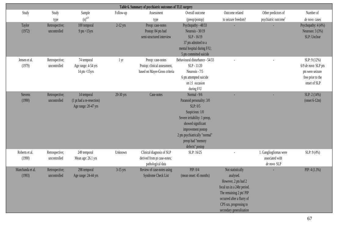
67
Table 6. Summary of psychiatric outcomes of TLE surgery
Study Study Sample Follow-up Assessment Overall outcome Outcome related Other predictors of Number of
type (n)a,b
type (preop/postop) to seizure freedom? psychiatric outcomec
de novo cases
Taylor Retrospective; 100 temporal 2-12 yrs Preop: case-notes Psychopathy - 48/33 - - Psychopathy: 4 (4%)
(1972) uncontrolled 9 pts <15yrs Postop: 84 pts had Neurosis - 30/19 Neuroses: 3 (3%)
semi-structured interview SLP - 16/19 SLP: Unclear
37 pts admitted to a
mental hospital during F/U;
5 pts committed suicide
Jensen et al. Retrospective; 74 temporal 1 yr Preop: case-notes Behavioural disturbance - 54/33 - - SLP: 9 (12%)
(1979) uncontrolled Age range: 4-54 yrs Postop: clinical assessment, SLP - 11/20 6/9 de novo SLP pts
14 pts <15yrs based on Mayer-Gross criteria Neurosis - 7/5 pts were seizure
6 pts attempted suicide free prior to the
on ≥1 occasion onset of SLP
during F/U
Stevens Retrospective; 14 temporal 20-30 yrs Case-notes Normal - 9/6 - - SLP: 2 (14%)
(1990) uncontrolled (1 pt had a re-resection) Paranoid personality: 3/0 (onset 6-12m)
Age range: 20-47 yrs SLP: 0/5
Suspicious: 1/0
Severe irritability: 1 preop,
showed significant
improvement postop
2 pts psychiatrically "normal"
preop had "memory
defects" postop
Roberts et al. Retrospective; 249 temporal Unknown Clinical diagnosis of SLP SLP: 16/25 - 1. Gangliogliomas were SLP: 9 (4%)
(1990) uncontrolled Mean age: 26.1 yrs derived from pt case-notes; associated with
pathological data de novo SLP
Manchanda et al. Retrospective; 298 temporal 3-15 yrs Review of case-notes using PIP: 0/4 Not statistically - PIP: 4 (1.3%)
(1993) uncontrolled Age range: 24-44 yrs Syndrome Check List (mean onset: 45 months) analysed.
However, 2 pts had 2
focal szs in a 24hr period.
The remaining 2 pts' PIP
occurred after a flurry of
CPS szs, progressing to
secondary generalisation

68
Table 6 (continued)
Study Study Sample Follow-up Assessment Overall outcome Outcome related Other predictors of Number of
type (n)a,b
type (preop/postop) to seizure freedom? psychiatric outcomec
de novo cases
Naylor et al. Retrospective; 47 = 38 SAH/9 "other" Mean: 1.9 yrs Structured clinical interview 38 SAH - - 38 SAH:
(1994) uncontrolled 9 "other": (PSE) & review of case-notes; Mood disorder: 2/5 Mood disorder: 5 (13%)
2 pts: intially treated with SAH ICD 10 diagnosis Anxiety disorder: 0/2 Anxiety disorder: 2 (5%)
underwent ATLRs Personality disorder: 2/5 Personality disorder: 1 (3%)
1 pt: intially treated with SAH SLP: 1/1 9 "other"
had an extra-TL resection Organic delusional disorder: 2/2 Mood disorder: 1 (11%)
3 pts: TL lesionectomy Delirium: 1/0
2 pts: ATLR Eating disorder: 1/1
1 pt: previous ATLR had a Cannabis dependent syndrome: 1/0
frontal & re-tailored TL Alcohol dependent syndrome: 2/1
resection 9 "other"
Mean age: 29.0 ± 8.9 yrs Mood disorder: 1/1
Acute & transcient psychosis: 1/0
Kanemoto et al. Retrospective; 38 TLE pts who underwent 24 months Case-notes; 1. Higher frequency (5x) of - PIP & L-sided surgery Unclear
(1998) controlled TLE surgery DSM-IV preop PIP pts in the postop MD
MD mean age range: 26.8 ± 3.8 yrs (38%) than the non-MD (7%) group
Non MD mean age range: 26.1 ± 6.5 yrs
Altshuler et al. Retrospective; 62 = 49 ATLR (2/49 had two TL Mean: 10.9 yrs Structured interview (SCID); Depression: 17/14 Yes - 7/8 (88%) pts without 1. Surgical group had a Depression: 5 (10%)
(1999) controlled surgeries; the second were DSM-III-R diagnosis depression recurrence higher lifetime history 4/5 de novo cases
lateral neocortical extensions postsurgery were sz free of depression than developed within 1 yr
of a previous ATLR); controls
13 non-surgical group
Mean age:
Surgical group: 40.0 ± 5.3 yrs
Non-surgical group: 38.9 ± 9.9 yrs
Lendt et al. Retrospective; 56 = 28 surgical/28 medical 3m Child Behaviour Behavioural problems Yes - better seizure outcome - -
(2000) controlled (13 temporal resections, Checklist (CBCL) in surgical group (%): 39/25. predicted significant
11 extra-TL, Behavioural changes improvement in postop
2 hemispherectomies, significantly worsened behaviour problems
2 callosotomies) in the medical group
Age range: 4-16 yrs

69
Table 6 (continued)
Study Study Sample Follow-up Assessment Overall outcome Outcome related Other predictors of Number of
type (n)a,b
type (preop/postop) to seizure freedom? psychiatric outcomec
de novo cases
Anhoury et al. Retrospective; 109 temporal 12m Case-notes Mood disorder: 16/35 No, but seizure freedom was 1. Poor psychiatric outcome Mood disorder: 19 (17%)
(2000) uncontrolled Mean age: 30.1 ± 7.4 yrs Anxiety disorder: 12/17 significantly reduced by the was associated with preop Anxiety disorder: 5 (5%)
SLP: 3/3 presence of a preop diag psychiatric history & Emotional lability: 7 (6%)
Emotional lability: 5/12 of anxiety bilateral interictal EEG NES: 4 (4%)
NES: 0/4 discharges
31 (28.4%) had treatment 2. Bilateral interictal
for their psychopathology. EEG abnormalities
10 (9.2%) required psychiatric related to de novo
admission psychopathology
3. Postop emotional lability
positively related to size
of resection
4. History of affective disorders
associated with postop anxiety
Kohler et al. Retrospective; 59 temporal 1-3 & 12m Case-notes; Fear aura (preop/1-3m/12m) Yes - mood & anxiety disorders 1. Fear aura preoperatively Not specified,
(2001) uncontrolled (22 fear aura; 22 other aura; DSM-IV diagnosis Mood disorder: 13/15/13 more common in pts with associated with mood & despite clear occurrence
15 no aura) Anxiety disorder: 2/3/5 persistence of anxiety disorders 12 months of de novo cases
Mean age: 33.0 ± 10.6 yrs Psychiatric meds (preop/12m) szs after surgery
Fear: 5/16 2. In the sz free sample at
Other aura: 2/6 12 months, mood & anxiety
No aura: 2/2 disorders were more common
in the preoperative fear
aura group
Inoue et al. Retrospective; 226 = 196 temporal (166 ATLR; >2 yrs Case-notes; 78 (74 TLE) pts had a - - Affective disorder: 9 (4%) -
(2001) uncontrolled 25 SAH); 20 FLE; 10 other ICD-10 diagnosis psychiatric diagnosis before resolved within 1-2 months.
Age range: 15-55 yrs &/ after surgery; 22 preop SLP: 5 (2%) - onset within 2
(mean: 26.9 yrs) psychiatric diagnosis only; 2 postoperative years
39 psychiatric disorders PIP: 1 (0.4%)
persisted after surgery; Adjustment disorder: 1 (0.4%)
17 de novo psychiatric Personality disorder: 1 (0.4%)
disorders
Kanner et al. Retrospective; 100 temporal (ATLR) Mean: 8.3 yrs Structured clinical interview Preop/2 yrs postop (n) N/A focus of this study was - De novo mood/anxiety
(2009) uncontrolled Mean age: 31.2 ± 3.1 yrs Range: 2-14 yrs (SCID); DSM-IV diagn. Axis I diagnosis: 56/24 identifying predictors of seizure disorders not reported;
Mood disorder: 46/18 outcome: seizure freedom at 2yrs NES: 7 (7%)
Anxiety disorder: 25/8 was significantly reduced by
Comorbid mood & anxiety: 21/7 the presence of a lifetime
PIP: 2/0 preoperative psychiatric
NES: 0/7 diagnosis
2 yrs postop: 44% = psychiatric
medication

70
Table 6 (continued)
Study Study Sample Follow-up Assessment Overall outcome Outcome related Other predictors of Number of
type (n)a,b
type (preop/postop) to seizure freedom? psychiatric outcomec
de novo cases
Metternich et al. Retrospective; 113 = 95 temporal; 18 FLE 12m Self report: BDI Pre- & postop BDI data not 1. Yes - sz free pts had -
(2009) uncontrolled surgical procedures: SAH/LesX Overall BDI scores used in reported. significantly lower BDI scores
Mean age: 38.5 ± 13.2 yrs analysis, rather than clinical The focus of this paper was 2. Low preop BDI scores
cut-offs identifying predictors of seizure predicted sz freedom
outcome
1 yr postop: 11% = anti-
depressant medication; unrelated
to sz outcome
Guarnieri et al. Retrospective; 186 = TLE+HS 1-10.5 yrs Clinical interview; Preop (%) only reported: N/A focus of this study was - -
(2009) uncontrolled Mean age: 35.8 yrs (Mean F/U: 6.1 yrs) DSM-IV diagnosis Axis I diagnosis: 41 identifying predictors of seizure
(8 (4%): bilateral HS) Mood disorder: 19/ outcome: seizure freedom
Anxiety disorder: 5/ during F/U was not associated
IDD: 6/ with preop Axis I disorders.
IP: 8% However, pts with preop anxiety
PIP: 3% disorder or Axis II disorders
Axis II diag: 12 were significantly less likely
to be rendered seizure free
Adams et al. Retrospective; 72 = TLE+HS 12m Clinical interview; Preop (%) only reported: N/A focus of this study was - -
(2012) uncontrolled Age range: 16-71 yrs DSM-IV-TR Lifetime MD: 15 identifying predictors of seizure
Mean age: 37 yrs Lifetime psychosis: 4 outcome: seizure freedom at 12m
Lifetime diag: 38 was not significantly reduced by
(inc. anxiety, personality the presence of a lifetime
disorders & substance abuse) preoperative psychiatric
diagnosis in TLE+HS pts
Lackmayer et al. Retrospective; 45 TLE+MTS 12, 24, 36m BDI (adapted German version) Preop (%) only reported: N/A focus of this study was - -
(2013) uncontrolled Age range: 25-49 yrs (BDI < 11 = non-depressed; BDI ≥ 11: 44% identifying predictors of seizure
BDI ≥ 11 = depressed). BDI ≥ 18: 22% outcome: seizure freedom at 12,
Further analysis: BDI ≥ 18 (moderate depression) 24 or 36m was not significantly
was used to assess prognostic impact of moderate depressive reduced by the presence of
symptoms on seizure outcome preoperative depressive symptoms
(BDI ≥ 11 or 18) in TLE+MTS pts

71
Table 6 (continued)
Study Study Sample Follow-up Assessment Overall outcome Outcome related Other predictors of Number of
type (n)a,b
type (preop/postop) to seizure freedom? psychiatric outcomec
de novo cases
Hermann et al. Prospective; 41 temporal 1, 3 & 6m Self report: MHI Specific data for pre- & post- Yes - no change in - -
(1989) uncontrolled Mean age: 31.4 ± 8.9 yrs surgery MHI scores not given. psychopathology for pts with
continued szs (despite sig.
reduction ≥ 75%); whereas
pts with improved mental
health were sz free at
at 3 months F/U
Hermann et al. Prospective; 97 temporal 6-8m Self report: MMPI, WPSI N/A - The focus of this study Yes - total sz freedom predicted Preoperative total -
(1992) uncontrolled Mean age: 30.5 ± 9.4 yrs & GHQ was on the predictors of overall postoperative psychological adjustment
psychosocial adjustment psychological adjustment
Leinonen et al. Prospective; 57 = 54 temporal/3 temporal 2 wks, 3 & 12m Clinical interview; SLP: 0/3 Not statistically analysed. - SLP: 3 (5%)
(1994) uncontrolled & extra-TL DSM-III-R diagnosis & GHQ PIP: 4/2 However, the 2 pts with (mean onset: 3.8 months)
Age range: 17-52 yrs postoperative cessation of PIP
(mean: 33.5 yrs) had a marked improvement in
their postop sz frequency
Blumer et al. Prospective; 50 = 44 temporal & 6 frontal 3m & 6 monthly Semi-structured interview; IDD: 25/28 Yes - poor seizure outcome - IDD: 8 (18%)
(1998) uncontrolled Age Range (mean): thereafter; epilepsy questionnaire 28 (64%) pts required associated with psychiatric 2/8 with concomitant psychotic
TLE pts: 12-59 yrs (36 yrs) mean: 2 yrs & neurobehavioural inventory psychiatric medication morbidity features; onset within 8 months
FLE pts: 37-46 yrs (41 yrs) SLP: unclear/6 SLP: 6 (12%) - all TLE pts;
onset within 6 months postop
Ring et al. Prospective; 60 temporal 6 wks & 3m Structured clinical interview; Preop/6wks/3m (n) - Left-sided pathology Unclear, although authors
(1998) uncontrolled Age range: 16-48 yrs DSM-IV diagnosis Depression: 13/14/20 associated with de novo report that at 6 wks after
(mean: 27 ± 7.4 yrs) Anxiety: 11/25/5 anxiety surgery 50% of pts with no
Emotional lability: ?/27/5 psychopathology at baseline
had developed symptoms of
anxiety or depression.

72
Table 6 (continued)
Study Study Sample Follow-up Assessment Overall outcome Outcome related Other predictors of Number of
type (n)a,b
type (preop/postop) to seizure freedom? psychiatric outcomec
de novo cases
Derry et al. Prospective; 39 temporal 2 yrs Self report: psychosocial (WPSI) Depression: 16/11 Yes - poor seizure outcome Poor preop emotional Depression: 4 (10%)
(2000) uncontrolled Age range: 18-51 yrs & depression (CES-D) scales predicted poor postop adjustment (particularly for
(mean: 31.2 ± 1.0 yrs) depression score older pts, with neurological
deficit(s), preop history of
generalised szs, family history
of psychiatric illness &/ szs)
predicted higher postop
depression scores
Suchy et al. Prospective; 60 = 30 temporal & 30 frontal Mean: 7.3m Self report: BDI Temporal pts: No - change in BDI scores 1. Males reported lower -
(2001) uncontrolled Mean age: (≥ 10 BDI = depressed) Mean BDI: 8.80/6.07 unrelated to postoperative sz postsurgical BDI scores
TLE pts: 28.3 ± 6.3 yrs Frontal pts: status 2. Frontal pts were more likely
FLE pts: 27.9 ± 8.9 yrs Mean BDI: 8.80/7.47 to experience extreme changes
in mood compared
to temporal pts
Koch-Stoecker Prospective; 100 temporal 24m Structured clinical interview Preop/24m (%) Not statistically analysed. 1. Pts with a preop PD & axis I -
(2002) uncontrolled Age range: 24.3-43.1 yrs (SCID); DSM-III-R diagn. Axis I diagn. only: 11/11 diagn. were more likely to
(mean: 33.7 yrs) The defining outcome in this Axis I & II diag: 32/32 require postop psychiatric
At least 4 pts: ≥ 1 TL resection study relates to the number Axis II diag. only: 29/29 hospitalisation
of pts requiring psychaitric 14% (all with preop PDs) 1. Pts with a preop PD without
admission postoperatively were hospitalised an axis I diagn. were more
likely to require postop
psychiatric hospitalisation
Malmgren et al. Prospective; 70 = 54 temporal; 3, 12 & 24m Clinical interview; Preop/within 24m postop - Postop anxiety/depression Not specified,
(2002) uncontrolled 16 extra-TL DSM-III-R & Linqvist-Malmgren Affective disorder: 4/16 associated with preop history despite clear occurrence
Age range: 18.3-62.2 yrs diagnosis Anxiety disorder: 1/12 of anxiety/depression or of de novo cases
(mean: 35.1 yrs) SLP: 1/1 AE disorder
AE disorder: 16/34
Frontal lobe syndrome: 11/11
Spencer et al. Prospective; 396 (temporal & extra- 3, 12 & 24m Self report: BDI & BAI Preop/3m/12m/24m (%) No - -
(2003) uncontrolled TL, no further details (BDI & BAI ≥ 16 = BAI ≥ 16 : 26/10/10/13
provided). moderate-severe symptoms) BDI ≥ 16: 24/10/13/13
Age range: 14-66 yrs
(mean: 36.6 ± 11.3 yrs)
Quigg et al. Prospective; 107 = 90 temporal; 12m Clinical interview (CDI) & Depression (%): 33/44 - 1. Presurgical depressive Not specified,
(2003) uncontrolled 17 extra-TL MMPI-2 4 pts attempted suicide within morbidity predicted higher despite clear occurrence
Mean age: 32.5 ± 1.4 yrs 12m of F/U; postop depression scores of de novo cases
3/4 pts completed suicide; 2. Right sided surgery associated
all patients were sz free with increased postop
depression scores

73
Table 6 (continued)
Study Study Sample Follow-up Assessment Overall outcome Outcome related Other predictors of Number of
type (n)a,b
type (preop/postop) to seizure freedom? psychiatric outcomec
de novo cases
Wrench et al. Prospective; 60 = 43 temporal, 1 & 3m Semi-structured Preop/1m/3m (%) No 1. Site of surgery (temporal) Depression (TL group; 1m/3m)
(2004) uncontrolled 17 extra-TL interview (Austin CEP); Depression (TL group): 33/26/30 associated with increased 5 (13%)/4 (13%)
Mean age: DSM-IV diagnosis Depression (ETL group): 53/0/17 depression & anxiety at Anxiety (TL group; 1m/3m)
TLE pts: 35 ± 10 yrs Anxiety (TL group): 23/42/24 1 month F/U 10 (26%)/5 (15%)
extra TL pts: 37 ± 10 yrs Anxiety (ETL group): 18/6/17 2. Poor psychosocial Anxiety (ETL group; 1m/3m)
adjustment associated with 0/2 (17%)
postop depression (particulary
in the TL group at 1m F/U)
Reuber et al. Prospective; 94 = 76 surgical (60% SAH, 12m (surgical) Self report: BDI & SRAS Surgical group: Preop/12m (%) Yes - improved symptoms 1. Pts with higher presurgical Depression (surgical group):
(2004) controlled 15% ATLR & 25% extended 16m (medical) (> 12 BDI = probably depressed; BDI > 12: 29/16 of depression with better depression scores 3 (4%)
temporal lesionectomy); > 35 SRAS = abnormal anxiety) SRAS > 35: 48/39 sz control regardless of demonstrated most Anxiety (surgical group):
18 medical treatment; Started antidepressant treatment (surgical/medical) improvement in depression 10 (13%)
Mean age: therapy postop: 3 mode if they were sz free Depression (medical group):
Surgical group: 35.0 ± 9.0 yrs Started anxiolytic postsurgery 6 (35%)
Medical group: 35.8 ± 10.1 yrs therapy postop: 17 2. Increase in anxiety scores Anxiety (medical group):
Medical group: Preop/16m (%) only for pts that experienced 4 (22%)
BDI > 12: 18/41 <50% improvement in sz frequency
SRAS > 35: 39/56
Started antidepressant
therapy postop: 5
Started anxiolytic
therapy postop: 5
Devinsky et al. Prospective; 360 = 322 temporal, 3, 12 & 24m Clinical interview (CIDI) Preop/24m (%) Yes - moderate to severe Presurgical depressive Depression: 16 (6.1%)
(2005) uncontrolled 38 extra-TL ICD-10 & DSM-IV diagnosis; BDI ≥ 16: 22.1/11.7 depressive symptoms were symptoms strongest predictor Anxiety: 18 (6.9%)
Mean age: 37.6 ± 11.1 yrs Self-report: BDI & BAI: BAI ≥ 16: 24.7/13 reported in 17.6% with of postop depression SLP: 3 (1.1%)
(scores ≥ 16 = moderate - severe) CIDI diagnosis: preop/24m (n) continued seizures vs 8.2% (i.e. ≥ 16 BDI score)
Anxiety disorder: 59/29 who were sz free at 24 months
Depression: 75/26
SLP: 6/3
Of note, authors state at 24m
F/U, pts with preop SLP were
lost to FU
Cankurtaran et al. Prospective; 22 temporal 3 & 6m Clinical interview (SCID-I); Preop/3m/6m - - 3 months
(2005) uncontrolled Age range: 18-49 yrs DSM - IV diagnosis; Psychiatric dignosis (SCID-I): Depression: 4 (18.2%)
(mean: 30.0 ± 9.1 yrs) BPRS, HDRS & HARS 6 (27%)/7 (32%)/2 (9%): Anxiety: 2 (9%)
Depression: 2/4/2 Adjustment disorder: 1 (5%)
Anxiety: 4/2/0 6 months
Adjustment disorder: 0/1/0 Depression: 1 (5%)
Self-report measures: Preop/3m/6m (mean)
BPRS: 6.0/8.3/4.0
HDRS: 5.3/8.0/4.1
HARS: 6.5/9.5/4.7
Psychiatric meds required in 4 (18%)
pts at 3m F/U and 7 (32%) at 6m F/U

74
Table 6 (continued)
Study Study Sample Follow-up Assessment Overall outcome Outcome related Other predictors of Number of
type (n)a,b
type (preop/postop) to seizure freedom? psychiatric outcomec
de novo cases
Mattsson et al. Prospective; 57 = 47 temporal; 2-8 yrs Anxiety scale (KSP) Specific data for pre- & No significant differences in - -
(2005) uncontrolled 10 extra-TL postsurgery KSP scores not KSP scores between Engel I
Mean age: 39.0 ± 10.0 yrs reported. However, somatic (sz free/auras) & Engel II-IV
& psychic trait anxiety (CPS ± secondary
normalised in pts with generalisation) groups
Engel I outcome
Pintor et al. Prospective; 70 = 59 ATLR/11 SAH 1, 6 & 12m Clinical interview (SCID-IV); Preop/1m/6m/12m (%) No 1. Any (lifetime/during Psychiatric diagnosis: 3 (4.3%)
(2007) uncontrolled Mean age: 31.1 ± 8.3 yrs DSM - IV diagnosis Psychiatric disorder: (Additionally, no significant presurgical evaluation) (no further details given)
47/43/32/25 difference in sz outcome & psychiatric history related
presence of a preop psychiatric to psychiatric disorders
diagnosis) post-surgery
2. Gender: females more
likely to have postoperative
psychiatric diagnoses
Meldolesi et al. Prospective; 52 temporal 12 & 24m Self report: BDI, STAI, Specific data for pre- & post- No Both (1) younger age and -
(2007) uncontrolled Mean age: 33.0 ± 9.4 yrs MMPI & STAXI surgery psychiatric prevalence (2) shorter duration of epilepsy
(BDI ≥ 16 = clinical depression) rates not reported. However, were related to improved
anxiety & anger significantly postoperative anger scores
improved; depression reduced,
but not significantly
Witt et al. Prospective; 151 = 125 temporal; 12m Self report: BDI, FPZ Mean score: Preop/12m Yes - significantly lower Right sided surgery associated Depression: 13 (8.6%), of which
(2008) uncontrolled 26 extra-TL (BDI ≥ 17= clinical depression) BDI: 9.4/7.9 neuroticism, organic psych- with decreased postop 6 pts were sz free
Mean age: FPZ: syndrome & BDI scores in sz depression scores
TLE pts: 37.9 ± 12.2 yrs Extraversion: 52.7/52.5 free group
extra-TL pts: 33 ± 9.9 yrs Neuroticism: 80.6/74.2
Organic Psycho-
Syndrome: 71.4/65.5
Addiction: 17.1/16.6
Salgado et al. Prospective; 36 temporal 6 &12m Self report: Mean score: Preop/6m/12m - - -
(2009) uncontrolled Age range: 22.9-41.1 yrs QOLiE-31; STAI; BDI; QOLiE-31: 63.7/84.3/82.7
(mean: 32 ± 9.1 yrs) PEQ; PLCQ STAI: 45.9/37.1/37.3
BDI: 9.6/5.8/4.2
PEQ: 36.1/-/-
PLCQ: -/51.6/58.8

75
Table 6 (continued)
Study Study Sample Follow-up Assessment Overall outcome Outcome related Other predictors of Number of
type (n)a,b
type (preop/postop) to seizure freedom? psychiatric outcomec
de novo cases
Guangming et al. Prospective; 62 temporal (all CAH) 12 & 24m Self report: SCL-90-R Preop/12m/24m (mean score) Yes - significantly lower - -
(2009) uncontrolled Age range: 16-46 yrs SCL-90-R: 2.18/1.95/1.79 SCL-90-R scores attained at 12m
(mean: 28.8 ± 7.6 yrs) & 24m F/U for pts with Engel I
outcome
Wrench et al. Prospective; 60 = 38 MTR/22 NMTR = 9 1, 3, 6 & 12m Clincal interview (MSE); MTR group: No - postoperative depression 1. History of preoperative Depression: 5 (8.3%);
(2011) uncontrolled lateral TL corticectomies DSM-IV diagnosis; Preop/1m/3m/6m/12m (%) was unrelated to sz outcome depression associated with all pts had a MTR
10 frontal; 2 occipital; semi-structured interview Depression: 40/16/20/26/38 for the sample as a whole or post-surgical depression (4/5 pts onset within 3m)
1 temporoparietal (Austin CEP) NMTR group: at post-surgical time-point 2. Poor postoperative family
corticectomy. Preop/1m/3m/6m/12m (%) dynamics associated with
Age range: 18-60 yrs Depression: 50/14/20/25/24 postoperative depression
(mean: 35 ± 10.9 yrs) Irrespective of surgical 3. Type of surgical intervention
intervention, 13 (22%) on SSRI (MTR) associated with
therapy postoperatively de novo depression:
13% MTR vs 0% NMTR
Barbieri et al. Prospective; 12 = 8 temporal; 2 FLE; 6 &12m Clinical interview; Mean preop/6m/12m scores Small n precluded statistical - -
(2011) uncontrolled 1 occipital; 1 temporo-frontal DSM-IV-TR diagn. SANS: 33/39/34.4 analysis
Mean age: 36.3 ± 8.2 yrs SANS; SAPS SAPS: 31/34/28.5
Hamid et al Prospective; 256 = temporal and extra-TL 3, 12, 24, 48 & 60m Self report: BDI Preop/60m (%) Yes, only pts with excellent - Depression: 10 (4%)
(2011) uncontrolled surgeries (BDI ≥ 15= moderate - severe not depressed: 64.1/77.3 (vs. fair/poor) or good (vs. poor) Notably, 6/10 pts did have a
Age range: 27.3-48.1 yrs depression) mild depression: 13.3/7.8 sz control had significant previous history of affective
(mean: 37.7 ± 10.4 yrs) moderate-severe: 22.7/14.8 reduced mean change in BDI disorders
BDI scores at 60m
Hellwig et al. Prospective; 84 = 55 temporal; 21 extra-TL; 3 & 12m Clinical interview; Preop/3m (n) Yes, significant association - 3 month F/U
(2011) uncontrolled 8 temporal & extra-TL (N.B. 12m F/U data was ICD-10 & ILAE neuropsychiatric Psychiatric diag: 48/46 between the remission of Psychiatric diag: 14 (17%)
Age range: 23.8-49.2 yrs only avaliable on 67pts classification Affective disorder: 29/27 affective disorder and Engel I Affective disorder: 12 (14%)
Mean age: 36.5 ± 12.7 yrs which is not clearly Irrespective of surgical or II seiuzre outcome at 3m. 3 pts (25%) had a preoperative
reported) intervention, 9 (11%) were However, no sigificant difference personality disorder diagnosis.
prescribed psychotrophic between Engel I and II-IV were 6 pts showed remission of
medication. observed at 3 or 12m affective disorder within 12m
76
Table 6 (continued)
Study Study Sample Follow-up Assessment Overall outcome Outcome related Other predictors of Number of
type (n)a,b
type (preop/postop) to seizure freedom? psychiatric outcomec
de novo cases
Filho et al. Prospective; 115 = TLE+MTS Within the first year Clinical interview; Lifetime preop/postop (%) N/A 1. De novo psychosis (type: PIP/ Psychiatric diag: 11 (10%)
(2012) uncontrolled Mean age: 36.9 ± 10.7 yrs following surgery DSM-IV (Axis I & II were Psychiatric diag: 41/27 The preop psychiatric group IP unspecified) was associated MD: 3 (4%)
considered) MD: 23/12 had significantly more pts with L MTS Anxiety: 3 (4%)
Anxiety: 10/9 with an unfavourable sz 2. Preop MDD and IP were Psychosis: 5 (7%); type (PIP/IP)
PIP: 3/? outcome associated with the continuance unspecified.
IP: 3/7 of postop psychopathology 27 (54%) of preop cases
3. Contralateral epileptiform reported complete remission
discharges were associated of psychopathology postop
with continued postop
psychiatric morbidity
4. Older age at surgery related
to de novo psych
Filho et al. Prospective; 115 = TLE+MTS Within the first year Clinical interview; Lifetime preop/postop (%) N/A - -
(2012a) uncontrolled Mean age: 36.9 ± 10.7 yrs following surgery DSM-IV (Axis I) Psychiatric diag: 41/27 The preop psychiatric & MDD 27 (54%) of preop cases
(same sample as 2012 paper) MD: 23/12 groups had significantly poorer reported complete remission
Anxiety: 10/9 postop sz outcome of psychopathology postop
PIP: 3/?
IP: 3/7
Engman et al. Prospective; 50 = 39 TLE & 11 FLE 24m KSP & HADS Lifetime preop/postop (%) No - there were no significant - -
(2012) uncontrolled Psychiatric diag: 24/? associations between change in
Specific data for KSP & HADS KSP/HADS & seizure outcome
were not reported. Statistical for either the TLE/FLE groups
analyses were based on change
from baseline (preop) to 24m F/U
da Conceição et al. Prospective; 145 TLE+MTS ≥ 24m Clinical interview; Preop (%) only reported: N/A focus of this study was - -
(2013) controlled (8 (6%) = bilateral MTS) DSM-IV Psychiatric diag: 42 identifying the surgical outcome
Age range: 18-63 yrs MD: 61 of pts with a lifetime psych
Mean age: 37.8 ± 10.3 yrs Anxiety: 13 history: Seizure outcome did
PIP: 12 not significantly differ between
IP: 8 pts with/without preoperative
psychiatric morbidity (n=94)
ATLR, anterior temporal lobe resection; AE disorder, Astheno-Emotional disorder; Austin CEP, Austin Comprehensive Epilepsy Program; BAI, Beck Anxiety Inventory; BDI, Beck Depression Inventory; BPRS, Brief Psychiatric Rating Scale; CAH, corticoamygdalohippocampectomy; CDI, clinical depression index; CES-D, Centre for Epidemiological Studies Depression Scale; CIDI, Composite International Diagnostic Interview; Diagn, diagnosis; DSM, Diagnostic and Statistical
Manual for Mental Disorders; DSM-IV-TR, Diagnostic and Statistical Manual of Mental Disorders - Text Revision; ETL, extra-temporal; F/U, follow-up; FLE, frontal lobe epilepsy; FPZ, Fragebogen zur Persönlichkeit bei zerebralen Erkrankungen;
GHQ, General Health Questionnaire; HADS, Hospital anxiety and depression scale; HARS, Hamilton Anxiety Rating Scale; HDRS, Hamilton Depression Rating Scale; HS, hippocampal sclerosis; ICD, International Classification of Disease; IDD, interictal dysphoric disorder; IP, interictal psychosis; KSP, Karolinska Scales of Personality; LesX, lesionectomies; m, months; MD, mood disorder; MHI, Mental Health Inventory; MMPI, Minnesota Multiphasic Personality Inventory; MMPI-2,
Minnesota Multiphasic Personality Inventory, scale 2; MSE, mental state examination; MTR, mesial temporal resection; MTS, mesial temporal sclerosis; n, sample size; NMTR, nonmesial temporal resection; PD, personality disorder; PEQ,
Preoperative Expectations Questionnaire; PIP, postictal psychosis; PSE, Present State Examination; PLCQ, Postoperative Life Changing Questionnaire; Pts, patients; QOLiE-31, quality of life questionnaire; RCI, reliable change index; SAH, selective amygdalohippocampectomy; SANS, Scale for the Assessment of Negative Symptoms; SAPS, Scale for the Assessment of Positive Symptoms; SCID, Structured Clinical Interview for DSM; SCL-90-R, Self-Report Symptom Inventory;
SLP, schizophrenia-like psychosis; SRAS, Self-Rating Anxiety Scale; STAI, Spielberger State-Trait Anxiety Inventory; STAXI, Spielberger State-Trait Anger Expression Inventory; Sz(s), seizures; TL, temporal lobe; TLE, temporal lobe epilepsy;
WPSI, Washington Psychosocial Seizure Inventory. (a = sample size that was used in statistical analysis. Thus, sample size may not always equal the size reported in research article; b = age data as reported in article; c = only listed if statistically significant predictor).

77
Twenty-seven studies were prospective but uncontrolled; fourteen were retrospective but
uncontrolled; three were retrospective and controlled and two prospective and controlled.
Control groups consisted of patients deemed not eligible for surgery (Altshuler et al., 1999;
da Conceição et al., 2013), continued on medical treatment (Lendt et al., 2000; Reuber et al.,
2004) or who did not develop a postoperative mood disorder (Kanemoto, Kawasaki & Mori,
1998). The majority of retrospective studies (n=10) had a follow-up of more than 1 year
(range 3 months – 30 years). However, one retrospective study did not report their follow-up
duration (Roberts et al., 1990). Unsurprisingly, prospective studies in comparison had a
shorter follow-up ranging from 2 weeks to 8 years.
Depression (twenty-nine studies), anxiety (twenty-three studies) and interictal psychosis
(seventeen studies) were the most frequent outcomes explored. These outcomes were
assessed mainly through reviewing case-notes, via structured clinical interview and/or self-
report measures. Other outcomes received less attention: postictal psychosis (nine studies),
personality disorders (seven studies), non-epileptic seizures (three studies), psychosocial
adjustment (one study), behavioural problems (one study) and interictal dysphoric disorder
(IDD; one study).
2.5 Depression
The prevalence of preoperative depressive symptoms in TLE patients was highly variable
owing to divergent methodologies and diagnostic classifications (range 5-61%) (Naylor, et
al., 1994; Ring, Moriarty & Trimble, 1998; Altshuler et al., 1999; Derry, Rose & McLachlan,
2000; Kohler et al., 2001; Wrench, Wilson & Bladin, 2004; Cankurtaran et al., 2005; Kanner
et al., 2009; Guarnieri et al., 2009; Wrench et al., 2011; Adams et al., 2012; Filho et al., 2012;

78
da Conceição et al., 2013; Lackmayer et al., 2013). Cases of attempted and completed
suicide have been reported, paradoxically in patients who have been rendered seizure free
(Jensen & Larsen, 1979; Quigg et al., 2003).
There were inconsistent findings comparing depression before and after temporal lobe
surgery. A number of studies reported improvements in depression after surgery, defined
either as a reduction in the number of patients meeting the clinical criteria for depression
(Ring et al., 1998; Altshuler et al., 1999; Kanner et al., 2009; Wrench et al., 2011; Filho et al.,
2012), or significant improvements in rating scales measuring depressive symptomatology
(Derry et al., 2000; Reuber et al., 2004; Hamid et al., 2011).
In contrast, Anhoury and colleagues (2000) found no change in prevalence rates of mood
disorders pre- and post-surgery. In two studies it was unclear whether depression prevalence
rates had improved or worsened after surgery, as the frequency of patients that developed de
novo depression was not reported (Ring et al., 1998; Kohler et al., 2001).
Although depression improved in some patients, de novo cases (including IDD) were
reported in a minority of cases (Naylor et al., 1994; Blumer et al., 1998; Altshuler et al.,
1999; Anhoury et al., 2000; Derry et al., 2000; Reuber et al., 2004; Wrench et al., 2004;
Cankurtaran et al., 2005; Devinsky et al., 2005; Witt, Hollmann & Helmstaedter, 2008;
Wrench et al., 2011; Hamid et al., 2011; Filho et al., 2012).
In studies that only included temporal lobe surgical patients, de novo depression prevalence
rates ranged from 4% (Reuber et al., 2004) to 18% (Cankurtaran et al., 2005). The research
findings reviewed suggest that de novo depression frequently occurs within 3-12 months after
surgery (Altshuler et al., 1999; Cankurtaran et al., 2005) and tends to persist (range 1-11

79
months; Wrench et al., 2011). However, determining the evolution of depression is
challenging, owing to the varied and limited time frames in which psychiatric symptoms have
been assessed.
Regarding predictors of post-surgical depression, ten studies demonstrated that improvements
in depression after surgery were related to significant gains in seizure control (Blumer et al.,
1998; Altshuler et al., 1999; Derry et al., 2000; Kohler et al., 2001; Reuber et al., 2004;
Devinsky et al., 2005; Witt et al., 2008; Metternich et al., 2009; Hamid et al., 2011). This,
however, was not a consistent finding (Suchy & Chelune, 2001; Spencer et al., 2003; Wrench
et al., 2004; Meldolesi et al., 2007; Wrench et al., 2011; Engman et al., 2012).
There is little evidence to suggest that post-surgical depression is associated with the
laterality of the surgical resection. Only one study found that patients undergoing a right
temporal lobe resection were at greater risk (Quigg et al., 2003), but this has not been
confirmed by others (Ring et al., 1998; Blumer et al., 1998; Altshuler et al., 1999; Derry et
al., 2000; Kohler et al., 2001; Wrench et al., 2004; Devinsky et al., 2005; Wrench et al., 2011;
Hamid et al., 2011).
Significant preoperative risk factors for postoperative depression include: pre-existing history
of affective disorders (depression or anxiety) (Malmgren et al., 2002; Quigg et al., 2003;
Devinsky et al., 2005; Wrench et al., 2011; Filho et al., 2012); fear auras (Kohler et al.,
2001); temporal versus extra-temporal surgery (Wrench et al., 2004) and within this group,
mesial versus lateral resections (Wrench et al., 2011). Female gender (Suchy et al., 2001),
poor psychosocial adjustment (Derry et al., 2000; Wrench et al., 2004) and negative family
dynamics (Wrench et al., 2011) have also been found to place patients at higher risk.

80
2.6 Anxiety
The prevalence of preoperative anxiety symptoms was also extremely variable, ranging from
0-48% (Naylor et al., 1994; Ring et al., 1998; Anhoury et al., 2000; Kohler et al., 2001;
Malmgren et al., 2002; Spencer et al., 2003; Reuber et al., 2004; Wrench et al., 2004;
Cankurtaran et al., 2005; Devinsky et al., 2005; Kanner et al., 2009; Guarnieri et al., 2009;
Salgado & Cendes, 2009; Filho et al., 2012; da Conceição et al., 2013), and as for depression,
this is likely to be due to different diagnostic and psychopathological measures used. For
instance, Devinsky et al. (2005) reported that preoperatively a quarter of patients had
moderate to severe anxiety according to rating scale data, whereas 17% met the clinical
criteria for an anxiety disorder following a structured clinical interview (Composite
International Diagnostic Interview, CIDI).
The majority of studies demonstrated reduced prevalence of anxiety postoperatively when
assessed between 3 months – 12 years after surgery (Taylor, 1972; Jensen et al., 1979;
Spencer et al., 2003; Cankurtaran et al., 2005; Devinsky et al., 2005; Kanner et al., 2009;
Salgado et al., 2009). Two studies however have suggested that anxiety symptoms may
present early and resolve within the first few weeks following surgery (Ring et al., 1998;
Kohler et al., 2001). Ring and co-workers (1998) reported that anxiety symptoms rose 6
weeks after TLE surgery in their surgical cohort, and reduced to about half the pre-surgical
prevalence rate by 3 months. Similarly, Wrench et al. (2004) found anxiety diagnoses peaked
4 weeks after temporal lobe surgery, remitting to pre-surgical levels 3 months later.

81
Four studies found an increase in the prevalence of anxiety disorders postoperatively (Naylor
et al., 1994; Anhoury et al., 2000; Kohler et al., 2001; Malmgren et al., 2002) and one study
demonstrated no significant change in self-reported anxiety symptoms (Reuber et al., 2004).
Seven studies reported de novo anxiety cases, ranging from 3-26% (Taylor, 1972; Naylor et
al., 1994; Anhoury et al., 2000; Reuber et al., 2004; Wrench et al., 2004; Cankurtaran et al.,
2005; Hamid et al., 2011; Filho et al., 2012). The highest prevalence rate was noted one
month after temporal lobe surgery (26%) (Wrench et al., 2004).
Evidence for predictors of post-surgical anxiety indicates patients with a previous history of
affective disorders (depression/anxiety) (Anhoury et al., 2000; Malmgren et al., 2002) are
more susceptible to postoperative anxiety. As with depression, TLE patients with
preoperative fear auras were identified in one study as at greater risk of post-surgical anxiety
disorders despite becoming seizure free (Kohler et al., 2001).
Data relating to the predictive value of other clinical characteristics is unconvincing. Some
authors have reported an association between temporal lobe (versus extra-temporal lobe)
surgery and postoperative anxiety (Wrench et al., 2004), but this has not been replicated
(Devinsky et al., 2005). There is conflicting evidence as to whether postoperative anxiety is
influenced by seizure outcome. Two studies reported that anxiety disorders/symptoms were
more common in patients with persistent seizures (Kohler et al., 2001), or less than 50%
reduction (Kanemoto et al., 1998), but another did not support such an association (Devinsky
et al., 2005). Of those studies considering surgical laterality, only one reported an association
between left temporal lobe resection and the development of de novo postoperative anxiety
(Ring et al., 1998).

82
2.7 Interictal psychosis
Reported preoperative prevalence rates are widely divergent (0-16%) (Taylor, 1972; Jensen et
al., 1979; Roberts et al., 1990; Leinonen, Tuunainen & Lepola, 1994; Naylor et al., 1994;
Anhoury et al., 2000; Guarnieri et al., 2009; Malmgren et al., 2002; Barbieri et al., 2011;
Filho et al., 2012; da Conceição et al., 2013). Arguably, the high baseline prevalence of
interictal psychosis may have been inflated by earlier retrospective studies that included
patients directly referred for surgery from psychiatric institutions (Taylor, 1972; Jensen et al.,
1979).
Patients with pre-existing interictal psychosis are often unlikely to be considered for surgical
intervention for their seizures (Taylor, 1972; Jensen et al., 1979; Roberts et al., 1990;
Anhoury et al., 2000; Malmgren et al., 2002; Foong et al., 2007). However, a recent small
prospective study, but the largest reported, (Barbieri et al., 2011) found that positive
symptoms (hallucinations/delusions) worsened for a subgroup of patients (n=4/12) in the first
6 months and later improved to preoperative levels at 12 month follow-up. Negative
symptoms (social withdrawal/emotional blunting) either persisted or remained unchanged in
the majority of their patients at one year follow-up. No patients with worsening psychotic
symptoms required hospitalisation.
There are a number of reports of an interictal psychosis presenting for the first time following
TLE surgery (Jensen et al., 1979; Roberts et al., 1990; Stevens et al., 1990; Leinonen et al.,
1994; Blumer et al., 1998; Inoue & Mihara, 2001; Devinsky et al., 2005). Two prospective
studies reviewed reported de novo interictal psychosis cases (Blumer et al., 1998; Devinsky
et al., 2005). Blumer et al.’s (1998) study of temporal and frontal lobe resections

83
demonstrated a 12% prevalence rate of de novo interictal psychosis at 6 months post-surgery.
Notably, all de novo cases had undergone a temporal lobe resection. Devinsky et al.’s (2005)
uncontrolled study of temporal and frontal resections reported a much lower rate of de novo
interictal psychosis (1.1%) at 24 months follow-up. It is possible that the de novo interictal
psychosis prevalence rate may have been underestimated due to the high attrition rate of this
study.
There is no clear evidence of a relationship between the development of psychotic symptoms
and postoperative seizure control (Jensen et al., 1979; Leinonen et al., 1994; Blumer et al.,
1998; Inoue et al., 2001), or that surgical laterality is a risk factor. The pathological
mechanisms mediating the development of de novo psychotic symptoms have not been
clarified. Stevens (1990) proposes that aberrant reinnervation with axonal sprouting in the
projection sites of the surgical area explains the latency (2-24 months) between surgical
resection and the development of de novo cases. However, a histopathological study
(Roberts et al., 1990) found a significant association between developmental lesions
(gangliogliomas) in the temporal lobe and de novo interictal psychosis. In contrast, Anhoury
et al. (2000) failed to find that de novo interictal psychosis patients had developmental
pathological abnormalities. The authors highlight, however, that very few patients included
in the ‘developmental pathology’ group had gangliogliomas.
2.8 Postictal psychosis (PIP)
Given the limited research on PIP, case studies will be reviewed, although they are not
presented in Table 6.

84
PIP is a recognised psychiatric complication of temporal lobe epilepsy (TLE); usually
manifesting after a cluster of complex partial or generalised seizures (Logsdail & Toone,
1988). Psychotic symptoms tend to emerge following a symptom-free (lucid) period ranging
from 12 to 72 hours (Kanner et al., 1996) and are transient; typically lasting several days
(Leutmezer et al., 2003), but may be up to weeks (Devinsky et al., 1995).
Incidence rates of PIP in studies of patients undergoing video-electroencephalography
recordings (VEEG) are ~7% (Kanner et al., 1996; Alper et al., 2001). Patients who have a
recurrent PIP may be at risk of developing chronic interictal psychosis (Tarulli, Devinsky &
Alper, 2001).
The clinical risk factors reported are diverse, although widespread cerebral
disturbance has been consistently reported (Ferguson et al., 1969; So et al., 1990; Devinsky et
al., 1995; Mathern et al., 1995; Kanemoto, Kawasaki & Kawai, 1996; Kanner et al., 1996;
Seeck et al., 1999; Takeda et al., 2001; Christodoulou et al., 2002; Alper et al., 2008; Kanner
& Ostrovskaya, 2008; Falip et al., 2009; Schulze-Bonhage & van Elst, 2010; Oshima,
Motooka & Kanemoto et al., 2011; Kuba, Brázdil & Rektor et al., 2012)
There is limited data of post-surgical outcome of patients with PIP in relation to psychiatric
status (Kanemoto et al, 1998; Kanemoto et al., 2001) or seizure outcome (Alper et al., 2008;
Kanner et al., 2008; Falip et al., 2009). Two studies suggest that a history of PIP is
associated with postsurgical de novo psychopathology, particularly mood disorders
(Kanemoto et al., 1998; Kanemoto et al., 2001).

85
2.9 Methodological limitations
The literature review highlights a number of methodological problems that make it difficult
to draw definite conclusions and undoubtedly contribute to the conflicting findings. Firstly,
many studies provide little or no information regarding pre-surgical psychiatric status
(Chovaz et al., 1994; Kanemoto et al., 1998; Kanemoto et al., 2001; Mayanagi et al., 2001;
Moss et al., 2009). Even when reported, pre-surgical psychiatric evaluations are usually only
sought if there is a positive psychiatric history or if patients report peri-operative psychiatric
symptoms (Malmgren et al., 2002; Metternich et al., 2009). If a neuropsychiatric assessment
is not part of the pre-surgical evaluation, cases and symptoms may be missed (Boylan et al.,
2004; Kanner et al., 2008). Patients and families often under-report psychiatric
symptomatology, viewing this as a “natural reaction” to difficult life circumstances (Kanner,
2008). Some surgical candidates may not disclose past or current psychopathology in the
fear that it would disqualify them from surgical consideration (Kanner et al., 2000).
Moreover, psychiatric comorbidities in TLE patients are often unrecognised or overlooked in
routine neurological consultations and go untreated despite persisting symptoms severe
enough to warrant pharmacotherapy (Kanner et al., 2000; Boylan et al., 2004). The lack of a
psychiatric evaluation preoperatively, as part of standard clinical care, may in turn inflate the
reporting of de novo figures postoperatively (type I error).
Another important consideration is the time-frame during which mental health is assessed.
Psychiatric comorbidity rates may differ depending on whether the evaluation focuses on
lifetime prevalence of psychiatric dysfunction (Anhoury et al., 2000; Guarnieri et al., 2009;
Kanner et al., 2009; Adams et al., 2012; Filho et al., 2012) or sets a specific period (e.g. the 6
months immediately before surgery). Conceivably, psychiatric status in the months

86
preceding surgery, when patients are undergoing complicated assessments that include
anxiety-provoking measures (sleep deprivation, medication reduction, continuous video-EEG
recording) that could lead to life-altering neurosurgery may not represent stable mood states
(Glosser et al., 2000).
Several studies had short follow-up periods (≤ 6 months) which may distort the profiling of
postoperative psychopathology. It may be erroneously concluded that patients are no longer
at risk of de novo psychiatric morbidity or an exacerbation of symptoms after this time-frame
(Herman et al., 1989; Ring et al., 1998; Lendt et al., 2000; Wrench et al., 2004; Cankurtaran
et al., 2005). Conversely, other studies where follow-up commenced after 6 months
(Hermann et al., 1992; Anhoury et al., 2000; Metternich et al., 2009; Salgado et al., 2009), or
even several years after surgery (Taylor et al., 1972; Stevens 1990; Naylor et al., 1994;
Altshuler et al., 1999; Derry et al., 2000; Suchy et al., 2001; Inoue et al., 2001; Mattsson et
al., 2005), will miss the emergence of early psychiatric complications. For example, Wrench
et al. (2011) found the majority of clinically depressed patients (70%) were diagnosed within
3 months postoperatively and in 65% of diagnosed cases, the depression persisted for at least
6 months.
The application of diverse diagnostic criteria, instruments and clinical cut offs in assessing
psychiatric status complicates comparison across studies. Self-reported measures are not
comparable with results of psychiatric diagnostic procedures using semi-structured interviews
of patients and families, or rating scales completed by a neuropsychiatrist (Glosser et al.,
2000). A thorough psychiatric evaluation does not only consider transient mood states
reflected by self-reported indices, but the full context of the presenting symptoms; including
the history, other behavioural characteristics, and corroborative information from the family
or carer(s). Although no single method is ideal to assess psychiatric functioning pre- or post-

87
surgery (Guangming et al., 2009), a composite of measures may be the best approach. Even
when standardised classification systems are adopted, for example the DSM or ICD, many
argue that neither has been validated on patients with neurological conditions (Glosser et al.,
2000; Anhoury et al., 2000). Furthermore, the clinical constellation of symptoms evident in
patients with epilepsy may not conform to a single diagnostic category (Glosser et al., 2000;
Meldolesi et al., 2007).
Few studies had a control group (Kanemoto et al., 1998; Altshuler et al., 1999; Lendt et al.,
2000; Reuber et al., 2004; da Conceição et al., 2013) and given the high psychiatric morbidity
in patients with epilepsy, it is difficult to determine whether surgery per se is a risk factor.
Control groups were restricted to patients who were denied surgery (Altshuler et al., 1999;
Lendt et al., 2000; da Conceição et al., 2013), did not develop a postoperative mood disorder
(Kanemoto et al., 1998) or who did not go on to have surgery; presumably due to high risks
of neuropsychological impairment and low odds of seizure freedom (Reuber et al., 2004).
Furthermore, a number of studies used heterogeneous samples that included temporal, extra-
temporal and palliative procedures yielding limited data regarding temporal lobe surgery.
Regarding postictal psychosis, few studies have investigated the relationship between PIP and
post-surgical psychiatric outcome. Of these, the relationship between the number of PIP
episodes (i.e. PIP severity) and postoperative psychiatric morbidity has not been examined.
No study has systematically explored the association of PIP with neuropsychological test
performance, apart from studies reporting unspecified bi-temporal memory dysfunction (So
et al., 1990; Mathern et al., 1995; Christodoulou et al., 2002; Falip et al., 2009), or the
relationship with postoperative neuropsychological/seizure outcome.

88
2.10 Statistical analysis
Statistical approaches employed are also open to criticism. Research on the predictors of
postoperative psychopathology is dominated by Chi-squared (X2) or Fisher exact analyses
(see, Naylor et al., 1994; Kanemoto et al., 1998; Ring et al., 1998; Kohler et al., 2001;
Malmgren et al., 2002; Wrench et al., 2004; Devinsky et al., 2005; Pintor et al., 2007; Witt et
al., 2008, for examples) as the outcome variables are normally categorical (i.e.
presence/absence of psychopathology).
Although exploratory univariable analyses (i.e. multiple X2 tests) are sufficient for
identifying possible preoperative predictors of postoperative psychiatric morbidity, they fail
to quantify or adjust for the association between variables of interest in the presence of other
preoperative factors. Moreover, the performance of numerous univariable analyses also
increases the chance of finding significant results purely by chance (type I error). A further
disadvantage of this non-parametric test is that all predictor(s) variables are required to be
categorical.
These difficulties can be avoided by using multivariable analyses and some studies have
adopted this approach (Blumer et al., 1998; Anhoury et al., 2000; Wrench et al., 2011; Filho
et al., 2012; Filho et al., 2012a, for examples). Logistic regression is a multivariable
statistical technique with the advantage that predictor variables can be continuous or
categorical. Therefore, the relationship between a continuous independent variable (e.g.
years of epilepsy) and the outcome (e.g. de novo psychopathology) does not need to be
partitioned into arbitrary categories (‘per 10 year’ increase in epilepsy duration). This
approach also enables the quantification of associations between variables and examines the

89
odds of the outcome (de novo psychopathology) per unit increase/decrease in the predictor
(per year of epilepsy duration).
Following the identification of significant univariable associations in the primary analysis, a
multivariable logistic regression analysis can be performed to identify any independent
associations, whilst controlling for other preoperative factors; thereby minimising the risk of
a type I error.
Reducing the chance of a type I error can also be achieved by including the fewest possible
predictors in a multivariable model. The suggested guideline is a ratio of 1 predictor per 10
positive cases (of postoperative psychopathology) (Field, 2009). Apart from the number of
predictors, the method (e.g. hierarchical/forced/stepwise) in which predictors are entered into
a model can also reduce the type I error rate. However, some investigators have exceeded
this ratio and not specified the method used in their predictive analysis (see Filho et al., 2012,
for example).
The majority of self-report longitudinal data collected in psychiatric research has some form
of hierarchical structure, with repeated measurements (at Level 1) nested/clustered within
individuals (at Level 2) (see Figure 3).
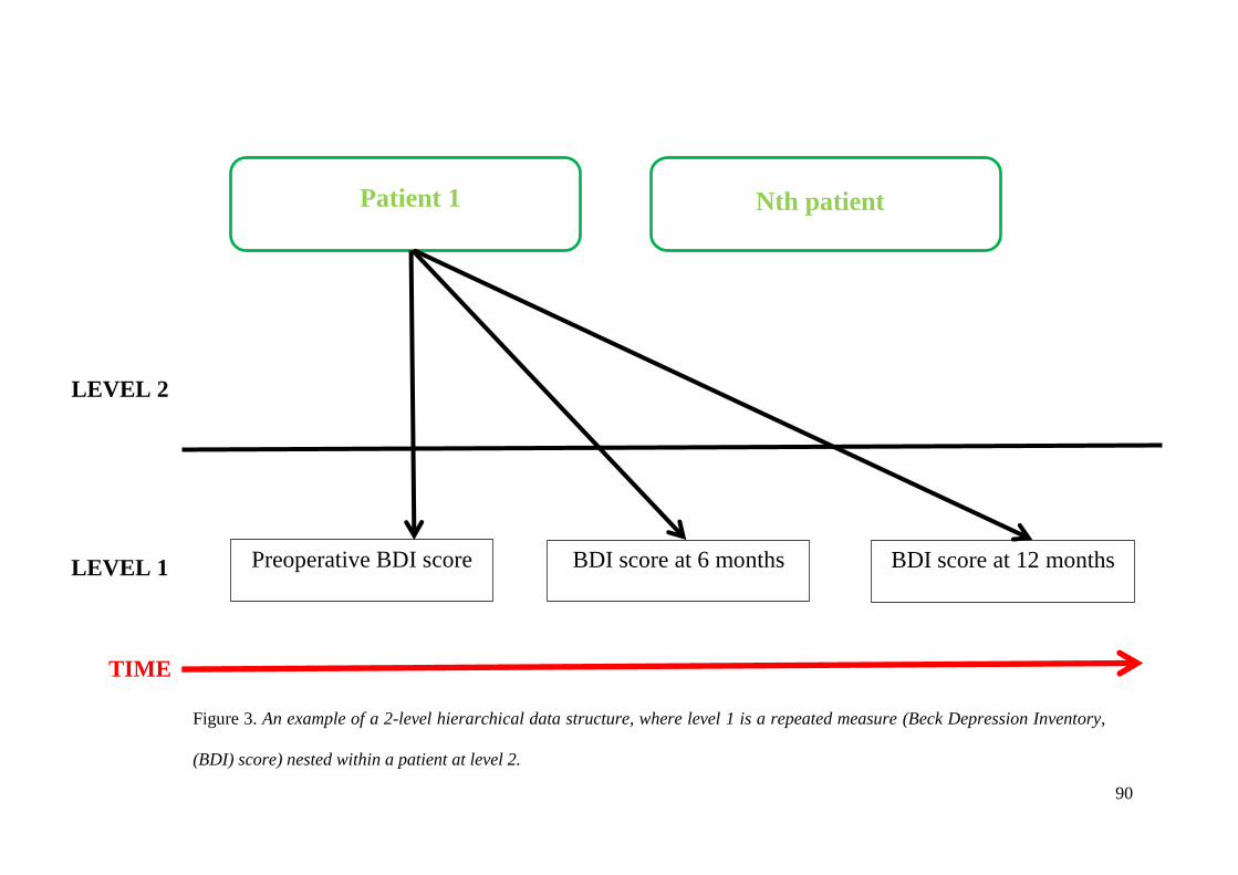
90
LEVEL 1
LEVEL 2
Preoperative BDI score BDI score at 6 months BDI score at 12 months
TIME
Patient 1 Nth patient
Figure 3. An example of a 2-level hierarchical data structure, where level 1 is a repeated measure (Beck Depression Inventory,
(BDI) score) nested within a patient at level 2.

91
This inherent hierarchical structure therefore should be reflected in the statistical models that
are used to analyse such data (Paterson & Goldstein, 1991). However, self-report data from
longitudinal research is usually analysed using a conventional generalised linear model
(GLM), such as repeated-measures analysis of variance (rANOVA) (see: Suchy et al., 2001;
Spencer et al., 2003; Quigg et al., 2003; Reuber et al., 2004; Cankurtaran et al., 2005;
Meldolesi et al., 2007; Witt et al., 2008; Metternich et al., 2009, for examples). However,
rANOVA has a number of assumptions that when violated invalidate the model’s accuracy,
including:
1. Complete/balanced dataset – equal number of patients at each post-surgical follow-up
2. Sphericity – The variances in a characteristic of interest (e.g. Beck Depression
Inventory; BDI) for each follow-up time are equal
3. Independence of observations – No correlation between residual scores in a
characteristic of interest (e.g. BDI) between follow-up times
Due to sample attrition data sets may be unbalanced, violating assumption 1. To circumvent
this problem, subjects with missing values may be removed from analysis reducing statistical
power and introducing sample bias (e.g. listwise deletion; all subjects with missing value(s)
for a characteristic of interest are deleted from the dataset) or imputation techniques
employed (e.g. last observation carried forward), resulting in biased treatment estimates (see
Gueorguieva & Krystal, 2004, for further discussion).
The assumption of sphericity is often violated in longitudinal research designs as consecutive
observations on the same subject tend to be correlated more highly than observations on the

92
same subject taken further away in time (Gueorguieva et al., 2004). When this occurs,
standard errors of the parameter estimates are reduced, inflating the type I error rate, leading
to spuriously significant results.
Similarly, each subject’s residual scores (assumption 3) in a characteristic of interest are
likely to be auto-correlated as the same subject is measured on several occasions. Thus, any
unexplained subject-specific time-invariant effect in the residuals will create a correlation
across measurement occasions (Singer & Willet, 2003). This problem results in a downward
bias in the estimation of standard errors, leading to an increased familywise error rate (Heck,
Thomas & Tabata, 2010).
Only one prospective study reviewed (see, Hamid et al., 2011) has accounted for the
hierarchical structure inherent in self-report longitudinal designs, by employing a multilevel
model, specifically individual growth curve (IGC) modeling. This form of statistical
modeling is necessary to describe the development/change of a variable of interest (e.g. BDI)
over time. A multilevel model decomposes the variance in a characteristic of interest (e.g.
BDI) and models whether the variability and rate of change varies across individuals in a
systematic way, according between-subject predictors of interest (e.g. epilepsy lateralisation,
history of psychopathology etc.). Therefore, it should be applied to longitudinal data where
the primary interest is in modeling the structure and predictors of change over time (Luke,
2004).

93
Chapter 3. NEUROIMAGING CORRELATES OF
PSYCHOPATHOLOGY IN TLE
3.1 Neuroimaging in TLE
Neuroimaging techniques have been exhaustively applied to the investigation of TLE,
resulting in a wealth of scientific data and clinical applications (Duncan, 1997; Duncan,
2010). Structural MRI studies in mTLE show atrophy of the hippocampus, in addition to
other mesial (enthorinal cortex, perirhinal cortex, amygdala, fusiform gyrus) and extra-
temporal regions (striatum, cingulate gyrus, frontal/parietal neocortex and bilateral thalamus)
(see Keller & Roberts, 2008; Li, Zhang & Shang, 2012, for reviews). Using diffusion tensor
imaging (DTI), which captures the direction and integrity of white matter (WM) tracts, the
connections between these regions have been found to be abnormal, particularly in the limbic
and transcallosal WM (see Otte et al., 2012, for meta-analysis).
These structural abnormalities are paralleled in the functional neuroimaging literature. FDG-
PET studies typically identify interictal regions of reduced glucose metabolism in the mesial
temporal lobe and adjacent neocortex, insular cortex, putamen and thalamus in the interictal
state. During seizures captured with ictal SPECT, increased perfusion has been reported in
the ipsilateral mesial and lateral temporal lobe, striatum and bilateral thalamus. Decreased
perfusion is seen in the frontal cortex and precuneus (Richardson, 2012, for review & Figure
4).

94
Figure 4. A schematic summary of the widespread abnormalities reported in neuroimaging
studies of mTLE patients. (A) coronal (B) medial view of ipsilateral right hemisphere
(C) axial section (D) lateral view of ipsilateral right hemisphere. Adapted from
Richardson, 2012

95
As outlined in Chapter 1, imaging methods are used extensively for TLE surgical planning.
Integration of structural and functional data into surgical image-guidance systems is currently
underway, with the aim of improving seizure freedom rates and reducing postoperative
neurological deficits (Duncan, 2010, for review).
Over the last two decades, a cumulative literature has linked structural and functional
neuroimaging abnormalities with cognitive impairment in TLE. More recently, imaging
studies have attempted to predict the impact of TLE surgery on neuropsychological
(memory/language) outcome (see Bell et al., 2011, for review).
Less attention has been focussed on delineating the neuroimaging correlates of psychiatric
comorbidities in TLE, or identifying preoperative cerebral markers that increase the risk of
postoperative psychopathology. These studies are considered in this chapter.
3.2 Systematic literature review
A literature search was conducted using Medline, Embase and PsychINFO until June 2013
with the following search terms: temporal lobe epilepsy, neuroimaging (e.g., structural and
functional), neurosurgical procedures and mental disorders (e.g., mood/anxiety disorders, and
psychosis). Previous literature reviews were excluded (Kanner et al., 2002; Kanner, 2004;
Theodore, 2004; Gilliam, 2005; Charyton, et al., 2010; Kanner et al., 2012a; Li, Zhang &
Shang, 2012; Valente & Filho, 2013), although reference lists were checked to ensure no
additional studies had been missed. The search yielded 2,547 articles relating to
neuroimaging and TLE. Of these, 7% (n=179) investigated psychiatric comorbidity pre-
and/or postoperatively. Studies on MEG, resting-state, palliative neurosurgical procedures,

96
stimulation studies, and studies not published in peer reviewed English language journals
were excluded. Fifty one studies met these inclusion criteria and therefore were included,
summarised in Appendix 1.
3.2.1 Depression
Structural imaging studies
Preoperative studies report increased depressive morbidity in TLE in association with
enlarged amygdala volume (Tebartz van Elst, Woermann, Lemieux & Trimble, 1999;
Richardson et al., 2007) or preserved contralateral amygdala T2 relaxometry (Briellmann,
Hopwood & Jackson, 2007). The syndrome mTLE has also been associated with depression
(Quiske et al., 2000; Salgado, Yasuda & Cendes, 2010), but this is not a consistent finding
(Adams et al., 2008; Wrench et al., 2009; Shamim et al., 2009). Left-sided mTLE has been
suggested as a risk factor (Salgado et al., 2010), but this has not been confirmed by others
(Quiske et al., 2000; Baxendale, Thompson & Duncan, 2005; Paparrigopoulos et al., 2008;
Shamim et al., 2009; Finegersh et al., 2011; Butler et al., 2012).
Reduced hippocampal volume has been variably associated with greater depressive
symptomatology. An early study by Quiske et al. (2000) reported that patients with
hippocampal sclerosis (mTLE-HS) had higher BDI scores than neocortical TLE patients,
independent of the lateralisation of the lesion. Notably, the mean BDI scores in the mTLE-
HS group were in the ‘mild’ range. This finding has been replicated, although the atrophy
was specific to the hippocampus contralateral to the seizure onset (Baxendale et al., 2005;
Shamim et al., 2009). A subsequent study using 3D surface mapping, also found

97
significantly increased contralateral hippocampal atrophy in depressed (BDI ≥14) mTLE
patients (Finegersh et al., 2011). Others however have not found an association between
hippocampal volumetry and depression (Richardson et al., 2007; Briellmann et al., 2007;
Wrench et al., 2009).
Alternative imaging modalities have offered insights into the relationship of epileptogenicity
of the limbic structures and depression (Kanner et al., 2012a). A study of MR spectroscopy
in 31 TLE patients found that the extent of voxels in the hippocampi with decreased NAA
(creatine/N-acetylaspartate metabolite ratio; biomarker of hippocampal dysfunction) was
linearly associated with the severity of depression symptoms (Gilliam et al., 2007). No other
variable was associated with depression, including seizure rate, number of AEDs, or self-
assessed social and vocational disability. The extent of hippocampal dysfunction (NAA)
explained 57% of the variance in mood state. Decreased NAA has been associated with
cerebral regions of interictal spiking and seizure onset (Garcia et al., 1997), suggesting that
chronic hyper-excitability of the hippocampus may negatively influence the limbic network
toward a depressed state (Gilliam et al., 2007).
Studies investigating extra-limbic structural correlates of depressive morbidity in TLE are
limited in contrast with the primary depression literature (see Drevets & Price, 2012, for
review). Butler and colleagues (2012) used a measure of cortical thickness (FreeSurfer) and
demonstrated bilateral orbito-frontal cortex (OFC) thickening in mTLE patients with
increasing depressive symptoms compared to healthy controls. However, the role and
morphological changes of the OFC in TLE patients with depressive symptoms is unclear due
to the lack of a TLE-only control group.

98
Only one structural neuroimaging study has investigated neuroanatomical markers of de novo
depression following TLE surgery. In an a priori region-of-interest (ROI) analysis, Wrench
and colleagues (2009) manually segmented the hippocampi and found that reduced
preoperative contralateral hippocampal volume was a significant risk factor in the
development of de novo depression within the first year of surgery in mTLE patients.
Functional imaging studies
Decreased serotonergic function has been identified as a pivotal pathogenic mechanism of
depression, evidenced by the efficacy of serotonin reuptake inhibitors (SSRIs) in treating
depression (Kanner et al., 2002). A number of studies using serotonin receptor ligands for
PET have yielded important insights into the neurobiology of depression in TLE (Kanner et
al., 2012a). Investigations in TLE patients have reported a significant inverse relation
between BDI score and reduced ipsilateral hippocampal binding, after partial volume
correction (Giovacchini et al., 2005; Theodore et al., 2007).
Serotonergic abnormalities have also been demonstrated in extra-temporal structures
including the anterior cingulate cortex (ACC) (Savic et al., 2004; Hasler et al., 2007; Lothe et
al., 2008), insular cortex (Lothe et al., 2008; Martinez et al., 2013) and ipsilateral (Salzberg et
al., 2006) and bilateral (Bromfield et al., 1992; Lothe et al., 2008) frontal regions (Salzberg et
al., 2006).
Schmitz and colleagues (1997) found a negative relationship between BDI score and
contralateral temporal and bilateral frontal perfusion in left-sided focal patients using
interictal SPECT. A similar study of TLE patients meeting the diagnostic criteria for a major

99
depressive episode failed to find any cerebral areas of hypo-perfusion compared to healthy
controls (Ring et al., 1999).
The majority of studies have reported that epilepsy-related variables, for example seizure
laterality (Victoroff et al., 1994; Theodore et al., 2007; Hasler et al., 2007), age of epilepsy
onset/duration (Savic et al., 2004; Salzberg et al., 2006; Theodore et al., 2007) or pathology
(Salzberg et al., 2006; Theodore et al., 2007; Hasler et al., 2007) are unrelated to preoperative
depressive morbidity.
An FDG-PET study suggested that ipsilateral OFC metabolism may be a predisposing factor
for developing depression in TLE and also following TLE surgery (Salzberg et al., 2006).
However, it is unclear whether reduced metabolism in this region is a predisposing factor for
de novo depression; particularly as hypo-metabolism was found in the same area in depressed
patients preoperatively (Salzberg et al., 2006).
3.2.2 Anxiety
Structural imaging studies
In a cross-sectional study, relative preservation of the right amygdala was found in patients
with refractory partial epilepsy and associated state anxiety (Satishchandra et al., 2003).
Interestingly, patients with high anxiety scores had a significantly earlier onset and longer
duration of epilepsy, suggesting neurobiological and/or psychological processes may explain
and contribute to this association. However, in a longitudinal study of surgical epilepsy
patients, no relationship between amygdalar volume and a preoperative history of anxiety
was reported (Halley et al., 2010). This discrepancy may be due to methodological

100
differences between studies. Satishchandra et al. (2003) selected a homogenous sample of 16
patients with or without comorbid anxiety symptoms, defined using the Schedules for Clinical
Assessment in Neuropsychiatry (SCAN). Halley et al. (2010) included patients who met the
diagnostic criteria for anxiety disorders (DSM-IV). Additionally, patients with comorbid
anxiety symptoms assessed by Satishchandra et al. (2003) showed a significantly earlier onset
and longer duration of epilepsy, potentially increasing the chance of structural plasticity
effects. Patient groups in Halley et al.’s (2010) study did not differ statistically on these
variables.
One preliminary study has investigated the relationship between amygdala volume and
postoperative anxiety (Halley et al., 2010). Relative to healthy volunteers, resection of an
ipsilateral amygdala of normal volume was significantly associated with postoperative
anxiety in patients who underwent mesial temporal lobe resections, regardless of seizure
outcome. There was no association between amygdala volume and anxiety in patients with
non-mesial resections or between contralateral amgdala volumes and anxiety in patients with
mesial temporal resections.
Functional imaging studies
Similar to depression, few studies of the functional neurobiological markers of de novo
anxiety have been published. Bonelli et al. (2009) used fMRI to investigate the role of the
amygdala in processing emotions in TLE-HS patients and whether this was a potential
biomarker for emotional disturbance following surgery. They found that postoperatively
right TLE patients demonstrated significant correlations between preoperative ipsilateral
amygdala activation on viewing fearful faces and postoperative change in anxiety and
depression levels; with greater preoperative activation being related with worsening severity

101
of anxiety (and depression) following ATLR. There was no association between
postoperative mood disturbance and seizure outcome at 4 months follow-up.
3.2.3 Interictal Psychosis
Structural imaging studies
Early MRI studies concentrated on whether gross structural abnormalities in primary
schizophrenia were also found in epilepsy patients with interictal psychosis (Conlon &
Trimble, 1988), but these were limited in terms of imaging sensitivity and specificity.
More recently, advances in imaging techniques coupled with volumetric findings in
idiopathic schizophrenia has led to investigation of hippocampal and amygdala abnormalities
in patients with epilepsy and interictal psychosis, with variable results. Some have reported
bilateral amygdala enlargement (Tebartz van Elst et al., 2002), left-sided (Marchetti et al.,
2003) or bilateral (Maier et al., 2000; Sundram et al., 2010) volume loss in the temporal lobes
of patients with interictal psychosis, but these are inconsistent findings (Marsh et al., 2001;
Flugel et al., 2005; Flugel et al., 2006; Flugel et al., 2006a). A sub-group analysis however,
revealed significant reductions in the magnetization transfer ratio (MTR; an index of signal
loss/macromolecular structural integrity) in the left superior and middle temporal gyri in
MRI-negative TLE psychotic patients, in the absence of any volumetric differences (Flugel et
al., 2005). Furthermore, MTR imaging has been useful in investigating structural correlates
of cognitive impairment (Flugel et al. 2006). For example, lower vocabulary test scores were
correlated with MTR reductions in the fusiform gyrus of the left temporal lobe in TLE-
interictal psychosis patients (Flugel et al. 2006).

102
Frontal cortical thinning (Gutierrez-Galve et al., 2012) and more widespread cortical and
subcortical abnormalities have been reported in epilepsy patients with psychosis, suggesting
pathogenic similarities with primary schizophrenia (Marsh et al., 2001; Sundram et al., 2010).
However, others have failed to find diffuse cortical pathology and propose that interictal
psychosis is a nosological entity distinct from schizophrenia (Rusch et al., 2004).
In contrast to the schizophrenia literature (Fitzsimmons, Kubicki & Shenton, 2013), limited
research has examined WM abnormalities in patients with TLE and interictal psychosis. One
study using diffusion tensor imaging (DTI; non-invasive information regarding the
microstructure of WM) and a ROI approach has demonstrated subtle abnormalities in the
fronto-temporal WM of patients with interictal psychosis (Flugel et al., 2006a). Specifically,
fractional anisotropy (FA; directionality and coherence of axonal fibres) was significantly
reduced in bilateral frontal and temporal regions, with significantly increased mean
diffusivity (MD; quantitative measurement of the diffusion of water molecules) in frontal
regions bilaterally, compared to non-psychotic TLE patients. Furthermore, these structural
abnormalities were significantly related to clinical and neuropsychological impairments
(Flugel et al., 2006a). A small whole brain VBM study has also found pronounced WM
reductions in bilateral temporal and extra-temporal regions in patients with TLE and
comorbid psychosis (Sundram et al., 2010).
Functional imaging studies
Few functional imaging studies have directly compared psychotic and non-psychotic epilepsy
patients (Gallhofer et al., 1985; Marshall et al., 1993; Mellers et al., 1998). Mellers et al.
(1998) reported reductions in cerebral blood flow as measured by SPECT in the left superior
temporal gyrus associated with performance on a verbal fluency task. Marshall et al. (1993)

103
found hypo-metabolism in the left medial temporal region; while a third study highlighted
lower regional oxygen extraction in the fronto-temporal regions using PET (Gallhofer et al.,
1985). These studies involved small and heterogenous samples.
3.2.4 Postictal Psychosis (PIP)
Neuroimaging studies of post-ictal psychosis (PIP) are rare (Butler et al., 2012). Results
from structural imaging studies have reported bilateral pathology, generalised cerebral
atrophy (Morrow et al., 2006), preserved ipsilateral anterior hippocampal volume (Briellman
et al., 2000) and cortical abnormalities in PIP patients (DuBois et al., 2011). In the largest
MRI study to date, right rostral thickening and thinning of the right angular gyrus and left
temporal gyrus distinguished PIP patients from epilepsy-only and healthy controls (DuBois et
al., 2011).
Results from functional imaging studies have indicated that widespread or diffuse brain
dysfunction may be key factors. Case reports using SPECT during an acute episode have
found increased perfusion in contralateral basal ganglia (Fong et al,. 2000); bi-temporal and
bi-frontal areas (Leutmezer et al., 2003); and widespread ipsilateral fronto-temporal networks
(Nishida et al., 2006). Interictal bitemporal hypometabolism has also been documented in
single case studies (Seeck et al., 1999; Kuba et al., 2012).
3.2.5 Conclusion
Depression was the most common psychiatric comorbidity explored in the brain imaging
studies reviewed. The experimental findings described indicate that similar structures

104
involved in primary depression (amygdala, hippocampus, OFC) (Drevets et al., 2012) are also
abnormal in patients with TLE and co-morbid depression. However, the heterogeneity of
epilepsy syndromes, diagnostic methodologies (DSM/BDI), scanning techniques
(ROI/VBM/partial volume correction), radioactive ligands, duration of psychopathology,
possible drug confounds (psychiatric/AEDs), small sample sizes, cross-sectional designs and
lack of adequate controls, impedes the establishment of firm and consistent conclusions and
comparisons with the primary depression literature.
Only one structural ROI study has investigated neuroanatomical markers of de novo
depression following TLE surgery. Wrench et al. (2009) reported preoperative contralateral
hippocampal volume loss was a significant risk factor in the development of de novo
depression within the first year of surgery in mTLE patients. Further research is warranted
into the structural neuroanatomical markers of de novo depression following TLE surgery
using a whole brain fully automated computerised volumetric method (e.g. VBM). The
advantage of VBM is that is not restricted to the study of one brain region at a time (Keller &
Roberts, 2008), and therefore can examine the possible role of extra-limbic structures, such as
the OFC, in the development of de novo depression.
Anxiety disorders have been described as the “forgotten psychiatric comorbidity in epilepsy”
(Kanner, 2011, p. 90). This view is supported by the lack of neuroimaging research into the
condition (n=3/51; see Tables 7 & 10). Both structural studies (Satishchandra et al., 2003;
Halley et al., 2010) used ROI analyses of the mesial temporal structures, but found
contrasting results, which may be due to methodological differences.
One study investigated the relationship between amygdala volume and postoperative anxiety
(Halley et al., 2010). Relative to healthy volunteers, resection of an ipsilateral amygdala of
105
normal volume was associated with postoperative anxiety in patients who underwent mesial
temporal surgeries. However, the sample size was small (Fisher exact analyses were
performed) and requires replication with larger cohorts.
The low prevalence of inter- and post-ictal psychosis prohibits well-powered imaging studies
into the underlying pathophysiological mechanisms of the psychoses of epilepsy (Foong et
al., 2007). Some suggest that interictal psychosis and primary schizophrenia share
pathogenic correlates (Marsh et al., 2001; Sundram et al., 2010), but the literature is
unconvincing (Rusch et al., 2004). Patients with inter-ictal psychosis rarely undergo surgical
intervention and de novo cases are rare (Foong et al., 2007), therefore predictive
neuroimaging studies have not been conducted.
The majority of PIP studies are dominated by single/multiple case studies following
intracranial electrode implantation for different epilepsy syndromes (Morrow et al., 2006;
Fong et al., 2000; Leutmezer et al. 2003; Seeck et al., 1999; Kuba et al. 2012). Collectively,
intracranial and functional imaging research has implicated widespread brain dysfunction in
PIP patients (Fong et al,. 2000; Leutmezer et al., 2003; Nishida et al., 2006; Seeck et al.,
1999; Kuba et al., 2012). Despite diffuse cortical abnormalities, patients with a history of
PIP are not excluded from surgical consideration and it has been argued that there may be an
indication to consider resective surgery in order to prevent the evolution into chronic
(interictal) psychosis (Kanner, 2009). To date, no study has investigated whether the
preoperative neuroimaging correlates of PIP are predictive of poor surgical
(psychiatric/seizure) outcome.

106
Overall, evidence from structural and functional imaging studies to date indicates that
abnormalities extending beyond the epileptogenic temporal lobe may have a role in the
development and maintenance of psychiatric symptoms.

107
Chapter 4. PSYCHOPATHOLOGY & RELATIONSHIP TO
COGNITIVE FUNCTION
4.1 Memory Function in TLE
There has been extensive research regarding memory functioning in TLE (Hamberger &
Drake, 2006; Hoppe, Elger & Helmstaedter, 2007; Hermann et al., 2009; Bell et al., 2011).
Helmstaedter and Kockelmann (2006) reported approximately 70% of TLE patients have
declarative memory dysfunction, and memory impairment has been viewed as the ‘signature’
cognitive deficit in TLE (Bell et al., 2011). However, it is increasingly recognised that TLE
patients may exhibit cognitive difficulties indicating more widespread cerebral disruption,
extending beyond the epileptogenic temporal lobe.
4.2 Cognitive Function in TLE
Hermann and colleagues (1997) examined a broad range of cognitive abilities and found that
neuro-pathologically confirmed TLE+HS (n=66) patients demonstrated a pattern of
generalised cognitive disruption compared to TLE patients without HS (n=41). Weaker
performance was noteworthy for intelligence, language, and visuo-spatial functions.
Academic under-achievement was a feature of the TLE+HS group. More recently, a study by
the same group reported diffuse cognitive morbidity in TLE patients (n=96) compared with
healthy controls (n=82) (Oyegbile et al., 2004). TLE patients exhibited not only poorer
memory function, but poor performance across measures of intelligence, language, executive
function and motor speed (see Figure 5). Some cognitive abilities, namely confrontation

108
naming and speeded motor dexterity were affected at least as severely as memory function.
This pattern was confirmed in patients with unilateral left and right TLE. The degree of
cognitive morbidity was positively associated with the chronicity of epilepsy and this
relationship remained significant after controlling for AEDs, history of status epilepticus,
initial precipitation injury and number of lifetime SGTCS, but was more evident in patients
with less than 12 years formal education (Oyegbile et al., 2004).
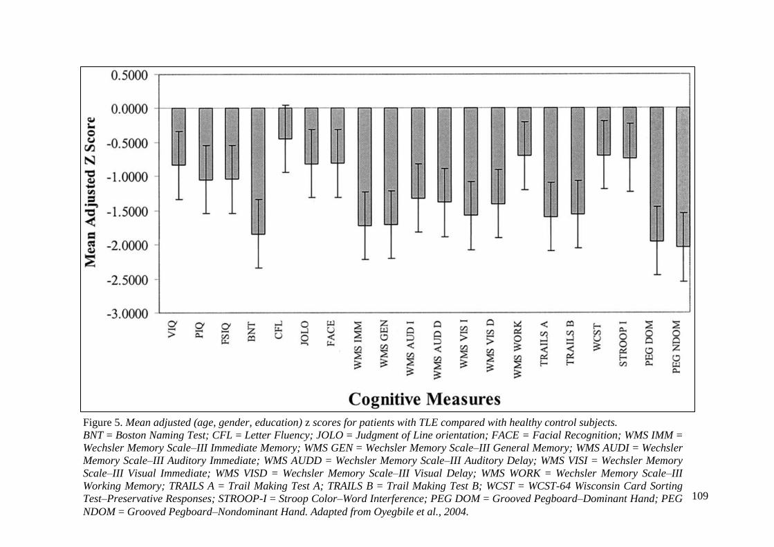
109
Figure 5. Mean adjusted (age, gender, education) z scores for patients with TLE compared with healthy control subjects.
BNT = Boston Naming Test; CFL = Letter Fluency; JOLO = Judgment of Line orientation; FACE = Facial Recognition; WMS IMM =
Wechsler Memory Scale–III Immediate Memory; WMS GEN = Wechsler Memory Scale–III General Memory; WMS AUDI = Wechsler
Memory Scale–III Auditory Immediate; WMS AUDD = Wechsler Memory Scale–III Auditory Delay; WMS VISI = Wechsler Memory
Scale–III Visual Immediate; WMS VISD = Wechsler Memory Scale–III Visual Delay; WMS WORK = Wechsler Memory Scale–III
Working Memory; TRAILS A = Trail Making Test A; TRAILS B = Trail Making Test B; WCST = WCST-64 Wisconsin Card Sorting
Test–Preservative Responses; STROOP-I = Stroop Color–Word Interference; PEG DOM = Grooved Pegboard–Dominant Hand; PEG
NDOM = Grooved Pegboard–Nondominant Hand. Adapted from Oyegbile et al., 2004.

110
Similarly, a cross-sectional study of TLE patients reported a large proportion (86%) had
cognitive impairment compared to normative test data (Wang et al., 2011). Of these, 78%
demonstrated neuropsychological morbidity in at least one domain, in order of magnitude:
semantic memory (87%), language (78%), psycho-motor speed (69%), verbal episodic
memory (64%) and executive function (55%).
4.3 Executive function in TLE
The frontal lobes play a crucial role in higher-level cognitive processes and are a key
substrate for executive function (Gilbert & Burgess, 2008). Executive function refers to the
ability to maintain an appropriate problem solving set for the attainment of future goals, and
encompasses: decision making, response inhibition, concept formation, cognitive flexibility,
planning, sustained attention (Zamarian et al., 2011). Lesion and neuroimaging evidence has
consistently reinforced the role of the pre-frontal cortex (PFC) in executive function (Alvarez
& Emory, 2006). More recently, however, studies have shown that executive function is
sustained by a distributed neural circuit composed of multiple sectors of the PFC interacting
with other cortical and subcortical regions that are themselves richly interconnected (Clark,
Chamberlain & Sahakian, 2009). For example, patients with focal lesions in the basal
ganglia exhibit cognitive switching difficulties (Cools, Ivry & D’Esposito, 2006), and lesions
in the caudate nucleus have been shown to be associated with impaired executive function
(Nys et al., 2006).
There are numerous direct reciprocal connections between the PFC and the medial temporal
lobe (Simmons & Spier, 2003), and neuropsychological studies indicate that executive
dysfunction in TLE is not uncommon (Stretton et al., 2012). In a seminal study, Hermann et

111
al. (1988) used the Wisconsin Card Sorting Task (WCST) in 35 TLE patients. The WCST is
a well-established executive function measure that taps planning, organisation and the use of
environmental feedback to shift cognitive set. Fifty-seven per cent of the TLE group (n=20)
performed in a manner suggestive of frontal-lobe pathology (increased preservative
responses), compared to 17% of a primary generalised epilepsy control group. Consistent
with these findings, Corcoran and Upton (1993) used the modified WCST in TLE+HS, TLE-
only and FLE patients. TLE+HS patients took significantly longer to complete the task and
made more perseverative errors than the TLE-only and FLE groups. Based on the number of
perseveration errors, 75% of TLE+HS patients were categorised as having ‘frontal lobe
damage’ compared to 28% of the FLE group. Similarly, Strauss, Hunter and Wada (1993)
assessed 77 pathologically heterogeneous unilateral TLE patients with the WCST.
Perseveration errors were significantly greater in left TLE patients, but only if TLE onset
occurred before the age of one year. Set-shifting ability in right TLE patients was less severe,
but occurred independently of age of onset. Subsequent studies have also reported executive
impairment (increased perseveration errors on the WCST) in TLE patients (Horner et al.,
1996; Oddo et al., 2003; Oyegbile et al., 2004; Kim et al., 2007; Zamarian et al., 2011),
particularly those with HS (Giovagnoli et al., 2001).
There is limited research using other measures of executive function in TLE (Stretton et al.,
2012). Labudda et al. (2009) assessed decision-making in 20 unilateral mesial TLE patients
using a computerised version of the Iowa Gambling Task (IGT). This test is designed to
examine how feedback moderates the decision-making process. Compared to healthy
controls, mTLE patients were significantly impaired in their decision making, with a
preference for increased disadvantageous decisions. Sub-group analysis revealed that those

112
patients with the poorest decision-making performed less well on other tests of executive
function.
Zamarian et al. (2011) used a battery of neuropsychological tests to assess different aspects of
executive function in mTLE. Relative to healthy controls (n=20), unilateral TLE patients
(n=28) performed poorly on tests of working memory (Digit Span Backwards), cognitive
flexibility (Trail Making Test), categorical verbal fluency, set-shifting (WCST; perseveration
errors), categorisation (WCST; completed categories) and planning.
One possible explanation for the observed executive skills weakness in association with TLE
is that structural/functional abnormalities may extend beyond the temporal lobe, and these
extra-temporal abnormalities may have additional cognitive consequences (Bell et al., 2011).
Few neuroimaging studies have examined the relationship between executive function and
TLE (Stretton et al., 2012). An FDG-PET study demonstrated that hypometabolism in
prefrontal structures were related to executive impairment in patients with TLE (Jokeit et al.,
1997). A neuropsychological test battery tapping executive function was administered to 96
TLE patients within 3 months of scanning. Prefrontal hypometabolism was reported in 28%
of patients. Patients with prefrontal hypometabolism (versus those without) performed
significantly more poorly on measures of frontal lobe function; the degree of hypometabolism
was positively related to poorer performance.
Keller and colleagues (2009) investigated executive function in TLE using quantitative MRI
(VBM) in 43 unilateral TLE patients and 30 health controls. Relative to healthy controls,
patients with TLE had atrophy in the ipsilateral hippocampus and bilateral PFC. Executive
function as measured by phonemic fluency was positively correlated with the left dorsal PFC
and left hippocampal volumes.

113
The range of cognitive impairments observed in TLE has led to attempts to identify cognitive
phenotypes (Hermann et al., 2007; Dabbs et al., 2009). Hermann et al. (2007) applied a
cluster analysis to the test scores 96 chronic TLE patients and 82 healthy controls. Three
cognitive profile types were identified: (1) minimally impaired (47%); (2) memory impaired
(24%); and (3) memory, executive and speed impaired (29%). The latter subset presented
with reduced cortical thickness in temporal and extra-temporal structures, compared to
healthy controls and the less cognitively compromised TLE patients (Hermann et al., 2007;
Dabbs et al., 2009). The clinical and demographic features between the cluster groups were
not strikingly different given the divergent nature of the cognitive and imaging results (Dabbs
et al., 2009). Notably, the authors did not consider possible psychopathological differences
between the groups.
The mechanism responsible for frontal lobe dysfunction in TLE remains unclear (Devinsky et
al., 2005). Suggestions include more widespread brain pathology as discussed above, the
secondary spread of epileptic activity to frontal brain circuitry, and the propagation of
temporal lobe hypo-metabolism to the thalamus, secondarily affecting the frontal lobes
(Hermann et al., 1988; Bell et al., 2011). More research is needed to understand the
widespread cognitive sequelae of TLE, including whether psychopathology and cognition are
associated, and what could be the potential mechanisms underlying such an association
(Gilliam, Albertson & Driscoll, 2011; Wilson, 2011).

114
4.4 Relationship between Psychopathology and Cognitive
Function in TLE
Evidence from both neuropsychological and neuroimaging studies suggest that temporal and
extra-temporal cognitive function may be compromised in TLE (Stretton et al., 2012). It is
also recognised that neurocognitive impairment, including compromised executive function,
attention and concentration, memory and processing speed occur in primary major depressive
disorder (MDD) (see Austin, Mitchell & Goodwin 2001; Clark et al., 2009; McClintock et
al., 2010).
Gorwood et al. (2008) assessed memory function in a large primary care cohort (n=8,229)
who fulfilled the DSM-IV criteria for MDD. Using structural equation modelling, they found
that prose recall was negatively associated with the number of past depressive episodes.
Memory performance was cumulatively impaired by 2-3% for each depressive episode up to
four episodes. The authors suggest that depression is directly involved in hippocampal
atrophy, a hypothesis supported by consistent neuroimaging evidence of hippocampal
atrophy in MDD (Cambell et al., 2004); the most common histo-pathological substrate of
TLE (Kim et al., 2001).
In parallel to neuropsychological evidence in TLE, executive dysfunction in MDD is evident
across a range of paradigms (Rogers et al., 2004). Martin et al. (1991) found that MDD
patients performed significantly less well on the Digit Span subtest of the WAIS-R compared
to dysthymic and healthy subjects. Additionally, depressive symptom severity as measured
by BDI score was found to be an independent predictor of total errors, perseverative
responses and failure to maintain set on the WCST. In a larger study, Grant et al. (2001)
administered the WCST to 123 non-medicated depressed outpatients. Relative to healthy

115
controls, depressed patients completed fewer categories and exhibited increased perseverative
errors. Stordal et al. (2004) compared the performance of 45 recurrent MDD patients to
healthy controls on a neuropsychological test battery tapping sub-components of executive
function. MDD patients performed significantly less well on 80% of the tasks, including
measures of verbal fluency, inhibition, working memory and set-shifting.
Despite the prevalence of depression and widespread neuropsychological morbidity in TLE,
and the historical association of depression and diffuse cognitive dysfunction (Clark et al.,
2009; McClintock et al., 2010), few studies have directly investigated the possible
relationship between depressed mood and cognition in TLE. It is clear that to fully
understand the cognitive and psychiatric comorbidities in TLE, they should not be studied in
isolation (Gilliam et al., 2011).
In an early investigation, Hermann and co-workers (1991) examined the relationship between
WCST performance and depressive symptoms in 64 unilateral TLE patients. Overall, 45% of
TLE patients obtained in excess of 19 perseveration responses, indicating clinically
significant frontal lobe dysfunction. As predicted, there was a positive relationship between
frontal lobe function (perseverative responses) and dysphoric mood state, particularly for left-
sided patients.
Corcoran and colleagues (1993) explored the determining factors of memory complaints in
epilepsy patients and included indicators of mood. Those reporting significant memory
difficulties were significantly more depressed and anxious compared to non-complainers. A
relationship between depressive morbidity and diminished verbal and visual memory
performance was also observed. This latter finding possibly suggests that bi-temporal
cognitive functions may be compromised in epilepsy patients with comorbid
116
psychopathology. Consistent with this hypothesis, Wishart et al. (1993) reported that
depressive symptoms were related to performance deficits in attention/concentration, verbal
and visual memory in a heterogeneous epilepsy cohort.
In the only study to specifically investigate the relationship between cognition and mood state
in newly diagnosed epilepsy patients (n=51), Pulliainen et al. (2000) found no association
between emotional state and cognitive function. However, the variability of depressive
symptoms was within the normal range, and none of the patients had major depression. Thus,
this negative finding only relates to epilepsy patients with sub-clinical depressive symptoms.
It may also indicate that a longer duration of epilepsy with poorer controlled seizures may be
a pertinent variable.
Paradiso et al. (2001) found that after controlling for seizure frequency, clinically depressed
psychotropic-naive TLE+HS patients (n=24) exhibited poorer performance on measures of
IQ, language, visuo-spatial skills, memory and executive function, compared to TLE-only
controls (n=46). Sub-group analyses revealed that the deleterious impact of depression was
accentuated in patients with left (n=15) compared to right TLE (n=9).
In a retrospective analysis of 84 medically refractory TLE patients, Dulay et al. (2004)
examined the relationship between the severity of depression and performance on measures
of auditory memory and learning. They found that the severity of depressive symptoms was
inversely associated with auditory memory test performance, particularly for left-sided TLE
patients. Although suggestive of an interaction, the authors warrant caution due to limited
statistical power. In a well-powered prospective study, Helmstaedter et al. (2004) examined
the interaction of depressive mood (BDI >12) and memory, as a function of seizure
lateralisation and localisation (mesial/lateral, left/right). They found a main effect of

117
laterality with left-sided TLE patients demonstrating verbal learning deficits and right-sided
patients exhibiting visual learning deficits. Furthermore, compared to lateral TLE patients,
mesial patients performed more poorly. Correlational analyses revealed interactions between
memory and mood only for the left lateral TLE group, and this was present for both measures
of verbal and nonverbal memory. The authors propose that epileptic activity from the left
temporo-lateral lobe may establish a pathological circuit that includes the frontal brain
regions, resulting in both memory and mood dysfunction.
Tracy et al. (2007) investigated whether depressed TLE patients exhibited increased cognitive
deficits relative to non-depressed epilepsy controls. Contradicting previous reports (Corcoran
et al., 1993; Wishart et al., 1993; Parasido et al., 2001; Dulay et al., 2004; Helmstaedter et al.,
2004), they found depression (BDI ≥ 10) was not related to memory performance, nor did it
mediate the cognitive presentation of left or right TLE. However, the overall level of
depressive morbidity in this study was low for both the left (mean BDI=9, SD=7) and right
(mean BDI=7, SD=7) TLE groups. Furthermore, weak statistical power may have precluded
the determination of interaction effects. These factors may have mitigated any possible effect
of depressive morbidity on cognition to be detected.
4.5 Relationship between Preoperative Psychiatric Status and
Cognitive Outcome Following TLE Surgery
Since the case of H.M, who underwent a bilateral temporal lobe resection for the relief of
epilepsy and subsequently developed a dense amnesia (Corkin, 2013), a voluminous literature
has focussed on memory outcome following temporal lobe surgery (see, Helmstaedter, 2004;
Téllez-Zenteno et al., 2007; Baxendale, 2008; Baxendale, Thompson & Sander, 2013).

118
Sherman and colleagues (2011) conducted a systematic review of 23 neuropsychological
studies using single pooled estimates of gains and losses for each cognitive domain (memory,
language, executive function and attention) using a random effects model. Weighted
estimates indicated the risk to verbal memory with left-sided TLE surgery (44%) was twice
as high as the rate for right-sided surgery (20%). Naming was reduced in 34% of left-sided
TLE patients, with few patients showing gains (4%). Pooled data on IQ, executive function
and attention indicated few patients show declines postoperatively, but a substantial rate of
improvement in verbal fluency with left-sided TLE surgery was found (27%). The authors
however, did not subgroup their analyses according to preoperative psychiatric status.
To date, the relationship between pre-surgical psychopathology and cognitive change has
received little attention. One retrospective study reported that patients with a poor pre-
surgical mood status (BDI ≥16) who underwent a left ATLR had a greater decline in memory
test performance (auditory delayed recall) compared to non-depressed patients at the 7 month
follow-up (Busch et al., 2011). Differences in seizure outcome (Engel 1A: seizure freedom
or 1B: continued auras) and post-surgical mood change could not account for the observed
memory decline. The authors hypothesised that the greater decline in auditory memory
capacity observed in depressed patients following surgery may be related to limited
functional cognitive reserve.
Cognitive reserve refers to the proposal that individuals with neurological impairment utilise
the cognitive capacity of undamaged brain regions to compensate for existing cognitive
deficits. The higher the cognitive reserve capacity, as evinced by good cognitive test
performance on tests subserved by intact brain regions, the better the cognitive outcomes.
Previous neuroimaging evidence, as outlined in Chapter 3, indicated that patients with TLE
and comorbid psychopathology show additional widespread functional brain abnormalities.
Busch et al. (2011) accordingly suggested that depressed mood may be a biomarker of

119
reduced cognitive reserve. This study however, was limited by the relatively short follow-up,
the failure to account for the diagnosis of depression on postoperative memory decline, and
the inclusion of auras (Engel 1B), indicative of epileptic activity and limbic dysfunction,
which may have confounded the primary finding.
4.6 Conclusion
Until recently, episodic memory impairment was viewed as the ‘signature’ cognitive deficit
in TLE (Bell et al., 2011). The studies reviewed here suggest that the cognitive
complications of TLE can be heterogeneous, deleteriously affecting diverse abilities such as
language, attention and frontal lobe (executive) function.
The mechanism(s) responsible for executive dysfunction in TLE remains unclear. Our
understanding of this association may be increased by research investigating the possible
relationship between psychopathology and cognition in TLE, particularly as widespread
neuropsychological morbidity is evident in primary depressive disorder (see Figure 6). It is
clear that to fully understand the cognitive and psychiatric comorbidities in TLE, they should
not be considered in isolation. Further research is warranted into the relationship between
pre-surgical psychopathology and cognitive change.

120
Figure 6. A schematic summary of work reviewed. A) Epidemiological evidence suggests a bidrectional relationship between TLE and
psychopathology (Forsgen et al., 1990; Hesdorffer et al., 2000; 2006; 2012; Adelöw et al., 2012). B) TLE is associated with temporal and
extra-temporal (executive) cognitive dysfunction (Hermann et al., 1988; Corcoran et al., 1993; Strauss et al., 1993; Horner et al., 1996;
Hermann et al., 1997; Drake et al., 2000; Giovagnoli et al., 2001; Oddo et al., 2003; Oyegbile et al., 2004; Kim et al., 2007; Wang et al.,
2011; Zamarian et al., 2011); with limited evidence that cognitive morbidity is more pronounced in TLE patients with co-morbid depression
(Hermann et al., 1991; Corcoran et al., 1993; Wishart et al., 1993; Parasido et al., 2001; Dulay et al., 2004; Helmstaedter et al., 2004). C)
Similarly, MDD has been associated with diffuse neurocognitive impairment (Martin et al., 1991; Austin et al., 2001; Rogers et al., 2004;
Stordal et al., 2004; Clark et al., 2009; McClintock et al., 2010). D) Few Studies have directly investigated the possible relation between
cognitive and psychiatric morbidity in TLE. While memory decline is an established risk of TLE surgery, its association to pre-surgical
psychiatric status remains unclear.
A B
C
D

121
Chapter 5. HYPOTHESES AND APPROACH
Epilepsy surgery is considered an effective treatment for refractory TLE, and carries a 60-
70% chance of seizure freedom (Engel, 1996). For many surgical patients, the long-term
psychosocial gains with respect to employment and independent living can be significantly
more favourable than for patients who are medically treated (Mikati et al., 2006). Following
TLE surgery however, psychiatric symptoms may develop for the first time (de novo) or pre-
existing symptoms may be exacerbated. Understandably, this may tarnish an otherwise good
surgical outcome, resulting in significant distress for patients and their families (Moss et al.,
2009).
Evidence from the reviewed literature (see Chapter 2) indicates there is a high prevalence of
psychiatric disturbance in medically refractory TLE. Despite this, less than 3% of TLE
surgical outcome studies have investigated the psychiatric sequelae and morbidity associated
with surgery. This is disproportionate to the extent of the problem. Variable rates have been
reported for post-surgical depression, anxiety, interictal- and post-ictal psychoses. Until
recently, very few studies distinguished de novo postoperative presentations from pre-
existing conditions, making it difficult to accurately assess the impact of TLE surgery on
psychiatric morbidity.
Whilst it is well documented that pre-surgical psychiatric conditions such as affective
disorders (Devinsky et al., 2005; Wrench et al., 2011), increase the risk of post-surgical
psychiatric morbidity, it is less clear whether there are risk factors for the development of
postoperative de novo psychiatric disorders (Spencer et al., 2008). The pathogenic
mechanisms and identification of surgical candidates at risk of de novo depression represents
a current challenge in the practice of TLE surgery (Wrench et al., 2011a). Previous research
has indicated that depression following TLE surgery is likely to be multi-factorial, including

122
psychosocial adjustment difficulties (Wilson et al., 2004), but few studies have investigated
the neuroanatomical substrates of de novo depression (see Chapter 3). This is of interest
given the current evidence of structural and functional abnormalities in primary depression,
implicating orbital and medial prefrontal cortices and anatomically related structures within
the limbic, striatal, thalamic and basal forebrain structures (see Price et al., 2012 for review).
As outlined in Chapter 4, the relationship between cognitive function and psychiatric
outcome following TLE surgery has not been systematically studied to date. With the
exception of IQ (Hermann et al., 1992; Ney et al., 1998; Blumer et al., 1998; Quigg et al.,
2003), no study has examined whether pre-surgical memory deficits or the existence of more
extensive cognitive disturbance is predictive of post-surgical psychopathology. This is of
interest given the evidence of an interaction between depression and cognitive function (see
Figure 6). Neuropsychological and neuroimaging studies in primary depression have
reported executive and memory deficits (Austin et al., 2001), and frontal volumetric
abnormalities (Bremner et al., 2002). A PET study has implicated extra-temporal or frontal
dysfunction, including the inferior dorsolateral frontal cortex bilaterally, in TLE+HS patients
with preoperative co-morbid depressive symptoms and no prior exposure to antidepressant
treatment (Lothe et al., 2008). Salzberg et al. (2006) demonstrated that preoperative
ipsilateral orbitofrontal hypometabolism in surgical TLE patients is a significant risk factor
for the development of depression postoperatively. Preoperative bilateral independent spike
discharges are significantly associated with poor post-surgical psychiatric outcome (Anhoury
et al., 2000) and more recently, Wrench et al. (2009) found that patients with reduced
preoperative contralateral hippocampal volume are at significantly greater risk for the
development of postoperative depression. Collectively, these studies suggest that patients
with more diffuse epileptogenicity and more widespread cognitive disturbance maybe at
greater risk of developing de novo psychopathology.

123
The relationship between preoperative psychiatric status and postoperative seizure outcome
warrants further investigation. It has been suggested by some that a lifetime psychiatric
history may predict a poor seizure outcome following TLE surgery (Anhoury et al., 2000;
Kanner et al., 2009; Guarnieri et al., 2009).
Further exploration of the aforementioned topics would contribute to the surgical decision-
making process and enhance postoperative care.
5.1 Hypotheses
The main hypothesis formulated and explored here is that TLE patients with less localised
cerebral dysfunction, as supported by electrophysiological, neuro-radiological and cognitive
indicators, will be at risk for psychiatric disturbance preoperatively and have poorer
outcomes following TLE surgery. Specifically:
Hypothesis 1: Patients with TLE under consideration for surgical treatment will have
significant psychiatric comorbidity, with major depression as the most prevalent diagnostic
category.
Hypothesis 2: Surgical candidates with psychopathology will have evidence of extra-
temporal lobe dysfunction as measured by clinical and cognitive parameters.
Hypothesis 3: A lifetime psychiatric history will be associated with poorer surgical outcomes
in terms of seizure control, cognitive and psychiatric status.
Hypothesis 4: Preoperative indicators of more widespread cerebral pathology will be risk
factors for psychiatric morbidity following surgery.

124
5.2 Approach
These hypotheses were explored firstly using data derived from a retrospective survey with
methods and findings presented in chapters 6-9; and secondly via prospective studies
presented in chapters 11 and 12. The final chapter provides an overall summary of the
findings, their implications, and discusses methodological limitations and directions for
future research.
125
SECTION 2:
RETROSPECTIVE AND
PROSEPCTIVE STUDIES

126
Chapter 6. RETROSPECTIVE STUDY METHODOLOGY
This chapter describes common methods that were used for the studies detailed in Chapters 7-
9. Methods used specifically for one study are described in the relevant chapters. All
research studies were approved by the Research Ethics Committee of the UCL Institute of
Neurology and UCL Hospitals.
6.1 Sample
Patients who had undergone epilepsy surgery at the National Hospital for Neurology and
Neurosurgery between 1997 and 2007 were identified from a surgical database. Of the 384
patients identified, 104 were excluded from the study due to extra-temporal surgeries (n=31),
palliative procedures (n=12), previous neurosurgery (n=16) or incomplete psychiatric data
(n=45); the latter determined by the absence of a routine postoperative neuropsychiatric clinic
letter (see Figure 7). A single coder (RAP) systematically collated information from case the
notes of the remaining 280 temporal lobe surgical patients over a fourteen year period (1997-
2011). However, due to sample attrition (see Figure 8) and therefore reduced statisical
power, analyses were based on a 4-year follow-up.

127
384 surgical patients:
1997-2007
Excluded patients, n=104
Extra TL surgery,
n=31
Previous neurosurgery,
n=16
Palliative surgeries,
n=12
Incomplete psychiatric
data, n=45
280 TLE patients:
1997-2007
Figure 7. Case selection process.

128
Figure 8. Sample attrition of the TLE patients (n=280) who underwent surgery between 1997 and 2007. Data analyses based on 4-year follow-up (red line).

129
6.2 Epilepsy
Seizure related variables were recorded from a standardised template completed during the
pre-surgical evaluation. The following seizure related variables were recorded; a history of
febrile convulsion(s), secondary generalised tonic-clonic seizures (SGTCS), status
epilepticus, head trauma, CNS infection (meningitis/encephalitis) and family history of
epilepsy. Postoperative seizure outcome was taken from a surgical follow-up database and
classified according to the International League Against Epilepsy (ILAE) surgical
classification scheme (Wieser et al., 2001; see Table 7).
Table 7. ILAE surgical classification scheme. Adapted from Wieser et al., 2001
Outcome Definition
Classification
1 Completely seizure free; no auras
2 Only auras; no other seizures
3 One to three seizures per year ± auras
4 Four seizure days per year to 50%
reduction of baseline seizure days ± auras
5 Less than 50% reduction of baseline
seizure days to 100% increase of baseline
seizure days ± auras
6 More than 100% increase of baseline
seizure days ± auras

130
6.3 Neuropsychiatric classification
All patients were assessed by a neuropsychiatrist as part of their pre-surgical evaluation.
Psychiatric diagnoses pre- and postoperatively were made by a neuropsychiatrist following a
clinical interview as described previously (Anhoury et al., 2000), and written documentation
of psychiatric treatment (CBT/psychotherapy/psychiatric medication) was recorded. There
were a few cases (n=5) of psychiatric ambiguity, for example a psychiatric medication was
mentioned in a neurological clinical letter without prior introduction in the neuropsychiatry
notes. In such instances, clarification was sought from the treating neurologist.
The presence or absence of diagnosed family psychopathology (first-degree relative) was also
documented. Psychiatric conditions were categorised according to the DSM-IV-R Axis I
diagnoses, namely mood and anxiety disorders. The presence of postictal psychosis (PIP),
interictal psychosis (IP) and non-epileptic seizures (NES) was also recorded. For comorbid
psychopathology, a positive entry was made into each diagnostic category. Medication
related psychiatric symptoms diagnosed by the neuropsychiatrist, defined as psychopathology
temporally related to AED change(s) that resolved following AED titration/discontinuation,
were excluded.
Psychiatric outcome was derived from neuropsychiatry clinic letters and documentation of
psychiatric treatment. The emergence or continuation of postoperative psychopathology was
recorded according to when a formal diagnosis was documented by the neuropsychiatrist, and
classified according to the DSM-IV-R pre-surgically, but with the addition of adjustment
disorders.

131
6.4 De novo neuropsychiatric cases
De novo psychopathology was only coded if a patient had no preoperative psychiatric history
and could only be termed as such at one mutually exclusive post-surgical time-frame. All
patients had a clinical neuropsychiatric evaluation at 3 and 6 months post-surgically.
Psychiatric symptomatology emerging after this would be identified by the general
practitioner or the treating neurologist resulting in a review by the epilepsy surgical team’s
neuropsychiatrist. Subsequently, documented psychiatric disorders were then coded as
appropriate into the following postoperative time-frames: 12, 24, 36 and 48 months.
6.5 Neuropsychological tests
Cognitive data was derived from neuropsychological assessments routinely performed pre-
operatively and between three and six and twelve months post-operatively.
General ability:
Intellectual level was measured using the Wechsler Adult Intelligence Scales (WAIS-
R/WAIS-III). Patients also underwent the Nelson Adult Reading Test (NART; Nelson, 1982)
preoperatively. This measures reading competence and has been validated as a measure of
intellectual potential (Bright et al., 2002; McGurn et al., 2004). A discrepancy between
NART predicted IQ and assessed IQ in favour of the former measure indicates intellectual
decline, and serves as an indicator of extra-temporal cerebral disturbance.

132
Memory
Scores from the List and Design Learning subtests from the Adult Memory and Information
Processing Battery (AMIPB; Coughlan & Hollows, 1985) were used. The learning
paradigms in particular have been demonstrated as sensitive to mesial temporal lobe
pathology and surgical treatment (Baxendale et al., 2008).
List Learning Task
The patient is read a list of 15 high-frequency words on five occasions. The patient has to
recall as many words as possible after each trial. The total number of words recalled is
recorded (List A1-5; max=75). A second list of 15 distractor words is then presented. The
patient is required to recall as many as possible from the second list (List B; max=15) and
then from the original list (A6; max=15).
Design Learning Task
The format parallels that of the List-learning task. The patient is presented with a 9 element
abstract design (Design A) and asked to reproduce it from memory on a 4 x 4 dot grid over 5
trials. A distractor design (Design B) is then presented. The patient is then asked to
reproduce the original design without further exposure. The total number of elements
recalled over the 5 trials is recorded (Design A1-5; max=45). The delayed recall of Design A
(Design A6; max=9), is also recorded.
Performance levels for List- and Design Learning were translated into Z-scores and used as
measures of hippocampal integrity.

133
Executive Functions
Sufficient data was available for only one measure considered sensitive to frontal lobe
pathology, namely phonemic fluency (Robinson et al., 2012). The performance indicator
employed was the total number of words beginning with “S” produced in 60 seconds; names
of people, places and repetitions are not permitted.
6.6 Aetiology
The aetiology for each case (n=280) was identified as either (1) hippocampal sclerosis, (2)
developmental pathology (encompassing dysembryoplastic neuroepithelial tumours
[DNETs], microdysgenesis, cortical dysplasias and angiomas), (3) vascular pathology
(encompassing cavernous malformation and arteriovenous malformation), and (4) “other”
pathology (encompassing gliosis, gliomas, hamartoma and nonspecific pathology). For
instances of more than one pathology, most usually hippocampal sclerosis and
microdysgenesis, a positive entry was made in both diagnostic categories.
6.7 MRI & video-EEG data
Temporal lobe laterality was determined using preoperative MRI scans and VEEG reports.
Preoperative telemetry reports were used to classify cases regarding the presence or absence
of discordant interictal EEG abnormalities. Discordant interictal EEG was based on any
epileptiform discharges that conflicted with neuro-radiological findings (e.g. independent bi-
temporal or contralateral spikes/multifocal spikes/fronto-temporal discharges in the presence
of unilateral HS) as defined by VEEG reports.

134
6.8 Statistical Analyses: Retrospective and Prospective Studies
For Chapters 7-11, all statistical analyses were conducted with SPSS for Windows (Inc,
Chicago, IL, U.S.A.), and a criterion level of p ≤ 0.05 was set for statistical significance.
6.8.1 Kolmogorov-Smirnov test
For parametric testing a key assumption is that the sampling distribution is normally
distributed (Field, 2009). For continuous variables, the Kolmogorov-Smirnov test (K-S test)
was used to assess the assumption of normality. The K-S test compares the scores in the
sample to a normally distributed set of scores with the same mean and standard deviation.
When the K-S test was non-significant (p > 0.05), indicating that the sample is not
significantly different from a normal distribution, parametric tests were employed. However,
if this test was significant (≤ 0.05), non-parametric analyses were conducted.
6.8.2 Mann-Whitney U test
If the assumption of normality was not met and there were two samples, the Mann-Whitney
U test was employed. This test ranks the data (lowest to highest) for each sample, and then
calculates whether the difference in ranks between the samples is greater than expected if the
null hypothesis were correct (Field, 2009).

135
6.8.3 Chi-squared test (X2)
For categorical variables the X2
test was used. This test examines whether the frequencies
observed in certain categories would be expected by chance. For small samples, Fisher Exact
Test (FET) was employed (Field, 2009).
6.8.4 Logistic Regression
As discussed in Chapter 2.10, logistic regression is a preferable uni- and multivariable
technique compared to X2, as predictor variables can be continuous and/or categorical, and
the association between predictor(s) and the categorical outcome variable can be quantified.
Although there are many similarities between linear and logistic regression, the former
assumes that there is a linear relationship between the variables (i.e. the mean values of the
outcome variable for each increment of the predictor(s) lie along a straight line). However,
when the outcome is dichotomous this assumption is violated (Miles & Shevlin, 2011).
Therefore, in logistic regression the raw data is transformed using the logarithmic
transformation and thereby expresses a non-linear relationship in a linear way.
For any mathematical modelling approach, equation 1 applies, where Y denotes the
dependent variable as a function (f) of independent/predictor variables(s), X.
(Equation 1)
Y = f (X)

136
In logistic regression, Y will be equal to the logarithm/logit (ln) of the odds (the probability
of an event occurring (p) divided by the event not occurring (1-p)), as described below:
(Equation 2)
Ln(p/1-p) = f (X)
Crucial to the interpretation of logistic regression is the odds ratio (OR), which is an indicator
of the change (Δ) in odds (the probability of an event occurring divided by the event not
occurring), resulting from one unit change in the predictor.
(Equation 3)
Δ odds = odds after a unit change in the predictor/original odds
If the value is greater than 1, it indicates that as the predictor increases, the odds of the
outcome occurring increase. Conversely, a value less than 1 indicates that as the predictor
increases, the odds of the outcome decrease.
For the studies outlined in chapters 7 and 9, the logit transformation was inverted by
exponentiating (exp) (taking the anti-logarithm of both sides of equation 2) of the odds of the
event (Field, 2009):
(Equation 4)
p/1-p = exp (f (X))

137
p = (1-p) exp (f (X))
p(1+exp (f (X)) = exp (f (X))
p = exp (f (X)) / [1+exp (f (X)]
This permitted the probability/chance of the event to be calculated. In addition, the
contribution of each predictor and their combined effect on the chance (rather than odds) of
an outcome could be assessed.
6.8.5 Spearman’s Correlation Coefficient
The Spearman’s correlation coefficient, rs, was used to investigate the correlation between
non-normally distributed data (Field, 2009). For significant correlations, rs was squared
(coefficient of determination, Rs2) giving a measure of the proportion of shared variance
between the two ranked variables.

138
Chapter 7.
Study 1. Predictors of Psychiatric and Seizure Outcome following
Temporal Lobe Epilepsy Surgery
7.1 Study Aims
The aims of this study were (1) to investigate psychiatric morbidity in pre-surgical TLE
patients, and to assess whether depression was the most prevalent diagnostic category. (2)
Examine whether TLE patients with lifetime psychopathology had evidence of extra-
temporal lobe dysfunction. (3) To assess whether lifetime psychopathology was associated
with poorer surgical outcomes in terms of seizure control, cognitive and psychiatric status.
(4) To explore whether preoperative indicators of diffuse cerebral pathology were risk factors
for psychiatric (de novo) morbidity following surgery.
7.2 Methods
The study sample consisted of 280 patients who had undergone TLE surgery at the National
Hospital for Neurology and Neurosurgery between 1997 and 2007 (see Figure 7).
Demographic and clinical characteristics according to seizure laterality are summarised in
Table 8; and these were comparable to those reported in previous surgical series (Anhoury et
al., 2000; Wrench et al., 2009).
Neuropsychological tests
Poor test performance on both the List- and Design learning tests was taken as one indicator
of more widespread brain dysfunction. It was anticipated that lifetime psychopathology
would be associated with verbal and visual (bi-temporal) memory deficits and this cognitive

139
profile would place patients at greater risk of de novo psychopathology. For the purposes of
X2 and logistic regression analyses, patients were dichotomised into those who demonstrated
preoperative bi-temporal memory deficits (globally weak memory) and those who did not.
Globally weak memory was based on a preoperative score of at least 1.5 standard deviations
below the mean for both verbal and non-verbal memory (see Figure 9a & b for distribution
statistics and justification).
Post-operative memory decline was defined as a performance decline of at least one standard
deviation on the List and Design Learning tests.
Executive skills deficits were also hypothesised to be associated with preoperative
psychopathology, and considered a pertinent risk factor of developing a de novo psychiatric
disorder postoperatively. Phonemic fluency performance was used as the indicator of
executive function.
A
Skewness: -0.4
Median: -1.26
IQR: -2.3, -0.33
B
Skewness: -0.5
Median: -0.25
IQR: -1.20, -0.51
Figure 9. Given the negative skew for both (A) verbal (List learning) and (B) non-verbal
(Design learning) memory measures, ‘globally (bi-temporal) weak memory’ was based on a
preoperative score of at least 1.5 standard deviations (dashed red line) below the mean,
ensuring that the most clinically impaired TLE memory patients were correctly identified.
IQR, interquartile range

140
Table 8. Demographic and clinical characteristics of 280 patients who underwent TLE
surgery.

141
7.3 Statistical Analyses
Chi-square (X2) analysis was used to examine whether 1) lifetime psychopathology was
associated with widespread cerebral dysfunction, as measured by clinical and cognitive
parameters and 2) whether de novo psychiatric disorders were related to postoperative
cognitive decline or the presence of seizure freedom within either 12 or 48 month follow-up.
Logistic Regression
The variables entered into the logistic regression analyses were selected on the basis of
previous literature and those which had not previously been explored.
In univariable and multivariable logistic regression analyses, the independent pre-surgical
variables to predict the odds of a de novo psychiatric diagnosis within four years follow-up
were: globally weak memory (i.e. bi-temporal memory deficits); phonemic fluency score;
discordant interictal EEG abnormalities; history of generalised seizures (SGTCS); history of
status epilepticus; epilepsy duration; side of resection; family history of psychopathology.
The same independent variables were used to predict the odds of seizure freedom at one and
four years with the exception that pre-surgical psychiatric diagnosis replaced family history
of psychopathology.
142
7.4 Results
Psychiatric History
Preoperatively, 29% of TLE patients were diagnosed as having a current or past history of a
psychiatric disorder (n=81), with some individuals meeting the criteria for more than one
disorder (n=14/280; 5%). The most prevalent preoperative psychiatric diagnosis was mood
disorders (51/280; 18%), particularly depression (n=43/280; 15%), with lower rates for
postictal psychosis (n=20/280; 7%), anxiety disorders (n=13/208; 5%), NES (n=9/280; 3%)
and interictal psychosis (n=2/280; 0.7%).
A positive family psychiatric history (25% vs 11%; X2(1)=8.4, p=0.005) and history of head
trauma (21% vs 11%; X2(1)=4.7, p=0.03) were associated with lifetime psychopathology in
TLE patients. Multivariable analysis revealed that the presence of both factors were
independently associated with increased odds of lifetime psychopathology (head trauma: OR:
2.2, 95%CI: 1.1-4.4, p=0.03; family psychiatric history: OR: 2.7, 95%CI: 1.4-5.2, p=0.004).
There were no other statistical associations between lifetime psychopathology and clinical or
cognitive factors (see Tables 9 & 10, respectively).
Postoperative Psychopathology
One hundred and five patients (38%; n=280) had clinically significant mental health
difficulties within four years following temporal lobe surgery. Sixty-seven per cent of
patients (n=54/81) with a preoperative psychiatric history continued to experience significant
psychiatric symptoms of diagnostic severity within four years following TLE surgery. Of
these patients, 61% (n=49/81) continued to receive psychiatric treatment (pharmacological
and/or psychological); 12% (n=10/81) required psychiatric hospitalisation and one patient

143
committed suicide within 3 months postoperatively. Patients with a preoperative psychiatric
history had over six times the odds of developing a postoperative psychiatric disorder (OR:
6.12, 95%CI: 3.49-10.76, p<0.001).
For patients with pre- and postoperative psychopathology (n=54), the most frequent
psychiatric condition was mood disorder (depression, n=38; bipolar II disorder, n=1;
dysthymia, n=1), followed by anxiety disorders (not otherwise specified, n=6; generalised
anxiety disorder, n=4; social phobia, n=1) and interictal psychosis (n=4). A few patients
developed postoperative NES (n=2), postictal psychosis and adjustment disorder (n=1,
respectively). Five patients (n=5/54; 9%) had comorbid psychopathologies.
De Novo Psychopathology
Fifty-one patients (18%; n=51/280) developed a de novo psychiatric condition within 48
months postoperatively (see Table 11 & Figure 9c). Of these, nine patients (n=9/51; 18%)
developed two diagnosed psychiatric disorders during follow-up. Table 11 presents the point
prevalence of de novo psychopathologies at specific time-intervals following TLE surgery.
Forty-nine per cent of cases (n=25/51) presented within 6 months and 90% (n=46/51) had de
novo psychopathology that persisted for at least 6 months (IQR: 12-30 months).
Overall, the most prevalent postoperative de novo psychiatric conditions were mood disorder
(all depression, n=33), followed by anxiety disorders (not otherwise specified, n=6; panic
disorder without agoraphobia, n=3; generalised anxiety disorder, n=2; posttraumatic stress
disorder, n=1; obsessive-compulsive disorder, n=1), interictal psychosis, adjustment disorder
(n=4, respectively), NES (n=3) and adjustment disorders (n=3). Forty patients (78%,
n=40/51) required psychiatric treatment (pharmacological and/or psychological) within four
year follow-up.

144
Predictors of De Novo Psychopathology
In univariable analyses, a history of SGTCS was a significant predictor of de novo
psychopathology within 4 years (OR: 2.53, 95%CI: 1.03–4.82, p=0.04). In the multivariable
analysis, a history SGTCS remained a significant predictor of de novo psychopathology after
adjusting for preoperative cognitive status, discordant interictal EEG abnormalities, side of
resection, epilepsy duration, and a history of status and familial psychopathology (OR: 2.73,
95%CI: 1.14–6.57, p=0.02) (see Tables 12 & 13).

145
Table 9. Demographic and clinical characteristics of patients with (n=81) and without (n=199)
a lifetime psychiatric diagnosis who underwent TLE surgery.
Variable Lifetime No
psychiatric psychiatric
history history
(n = 81) (n = 199)
Age of seizure onset, y,1
9 (3, 15.5) 9 (3, 16)
Epilepsy duration, y,1
24 (17, 34) 24 (16.5, 30)
Predisoposing Factors†
History of febrile convulsions 36 (44%) 100 (50%)
Family history of epilepsy 19 (24%) 45 (22%)
History of status epilepticus 15 (19%) 22 (11%)
History of head trauma 17 (21%) 22 (11%)
History of CNS infection 3 (4%) 14 (7%)
Side of seizure focus (L/R) 42/39 (52%/48%) 102/97 (51%/49%)
Family history of psychopathology 20 (25%) 22 (11%)
Presurgical Seizure Type†*
Simple partial 61 (75%) 143 (72%)
Complex partial 81 (100%) 195 (98%)
Secondary generalised 57 (70%) 140 (70%)
Medication
Total number of AEDs trialled preoperatively1
5 (3, 6) 4 (3, 6)
Monotherapy at the time of surgery† 10 (12%) 24 (12%)
Polytherapy at the time of surgery† 71 (88%) 175 (88%)
Age at surgery, y,1
33 (28, 41) 33 (28, 40)
EEG Findings†
Discordant interictal EEG 41 (50%) 84 (44%)
Histopathological Findings†
Hippocampal sclerosis 66 (81%) 160 (80%)
Developmental pathology 14 (17%) 36 (18%)
Vascular pathology 4 (5%) 9 (5%)
Other 6 (7%) 10 (5%)
Dual pathology 9 (11%) 16 (8%)
† Values are number of patients (%)
* May have >1 seizure type1
Median (IQR)2
Mean (SD)
AED=anti-epileptic drugs3
For avaliable data
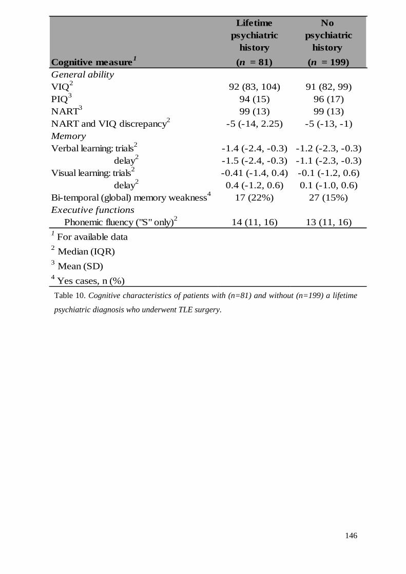
146
Table 10. Cognitive characteristics of patients with (n=81) and without (n=199) a lifetime
psychiatric diagnosis who underwent TLE surgery.
Lifetime No
psychiatric psychiatric
history history
Cognitive measure1
(n = 81) (n = 199)
General ability
VIQ2
92 (83, 104) 91 (82, 99)
PIQ3
94 (15) 96 (17)
NART3
99 (13) 99 (13)
NART and VIQ discrepancy2
-5 (-14, 2.25) -5 (-13, -1)
Memory
Verbal learning: trials2
-1.4 (-2.4, -0.3) -1.2 (-2.3, -0.3)
delay2
-1.5 (-2.4, -0.3) -1.1 (-2.3, -0.3)
Visual learning: trials2
-0.41 (-1.4, 0.4) -0.1 (-1.2, 0.6)
delay2
0.4 (-1.2, 0.6) 0.1 (-1.0, 0.6)
Bi-temporal (global) memory weakness4
17 (22%) 27 (15%)
Executive functions
Phonemic fluency ("S" only)2
14 (11, 16) 13 (11, 16)1
For available data 2 Median (IQR)
3 Mean (SD)
4 Yes cases, n (%)

147
Table 11. Point prevalence of diagnosed de novo psychopathology following TLE surgery (frequency of de novo diagnostic categories given in
parentheses).
De novo
Overall
Total
Mood disorder 3.6 (10) 1.8 (5) 2.9 (8) 2.2 (6) 0 (0) 1.6 (4) 12 (33)
Anxiety disorder 0.7 (2) 0.7 (2) 2.2 (6) 0.7 (2) 0.4 (1) 0 (0) 4.6 (13)
Adjustment disorder 1.1 (3) 0 (0) 0.4 (1) 0 (0) 0 (0) 0 (0) 1.4 (4)
Interictal psychosis 0 (0) 0.4 (1) 0.7 (2) 0 (0) 0.4 (1) 0 (0) 1.4 (4)
Postictal psychosis 0.4 (1) 0.4 (1) 0 (0) 0.4 (1) 0 (0) 0 (0) 1.1 (3)
NES 0.4 (1) 0 (0) 0 (0) 0.7 (2) 0 (0) 0 (0) 1.1 (3)
psychopathologies (%) 6 months 12 months 24 months 36 months 48 months 3 months

148
Figure 9c. Histogram of point prevalence of patients (pts) diagnosed de novo psychopathologies following TLE surgery.

149
Table 12. Characteristics of TLE patients (pts) who developed a de novo psychiatric diagnosis within four postoperative years (n=51), versus those who did not
(n=229).
Pts without a de novo psychiatric Pts with a de novo psychiatric Overall (n=280)
diagnosis within 4yrs after surgery (n=229) diagnosis 4yrs within after surgery (n=51)
Pre-op cognitive deficits:
Globally impaired memory (n=259)* 37 (17.6%) 7 (14.3%) 44 (17%)
Verbal fluency (n=247)2
14 (10, 17) 12 (10, 18) 14 (10, 17)
EEG abnormalities:
Discordant interictal EEG (n=274)* 97 (43.3) 27 (54.0) 124 (45.3)
Chronicity & severity of epilepsy:
History of SGTCS (n=280)* 155 (67.7%) 42 (82.4%) 197 (70.4%)
History of status (n=280)* 30 (13.1%) 7 (13.7%) 37 (13.2%)
Epilepsy duration (n=280)1,2
24 (16.75, 33) 24 (17, 29) 24 (17, 32)
Other:
Right TL resection (n=136) 110 (48.0%) 26 (51.0%) 136 (48.6%)
Left TL resection (n=144) 119 (52.0%) 25 (49%) 144 (51.4%)
Family history of diagnosed 32 (14.0%) 10 (19.6%) 42 (15.0%)
psychopathology (n=280)*
* = yes cases1
= yrs2
= median (IQR)

150
Table 13. Uni- and multivariable logistic regression analyses examining whether preoperative factors are predictive of patients who develop de novo
psychopathology (n=51) versus those who do not (n=229) within 4 years following TLE surgery.
Univariable analysis Multivariable analysis
Odds Ratio (95% CI) P-value Odds Ratio (95% CI) P-value
Pre-op cognitive deficits:
Globally impaired memory (Yes vs No) 0.78 (0.33-1.87) 0.60 0.78 (0.31-2.0) 0.60
Verbal fluency1
0.99 (0.93-1.10) 0.70 0.93 (0.66-1.30) 0.70
EEG abnormalities:
Discordant interictal EEG (Yes vs No) 1.54 (0.83-2.85) 0.17 1.31 (0.66-2.60) 0.44
Chronicity & severity of epilepsy:
History of SGTCS (Yes vs No) 2.53 (1.03-4.82) 0.04 2.73 (1.14-6.57) 0.02
History of status (Yes vs No) 1.01 (0.44-2.56) 0.91 0.84 (0.31-2.28) 0.84
Epilepsy duration2
0.93 (0.80-1.07) 0.29 0.90 (0.77-1.06) 0.23
Other:
Side of resection (L vs R) 0.89 (0.48-1.63) 0.70 0.87 (0.44-1.41) 0.67
Family Hx of diagnosed psychopathology (Yes vs No) 1.50 (0.68-3.30) 0.31 1.38 (0.59-3.22) 0.461 = for each 5 word increase
2 = for each 5 year increase

151
Cognitive Outcome
A decline in memory test performance of at least one standard deviation was recorded for 67
patients for verbal memory, 54 patients for visual memory with 24 declining on both
measures. We found no relationship between postoperative memory decline (at 12 month
follow-up) and a lifetime psychiatric history, or the development of de novo psychopathology
or within 12 or 48 month follow-up (p≥0.05).
Seizure Outcome
From patients with available seizure outcome data, 170 (61%; n=170/278) were seizure free
(ILAE 1) 12 months following surgery and 127 (49%; n=127/258) remained seizure free
during four years follow-up. There was no relationship between seizure outcome (ILAE 1 vs
2-6) and de novo psychiatric disorders within 12 or 48 months postoperatively (X2(1) = 0.29,
p=0.61; X2(1) = 0.36, p=0.63, respectively, see Table 14 for further breakdown).
Predictors of Seizure Freedom
The odds ratio (OR) of being seizure free at 12 months, and during four years following TLE
surgery, was significantly lower for patients with a history SGTCS (OR: 0.52, 95%CI: 0.28–
0.98, p=0.04; OR: 0.47, 95%CI: 0.25–0.90, p=0.02, respectively) and also those with a
preoperative psychiatric diagnosis (OR: 0.46, 95%CI: 0.26–0.82, p=0.009; OR: 0.53, 95%CI:
0.28–0.98, p=0.04, respectively). Both SGTCS and a preoperative psychiatric diagnosis were
independent predictors of seizure outcome. Thus, compared with patients without a history
of SGTCS, those with preoperative SGTCS had less than half the odds (OR: 0.47) of
remaining seizure free the four year follow-up period (see Tables 15-18). The presence of a
lifetime psychiatric disorder remained a strong predictor of a poor surgical outcome:

152
compared to patients without a prior history of psychopathology, those with a psychiatric
history had nearly half (OR 0.53) the odds of attaining seizure freedom during the four year
follow-up interval (see Figure 10). For clinical translation, we assessed the contribution of
each predictor and their combined effect on the chance (rather than odds) of seizure freedom
at 12 months, and remaining seizure free during 48 month follow-up (see Figure 11).

153
ILAE outcome
(%) 1 2 3 4 5 6
12 months de novo 61 13 10 14 2 0
no psych 61 13 12 13 1 0
24 months de novo 56 10 14 16 4 0
no psych 68 9 10 11 2 0
36 months de novo 65 6 8 17 4 0
no psych 67 6 10 11 6 0
48 months de novo 66 6 9 15 4 0
no psych 69 8 7 10 6 0Table 14. ILAE seizure outcome for patients who developed de novo psychopathology versus those who did not (no psych) at 4 yearly
intervals following TLE surgery.

154
Table 15. Characteristics of TLE patients (pts) who remained seizure free (ILAE=1) (n=170) versus those who did not (n=108) after TLE surgery at 12 month
follow-up.
Pts did not remain seizure free Pts who remained seizure free Overall (n=278)
12m after surgery (n=108) 12m after surgery (n=170)
Pre-op cognitive deficits:
Globally impaired memory (n=257)* 16 (16.0%) 28 (17.8%) 44 (17.1%)
Verbal fluency (n=247)2
14 (10, 17) 13 (10, 17) 14 (10, 17)
EEG abnormalities:
Discordant interictal EEG (n=272)* 50 (47. 2%) 73 (44.0%) 124 (45.3%)
Chronicity & severity of epilepsy:
History of SGTCS (n=278)* 83 (76.9%) 113 (66.5%) 197 (70.5%)
History of status (n=278)* 14 (13.0%) 22 (12.9%) 36 (12.9%)
Epilepsy duration (n=280)
1,223 (17, 32) 24 (16.4, 31) 24 (17, 31.4)
Other:
Right TL resection (n=136) 48 (44.4%) 88 (51.8%) 136 (48.9%)
Left TL resection (n=144) 60 (55.6%) 82 (48.2%) 142 (51.1%)
Preoperative psychiatric diagnosis (n=278)* 40 (37.0%) 40 (23.5%) 80 (28.8%)
* = yes cases1
= yrs2
= median (IQR)

155
Table 16. Uni- and multivariable logistic regression analyses examining whether preoperative factors are predictive of seizure freedom (ILAE=1) after TLE
surgery at 12 month follow-up.
Univariable analysis Multivariable analysis
Odds Ratio (95% CI) P-value Odds Ratio (95% CI) P-value
Pre-op cognitive deficits:
Globally impaired memory (Yes vs No) 1.14 (0.58, 2.23) 0.70 1.16 (0.55, 2.43) 0.69
Verbal fluency1
0.94 (0.73, 1.19) 0.62 0.91 (0.68, 1.19) 0.50
EEG abnormalities:
Discordant interictal EEG (Yes vs No) 0.87 (0.53, 1.43) 0.60 0.89 (0.51, 1.55) 0.68
Chronicity & severity of epilepsy:
History of SGTCS (Yes vs No) 0.59 (0.34, 1.03) 0.06 0.52 (0.28, 0.98) 0.04
History of status (Yes vs No) 0.99 (0.48, 2.04) 0.99 1.15 (0.50, 2.61) 0.73
Epilepsy duration2
0.98 (0.88, 1.09) 0.77 1.00 (0.88, 1.13) 0.99
Other:
Side of resection (L vs R) 0.74 (0.45, 1.21) 0.23 0.82 (0.47-1.43) 0.49
Preoperative psychiatric diagnosis (Yes vs No) 0.52 (0.30 - 0.88) 0.01 0.46 (0.26-0.82) 0.0091 = for each 5 word increase
2 = for each 5 year increase

156
Table 17. Characteristics of TLE patients (pts) who remained seizure free (ILAE=1) (n=127) versus those who did not (n=131) after TLE surgery during four
years follow-up.
Pts did not remain seizure free Pts who remained seizure free Overall (n=258)
within 4yrs after surgery (n=131) within 4yrs after surgery (n=127)
Pre-op cognitive deficits:
Globally impaired memory (n=237)* 18 (15.1%) 22 (18.6%) 40 (16.9%)
Verbal fluency (n=226)2
14 (10, 18.5) 14 (10, 17) 14 (10, 17.3)
EEG abnormalities:
Discordant interictal EEG (n=253)* 58 (44.6%) 55 (44.7%) 113 (44.7%)
Chronicity & severity of epilepsy:
History of SGTCS (n=258)* 101 (77.0%) 82 (64.6%) 183 (70.9%)
History of status (n=258)* 20 (15.3%) 15 (11.8%) 35 (13.6%)
Epilepsy duration (n=258)
1,224 (17, 32) 23.3 (15, 30) 24 (17, 31.1)
Other:
Right TL resection (n=136) 57 (43.5%) 66 (52%) 123 (47.7%)
Left TL resection (n=144) 74 (56.5%) 61 (48%) 135 (52.3%)
Preoperative psychiatric diagnosis (n=258)* 44 (33.6%) 27 (21.3%) 72 (27.5%)
* = yes cases1
= yrs2
= median (IQR)

157
Table 18. Uni- and multivariable logistic regression analyses examining whether preoperative factors are predictive of seizure freedom (ILAE=1) during four
years after TLE surgery.
Univariable analysis Multivariable analysis
Odds Ratio (95% CI) P-value Odds Ratio (95% CI) P-value
Pre-op cognitive deficits:
Globally impaired memory (Yes vs No) 1.28 (0.65 - 2.54) 0.47 1.14 (0.53 - 2.43) 0.72
Verbal fluency1
0.90 (0.70 - 1.15) 0.42 0.83 (0.62-1.10) 0.20
EEG abnormalities:
Discordant interictal EEG (Yes vs No) 1.00 (0.61 - 1.64) 0.98 1.00 (0.56 - 1.77) 0.99
Chronicity & severity of epilepsy:
History of SGTCS (Yes vs No) 0.54 (0.31 - 0.93) 0.03 0.47 (0.25 - 0.90) 0.02
History of status (Yes vs No) 0.74 (0.36 - 1.52) 0.41 1.02 (0.45 - 2.31) 0.96
Epilepsy duration2
0.98 (0.96 - 1.00) 0.14 0.90 (0.79 - 1.03) 0.14
Other:
Side of resection (L vs R) 0.71 (0.43 - 1.16) 0.17 0.82 (0.46 - 1.47) 0.51
Preoperative psychiatric diagnosis (Yes vs No) 0.53 (0.30 - 0.93) 0.03 0.53 (0.28 - 0.98) 0.041 = for each 5 word increase
2 = for each 5 year increase

158
Increase odds
Decrease odds
Figure 10. Clustered boxplot depicting the odds seizure freedom (ILAE=1) at 12 and during 48 month after TLE surgery, as a function of preoperative clinical
characteristics. Red boxes indicate that a history of SGTCS and/or a preoperative psychiatric diagnosis significantly reduced the odds of seizure freedom at
12 and during 48 month follow-up for TLE patients.

159
Figure 11. The modifying effect of preoperative clinical factors on a patient’s chance (%) of being seizure free at 12 months and remaining seizure free
during 48 months after TLE surgery.

160
7.5 Discussion
The results are in keeping with previous reports that TLE patients are affected by psychiatric
problems (Gaitatzis, et al., 2004; Swinkels et al., 2005). As predicted and consistent with
earlier reports, depression was the most prevalent preoperative psychiatric comorbidity in
TLE patients (Ring et al., 1998; Anhoury et al., 2000; Kohler et al., 2001; Wrench et al.,
2004; Devinsky et al., 2005; Kanner et al., 2009; Guarnieri et al., 2009; Adams et al., 2012;
Filho et al., 2012; da Conceição et al., 2013). This study also found that psychopathology
was prevalent following TLE surgery. Almost 50% (n=51/105) of patients with
postoperative psychopathology presented with de novo presentations, particularly in the first
two post-surgical years. De novo psychiatric morbidity persisted for at least 6 months and
the majority of patients (78%; n=40/51) required psychiatric treatment. Contrary to
expectations, preoperative cognitive variables indicative of diffuse cerebral pathology were
not associated with pre- or postoperative (de novo) psychopathology.
TLE patients are considered at high risk of psychopathology, particularly depression due to
limbic and/or frontal lobe dysfunction (Swinkels et al., 2005). The significant relationship
between a history of head trauma and preoperative psychopathology lends some support to
the role of extra-temporal pathology. Traumatic brain injury (TBI) predominantly causes
damage to the fronto-temporal regions (Stuss et al., 2011), and is associated with executive
function impairments (Manly & Murphy, 2012, for review). Although the current study
failed to identify executive dysfunction in TLE patients with comorbid psychopathology, this
may be due to methodological weaknesses. Earlier investigators reporting this association
used attention and/or set-shifting tasks namely, the Trail Making Test (Paradiso et al., 2001)
or Wisconsin Card Sorting Test (Hermann et al., 1991; Paradiso et al., 2001). However,
owing to the retrospective nature of this study, data available with respect to executive skills

161
was limited, with reliance on only one measure (phonemic fluency). Conceivably, TBI may
result in widespread disruption of fronto-limbic pathways, resulting in less focal cognitive
pathology, which in turn may increase the likelihood of psychiatric morbidity in TLE
patients.
The study found preoperative psychiatric history was predictive of postoperative
psychopathology consistent with previous studies (Anhoury et al., 2000; Wrench et al.,
2011). Those with a psychiatric history in our series had over six times the odds of a post-
surgical psychiatric condition. Similar to previous reports (Anhoury et al., 2000; Kanner et
al., 2009), the most common postoperative psychiatric disorders were mood disorders,
namely depression (25%; n=71/280) followed by anxiety disorders (9%; n=24/280).
Replicating the primary psychiatric literature (Dean et al., 2010), a positive psychiatric family
history independently increased the odds of a lifetime psychiatric diagnosis in TLE patients.
Closer examination of the data revealed that nearly half of patients (n=9/20; 45%) had
different psychopathologies compared to their first-degree relative(s). This is a novel finding
in TLE and is consistent with a recent meta-analysis that reported familial psychiatric risk can
extend across diagnostic boundaries and can be heterotypic (different from parental
diagnosis) (Rasic et al., 2013). The mechanistic link of heterotypic familial transmission may
be explained by findings from molecular genetic studies, indicating that many psychiatric
disorders share a substantial proportion of their genetic susceptibility (Smoller et al., 2013;
Serretti et al., 2013). Whether genetic pleiotropy is responsible for the observed divergent
relation between familial-offspring psychopathology remains speculative.
Regarding whether preoperative indicators of cerebral pathology were risk factors for
psychiatric (de novo) morbidity, a history of SGTCS was the only independent preoperative
predictor. Additionally, the study confirmed previous findings that seizure remission

162
following resective TLE surgery is significantly reduced for patients with a pre-surgical
history of SGTCS (Jansky et al., 2005). More widespread interictal cerebral hypometabolism
on PET has been demonstrated in TLE patients with a history of SGTCS compared to those
with focal-only seizures (Savic et al., 1997). Similarly, patients who develop de novo
psychopathology have widespread frontal (Salzberg et al., 2006) and bi-temporal
abnormalities (Anhoury et al., 2000). Taken together, our findings suggest that a history of
SGTCS may reflect more widespread epileptogenicity, and thereby is a negative predictor for
seizure and psychiatric outcome of TLE surgery.
None of the other seizure related factors selected predicted de novo post-surgical psychiatric
morbidity. Previous studies examining risk factors for poor psychiatric outcome have
reported conflicting findings (Foong et al., 2007; Spencer et al., 2008). Some have suggested
that the risk factors for poor psychiatric outcome include localisation of surgery (temporal vs
extra-temporal) (Wrench et al., 2000), laterality of resection (Ring et al., 1998), bilateral EEG
abnormalities (Anhoury et al., 2000), or seizure outcome (Blumer et al, 1998; Devinsky et al.,
2005; Hamid et al., 2011).
Regarding laterality of resection, some studies have delineated the type of postoperative
psychiatric diagnosis in their analyses suggesting that left-sided temporal lobe surgery was
associated with the development of anxiety (Ring et al., 1998) or that right-sided surgery was
associated with postoperative depression (Quigg et al., 2003). However, for the regression
analyses in this study, all types of de novo post-surgical psychiatric conditions were pooled,
which may have decreased the ability to detect an effect of lateralisation (i.e. increase the
chance of a type II error).
In this study, postoperative de novo psychopathology was not associated with seizure
outcome. It could be argued that the use of a chi-squared analysis which did not allow for

163
subtyping postoperative de novo psychiatric morbidity, may account for this result.
However, a number of studies that have subtyped post-surgical psychiatric disorders have
also failed to find an association with seizure outcome (Wrench et al., 2011; Anhoury et al.,
2000).
Paradoxically, the majority of de novo psychopathologies (66%) occurred in the context of
seizure freedom (ILAE 1, see Table 20). A possible explanation is that this may due to the
well described “burden of normality” (Wilson, Bladin & Saling, 2001). According to this
model, post-surgical seizure freedom may provoke life-changing and profound psychosocial
adjustment; a lack of preparedness and skills to cope with these new challenges may render
the patient at greater risk to significant postoperative psychopathology (Wilson, Bladin,
Saling, 2004). Evidence relating to this concept, and its link to de novo psychopathology, is
still limited and requires further research.
This investigation did not replicate the finding that bilateral EEG abnormalities were related
to poor psychiatric outcome (Anhoury et al., 2000). However, a caveat of this research was
that epileptic spike counts were not performed in a uniform manner over time precluding
quantification of interictal spike ratios. Prospective studies exploring this would be
beneficial.
The pre-surgical cognitive variables used in this study, namely preoperative memory and
executive skills weakness, did not predict those individuals at increased risk of developing a
de novo psychiatric diagnosis. However as previously stated, data available with respect to
executive skills was limited, with reliance on only one measure (phonemic fluency). Hence,
the degree of widespread cerebral dysfunction in this surgical cohort could not be
exhaustively examined. It would be valuable for prospective studies to explore the
relationship of executive skills deficits, termed the ‘extra-temporal neuropsychological

164
profile’ (Keller et al, 2009), to psychiatric outcome using a more thorough assessment of this
important cognitive domain.
An important finding was that a lifetime psychiatric diagnosis significantly lowered the odds
of attaining seizure freedom in the 4 years following TLE surgery. This lends further support
to the findings of three previous studies with smaller cohorts (Anhoury et al., 2000; Kanner et
al., 2009; Guarnieri et al., 2009) that have reported a strong association between a lifetime
psychiatric history and the failure to achieve seizure freedom. In the earliest study, the case-
notes of 109 patients TLE surgical patients were reviewed one year following surgery
(Anhoury et al., 2000), and another series of 100 consecutive temporal lobectomy patients
were reviewed 2 years postoperatively (Kanner et al., 2009). In a larger series of 186 TLE
cases, it was reported that those diagnosed with a preoperative anxiety or personality disorder
were significantly less likely to attain seizure freedom (Guarnieri et al., 2009). This study
employed a clinical diagnosis of pre- and postoperative psychopathology rather than self-
report measures. No single method is ideal to assess psychiatric functioning pre- or post-
surgery (Guangming et al., 2009), but the merit of a psychiatric evaluation is that it not only
consider transient mood states reflected by self-reported indices, but the full context of the
presenting symptoms including the history, other behavioural characteristics, and
corroborative information from the family or carer(s). This is supported by evidence that
TLE+HS patients with high depressive symptoms did not have poor seizure outcome within
three post-surgical years (Lackmayer et al., 2013).
Although explanations for poor seizure outcome associated with pre-surgical psychiatric
history could include poor AED compliance, pro-convulsant psychotropic medication and/or
that persisting seizures are non-epileptic, these explanations are unlikely. Patients were under
regular postoperative neurological review whereby AED compliance was monitored by
inquiry of patients and relatives, supplemented with anticonvulsant drug levels. Pro-

165
convulsant psychiatric medications were purposively avoided. Based on discordant pre- and
post-operative seizure semiology, three de novo NES cases were diagnosed clinically. Where
there was doubt concerning the nature of postoperative seizures (n=15), patients were
referred for VEEG monitoring; 93% (n=14) of cases were confirmed as epileptic events.
The findings do not imply that TLE patients with a preoperative co-morbid psychiatric
diagnosis should be excluded from surgical consideration, rather they suggest that a
psychiatric history is a pertinent risk factor for seizure freedom. In agreement with Kanner et
al. (2009), it is possible that a psychiatric history may be a biomarker of a more severe
seizure disorder or indicative of more diffuse cerebral pathology. Support for this hypothesis
comes from a retrospective study of 780 patients with new-onset epilepsy, which found
patients with psychiatric comorbidity, particularly depression, had more than twice the odds
of developing pharmacoresistance (Hitiris et al., 2007). Similarly, a prospective study of 138
patients with newly diagnosed epilepsy syndromes prior to AED treatment reported that
greater neuropsychiatric symptomatology was independently predictive of AED non-
responsiveness (Petrovski et al., 2010). Whereas Adams et al. (2008) found that patients with
refractory non-lesional (versus lesional) epilepsy are more than twice as likely to have a
psychiatric diagnosis, particularly depression. Thus, a possible explanation for our findings
is that these putative diffuse epileptogenic zones may become “unmasked” once the dominant
temporal ictal focus is resected, resulting in seizure recurrence postoperatively.
As reviewed in Chapter 3, it has also been suggested that TLE and depression may share
common widespread neuro-anatomical substrates. Extra-temporal or frontal abnormalities
have been implicated in neuroimaging studies in patients with primary depression (Sheline,
2003; Price et al., 2012) and those with TLE and co-morbid depression (Salzberg et al., 2006;
Lothe et al., 2008). One study found that a smaller contralateral hippocampal volume was
related to de novo depression following mesial TLE surgery (Wrench et al., 2009). Clearly,

166
prospective studies utilising in vivo imaging techniques with neuropsychological and
psychiatric measures to further elucidate the neural correlates of de novo postoperative
psychopathology is warranted.
There were several limitations to this study due to its retrospective nature. Missing and
incomplete preoperative data may have introduced sample bias. It was not possible to
statistically investigate longer-term follow-up due to steep sample attrition after 4 years. This
was due to patients being referred back to local services and that 4 years was the maximum
follow-up time available given the end point of our study in 2007. However, pre- (OR: 1.64,
95%CI: 0.89–3.04, p=0.12) or postoperative psychiatric morbidity (OR: 1.32, 95%CI: 0.69–
2.55, p=0.40) did not predict sample attrition. The limited neuropsychological data did not
allow us to explore the relationship between pre-surgical executive deficits and pre- or
postoperative (de novo) psychiatric morbidity in greater detail. Although all patients in this
study had clinical pre- and post-surgical neuropsychiatric evaluations at 3 and 6 months, it is
possible that psychiatric symptoms may have emerged after routine post-surgical
neuropsychiatric screening. Research indicates that psychiatric morbidity in TLE patients is
often unrecognised or overlooked in routine neurological consultations (Kanner, Kozak &
Frey, 2000; Kanner, 2003). Therefore, the reported postoperative psychopathology
prevalence rates may represent an underestimation. In addition, a lack of statistical power
precluded the subtyping of preoperative psychopathology. Finally, due to a lack of
systematic recording of psychopathology onset, it was not possible to delineate whether on-
going (versus a history) of psychopathology at the time of surgery modifies the relationship
with seizure outcome.
In summary, this study provides support for the hypotheses outlined. The data suggests that a
lifetime history of psychopathology is related to diffuse cerebral pathology and genetic
factors. The finding that post-surgical de novo psychiatric disorders are a significant and
167
persisting complication, particularly in the first 2 years, following TLE surgery has clinical
implications. Hence it is imperative that patients, especially those with a history of SGTCS,
and their families are counselled about the possible development or exacerbation of
psychiatric symptoms following surgery. However, a recent survey found that only 16% of
adult epileptologists and 13% of adult neurosurgeons discussed the possible psychiatric
complications of TLE surgery during their pre-operative consultation (Demase et al., 2009).
The data suggests that the identification and treatment of preoperative psychopathology may
lead to substantial gains in postoperative seizure outcome; although the mechanisms
mediating poorer postoperative seizure outcome in patients with presurgical psychopathology
are unknown at this time, studies in animal models of epilepsy and humans with TLE (with
and without depression) have suggested the existence of common serotinergic pathogenic
mechanisms operant in psychopathology and epilepsy (see Kanner et al., 2005, for review).
Thus, it is speculated that serotinergic agents (e.g. SSRIs) for presurgical mood/anxiety
disorders may also improve postoperative seizure outcome. Finally, the finding that a
preoperative psychiatric history, in surgical sample deemed comparable to other surgical
cohorts (Anhoury et al., 2000; Wrench et al., 2009), significantly lowers the odds of seizure
freedom 4 years following TLE surgery underscores the importance of a pre-surgical
psychiatric evaluation. The inclusion of neuropsychiatric assessments in the pre-surgical
evaluation may lead to an increase in the power of prognostic models used to predict the
neurological outcome of TLE surgery.

168
Chapter 8.
Study 2. Neural Correlates of De novo Depression Following Left
Temporal Lobe Epilepsy Surgery: A Voxel Based Morphometry
Study of Pre-surgical Structural MRI
8.1 Study Aims
As outlined in Chapter 3, neuroimaging has revolutionised the investigation and treatment of
TLE, and has enabled significant advances towards elucidating the pathophysiology of mood
disorders. It also provides a means to further explore the hypothesis that more widespread
cerebral pathology places TLE patients at increased risk of psychiatric (de novo) morbidity
following surgery. To date, only a stereological region-of-interest (ROI) analysis has
addressed this.
The aim of this study was to investigate whether pre-surgical abnormalities in cerebral
volume are associated with de novo depression following mTLE surgery.

169
8.2 Methods
Study Sample
Eighty-seven TLE+HS (43 left, 44 right) patients had preoperative MRI imaging available to
which voxel-based morphometry (VBM; Ashburner & Frston, 2000) had been applied.
In the right TLE group, 14 (32%; n=14/44) had a lifetime psychiatric history and only two
(5%; n=2/44) patients developed de novo depression. The small number of de novo
depression cases in the right TLE group precluded meaningful comparisons, and only left-
sided TLE patients were examined in this study.
In the left TLE group, 12 patients (28%; n=12/43) had a lifetime psychiatric history (see
Figure 12, for diagnostic categories) and 1 patient (2%; n=1/43) developed de novo PIP.
These 13 (30%; n=13/43) patients were excluded from the analysis as the study focus was on
de novo depression. Of the remaining 30 left TLE patients, 25 (83%; n=25/30) had neither
pre- nor postoperative psychopathology (TLE-control group), and 5 (17%; n=5/30) developed
de novo depression. Demographic and clinical characteristics are summarised in Table 19.
Neuropsychiatric evaluation (including de novo cases), epilepsy related variables,
neuropsychological assessment, aetiological and MRI/video-EEG classification data have
been described previously (see Chapter 6.3, 6.4, 6.2, 6.5 & 6.6, 6.7 respectively).
Magnetic Resonance Imaging
All subjects underwent an MRI scan on a 1.5-T GE Signa scanner (GE Medical Systems,
Milwaukee, Wisconsin). A T1-weighted inversion recovery three-dimensional fast spoiled
gradient echo (SPGR) (TE = 4.2 msec, TR = 15.5 msec, inversion time = 450 msec, flip angle
= 20°, matrix = 256 × 192, FOV = 24 × 18 cm2) were obtained for each subject. All images

170
were acquired in the coronal plane with 28 contiguous, 5-mm-thick slices. Images were
processed and analysed using Statistical Parametric Mapping software (SPM8)
(www.fil.ion.ucl.ac.uk).
Segmentation of the T1 images was performed using the “New Segmentation” algorithm of
SPM8. The grey matter (GM), white matter (WM) and CSF tissue maps where normalized to
MNI space using the diffeomorphic anatomical registration through exponentiated lie algebra
(DARTEL) toolbox. GM segmentations were modulated by the Jacobian determinants
derived from the registration step, in order to preserve each subject’s tissue volume after
warping. Finally, images were smoothed by an 8-mm full width at half maximum isotropic
Gaussian kernel.
Voxel-wise grey matter volume (GMV) differences between patients that developed de novo
post-surgical depression and patients with no pre- or postoperative psychiatric diagnoses
were investigated, using independent samples t-tests. To account for differences in brain
sizes, images were globally normalized using each subject’s whole brain volume. Age, and
gender were used as regressors of no interest (Takahash et al., 2011), as well as SGTCS
which has been reported as a confound (see Chapter 7). Differences at a threshold of
p<0.001, uncorrected for multiple comparisons are reported with a cut off of 50 contiguous
voxels of activation. This threshold was chosen in order to avoid type II errors (false
negatives), while ensuring a desirable balance with Type I errors (false positives) (Liberman
& Cunningham, 2009). This approach was justified given the small sample size.
Neuropsychological Measures
It was anticipated that de novo depression would be associated with extra-temporal cerebral
dysfunction as indicated by a larger discrepancy between NART predicted and assessed IQ,
and reduced phonemic fluency.

171
Figure 12. Preoperative lifetime psychiatric history of the 12 (28%) left TLE patients (pts) excluded from the VBM analysis.
Personality disorder = emotionally unstable.

172
De novo Depression (n=5) Left TLE controls (n=25) P-value
Gender (female)a
2 (40%) 14 (56%) 0.64
Age of seizure onset, years2
2 (0.7-9.3) 5 (1.2-9.5) 0.40
TLE duration, years2
26 (20-33) 30 (19.5-35.1) 0.71
Predisposing Factorsa
History of FC 4 (80%) 13 (52%) 0.40
Family history of epilepsy 0 (0%) 6 (24%) 0.60
Family histoy of psychopathology 1 (20%) 3 (12%) 0.54
History of status epilepticus 0 (0%) 1 (4%) 1.00
History of CNS infection 0 (0%) 1 (4%) 1.00
History of head trauma 1 (20%) 2 (8%) 0.43
Presurgical seizure type†a
Simple partial 3 (60%) 9 (64%) 1.00
Complex partial 5 (100%) 25 (100%) n/a
SGTCS 5 (100%) 19 (76%) 0.60
Medication
Lifetime number of AEDs 6 (2) 5 (2) 0.38
trialled preoperatively1
Monotherapy at the time of 0 (0%) 5 (20%) 0.60
surgerya
Polytherapy at the time of 5 (100%) 20 (80%) 0.60
surgerya
Age at surgery, years1
29 (5.4) 35.1 (9) 0.15
EEG Findingsa
Discordant interictal EEG 5 (100%) 16 (70%) 0.29
Histopathological findingsa
Hippocampal sclerosis 5 (100%) 25 (100%) n/a
Developmental pathology 2 (40%) 3 (12%) 0.18
Vascular pathology 0 (0%) 1 (4%) n/a
Other 0 (0%) 0 (0%) n/a
Dual pathology 2 (40%) 4 (16%) 0.25
Preoperative cognitive variablesb
VIQ1
86 (17) 92 (12) 0.33
NART2
89 (73-100) 100 (89-107) 0.17
NART/VIQ discrepancy2
-13 (-5, -3) -5 (-9, -2) 0.58
Verbal learning: trials1
-2.08 (0.97) -1.21 (1.07) 0.11
Visual learning: trials1
-1.07 (2.09) -0.39 (1.32) 0.34
Phonemic fluency2
-0.21 (-2.31, 1.17) -0.21 (-1.48, 0.85) 0.73aValues are number of patients (%)
bFor available data
1Mean (SD)
2Median (range)
†May have >1 seizure type
AEDs, anti-epileptic drugs
CNS, central nervous system
FC, febrile convulsions
SGTCS, secondary generalised tonic-clonic seizures
Table 19. Clinical and demographic characteristics of patients who developed de novo depression
(n=5) compared to those who did not (n=25) following TLE surgery.

173
Memory Decline
Postoperative memory decline, as described in Chapter 7, was defined as a performance drop
of at least 1 SD on the List and Design learning tests.
8.3 Statistical Analyses
Chi-square (X2; with Fisher Exact Test (FET) when warranted) was used to evaluate
comparisons between patients who developed de novo depression compared to those who did
not (TLE-controls), and to explore whether memory decline was associated with de novo
depression. Differences between continuous variables were evaluated using the independent
samples t-test, with the exception of variables that were non-normally distributed as defined
by the Kolmogorov-Smirnov test (Field, 2009), for which Mann-Whitney U tests were
employed.
8.4 Results
De Novo Depression
Five TLE patients (17%; n=5/30) developed de novo depression within four years following
surgery. Sixty per cent (n=3/5) presented within 12 months, all cases (100%; n=5/5)
persisted for at least 6 months (range: 6-60 months), and required psychiatric medication.
One patient (20%; n=1/5) attempted suicide on two separate occasions, necessitating
hospitalisation admissions within the 4-year follow-up. All patients were prescribed selective
serotonin reuptake inhibiters (SSRIs: paroxetine, fluoxetine or citalopram) in isolation.

174
Clinical Characteristics
The two groups did not differ in terms of age of seizure onset, epilepsy duration, history of
febrile convulsions, status epilepticus, head trauma or CNS infection, frequency of discordant
interictal EEG abnormalities or family history of epilepsy or psychopathology. No
differences in the frequency of simple partial, complex partial or history of SGTCS were
determined (see Table 19).
VBM Findings
Pre-surgical MRI revealed significant areas of atrophy within bilateral OFC, and left
cingulate gyrus and left thalamus in TLE patients that developed de novo depression
following an ATLR (see Figure 13).
Seizure Outcome
Forty three per cent (n=13/30) remained seizure-free (ILAE=1) during four years follow-up.
There was no relationship between seizure outcome (ILAE 1 vs 2-6) and de novo depression
within 48 months postoperatively (FET; p=1.0).
Cognitive Outcome
From patients with available cognitive outcome data, a decline in memory test performance
of at least 1 SD was recorded in 6 (n=6/26; 61%) patients for verbal memory and 5 (n=5/25;
58%) patients for visual memory. We found no relationship between postoperative memory
decline and the development of de novo depression within the 12 month follow-up (p >0.05).

175
Figure 13. Bilateral OFC (slice:-20) and ipsilateral cingulate gyrus (slice:10) and thalamic atrophy (slices: -2, 4 & 10) in left-sided TLE+HS
patients who developed de novo depression (n=5) compared to patients with no pre- or postoperative psychopathology (n=25) (p<.001, unc);
controlling for age, gender and SGTCS.
176
8.5 Discussion
Preoperative bilateral OFC, ipsilateral cingulate and thalamic atrophy was identified in
patients with left hippocampal sclerosis who developed de novo depression following TLE
surgery, compared to patients without pre- or postoperative psychopathology. In keeping
with previous literature (Chapter 7; Altshuler et al., 1999; Cankurtaran et al., 2005), the
majority of cases developed within the first postoperative year. All de novo depression
presentations persisted for at least 6 months and required psychiatric intervention, thus
reinforcing the recommendation that it is neither sufficient nor accurate to measure the
efficacy of surgery solely by examining seizure outcome (Wrench et al., 2011).
These findings are consistent with meta-analytic reports in the primary affective disorder
literature of reduced GMV in the OFC, cingulate gyrus (Kempton et al., 2011; Arnone et al.,
2012; Lai, 2013) and thalamus (Kempton et al., 2011) compared to healthy controls. It has
been postulated that major depressive disorder (MDD) results from dysfunction within a
neural network involving these structures (Drevets et al., 2012).
The finding of OFC atrophy also lends support to previous reports of OFC abnormalities,
namely focal hypo-metabolism in the ipsilateral orbitofrontal cortex in TLE patients with
postoperative depression (Salzberg et al., 2006). However, there are important
methodological differences compared to earlier work. Firstly, to explore the development of
de novo depression, patients with a preoperative psychiatric history (n=12), and those who
developed de novo psychopathology other than depression (PIP; n=1), were excluded from
the imaging analyses. This allows confidence that the de novo depression group was
psychiatrically homogenous.
Secondly, as Salzberg and colleagues (2006) did not conduct a volumetric analysis of the
OFC, it is uncertain whether the reported ipsilateral OFC hypo-metabolism in the de novo

177
depression group was related to structural (i.e. reduced GMV) abnormalities in the region.
Therefore, further correlational studies investigating OFC atrophy and hypo-metabolism in de
novo depression patients will be needed to clarify this.
This study did not replicate the finding that reduced preoperative contralateral hippocampal
volume was a significant risk factor in the development of de novo depression in mTLE
patients (Wrench et al., 2009). This divergent result may be due to methodological
differences between studies. Wrench and co-workers (2009) manually segmented the
hippocampi, which relies on investigator expertise and is associated with potential biases
(Keller et al., 2008). A fully automated, whole brain VBM method (Ashburner et al., 2000)
was employed which is a powerful and unbiased tool that overcomes the limitations of the
ROI approach (Lai, 2013). Moreover, DARTEL was used in the image pre-processing step,
which improves realignment of temporal lobe inner structures, particularly the hippocampus
(Yassa & Stark, 2009).
Although this investigation did not replicate the previous finding that a history of SGTCS
was an independent predictor of de novo psychopathology (see Study 1), this may be due to
the small sample size. Notably, the areas of significant atrophy in patients who developed de
novo depression were mainly ipsilateral to the epileptic focus. This suggests that the
propagation of epileptic seizures that extend beyond the seizure onset zone (e.g. SGTCS)
may also contribute to the pathogenesis of depression. More widespread interictal cerebral
metabolism on PET has been reported in TLE patients with a history of SGTCS compared to
those with focal–only seizures (Savic et al., 1997). Future studies with larger cohorts are
needed to assess whether the history or frequency of SGTCS mediates the degree of GMV
loss in patients who develop de novo depression.

178
No clinical or other pathological factors distinguished the patient groups. Previous studies
examining risk factors for de novo depression have reported conflicting findings (Cleary et
al., 2013). Some have suggested that temporal (versus extra-temporal) epilepsy surgery
(Wrench et al., 2004) or poor seizure outcome (Blumer et al., 1998; Devinsky et al., 2005)
increases the risk of de novo depressive morbidity.
Regarding localisation of resective surgery, as only patients with unilateral left-sided HS
were included it is not possible to generalise the findings to right-sided TLE-HS patients,
those with neocortical TLE or extra-temporal patients.
Similar to previous research, this study found no relationship between de novo depression
and seizure freedom within four post-operative years (ILAE=1 vs 2-6) (Wrench et al., 2009).
Conceivably, low mood may be temporally linked to seizure recurrence. However, three of
the five patients were seizure free at the time of the de novo depression. This suggests that de
novo depression following TLE surgery cannot be solely attributed to a failed procedure.
Larger cohort studies are warranted to further examine the relationship between de novo
depression and the recurrence of conscious-altering seizures (ILAE 1-2 vs ≥3). Furthermore,
the genesis of de novo depression may be influenced by surgical morbidity other than seizure
recurrence. No patient in this study developed a neurological deficit such as hemiparesis; a
rare occurrence in temporal lobe resections (Hader et al., 2013), and there was no relationship
between memory decline and the development of de novo depression.
The research literature suggests that de novo depression frequently occurs within 3-12
months after surgery (see Chapter 2). In this study 40% (n=2/5) of the cohort developed de
novo depression at between 24 and 48 months. There are several possibilities for this
divergent finding. First, psychiatric comorbidities TLE are often unrecognised or overlooked
in routine neurological consultations and remain undertreated (Kanner et al., 2000; Rai et al.,

179
2012). Second, patients and families often under-report psychiatric symptomatology,
viewing them as a “natural reaction” to difficult life circumstances (Kanner et al., 2002).
This problem is complicated further as patient disclosure can be modified by context. For
example, a cross-sectional study of 574 epilepsy patients found that 0% spontaneously
reported depressive symptoms, whereas 31% reported depressive morbidity when
administered a self-report questionnaire (Carreno et al., 2008). It is of course also possible
that depression would have developed had surgery not been undertaken.
The pre-surgical cognitive variables used in this study, namely preoperative memory and
executive skills weakness were not associated with the development of de novo depression.
However, data available with respect to executive function was limited as discussed in Study
1, with reliance on one measure (phonemic fluency). Hence, the degree of widespread
cerebral dysfunction in this surgical cohort could not be exhaustively examined.
There are several limitations of this study. Although the de novo depression group is similar
in size to previous studies (Salzberg et al., 2006; n=5; Wrench et al., 2009; n=4), nonetheless
the small sample size results in limited statistical power. However, as all patients had a
clinical psychiatric evaluation pre-surgically, which considered the full context of
psychopathology, including medication-related and peri-ictal symptoms, it is with confidence
that the diagnosis of de novo depression can be made. Whilst no single method is optimal to
assess psychiatric state in TLE (Guarnieri et al., 2009), an evaluation by an experienced
neuropsychiatrist is considered the gold standard (Jones et al., 2005). Although the
retrospective study does not allow comment on the qualitative nature of the de novo
depressive episodes, the long duration of treatment (6-60 months) merits consideration. It is
conceivable that these cases were refractory to SSRI therapy. However, the majority of
patients (80%) remained on their initial anti-depressant for the entire course of their
depression and there was little variance in drug titration over this period. Therefore, it is

180
likely that patients were not symptomatic during this period, but rather remained on
psychiatric medication due to mood stabilising benefits. It was not possible to investigate
follow-up longer than 4 years due to sample attrition. Despite all patients undergoing post-
surgical postoperative neuropsychiatric evaluations at 3 and 6 months, it is possible that
psychiatric symptoms may have emerged later or were missed, as research indicates that
psychiatric morbidity in TLE is often unrecognised or overlooked in routine neurological
consultations (Kanner et al., 2000). These scenarios however, would have been the same for
both patient groups.
A possible confounding factor in this study was the impact of AEDs on mood. The
psychotropic properties of AEDs are well recognised and a minority have been linked to
depressive morbidity and suicide (FDA, 2008). Whereas other AEDs demonstrate mood
stabilising-protective effects, such as valproate and carbamazepine (Britton & Shih, 2010).
Therefore, it is conceivable that the findings are linked to changes in AED therapy following
surgery, rather than pre-surgical structural abnormalities. This is an unlikely explanation as it
is clinical practice that within the first 12 months, unless seizure frequency worsens, patients
remain on the same preoperative AED regimen. There was no relationship between change
of AED regimen and group membership (TLE-controls: n=3/25, 12% versus De novo
Depression: n=1/5, 5%; p=0.60).
In conclusion, this study shows pre-surgical structural brain differences in those patients that
develop depression after left ATLR and confirms de novo depression as a significant and
persisting complication of TLE surgery in left-sided TLE+HS patients. The findings support
the hypothesis that structural abnormalities remote from the epileptogenic focus are relevant
to the development of post-surgical de novo depression.

181
Chapter 9.
Study 3. Postictal Psychosis in Temporal Lobe Epilepsy: Risk
Factors & Post-surgical Outcome?
9.1 Study Aims
As highlighted in Chapter 3, the pathophysiological mechanisms of PIP are not fully
understood and most evidence has been derived from single and multiple case studies. The
aims of this study therefore were to examine further the risk factors associated with PIP in
TLE patients, including EEG, cognitive and neuropathological factors, and to investigate
whether a history or frequency of PIP predicted psychiatric, cognitive or seizure outcome
following TLE surgery.

182
9.2 Method
Study Sample
Of the 280 TLE cases, 20 patients with a history of PIP (TLE+PIP) were identified and age-
matched with 60 TLE controls with no history of psychopathology (TLE-only; n=60). The
sample was selected blind for neurological history, family history of seizures and
psychopathology, VEEG and MRI results. Demographic and clinical characteristics are
summarised in Table 20.
Neuropsychiatric Classification
For the TLE+PIP group, the number of PIP was episodes documented. None of these PIP
episodes were as a result of drug reduction/withdrawal during pre-surgical VEEG. Any
family psychiatric history was also documented. For comorbid psychopathology (e.g. a
history of PIP and depression), a positive entry was made into each diagnostic category.
For the TLE+PIP group, de novo psychopathology was only coded if a patient had no
preoperative psychiatric history of that psychiatric disorder. For example, if a TLE+PIP
patient had preoperative and postoperative depression, but developed and anxiety disorder
post-surgically, only the latter condition would be considered de novo. For the TLE control
group, all postoperative psychiatric conditions were coded as de novo as these patients had
no previous psychiatric history. All de novo cases could only be termed as such at one
mutually exclusive time frame. All patients had neuropsychiatric evaluation only at 3 and 6
months post-surgically and subsequently diagnosed psychiatric disorder(s) were then coded
as appropriate into the following postoperative time-frames: 12, 24, 36 and 48 months.

183
Neuropathology
Neuropathological examination was undertaken by a Consultant Clinical Neuropathologist
(Dr Maria Thom). All tissue from each surgical resection was routinely-processed and
embedded in paraffin. Sections were cut at 5-14 microns and were examined with H&E and
Luxol fast blue/cresyl violet (LFB/CV) stain. The immunohistochemistry selected for each
case varied according to the underlying pathology, but more commonly used markers
included GFAP (1:1500, polyclonal Dako, Glostrup, Denmark), CD68 (1:100, monoclonal
Dako, heat pre-treatment) , Neurofilaments (Sternberger Mononclonals (SMI32&31), USA,
both 1: 500) and Dynorphin (1:100 AbD Serotec, Kidlington, Oxford, UK).
The pathology findings in the cases were groups as follows (1) hippocampal sclerosis (HS)
alone; (2) HS with additional focal cortical dysplasia (FDC type IIIa); (3) glioneuronal
tumour (dysembryoplastic neuroepithelial tumour (DNT) or ganglioglioma); (4) cavernomas
or other vascular formations; (5) Non-specific gliosis only, with no specific pathological
lesion identified and (6) dual pathology comprising of cases with HS and another lesions (e.g.
cavernoma, DNT, but HS+FCDIIIa was not regarding as dual pathology) (Blumcke et al.,
2011).
Video-EEG Data
Pre-surgical VEEG reports were used to classify cases demonstrating ‘localised’ or ‘non-
localised’ ictal or interictal epileptiform abnormalities. Specifically, non-localised ictal or
interictal EEG was based on any epileptiform discharges that were discordant with neuro-
radiological findings (e.g. independent bi-temporal spikes/multifocal spikes/fronto-temporal
discharges in the presence of unilateral HS) as defined by VEEG reports.

184
Table 20. Demographic and clinical characteristics of TLE+PIP and TLE-only patients who
underwent TLE surgery.
Variable TLE+PIP TLE-only All
(n = 20) (n = 60) (n = 80)
Age of seizure onset, y1
6 (2.1-14) 9 (4-15) 8.5 (3-15)
Epilepsy duration, y1
29 (21-40.3) 24 (16-30) 25 (17-32)
Seizure laterality (R/L) 40/60% 55/45% 51/48%
Predisposing Factors
History of febrile convulsions 55% 50% 51%
History of status epilepticus 30% 20% 23%
History of head trauma 20% 12% 14%
History of CNS infection 10% 7% 8%
Family history of epilepsy 25% 15% 18%
Pre-surgical Seizure Type*
Simple partial 75% 78% 78%
Complex partial 100% 80% 99%
History of SGTCS 65% 80% 76%
Medication
Total number of AEDs trialled preoperatively1
5 (3-7) 4 (3-6) 5 (3-6)
Monotherapy at the time of surgery 5% 10% 9%
Polytherapy at the time of surgery 95% 90% 91%
Presurgical Investigations
SEEG 5% 7% 6%
Ictal SPECT 5% 0% 1%
PET 0% 0% 0%
Localised ictal EEG 40% 65% 58%
Localised interictal EEG 25% 41% 38%
Pre-surgical Psychiatric History
Mood disorder 25% 0% 6%
Anxiety disorder 15% 0% 4%
Interictal psychosis 0% 0% 0%
Psychiatric treatment 60% 0% 15%
Suicidal ideation 15% 0% 4%
Suicidal attempts 0% 0% 0%
Psychiatric hospitalisation 55% 0% 14%
Family history of psychopathology 25% 7% 11%
Histopathological Findings3
Hippocampal sclerosis 71% 67% 68%
HS + FCDIIIa 12% 9% 10%
Glioneuronal tumour 12% 15% 14%
Cavernomas/vasucular malformations 0% 4% 3%
Non-specific gliosis only 0% 0% 0%
Dual pathology 6% 4% 5%
* May have >1 seizure type1
Median (IQR)2
Mean (SD)3
For available data
AED=anti-epileptic drugs

185
Neuropsychological Measures
Measures of general ability and memory have been detailed previously (see Chapter 6.5)
Contralateral memory weakness:
As previous research has suggested that contralateral memory dysfunction is a risk factor for
PIP (So et al., 1990; Savard et al., 1991; Mathern et al., 1995; Christodoulou et al., 2002;
Falip et al., 2009), the contralateral memory score for each case was derived (i.e. Z-score on
verbal memory (List Learning) in right TLE patients and visual memory (Design Learning)
Z-score in left TLE patients). This composite measure of contralateral memory function (i.e.
overall group mean contralateral Z-score) was compared between the groups.
Semantic Processing:
Semantic memory has previously been shown to be impaired in interictal psychotic TLE
patients compared to TLE controls (Flugel et al., 2006). Therefore performance on semantic
processing tasks was included in the current investigation. Data from four tests were
available that measured semantic processing and that have been shown to involve neural
networks involving more lateral temporal regions. Two were taken from the WAIS, namely
the Vocabulary and Similarities subtests. In addition, data from the Graded Naming Test
(GNT) (McKenna, 1980), which requires the patient to name 30 black and white drawings
that are arranged in a hierarchy of difficulty, and a category (‘animal’) fluency test were used.
Neuropsychological and neuroimaging evidence suggest that performance on category
fluency depends upon the left (anterior) temporal cortex (Jefferies, 2013).

186
Executive Functions:
For two measures working memory was considered a major component, namely the Digit
Span Test from the WAIS and the phonemic fluency test. Working memory is an early and
key stage in most cognitive processing, but is fundamental to executive functioning and
evidence supports a fronto-parietal cerebral network (Schlosser et al., 2006).
Memory Decline
The difference in pre- and postoperative List- and Design Leaning performance was
calculated by a change in Z-scores at 3 and 12 months following TLE surgery. Patients’
ipsilateral and contralateral Z-scores, according to resection laterality, were then entered into
linear regression analyses.
9.3 Statistical Analyses
Chi-square (X2; with Fisher Exact Test (FET) when warranted) was used to evaluate
comparisons between TLE+PIP and control patients (TLE-only) on categorical variables.
Differences between continuous variables were evaluated using independent samples t-test,
or Mann-Whitney U, as appropriate.
Uni- and multivariable logistic regression was used to examine whether the frequency of PIP
predicted postsurgical (de novo) psychiatric outcome. Specifically, dummy variables with
four levels (Field, 2009) were computed to control for the possible confounding influence of
a history of SGTCS and co-morbid psychopathology other than PIP (e.g. PIP and depression),
as both have been demonstrated as independent predictors of postoperative psychopathology
(see Study 1). This dummy (predictor) variable was entered into multivariable logistic
regression analysis.

187
Univariable logistic regression was also used to explore whether a history (yes vs no) or
frequency (0 vs 1 vs ≥2) of PIP predicted seizure freedom (ILAE=1) at 12 or 48 months.
Whereas, linear regression explored whether a history (yes vs no) or frequency (0 vs 1 vs ≥2
episodes) of PIP predicted ipsilateral or contralateral memory decline at 3-6 and 12-18
months postoperatively.
9.4 Results
Clinical Characteristics
The two groups did not differ in terms of age of seizure onset, epilepsy duration, history of
febrile convulsions, status epilepticus, head trauma, CNS infection or family history of
epilepsy.
There were no differences in the frequency of simple partial, complex partial or history of
SGTCS or lateralisation of seizure onset.
TLE+PIP patients had significantly less localised ictal EEG recordings (see Chapter 6.7 for
definition) than controls (40% vs 65%; X2(1)=3.8, p=0.05). The interictal EEG indicated a
similar but non-significant trend (TLE+PIP: 25% vs controls: 41%, p=0.20).
A family psychiatric history was significantly more common in the TLE+PIP patients (25%)
compared to the controls (7%) (p=0.04, FET). Univariable logistic regression analysis
indicated that TLE+PIP patients had over four and a half times the odds of a positive family
psychiatric history compared to controls (n=4/20 vs 4/60; OR: 4.67, 95%CI: 1.11-19.55,
p=0.04).
188
Neuropsychology
Table 21 indicates both groups demonstrated similar neuropsychological profiles with weak
naming and memory. There were no significant differences in cognitive findings between the
groups (p values >.05).
De Novo Psychopathology
Table 22 presents the point prevalence of de novo psychopathologies for each group at
specific time-intervals following TLE surgery.
TLE+PIP Group
Nine of 20 (45%) patients with a history of PIP developed de novo psychopathology during
the four year follow-up. De novo psychopathologies were most apparent within the first
post-surgical year (IQR: 3-33 months) and the majority (78%; n=7/9) persisted for at least 6
months (IQR: 5-33 months).
The most common de novo psychiatric condition was mood disorders (depression, n=3;
mixed affective state, n=1; bipolar disorder, n=1) followed by interictal psychosis (n=3) and
anxiety disorders (not otherwise specified, n=1). Six patients (67%; n/9) required psychiatric
treatment (pharmacological and/or psychological) within four year follow-up.
Predictors of De Novo Psychopathology: Group Analysis
In univariable analyses, only a history of PIP was a significant predictor of de novo
psychopathology within 4 years (OR: 3.0, 95%CI: 1.01-8.65, p=0.05). In the multivariable
analysis, a history of PIP remained a significant predictor of de novo psychopathology after
189
controlling for preoperative psychiatric status and a history of SGTCS (OR: 5.06, 95%CI:
1.23-20.82, p=0.03).
Recurrent Episodes of PIP and De Novo Psychopathology
Patients with only one episodes of PIP did not have significantly increased odds of de novo
psychiatric morbidity within 4 years after surgery (OR: 1.36, 95% CI: 0.31-5.85, p=0.68).
However, patients with two or more PIP episodes had significantly increased risk of de novo
psychiatric morbidity within 4 postoperative years, even after controlling for SGTCS and co-
morbid psychopathology (OR: 9.11, 95%CI: 1.53-54.10, p=0.02). For clinical translation, the
increased chance (rather than odds) of de novo psychopathology was assessed as a function
of PIP episodes (0 vs 1 vs ≥2), after adjusting for SGTCS and co-morbid psychiatric
conditions (see Figure 14).
Histopathology
There were no significant differences in histopathological findings between TLE+PIP and
control patients (see Table 20).
Cognitive Outcome
Linear regression analyses indicated that neither a history of PIP or the number of episodes (0
vs 1 vs ≥2) predicted ipsilateral or contralateral memory decline at 3- or 12 month follow-up
(p values >.05).

190
Table 21. Preoperative cognitive performance of TLE+PIP and TLE-only patients.
TLE+PIP TLE-only
(n = 20) (n = 60)
Cognitive measure1
Mean (SD) centile*
Mean (SD) centile*
VIQ 93 (10) 32nd 94 (15) 34th
PIQ 95 (14) 37th 96 (19) 39th
NART 97 (12) 37th 100 (14) 50th
NART and VIQ discrepancy -2.8 (8.8) - -5.2 (9.8) -
Memory
Verbal learning: trials 43 (10) 5-10th 46 (10) 10-25th
delay 8 (3) 10th 8 (3) 10th
Visual learning: trials 30 (8) 10th 31 (9) 10-25th
delay 6 (3) 5-10th 6 (3) 5-10th
Contralateral memory weakness -0.5 (1.4) - -0.5 (1.1) -
Semantic knowledge
Vocabulary (scaled score) 8 (2) 21st 8 (3) 21st
Similarities (scaled score) 9 (2) 32nd 9 (3) 32nd
Graded Naming Test score 15 (6) 10th 16 (5) 10-25th
Category fluency score 17 (6) 50-55th 18 (5) 55-63rd
Executive functions
Working memory (Digit Span) 10 (2) 50th 10 (3) 50th
Phonemic fluency 15 (3) 37th-45th 16 (6) 45th-50th1
For available data *
Based on group mean age, y
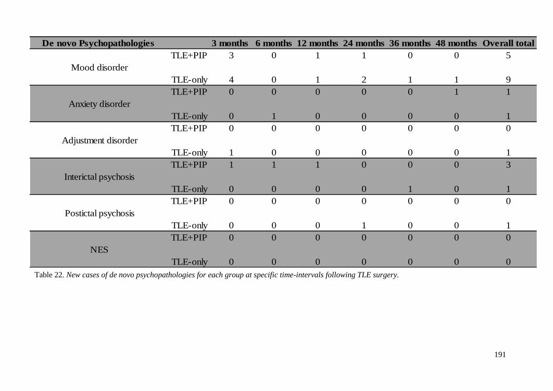
191
De novo Psychopathologies 3 months 6 months 12 months 24 months 36 months 48 months Overall total
TLE+PIP 3 0 1 1 0 0 5
Mood disorder
TLE-only 4 0 1 2 1 1 9
TLE+PIP 0 0 0 0 0 1 1
Anxiety disorder
TLE-only 0 1 0 0 0 0 1
TLE+PIP 0 0 0 0 0 0 0
Adjustment disorder
TLE-only 1 0 0 0 0 0 1
TLE+PIP 1 1 1 0 0 0 3
Interictal psychosis
TLE-only 0 0 0 0 1 0 1
TLE+PIP 0 0 0 0 0 0 0
Postictal psychosis
TLE-only 0 0 0 1 0 0 1
TLE+PIP 0 0 0 0 0 0 0
NES
TLE-only 0 0 0 0 0 0 0
Table 22. New cases of de novo psychopathologies for each group at specific time-intervals following TLE surgery.
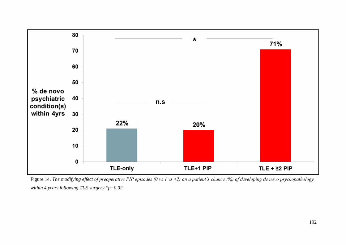
192
Figure 14. The modifying effect of preoperative PIP episodes (0 vs 1 vs ≥2) on a patient’s chance (%) of developing de novo psychopathology
within 4 years following TLE surgery.*p=0.02.
n.s
*

193
Seizure Outcome
There were no significant differences in the number of patients who were seizure free
(ILAE=1) at 12 months (TLE-only: 62% vs TLE+PIP: 50%, p=0.44) or remained seizure free
over 4 years (TLE-only: 45% vs TLE+PIP: 35%, p=0.6). In addition, there were no
significant associations in seizure freedom and the number of PIP episodes (p values >.05).
9.5 Discussion
In keeping with previous findings, this study found a greater prevalence of non-localised ictal
epileptiform activity in TLE+PIP patients (So et al., 1990; Kanner et al., 2008; Falip et al.,
2009) compared to those without PIP. This suggests that more widely distributed
epileptogenic networks may contribute to the development of PIP. This finding also supports
the emerging evidence from functional neuroimaging studies that more diffuse brain
abnormalities can be detected in PIP patients’ interictally (Seeck et al., 1999; Takeda et al.,
2001). There have also been some reports of diffuse structural brain abnormalities in patients
with PIP (Morrow et al., 2006; DuBois et al., 2011). However, these changes may be subtle
and warrant more advanced or sensitive imaging techniques.
There were no histopathological differences between the TLE+PIP and TLE-only groups. In
contrast, a small study (n=6) found PIP to be associated with temporal lobe dysplasia
(Briellman et al., 2000). The sample employed here was larger and applied a refined clinico-
pathological classification system (Blumcke et al., 2011).
There were no significant differences in the other epilepsy variables between the groups.
This is consistent with previous retrospective (Devinsky et al., 1995; Falip et al., 2009) and
prospective (Alper et al., 2008) studies. However, investigators have indicated that SGTCS
(Devinsky et al., 1995; Alper et al., 2008), status epilepticus (Falip et al., 2009) and
194
encephalitis (Devinsky et al., 1995; Alper et al., 2008) are risk factors for PIP. However, this
current study lacked reliable information to subtype CNS infections (e.g.
meningitis/encephalitis) or quantify the frequency of SGTCS and status epilepticus, which
may explain the lack of association.
The finding that a positive family psychiatric history was significantly associated with PIP,
and the most common diagnosis was mood disorders, is consistent with previous research
(Alper et al., 2008). Notably, no first- or second degree relative included in the analysis had
a diagnosis of psychosis/schizophrenia. The genetic mechanism(s) underlying this
association is unclear.
Preoperative cognitive measures did not differentiate between our TLE+PIP and the TLE-
only controls. While most previous studies do not report the neuropsychological status of
PIP patients, a few have documented bi-temporal neuropsychological dysfunction (So et al.,
1990; Savard et al., 1991; Mathern et al., 1995; Christodoulou et al., 2002; Falip et al., 2009).
However, it may be difficult to compare the current findings given the smaller sample sizes
of previous studies, and the unspecified nature of the cognitive measures used.
Postsurgical Outcome
Consistent with previous reports (Kanemoto et al., 1998; Kanemoto et al., 2001), a history of
PIP was associated with postsurgical de novo psychopathology. However, this study extends
previous findings in several ways. This study had a much larger sample size, identified
patients with co-morbid pre-surgical psychiatric diagnoses and/or SGTCS and controlled for
these possible confounds in the analyses. Furthermore, the study determined that patients
with a history of two or more PIP episodes had a significantly increased risk of developing de
novo psychopathology within four postoperative years (see Figure 14). Notably, the 3
patients in the TLE+PIP group who developed a postoperative chronic interictal psychosis

195
had a history of more than 2 PIP episodes (range: 2-6). This is concordant with Tarulli et
al.’s (2001) findings that 5 out of 6 of their patients with interictal psychosis had a previous
history of at least 2 or more PIP episodes. The findings of this investigation may suggest that
resective TLE surgery should not be delayed in patients with a history of PIP (Kanner et al.,
2009).
Few studies have examined the postsurgical seizure outcome of PIP patients (Kanner et al.,
2008). To date, this is the first study to investigate surgical outcome (ILAE=1) in TLE+PIP
patients compared to controls over a four year period (at yearly intervals). It was found that
postsurgical seizure outcome (ILAE 1 vs 2-6) did not significantly differ between the groups
at 12 or during 48 months, nor did the number of PIP episodes predict seizure freedom.
Alper et al. (2008) also found no difference between seizure freedom rates in PIP versus non-
PIP epilepsy patients. In Kanemoto et al.’s (1998) study, whilst the majority (81%) of the 52
TLE patients in their study achieved an Engel class I, the surgical outcomes of 5 PIP patients
were not specifically reported. In contrast, a retrospective controlled study found patients
without a history of PIP (n=17/20; 85%) were significantly more likely to become seizure
free compared to a small sample of PIP patients (n=2/3; 67%) (Kanner et al., 2008). The lack
of consensus with regards to seizure freedom in patients with PIP may reflect methodological
differences (e.g. sample size and/or seizure classification systems).
Neither a history nor frequency of PIP episodes significantly predicted ipsi- or contralateral
memory decline at 3- and 12 months. Future research to investigate whether extra-temporal
(executive function) decline is evident in PIP following TLE surgery is warranted.
There are several limitations of this study due to its retrospective design, including limited
follow-up (four years), the possibility that after 3 and 6 months psychiatric symptoms may
have emerged or were missed (Kanner et al., 2000). Furthermore, this study was unable to

196
determine whether the development of de novo psychopathology was modified by the
temporal relationship between the onset of PIP and surgical intervention. A lack of statistical
power precluded an investigation into the possible linear relationship between PIP episodes
and de novo psychopathology or seizure outcome. Exploration of subtypes of de novo
psychiatric morbidity was precluded with owing to a small sample size, but this could be
addressed by larger cohort studies.
In conclusion, this study demonstrates that preoperative PIP is associated with poorly
localised ictal EEG indicating that surgical candidates may require the recording of several
seizures during VEEG to determine the extent of the epileptogenic zone (Kanner et al., 2008).
The data also suggests that genetic predisposing factors may increase the risk of PIP. Lastly,
patients with recurrent episodes of PIP are at greater risk of developing de novo
psychopathology following surgery. TLE patients with recurrent episodes of PIP should be
counselled about the increased psychiatric risks following TLE surgery, and closely
monitored for postsurgical psychiatric morbidity.

197
Chapter 10. PROSPECTIVE STUDIES
10.1 Introduction
The previous studies failed to find an association between psychopathology and cognitive
variables in TLE and this may be due to a number of factors. Firstly, the pooling of all pre-
surgical psychiatric diagnoses (depression, anxiety, psychoses, NES) in statistical analyses
(see Study 1), may have resulted in a type II error. Second, the retrospective nature of the
data used limited a thorough investigation of the ‘extra-temporal neuropsychological profile’
(Keller et al, 2009), and the relation to pre- and/or postoperative (de novo) depressive
morbidity (see Studies 1 and 2). Third, epilepsy-related factors indicative of more
widespread cerebral dysfunction were not systematically quantified in patients’ notes and
thus, were categorised binomially (yes versus no) (see Studies 1-3), potentially reducing
sensitivity and statistical power. Fourth, the date of psychiatric onset and severity were not
consistently available in previous data-sets. In the prospective studies described here an
attempt was made to address the above deficiencies. In particular, the postoperative course of
Axis I psychopathology and the relationship to cognitive measures was more closely
investigated.
10.2 Sample
Patients under consideration for epilepsy surgery from September 2009 to December 2012 at
the NHNN, who fulfilled the following inclusion criteria, and were able to provide informed
consent, were invited to participate in the study (see Appendix 2 for the patient information
sheet and consent form).

198
10.3 Inclusion Criteria
1. Age ≥ 18 years
2. IQ > 70
3. Probable TLE
4. Fluent English speakers
5. No history of previous neurosurgery
One hundred and six patients with epilepsy were recruited, of whom 87 (82%) subjects
remained in the study and participated in the full assessment protocol. Of these, 49 (56%)
patients had TLE surgery (see Figure 15).
10.4 Procedure
All patients were seen preoperatively (n=106) and those who underwent TLE surgery (n=49),
were seen at 6 months (n=41), 12 months (n=29) and 24 months (n=9) postoperatively.
10.5 Pre-surgical Clinical Measures
The following data was collected.
10.5.1 Clinical History
All patients underwent a detailed medical assessment as part of their pre-surgical evaluation.
Records of birth history (history of febrile convulsions; CNS infection), seizure related
variables (head trauma, age of epilepsy onset, epilepsy duration, seizure types and frequency,
seizure semiology, history of status epilepticus); family history and social status were
recorded.

199
106 surgical candidates recruited:
2009-2012
Excluded patients, n=19
Multifocal epilepsy
n=3
Non-TLE focal
epilepsies
n=10
87 TLE patients:
Figure 15. Case selection process.
Bilateral TLE
n=2
Idiopathic epilepsy
n=4
Awaiting/declined
TLE surgery:
n=38
n=49
TLE surgery:
n=49

200
10.5.2 MRI Scan
All patients underwent MRI scanning with hippocampal volumetry. MRI studies were
performed on a 3T General Electric Excite HDx scanner (General Electric, Milwaukee,
Wisconsin, U.S.A.), using a body coil for transmission, and eight channel phased array coil
for reception. Standard imaging gradients with a maximum strength of 40 mTm−1
and slew
rate 150 Tm−1
s −1
were used. Standard clinical sequences were performed and included a
coronal T1-weighted volumetric acquisition with 170 contiguous 1.1 mm thick slices (matrix
256 x 256, in-plane resolution 0.9375 x 0.9375mm).
Manual segmentation of the hippocampi was performed using MRreg software by trained
neuro-radiographers (Lemieux, Liu, Duncan, 2000). This involves manual segmentation of
the entire hippocampi using a mouse-driven cursor according to the anatomical boundaries
specified by Watson et al. (1992). Following the tracing, MRreg automatically calculates the
hippocampal volume by summing the trace area of each slice and multiplying by the slice
thickness. The hippocampal volumes were corrected for total brain size by dividing by the
cerebral volume.
10.5.3 Prolonged Scalp Video-EEG
The diagnosis of TLE was established by prolonged scalp video-EEG monitoring with
electrode placement according to the extended International 10-20 system (Klem et al.,
1999), including bilateral anterior temporal and bilaterally placed sphenoidal electrodes. For
the majority of patients, provocative seizure-inducing procedures (i.e. drug reduction, sleep
deprivation, exercise) were performed.
Pre-surgical video-EEG reports were classified by a clinical electro-physiologist into cases
demonstrating ‘localised’ or ‘non-localised’ ictal or interictal epileptiform abnormalities

201
dependent on the neuro-radiological findings (e.g. bilateral temporal, extra-temporal,
multifocal, bi-frontal and unilateral temporal, widespread fronto-temporal discharges in the
presence of unilateral HS).
10.5.4 Neuropsychological Assessment
Preoperative neuropsychological data was collected in the following domains preoperatively
and 6- and 12 months postoperatively:
5. Intellectual functioning
(i) Wechsler Adult Intelligence Scale-III (WAIS-III) (Wechsler, 1997).
(ii) The National Adult Reading Test (NART) (Nelson & Willison, The Revised
National Adult Reading Test - Test Manual, 1991).
6. Semantic knowledge
(i) Category fluency: Patients were required to name as many exemplars from the
category “animals” as they could in 60 seconds.
(ii) Graded Naming Test (GNT) (McKenna, 1980).
7. Memory
The List- and Design- learning tasks from the BIRT Memory and Information
Processing Battery (BMIPB; Coughlan, Oddy, & Crawford, 2007) were used as
measures of verbal and non-verbal memory, respectively. This is the successor to the
AMIPB used in the retrospective studies (see Chapters 6 and Studies 1-3).
Performance on List- and Design learning were translated to Z-scores and used as
measures of hippocampal integrity.

202
Contralateral memory weakness:
As described in Chapter 9, the contralateral memory weakness for each patient was
calculated.
8. Executive functions
Rationale
Chapter 4 highlighted the limited understanding of more widespread cognitive sequelae in
TLE, particularly frontal lobe dysfunction, and the relationship to psychopathology (see
Figure 6). The retrospective studies only provided a single indicator of executive
function, namely phonemic fluency. To address this, additional neuropsychological
measures assessing different aspects of executive function were employed (planning, set-
shifting, categorisation, verbal working memory, inhibition).
(i) The Spatial Working Memory (SWM) subtest from the Cambridge Automated
Test Battery (CANTAB) (Sahakian & Owen, 1992). The CANTAB is a
computersied neuropsychological assessment that was orginally developed to
diagnose dementia in geriatric cohorts (Fray et al., 1996). The task stimuli are
nonverbal and language proficiency is necessary only to understand the
instructions. The validity of the CANTAB for assessing brain-behaviour relations
in adults has been supported by numerous studies of patients with brain lesions,
degenerative disorders and psychiatric illness (see
http://www.camcog.com/bibliography.asp).
The SWM sub-test was chosen as it reliably differentiates between patients with
frontal versus temporal lobe pathology (Owen et al., 1996), and it has an
acceptable test/retest coefficient (R=0.68) (Lowe & Rabbitt, 1998). This self-

203
ordered computerised test measures executive function (search strategy) and the
updating and monitoring of spatial information in working memory (WM). The
screen displays a number of coloured boxes (see Figure 16a).
Patients were required to ‘search’ through the boxes on the screen in order to
locate blue tokens hidden inside and once located, to place the token in an empty
column on the right hand side of the screen (see Figure 16b & c).
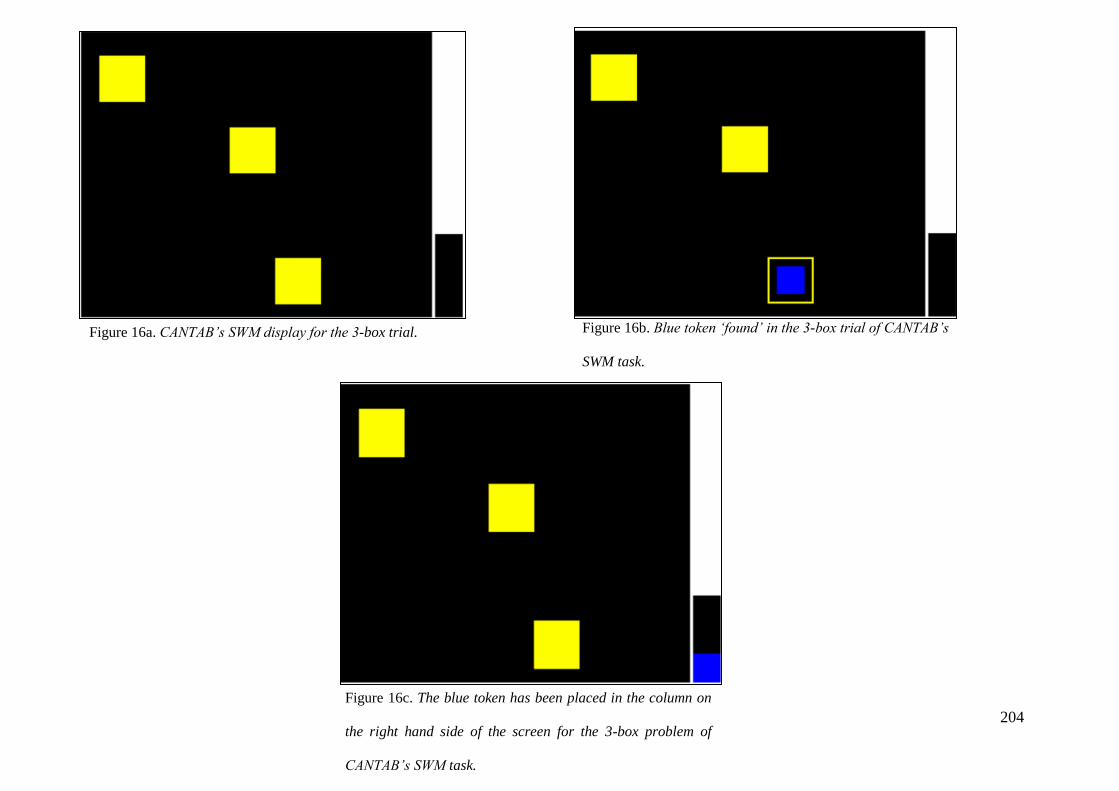
204
Figure 16a. CANTAB’s SWM display for the 3-box trial. Figure 16b. Blue token ‘found’ in the 3-box trial of CANTAB’s
SWM task.
Figure 16c. The blue token has been placed in the column on
the right hand side of the screen for the 3-box problem of
CANTAB’s SWM task.

205
The patients were instructed that once a token had been found within a particular
box, then that box would not be used again to hide any of the remaining tokens for
that particular trial. There are four example trial stages with 3-boxes, followed by
three levels of difficulty: 4-, 6- and 8-box problems, all of which contain four
trials; allowing for a graded approach that avoids ceiling or floor effects (Lezak et
al., 2012). Two types of performance indices were recorded at each level of
difficulty (4-, 6- and 8-box problems). The ‘between errors’ score indicates when
a patient has returned to a box in which a blue token had already been located
during a previous searching sequence. This is a stringent measure of short-term
WM failure and perseveration (Flugel et al., 2006). A ‘strategy score’, was also
recorded for performance at the more difficult levels (i.e. 6- and 8-box levels), and
demonstrates the adoption of a systematic searching approach (beginning with the
same box once a token has been found). The total score provides a composite
measure of strategy with a high score (many sequences beginning with a different
box) representing low strategy use and a low score (many sequences starting with
the same box) representing efficient strategy use and executive functions (Flugel
et al., 2006). Previous research indicates that these performance indices are
sensitive to neurocognitive dysfunction in patients with primary depressive
disorder (Porter et al., 2003).
Based on normative data provided by CANTAB from over 2000 control subjects
(aged 4-90) who participated in various research studies (see,
http://www.camcog.com/faqs.asp), the ‘between errors’ and ‘search strategy’
scores were converted to Z-scores for subsequent statistical analyses.
(ii) Wisconsin Card Sorting Task (WCST) (Berg, 1948) is a widely utilised
neuropsychological measure which was originally developed to assess “human

206
abstraction” and “shift of set”. It has been shown to examine executive functions
mediated by the frontal lobes, such as problem-solving, strategic planning, use of
environmental feedback to shift-set, and inhibition of impulsive responding
(Demakis, 2003). A meta-analysis of neuroimaging studies in a highly selected
cohort of frontal-lobe damaged patients, reported that the WCST is associated
fronto-parietal activation patterns, particularly in the lateral and ventral pre-frontal
cortices bilaterally (Buchsbaum et al., 2005).
A shortened version has been developed (WCST-64: Kongs et al., 2000), with
psychometric properties commensurate with the full version. Generalizability
coefficients ranging from 0.60 to 0.85 are documented, and scores have been
shown to discriminate between control groups and individuals with various
neurological disorders (Paolo et al., 1996; Kongs et al., 2000).
Patients were shown four specific stimulus cards, composed of geometric
elements, laid out in a row. Ordinarily, participants are given 64 cards and asked
to sort them one at a time by placing each response card on top of one of the four
stimulus cards, according to a given criterion (colour, shape or number).
However, pilot data indicated that this was too difficult for TLE patients.
Therefore, to minimise patient distress and a premature floor effect, patients were
given 36 cards and asked to sort them one at a time by placing each response card
on top of one of the four stimulus cards, according to a given criterion (colour,
shape or number). No instructions regarding card sorting was given by the tester.
After each card was sorted, the tester informed the patient if they correctly or
incorrectly sorted the card (according to the sorting rule only known by the
experimenter). After six cards, the tester told the patient that the sorting rule had
changed. The test continued until all cards were sorted. Two scores were

207
calculated: the number of categories completed (maximum 6) and the number of
perseveration errors (i.e. the number of cards classified on the basis of a previous
sorting criterion). The latter was used in statistical analyses as an indicator of
executive dysfunction; with higher preservation scores indicating poor set-shifting
ability.
(iii) Trail Making Test (TMT; Halstead, 1947) is comprised of two parts. In part A the
subject uses a pencil to connect a series of 25 encircled numbers in numerical
order. In part B, the subject connects 25 encircled numbers and letters in
numerical and alphabetical order, alternating between numbers and letters. For
example, the first number “1” is followed by the first letter “A”, followed by the
second number “2” and the second letter “B” and so on. The numbers and letters
are placed in a semi-random fixed order, as to avoid overlapping lines being
drawn by the subject (Bowie & Harvey, 2006). The primary variables of interest
are the total time to completion for parts A and B. Part A is a test of visual search
and motor speed skills, whereas part B is also considered to test higher level
cognitive skills such as mental flexibility, and the ability to modify a plan of
action or to maintain two trains of thought simultaneously (Crowe, 1998;
Arbuthnott & Frank, 2000). The TMT has excellent inter-rater reliability and
psychometric properties (Strauss, Sherman & Spreen, 2006).
(iv) Digit Span (WAIS-III; Wechsler, 1997): Requires repetition of orally presented
digit strings (digit forward, e.g., 1, 6, 8) and then recall of digits in reverse order
(digit backwards e.g., 8, 6, 1). Digit sequences range from 2 to 8 with two trials
per sequence. A well-powered neuroimaging study, using voxel-based lesion-
symptom mapping of patients with focal brain damage (n=241), reported a
significant relationship between digit span performance and activations in the left

208
frontal and parietal cortices (Glascher et al., 2009). Digit span score was used to
assess verbal working memory (Flugel et al., 2006) and executive function.
(v) Dysexecutive Questionnaire (DEX; Burgess et al., 1996; see Appendix 3): The
DEX is a 20-item questionnaire constructed to sample a range of ‘everyday’
symptoms of dysexecutive (or ‘frontal lobe’) syndrome, and forms part of the
Behavioural Assessment of the Dysexecutive Syndrome test battery (BADS;
Wilson et al., 1996). The questions sampled include changes in (1) emotional
responsivity or personality (2) motivation, (3) behaviour and (4) cognition. The
DEX assesses a number of characteristics, including abstract thinking problems,
impulsivity, confabulation, planning problems, euphoria, lack of insight, apathy
disinhibition, distractabiity, knowledge-response dissociation, lack of concern,
and disregard for social rules (Lezak et al., 2012). Each item was scored on a 5-
point Likert scale, ranging from ‘never’ to ‘very often’, with a high score
indicating higher frequency of dysexecutive behaviour in everyday life. One
version of the questionnaire was completed by someone who knew the patient
well (usually either a relative or carer; DEX-I), and the other version was
completed by the patient (DEX-S). Internal consistency is reported to be high
(>0.90), and investigators have reported a significant positive relation between the
DEX and measures of executive dysfunction (Bennett, Ong & Ponsford, 2005).
(vi) Phonemic fluency: This measure used in the retrospective studies was also
employed again, with patients required to name as many words beginning with
“S” in a 60 second epoch. Research has demonstrated this task is highly sensitive
and specific to frontal lobe damage (Robinson et al., 2012); with structural and
functional imaging showing that frontal lobe pathology disproportionately impairs
phonemic fluency, while temporal lobe damage has a greater effect on semantic

209
fluency (Bird et al., 2010).
10.5.5 Neuropsychiatric Assessment
As part of standard clinical care, all patients were assessed by neuropsychiatrist as part of
their pre-surgical evaluation. A single psychiatrist conducted clinical interviews and
diagnoses were determined using the DSM-IV-TR axis I (American Psychiatric Association,
2000) and ILAE criteria (Krishnamoorthy, Trimble & Blume, 2007). For comorbid
psychopathology, a positive entry was made into each diagnostic category. Medication
related psychiatric symptoms, defined as psychopathology temporally related to AED
change(s) that resolved following AED titration/discontinuation, were excluded. Information
regarding whether any first-degree relation had a history and/or current psychiatric diagnosis
was documented. Data concerning lifetime history of psychiatric treatment, defined as any
past treatment with psychiatric drugs, past psychiatric hospitalisation, and deliberate self-
harm/suicide attempts were also documented on first clinical interview.
All patients were seen by the same psychiatrist six months following surgery. The
continuance (or remission) of lifetime psychopathology was recorded and classified
according to the DSM-IV-TR (American Psychiatric Association, 2000) as for the pre-
surgical ones, but with the addition of adjustment disorders. Post-surgical psychiatric
hospitalisation and deliberate self-harm/suicidal ideation or attempts were documented in
detail. De novo psychopathology was only coded if a patient had no preoperative psychiatric
history and could only be termed as such at one mutually exclusive post-surgical time-frame.
Psychiatric symptomatology emerging after 6 months was identified by the general
practitioner or the treating neurologist resulting in a review by the epilepsy surgical team’s
neuropsychiatrist. Subsequently documented psychiatric disorders were then coded as
appropriate into the following postoperative time-frames: 12 or 24 months.
210
10.5.6 Psychiatric Rating Scales
All patients completed the The Beck Depression Inventory-Fast Screen (BDI-FS) (Beck,
Steer, & Brown, BDI-Fast Screen for medical patients: manual, 2000; see Appendix 4) and
the Beck Anxiety Inventory (BAI) (Beck & Steer, Beck Anxiety Inventory Manual, 1990; see
Appendix 5). These measures were completed preoperatively (within 6 months of TLE
surgery), and 6, 12 and 24 months postoperatively.
Becks Depression Inventory
This measure is comprised of seven items from the 21-item Beck Depression Inventory-II
(Beck, Steer, & Brown, Manual for the Beck Depression Inventory-II, 1996) pertaining to (1)
feelings of sadness, (2) discouragement about the future (pessimism), (3) personal failures,
(4) perceived decreases in self confidence, (5) a sense of being overly self-critical, (6) the
ability to derive pleasure from activities (anhedonism) and (7) suicidal ideation. Somatic
items are excluded to increase specificity for medical patients. Items are rated for the past
two weeks on a four-point likert scale (0-3), yielding scores ranging from 0-21. The
suggested cut-off scores are 0-3 (minimal symptoms); 4-6 (mild symptoms); 7-9 (moderate
symptoms); 10-21 (severe symptoms). The BDI-FS has been reported as a psychometrically
rigorous depression screening tool in a variety of medical populations (Nezu et al., 2010).
Becks Anxiety Inventory
This scale measures 21 symptoms associated with anxiety. Patients are asked to rate on a
four-point likert scale ranging from 0 (“not at all”) to 3 (“severely, I could barely stand it”)
how much these symptoms have bothered them over the past week. Scores range from 0-63.
Recommended cut-offs were employed: 0-7 (minimal anxiety); 8-15 (mild anxiety); 16-25

211
(moderate anxiety); 26-63 (severe anxiety). The BAI has a high internal consistency
(Cronbachs α =.92) and a test-retest reliability of .75 (Beck, Epstein, Brown, & Steer, 1988).

212
Chapter 11.
Study 4. Temporal Lobe Epilepsy & Depressed Mood:
Correlations with Cognitive and Clinical Factors
11.1 Study Aims
Chapter 4 highlighted the neuropsychological evidence suggesting that temporal and extra-
temporal cognitive function may be compromised in the presence of TLE and primary
depressive disorder. Few studies, however, have directly investigated the relation between
depressed mood and cognition in TLE.
The aims of this prospective study were two-fold: (1) determine whether depression is the
most prevalent psychiatric diagnostic category in TLE patients under consideration for
surgical treatment and (2) further examine the hypothesis that TLE surgical candidates with a
current diagnosis of depression have evidence of extra-temporal dysfunction as measured by
cognitive and clinical factors.

213
11.2 Methods
Study Sample
Eight-seven TLE patients were assessed for epilepsy surgery between 2009 and 2012 (see
Figure 15). Given that an aim of this study was to determine whether patients with a current
diagnosis of depression had evidence of extra-temporal dysfunction, twenty-four patients
(28%; n=24/87) were excluded due to a past history of psychopathology. Of the remaining
63 patients, 12 (19%; n=12/63) had a current diagnosis of depression and 51 (81% n=51/63)
patients had no psychiatric history (TLE-control group).
Procedure
As described in Chapter 10, pre-surgical evaluations consisted of 1) a detailed medical
assessment, 2) high-resolution MRI of the brain with volumetric hippocampal measurements,
3) prolonged VEEG recording, 4) neuropsychological testing and 5) a neuropsychiatric
clinical interview, conducted by the same psychiatrist in all patients.
11.3 Statistical Analyses
For comparing frequencies in demographic and clinical variables between patients with a
current diagnosis of depression versus those with no formal psychiatric diagnosis, Chi-square
(X2; with Fisher Exact Test (FET) when warranted) was used. Univariable logistic regression
was conducted to quantify the odds of group membership (‘depressed’ versus ‘TLE-
controls’) for any significant association identified using X2.
Differences between continuous variables were evaluated as appropriate using independent
samples t-test, or Mann-Whitney U test.

214
11.4 Results
Lifetime and Current Psychopathology
The lifetime and current psychopathologies of the sample as a whole (n=87) are presented in
Figure 17. A mood disorder was the most prevalent lifetime (n=27/87; 31%) or current
(n=12/17; 71%) psychiatric diagnosis (all depression). Nine patients (n=9/87; 10%) had a
lifetime diagnosis other than depression (n=6 anxiety; n=5, PIP; n=1, interictal psychosis;
n=2 NES); five (n=5/9; 56%) of which had lifetime comorbid psychopathologies (n=3,
depression and anxiety disorder; n=1, depression and NES; n=1, anxiety and PIP).
Demographic and clinical variables for each group are presented in Table 23. No significant
differences were found between the depressed and non-depressed groups in terms of
demographic variables, predisposing factors, seizure frequency or type, MRI findings or
hippocampal volumes.
Depressed TLE patients had significantly less localised ictal EEG recordings than controls
(40% vs 74%; X2(1)=4.3, p=0.05). Univariable logistic regression analysis indicated that
TLE patients with non-localised ictal EEG patterns had over four times the odds of being in
the ‘depressed’ group compared to patients with localised ictal EEG recordings (OR: 4.25,
95%CI: 1.02, 17.70, P=0.05). A similar trend was observed for localised interictal EEG, but
this did not reach statistical significance (64% vs 83%; OR: 2.71, 95%CI: 0.64-11.52,
p=0.18).
Patients with diagnosed depression exhibited significantly greater self-reported depressive
and anxiety symptoms compared to TLE-controls (p values >.05; see Table 23). All
depressed patients were receiving psychiatric treatment (p<0.001).

215
Figure 17. Lifetime (blue bars) and current (maroon bars) psychiatric diagnoses of TLE patients (n=87).

216
Table 23. Demographic and clinical characteristics of TLE patients with a current diagnosis of
depression (n=12) versus those with no psychiatric history (n=51).
217
Cognitive Performance
The performances of the groups on neuropsychological tests are shown in Table 24.
The Mann-Whitney U test revealed that the discrepancy between premorbid and assessed IQ
was significantly greater in the depressed group compared to controls, (U=161, z = -2.1,
p=0.04).
Reported dysexecutive symptoms by both patients (t(60) = -4.33, p<0.001) and their
informants (t(51) = -2.00, p=0.05) were significantly increased for depressed patients. The
difference between patient and informant DEX ratings did not significantly differ according
to group (t(51) = 10.4, p=0.18).
Exploratory correlational analyses revealed there was a significant positive association
between preoperative depressive symptomatology (BDI-FS) and patient and informant
dysexecutive symptoms (rs = 0.61, p = <0.001; Rs2 = 37%; see Figure 18; rs = 0.30, p = 0.04;
Rs2 = 8%; see Figure 19, respectively). There were no significant associations between the
premorbid and assessed IQ discrepancy, BDI-FS or reported DEX measures (Table 25a); and
accordingly partial correlations were not performed.
Patient and informant DEX ratings were correlated with the five neuropsychological
measures of executive functioning to determine the extent to which they were assessing a
common construct. The DEX measures were significantly related (rs = 0.39, p = <0.01; Rs2 =
15%); however only weak non-significant inter-correlations were found with
neuropsychological measures of executive function (see Table 25b).
There were no other significant differences in cognitive findings between the groups (p
values >.05; see Table 26).

218
Interaction: Depression and Seizure Focus
Due to small sample sizes and incomplete data, statistical analyses examining cognitive
performance as a function of laterality and depression were not performed. Descriptive
statistics for each subgroup, however, are presented in Table 27. Depressed patients
performed less well on 13 out of 18 tasks (72%), particularly for left-sided depressed patients
(poorer performance: 9/13 tasks).

219
Table 24. Preoperative cognitive performance of TLE patients with a current diagnosis of depression
(n=12) versus those with no psychiatric history (n=51).
Variable1
Depression TLE-controls
(n=12) (n=51) p-value
NART2
100 (87, 111) 97 (83, 108) 0.70
VIQ2
87 (79, 99) 91 (82, 102) 0.40
NART/VIQ discrepancy2
-7 (-12, -5) -2 (-10, 6) 0.04
Verbal learning: trials3
-1.2 (1.1) -1.0 (1.2) 0.70
delay2
-0.4 (-1.9, 0) -1.0 (-1.6, 0) 0.83
Visual learning: trials3
-0.8 (1.2) -0.7 (1.4) 0.83
delay2
-1.3 (-2.1, 0.4) -0.4 (-2.1, 0.4) 0.64
Contralateral memory weakness2
-0.9 (1.3) -0.8 (1.2) 0.83
Semantic knowledge
Graded Naming Test3
18 (6) 17 (5) 0.57
Category fluency3
18 (5) 18 (5) 1.00
Executive functions
Wisconsin Card Sorting Test (Perseveration error)2
2 (0-2) 1 (0-4) 0.96
Trail making B (secs)2
73.5 (65.5, 101) 64 (54, 89) 0.20
Phonemic fluency ("S" only)3
13 (9) 13 (5) 0.93
Working memory (Digit Span)2
8 (6-9) 8 (7-10) 0.60
Spatial WM (between errors)2
-0.7 (-1.5, 0.1) 0 (-0.7, 0.5) 0.12
Spatial WM (strategy)3
0.1 (1.0) 0.08 (1.2) 0.95
DEX-informant3
24 (12) 16 (10) 0.05
DEX-subject3
32 (14) 17 (10) <0.0011For available data2Median (IQR)3Mean (SD)
† Values are number of patients (%)

220
Figure 18. Positive correlation between preoperative depressive symptoms (BDI-FS) and patient reported dysexecutive symptoms.

221
Figure 19. Positive correlation between preoperative depressive symptoms (BDI-FS) and informant reported dysexecutive symptoms.

222
Table 25a. Spearman’s Rho correlations between preoperative depressive symptoms (BDI-FS), premorbid and assessed IQ discrepancy, and
patient (DEX-S) and informant (DEX-I) reported dysexecutive symptoms.
*p<0.05; **p<0.01; ***p<0.001
Spearman's Rho Preoperative
BDI-FS DEX-S DEX-I NART-VIQ discrepancy
BDI-FS 1.00
Preoperative DEX-S 0.61*** 1.00
DEX-I 0.30* 0.39** 1.00
NART-VIQ discrepancy -0.18 -0.24 -0.17 1.00
Spearman's Rho Preoperative
DEX-S DEX-I
DEX-S 1.00 0.39*
DEX-I 0.39* 1.00
WCST (perseveration errors) 0.09 0.02
TMT (B) 0.28 0.28
Preoperative Digit Span -0.02 -0.22
CANTAB:
Between errors -0.27 -0.18
Strategy score -0.22 -0.06
Phonemic fluency 0.10 -0.05
Table 25b. Spearman’s Rho correlations between patient and informant DEX ratings, and
preoperative neuropsychological measures of executive functioning.
*p<0.01

223
Table 26. Preoperative cognitive performance of TLE patients with a current diagnosis of depression versus those with no psychiatric
history, as a function of seizure focus. Bold text indicates poorest performance.
Variable1,2
Left-TLE Right-TLE Left-TLE Right-TLE
Depression (n=4) Depression (n=8) Controls (n=27) Controls (n=24)
NART 98 (74, 115) 100 (84, 107) 95 (87, 103) 98 (82, 109)
VIQ 92 (65, 109) 84 (83, 93) 96 (82, 101) 91 (82, 104)
NART and VIQ discrepancy -6.5 (-8.5, -5.0) -10 (-18, -4) -2 (-6, 7) -4 (-12, 5)
Verbal learning: trials -2.3 (-2.3, -1.4) -0.9 (-2.3, 0.23) -1.0 (-2.1, 0.23) -0.9 (-1.6, 0.5)
delay -1.7 (-2.9, -0.7) -0.1 (-2.4, 0.76) -1.1 (-1.5, 0.0) -0.8 (-1.7, 0.0)
Visual learning: trials -1.0 (-2.7, 0.3) -1.0 (-1.7, 0.6) -0.2 (-1.4, 0.6) -0.5 (-1.9, 0.6)
delay -1.7 (-2.8, 0.1) -0.1 (-5.4, 0.4) 0.1 (-1.2, 0.5) -0.9 (-2.7, 0.4)
Contralateral memory weakness -1.0 (-2.8, 0.1) -1.0 (-5.4, 0.4) 0.1 (-1.2, 0.5) -0.9 (-2.7, 0.4)
Semantic knowledge
Graded Naming Test 22 (11, 22) 15 (14, 17) 16 (13, 21) 16 (14, 20)
Category fluency 16 (13, 16) 18 (14, 23) 18 (16, 21) 18 (13, 22)
Executive functions
Wisconsin Card Sorting Test (Perseveration error) 2 (2, 2) 1 (0, 3) 1 (0, 3) 1 (1, 4)
Trail making B (secs) 68 (64, 68) 71 (58, 75) 61 (54, 72) 80 (80, 128)
Phonemic fluency ("S" only) 7 (1, 7) 13 (4, 22) 13 (9, 17) 14 (9, 17)
Working memory (Digit Span) 8 (3, 13) 8 (6, 8) 8 (7, 10) 7 (6, 10)
Spatial WM (between errors) n/a -0.7 (-1.1, -0.1) 0.2 (-0.5, 0.9) -0.5 (-1.3, 0.2)
Spatial WM (strategy) n/a 0.1 (-0.6, 1.2) 0.5 (-0.8, 1.7) -0.5 (-1.0, 0.2)
DEX-informant 29 (16, 29) 23 (9, 29) 12 (6, 28) 15 (13, 22)
DEX-subject 35 (25, 46) 24 (15, 32) 16 (10, 22) 16 (10, 27)1For available data2Median (IQR)
n/a, not applicable

224
11.5 Discussion
The present findings corroborate earlier investigations demonstrating increased psychiatric
morbidity in TLE patients (Gaitatzis, et al., 2004; Swinkels et al., 2005; Grabowska-Grzyb et
al., 2006), with depression the most prevalent pre-surgical psychiatric comorbidity (Ring et
al., 1998; Anhoury et al., 2000; Kohler et al., 2001; Wrench et al., 2004; Devinsky et al.,
2005; Kanner et al., 2009; Guarnieri et al., 2009; Adams et al., 2012; Filho et al., 2012; da
Conceição et al., 2013). The data lends some support to the hypothesis that TLE patients
with a current diagnosis of depression have evidence of extra-temporal dysfunction.
TLE patients are considered at high risk of depression due to limbic and/or frontal lobe
dysfunction (Swinkels et al., 2005). The hypothesis that brain areas outside the epileptic
focus may play a pathogenic role is supported by the relationship found between depression
and less ‘localised’ ictal epileptiform activity. Video-EEG reports for these patients noted
widespread, ipsilateral fronto-temporal ictal patterns. The role of extra-temporal regions is
further strengthened by structural (Baxendale et al., 2005; Shamim et al., 2009; Wrench et al.,
2009; Finegersh et al., 2011) and functional (Schmitz et al., 1997; Savic, et al., 2004; Hasler
et al., 2007; Lothe et al., 2008; Martinez et al., 2013) neuroimaging studies indicating that
more diffuse cerebral pathology/dysfunction is found in TLE patients with depressive
morbidity. Butler and co-workers (2012) have shown that the orbitofrontal cortex (OFC),
one of the first propagation sites of mesial temporal epileptic discharges (Lieb et al., 1991), is
bilaterally thickened in depressed mesial TLE patients. Furthermore, the severity of the
depression was positively correlated with the cortical thickness of these regions. The
presence of a more extensively damaged cerebrum, as indicated by non-localised ictal
epileptiform abnormalities, may also explain the association between a history of depression
and a poorer response to anti-epileptic medication (Hitiris et al., 2007), and epilepsy surgery

225
(Study 1; Anhoury et al., 2000; Kanner et al., 2009). The correlational nature of this current
study, and the evidence of a bidirectional relationship between depression and epilepsy
(Forsgren et al., 1990; Hesdorffer et al., 2000; Hesdorffer et al., 2006; Hesdorffer et al.,
2012), indicates longitudinal research is required to conclusively demonstrate that depression
is causally associated with non-localised ictal EEG patterns.
None of the other demographic or epilepsy-related factors distinguished group membership.
Previous studies examining risk factors for depression in TLE have reported conflicting
findings (Foong et al., 2007). The role of gender is uncertain with higher rates reported in
both males (Altshuler et al., 1999; Strauss et al., 1992) and females (Paradiso et al., 2001),
with others finding no effect (Grabowska-Grzyb et al., 2006). The age of TLE onset,
duration and seizure laterality do not appear to influence depression (Foong et al., 2007);
suggesting other biological (e.g. concomitant frontal lobe dysfunction) and/or psychosocial
determinants may be of importance.
Parasido and co-workers (2001) found that seizure frequency was increased in the presence
of depression, particularly for right-sided patients (Paradiso et al., 2001). However, due to
limited statistical power such interactional effects were unable to be investigated in this
current examination, and require replication with larger cohorts.
This study provides some support for previous research indicating that depressed mood is
significantly associated with neuropsychological dysfunction remote from the epileptogenic
focus (Hermann et al., 1991; Corcoran et al., 1993; Paradiso et al., 2001; Helmstaedter et al.,
2004). Depressed TLE patients demonstrated a significantly larger discrepancy between
premorbid and assessed IQ, an indicator of more global cognitive decline and, an increased
incidence of dysexecutive behaviours in daily life. The failure to find greater performance
226
deficits in association with depression conflicts with previous work (Paradiso et al., 2001),
including a more global ‘blunting’ of cognitive performance reported in the neuropsychiatric
literature (Austin et al., 2001; Castaneda et al., 2008; Clark et al., 2009; McClintock et al.,
2010).
Antidepressant treatment likely complicates the cognitive presentation. The relation between
cognition and depression appears modified by antidepressant treatment. Animal and human
studies indicate that selective serotonin reuptake inhibitors (SSRIs) have neuro-protective and
pro-cognitive effects (Gualtieri et al., 2006; Taler et al., 2013). Taler et al. (2013) reported
that following SSRI (sertraline) treatment, mice exhibited significant improvements in spatial
memory, using the Morris Water Maze task. This effect was positively associated with the
up-regulation of brain derived neurotrophic factor (BDNF) in the hippocampus; a protein
involved in neurogenesis and differentiation. Furthermore, widespread cognitive
impairments have been shown to normalise following successful SSRI therapy in unipolar
MDD patients (Gualtieri et al., 2006). Notably, all depressed patients in the current study
were receiving SSRI monotherapy. Thus, the finding of a specific cognitive impairment may
not be in direct conflict with previous reports (Paradiso et al., 2001); but rather supports the
view that the relationship between global cognitive dysfunction and depression is modulated
by psychotropic (SSRI) medication.
Contrary to previous reports, we did not replicate the finding that executive function test
performance was poorer in relation to depressed mood. Hermann et al. (1991) reported that
45% of TLE patients had clinically significant frontal lobe dysfunction as assessed by
perseveration errors on the WCST. Hermann and colleagues (1991) also reported a positive
relationship between frontal lobe function (perseverative errors) and dysphoric mood state,
particularly for left-sided patients. This divergent finding may be due to methodological
differences between studies. Hermann et al.’s (1991) cohort was non-lesional; 88% requiring

227
bi-temporal and bi-frontal intracranial recordings. Most patients in the current study were
lesional cases (n=54; 84%), and only 8 (13%) patients required invasive EEG recordings as
part of their pre-surgical evaluation.
Depressed TLE patients in this study showed awareness of executive function deficits in
daily life as measured by the DEX and experienced more difficulties than TLE-only patients,
as demonstrated by the good agreement between patient and informant DEX ratings. The
lack of an association however, between cognitive test performance and reported
dysexecutive behaviours in depressed TLE patients requires consideration.
Burgess et al. (1998) proposed that neuropsychological tests may not always reflect the
executive problems that patients encounter in real-life situations (i.e. low ecological validity).
This may be due to the structured nature of cognitive tests (i.e. rules and goals are set, and
behavioural initiation is prompted; van Beilen et al., 2006). In contrast, activities of daily
living are usually unstructured, often without a clearly defined goal or solution, and with an
unspecified behavioural trajectory. Thus, as reported previously (Burgess et al., 1998; Koerts
et al., 2012), the lack of an association between reported dysexecutive behaviours and
executive test performance may reflect the low ecological validity of the cognitive measures.
In addition, other cognitive tasks such as the Iowa Gambling Task (Bechara et al., 1994),
which have been modelled on real-life decision-making, may be better related to everyday
executive function than the tests used in the current study. Labudda et al. (2009) found that
TLE patients (with no information regarding depressive morbidity) were significantly
impaired in their decision-making compared to healthy controls. Notably, those with a
preference for disadvantageous decisions also performed less well on other tests of executive
function.

228
An alternative explanation is that the DEX may not be directly assessing difficulties with
executive functions in daily life, but rather indicating the associated negative affect (Gerstorf
et al., 2008). This could therefore explain the current finding that depressed TLE patients
reported significantly more problems with real-life executive tasks than TLE-only patients,
even though there were no differences with regard to neuropsychological test performance.
However, the positive relation between informant DEX ratings and patient BDI-FS symptoms
argues against a subjective bias in the reporting of executive difficulties by depressed
patients.
This study extends previous findings in several ways. First, temporal and extra-temporal
cognitive function was examined. Previous studies have either limited investigation to
memory function (Corcoran et al., 1993; Wishart et al., 1993; Dulay et al., 2004;
Helmstaedter et al., 2004) or used a narrow neuropsychological test battery to assess
depressed TLE samples (Hermann et al., 1991; Paradiso et al., 2001).
Although no single method is optimal to assess psychiatric state in TLE (Guarnieri et al.,
2009), this study employed clinical rating scale (BDI-FS) in addition to a clinical assessment.
The use of a clinical diagnosis of depression is important, as patients had elevated depressive
symptoms (median BDI-FS = 9); prior work with minimal or mildly depressed TLE cohorts
failed to relate depression and cognitive function (Pulliainen et al., 1999; Tracy et al., 2007).
Furthermore, both approaches allow confidence that the depressed and control groups were
psychiatrically homogenous. The removal of TLE patients with a past history depression also
ensured that the findings were not complicated by cognitive deficits (sustained
attention/executive function) that may persist following remission/recovery (Porter et al.,
2003).

229
A limitation of this study is the relatively small number of TLE patients with current
depression. This precluded statistical investigation into whether depression and seizure
laterality interact and adversely impact cognitive performance. However, the suggestion that
depression deleteriously impacts the neuropsychological status of depressed left-sided TLE
patients (Paradiso et al., 2001; Dulay et al., 2004; Helmstaedter et al., 2004), receives
cautious support. Studies with larger sample sizes are needed, particularly as cognitive
deficits on left-sided tasks are more easily discerned than right-sided measures (Helmstaedter,
2004). Another limitation is that no correction for multiple comparisons was performed.
Given the limited research in this area, a Bonferroni correction was considered too
conservative, and may have increased the chance that genuine differences between the groups
would not have been detected (Type II error).
In conclusion, depression is common in TLE and is associated with clinical and cognitive
markers of more widespread cerebral dysfunction. Depressed TLE patients and their
informants are aware of problems with executive functions in daily life, and report
considerably more difficulties than TLE-controls. This may explain why depression is a
powerful predictor of quality of life in epilepsy, whereas seizure-related factors have little
predictive value (Boylan et al., 2004). Reported dysexecutive behaviours however, did not
correspond with the results of neuropsychological measures. This lack of an association
suggests that standard cognitive measures of executive function do not reveal the extent of
problems patients encounter in their everyday life, and highlight the need for ecologically
valid assessment tools. The findings suggest that the DEX questionnaire, which is easy to
administer and yields valuable informant information, may have clinical utility in the
assessment of executive dysfunction in TLE. Finally, longitudinal investigation is required to
conclusively demonstrate that depression is causally associated with additional
neuropsychological morbidity in TLE.

230
Chapter 12.
Study 5. Profiling and Predicting Psychiatric Outcome Following
TLE surgery
12.1 Study Aims
The literature reviewed in Chapter 2, did not yield consistent or reproducible insights into the
possible predictors of poor psychiatric outcome following TLE surgery. This may be due to
the investigation of heterogeneous samples, limited information regarding preoperative
psychiatric status, short-term follow-up, the application of diverse diagnostic criteria and/or
inappropriate selection of statistical analysis techniques (see Chapter 2.9).
Study 1 found 18% of TLE patients experienced deterioration in psychiatric status
postoperatively. De novo psychiatric disorders were persistent (lasting longer than 6
months), and the majority of patients required psychiatric treatment. Neither seizure nor
cognitive outcomes were related to de novo psychiatric morbidity, but this may have been
due to methodological weaknesses.
As hypothesised, previous studies have demonstrated that preoperative indicators of less focal
epileptogenicity were predictive of poor post-surgical psychiatric outcome, including a
history of SGTCS (Study 1), recurrent PIP episodes (Study 3) and more diffuse structural
abnormalities (Study 8).
The aim of this prospective study was to use multilevel modelling (MLM) to further explore
predictor(s) of poor psychiatric outcome following TLE surgery. Specifically, (1) to examine
whether TLE patients with evidence of pre-surgical extra-temporal lobe dysfunction are at
increased psychiatric risk following surgery. (2) To determine whether preoperative

231
depressed mood is predictive of poor psychiatric, cognitive and seizure outcome.
12.2 Methods
Study Sample
Forty-nine patients who underwent TLE surgery from September 2009 to December 2012 at
the NHNN, participated in the study (see Figure 15). Demographic and clinical
characteristics are summarised in Tables 27 and 28.
Procedure
As described in Chapter 10, the following pre-surgical evaluations consisted of 1) a detailed
medical assessment, 2) high-resolution MRI of the brain with volumetric hippocampal
measurements, 3) prolonged VEEG recording, 4) neuropsychological testing and 5) a
neuropsychiatric clinical interview, conducted by the same psychiatrist in all patients.
Measures of intellectual function, semantic knowledge, memory and executive function were
completed preoperatively and at 6 (n=41), 12 (n=29) and 24 months (n=9) following surgery.
The BDI-FS and BAI were administered at the same time intervals as the cognitive tests and
were used as the main measures of mood status (i.e. outcome variables).

232
Variable TLE patients
(n=49)
Female† 32 (65%)
Chronological age, yrs1
38 (12)
Age of TLE onset, yrs1
15 (11)
TLE duration, yrs1
23 (14)
Predisposing Factors†
History of febrile convulsions 16 (33%)
History of status epilepticus 4 (8%)
History of head trauma 7 (14%)
History of CNS infection 3 (6%)
Family history of epilepsy 11 (22%)
Seizure Type2
Simple partial† 42 (86%)
Complex partial† 49 (100%)
SGTCS† 18 (37%)
Seizure frequency SGTCS/month1
1 (3)
Seizure frequency/month3
8 (1-40)
Medication
Total number of AEDs trialled preoperatively1
3 (1)
Monotherapy† 2 (4%)
Polytherapy† 47 (96%)
MRI†
Normal 6 (12%)
HS 29 (59%)
FCD 2 (4%)
Cavernoma 3 (6%)
DNT 9 (18%)
Other (abn. gyral folding/epidermoid) 7 (14%)
Dual pathology 7 (14%)
Hippocampal Volumes4,5
Ipsilateral HC volume1
2.23 (0.61)
Contralateral HC volume1
2.71 (0.44)
Video-EEG Findings†,5
Localised ictal EEG 29 (59%)
Localised interictal EEG 36 (74%)
Psychiatric variables
Family history of psychopathology† 16 (33%)
Lifetime psychopathology† 18 (37%)
Lifetime mood/anxiety disorder† 15 (31%)
Current mood/anxiety disorder† 9 (18%)
Psychiatric treatment† 10 (18%)
BDI-FS3,6
3 (0-17)
BAI3
9 (0-38)
Surgical procedures†
Right ATLR 24 (49%)
Left ATLR 22 (45%)
Right Lesionectomy 1 (2%)
Left lesionectomy 2 (4%)
ATLR, anterior temporal lobe resection CNS, central nervous system
DNT, dysembryoplastic neuroepithelial tumour;
FCD, focal cortical dysplasia; HC, hippocampal;
HS, hippocampal sclerosis; † Values are number of patients (%); 1Mean (SD);
2May have >1 seizure type;
3Mean (range);
4Corrected for intracranial volume;
5For available data;
6Rounded to nearest whole number
Table 27. Demographic and clinical characteristics of 49 patients who
underwent TLE surgery.

233
Table 28. Cognitive characteristics of 49 patients who underwent TLE surgery.
Variable1-3
TLE patients Performance
(n=49) level:
NART 95 (14) average
VIQ 92 (14) average
NART and VIQ discrepancy -2.7 (11) -
Verbal learning: trials -0.9 (1.2) low average
delay -0.8 (1.2) low average
Visual learning: trials -0.6 (1.1) low average
delay -1.1 (1.7) low average
Semantic knowledge
Graded Naming Test 16 (5) low average
Category fluency 18 (5) average
Executive functions
Wisconsin Card Sorting Test (Perseveration error)3
2 (0-11) -
Trail making B (secs) 78 (38) -
Phonemic fluency 14 (6) average
Working memory (Digit Span) 8 (3) low average
Spatial WM (between errors) -0.5 (1.2) -
Spatial WM (strategy) 0.1 (1.7) -
DEX-informant 20 (0-59) -
DEX-subject 20 (2-47) -
-, not applicable; 1For available data
2Mean (SD);
3Mean (range)

234
12.3 Statistical Analyses
Multilevel Modeling (MLM)
A mixed-model repeated measures analysis was implemented using Stata 10 (StataCorp,
2007). This statistical program was chosen as it contains specific commands (i.e. xtmixed)
for fitting multilevel models and saves syntax, data organisation and output formats, as a “do-
file”. This allows the same model or set of commands to be applied to different outcomes of
interest (e.g. BDI-FS and BAI) in one step (by re-running the do-file). Thus, do-files
facilitate an efficient and reproducible workflow reducing data management time, and Stata’s
computational speed afford significant advantages over other statistical packages (Long,
2009).
The advantages of multilevel modeling (MLM) have been described elsewhere (see Chapter
2.10). MLM is the preferred technique to longitudinal data where the primary interest is in
modeling the structure and predictors of change over time in an outcome of interest (Luke,
2004), for example mood rating scores following TLE surgery.
A multilevel model decomposes the variance in a characteristic of interest (e.g. BDI) and
models whether the variability and rate of change over time varies across individuals in a
systematic way, according between-subject predictors of interest (e.g. epilepsy lateralisation,
history of psychopathology etc.). Although MLM computations are dealt with by statistical
programs (Stata 10), the following theory is outlined to aid interpretation of the following
results.

235
Fixed and Random Coefficients
In normal general linear regression models (e.g. ANOVA), it is assumed that the regression
parameters (intercept, B0; slope, B1) are fixed as described below (Field, 2009):
Yi = B0 + B1X1i + ɛi
Note the outcome (Y; e.g. BDI), the predictor X and the error (ɛ) all vary as a function of i,
which represents the time points in the dataset (e.g. preoperative, 6 and 12 months etc.) – the
level 1 variable (see Chapter 2.10, Figure 3). Therefore, if we wanted to know a patient’s
BDI at 6 months, we could replace the i’s with the level 1 variable of interest:
BDI(6m) = B0 + B1X1(6m) + ɛ(6m)
The inherent assumption here is that the model holds true across the entire sample. Thus, for
every patient in the sample we can predict the outcome (BDI at 6 months) using the same
values for the intercept (B0) and slope/gradient (B1) (Field, 2009), as described by the single
(red) regression line in Figure A below:

236
The cornerstone of multilevel modeling is that each individual has their own
regression/growth parameters (B0; intercept and B1; slope), and therefore growth parameters
can vary across patients – at level 2 (see Chapter 2.10, Figure 3).
Random Intercept Only Model
To include a random intercept for BDI, a component is added to the intercept that measures
the variability in intercepts, u0j. Therefore, the intercept term changes from B0 to become
B0 + u0j. This term estimates the intercept of the overall model fitted to the data, B0, and the
variability of intercepts around that overall model, u0j:
Figure A. Schematic of a fixed effect simple linear regression model; blue dots = patient
data points

237
Yij = (B0 + u0j) + B1X1ij + ɛij
The j’s in the equation denote the levels of the variable over which the intercept varies (i.e.
across patients) – the level 2 variable (see Chapter 2.10, Figure 3). Therefore, to estimate
patient 1’s BDI at 6 months, the equation becomes idiosyncratic to patient 1 (see Figure B):
BDI(6m patient1) = (B0 + u0patient1) + B1X16m, patient 1 + ɛ6m, patient 1
Figure B. Schematic of a fixed effect simple linear regression model (red line) and the
regression line for patient 1 (purple line). The difference between the model and patient’s
intercept (B0 model - B0 patient1) is depicted by u0j; blue dots = patient data points

238
Random Intercept and Slope Model
As described above, the intercept and slope for the overall model (B0 and B1) is estimated,
but the variability in the intercepts, u0j and slopes, u1j is included:
Yij = (B0 + u0j) + (B1 + u1j)X1ij + ɛij
Now patient 1’s BDI at 6 months with a varying intercept and slope can be estimated (see
Figure C):
BDI(6m patient1) = (B0 + u0patient1) + (B1+u1patient 1)X16m, patient 1 + ɛ6m, patient 1
Figure C. Schematic of a fixed effect simple linear regression model (red line) and the
regression line for patient 1 (purple line). The difference between the model and patient’s
intercept (B0 model - B0 patient1 = u0j) and slope (B1 model – B1 patient1 = u1j) is depicted;
blue dots = patient data points

239
Hypothesis Testing
Although MLM is a flexible technique, hypothesis testing is still required to evaluate whether
a random intercept only, or random intercept and random slope model is the best fit to
the observed data. This decision is guided by the likelihood-ratio test. This test compares the
overall fit of the two candidate models (random intercept only versus random intercept and
random slope), and whether the null hypothesis that the addition of a random slope makes no
significant difference to the overall fit of the model (compared to the random intercept only
model), can be rejected (Heck et al., 2010). For this form of hypothesis testing full
maximum-likelihood estimation (MLE) must be used (Field, 2009).
Maximum Likelihood Estimation
The method of MLE is the most popular approach to statistical estimation (Singer et al.,
2003). Conceptually, the aim of MLE is to determine the unknown population parameters
that maximise the probability of observing a particular sample of data.
The derivation of a MLM estimate for a population parameter is accomplished by the
construction of the likelihood function – an expression that describes the probability of
observing the sample data as a function of the model’s unknown parameters. The statistical
program (Stata) then numerically examines the relative performance of potentially competing
estimates until those that maximise the likelihood function are found (see Singer et al., 2003,
for further discussion).
Covariance Structure
Fitting a multilevel model requires the specification of a covariance structure to the dataset
(Field, 2009). This part of the model represents the variation/error in measuring each

240
individual over time (i.e. level 1 residual errors). By definition, these errors are unobserved
and require assumptions about the distribution of the level 1 residuals to be invoked, from
occasion to occasion (preoperative – 6m; preoperative – 12m), and from patient to patient
(Singer et al., 2003).
The covariance structure specifies the form of the variance-covariance matrix (a matrix in
which the diagonal elements are variances and off diagonal elements are the covariance) and
is the starting point to estimate model parameters. Therefore, different results are yielded
depending on which covariance structure is chosen. Specifying a structure that is too simple
will increase the chance of a Type I error (erroneously concluding a parameter is significant).
Whereas, specifying one that is too complex may result in a Type II error (erroneously
concluding parameters are non-significant) (Field, 2009). There are four common structures:
(1) unstructured, (2) identity, (3) diagonal and (4) auto-regressive.
For longitudinal data, an auto-regressive structure is preferred as it assumes that correlations
between repeated measurements is highest at adjacent time-points and decreases over time
(e.g. preoperative and 6 months BDI-FS score is more related than preoperative and 12 month
BDI-FS score). An auto-regressive structure was adopted for all the models in this thesis.
Assumptions of Multilevel Models:
1. Variable types: All predictor variables must be quantitative or categorical (with only
two categories). The outcome variable must be quantitative, continuous and
unbounded (i.e. no constraints on the variability of the outcome).
2. Non-zero variance: The predictors should have variation in value (i.e. they do not
have variances of zero).

241
3. No perfect multicollinearity: No perfect linear relationship between two or more of
the predictors (r ≥ 0.9).
4. Normally distributed standardised residuals: The residuals in the model are
random and normally distributed (i.e. no more than 5% of the data will have ≥ ±2
standard deviations from the mean).
Taxonomy of Statistical Models:
1. Unconditional Means Model: Instead of describing the change in outcome (BDI-
FS/BAI) over time, this model simply describes and partitions the outcome variation.
The primary reason for fitting this model is to estimate the (between- (σ2
B) and
within-subject (σ2
w) variance). For each outcome variable (BDI-FS/BAI), the model
produces the interclass correlation (ICC) that describes the amount of variance in the
outcome attributed to differences between people (at level 2):
ICC = (σ2
B / σ2
B + σ2w)*100
Thus, the ICC evaluates the necessity of modeling the nested data structure (i.e. any
significant variation in individual initial status of the outcome variable). If the ICC is
low (i.e. <25%), incorporating random coefficients (intercepts and/or slopes) are not
required and a multilevel model strategy has no significant gains than traditional
methods (repeated measures ANOVA) (Shek et al., 2011).
2. Does the slope vary randomly across patients?
This model focusses on defining the shape of patients’ growth trajectories (i.e. how
BDI-FS/BAI changes over time) and determines whether the initial intercept and/or

242
slope varies across patients (Heck et al., 2010). In this step, two models are generated
– the random intercept only and random intercept and slope models.
At this point, checking the model’s assumptions (described above, assumptions 1-4) is
crucial for determining the tenability of the model and subsequent conclusions. For
multilevel modeling, Singer et al. (2003) suggest that the visual inspection of
standardised residuals (using probability plots) is the most efficient way to ensure the
distribution is normal (see assumption 4). For both self-report indices (BDI-FS/BAI),
there were marked deviations from the predicted line (more than 5% of the data had ≥
± 2 standard deviations from the mean) (Field, 2009); indicating that the assumption
of normality of residuals has been violated (see Appendix 6a & c). Therefore, for
both outcomes (BDI-FS/BAI) the data was transformed using the logarithmic function
(Weiss, 2005). For a logarithmic transformation, no observation in the data set can be
0 (see Weiss, 2005, for discussion). Therefore, all values were increased by one unit
so that 0 values had been increased sufficiently to permit a logarithmic
transformation.
Following the power transformation, the log standardised residuals probability plots
were visually inspected to evaluate whether the assumption of normality has been met
(i.e. the data points lie along the predicted line). For BDI-FS and BAI, the residuals
were normally distributed (i.e less than 5% of the data deviated ≥ ± 2 standard
deviations from the prediction line; see Appendix 6b & d).
The likelihood-ratio test was then computed to assess which model captured more
variability in the outcome (random intercept only versus random intercept and
slope). For both outcome measures (BDI-FS/BAI), the random intercept only model

243
was sufficient (all p-values for the likelihood-ratio test >.05) and was used in the
subsequent uni- and multi-variable analyses (steps 3 & 4).
3. Adding Predictors: Univariable analyses
For each logarithmic (log) random intercept only model (BDI-FS/BAI) the following
between-subject predictors (n=30) were added singly (see Table A); affording the
assessment of whether a level 2 variable (e.g. gender) effects the outcome (e.g. BDI-
FS/BAI), adjusting for the main effect of time.
Two cross-level interaction terms were also included in each model to examine
whether a level 2 variable (e.g. gender) had a significant effect on the outcome
variable (BDI-FS/BAI) over different levels of the level 1 variable (i.e. time at 6 or 12
months, relative to preoperative score).

244
Variable*
Gender
Age of TLE onset, yrs
TLE duration, yrs
TLE laterality
Hippocampal volumes1
Ipsilateral HC volume
Contralateral HC volume
Pathology
Hippocampal sclerosis vs other
Video-EEG Findings2
Localised ictal EEG
Localised interictal EEG
Cognitive variables2
Preoperative VIQ
NART/VIQ discrepancy
Verbal learning: trials
delay
Visual learning: trials
delay
Contralateral memory weakness
Semantic knowledge
Graded Naming Test score
Category fluency score
Executive functions
Phonemic fluency score
Trail making B (secs)
Wisconsin Card Sorting Test (Perseveration error)
Working memory (Digit Span)
Spatial WM (between errors)
Spatial WM (strategy)
DEX-informant
DEX-subject
Psychiatric variables
Family history of psychopathology
Lifetime psychopathology
Lifetime mood/anxiety disorder
Current mood/anxiety disorder
Table A. Preoperative predictors of change in mood rating scales
following TLE surgery used in univariable analyses.

245
For predictor selection into a multivariable model, variables from step 3 were retained
if the p-value was ≤ 0.20. This less conservative p-value was adopted as a Type I
error rate of 5% is too conservative for predictors that may have borderline
significance in a univariable model, but may be significant when controlling for
covariates in a multivariable model (Field, 2009). Only significant (p ≤ 0.05)
interaction terms are reported as these coefficients are not included in a multivariable
model and describe how a between-subject factor differentially affect the outcome
over different time points.
As described previously, for each of the univariable log-random intercept only model
per outcome (BDI-FS/BAI), standardised residuals probability plots were computed
and all were normally distributed.
4. Adding Predictors: Multivariable analyses
For each outcome variable (BDI-FS/BAI) a “final” model assessed whether predictors
retained from step 3 (univariable analyses) remained significant after controlling for
other possible covariates (i.e. the degree to which a predictor affects the outcome, if
the effects of all the other predictors are held constant). Employing a multivariable
model also controls for the inflated family-wise error rate that occurs after running
multiple univariable analyses (i.e. 1/20 or 5% of the uni-variable models will be
significant purely by chance).
As suggested by Singer et al. (2003), a taxonomic modelling structure was employed to
identify preoperative predictors of change in mood rating scales (BDI-FS/BAI) at 6- and 12
months compared to baseline (i.e. preoperative scores). Twenty-four month follow-up was
not included in the model due to steep sample attrition (see Figure 20).

246
Logistic Regression
Logistic regression analyses were used to examine whether postoperative mood rating scores
(BDI-FS and BAI) or change (i.e. postoperative minus preoperative) in mood rating scales at
12 months, were related to seizure outcome (ILAE=1).
Univariable logistic regression analyses were also used to investigate whether de novo
psychopathology was related to preoperative factors; specifically, seizure laterality, TLE
duration, presence of SGTCs during the pre-surgical evaluation or a family history of
psychopathology.
Limited statistical power precluded analyses regarding the relation between depressive
symptoms (BDI-FS) and cognitive outcome at 12 month follow-up (see Appendix 9A-F).
12.4 Results
Preoperative Psychopathology: Lifetime and Current
Eighteen patients (n=18/49; 37%) had a lifetime psychiatric history (depression, n=13/18;
panic disorder without agoraphobia, n=2; generalised anxiety disorder, n=3; PIP, n=3; non-
epileptic seizures (NES), n=1). Of which, four patients (n=4/18; 22%) had more than one
lifetime psychiatric diagnosis (depression and panic disorder without agoraphobia, n=1;
depression and NES, n=1; depression and generalised anxiety disorder, n=2).
Fifty per cent (n=9/18) of patients with a lifetime psychiatric history had a psychiatric
diagnosis at the time of TLE surgery (depression, n=6/9; 67%; anxiety disorders, n=3/9; 33%:
panic disorder without agoraphobia, n=1; not otherwise specified, n=2). Eight patients
(n=8/9; 89%) were receiving psychotropic medication (see Figure 21).

247
Of the nine patients with a clinically significant mental health problem at the time of surgery,
seven (n=7/9; 78%) were seen six months postoperatively and five (n=5/9; 56%) at the
twelve month follow-up. During this time period, no patient had remission of their pre-
surgical psychiatric disorder and all patients continued on psychotropic medication.

248
Figure 20. Sample attrition of the TLE patients (n=49) who underwent TLE surgery.

249
Figure 21. Lifetime (blue bars) and current (maroon bars) psychiatric diagnoses of patients (n=49) who underwent TLE surgery.

250
De Novo Psychopathology
Of the 31 patients with no history of psychopathology, five (16%) developed de novo
psychopathology within 12 postoperative months (depression, n=2; generalised anxiety
disorder, n=3; adjustment disorder, n=1; NES, n=1; see Figure 22); of which two patients
(n=2/5) had de novo comorbid psychopathologies (depression and generalised disorder, n=1;
depression and adjustment disorder, n=1), within 6 months follow-up. All patients (n=5;
100%) required psychotropic medication (SSRI: citalopram; anxiolytic; diazepam) and one
de novo case (depression and generalised disorder) reported prominent suicidal ideation in
the early (< 3 months) postoperative period in the context of seizure freedom.
De novo psychopathology (n=5 cases) was unrelated to seizure laterality (or: 0.67, 95%CI:
0.10-4.39, p=0.67), TLE duration (OR: 0.98, 0.91-1.05, p=0.50) or a family history of
psychopathology (OR: 3.67, 95%CI: 0.53-23.98, p=0.19). The odds of a de novo psychiatric
disorder within 12 postoperative months were increased for patients with pre-surgical
SGTCs, compared to those without (80% vs 32%; OR: 8.5, 95%CI: 0.87-83.9, p=0.06),
although the results were borderline significant.

251
Figure 22. Point prevalence of diagnosed de novo psychopathologies at 6- (blue bars) and 12 (red bars) month follow-up following TLE surgery.

252
Preoperative Mood Rating Scores
Of the pre-surgical TLE cohort (n=49), 33 (67%) patients had minimal depressive symptoms,
8 (16%) were mildly depressed, 2 (4%) were moderately depressed, and 6 (12%) reported
severe depressive morbidity. Twenty-three (47%) patients reported minimal anxiety, 18
(37%) were mildly anxious, 6 (12%) were moderately anxious and 2 (4%) patients reported
severe anxiety symptoms, (see Figure 23).
Temporal Profile of Psychiatric Rating Scales
At a group level, there was a non-significant decrease in mean log-BDI-FS scores following
TLE surgery compared to the mean preoperative (baseline) score (log-random intercept only
BDI-FS model at 6 months: B = -0.04, 95%CI: -0.31 – 0.22, p=0.74; log-random intercept
only BDI-FS model at 12 months: B = -0.14, 95%CI: -0.44 – 0.16, p=0.35) (see Figure 24A).
Postoperative anxiety ratings (log-BAI) significantly decreased following TLE surgery
relative to the mean preoperative (baseline) level, particularly at 6 month follow-up (log-
random intercept only BAI model at 6 months: B = -0.40, 95%CI: -0.69 – -0.11, p=0.005;
log-random intercept only BAI model at 12 months: B = -0.42, 95%CI: -0.74 – -0.01, p=0.01)
(see Figure 24B).
However, inspection of empirical growth plots of patient mood ratings over time (Singer et
al., 2003) indicates a large degree of inter-subject variation (see Figures 25 and 26). For
example, patient 1 had minimal preoperative depressive symptoms (BDI-FS=0) that did not
change over time, whereas patient 38’s pre-surgical depression rating was severe (BDI-FS=
12), decreasing to minimal rates postoperatively. This variability was also evident in BAI
ratings. For example, patient 63 had mild (BAI=8) anxiety before surgery, which linearly
increased (6 month BAI=24, moderate; 12 month BAI=36, severe) during follow-up, whereas

253
patient 101 had severe preoperative anxiety (BAI=38) that sharply decreased following
surgery (6 month BAI=4, minimal; 12 month BAI=8, mild). This variance in preoperative
mood ratings was quantified by the unconditional means models for BDI-FS (ICC=0.48;
48%) and BAI (ICC=0.51; 51%). These ICC values indicate that a large degree of variation
in pre-surgical mood rating scores are attributable to preoperative between-subject (level 2)
factors; necessitating a prediction model that permits the incorporation of random coefficients
(intercepts and/or slopes), namely multilevel modeling (Shek & Ma, 2011).
Predicting Change in Mood Rating Scales Following TLE
Surgery
Univariable log-random intercept only models for BDI-FS and BAI are described to aid
adjusting for the main effect of time, are presented in Tables 29 and 30.
There were no significant cross-level interactions for any of the univariable log-random
intercept only models (data not shown; all p-values > 0.05); demonstrating that none of the
preoperative between-subject (level 2) factors significantly affected mood ratings (log-BDI-
FS/log-BAI) over different levels of the level 1 variable (i.e. time at 6 or 12 months, relative
to preoperative score).

254
Figure 23. Preoperative mood rating scores (BDI-FS & BAI) for the TLE surgical cohort (n=49).

255
-4
-2
0
2
4
6
8
10
BDI-FS
Preop 6m 12
A
-5
0
5
10
15
20
BAI
Preop 6m 12m
B
Figure 24. Change in mean BDI-FS (A) and BAI (B) ratings following TLE surgery.

256
Figure 25. Empirical growth plots of mean BDI-FS rating for each patient (n=49) following TLE surgery. The single dots indicate patients
that were only seen preoperatively (n=6).
Mea
n B
DI-
FS s
core

257
Mea
n B
AI s
core
Figure 26. Empirical growth plots of mean BAI rating for each patient (n=49) following TLE surgery. The single dots indicate patients
that were only seen preoperatively (n=6).

258
Variable* Beta Coefficient (B) Standard Error B p-value 95% CI
Gender -0.14 0.27 0.60 -0.67,0.38
Age of TLE onset, yrs 0.00 0.01 1.00 -0.02, 0.02
TLE duration, yrs 0.01 0.01 0.48 -0.01, 0.02
TLE laterality -0.27 0.25 0.29 -0.77, 0.23
Hippocampal volumes1
Ipsilateral HC volume -0.23 0.21 0.28 -0.64, 0.18
Contralateral HC volume -0.17 0.30 0.57 -0.74, 0.40
Pathology
Hippocampal sclerosis vs other 0.03 0.26 0.90 -0.47, 0.54
Video-EEG Findings2
Localised ictal EEG -0.16 0.29 0.58 -0.73, 0.41
Localised interictal EEG -0.52 0.32 0.10 -1.17, 0.11
Cognitive variables2
Preoperative VIQ -0.01 0.01 0.10 -0.03, 0.01
NART/VIQ discrepancy -0.02 0.01 0.06 -0.04, 0.01
Verbal learning: trials -0.10 0.11 0.35 -0.32, 0.11
delay -0.06 0.10 0.53
Visual learning: trials -0.02 0.12 0.84 -0.26, 0.21
delay -0.06 0.77 0.38 -0.21, 0.08
Contralateral memory weakness 0.04 0.13 0.73 -0.20, 0.29
Semantic knowledge
Graded Naming Test score -0.02 0.03 0.46 -0.07, 0.03
Category fluency score -0.05 0.02 0.05 -0.10, 0.01
Executive functions
Phonemic fluency score 0.01 0.02 0.67 -0.04, 0.06
Trail making B (secs) 0.01 0.01 0.12 -0.001, 0.01
Wisconsin Card Sorting Test (Perseveration error) 0.16 0.05 0.001 0.06, 0.26
Working memory (Digit Span) -0.05 0.05 0.27 -0.16, 0.04
Spatial WM (between errors) -0.03 0.12 0.81 -0.30, 0.20
Spatial WM (strategy) -0.01 0.12 0.96 -0.24, 0.23
DEX-informant 0.02 0.01 0.004 0.01, 0.05
DEX-subject 0.04 0.01 <0.001 0.03, 0.07
Psychiatric variables
Family history of psychopathology 0.45 0.27 0.09 -0.07, 1.00
Lifetime psychopathology 1.20 0.23 <0.001 0.75, 1.67
Lifetime mood/anxiety disorder 1.20 0.25 <0.001 0.71, 1.68
Current mood/anxiety disorder 1.16 0.32 <0.001 0.54, 1.80
*Univariable log-random intercept only models;
therefore all values in the table (apart from p-values)
are log transformed1Corrected for intracranial volume
2For available data
Table 29. Univariable logarithmic random intercept only models for BDI-FS, adjusting for the
main effect of time.

259
Variable* Beta Coefficient (B) Standard Error B p-value 95% CI
Gender -0.35 0.27 0.21 -0.89, 0.19
Age of TLE onset, yr 0.01 0.01 0.58 -0.01, 0.03
TLE duration, yr 0.003 0.009 0.68 -0.01, 0.02
TLE laterality -0.10 0.27 0.70 -0.63, 0.43
Hippocampal volumes1
Ipsilateral HC volume 0.06 0.22 0.76 -0.37, 0.51
Contralateral HC volume -0.08 0.31 0.78 -0.70, 0.53
Pathology
Hippocampal sclerosis vs other -0.03 0.28 0.90 -0.57, 0.51
Video-EEG Findings2
Localised ictal EEG -0.14 0.33 0.66 -0.79, 0.50
Localised interictal EEG -0.58 0.36 0.10 -1.28, 0.12
Cognitive variables2
Preoperative VIQ -0.02 0.01 0.01 -0.04, 0.00
NART/VIQ discrepancy -0.03 0.01 0.01 -0.05, -0.01
Verbal learning: trials -0.04 0.12 0.68 -0.28, 0.18
delay -0.01 0.11 0.91 -0.23, 0.21
Visual learning: trials -0.13 0.12 0.28 -0.38, 0.11
delay -0.10 0.08 0.20 -0.26, 0.05
Contralateral memory weakness -0.03 0.13 0.78 -0.29, 0.22
Semantic knowledge
Graded Naming Test -0.02 0.02 0.31 -0.08, 0.02
Category fluency -0.05 0.03 0.07 -0.10, 0.01
Executive functions
Phonemic fluency 0.01 0.02 0.90 -0.05, 0.05
Trail making B (secs) 0.01 0.00 0.03 0.00, 0.01
Wisconsin Card Sorting Test (Perseveration error) 0.05 0.05 0.33 -0.05, 0.16
Working memory (Digit Span) -0.13 0.05 0.01 -0.23, -0.02
Spatial WM (between errors) -0.12 0.11 0.28 -0.36, 0.10
Spatial WM (strategy) -0.09 0.12 0.47 -0.32, 0.15
DEX-informant 0.01 0.01 0.37 -0.01, 0.03
DEX-subject 0.05 0.01 <0.001 0.03, 0.07
Psychiatric variables
Family history of psychopathology 0.40 0.30 0.18 -0.18, 0.96
Lifetime psychopathology 0.72 0.27 0.009 0.18, 1.25
Lifetime mood/anxiety disorder 0.59 0.29 0.04 0.03, 1.15
Current mood/anxiety disorder 0.57 0.36 0.12 -0.14, 1.30
*Univariable log-random intercept only models;
therefore all values in the table (apart from p-values)
are log transformed1Corrected for intracranial volume
2For available data
Table 30. Univariable logarithmic random intercept only models for BAI, adjusting for the main
effect of time.
260
From the univariable step (Table 29), the following variables were entered (as single block)
into the multivariable BDI-FS log-random intercept only model, based on the criteria p-value
≤ 0.20. This less conservative p-value was adopted as a Type I error rate of 5% is too
conservative for predictors that may have borderline significance in a univariable model, but
may be significant when controlling for covariates in a multivariable model (Field, 2009).
1. Localised interictal EEG
2. Preoperative VIQ
3. NART/VIQ discrepancy
4. Category fluency
5. Trail Making B
6. Wisconsin Card Sorting Test (Perseveration errors)
7. DEX-informant
8. DEX-subject
9. Family history of psychopathology
10. Lifetime psychopathology
11. Lifetime mood/anxiety disorder
12. Current mood/anxiety disorder
With the addition of the main effects of time (at 6 and 12 month BDI-FS score, relative to the
preoperative score) and the intercept (B0), 15 parameters required estimation. However, due
to steep sample attrition and missing BDI-FS data postoperatively (see Figure 20), the model
lacked statistical power permitting the inclusion of only 8 predictors (including the two main
261
effects of time and the intercept (B0)). To avoid over-fitting/saturating the model, which
increases the chance of a type I error (Field, 2009); quantitative predictors were entered into a
correlation matrix (see Table 31).
There was a large degree of multicollinearity between preoperative VIQ and (1) category
fluency (rs = -0.51, p = <0.01; Rs2 = 26%), (2) Trail making B (rs = -0.60, p = <0.01; Rs
2 =
36%), (3) DEX-subject (rs = -0.43, p = <0.01; Rs2 = 18%) and (4) NART-VIQ discrepancy (rs
= 0.36, p = <0.05; Rs2 = 23%), so preoperative VIQ was removed. DEX-S and DEX-I were
positively correlated (rs = 0.34, p = <0.05; Rs2 = 12%), so the latter was removed from the
model as more data was collected for DEX-S (n=48/49; 98%) compared to DEX-I (46/49;
94%), and there was good agreement between patient (Mean: 20, SD: 12) and informant
(Mean: 20, SD:13) ratings (t(92) = 0.25, p=0.81).
Given the study aims and limited statistical power, the following predictors were selected and
entered into the BDI-FS log-random intercept only multivariable model:
1. Category fluency
2. Wisconsin Card Sorting Test (perseveration error)
3. DEX-subject
4. Current mood/anxiety disorder
5. Lifetime mood/anxiety disorder
The model’s residuals were checked and the assumption of normality had not been violated
(see Appendix 7). Therefore, the (non-log) BDI-FS random intercept only model was
selected. Predictors were considered significant at p = 0.05 (two-tailed) and results are
presented in Table 32.

262
Table 31. Correlation matrix of quantitative variables considered for multivariable BDI-FS analysis.
*p<0.05; **p<0.01
Spearman's Rho Preoperative
VIQ NART and VIQ discrepancy Category fluency TMT (B) WCST (Perseveration errors) DEX-I DEX-S
VIQ 1.00
NART/VIQ discrepancy 0.36* 1.00
Category fluency 0.51** 0.22 1.00
TMT (B) -0.60** -0.49** -0.45** 1.00
Preoperative WCST (Perseveration errors) -0.29 0.32 -0.23 -0.60 1.00
DEX-I -0.23 -0.12 -0.17 0.32 0.15 1.00
DEX-S -0.43** -0.29* -0.24 0.25 0.22 0.34* 1.00

263
Variable* Beta Coefficient (B) Standard Error B p-value 95% CI
Time 1 (6 months)† -0.18 0.76 0.81 -1.67, 1.30
Time 2 (12 months)† -0.47 0.84 0.57 -2.14, 1.18
Cognitive variables1
Category fluency -0.08 0.07 0.23 -0.22, 0.05
WCST (Perseveration error) 0.41 0.13 0.002 0.15, 0.67
DEX-S 0.11 0.03 0.001 0.04, 0.17
Psychiatric variables
Current mood/anxiety disorder 3.70 1.56 0.02 0.60, 6.73
Lifetime mood/anxiety disorder 1.61 0.95 0.09 -0.24, 3.48
*Multivariable variable random intercept only model
†Compared to preoperative BDI score1For available data
Table 32. Multivariable random intercept only model for BDI-FS, adjusting for the main effect of time.

264
Multivariable Model: Predicting Change in Depressive Symptoms
Following TLE Surgery
Similar to the group level results, there was a non-significant decrease in mean BDI-FS rating
scores following TLE surgery compared to preoperative (baseline) levels, controlling for
preoperative cognitive and psychiatric variables (random intercept only BDI-FS model at 6
months: -0.81, 95%CI: -1.67 – 1.3, p=0.81; random intercept only BDI-FS model at 12
months: -0.47, 95%CI: -2.47, 1.18, p=0.57).
The multivariable analysis indicated that two cognitive variables, WCST perseveration errors
and DEX-subject ratings, were independent predictors of postoperative BDI-FS score,
adjusting for time, category fluency and a current or lifetime affective (mood/anxiety)
disorder. The significant beta coefficient (B: 0.41) specifies that for every preoperative
WCST perseveration error, postoperative BDI-FS increases by a factor of 0.41, adjusting for
time and other included covariates. Similarly, after the effects of all the other predictors are
held constant, postoperative depressive morbidity increases by a factor of 0.11 per unit
increase in pre-surgical patient-reported dysexecutive behaviours.
The presence of a current affective (depression/anxiety) diagnosis remained a strong
predictor of postoperative BDI-FS, after adjusting for time and other pre-surgical factors:
compared to patients without a current affective diagnosis, patients with a diagnosis had a
substantial increase in post-surgical depressive symptoms (B = 3.70, 95%CI: 0.60-6.73,
p=0.02). There was a similar trend for a lifetime diagnosis, but this failed to reach
significance (B = 1.61, 95%CI: -0.24, 3.48, p=0.09).
265
Seizure Outcome: Relation to Depressive Symptoms
Twenty three patients (79%; n=23/29) were seizure free (ILAE 1) at 12 month follow-up.
There was no relationship between preoperative depressive symptoms (BDI-FS) and seizure
outcome (ILAE 1 vs 2-6) at 12 months (OR: 1.15, 95%CI: 0.84-1.56, p=0.38, see Figure 27
for further breakdown), or change in depressive symptoms at 12 months compared to
preoperative levels (OR: 0.92, 95%CI: 0.76-1.10, p=0.34).
Cognitive Outcome: Relation to Depressive Symptoms
Small samples and limited variation precluded statistical investigation into the relation
between preoperative depressive symptoms (BDI-FS) and neuropsychological outcomes at 12
month follow-up (see Appendix 9A-F). For descriptive purposes however, cognitive
outcomes (memory/executive function) for those with a preoperative current diagnosis of
depression (n=6) were compared to patients with no psychiatric history (n=31) are presented
in Table 33.

266
Figure 27. ILAE rating for TLE patients (n=29) following surgery.
Table 33. Change in neuropsychological status (preop- minus postoperative) for patients
with a current pre-surgical depressive diagnosis (n=6) compared to TLE patients with no
psychiatric history (n=31).
267
Multivariable Analyses: BAI
From the univariable step (see Table 30), the following variables were entered (as single
block) into the multivariable BAI log-random intercept only model, based on the criteria p-
value ≤ 0.20.
1. Localised interictal EEG
2. Preoperative VIQ
3. NART/VIQ discrepancy
4. Design learning (delay)
5. Category Fluency
6. Trails Making B
7. Digit Span
8. DEX-subject
9. Family history of psychopathology
10. Lifetime psychopathology
11. Lifetime mood/anxiety disorder
12. Current mood/anxiety disorder
Similar to the BDI multivariable analysis, with the addition of the main effects of time (at 6
and 12 months, relative to baseline) and the constant (B0), 15 parameters required estimation.
However, due to sample attrition and missing BAI data postoperatively (see Figure 20), the
model lacked statistical power permitting the inclusion of only 8 predictors (including the

268
two main effects of time and the constant (B0)). To avoid over-fitting/saturating the model,
which increases the chance of a type I error (Field, 2009); quantitative predictors were
entered into a correlation matrix (see Table 34).
There was a large degree of multicollinearity between preoperative VIQ and all other
variables considered, so the former was removed (see Table 34). Given the aims of the study
and limited statistical power, the following predictors from the correlation matrix were
selected and entered into a multivariable model:
1. Trails making B
2. Digit Span
3. DEX-subject
4. Lifetime mood/anxiety disorder
5. Current mood/anxiety disorder
The model’s residuals were checked and the assumption of normality had not been violated
(see Appendix 8). Therefore, the (non-log) BAI random intercept only model was selected.
Predictors were considered significant at p = 0.05 (two-tailed) and results are presented in
Table 35.

269
Table 34. Correlation matrix of quantitative variables considered for multivariable BAI analysis.
*p<0.05; **p<0.01
Spearman's Rho Preoperative
VIQ NART & VIQ discrepancy Design learning (delay) Category fluency TMT (B) Digit span DEX-S
VIQ 1.00
NART/VIQ discrepancy 0.36* 1.00
Design learning (delay) 0.41** 0.26 1.00
Preoperative Category fluency 0.51** 0.22 0.26 1.00
TMT (B) -0.60** -0.49** -0.21 -0.45** 1.00
Digit span 0.68** 0.34* -0.38** -0.49** -0.63** 1.00
DEX-S -0.43** -0.29* -0.17 -0.24 0.25 -0.16 1.00

270
Table 35. Multivariable random intercept only model for BAI, adjusting for the main effect of time.
Variable* Beta Coefficient (B) Standard Error B p-value 95% CI
Time 1 (6 months)† -3.73 1.19 <0.001 -6.06, -1.39
Time 2 (12 months)† -3.91 1.40 <0.001 -6.64, -1.18
Cognitive variables1
TMT B 0.07 0.03 0.18 0.10, 0.13
Digit span 0.26 0.46 0.94 -0.64, 1.15
DEX-S 0.23 0.06 0.01 0.10, 0.36
Psychiatric variables
Lifetime mood/anxiety disorder -1.32 3.14 0.47 -7.50, 4.83
Current mood/anxiety disorder -1.16 2.41 0.39 -5.90, 3.56
*Multivariable variable random intercept only model
†Compared to preoperative BAI score1For available data

271
Multivariable Model: Predicting Change in Anxiety Symptoms
Following TLE Surgery
Replicating the group level results, there was a significant decrease in mean BAI rating
scores following TLE surgery compared to preoperative (baseline) levels, controlling for
preoperative cognitive and psychiatric variables (random intercept only BAI model at 6
months: -3.73, 95%CI: -6.06 – -1.39, p<0.001; random intercept only BAI model at 12
months: -3.91, 95%CI: -6.64 – -1.18, p<0.001). The small degree of change (-0.18) in the
beta coefficients between six (B: -3.73) and twelve (B: -3.91) months suggests that there is a
significant reduction in mean BAI rating at 6 months, but no additional reduction from 6- to
12 month follow-up. To investigate this further, the reference (baseline) category in the
multivariable BAI model was changed from the preoperative mean BAI score to the mean
BAI score at 6 months, and the model was re-run. As suspected, the mean preoperative BAI
level was significantly higher (+3.73 points) relative to the 6 month BAI score (B: 3.73,
95%CI: 1.39 - 6.06, p=0.002); but this was not the case for the mean BAI score at 12 months
(B: -0.18, 95%CI: -3.00, 2.62, p=0.90).
The multivariable analysis also indicated that the rate of change (decrease) in mean BAI
score over time is significantly reduced in the context of preoperative dysexecutive
behaviours, after adjusting for other cognitive (digit span, trail making B) and psychiatric
variables (B: 0.23, 95%CI: 0.10, 0.36, p=0.01). None of the other variables included
significantly predicted self-reported anxiety symptoms following TLE surgery.
272
Seizure Outcome: Relation to Anxiety Symptoms
There was no relationship between preoperative anxiety symptoms (BAI) and seizure
outcome (ILAE 1 vs 2-6) at 12 months (OR: 1.04, 95%CI: 0.91-1.20, p=0.57, see Figure 27
for further breakdown), or change in BAI at 12 months compared to preoperative levels (OR:
0.84, 95%CI: 0.66-1.60, p=0.14).
12.5 Discussion
The findings are consistent with previous reports that patients with TLE are affected by
psychiatric problems pre- and postoperatively, particularly affective disturbance (Gaitatzis, et
al., 2004; Swinkels et al., 2005; Wrench et al., 2011). Although anxiety, and to a lesser
extent depressive symptoms, improved following TLE surgery; the magnitude of this
improvement was significantly moderated by preoperative extra-temporal lobe dysfunction,
as predicted (i.e. patients with increasing preoperative dysexecutive dysfunction had
worsening depressive and anxiety symptoms following surgery). Support for the hypothesis
that an affective disorder at the time of surgery is predictive of postoperative depressive
morbidity was confirmed (Malmgren et al., 2002; Quigg et al., 2003; Devinsky et al., 2005;
Pintor et al., 2007; Wrench et al., 2011). Demographic and epilepsy-related factors had little
predictive value. Contrary to expectation, preoperative psychiatric status was unrelated to
seizure freedom (ILAE=1).
The current results confirm that psychopathology is a common comorbidity in TLE (Study 1;
Wrench et al., 2011). Over a third of patients (n=18/49; 37%) had a psychiatric diagnosis
before surgery, particularly depression (Glosser et al., 2000; Devinsky et al., 2005; Filho et

273
al., 2012; da Conceição et al., 2013). This finding coupled with the detrimental impact of
depression on patients’ quality of life (Boylan et al., 2004), and it’s deleterious interaction
with seizure freedom following TLE surgery (Study 1; Anhoury et al., 2000; Kanner et al.,
2009; Guarnieri et al., 2009), underscore the need for a thorough pre-surgical psychiatric
assessment in patients with TLE. It is also noteworthy that the prevalence rate of presurgical
psychopathology in this study (37%) is higher than the general population (25%;
http://www.hscic.gov.uk/pubs/psychiatricmorbidity07).
The reported rate of de novo psychopathology (n=5/31; 16%) is not dissimilar to earlier work
using a psychiatric diagnostic interview pre- and postoperatively (Hellwig et al., 2012).
Anxiety and depression were the most common de novo diagnoses, with all cases emerging
within 6 postoperative months, and necessitating psychotropic medication (Study 1; Blumer
et al., 1998; Altshuler et al., 1999; Wrench et al., 2011). Neither seizure laterality, epilepsy
duration nor a family history of psychopathology predicted de novo psychiatric disturbance.
Regarding seizure lateralisation, previous studies have reported an increased risk of de novo
psychopathology following right (Glosser et al., 2000) and left-sided TLE surgery (Ring et
al., 1998); with others finding no laterality effect (Devinsky et al., 2005). These conflicting
results suggest that other biological (e.g. concomitant frontal lobe dysfunction/extent of
resection, with a less conservative approach for the non-dominant temporal lobe) and/or
psychosocial factors may be important determinants of psychiatric health following TLE
surgery. This hypothesis is supported by Wrench et al. (2011) who found that the ‘burden of
normality’ symptoms (Wilson et al., 2001) and poor postoperative family dynamics were
related to the development of depression following mesial TLE surgery. The significance of
biological factors influencing postoperative psychiatric status is evinced by the current
finding that a history of secondary generalised seizures (SGTCS), a biomarker of more

274
widespread cerebral pathology (Savic et al., 1997), increased the likelihood de novo
psychopathology (see Study 1).
Contradicting previous findings, there was no significant improvement in depressive
symptoms following TLE surgery (Devinsky et al., 2005; Pintor et al., 2007; Hamid et al.,
2011). This may be due to methodological differences between studies. First, this study
measured change in depressive symptomatology using the BDI-FS; a rating scale purposely
developed to be used with patients whose medical symptoms/medication side effects may
confound the interpretation of depressive disorder symptoms (Benedict et al., 2003). It is
conceivable that previous studies, using the standard BDI, may have detected alterations in
seizure or medication status following TLE surgery, rather than changes in depressive
symptoms per se (Devinsky et al., 2005; Pintor et al., 2007; Hamid et al., 2011). This may
explain the improvement in mood following surgery in seizure-free cohorts (Devinsky et al.,
2005; Hamid et al., 2011). Second, the application of multilevel modeling afforded
investigation of postoperative BDI-FS change, adjusting for patient variability in baseline
(preoperative) depression levels (BDI-FS range: 0-17; see Figure 27). Furthermore, this
statistical technique reduced the chance of a Type I error (i.e. erroneously concluding BDI-FS
score reduces postoperatively), by accounting for the auto-correlation in residual scores
produced from measuring the same subject on several occasions (Heck et al., 2010).
There was an overall improvement in anxiety symptoms following surgery, confirming
previous reports (Devinsky et al., 2005; Pintor et al., 2007). Mean preoperative anxiety
declined from ‘mild’ to ‘minimal’ levels after TLE surgery, with a steeper decline in the
immediate postoperative period (≤ 6 months). Although unrelated to seizure control, this
temporal pattern suggests that neurobiological and/or psychological factors that improve
emotional well-being are active within in the early postoperative epoch (Devinsky et al.,

275
2005). Candidate neurobiological anxiety-protective factors include an enlarged ipsilateral
amygdala volume (Halley et al., 2010) and/or the absence of pre-surgical fear auras (Kohler
et al., 2001); whereas a reduced fear of seizures, improved sense of self-control and/or a
positive change in family dynamics may be important psychosocial factors. Alternatively,
this initial drop may be due to a placebo response (transient effect of surgery) and/or social
desirability effects. These latter explanations seem unlikely however, as depressive
symptoms did not follow the same postoperative trajectory, despite being collected on the
same day.
A current challenge in the practice of TLE surgery is the prediction of individual psychiatric
outcome (Wrench et al., 2011a). Research into possible predictors of poor post-surgical
psychopathology has provided mixed results (Foong et al., 2007; Macrodimitris et al., 2011;
Wrench et al., 2011a). Neuroimaging studies have linked key components of the limbic
system to postoperative mood disturbance, including the OFC (Study 2; Salzberg et al.,
2006), the hippocampus (Wrench et al., 2009) and the amygdala (Halley et al., 2010). To
date, little research has examined whether TLE patients with evidence of widespread pre-
surgical functional deficits are at increased psychiatric risk following surgery.
This study found that pre-surgical cognitive indices of widespread cerebral disturbance were
significant predictors of poor mood outcome following TLE surgery. Increased WCST
perseveration errors and self-reported dysexecutive behaviours were independently predictive
of increased depressive morbidity following surgery. Multivariable analyses indicated that
these factors remained significant after controlling for the effects of other confounding
variables (time, category fluency and lifetime affective history). Correlational studies have
demonstrated that depressed TLE patients exhibit frontal lobe dysfunction (see Study 4;
Hermann et al., 1991; Paradiso et al., 2001) consistent with the unipolar depression literature

276
(Rogers et al., 2004). The current finding extends correlational approaches, as executive
dysfunction was predictive of the development of depressive morbidity. This suggests that
executive impairment directionally associated with mood disturbance in TLE and is not a
reactive functional change. It is acknowledged however that a confounding factor not
included in this study may also mediate this relationship. The mechanism responsible for
frontal lobe dysfunction in TLE remains unclear (Devinsky et al., 2005). Suggestions include
more widespread brain pathology, the secondary spread of epileptic activity from the
epileptogenic zone impacting on frontal brain circuitry and the propagation of temporal lobe
hypo-metabolism to the thalamus, secondarily affecting the frontal lobes (Hermann et al.,
1988; Hermann et al., 1991; Bell et al., 2011). Indeed evidence from the developmental
literature suggests that executive dysfunction is evident before the clinical manifestation of a
seizure disorder. For example, Hesdorffer et al. (2004) found that attention deficit
hyperactivity disorder (a neurobehavioural disorder characterised by marked executive
dysfunction) was a risk factor for epilepsy in a paediatric population. Despite pathogenic
ambiguity, this study indicates that pre-surgical executive compromise in TLE serves as a
valuable predictor of post-surgical depressive morbidity.
This study also contributes to the clinical question of whether TLE patients with varying
degrees of pre-surgical affective morbidity have the same risk of postoperative mood
disturbance following surgery. Psychiatric assessment, in addition to patients’ rating scale
scores, allowed investigation into whether affective disorder refractoriness influenced
psychiatric outcome. Patients with an affective disorder at the point of surgery had a greater
risk of post-surgical depression than recovered affective disorder TLE patients. The
mechanism by which the severity of affective disorder influences postoperative mood is
uncertain. Multiple factors are likely to contribute including, duration/type of psychiatric
277
treatment, locus of control and self-esteem, the development of strategies to cope with
challenging or adverse life events and/or degree of social support.
The lack of an association between pre-surgical depression and seizure outcome warrants
consideration. Using a structured psychiatric interview, Anhoury et al. (2000) found that
TLE patients with a past psychiatric history were less likely to gain seizure freedom or 90%
reduction following surgery. This interaction has also been replicated by a number of others
(Study 1; Kanner et al., 2009; Guarnieri et al., 2009). However, investigators using a self-
report measure of depression have yielded conflicting results (Metternich et al., 2009;
Lackmayer et al., 2013). An elevated BDI-FS score indicates an increased probability of
having an actual depressive disorder, but is not a diagnostic measure (Jones et al., 2005).
Thus, the severity of depressive morbidity (trait versus state) may play a modulatory role in
the relation between depression and seizure outcome, accounting for divergent findings.
Postoperative anxiety symptoms also worsened in the context of pre-surgical executive
dysfunction. Multivariable analysis confirmed that preoperative patient-reported
dysexecutive behaviours significantly predicted increased post-surgical anxiety, after the
effects of other predictors were held constant (time, trail making, digit span and lifetime
affective history). Animal and human studies have pointed to an association between anxiety
and the amygdala (Phelps et al., 2005; Damsa et al., 2008). Resection of an amygdala of
normal volume as part of the resection (compared to healthy controls), has been associated
with postoperative anxiety in mesial TLE patients, regardless of seizure outcome (Halley et
al., 2010). Bonelli et al. (2009) reported a positive relationship in right TLE patients between
preoperative ipsilateral amygdala activation on viewing fearful faces and postoperative
change in anxiety levels; with greater preoperative activation being related with worsening
severity of anxiety following ATLR. However, converging structural and functional

278
neuroimaging also indicate that the prefrontal cortex (particularly the OFC) is implicated in
the pathophysiology of anxiety disorders (Milad & Rauch, 2007; Jackowski et al., 2012).
These findings are supported by neuropsychological evidence of executive dysfunction in
anxiety disorders (Ferreri, Lapp & Peretti, 2011). The OFC is involved in a variety of higher-
order executive tasks, including: control and inhibition of inappropriate
behavioural/emotional responses, decision making and maintaining behavioural flexibility.
Therefore, preoperative dysexecutive behaviour may indicate prefrontal lobe dysfunction,
which may predispose TLE patients to post-surgical anxiety.
There are several aspects of the study that limit interpretation and generalisability of the
findings. Despite using a linear mixed-effects model that is unaffected by missing data
(Gueorguieva et al., 2004), the sample size at 24 month follow-up was too small to yield
meaningful clinical interpretations. Statistical power also limited the number of predictors
that were included in the multivariable models, the ability to predict de novo psychiatric
presentations, and the investigation of cognitive decline. It was also not feasible, given the
prospective nature and limited time frame of the study, to collect data beyond a 24 month
follow-up period. However, postoperative mood disturbance is usually apparent within the
first year of surgery (Foong et al., 2007; Macrodimitris et al., 2011), and individual empirical
growth plots indicated a linear decrease in postoperative mood ratings. Given the scarcity of
neuroimaging studies investigating postoperative mood disturbance in TLE (Wrench et al.,
2009); it would be interesting to examine whether preoperative executive dysfunction is
associated with pre-surgical volumetric abnormalities in the prefrontal lobe.
In conclusion, this is the first longitudinal study to provide evidence that pre-surgical
cognitive indices of widespread cerebral dysfunction are predictive of poor psychiatric
outcome following TLE surgery. The high rate of psychopathology pre- and postoperatively

279
suggest that patients undergoing resective surgery, particularly those with a current affective
diagnosis and/or evidence of executive dysfunction, should be counselled about this potential
risk.

280
Chapter 13. Conclusions, limitations and future work
13.1 General Discussion
The central hypothesis formulated and examined here was that TLE patients with less
localised cerebral dysfunction, as supported by electrophysiological, neuro-radiological and
cognitive indicators, would be at risk for psychiatric disturbance preoperatively and have
poorer outcomes following TLE surgery. The ultimate aim was to provide findings with
clinical utility, particularly with respect to improving surgical decision making and prediction
of potential positive and negative outcomes.
In the subsequent discussion, I present the main findings of this thesis, outline the
methodological limitations (clinical and statistical), and propose future research directions.
Summary of the Main Findings
Preoperative Psychopathology
TLE patients undergoing pre-surgical evaluation had a high prevalence of psychopathology,
particularly affective morbidity (Studies 1, 4 and 5; Hypothesis 1 supported). Extending
previous work, it was found that preoperative psychiatric conditions were associated with a
positive family psychiatric history and less focal cerebral pathology (Studies 1, 2 and 4;
Hypothesis 2 supported). Evidence of less focal epileptogenicity (SGTCS) and lifetime
psychopathology were independent predictors of poor post-surgical seizure outcome (Study
1; Hypothesis 3 supported). This latter finding suggests that the inclusion of neuropsychiatric

281
assessments in the pre-surgical evaluation may lead to an increase in the power of prognostic
models to predict the neurologic outcome of TLE surgery.
Consistent with previous reports (Kanemoto et al., 1998; Kanemoto et al., 2001), a history of
PIP was associated with postsurgical de novo psychopathology (Hypothesis 4 supported).
However, Study 3 forms a distinct contribution to the field. Firstly, this was the first study to
systematically investigate the clinical and cognitive risk factors associated with PIP in TLE
patients, demonstrating that PIP is related to diffuse brain abnormalities. The novel finding
was that the frequency of PIP modulates the risk of de novo psychopathology. This study
may therefore, (1) aid clinical decision-making regarding early surgical intervention and (2)
increase psychiatric monitoring for postoperative de novo psychiatric morbidity.
De novo Psychopathology
Following TLE surgery psychiatric symptoms developed for the first time (de novo) and pre-
existing symptoms worsened (Study 1 and 5; Hypothesis 3, supported). De novo psychiatric
conditions emerged early during recovery and were persistent, necessitating psychotropic
medication and notably, were unrelated to seizure freedom (Study 1; Hypothesis 3, relating to
seizure outcome, not supported). Predictors of de novo depression included diffuse
epileptogenic networks (Study 1; Hypothesis 4, supported) and widespread pre-surgical
cerebral grey matter volumetric abnormalities (Study 2; Hypothesis 4, supported).
Using a novel statistical technique, predictors of post-surgical mood change in TLE were
determined (Study 5). These included evidence of pre-surgical extra-temporal lobe
dysfunction, particularly frontal lobe compromise (Hypothesis 4, supported). This study also

282
demonstrates that the time-course of affective morbidity is asynchronous; depressive
morbidity persists at 12 month follow-up, whereas anxiety abates early in the postoperative
period.
Collectively, this work reinforces the recommendation that it is neither sufficient nor accurate
to measure the efficacy of TLE surgery by solely examining seizure outcome (Wrench et al.,
2011a).
Psychopathology and Relationship to Cognitive Function
Confirming previous reports, Study 4 found that depressed mood is significantly associated
with neuropsychological dysfunction remote from the epileptogenic focus (Hermann et al.,
1991; Corcoran et al., 1993; Paradiso et al., 2001; Helmstaedter et al., 2004; Hypothesis 2,
supported). However, this study extends previous findings in several ways. First, temporal
and extra-temporal cognitive function was examined. Previous studies have either limited
investigation to memory function (Corcoran et al., 1993; Wishart et al., 1993; Dulay et al.,
2004; Helmstaedter et al., 2004) or used a narrow neuropsychological test battery to assess
depressed TLE samples (Hermann et al., 1991; Paradiso et al., 2001). The use of a large
number of frontal lobe measures suggests that behavioural reports (DEX subject and
informant) may be more sensitive than standard executive tests. Another novel finding was
that depressed TLE patients and their informants were aware of problems with executive
functions in daily life (Study 4; Hypothesis 2, supported).
This thesis is the first to examine the relationship between executive dysfunction and the
development of affective disturbance following TLE surgery (Hypothesis 4, supported).

283
Study 5 provides evidence that frontal lobe compromise (DEX-S score) is a valuable
predictor in the prognosis of mood deterioration following TLE surgery. This finding is also
consistent with Study 2 that found a relationship between bilateral OFC atrophy and the
development of de novo depression (Hypothesis 4, supported).
Collectively, these findings provide evidence for the hypothesis that executive dysfunction is
a pertinent risk factor for the development of pre- and post-surgical affective morbidity
(Hypothesis 4, supported). In addition, Studies 4 and 5 have provided a clinically useful
questionnaire (DEX-S) that is short to administer and is sensitive to executive dysfunction in
TLE. Thus, examination of this cognitive domain in the pre-surgical evaluation may lead to
an increase in the power of prognostic models used to predict the psychiatric outcome of TLE
surgery.
13.2 Methodological Limitations
13.2.1 Clinical
Sample
The sample used in this thesis consisted of adult patients with severe refractory TLE and
were referred to a tertiary epilepsy centre for pre-surgical evaluation. This limits the
generalisability of the findings to adult patients with medically refractory TLE.
Control Group
Although initial attempts were made to collect control data by assessing pre-surgical patients

284
twice (i.e. after the patient was placed on the surgical list and 6 months later, before surgery)
this proved challenging. Firstly, the delay between first assessment and surgery was often too
short for any possible change in psychiatric status to emerge. Secondly, those patients who
remained on the ‘waiting-for-surgery’ list for at least 6 months were patients who had less
localised epilepsy and required additional investigations, often in the form of intracranial
electrode implantation. This would have been an inadequate control group and introduced
sample bias.
The lack of a control group and the high psychiatric comorbidity in adult patients with
pharmaco-resistant TLE, limits the assessment into whether TLE surgery per se is a
psychiatric risk factor. Future studies comparing the psychiatric outcome of surgical TLE
patients with those treated medically are warranted.
In addition, as I did not include patients with chronic neurological and/or non-neurological
conditions. It is important to consider the psychiatric prevalence rates reported here within
this wider context. For example, it is noteworthy that the prevalence rates of pre- (29%) and
postoperative (de novo; 18%) psychopathology in the retrospective study (Chapter 7) are not
hugely dissimilar to those in the general population (25%;
http://www.hscic.gov.uk/pubs/psychiatricmorbidity07), or non-brain surgical cases such as
organ transplant recipients (range: 25-63%; Dimartini et al., 2008). Rates however are
inflated compared to chronic neurological (migrane; Rai et al., 2012) and non-neurological
(Type I diabetes: 10%; Perini et al., 1996) cohorts.
Non-blinded
For the retrospective studies, the pre- and postoperative psychiatric status was known by the

285
investigator (RAP). This may have led to bias in categorising VEEG data. This seems
unlikely however, as no significant association between ‘discordant interictal EEG’ and
lifetime psychopathology was discerned (Studies 1 and 2). In an effort to minimise selection
bias in Study 8, the sample was selected blind for neurological history, family history of
seizures and psychopathology, VEEG and MRI results.
One prospective study found a significant relation between less ‘localised’ ictal EEG patterns
and depressive morbidity (Study 4). Notably, VEEG analysis and categorisation for
prospective chapters were conducted by an independent, Clinical Electro-physiologist (Dr
Mary-Anne Wright) based at NHNN, blinded to pre- or postoperative psychiatric status. For
future studies, the investigator should be unaware of patients’ psychiatric morbidity. Where
this is not possible (e.g. the patients are known to the examiner), VEEG data analysis should
be repeated, preferably by a second examiner and inter-rater reliability coefficients reported.
Duration of TLE
The duration of TLE was determined from the date of diagnosis, as outlined in the medical
notes, and used in statistical analyses. However, it is possible that patient’s experienced
seizures long before their diagnosis. In these cases, the duration of TLE recorded would have
been shorter than the actual duration of seizures. To circumvent this issue, age of seizure
onset was also included in uni- and multivariable analyses.
Clinical History
Clinical data obtained from medical notes often relies on historical accounts from the patient,
family and/or carers, which may not always be accurate. To overcome this limitation in the

286
retrospective studies, strict criteria were applied for the inclusion of certain historical data.
For example, a positive history of febrile convulsion was only included if there was a hospital
record of the event, thereby minimising the risk of inaccurate data collection. This issue
limited the inclusion of the frequency/severity of particular variables. For example, epilepsy-
related factors indicative of more widespread cerebral dysfunction were not systematically
quantified in patients’ notes and thus, were categorised binomially (yes versus no) (Studies 1-
3), potentially reducing sensitivity and statistical power
TLE Surgery Type
All patients in this thesis underwent either an en bloc anterior temporal lobe resection or a
temporal lobe lesionectomy. Although this ensured a high degree of surgical homogeneity,
more tailored surgical approaches for mesial TLE patients that preserve the temporal
neocortex are undertaken (e.g., selective amygdalohippocampectomy, SAH). Systematic
reviews concerning comparative seizure and cognitive outcomes between surgical approaches
have been published (Schramm, 2008; Josephson et al., 2013). The psychiatric outcomes
however, have received limited investigation (Bujarski et al., 2013). Given the
aforementioned surgical approaches used in this thesis, this work cannot add or extend the
literature regarding the psychiatric outcome of tailored and more selective TLE resections.
Axis II Psychopathology
As outlined in Chapter 2, personality disorders in TLE are under-researched. One patient
(n=1/280; 0.4%) in the retrospective study and none of those included in the prospective
study had an Axis II diagnosis. Previous research suggests that epilepsy patients with
personality disorders, and organic personality traits, are at increased risk of poor psychiatric
287
and psychosocial outcomes (Inoue et al., 2001; Koch-Stoecker, 2002). To further explore the
relation between preoperative personality traits and psychiatric outcome, studies including a
personality inventory (e.g. Personality Assessment Schedule; Tyrer et al., 1979) would be
needed.
Intellectual Disability
Patients with an intellectual disability (IQ < 70) were excluded from the current investigation.
Behavioural/psychiatric disorders in patients with epilepsy and intellectual disability are not
uncommon, but assessment can be problematic due to cognitive and/or communication
deficits (Kerr et al., 2013). None of the included cognitive/psychiatric measures used in this
work have been validated in this clinical population, and at present, few exist. It is
acknowledged that this limits the generalisability of this work and represents an important
area of on-going research (Kerr et al., 2013).
Psychosocial Variables
In Study 5, multilevel modeling was applied to investigate predictors of change in psychiatric
mood ratings following surgery. The 2-level hierarchical data structure incorporated level 1
repeated-measure mood rating scores (BDI-FS/BAI) that were nested within patients at level
2. Patients however, are themselves nested within variant socio-economic, marital, ethnic and
employment categories. Patients will vary in their subjective perception of cognitive
morbidity, stigma, quality of life and expectations of surgery and perceived success. These
higher-level factors may have an additional impact on postoperative mood change and may
explain the current finding that change in mood ratings was not related to seizure outcome.

288
13.2.2 Statistical
Study design
A major limitation of the present work was the reliance on retrospective data (Studies 1-3).
This served to restrict the number of extra-temporal cognitive parameters included in
statistical analyses and reduced the specificity of the conclusions drawn. Although the
prospective study (Study 1) attempted to address these methodological limitations; the extent
to which widespread cerebral pathology predicts the development of de novo
psychopathology of diagnostic severity (rather than change in rating scores) could not be
assessed.
Furthermore, as continuous measures were not employed in the retrospective data collection
statistical power to investigate the relationship between ‘per unit change’ of an independent
variable on an outcome of interest was precluded.
Statistical Power
Although the studies included are among the largest to date investigating pre- and
postoperative psychiatric morbidity in TLE (Studies 1-5), a recurrent theme throughout this
work has been a lack of statistical power. For example, the suggestion that preoperative
depression deleteriously impacts neuropsychological status, particularly in left-sided patients
received some support (Study 4). However, only four patients were included in this group,
and the study was underpowered to detect a significant effect. In addition, statistical power
precluded the investigation into whether extra-temporal pre-surgical grey matter volumetric
abnormalities in right-sided TLE patients are risk factors for de novo depression. Well-

289
powered prospective studies are also needed to further investigate the possible relation
between pre- and postoperative psychiatric status and postsurgical cognitive change.
Moreover, a small sample size inflates the range of the associated confidence interval of a
statistical test. Large confidence intervals were particularly evident in Study 3 given the
small numbers of PIP cases available (n=20 TLE+PIP). However, there is an inverse square
root relationship between sample size and confidence interval range (Field, 2000; in order to
halve a confidence interval requires a quadruple sample size e.g. n=80 TLE+PIP patients);
this would not be feasible given a 7% incidence rate of PIP and the time restraints of the
study (Kanner et al., 1996; Alper et al., 2001). A method of overcoming this restriction
would be to combine data from different surgical epilepsy centres.
A linked issue regarding the small sample sizes available in this work (particularly in Studies
8, 9, 11) was the lack of power calculations or specification of an accepatble effect size.
Multiple Comparisons
The investigative nature of this work required multiple comparisons between groups to be
performed, depending on the research question. In an effort to control for Type I error
multivariable analyses were performed (Studies 1 and 4), a more conservative p-value
employed (Study 2), or dummy variables were used (Study 3). It is acknowledged that these
approaches may have reduced statistical power or increased the chance of a Type II error.
Although data reduction techniques could have been employed (e.g. principle components
analysis, Field, 2009), the weak inter-correlations between dysexecutive symptoms and
cognitive measures of executive performance (see Table 31b) argued against this approach.

290
13.3 Future Research Directions
Neuroimaging:
There is a growing body of evidence that brain abnormalities in mesial TLE are not limited to
the hippocampus and epileptogenic temporal lobe, but extend into several widespread areas
in both hemispheres (Berg et al., 2010). This paradigm shift was prompted by
neuropathological, electrocorticographic and neuroimaging evidence indicating that more
extensive cerebral damage accompanies hippocampal sclerosis (Bell et al., 2011), including
limbic (entorhinal/perirhinal cortices and the amygdala), subcortical (thalamus), and
neocortical regions (PFC) (Cataldi, Avoli & de Villers-Sidani, 2013; Maccotta et al., 2013).
Abnormalities in these brain structures/neural networks are also associated with the
pathophysiology of primary mood disorders (Drevets, Price & Furey, 2008).
Recently developed brain-imaging techniques (fMRI analysis of resting state connectivity)
have shown that the PFC is part of a larger “default system” of cortical areas that include the
dorsal PFC, mid- and posterior cingulate cortex, anterior temporal pole, and entorhinal and
parahippocampal cortex, which has been implicated in self-referential functions (Whitfield-
Gabrieli & Ford, 2012). Available evidence suggests the default mode network (DMN;
Raichle et al., 2001; Raichle & Snyder, 2007) is adversely affected in neuropsychiatric
conditions (Buckner et al., 2008; Broyd et al., 2009; Anticevic et al., 2012) and TLE
(Danielson et al., 2011; Cataldi et al., 2013, for reviews).
Preliminary evidence suggests that DMN dysfunction may provide a unifying biological
model in explaining the genesis psychiatric comorbidity in TLE. For example, fMRI

291
connectivity analysis has shown that treatment-naive TLE patients with depressive symptoms
had significantly reduced functional connectivity between the anterior PFC, the limbic
system and the temporal cortex compared to non-depressed mTLE patients and healthy
controls (Chen et al., 2012). This prefrontal-limbic system ‘disconnection’ may manifest in
increased dysexecutive behaviours in pre- and postoperative depressed TLE patients, as
found in this thesis (Studies 4 and 5). Thus, a dysfunctional distributed network (e.g. DMN)
may provide a complimentary rather than contradictory account for the widespread,
anatomically distinct regions (hippocampus, amygdala, OFC) implicated in the development
and maintenance of psychopathology in TLE. This hypothesis, however, remains speculative
and requires clinical studies designed to directly assess the correlation between the severity of
psychopathology in TLE and resting state network impairment.
Data on the Psychological Therapies for Psychopathology in TLE:
Despite promising pilot work in children with epilepsy (Bocher et al., 2013), the efficacy,
adaptability, and feasibility of psychological therapies (e.g. cognitive behavioural therapy,
CBT) in adult TLE cohorts is lacking (Charyton et al., 2010). This is surprising given the
large evidence base for non-pharmacological treatment in primary depressive and anxiety
disorders (NICE, 2009 NICE, 2011, respectively). Of the few studies in this area, there is an
emphasis on studying the influence of CBT interventions on reducing seizure severity or
frequency (Spector et al., 1999; Reiter & Andrews 2000) rather than on reducing comorbid
psychiatric symptoms. More recently, a pilot study (n=18) assessed change in depression
and anxiety symptoms using group CBT for adult patients with epilepsy (Macrodimitris et al.,
2011). There were significant improvements in depression and anxiety scores, and negative
automatic thoughts. Despite the limitations of this study (e.g. small, heterogenoues sample
size, psychiatric diagnostic interview was not performed, limited follow-up and no control

292
group, or inclusion of seizure frequency, which may have confounded the results), these
results suggest further investigation of CBT tehniques for the amelioration of psychiatric
morbidity is warranted. Thus, the recent funding of a randomised control trial assessing the
benefits of antidepressant treatment or CBT on mood, function and QOL in patients with
refractory epilepsy and MDD is welcomed
(http://clinicaltrials.gov/show/NCT00026637?displayxml=true).
Randomised controlled trials/meta-analyses:
There is still a need for randomised controlled trials into the psychiatric outcome treatment
options for refractory TLE patients. Although this may prove ethically unfeasible, given the
possible increased risk of mortality and severe disability due to unremitting seizure activity in
‘waiting-for-surgery’ group, it remains a shortcoming in the literature.
Owing to the limited studies into the psychiatric outcome of TLE surgery (sees Chapter 2),
meta-analytic studies have not been sufficiently powered to identify consistent predictors of
patients at high risk of postoperative (including de novo) psychopathology following TLE
surgery.

293
APPENDICES

294
Appendix 1. Structural studies: Mood disorders in epilepsy
Study Imaging Sample Epilepsy details Psychiatric details EEG Pathology
modality/ (n)
details
Tebartz van Elst et al. MRI 48: Duration, yrs: Onset: - TLE+I.E.D:
(1999). GE Signa Horizon 24 TLE+I.E.D TLE+I.E.D: - HS: 33%
Prospective; 1.5T; 1.5mm (6/24 also had) 22.4 [5-45] Duration: Other: 29%
controlled ROI: dysthymia) TLE-I.E.D: - None: 67%
Y (amygdala Age: 30.1 [18-49] yrs 24.5 [7-46] Assessment: Amyg. Atrophy: 21%
were manually 24 TLE-I.E.D Onset, yrs: DSM-IV TLE-I.E.D:
segemented) (5/24 also had - BDI-13 HS: 79%
Whole brain correction: dythymia) STAI Other: 0%
Y Age: 33.8 [19-56] yrs SDAS-21 None: Unclear
Controls: Syptom type: Amyg. Atrophy: 4%
20 HC -
Age: 36.5 Pharmacological
[22-58] yrs intervention:
None
Quiske et al. MRI 60 TLE Duration, yrs: Onset: - MTS:
(2000). (no further (31 L:29 R): MTS: - HS: 100%
Prospective; details) 43 MTS 10.51 ± 10.44 Duration: NTL:
uncontrolled Age: 37 ± 10 yrs; NTL: - CAV: 41%
17 NTL 20.71 ± 6.26 Assessment: Post-trauma: 18%
Age: 36 ± 10 yrs Onset, yrs: BDI Tumor: 23%
Controls: MTS: Syptom type: Hamartia: 18%
- 10.51 ± 10.44 -
NTL: Pharmacological
20.71 ± 6.26 intervention:
-
Baxendale et al. MRI 87 TLE: Duration, yrs: Onset: - HS: 100%
(2005).* GE Signa 31 RHS - -
Retrospective; 1.5T; 1.5mm Age: 37 ± 9.4 Onset, yrs: Duration:
uncontrolled 56 LHS RHS: -
Age: 33.9 ± 10 13.3 ± 11.6 Assessment:
Controls: LHS: HADS
- 10.8 ± 8.9 Syptom type:
-
Pharmacological
intervention:
-
Richardson et al. MRI & FDG-PET 18 TLE: Duration, yrs: Onset: - -
(2007).* GE Signa Age: 34.5 ± 13.2 20.9 ± 14.7 -
Retrospective; 1.5T; 1.5mm Controls: Onset, yrs: Duration:
uncontrolled ROI: - 13.7 ± 8.1 -
(Hippocampi & Assessment:
amygd.) BDI
Whole brain correction: Syptom type:
Y -
Pharmacological
intervention:
-

295
Appendix 1. Structural studies: Mood disorders in epilepsy, continued
Study Neuropathology Neuropsychology Post-surgical Findings
psychiatric outcome
Tebartz van Elst et al. - - Non-surgical study 1. No sig. differences in amyg.
(1999). volumes between patient groups
Prospective; (TLE+I.E.D vs TLE-I.E.D)
controlled 2. Sig. bilateral amyg. enlargement
in pts with dysthymia compared to
patient & HC; controlling for
I.E.D & gender did not change
the results
3. No correlation between
amgd. volumes & SDAS/STAI
4. Sig positive correlation between
L & R amygd. volume & BDI
Quiske et al. - Verbal IQ: Non-surgical study 1. Sig. ↑ BDI score for MTS
(2000). MTS: 99.84 ± 12.99 compared to NTL pts
Prospective; NTL: 105.53 ± 13.01 2. No sig. laterality effect
uncontrolled 3. No sig. interaction between
laterality & 'affected structure'
(MTS vs NTL)
Baxendale et al. - VIQ: Non-surgical study 1. No sig. difference in HADS
(2005).* RHS: 89.3 ± 11.6 between the groups
Retrospective; LHS: 90.7 ± 12.8 2. RHS group: HADS was positively
uncontrolled PIQ: correlated with greater degree of
RHS: 90.6 ± 13.5 hippocampal symmetry
LHS: 92.6 ± 15.6 (i.e. smaller L hippocampal volume)
3. No sig. correlations between
hippocampal symmetry & HADS
in the LHS group
Richardson et al. - - Non-surgical study 1. R & L amyg. volumes were
(2007).* postiviely associated with BDI
Retrospective; scores
uncontrolled 2. No sig correlation between BDI
score and hippocampal volumes
3. Non sig. correlation between
resting PET states & BDI score
for hippocampus/amyg.

296
Appendix 1. Structural studies: Mood disorders in epilepsy
Study Imaging Sample Epilepsy details Psychiatric details EEG Pathology
modality/ (n)
details
Gilliam et al. MRI spectroscopy 31 TLE: Duration, yrs: Onset: - -
(2007). (1H-MRSI) Age: 35.4 ± 10.8 yrs - 15 ± 12 yrs
Prospective; 4.1T Controls: Onset, yrs: Duration:
uncontrolled ROI: - 15 ± 12 yrs -
Y Assessment:
(Hippocampi) QOLIE-89;
POMS
Syptom type:
-
Pharmacological
intervention:
-
Briellman et al. MRI 34 TLE+HS: Duration, yrs: Onset: - TLE+ MDD:
(2007).* GE LX Horizon 15 TLE+MDD: TLE+MDD: - HS: 15 (100%)
Retrospective; 3T; 1.5mm Age: 42 ± 10 yrs 31 ± 17 Duration: TLE only:
controlled ROI: 19 TLE only: TLE only: (authors specify HS: 19 (100%)
(Hippocampi & Age: 35 ± 10 yrs 21 ± 12 that the majority
amygd.) Controls: Onset, yrs: of pts had ≥1
Whole brain correction: 55 TLE+ MDD: depressive episode)
Y 10 ± 12 Assessment:
TLE only: Austin CEP
14 ± 12 Interview
(DSM-IV
validated)
Syptom type:
TLE+ MDD:
5 (33%) had a
current diagn.
of MDD
Pharmacological
intervention:
TLE+ MDD:
9 (60%) were
on antidepressants

297
Appendix 1. Structural studies: Mood disorders in epilepsy, continued
Study Neuropathology Neuropsychology Post-surgical Findings
psychiatric outcome
Gilliam et al. - - Non-surgical study 1. Sig. positive correlation between
(2007). extent of 1H-MRSI abnormality &
Prospective; POMS depression scale score. Other
uncontrolled clinical factors (TLE duration/
work, driving, social scale/ GTC or
CPS sz rate/gender/No. of AEDs)
were unrelated to depression score
Briellman et al. - - Non-surgical study 1. TLE+MDD pts had a sig. longer TLE duration
(2007).* (on average, 10 yrs)
Retrospective; 2. TLE+MDD pts had sig. fewer antecedent events
controlled compared to TLE only pts
3. No sig. difference in hippocampal/amgdalal
volumes between MDD and non-MDD TLE pts
4. Preserved T2 relaxometry in the contralateral
amygdala in TLE+MDD compared to TLE-only pts

298
Appendix 1. Structural studies: Mood disorders in epilepsy
Study Imaging Sample Epilepsy details Psychiatric details EEG Pathology
modality/ (n)
details
Adams et al. MRI 312: Duration, yrs: Onset: - -
(2008). 1.5T 212 MTS - -
Retrospective; Age: 37 yrs Onset, yrs: Duration:
EP-controls TLE (other lesions) - -
Age: 35.5 yrs Assessment:
TLE (non-lesional) DSM-IV
Age: 36.8 yrs (not all pts had
ETLE (lesional) neuropsychiatric
Age: 37.7 yrs assessment)
ETLE (non-lesional) Syptom type:
Age: 34.5 yrs Lifetime diagn.
Controls: MTS
- Depression: 26.4%
Psychosis: 8.3%
TLE (other lesions)
Depression: 27.6%
Psychosis: 8.6%
TLE (non-lesional)
Depression: 41.9%
Psychosis: 5.4%
ETLE (lesional)
Depression: 35.7%
Psychosis: 8.6%
ETLE (non-lesional)
Depression: 41.2%
Psychosis: 4.8%
Pharmacological
intervention:
-

299
Appendix 1. Structural studies: Mood disorders in epilepsy, continued
Study Neuropathology Neuropsychology Post-surgical Findings
psychiatric outcome
Adams et al. - - Non-surgical study 1. No sig. difference between the prevalence
(2008). of any psychiatric disorder (depression/psychosis)
Retrospective; & TLE vs ETLE
EP-controls 2. Pts with non-lesional EP (both TLE & ETLE) had
sig. higher prevalence of psychiatric disorders
(69.2%) than those with lesional EP (inc. TLE &
ETLE pts) (52.9%)
3. No sig. differences in the prevalence of any
psychiatric disorder & pts with TLE vs ETLE
4. In multivariable analyses, the odds of any
psychiatric diagn. were 2.4 times higher for pts
with non-lesional focal EP vs lesional EP
(MTS & other lesions); although this was due to
pts with a lifetime diag. of depression
5. The laterality of the seizure foci (R/L/bilateral)
were not sig. predictors of psychopathology
6. No sig. difference between the lifetime
prevalence of depression between TLE (31.2%)
& ETLE (37.9%) pts
7. Pts with non-lesional EP exhibited higher
prevalence of depression (41.6%) compared to
those with lesional EP (28.5%)
8. Lifetime psychiatric diag. was not predicted
by MTS or another lesion
9. Multivariable analyses indicated that a lifetime
diag. of depression was only sig. predicted by
non-lesional EP. The odds of a lifetime diagnosis
of depression was almost double (OR: 1.96, 95%CI:
1.16-3.31, p=.01) for pts with non-lesional vs
lesional EP
10. Lifetime diag. of depression was not predicted
by MTS or another lesion
11. The laterality of the seizure foci (R/L/bilateral)
were not sig. predictors of depression
12. There were no sig. relationships between any
of the EP variables & a history of psychosis
13. Similarly, logistic regression did not find any
sig. EP variables (laterality/TLE vs ETLE/lesional vs
non-lesional/pathology type) predicted psychosis

300
Appendix 1. Structural studies: Mood disorders in epilepsy
Study Imaging Sample Epilepsy details Psychiatric details EEG Pathology
modality/ (n)
details
Paparrigopoulos et al. MRI 35 TLE Duration, yrs: Onset: - Mixed
(2008). GE Sigma Controls: 21.1 ± 9.1 - (no further details)
Retrospective; 1.5T; 1.5mm - Onset, yrs: Duration:
uncontrolled ROI: 10.8 ± 7.7 -
Y Assessment:
(Hippocampi & BDI & BAI
amygd.) (only postop
Whole brain correction: scores detailed)
Y Syptom type:
-
Pharmacological
intervention:
-
Wrench et al. MRI 42 TLE: Duration, yrs: Onset: - MTR:
(2009).* Siemens 26 MTR - - MTS: 77%
Prospective; Magnetom/ (23 ATLR & Onset, yrs: Duration: Other: 23%
controlled Signa Echospeed 3 PHG LesX) MTR: - non-MTR:
Superconducting Age: 36 ± 11.9 yrs 15.31 ± 11.9 Assessment: MTS: 0%
Imaging System 16 non-MTR non-MTR: Austin CEP Other: 100%
1.5T; 0.5x1x0.5 (6 NTLRx/8 FLRx/ 15.5 ± 11.2 Interview
Whole brain correction: 2 OLRx) (DSM-IV
Y Age: 33 ± 9.8 yrs validated);
Controls: BDI-II
41 Syptom type:
Age: 35 ± 12.9 yrs Depression
(diagnosed during
surgical evaluation):
MTR: 46%
non-MTR: 38%
Pharmacological
intervention:
-

301
Appendix 1. Structural studies: Mood disorders in epilepsy, continued
Study Neuropathology Neuropsychology Post-surgical Findings
psychiatric outcome
Paparrigopoulos et al. - - BDI: 6.1 ± 5.2 1. BDI scores sig correlated with intact HV &
(2008). BAI: 5.2 ± 6.4 absolute difference difference between the intact
Retrospective; 20 (57%) were sz free; & remnant HV for L sided TLE pts (trend for R-sided)
uncontrolled mean F/U period 2. BDI & BAI postop scores were not related to age,
unknown (range: age at surgery, onset/duration of EP
6m-14yrs postop) 3. Depressed pts (BDI >10) had smaller remnant
AV
4. BAI was negatively associated with the HV
remnant in left-sided resections
5. No sig laterality effect in BDI or BAI
Wrench et al. - - Depression: pre/postop: 1. History of febrile convulsions
(2009).* MTR: 46-42% sig. more common in MTR group
Prospective; non-MTR: 38-19% 2. Non-MTR group had sig. more
controlled De novo depression: frequent szs
MTR: 15% 3. Main effect of group & depression
non-MTR: 0% postop (MTR 42% vs non-MTR 19%)
4. No laterality effect for either
group
5. No relationship between L/R
hippocampal volumes and preop
depression for either group
MTR group:
1. No main effect of ipsilateral
hippocampal volume/sz outcome;
no interaction effects between
depression & seizure outcome
2. Main effect of contralateral hippo-
campus & postop depression
3. No main effect of contralateral
hippocampal volume and seizure
outcome
4. Trend (p=.06) between sz outcome,
postop depression & contralateral
hippocampal volume
De novo depression:
1. MTR group (15%) had sig. smaller
contralateral hippocampal volume
than those MTR pts without de novo
depression
non-MTR group:
1. No sig. difference in contra- or
ipsilateral hippocampal volumes
and postop depression
2. No sig. relationship between sz
outcome and hippocampal volume;
no interaction effects

302
Appendix 1. Structural studies: Mood disorders in epilepsy
Study Imaging Sample Epilepsy details Psychiatric details EEG Pathology
modality/ (n)
details
Shamim et al. MRI 55 TLE: Duration, yrs: Onset: - -
(2009).* GE Sigma Age: 35 [18-62] yrs - -
Prospective; 1.5mm (27 LTLE; 23 RTLE; Onset, yrs: Duration:
uncontrolled ROI: 5 bitemporal EP) - -
Manual Controls: Assessment:
tracing of the - BDI: 51 pts
hippocampi SCID: 34 pts
Syptom type:
Depression
Lifetime diagn.
of depression:
RTLE: 6 pts (43%)
LTLE: 8 pts (40%)
BDI & SCID: 31 pts
Pharmacological
intervention:
At the time of
study, no pts were
on antidepressant
therapy, although
they may have been
exposed previously
Salgado et al. VBM 48 MTLE: Duration, yrs: Onset: - -
(2010). Elscint Prestige Age: 39.18 ± 8.4 27.8 ± 10.4 -
Prospective; 2T Controls: Onset, yrs: Duration:
controlled Customised 96 9.7 ± 7.6 -
templates: Age: 37.11 ± 8.9 Assessment:
Y DSM-VI;
Modulation: SCID-I;
Y BDI
IGK: Syptom type:
8mm 24/48 (50%) MTLE
ROI: pts had depression
N at the point of
Whole brain correction: scanning
Y Pharmacological
intervention:
1/24 pt: anti-
depressant
therapy

303
Appendix 1. Structural studies: Mood disorders in epilepsy, continued
Study Neuropathology Neuropsychology Post-surgical Findings
psychiatric outcome
Shamim et al. - - Non-surgical study 1. Across the patient sample, pts with
(2009).* depression in SCID/BDI did not
Prospective; differ in hippocampi volume from pts
uncontrolled without a history of BDI score of
depression
2. No laterality effect of seizure
focus & BDI depression score/SCID
3. No relationship between L & R
depression scores on BDI &
hippocampi volumes
4. Severity of depression (BDI scores)
did not show a relation to hippo-
campi volumes
5. RTLE depressed pts on SCID had
Sig. lower L hippocampus volumes
compared to non-depressed RTLE pts
6. Pts with MTS on qualitative
reading did not have higher BDI/
frequency of SCID diagnoses of
depression
Salgado et al. - - Non-surgical study 1. No sig. differences MTLE onset/
(2010). duration/frequency of szs between pts
Prospective; with & without depression
controlled 2. No laterality effect of pathology
between groups
3. The L MTLE group had sig. more
severe BDI depression scores compared
to the R MTLE group
4. In MTLE + depression group,
there was no corrrelation between BDI
scores and GMV
Compared to HC group only:
MTLE pts with depression had
bilateral↓GMV volumes: hippocampus,
parahippocampal gyrus, uncus, inferior
& superior temporal gyrus.
Unilateral GMV reductions:
L thalamus, L postcentral gyrus,
L occipital gyrus & L fusiform gyrus &
R caudate body.

304
Appendix 1. Structural studies: Mood disorders in epilepsy
Study Imaging Sample Epilepsy details Psychiatric details EEG Pathology
modality/ (n)
details
Finegersh et al. MRI (RAM) 40 TLE: Duration, yrs: Onset: - BDI ≥ 14:
(2011). GE & Phillips Age: 34 [16-56] yrs 20 ± 15.5 - MTS: 8/11 (72%)
Retrospective; Achieva (all unilateral cases; Onset, yrs: Duration: BDI ≤ 14:
uncontrolled 1.5T=n 38; 3T=n 2 23 L: 17 R) - - MTS: 13/29 (45%)
Controls: Assessment:
- BDI-II
≥ 14 = depression
Syptom type:
BDI ≥ 14:
11 (28%) pts
Pharmacological
intervention:
No pts were
on antidepressant
therapy at the
point of scanning;
although some
cases were previously
exposed

305
Appendix 1. Structural studies: Mood disorders in epilepsy, continued
Study Neuropathology Neuropsychology Post-surgical Findings
psychiatric outcome
Finegersh et al. - - Non-surgical study 1. Pt age and EP duration were both
(2011). negative associated with IHC, but not
Retrospective; CHC volumes
uncontrolled 2. No sig. correlations between BDI
categories & IHC/CHC
3. Pts with a FC history were sig. older,
had a longer EP duration, smaller
IHC & more frequently had MTS
(compared to pts with an afebrile
history)
4. Pts with a history of FC had atrophy
in regions CA1 & CA3 subfields in the
CHC compared to pts without a history
of FC (after controlling for EP duration,
gender, seizure focus & BDI score).
Notably, FC positive pts did not have
IHC atrophy
5. Pts with a BDI ≥ 14 had atrophy in the
superoanterior portion of the CHC,
compared to pts with a BDI score of
≤ 14 (after controlling for FC, EP duration,
gender & seizure focus)
6. No sig. relationship between BDI
categories & seizure laterality

306
Appendix 1. Structural studies: Mood disorders in epilepsy
Study Imaging Sample Epilepsy details Psychiatric details EEG Pathology
modality/ (n)
details
Sanchez-Gistau et al. MRI 308: Duration, yrs: Onset: - -
(2012). 115 Lesional MTS: - -
Retrospective; Age: 35.36 ± 11.95 yrs Onset, yrs: Duration:
non-MTS lesional 106 Lesional non-MTS: Lesional MTS: -
& non-lesional pt Age: 35.44 ± 11.96 yrs 23.6 ± 12.1 Assessment:
controls 87 Nonlesional: Lesional non-MTS: DSM-IV (SCID-I);
Age: 35.24 ± 12.3 yrs 22.09 ± 12.86 HADS
Controls: Nonlesional: Syptom type:
- 19.89 ± 12.09 HAD-A/HAD-D
(within the last 12 m):
Lesional MTS: 7.65/11.71
Lesional non-MTS: 6.9/12.3
Nonlesional: 5.87/10.91
Pharmacological
intervention:
Total group:
Any: 19%
Antidepressants: 13%
Lesional MTS:
Any: 22%
Antidepressants: 13%
Lesional non-MTS:
Any: 19%
Antidepressants: 14%
Nonlesional:
Any: 17%
Antidepressants: 12%
Butler et al. MRI 36 MTLE: Duration, yrs: Onset: - -
(2012). GE EXCITE Mean age: 37 yrs - -
Prospective; 1.5T (22 L: 12 R: 2 bilateral) Onset, yrs: Duration:
controlled IGK: Controls: - -
15mm 45 Assessment:
Whole brain correction: Mean age: 40 yrs BDI-II
Y Syptom type:
-
Pharmacological
intervention:
-

307
Appendix 1. Structural studies: Mood disorders in epilepsy, continued
Study Neuropathology Neuropsychology Post-surgical Findings
psychiatric outcome
Sanchez-Gistau et al. - - Non-surgical study 1. 107 (35%) had a lifetime diagn. of MDD
(2012). 2. 66 (21%) had a MDD in the previous yr prior
Retrospective; to psychiatric evaluation
non-MTS lesional 3. Multivariate analysis revealed that MTS (as well
& non-lesional pt as female gender, primary education, anxiety
controls comorbidity & antidepressant therapy) were
independent predictors of lifetime MDD
(MTS vs non-MTS: OR: 2.46, 95%CI 1.28-4.73,
P=.006) after controlling for age/occupation/
duration of EP/sz freq/lateralisation).
4. The predictive value of the model (i.e. the
degree of lifetime MDD variance explained
by the predictors) = 83.1%
5. Multivariate analysis identified that being
married (vs. single/widow/divorced) was a sig.
protective factor against lifetime MDD
6. No sig. differences between lesional (=MTS &
lesional non-MTS) & nonlesional groups in
lifetime/current prevalence of mental health
difficulties
7. No sig. differences in HADS scores across the
groups
8. No effect of laterlity on lifetime prevalence
of MDD
Butler et al. - - Non-surgical study 1. TLE pts were sig. more depressed than HCs
(2012). 2. There was no sig. effect of laterality of TLE
Prospective; & BDI score
controlled Between group analysis:
BDI-thickness corelation diferred significantly
between pts and HCs in the bilateral OFC: the
correlation was positive in TLE pts & negative in HCs

308
Appendix 1. Functional studies: Mood disorders in epilepsy
Study Imaging Sample Number of Epilepsy details Epilepsy Psychiatric details EEG
modality/ (n) controls Onset, yrs
details (n)
Bromfield et al. PET (Neuropet); 23 CPS Duration, yrs: Onset: -
(1992).18
F-2-deoxyglucose (10L:8R:5bilateral) L-sided only: 5 (22%) pts had a
Prospective; Targets/ROI: Age: 30.5 ± 7.6 yrs BDI > 10: 25 yrs history of MDD
controlled ILT; IMT; IF; Controls: BDI < 10: 22.2 yrs Duration:
MT; TH; HCaud; 26 Onset, yrs: 3/5 L-sided pts
MF; ST; SF Age: 32.8 ± 7.6 yrs - with BDI > 10,
Whole brain correction: completed BDI
Y (ROI/CMR-glc) within 1 day of
scanning
Assessment:
SADS
(present/
lifetime
affective
symptoms);
BDI = 61%
assessed on
day of scan
Symptom type:
L-sided only:
BDI > 10: 5 pts
BDI < 10: 5 pts
Pharmacological
intervention:
-
Victoroff et al. PET; 53 CPS - Duration, yrs: - Onset: Ictal:
(1994).18
FDG (370 MBq) (Age: 30.1 yrs) - - L: 38%
Prospective; ROI: Controls: Onset, yrs: Duration: R: 40%
uncontrolled N; - - -
visual Assessment:
inspections DSM-III (SCID-P);
of scans Ham-D
Symptom type:
Ham: D (whole group):
6.1 ± 4.4
Pharmacological
intervention:
-

309
Appendix 1. Functional studies: Mood disorders in epilepsy, continued
Study Pathology Neuropathology Neuropsychology Post-surgical Ictal Interictal Findings
psychiatric outcome Blood flow/glucose Blood flow/glucose
metabolism metabolism
Bromfield et al. - - - Non-surgical study - 1. Pts with BDI > 10 1. No sig. differences in age/gender between
(1992). (all L-sided) had sig R-/ L-sided pts or controls
Prospective; reduced IF glucose 2. No R-sided pt scored in the depressed
controlled metabolism compared range (BDI > 10) (i.e. sig. laterality effect)
to nondepressed pts 3. L-sided pts with depressive symtoms
& controls (BDI > 10) exhibited reduced cerebral glucose
2. Nondepressed pts metabolism in the IF bilaterally
did not differ from
controls
Victoroff et al. - - - Non-surgical study - - 1. 33 (62%) had a lifetime history of ≥1
(1994). interictal depressive disorder
Prospective; 2. No sig association between depressive
uncontrolled episodes and age/gender/age of sz onset
3. No sig association between Ham-D
rating & laterality of ictal onset/
hypometabolism
Univariable analysis:
1. Sig association between a history of MDDs
& L TL hypometabolism
Multivariable analysis:
1. After adjusting for sex/L TL ictal onset/
degree of hypometabolism, there was a
non-sig trend between L TL hypometabolism
& MDDs
3. The combination of L TL hypometabolism
& 'high-degree' hypometabolism was sig
associated with a history of MDD

310
Appendix 1. Functional studies: Mood disorders in epilepsy
Study Imaging Sample Number of Epilepsy details Epilepsy Psychiatric details EEG
modality/ (n) controls Onset, yrs
details (n)
Schimitz et al. SPECT (GE neurocam); 31 (18R:13L) Duration, yrs: Onset: -
(1997).99M
Tc-HMPAO focal EP: - Some pts = pre-
Prospective; (av. dose: Age: 29.1 ± 9.2 yrs Onset, yrs: vious episodes
uncontrolled 606 MBq). Controls: 10.6 ± 6.9 of psychopathology;
Targets/ROI: - no further details
fronto- Duration:
temporal; -
basal ganglia; Assessment:
TL Conducted on
1.5T; 9mm the day of
IGK: scanning:
Y (4 cm2) BDI; LOI; BFQ;
SSSI
Symptom type:
BDI:
R-focal EP: 12.6 ± 19.7
L-focal EP: 8.3 ± 4.9
LOI:
R-focal EP: 25.6 ± 13.4
L-focal EP: 18.5 ± 5.8
SSSI:
R-focal EP: 1.5 ± 2.5
L-focal EP: 0.5 ± 0.3
Pharmacological
intervention:
Nil
Ring et al. SPECT (GE neurocam); 19 TLE (6R: 13L) Duration, yrs: Onset: -
(1999).99M
Tc-HMPAO 9 TLE+DEP: TLE+DEP: -
Prospective; (av. dose 500 MBq). Age: 33 ± 7.1 yrs 20 ± 8.6 Duration:
controlled Targets/ROI: 10 TLE-only: TLE-only: ≥ 3 wks
FL; PL; TL; Age: 31 ± 6.8 yrs 22 ± 6.2 Assessment:
caudate; TH; (n.b. 7/19 pts had Onset, yrs: DSM-IV;
cerebellum previous TL - HADS
Customised templates: resective surgery) Symptom type:
Y Controls: TLE+DEP:
40 HADS: 21 ± 2
Age range: 21-59 yrs TLE-only:
(used for semi- HADS: 4 ± 2
quantitative ROI Pharmacological
analysis) intervention:
TLE+DEP:
4 (44%) = anti-
depressant meds

311
Appendix 1. Functional studies: Mood disorders in epilepsy, continued
Study Pathology Neuropathology Neuropsychology Post-surgical Ictal Interictal Findings
psychiatric outcome Blood flow/glucose Blood flow/glucose
metabolism metabolism
Schimitz et al. - - All pts: Non-surgical study - L-focal EP: 1. No laterality effect with any psychiatric
(1997) VIQ: 92.4 ± 13.8 1.Negative association measure
Prospective; PIQ: 93.3 ± 16.5 between BDI scores 2. There were no sig. associations between
uncontrolled VIQ: & contralateral TL BFQ or SSSI & cRBF
R-focal EP: 93.1 ± 14.2 perfusion & bilateral 3. The higher the BDI scores in L-focal EP pts,
L-focal EP: 91.5 ± 13.9 frontal perfusion the lower the perfusion in frontal areas
PIQ: 2. Positive association bilaterally & contralateral temporal regions
R-focal EP: 92.1 ± 17 between BDI scores 4. Sig positive associations in R-focal EP
L-focal EP: 94.8 ± 16.5 & occipital cortex pts between LOI & cRBF (see interictal
perfusion findings)
R-focal EP: (n.b. multiple analyses were performed
1. Positive association without the use of Bonferroni corrections)
between LOI & ipsi-
lateral TL/thalamic &
basal ganglia perfusion
& bilateral frontal
perfusion
Ring et al. - - - Non-surgical study - Hyperperfusion: 1. Hyperperfusion in TLE+DEP pts were
(1999). TLE-DEP vs TLE-only: evident in L dorsolateral prefrontal cortex;
Prospective; L FL, L PL L TH L striatum; L TH; L temporo-parietal regions
controlled TLE-DEP vs HCs: compared to TLE-only controls
No sig differences 2. However, these areas of hyperperfusion
were within normal range compared to HCs
3. No areas of hypoperfusion were evident
in TLE-DEP vs TLE-only pts

312
Appendix 1. Functional studies: Mood disorders in epilepsy
Study Imaging Sample Number of Epilepsy details Epilepsy Psychiatric details EEG
modality/ (n) controls Onset, yrs
details (n)
Savic et al. PET (Siemens ECAT); 14 MTLE (8R:6L) Duration, yrs: Onset: Interictal:
(2004). [11
C]WAY-100 635 Age range: 25-56 yrs 24 ± 14 At the time of Fronto-temporal 3 (21%)
Prospective; (250 Mbq) & [18
F]FDG Controls: Onset, yrs: scanning 6 (43%) pts Bilateral: 1 (7%)
controlled Targets/ROI: 14 Range: 2-30 had IDD Ictal:
Hippocampi; Age range: 21-53 yrs Duration: Mesial TL: 14 (100%)
amygdala; OFC; -
insula; ACC; Assessment:
lateral TL; DLPFC; MADRS;
PL; cerebellum; supplementary
raphe nuclei anxiety score
1.5T; 1.2mm Symptom type:
Customised templates: MTLE:
Y MADRS: 13 ± 10
Whole brain correction: Anxiety: 7 ± 2
Y HC:
(PVC) MADRS: 5 ± 2
Anxiety: 2 ± 1
Pharmacological
intervention:
-
Giovacchini et al. PET (Advanced Tomograph) 22 TLE Duration, yrs: Onset:
(2005).18
F-FCWAY Age: 37 ± 11 yrs 23 ± 13 -
Prospective; (>18.5TBq/mmol) & 18
F-FDG Controls: Onset, yrs: Duration:
controlled Targets/ROI: 10 10 ± 15 -
Amygdala; Age: 35 ± 9 yrs Assessment:
hippocampus; BDI
FFG; PHG; Symptom type:
cerebellum; -
raphe Pharmacological
1.5T; 1.5mm intervention:
IGK: -
Y
Whole brain correction:
Y
(PVC)

313
Appendix 1. Functional studies: Mood disorders in epilepsy, continued
Study Pathology Neuropathology Neuropsychology Post-surgical Ictal Interictal Findings
psychiatric outcome Blood flow/glucose Blood flow/glucose
metabolism metabolism
Savic et al. HS (± gliosis): - - 2 TLE surgeries: - [18
F]FDG (n=11): 1. MTLE had sig higher depression and anxiety
(2004). 8 (57%) Depression continues 5 (45.5%) = ipsilateral scores relative to HCs
Prospective; Normal: for 1pt (on SSRI); hypometabolism 2. MTLE pts with high MADRS scores had
controlled 6 (43%) auras continue, although 6 (54.5) = normal sig lower BP in the ipsilateral ACC
less frequent MTLE vs HC: 3. No sig association between anxiety scores
Pts had reduced BP in & BP in the MTLE pts
the ipsil- & contralateral 4. The duration of MTLE/frequency of szs
hippocampi; ipsilateral were not sig correlated with 5HT1A BP/relative
cingulate; ipsilateral volume/relative metabolism in the epilepto-
insula; ipsilateral amygdala; genic hippocampus
ispilateral temporal neocortex;
raphe nuclei
MADRS:
Sig negative correlation
between BP of the ipsilateral
ACC and MADRS score
Giovacchini et al. MTS: 16 (73%) - - 14 (64%) had surgery; - Pre- PVC: (18
F-FCWAY) 1. Sig inverse correlation between BP & BDI
(2005). Normal: 6 (27%) psychiatric outcome Pts had sig reduced BP scores in TLE pts, before and after PVC, in the
Prospective; unspecified; 10 pts in ipsilateral TL regions; ipsilateral hippocampus only
controlled (71%) sz free at 1 yr contralateral hippo-
F/U campus & parahippo-
campus & bilateral insula
& raphe. No sig. differences
in FL, PL or OL compared to
HCs
Post- PVC: (18
F-FCWAY)
Sig reduced BP remained
in ipsilateral TL regions;
(n.b. lateral TL group
differences in BP = no
longer sig.) & bilateral
insula regions. No
differences in FL, PL or
OL compared to HCs
Correlational analyses:
1. No sig differences
between BP/CMRglu
between age/duration of
EP.
2. Sig positive relationship
between BDI & BP
(but not CMRglu) in the
ipsilateral hippocampus
only, pre- & post PVC

314
Appendix 1. Functional studies: Mood disorders in epilepsy
Study Imaging Sample Number of Epilepsy details Epilepsy Psychiatric details EEG
modality/ (n) controls Onset, yrs
details (n)
Salzberg et al. PET (PEN-PET) 23 TLE Duration, yrs: Onset: -
(2006). F-18 FDG 9 Preop TLE+DEP: Preop TLE+DEP: -
Retrospective; Age: 41 ± 4 21 ± 5 Duration:
uncontrolled 14 Preop TLE only: Preop TLE only: -
Age: 41 ± 4 29 ± 4 Assessment:
13 Postop TLE+DEP: Postop TLE+DEP: DSM-IV
Age: 45 ± 4 31 ± 5 Symptom type*:
10 Postop TLE only: Postop TLE only: Preop TLE+DEP:
Age: 35 ± 3 23 ± 2 MDD: 9 (39%)
Controls: Onset, yrs: Postop TLE+DEP:
- - MDD: 13 (27%)
Pharmacological
intervention:
Preop TLE+DEP:
2 (22%)
Postop TLE+DEP:
-
Theodore et al. PET (Advanced Tomograph) 45 TLE Duration, yrs: Onset: -
(2007). [18
F]FCWAY (370MBq) Age: 35.4 ± 11.4 yrs 19.9 ± 13.1 -
Prospective; Targets/ROI: Controls: Onset, yrs: Duration:
controlled Hippocampus; 10 15.6 ± 9.3 -
raphe nuclei Age: 32.2 ± 8.8 yrs Assessment:
IGK: BDI
Y Syptom type*:
Whole brain correction: BDI:
Y MTS: 10.4 ± 8.7
(PVC) Non-MTS: 8.8 ± 11.3
Pharmacological
intervention:
-
Hasler et al. PET (Advanced Tomograph) 37 TLE Duration, yrs: Onset: -
(2007).18
F-FCWAY (18.5TBq/mmol) 16 TLE+MDD: TLE+MDD: -
Prospective; Targets/ROI: Age: 38.5 ± 12 yrs 23.5 ± 16.9 Duration:
TLE-controls ACC; PCC; 21 TLE-only: TLE-only: -
anterior insula; Age: 35.4 ± 11.8 yrs 19.4 ± 11.9 Assessment:
hippocampus; Controls: Onset, yrs: DSM-V (SCID);
raphe nucleus - TLE+MDD: BDI
1.5T; 1.5mm 15 ± 11 yrs Symptom type:
Whole brain correction: TLE-only: TLE+MDD:
Y - 2.4 ± 1.6 MDD
(PVC) episodes
TLE+MDD:
BDI: 14.4 ± 13
TLE-only:
BDI: 5.3 ± 5.2
Pharmacological
intervention:
-

315
Appendix 1. Functional studies: Mood disorders in epilepsy, continued
Study Pathology Neuropathology Neuropsychology Post-surgical Ictal Interictal Findings
psychiatric outcome Blood flow/glucose Blood flow/glucose
metabolism metabolism
Salzberg et al. MTS: 16 (70%) - - De novo depression: - 1. Sig hypometabolism in the 1. No sig differences between preop
(2006). FCD: 2 (9%) 5 (22%) pts ipsilateral OFC in pts with a depressed and non-depressed pts in age/
Retrospective; Unknown: 5 (21%) No sig difference in history of DEP compared to pts gender/handedness/TLE duration/number
uncontrolled sz outcome between without a lifetime diagn of DEP of AEDs/prevalence of MTS
(1) pts with preop DEP 2. Sig hypometabolim in the 2. Pts with a lifetime diagn of DEP had
vs those without or ipsilateral OFC in pts in whom sig hypometabolism in the ipsilateral OFC
(2) pts with postop postop DEP developed (5 de novo compared to pts with no such history
DEP compared to those + 8 pre-existing cases) compared 3. Pts who developed (de novo) and/or
without to pts without postop DEP had a continuance of DEP postop, had sig
ipsilateral OFC hypometabolism
4. No sig difference between pts with/
without ipsilateral OFC regarding sz outcome
3 or 12 months (Engel Class I)
Theodore et al. MTS: 29 (64%) - - Non-surgical study - 1. Pts with unilateral TLE focus (L or R) 1. Pts with unilateral TLE focus demostrated
(2007). had sig reducted contralateral sig reduced [C] hippocampal BP compared to
Prospective; BP compared to controls controls
controlled 2. Sig inverse relationship 2. Sig negative correlation between [I]
between [I] hippocampal BP hippocampus & BDI scores; no effect of
& BDI score (after controlling for laterality
side of TLE focus) 3. Laterality of TLE/pathology/age of TLE onset
3. Pts with BDI > 20 (n=4) had sig reduced /TLE duration/age at scan/gender were
BP in the [I] hippocampus compared to not sig related to BDI
pts with BDI < 20 4. Pts with severe depressive symptoms (BDI
(medium & low groups) > 20) had sig lower BP in the [I] hippocampus
compared to pts with BDI scores of < 20
Hasler et al. TLE+MDD: - - Non-surgical study - TLE-MDD (lifetime) vs TLE-only: 1. No sig differences between age/gender/
(2007). MTS: 8 (50%) 1. TLE-MDD pts had sig MTS prevalence/laterality between the
Prospective; Other focal: 1 (6%) BP reductions in the ACC/ TLE groups
TLE-controls TLE-only: anterior insula/R hippo- 2. TLE-MDD had increased BDI scores compared
MTS: 10 (48% campus to TLE-only pts
Other focal: 2 (10%) 2. No sig in GM between 3. After bonferroni correction, TLE-MDD
TLE groups pts had sig lower BP in the ACC, R hippocampus
Current MDD vs TLE-only: & R medial & R superior TL (irrespective
TLE pts with current MDD of laterality of sz onset)
had sig reduced BP in the 4. Gender/age/current MDD/comorbid
hippocampus anxiety disorders were not sig effect
modifiers

316
Appendix 1. Functional studies: Mood disorders in epilepsy
Study Imaging Sample Number of Epilepsy details Epilepsy Psychiatric details EEG
modality/ (n) controls Onset, yrs
details (n)
Lothe et al. PET (CTI-SIEMENS) 24 MTS Duration, yrs: Onset: -
(2008). [18
F]MPPF (181.4 ± 32.1MBq) Age: 37.6 ± 11.2 yrs 30.5 No previous anti-
Prospective; Targets/ROI: Controls: Range: 10-55 depressant
uncontrolled Raphe nuclei; - Onset, yrs: exposure. No pts
Insula; post- - had current/lifetime
central gyrus; psychoapthology
cingulate; Duration:
IFG; MFG N/A
Customised templates: Assessment:
Y (for raphe BDI-2
nuclei) Symptom type:
IGK: 9 (38%) = BDI >11,
Y suggestive of MDD;
8/9 pts had RMTS
Pharmacological
intervention:
N/A

317
Appendix 1. Functional studies: Mood disorders in epilepsy, continued
Study Pathology Neuropathology Neuropsychology Post-surgical Ictal Interictal Findings
psychiatric outcome Blood flow/glucose Blood flow/glucose
metabolism metabolism
Lothe et al. 100% = MTS; - - 17 (71%) underwent - Global BP & total BDI score 1. No difference between pts > BDI greater/
(2008). 4 (17%) pts = MTS+ISWM surgery; 16 (94%) pts (flipped): lower than 11 and AEDs known to promote
Prospective; were sz free (Engel I); Non sig positive correlation the release of serotonin
uncontrolled psychiatric outcome between total BDI and BP in the 2. Positive correlation between total BDI
unspecified [C] insula score & BP in the [C] insula, [C] post-central
Voxel-based analyses & BDI gyrus & raphe nuclei
symptom classes (flipped): 3. Different depressive symptoms
1. Negative cognition & psych- correlated with BP in distinct brain regions
motor anhedonia positively
correlated with BP in the [C] insula
2. Somatic symptoms were
sig associated with BP in the [C]
mid-cingulate gyrus; MFG & IFG
bilaterally; [I] mTL
(=PHG & atrophic hippocampus)
ROI analyses & total BDI score (flipped):
Sig positive correlation between
[C] insula; [C] postcentral gyrus;
& raphe nuclei & BP & BDI total
ROI analyses & BDI symptom
classes (flipped):
1. Sig positive correlation between
negative cognition & psych-
motor anhedonia & BP in the
[C] insula
2. Somatic symptoms were
positively correlated with [C]
cingulate, [I] hippocampus &
[I] PHG
3. Negative cognition was positively
correlated with BP in the [C] post-
central gyrus
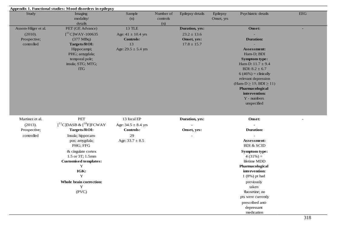
318
Appendix 1. Functional studies: Mood disorders in epilepsy
Study Imaging Sample Number of Epilepsy details Epilepsy Psychiatric details EEG
modality/ (n) controls Onset, yrs
details (n)
Assem-Hilger et al. PET (GE Advance) 13 TLE Duration, yrs: Onset: -
(2010). [11
C]WAY-100635 Age: 41 ± 10.4 yrs 23.2 ± 13.6 -
Prospective; (377 MBq) Controls: Onset, yrs: Duration:
controlled Targets/ROI: 13 17.8 ± 15.7 -
Hippocampi; Age: 29.5 ± 5.4 yrs Assessment:
PHG; amygdala; Ham-D; BDI
temporal pole; Symptom type:
insula; STG; MTG; Ham-D: 11.7 ± 9.4
ITG BDI: 8.2 ± 6.7
6 (46%) = clinically
relevant depression
(Ham-D ≥ 15; BDI ≥ 11)
Pharmacological
intervention:
Y - numbers
unspecified
Martinez et al. PET 13 focal EP Duration, yrs: Onset: -
(2013). [11
C]DASB & [18
F]FCWAY Age: 34.5 ± 8.4 yrs - -
Prospective; Targets/ROI: Controls: Onset, yrs: Duration:
controlled Insula; hippocam- 29 - -
pus; amygdala; Age: 33.7 ± 8.5 Assessment:
PHG; FFG BDI & SCID
& cingulate cortex Symptom type:
1.5 or 3T; 1.5mm 4 (31%) =
Customised templates: lifetime MDD
Y Pharmacological
IGK: intervention:
Y 1 (8%) pt had
Whole brain correction: previously
Y taken
(PVC) fluoxetine; no
pts were currently
prescribed anti-
depressant
medication

319
Appendix 1. Functional studies: Mood disorders in epilepsy, continued
Study Pathology Neuropathology Neuropsychology Post-surgical Ictal Interictal Findings
psychiatric outcome Blood flow/glucose Blood flow/glucose
metabolism metabolism
Assem-Hilger et al. HS (± hippocampal atrophy): - - Non-surgical study - (n.b continuous EEG recordings 1. Pts had sig reduced BP in [I] mesiotemporal
(2010). 5 (38%) not conducted) areas & lateral TL structures compared to the
Prospective; Hippocampal atrophy: 1. Pts had sig reduced BP in [C] hemisphere
controlled 3 (24%) epileptogenic lobe compared to 2. Compared to HCs, pts had sig reduced BP
Normal: 5 (38%) HC in the mesiotemporal structures only
2. Pts with hippocampal atrophy 3. No sig relationship between BP/AI
vs normal MRI pts did not sig & depression rating scales
differ in BP in any ROI 4. Sig positive correlation between hippo-
3. [I] BP was sig reduced in all campal AI & TLE duration
hippocampal ROIs compared
to [C] ROIs
4. Depression scores were
not sig correlated with BP in any
ROI
5. AI was not sig correlated with
depression scores
6. Sig positive correlation between TLE
duration & AI in the hippocampus
Martinez et al. - - - Non-surgical study - Lifetime MDD diag: 1. Mean regional [11
C]DASB binding &
(2013). pts with a history of MDD had sig. asymmetry did not differ between pts
Prospective; higher [11C]DASB asymmetry in insular & HCs
controlled cortex & a trend for FFG 2. Mean [18
F]FCWAY was sig decreased in
- adding side of focus did not [I] hippocampus & amygdala compared to
alter the results contralateral regions
3. MDD diagn. had a sig. effect on [11
C]DASB
asymmetry: pts with a history of MDD had
sig. higher asymmetry in insular cortex & a
trend for FFG - adding side of focus did not
alter the results
4. There was a trend for higher BDI to be
correlated with increased [11
C]DASB
asymmetry
5. Age of EP onset/EP duration/side of focus/
presence of MTS/LTG/CBZ/OXC did not have
sig. effects on [11
C]DASB binding in pts with TLE

320
Appendix 1. Structural studies: Anxiety disorders in epilepsy
Study Study Imaging MRI Sample Number of Epilepsy details Epilepsy Assessment EEG Pathology
type modality/ scanner (n) controls Onset, yrs type
details (n)
Satishchandra et al. MRI 16 'partial EP': 15 Duration, yrs: - Onset: - -
(2003).* GE Electric 8 EP+ANX - -
Prospective; 1.5T Mean age: 59.1 yrs Onset, yrs: Duration:
controlled ROI: 8 EP-only - -
Y Mean age: 50.9 yrs Assessment:
(amygd. Controls: SCAN
& hippocampi) 15 Symptom type:
Whole brain correction: -
Y Pharmacological
intervention:
-
Halley et al. Prospective; MRI Siemens medical 42: Duration, yrs: Onset: - -
(2010). controlled Siemens medical systems 63 & 26 MTLRx MTLRx: -
Prospective; systems 63 & Signa echospeed (26=ATLR & 3 para- 15 ± 12 Duration:
controlled Signa echospeed superconducting hippocampal gyral NMTRx: -
superconducting imaging system lesionectomies) 16 ± 11 Assessment:
imaging system Age 36 ± 12 yrs Onset, yrs: DSM IV;
ROI: 16 NMTRx - Austin CEP
Y (6 lateral temporal interview
(=manual corticectomies; Symptom type:
sementation 8 FLRx & 2 OLRx) Lifetime diagn.
of the Age: 33 ± 10 yrs of anxiety:
amygdalae) Controls: MTLRx:
41 7 (27%)
Age: 35 ± 13 yrs NMTRx
4 (165)
Pharmacological
intervention:
-
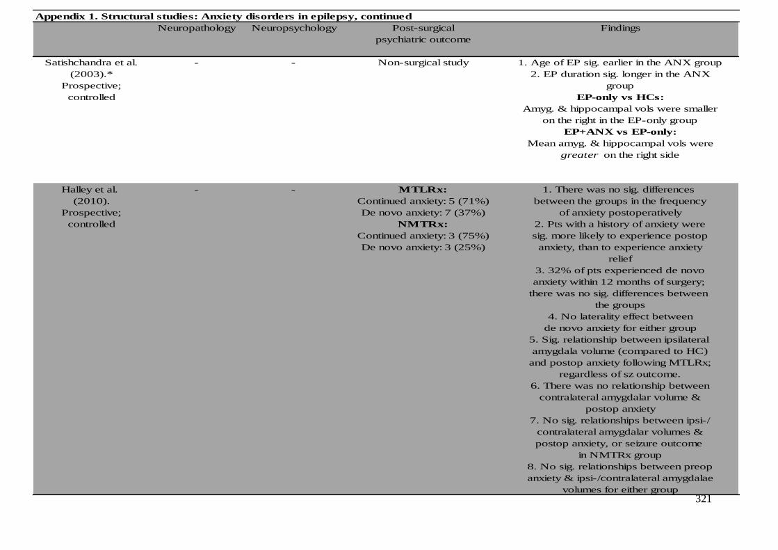
321
Appendix 1. Structural studies: Anxiety disorders in epilepsy, continued
Neuropathology Neuropsychology Post-surgical Findings
psychiatric outcome
Satishchandra et al. - - Non-surgical study 1. Age of EP sig. earlier in the ANX group
(2003).* 2. EP duration sig. longer in the ANX
Prospective; group
controlled EP-only vs HCs:
Amyg. & hippocampal vols were smaller
on the right in the EP-only group
EP+ANX vs EP-only:
Mean amyg. & hippocampal vols were
greater on the right side
Halley et al. - - MTLRx: 1. There was no sig. differences
(2010). Continued anxiety: 5 (71%) between the groups in the frequency
Prospective; De novo anxiety: 7 (37%) of anxiety postoperatively
controlled NMTRx: 2. Pts with a history of anxiety were
Continued anxiety: 3 (75%) sig. more likely to experience postop
De novo anxiety: 3 (25%) anxiety, than to experience anxiety
relief
3. 32% of pts experienced de novo
anxiety within 12 months of surgery;
there was no sig. differences between
the groups
4. No laterality effect between
de novo anxiety for either group
5. Sig. relationship between ipsilateral
amygdala volume (compared to HC)
and postop anxiety following MTLRx;
regardless of sz outcome.
6. There was no relationship between
contralateral amygdalar volume &
postop anxiety
7. No sig. relationships between ipsi-/
contralateral amygdalar volumes &
postop anxiety, or seizure outcome
in NMTRx group
8. No sig. relationships between preop
anxiety & ipsi-/contralateral amygdalae
volumes for either group

322
Appendix 1. Functional studies: Anxiety disorders in epilepsy
Study Imaging Sample Epilepsy details Psychiatric details EEG Pathology
modality/ (n)
details
Bonelli et al. fMRI 54 TLE Duration, yrs: Onset: - MTS: 100%
(2008).* GE Excite Age range: 18-62 yrs - -
Prospective; 3T; 1.5mm Controls: Onset, yrs: Duration:
controlled IGK: 21 - -
Y Age range: 22-62 yrs Assessment:
HADS
Symptom type:
HADS (D/A):
LTLE: 5.5/7
RTLE:4.5/9
HC: 1.5/5.5
Pharmacological
intervention:
-

323
Appendix 1. Functional studies: Anxiety disorders in epilepsy, continued
Study Neuropathology Neuropsychology Post-surgical Included contrasts (fMRI) Findings
psychiatric outcome Negative Positive
valence valence
Bonelli et al. - - Mean change in anxiety Correlational analysis with preop Happy vs neutral faces: 1. No sig correlations between pre- or post-
(2008).* scores (pre-postop): HADS & fMRI activation No sig activations operative change in HADS scores
Prospective; LTLE (n=10/26): -0.5 (-8 to +3) (fearful vs neutral faces): within TL at the group 2. No sig differences in T2 amygdala values
controlled RTLE (n=11/28): +1 (-5 to +7) 1. Sig positive correlation in levels; therefore between the R/L TLE groups
Mean change in depression RTLE pts between preop anxiety no correlational 3. In RTLE pts only, there was a sig positive
scores (pre-postop): scores & fMRI activation in L & R analyses with HADS correlation between preop fMRI activation in
LTLE (n=10/26): -1.5 (-6 to +1) amygdala, compared to LTLE pts data performed L & R amygdala & preop anxiety & depression
RTLE (n=11/28): +1 (-7 to +6) 2. Sig positive association in RTLE scores, compared to L TLE pts, when viewing
pts between preop depression fearful vs neutral faces
scores & fMRI activation in L & R 4. In RTLE pts only, there was a significant
amygdala, compared to LTLE pts positive correlation between preop fMRI
Correlational analysis with preop activation in the R amygdala and change
fMRI activation & change in HADS (increase) in postop anxiety & depression
4m postop ratings at 4m
(fearful vs neutral faces): 5. No sig correlation between sz outcome &
1. Sig positive correlation in changes in HADS scores at 4m F/U
RTLE pts between preop fMRI
activation in the R amygdala
& change (increase) in anxiety
scores
2. Sig positive correlation in
RTLE pts between preop fMRI
activation in the R amygdala
& change (increase) in depression
scores
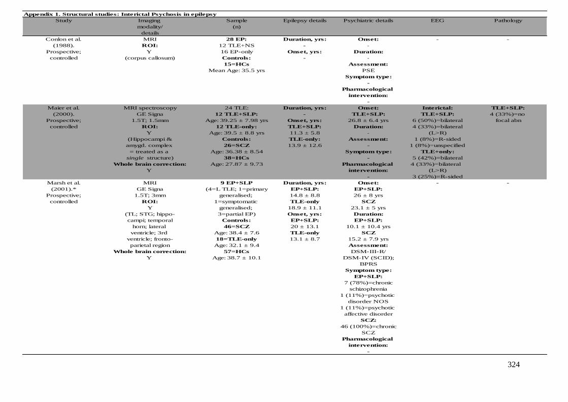
324
Appendix 1. Structural studies: Interictal Psychosis in epilepsy Appendix 1. Structural studies: Interictal Psychosis in epilepsy, continued
Study Imaging Sample Epilepsy details Psychiatric details EEG Pathology
modality/ (n)
details
Conlon et al. MRI 28 EP: Duration, yrs: Onset: - -
(1988). ROI: 12 TLE+NS - -
Prospective; Y 16 EP-only Onset, yrs: Duration:
controlled (corpus callosum) Controls: - -
15=HCs Assessment:
Mean Age: 35.5 yrs PSE
Symptom type:
-
Pharmacological
intervention:
-
Maier et al. MRI spectroscopy 24 TLE: Duration, yrs: Onset: Interictal: TLE+SLP:
(2000). GE Signa 12 TLE+SLP: - TLE+SLP: TLE+SLP: 4 (33%)=no
Prospective; 1.5T; 1.5mm Age: 39.25 ± 7.98 yrs Onset, yrs: 26.8 ± 6.4 yrs 6 (50%)=bilateral focal abn
controlled ROI: 12 TLE-only: TLE+SLP: Duration: 4 (33%)=bilateral
Y Age: 39.5 ± 8.8 yrs 11.3 ± 5.8 - (L>R)
(Hippocampi & Controls: TLE-only: Assessment: 1 (8%)=R-sided
amygd. complex 26=SCZ 13.9 ± 12.6 - 1 (8%)=unspecified
= treated as a Age: 36.38 ± 8.54 Symptom type: TLE+only:
single structure) 38=HCs - 5 (42%)=bilateral
Whole brain correction: Age: 27.87 ± 9.73 Pharmacological 4 (33%)=bilateral
Y intervention: (L>R)
- 3 (25%)=R-sided
Marsh et al. MRI 9 EP+SLP Duration, yrs: Onset: - -
(2001).* GE Signa (4=L TLE; 1=primary EP+SLP: EP+SLP:
Prospective; 1.5T; 3mm generalised; 14.8 ± 8.8 26 ± 8 yrs
controlled ROI: 1=symptomatic TLE-only SCZ
Y generalised; 18.9 ± 11.1 23.1 ± 5 yrs
(TL; STG; hippo- 3=partial EP) Onset, yrs: Duration:
campi; temporal Controls: EP+SLP: EP+SLP:
horn; lateral 46=SCZ 20 ± 13.1 10.1 ± 10.4 yrs
ventricle; 3rd Age: 38.4 ± 7.6 TLE-only SCZ
ventricle; fronto- 18=TLE-only 13.1 ± 8.7 15.2 ± 7.9 yrs
parietal region Age: 32.1 ± 9.4 Assessment:
Whole brain correction: 57=HCs DSM-III-R/
Y Age: 38.7 ± 10.1 DSM-IV (SCID);
BPRS
Symptom type:
EP+SLP:
7 (78%)=chronic
schizophrenia
1 (11%)=psychotic
disorder NOS
1 (11%)=psychotic
affective disorder
SCZ:
46 (100%)=chronic
SCZ
Pharmacological
intervention:
-

325
Appendix 1. Structural studies: Interictal Psychosis in epilepsy, continued
Study Neuropathology Neuropsychology Post-surgical Findings
psychiatric outcome
Conlon et al. - - Non-surgical study 1. EP-only pts showed sig. greater midcallosal
(1988). thickness than the TLE+NC & HCs
Prospective;
controlled
Maier et al. - - Non-surgical study 1. TLE-only & TLE+SLP groups = sig reduced NAA
(2000). bilaterally compared to HCs
Prospective; 2. No sig differences in NAA between the TLE groups
controlled 3. Total hippocampus/amygdala showed no sig
differences in any of the groups compared to HCs
Specific regions of focal volume reduction:
1. Bilateral volume reductions in specific areas of
the hippocampal-amgydala complex (anterior to the
fornix) bilaterally in TLE+SLP pts compared to HCs.
However, regional bilateral volume reduction in
these regions were not detected in TLE-only pts
when comapred to HCs
Marsh et al. - NART: Non-surgical study 1. HCs had sig higher NART-IQ score relative to
(2001).* EP+SLP: 103.4 ± 11.6 both psychotic groups
Prospective; SCZ: 106.4 ± 9.2 Relative to HCs:
controlled TLE-only: unknown 1. All pt groups had ventricular enlargement &
HCs: 111.3 ± 7.1 smaller TL, fronto-parietal and STG GM volumes;
VIQ: the extent of these abnormalities was greatest in
EP+SLP: 87.4 ± 16.3 EP+SLP pts (but differences between TLE-only,
SCZ: unknown SCZ & EP+SLP were n.s)
TLE-only: 98.1 ± 14.4 2. TLE-only pts had TL WMem deficits, hippocampal
HCs: unknown atrophy, which were [I] to the seizure focus
PIQ:
E+SLP: 85.6 ± 16.6
SCZ: unknown
TLE-only: 100.8 ± 16.4
HCs: unknown
FSIQ:
E+SLP: 86 ± 16.3
SCZ: unknown
TLE-only: 99.6 ± 15.2
HCs: unknown

326
Appendix 1. Structural studies: Interictal Psychosis in epilepsy Appendix 1. Structural studies: Interictal Psychosis in epilepsy, continued
Study Imaging Sample Epilepsy details Psychiatric details EEG Pathology
modality/ (n)
details
Tebartz van Elst et al. MRI 26 TLE+SLP/PIP Duration, yrs: Onset: TLE+SLP/PIP: TLE+SLP/PIP:
(2002).* GE Signa Controls: Groups were - R-sided: 6 (23%) None: 13 (50%)
Retrospective; 1.5T; 1.5mm 20=HCs matched Duration: L-sided: 8 (31%) R HS: 2 (8%)
controlled ROI: 24=TLE-only Onset, yrs: - Bilateral: 10 (38%) L HS: 6 (23%)
Y All groups were - Assessment: TLE-only: Bilateral HS: 0 (0%)
(Hippocampi & age- & gender matched ICD-10 R-sided: 9 (38%) Other: 5 (19%)
amygd.) Symptom type: L-sided: 9 (38%) TLE-only:
Whole brain correction: - Bilateral: 5 (21%) None: 7 (29%)
Y Pharmacological R HS: 6 (25%)
intervention: L HS: 10 (42%)
TLE+SLP/PIP: Bilateral HS: 1 (4%)
11 (42%) = anti- Other: 0 (0%)
psychotics
Marchetti et al. MRI 36 EP Duration, yrs: Onset: - EP:
(2003).* Philips Age: 40 ± 12 yrs - - MTS=22 (61%)
Prospective; Gyroscan (30 (83%)=MTLE) Onset, yrs: Duration: Cryptogenic=11 (31%)
controlled 1.5T; 1.2mm Controls: - - Tumour=1 (3%)
ROI: 30 HCs Assessment: Fahr disease=1 (3%)
Y Age: 42 ± 14 DSM-IV
(Hippocampi & Symptom type:
amygd.) -
Whole brain correction: Pharmacological
Y intervention:
-
Rusch et al. MRI (VBM) 26 TLE+SLP/PIP Duration, yrs: Onset: TLE+SLP/PIP: TLE+SLP/PIP:
(2004).* GE Signa Controls: Groups were - R-sided: 6 (23%) None: 13 (50%)
Retrospective; 1.5T; 1.5mm 20=HCs matched Duration: L-sided: 8 (31%) R HS: 2 (8%)
controlled Customised templates: 24=TLE-only Onset, yrs: - Bilateral: 10 (38%) L HS: 6 (23%)
Y All groups were - Assessment: TLE-only: Bilateral HS: 0 (0%)
IGK: age- & gender matched ICD-10 R-sided: 9 (38%) Other: 5 (19%)
12mm Symptom type: L-sided: 9 (38%) TLE-only:
Whole brain correction: - Bilateral: 5 (21%) None: 7 (29%)
Y Pharmacological R HS: 6 (25%)
intervention: L HS: 10 (42%)
TLE+SLP/PIP: Bilateral HS: 1 (4%)
11 (42%) = anti- Other: 0 (0%)
psychotics

327
Appendix 1. Structural studies: Interictal Psychosis in epilepsy, continued
Study Neuropathology Neuropsychology Post-surgical Findings
psychiatric outcome
Tebartz van Elst et al. - VIQ: Non-surgical study 1. VIQ was sig lower in the psychotic compared
(2002).* TLE+SLP/PIP: 86.4 ± 2.3 to non-psychotic TLE pts
Retrospective; TLE-only: 95.1 ± 3 2. Psychotic pts had sig smaller total brain volumes
controlled PIQ: compared to both non-psychotic pts & HCs
TLE+SLP/PIP: 89.7 ± 2.6 3. There was no sig difference in hippocampal
TLE-only: 93.6 ± 2.9 between the groups
4. TLE+SLP/PIP had a 16-18% bilateral enlargement
of the amygdala compared to pt and HC groups;
this finding was unchanged after removing pts
taking anti-psychotic medication
Marchetti et al. - - Non-surgical study 1. Mean left hippocampal volume was sig smaller
(2003).* in the EP group compared to HCs
Prospective; (in the presence of MTS)
controlled
Rusch et al. - VIQ: Non-surgical study 1. VIQ was sig lower in the psychotic compared
(2004).* TLE+SLP/PIP: 86.4 ± 2.3 to non-psychotic TLE pts
Retrospective; TLE-only: 95.1 ± 3 GM differences:
controlled PIQ: 1. There were no sig differences in GM concentration
TLE+SLP/PIP: 89.7 ± 2.6 between the psychotic (TLE+SLP/PIP) & non-
TLE-only: 93.6 ± 2.9 psychotic (TLE-only) groups
2. There were no sig differences between TLE+SLP
& TLE+PIP pts
3. The TLE-only group had a sig increase in GM
in the R TL

328
Appendix 1. Structural studies: Interictal Psychosis in epilepsy Appendix 1. Structural studies: Interictal Psychosis in epilepsy, continued
Study Imaging Sample Epilepsy details Psychiatric details EEG Pathology
modality/ (n)
details
Flugel et al. MRI 40 TLE: Duration, yrs: Onset: No sig. TLE+IP:
(2005).* (VBM - MTI analysis) 20 TLE+IP TLE+IP: TLE+IP: differences HS: 50%
Prospective; GE Signa Age: 39.1 ± 9.9 yrs 32.7 ± 9.2 26.4 [17-38] yrs; between the No lesion: 50%
controlled 1.5T; 1.5mm 20 TLE-IP TLE-IP: 20.1 yrs following groups TLE-IP:
Customised templates: Age: 38.7 ± 12.6 yrs 25.9 ± 14.7 TLE onset HS: 50%
Y Controls: Onset, yrs: Duration: No lesion: 50%
IGK: 23 HC Mean: TLE+IP:
12mm Age: 37 ± 12 yrs TLE+IP: Mean: 12.6 yrs
ROI: 6.3 yrs Assessment:
Y - only for TLE-IP: DSM-IV;
post-hoc analysis 12.8 yrs PANSS
Symptom type:
TLE+IP:
NSS: 23.2
PSS: 16
Pharmacological
intervention:
Neuroleptics: 80%

329
Appendix 1. Structural studies: Interictal Psychosis in epilepsy, continued
Study Neuropathology Neuropsychology Post-surgical Findings
psychiatric outcome
Flugel et al. - NART: Non-surgical study 1. No sig. differences in premorbid
(2005).* TLE+IP: 93.1 ± 13.6 IQ/age/sz freq
Prospective; TLE-IP: 97.7 ± 12.3 2. TLE+PIP pts had sig. earlier onset
controlled of TLE & history of SE
3. No EEG differences between
the groups (± IP/± lesion)
4. No sig. differences in MTR
between groups
5. No sig. differences in the
hippocampal volumes between
the groups
6. No sig differences in MTR
findings and clinical factors
(age/SE/TLE onset/psychosis
duration/current neoleptic meds/
PANSS scores)
No focal lesion subgroup analysis:
1. TLE+IP had sig. reduction of MTR
L middle & STG compared to
TLE-IP and HC
2. No sig. differences in MTR
between TLE-IP & HC groups
3. Sample size (n=8) was too small
to assess laterality effect

330
Appendix 1. Structural studies: Interictal Psychosis in epilepsy Appendix 1. Structural studies: Interictal Psychosis in epilepsy, continued
Study Imaging Sample Duration of Psychiatric details EEG Pathology
modality/ (n) Epilepsy, yrs
details
Flugel et al. MRI 40 TLE: Duration, yrs: Onset: No sig. TLE+IP:
(2006).* (VBM - MTI analysis) 20 TLE+IP TLE+IP: TLE+IP: differences HS: 50%
Prospective; GE Signa Age: 39.1 ± 9.9 yrs 32.8 ± 9.3 26.4 ± 6.5 yrs between the No lesion: 50%
controlled 1.5T; 1.5mm 20 TLE-IP TLE-IP: Duration: groups TLE-IP:
Customised templates: Age: 38.7 ± 12.6 yrs 25.9 ± 14.7 TLE+IP: HS: 50%
Y Controls: Onset, yrs: 12.5 ± 9.2 yrs No lesion: 50%
IGK: - TLE+IP: Assessment:
12mm 6.3 ± 5.7 DSM-IV;
TLE-IP: PANSS
12.8 ± 7 Symptom type:
TLE+IP:
NSS: 23.2
PSS: 16
Pharmacological
intervention:
Neuroleptics: 80%
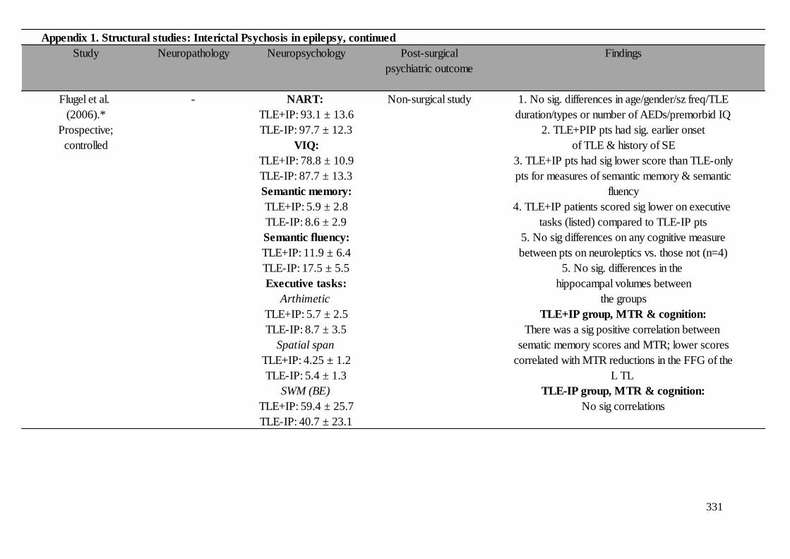
331
Appendix 1. Structural studies: Interictal Psychosis in epilepsy, continued
Study Neuropathology Neuropsychology Post-surgical Findings
psychiatric outcome
Flugel et al. - NART: Non-surgical study 1. No sig. differences in age/gender/sz freq/TLE
(2006).* TLE+IP: 93.1 ± 13.6 duration/types or number of AEDs/premorbid IQ
Prospective; TLE-IP: 97.7 ± 12.3 2. TLE+PIP pts had sig. earlier onset
controlled VIQ: of TLE & history of SE
TLE+IP: 78.8 ± 10.9 3. TLE+IP pts had sig lower score than TLE-only
TLE-IP: 87.7 ± 13.3 pts for measures of semantic memory & semantic
Semantic memory: fluency
TLE+IP: 5.9 ± 2.8 4. TLE+IP patients scored sig lower on executive
TLE-IP: 8.6 ± 2.9 tasks (listed) compared to TLE-IP pts
Semantic fluency: 5. No sig differences on any cognitive measure
TLE+IP: 11.9 ± 6.4 between pts on neuroleptics vs. those not (n=4)
TLE-IP: 17.5 ± 5.5 5. No sig. differences in the
Executive tasks: hippocampal volumes between
Arthimetic the groups
TLE+IP: 5.7 ± 2.5 TLE+IP group, MTR & cognition:
TLE-IP: 8.7 ± 3.5 There was a sig positive correlation between
Spatial span sematic memory scores and MTR; lower scores
TLE+IP: 4.25 ± 1.2 correlated with MTR reductions in the FFG of the
TLE-IP: 5.4 ± 1.3 L TL
SWM (BE) TLE-IP group, MTR & cognition:
TLE+IP: 59.4 ± 25.7 No sig correlations
TLE-IP: 40.7 ± 23.1

332
Appendix 1. Structural studies: Interictal Psychosis in epilepsy Appendix 1. Structural studies: Interictal Psychosis in epilepsy, continued
Study Imaging Sample Epilepsy details Psychiatric details EEG Pathology
modality/ (n)
details
Flugel et al. MRI (DTI) 38 TLE: Duration, yrs: Onset: No sig. TLE+IP:
(2006a). GE Signa 20 TLE+IP - TLE+IP: differences HS: 50%
Prospective; 1.5T; 1.5mm Age: 39.1 ± 9.8 yrs Onset, yrs: 26.7 ± 6.8 yrs between the No lesion: 50%
TLE-controls ROI: 18 TLE-IP TLE+IP: Duration: groups TLE-IP:
Y Age: Unspecified 6.2 ± 5.6 TLE+IP: HS: 50%
(MTG & MFG Controls: TLE-IP: 12.3 ± 9.4 yrs No lesion: 50%
bilaterally) 18 TLE-IP 12.8 ± 7 Assessment:
DSM-IV;
PANSS
Symptom type:
-
Pharmacological
intervention:
TLE+IP:
Neuroleptics: 75%

333
Appendix 1. Structural studies: Interictal Psychosis in epilepsy, continued
Study Neuropathology Neuropsychology Post-surgical Findings
psychiatric outcome
Flugel et al. - NART: Non-surgical study 1. TLE+IP had sig. lower VIQ/
(2006a). TLE+IP: 92.2 ± 13.7 semantic fluency/spatial span/
Prospective; TLE-IP: 97.94 ± 12.3 spatial Wmem
TLE-controls VIQ: 2. TLE+IP: Earlier age of TLE onset
TLE+IP: 78.28 ± 12.4 & SE history
TLE-IP: 87.68 ± 13.3 3. No sig. differences in the
Semantic Fluency: hippocampal volumes between
TLE+IP: 11.94 ± 6.4 the groups
TLE-IP: 17.52 ± 5.5 4. FA sig.↓ in bilateral frontal
Spatial Span: regions in TLE+IP pts (R>L) &
TLE+IP: 4.17 ± 1.1 bilateral TL regions
TLE-IP: 5.4 ± 1.3 5. MD was sig. ↑ in bilateral
Spatial WMem: frontal regions in TLE+IP pts
TLE+IP: 59.89 ± 27.2 TLE+IP only:
TLE-IP: 40.7 ± 23.1 1. Sig. positive correlation
between bifrontal & L temporal
FA with semantic fluency
2. L FL FA positively correlated
with VIQ
3. Bilateral FL FA positively
correlated with spatial span
4. Semantic fluency sig. predictor
of ↓ FA in L frontal & bilateral
temproal regions
5. Spatial span sig. predictor of
↓ FA in R FL
6. Sig. negative correlations were
found between age & bilateral
frontal & temporal reductions
in both groups
7. Negative symptom score
sig. predicted L FL FA reductions

334
Appendix 1. Structural studies: Interictal Psychosis in epilepsy Appendix 1. Structural studies: Interictal Psychosis in epilepsy, continued
Study Imaging Sample Epilepsy details Psychiatric details EEG Pathology
modality/ (n)
details
Adams et al. MRI 312: Duration, yrs: Onset: - -
(2008). 1.5T 212 MTS - -
Retrospective; Age: 37 yrs Onset, yrs: Duration:
EP-controls TLE (other lesions) - -
Age: 35.5 yrs Assessment:
TLE (non-lesional) DSM-IV
Age: 36.8 yrs (not all pts had
ETLE (lesional) neuropsychiatric
Age: 37.7 yrs assessment)
ETLE (non-lesional) Symptom type:
Age: 34.5 yrs Lifetime diagn.
Controls: MTS
- Depression: 26.4%
Psychosis: 8.3%
TLE (other lesions)
Depression: 27.6%
Psychosis: 8.6%
TLE (non-lesional)
Depression: 41.9%
Psychosis: 5.4%
ETLE (lesional)
Depression: 35.7%
Psychosis: 8.6%
ETLE (non-lesional)
Depression: 41.2%
Psychosis: 4.8%
Pharmacological
intervention:
-

335
Appendix 1. Structural studies: Interictal Psychosis in epilepsy, continued
Study Neuropathology Neuropsychology Post-surgical Findings
psychiatric outcome
Adams et al. - - Non-surgical study 1. No sig. difference between the prevalence
(2008). of any psychiatric disorder (depression/psychosis)
Retrospective; & TLE vs ETLE
EP-controls 2. Pts with non-lesional EP (both TLE & ETLE) had
sig. higher prevalence of psychiatric disorders
(69.2%) than those with lesional EP (inc. TLE &
ETLE pts) (52.9%)
3. No sig. differences in the prevalence of any
psychiatric disorder & pts with TLE vs ETLE
4. In multivariable analyses, the odds of any
psychiatric diagn. were 2.4 times higher for pts
with non-lesional focal EP vs lesional EP
(MTS & other lesions); although this was due to
pts with a lifetime diag. of depression
5. The laterality of the seizure foci (R/L/bilateral)
were not sig. predictors of psychopathology
6. No sig. difference between the lifetime
prevalence of depression between TLE (31.2%)
& ETLE (37.9%) pts
7. Pts with non-lesional EP exhibited higher
prevalence of depression (41.6%) compared to
those with lesional EP (28.5%)
8. Lifetime psychiatric diag. was not predicted
by MTS or another lesion
9. Multivariable analyses indicated that a lifetime
diag. of depression was only sig. predicted by
non-lesional EP. The odds of a lifetime diagnosis
of depression was almost double (OR: 1.96, 95%CI:
1.16-3.31, p=.01) for pts with non-lesional vs
lesional EP
10. Lifetime diag. of depression was not predicted
by MTS or another lesion
11. The laterality of the seizure foci (R/L/bilateral)
were not sig. predictors of depression
12. There were no sig. relationships between any
of the EP variables & a history of psychosis
13. Similarly, logistic regression did not find any
sig. EP variables (laterality/TLE vs ETLE/lesional vs
non-lesional/pathology type) predicted psychosis
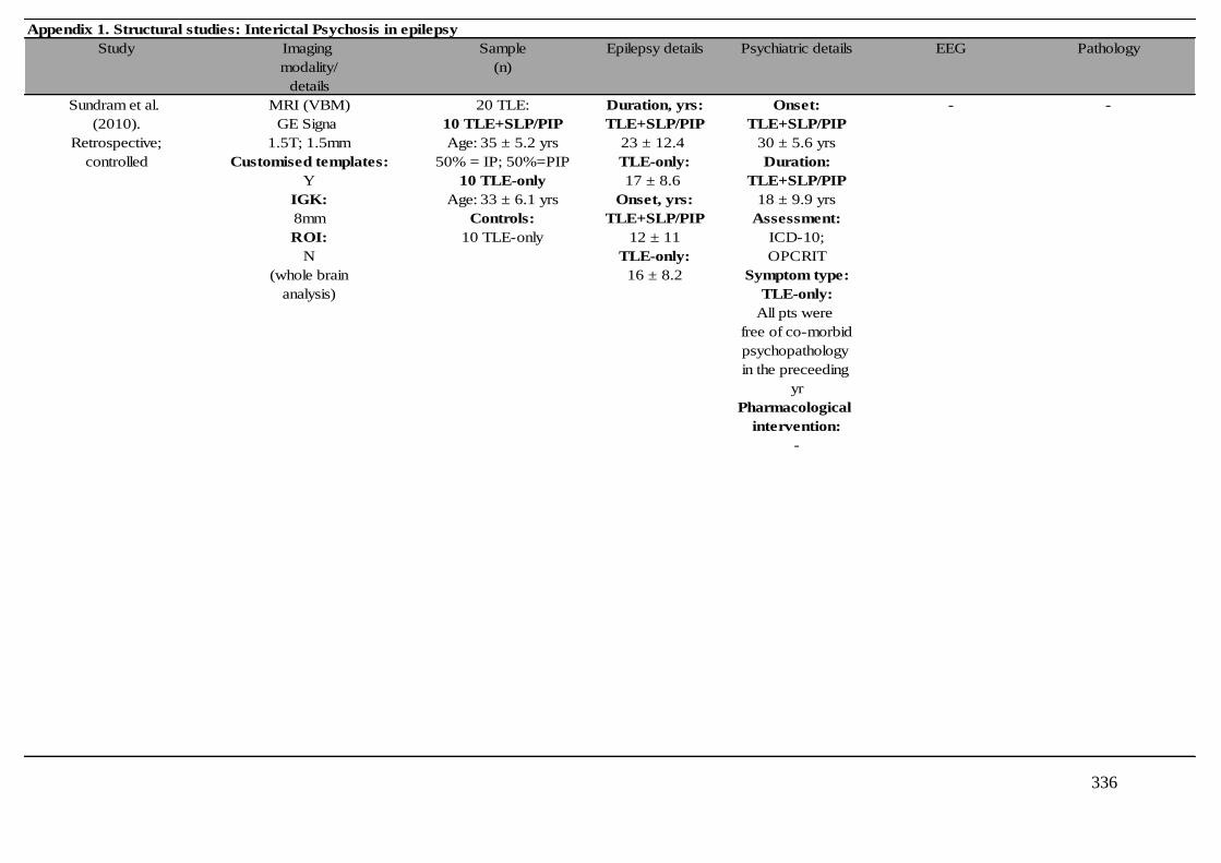
336
Appendix 1. Structural studies: Interictal Psychosis in epilepsy Appendix 1. Structural studies: Interictal Psychosis in epilepsy, continued
Study Imaging Sample Epilepsy details Psychiatric details EEG Pathology
modality/ (n)
details
Sundram et al. MRI (VBM) 20 TLE: Duration, yrs: Onset: - -
(2010). GE Signa 10 TLE+SLP/PIP TLE+SLP/PIP TLE+SLP/PIP
Retrospective; 1.5T; 1.5mm Age: 35 ± 5.2 yrs 23 ± 12.4 30 ± 5.6 yrs
controlled Customised templates: 50% = IP; 50%=PIP TLE-only: Duration:
Y 10 TLE-only 17 ± 8.6 TLE+SLP/PIP
IGK: Age: 33 ± 6.1 yrs Onset, yrs: 18 ± 9.9 yrs
8mm Controls: TLE+SLP/PIP Assessment:
ROI: 10 TLE-only 12 ± 11 ICD-10;
N TLE-only: OPCRIT
(whole brain 16 ± 8.2 Symptom type:
analysis) TLE-only:
All pts were
free of co-morbid
psychopathology
in the preceeding
yr
Pharmacological
intervention:
-

337
Appendix 1. Structural studies: Interictal Psychosis in epilepsy, continued
Study Neuropathology Neuropsychology Post-surgical Findings
psychiatric outcome
Sundram et al. - - Non-surgical study 1. No sig. differences in total brain/
(2010). GM/WM volumes between
Retrospective; between the groups
controlled 2. TLE+psychosis group had sig. ↓
GM volume in unilateral TL
structures (L parahippocampal gyrus
& hippocampus).
3. TLE+psychosis group had sig. ↓
GM volume in lateral temporal
lobes (bilateral inferior, middle &
superior temporal gyri & fusiform gyri)
4. TLE+psychosis group had sig. ↓
extratemporal bilateral GM atrophy
(insula, cerebellum, caudate
nuclei). Unilateral GM volume loss
seen in the R cingulum and L inferior
parietal lobule.
5. TLE+psychosis group had sig. ↓
WM in bilateral hippocampus,
parahippocampal gyrus, lateral
temporal lobes (middle & inferior
temporal gyri & fusiform gyri).
6. TLE+psychosis group had sig. ↓
WM in unilateral R superior temporal
gyrus.
7. TLE+psychosis group had sig. ↓
WM extratemporally (bilateral
cingulum, corpus callosum, thalamus.
Unilateral reductions: L lingual gyrus
& R midbrain)
found between age & bilateral
frontal & temporal reductions
in both groups
7. Negative symptom score
sig. predicted L FL FA reductions

338
Appendix 1. Structural studies: Interictal Psychosis in epilepsy Appendix 1. Structural studies: Interictal Psychosis in epilepsy, continued
Study Imaging Sample Epilepsy details Psychiatric details EEG Pathology
modality/ (n)
details
Gutierrez-Galve et al. SBM (FreeSurfer) 45: Duration, yrs: Onset: - TLE+IP:
(2012). GE Signa 22 TLE+IP TLE+IP: TLE+IP: Normal: 10 (46%)
Prospective; 1.5T; 1.2mm Age: 38.9 ± 9.7 yrs 39.1 ± 11.1 26.7 ± 6.3 yrs LHS: 6 (27%)
controlled Customised templates: 23 TLE-only: TLE-only: Duration: RHS: 4 (18%)
Y Age: 38.7 ± 12.1 yrs 25.7 ± 14.2 TLE+IP: Bilateral HS: 2 (9%)
IGK: Controls: Onset, yrs: 12.1 ± 9.1 yrs TLE-only:
10mm 20 TLE+IP: Assessment: Normal: 10 (44%)
ROI: Age: 36 ± 11.3 yrs 7 ± 7 Clinical LHS: 6 (26%)
22 cortical TLE-only: interview; RHS: 4 (17%)
parcellations 12.5 ± 7.2 (DSM-IV-TR) Bilateral HS: 3 (13%)
used PANSS
Whole brain correction: Symptom type:
Y PANSS (TLE+IP):
Positive: 16.4 ± 3.5
Negative: 24 ± 7.1
Pharmacological
intervention:
TLE+IP:
15 (68%) = anti-
psychotics
TLE-only:
None = anti-
psychotics
HC:
None had a formal
psychiatric history

339
Appendix 1. Structural studies: Interictal Psychosis in epilepsy, continued
Study Neuropathology Neuropsychology Post-surgical Findings
psychiatric outcome
Gutierrez-Galve et al. - NART: Non-surgical study 1. TLE+IP pts had a sig. earlier age of EP
(2012). TLE+IP: 90.6 ± 14.9 onset/higher prevalence of SE/reduced
Prospective; TLE-IP: 94.8 ± 14.2 current IQ/reduced working memory
controlled Current IQ: span & working memory manipulation,
TLE+IP: 76.1 ± 10.7 compared to TLE-only pts
TLE-IP: 85.6 ± 14.3 2. TLE+IP pts had sig. reduced cortical
Working memory span: thickness in the IFG, specifically the
TLE+IP: 4.2 ± 1.2 pars opercularis, compared to HC
TLE-IP: 5.3 ± 1.4 (after FDR correction)
Working memory 3. No sig. trend (p=.06) between TLE+IP
manipulation: and TLE-only pts in IFG cortical
TLE+IP: 60.6 ± 24.8 thickness
TLE-IP: 43.4 ± 23.7 4. Age of EP onset/duration of EP/
Story recall (immed): history of SE/age or duration of psycho-
TLE+IP: 16.5 ± 11.1 sis/dose of neuroleptic meds were
TLE-IP: 21.9 ± 11.8 not significantly associated with cortical
Story recall (delayed): thickness in either pt groups
TLE+IP: 13.6 ± 10.6 5. Cognitive scores were not sig.
TLE-IP: 16.4 ± 12.2 associated with cortical thickness
6. Sig. left-sided reduction in cortical
area in the IFG (pars opercularis) in
TLE+IP pts compared to TLE-only pts
7. In TLE+IP pts only, current IQ was
positively associated with frontal &
(superior frontal & rostral middle frontal)
& temporal (superior temporal) cortical
surface area (after FDR correction)
8. In TLE pts only, current IQ was
positively associated with cortical
frontal (superior frontal & rostral middle
frontal), temporal (superior temporal
& fusiform gyrus) and inferior parietal
volumes (after FDR correction)

340
Appendix 1. Functional studies: Interictal Psychosis in epilepsy
Study Imaging Sample Epilepsy details Epilepsy Psychiatric details EEG Pathology
modality/ (n) Onset, yrs
details
Gallhofer et al. PET 12 EP+SLP Duration, yrs: - Onset: EP-only: CT scans of
(1985). (oxygen-15 6 EP+SLP+meds: - - 3 (60%): R-sided abn the EP pts
Prospective; inhalation) Mean age: 42.3 yrs Onset, yrs: Duration: 2 (40%): L-sided abn did not reveal
controlled ROI: 6 EP+SLP-meds: - - EP+SLP: clear structural
Y 38.2 yrs Assessment: 3 (25%): R-sided abn lesions
Controls: PSE 1 (8%): L-sided abn
5=EP-only Symptom type: 8 (67%): Bilateral abn
5=HCs 4 (67%)=nuclear
schizophrenia
1 (17%)=psycho-
tic depression
1 (17%)=schizo-
phrenia with
1st rank symptoms
EP+SLP+meds:
2 (33%)=nuclear
schizophrenia
2 (33%)=depressive
psychosis
1 (17%)=paranoid
psychosis
1 (17%)=improved
& not psychotic
Pharmacological
intervention:
Of the EP+SLP
pts, 6 (50%) were
free from anti-
psychotics at the
time of scanning

341
Appendix 1. Functional studies: Interictal Psychosis in epilepsy, continued
Study Neuropathology Neuropsychology Post-surgical Ictal Interictal Findings
psychiatric outcome Blood flow/glucose Blood flow/glucose
metabolism metabolism
Gallhofer et al. - Mean IQ: Non-surgical study - HC vs. EP groups: 1. HC had higher values of oxygen utilization
(1985). EP+SLP+meds: 97 rCMRO2: HC > EP in & cerebral blood flow than the EP groups, with
Prospective; EP+SLP-meds: 96 L & R TL, L basal ganglia the most sig differences in the mid & posterior
controlled EP-only: 98 rCBF: HC > EP L TL, TLs
R TL, FTCB, basal 2. EP pts with psychosis had sig reduced oxygen
ganglia & limbic strip extraction (rOEF)in fronto-temporal regions
rOER: HC < EP in L
FTCB, R TL, FTCB &
limbic strip
EP-only analyses:
rOEF: EP-only <
EP+SLP in limbic strip
& TL, FTCB, basal ganglia
bilaterally
rOER: EP+SLP-meds <
EP+SLP+meds in L
TL, limbic strip &
bilaterally in the FTCB
& basal ganglia

342
Appendix 1. Functional studies: Interictal Psychosis in epilepsy
Study Imaging Sample Epilepsy details Epilepsy Psychiatric details EEG Pathology
modality/ (n) Onset, yrs
details
Marshall et al. SPECT 5 EP+SLP Duration, yrs: Onset: Groups were EP+SLP:
(1993).99M
Tc-HMPAO Age: 37.6 ± 7.7 yrs - - matched 3 (60%): atrophy
Prospective; (av. dose: Controls: Onset, yrs: Duration: on CT scans
controlled 550 MBq). 5 EP-only EP+SLP: - (groups were
ROI: Age: 38 ± 8.7 yrs 12.5 ± 12.1 Assessment: not matched on
Y EP-only: RDC; gross cerebral
9.9 ± 10.4 SADS pathology)
Symptom type:
EP+SLP:
No pt was psychotic
at the time of scanning
Pharmacological
intervention:
EP+SLP: 100%
on neuroleptics
Mellers et al. SPECT 12 EP+SLP Duration, yrs: Onset: Groups were -
(1998).99M
Tc-HMPAO Age: 43 ± 9.7 yrs EP+SLP: - matched
Prospective; (av. dose: Controls: 32.5 ± 10.4 yrs Duration:
controlled 550 MBq). 16 EP-only EP-only: EP+SLP:
ROI: Age: 40.5 ± 9.7 yrs 24.8 ± 12.2 yrs 13.5 ± 10.4 yrs
Y 11 SCZ Onset, yrs: SCZ:
(6 cortical Age: 38.7 ± 10.4 yrs - 24.8 ± 12.2 yrs
regions Assessment:
defined) DSM-III-R;
BPRS
Symptom type*:
BPRS:
EP+SLP: 5.5 ± 5.4
SCZ: 5.4 ± 5.2
EP-only: 1.4 ± 2.3
(assessed immediately
prior to scanning)
Pharmacological
intervention:
All SCZ & EP+SLP
pts were taking
neuroleptics

343
Appendix 1. Functional studies: Interictal Psychosis in epilepsy, continued
Study Neuropathology Neuropsychology Post-surgical Ictal Interictal Findings
psychiatric outcome Blood flow/glucose Blood flow/glucose
metabolism metabolism
Marshall et al. - VIQ range: Non-surgical study - EP+SLP vs EP-only: 1. EP+SLP pts had sig reduced rCBF in the
(1993). EP+SLP: 85-98 SLP pts had sig reduced L hemisphere, circumscribed to the medial
Prospective; EP-only: 96-116 rCBF in the L medial TL compared to EP-only pts
controlled PIQ range: TL region 2. Groups differed sig in gross brain pathology
EP+SLP: 80-107 & IQ
EP-only: 76-118
Mellers et al. - NART: Non-surgical study - 1. SCZ pts showed 1. EP+SLP pts had lower rCBF in the L STG
(1998). EP+SLP: 105 ± 14 greater activation of during a verbal fluency task compared to
Prospective; EP-only: 111 ± 9 the ACC compared EP-only and SCZ groups
controlled SCZ: 112 ± 6 to the EP groups 2. SCZ group had greater increase in rCBF in
2. EP+SLP pts showed the ACC than both epilepsy groups
reduced activity 3. The EP+SLP group performed sig worse
in the L STG during on the verbal fluency task compared to
verbal fluency the other groups (EP-only & SCZ)
compared to SCZ
& EP-only pts

344
Abbreviations for Appendix 1: * hippocampal volumes recorded, refer to paper for further details; cRBF, Cerebral regional blood flow; CT, Computerised tomography; d, days; DEP,
Depression; DES-II, Dissociative Experiences Scale version II; diagn, Diagnosis; DLPFC, Dorsolateral prefrontal cortex; DSM, Diagnostic Statistical Manual; DTI, Diffusion tensor imaging;
EP, Epilepsy; ETLE, Extra-temporal lobe epilepsy; ETOH, Alcohol; EZ, Epileptogenic zone; F/U, Follow-up; FA, Fractional anisotropy; FC, Febrile convulsions; FDG, Fludeoxyglucose; FDR,
False discovery rate; FFG, Fusiform gyrus; FG, Frontal gyrus; FL, Frontal Lobe; FLRx, Frontal lobe resections; FSIQ, Full scale IQ; FTCB, "Fronto-temporal bridge"; GM, Grey matter; GTC,
Generalised tonic clonic; HADS (D/A), Hamilton Depression Rating Scale (depression/anxiety); Hcau, Head of caudate; HC(s), Healthy controls; HS, Hippocampal sclerosis; HV, Hippocampal
volume; ICD, International classification of Diseases; ICV, Intracranial volume; IDD, Interictal dysphoric disorder; I.E.D, Intermittent Explosive Disorder; IED, Interictal epileptiform
discharges; IF, Inferior frontal; IFG, Inferior frontal gyrus; IG, Idiopathic generalised epilepsy; IGK, Isotropic Gaussian Kernel; IHC, Ipsilateral hippocampal volume; ITL, Inferior lateral
temporal; immed, immediate; IMT, Inferior mesial temporal; incl., including; IP, Interictal psychosis; ISWM, Increased T2-weighted signal in anterior temporal white matter; ITG, Inferior
temporal gyrus; L, Left; LOI, Leyton obsessional inventory inventory; LTG, Lamotrigine; m, month; MADRS, Montgomery Asberg Depression Rating Scale; MCST, Modified Card Sorting
Test; MD, Mean diffusivity; MDD, Major depressive disorder; meds, Medication; MF, Mid-frontal; MFG, Middle frontal gyrus; MINI, Mini International Neuropsychiatric Interview; MT, Mid-
temporal; MTG, Middle temporal gyri; MTI, Magnetization Transfer Imaging; MTLE, Mesial temporal lobe; MTLRx, Mesial temporal lobe resection; MTR, Magnetization Transfer Ratio;
MTS, Mesial temporal lobe sclerosis; N, No; N.S, Not significant; N/A, Not applicable; NAA, N-acetyl-aspartate; NART, National adult reading test; NMTRx, Non mesial temporal lobe
resection; No., Number; NOS, Not otherwise specified; NS, Nuclear schizophrenia; NTL, Neocortical temporal lobe lesions; NTLRx, Neocortical temporal lobe resections; OFC, Orbitofrontal
cortex; OL, Occipital lobe; OLRx, Occipital lobe resections; OPCRIT, Operational Criteria Checklist for Psychotic Illness; OXC, Oxcarbazepine; PANSS, Positive & Negative Syndrome Scale;
PET, Positron emission tomography; PHG, Parahippocampal gyrus; PHG LesX, Parahippocampal gyrus lesionectomy; PIM, Postictal manic; PIP, Postictal psychosis; PIQ, Performance IQ; PL,
Parietal Lobe; PNES, Psychogenic non-epileptic seizures; POMS, Profile of Mood States; Pre-TLE-op, Before temporal lobe epilepsy surgery; PSE, Present State Examination; psych.,
Psychiatric; pts, patients; PVC, Partial volume correction; QOLIE-89, Quality of Life in Epilepsy Inventory-89; R, right; RAM, Radial atrophy mapping; rCBF, Regional cerebral blood flow;
rCMRO2, Regional oxygen metabolism; RDC, Research Diagnostic Criteria; rOER, Regional oxygen extraction ratio; SADS, Schedule for Affective Disorders and Schizophrenia; SBM,
Surface-based morphometry; SCID, Structured Clinical Interview for DSM-IV; SCIP-P, Adapted SCID for patients with epilepsy; SCZ, Schizophrenia; SDAS-21, Social Dysfunction and
Aggression Scale; SDQ-20, Somatoform Dissociation Questionnaire-20; SE, Status epilepticus; secs, Seconds; SF, Superior frontal; sig., Significant; SMA, Supplementary motor area; SPECT,
Single-photon emission computed tomography; SPM, Statistical parametric mapping; SSSI, Social stress & support interview; ST, Superior temporal; STAI, State-Trait Anxiety Inventory; STG,
Superior temporal gyrus; SWM (BE), Spatial working memory (between errors), sz, Seizure; sz freq, seizure frequency; TH, Thalamus; TL, Temporal lobe; TLE, temporal lobe epilepsy; TMT,
Trail Making Test; Uni-, Unilateral; VBM, Voxel-Based Morphemetry; VIQ, Verbal IQ; Wmem, working memory; WM, White matter; Y, Yes; yr(s); year(s).
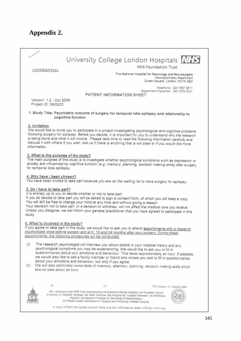
345
Appendix 2.

346

347

348

349
Appendix 3.

350

351
Appendix 4.

352
Appendix 5.

353
Appendix 6.
a b
BDI-FS probability plots. a) Indicating that there were marked deviations from the predicted line;
the assumption of normally distributed residuals has been violated. b) Normally distributed BDI-FS
residuals following a logarithmic transformation
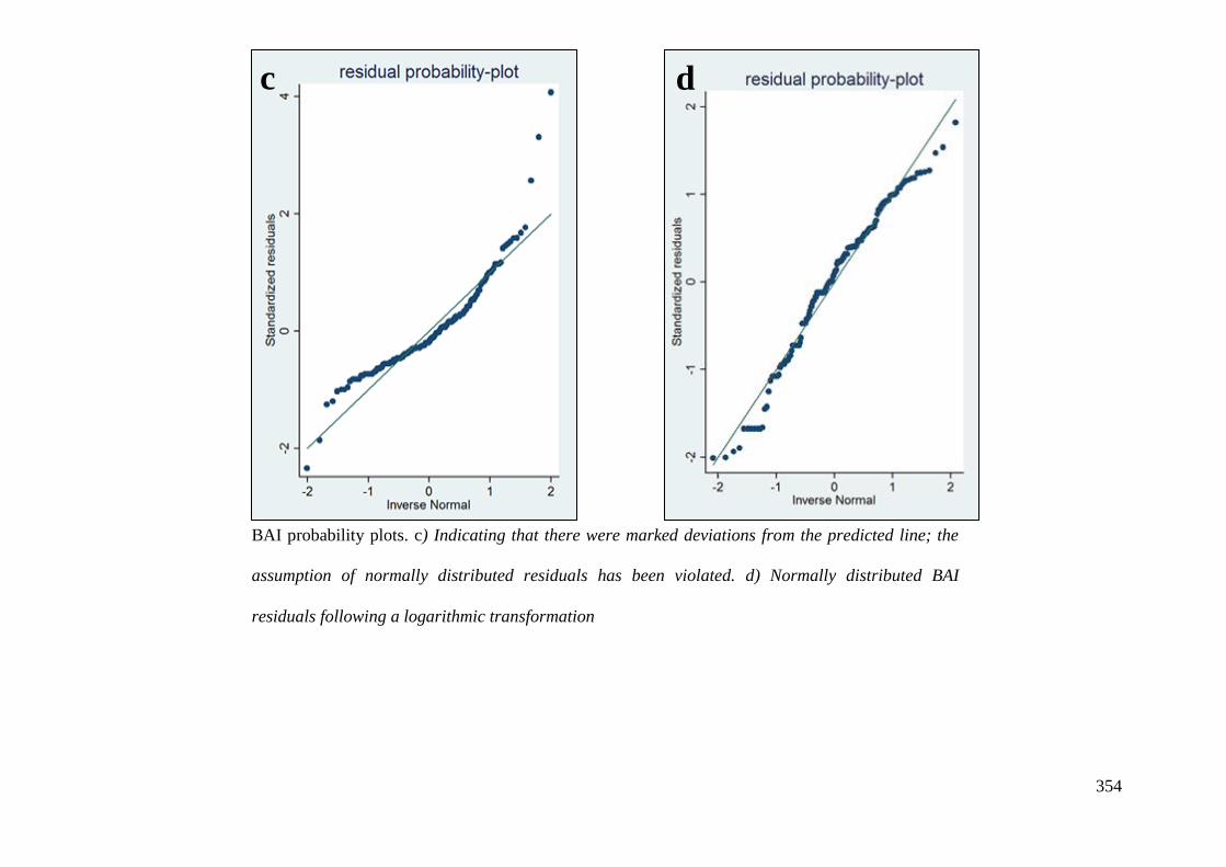
354
c d
BAI probability plots. c) Indicating that there were marked deviations from the predicted line; the
assumption of normally distributed residuals has been violated. d) Normally distributed BAI
residuals following a logarithmic transformation

355
Appendix 7.
Multivariable (a) non-log and (b) log- BDI-FS random intercept only models.
a b

356
Appendix 8.
Multivariable (a) non-log and (b) log- BAI random intercept only models.
a b

357
Appendix 9.
A

358
B

359
C
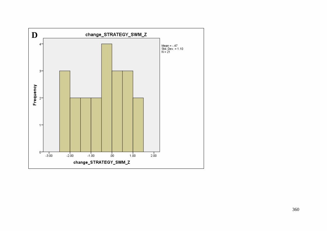
360
D

361
E

362
F

363
REFERENCES
Adams SJ, O'Brien TJ, Lloyd J, Kilpatrick CJ, Salzberg MR, Velakoulis D. Neuropsychiatric
morbidity in focal epilepsy. Br J Psychiatry. 2008 Jun;192(6):464-9.
Adams SJ, Velakoulis D, Kaye AH, Corcoran NM, O'Brien TJ. Psychiatric history does not
predict seizure outcome following temporal lobectomy for mesial temporal sclerosis.
Epilepsia. 2012 Oct;53(10):1700-4.
Adelöw C, Andersson T, Ahlbom A, Tomson T. Hospitalization for psychiatric disorders
before and after onset of unprovoked seizures/epilepsy. Neurology. 2012 Feb 7;78(6):396-
401.
Alper K, Devinsky O, Westbrook L, Luciano D, Pacia S, Perrine K, Vazquez B. Premorbid
psychiatric risk factors for postictal psychosis. J Neuropsychiatry Clin Neurosci. 2001
Fall;13(4):492-9.
Alper K, Kuzniecky R, Carlson C, Barr WB, Vorkas CK, Patel JG, Carrelli AL, Starner K,
Flom PL, Devinsky O. Postictal psychosis in partial epilepsy: a case-control study. Ann
Neurol. 2008 May;63(5):602-10.
Altshuler L, Rausch R, Delrahim S, Kay J, Crandall P. Temporal lobe epilepsy, temporal
lobectomy, and major depression. J Neuropsychiatry Clin Neurosci. 1999 Fall;11(4):436-43.
Alvarez JA, Emory E. Executive function and the frontal lobes: a meta-analytic review.
Neuropsychol Rev. 2006 Mar;16(1):17-42.
American Psychiatric Association. (2000). Diagnostic and statistical manual of mental
disorders (4th ed., text rev.). Arlington, VA: American Psychiatric Publishing.
Anhoury S, Brown RJ, Krishnamoorthy ES, Trimble MR. Psychiatric outcome after temporal
lobectomy: a predictive study. Epilepsia. 2000 Dec;41(12):1608-15.
Anticevic A, Cole MW, Murray JD, Corlett PR, Wang XJ, Krystal JH. The role of default
network deactivation in cognition and disease. Trends Cogn Sci. 2012 Dec;16(12):584-92.
Arbuthnott K, Frank J. Trail making test, part B as a measure of executive control: validation
using a set-switching paradigm. J Clin Exp Neuropsychol. 2000 Aug;22(4):518-28.
Arnone D, McIntosh AM, Ebmeier KP, Munafò MR, Anderson IM. Magnetic resonance
imaging studies in unipolar depression: systematic review and meta-regression analyses. Eur
Neuropsychopharmacol. 2012 Jan;22(1):1-16.
Ashburner J, Friston KJ. Voxel-based morphometry--the methods. Neuroimage. 2000
Jun;11(6 Pt 1):805-21.
Assem-Hilger E, Lanzenberger R, Savli M, Wadsak W, Mitterhauser M, Mien LK, Stögmann
E, Baumgartner C, Kletter K, Asenbaum S. Central serotonin 1A receptor binding in
temporal lobe epilepsy: a [carbonyl-(11)C]WAY-100635 PET study. Epilepsy Behav. 2010
Nov;19(3):467-73.
Austin MP, Mitchell P, Goodwin GM. Cognitive deficits in depression: possible implications
for functional neuropathology. Br J Psychiatry. 2001 Mar;178:200-6.

364
Baker GA. The psychosocial burden of epilepsy. Epilepsia. 2002;43 Suppl 6:26-30.
Barbieri V, Gozzo F, Schiariti MP, Lo Russo G, Sartori I, Castana L, Scarone S, Gambini O.
One-year postsurgical follow-up of 12 subjects with epilepsy and interictal psychosis.
Epilepsy Behav. 2011 Oct;22(2):385-7.
Baxendale S, Thompson P, Harkness W, Duncan J. Predicting memory decline following
epilepsy surgery: a multivariate approach. Epilepsia. 2006 Nov;47(11):1887-94.
Baxendale S, Thompson PJ, Duncan JS. Neuropsychological function in patients who have
had epilepsy surgery: a long-term follow-up. Epilepsy Behav. 2012 Jan;23(1):24-9.
Baxendale S, Thompson PJ, Duncan JS. The role of the Wada test in the surgical treatment of
temporal lobe epilepsy: an international survey. Epilepsia. 2008 Apr;49(4):715-20.
Baxendale S, Thompson PJ, Sander JW. Neuropsychological outcomes in epilepsy surgery
patients with unilateral hippocampal sclerosis and good preoperative memory function.
Epilepsia. 2013 Sep;54(9):e131-4.
Baxendale S. The impact of epilepsy surgery on cognition and behavior. Epilepsy Behav.
2008 May;12(4):592-9.
Baxendale S. The role of functional MRI in the presurgical investigation of temporal lobe
epilepsy patients: a clinical perspective and review. J Clin Exp Neuropsychol. 2002
Aug;24(5):664-76.
Baxendale SA, Thompson PJ, Duncan JS. Epilepsy & depression: the effects of comorbidity
on hippocampal volume--a pilot study. Seizure. 2005 Sep;14(6):435-8.
Bechara A, Damasio AR, Damasio H, Anderson SW. Insensitivity to future consequences
following damage to human prefrontal cortex. Cognition. 1994 Apr-Jun;50(1-3):7-15.
Beck AT, Epstein N, Brown G, Steer RA. An inventory for measuring clinical anxiety:
psychometric properties. J Consult Clin Psychol. 1988 Dec;56(6):893-7.
Beck, A., & Steer, R. (1990). Beck Anxiety Inventory Manual. San Antonio: Psychological
Corporation.
Beck, A., Steer, R., & Brown, G. (1996). Manual for the Beck Depression Inventory-II. San
Antonio, TX: Psychological Corporation.
Beck, A., Steer, R., & Brown, G. (2000). BDI-Fast Screen for medical patients: manual. San
Antonio, TX: Psychological Corporation.
Bell B, Lin JJ, Seidenberg M, Hermann B. The neurobiology of cognitive disorders in
temporal lobe epilepsy. Nat Rev Neurol. 2011 Mar;7(3):154-64.
Benedict RH, Fishman I, McClellan MM, Bakshi R, Weinstock-Guttman B. Validity of the
Beck Depression Inventory-Fast Screen in multiple sclerosis. Mult Scler. 2003 Aug;9(4):393-
6.
Bennett PC, Ong B, Ponsford J. Measuring executive dysfunction in an acute rehabilitation
setting: using the dysexecutive questionnaire (DEX). J Int Neuropsychol Soc. 2005
Jul;11(4):376-85.

365
Berg AT, Berkovic SF, Brodie MJ, Buchhalter J, Cross JH, van Emde Boas W, Engel J,
French J, Glauser TA, Mathern GW, Moshé SL, Nordli D, Plouin P, Scheffer IE. Revised
terminology and concepts for organization of seizures and epilepsies: report of the ILAE
Commission on Classification and Terminology, 2005-2009. Epilepsia. 2010 Apr;51(4):676-
85.
Berg EA. A simple objective technique for measuring flexibility in thinking. J Gen Psychol.
1948 Jul;39:15-22.
Binder JR, Swanson SJ, Hammeke TA, Morris GL, Mueller WM, Fischer M, Benbadis S,
Frost JA, Rao SM, Haughton VM. Determination of language dominance using functional
MRI: a comparison with the Wada test. Neurology. 1996 Apr;46(4):978-84.
Bird CM, Papadopoulou K, Ricciardelli P, Rossor MN, Cipolotti L. Monitoring cognitive
changes: psychometric properties of six cognitive tests. Br J Clin Psychol. 2004 Jun;43(Pt
2):197-210.
Blocher JB1, Fujikawa M, Sung C, Jackson DC, Jones JE. Computer-assisted cognitive
behavioral therapy for children with epilepsy and anxiety: a pilot study. Epilepsy Behav.
2013 Apr;27(1):70-6.
Blumcke I, Thom M, Aronica E, Armstrong DD, Vinters HV, Palmini A, Jacques TS,
Avanzini G, Barkovich AJ, Battaglia G, Becker A, Cepeda C, Cendes F, Colombo N, Crino
P, Cross JH, Delalande O, Dubeau F, Duncan J, Guerrini R, Kahane P, Mathern G, Najm I,
Ozkara C, Raybaud C, Represa A, Roper SN, Salamon N, Schulze-Bonhage A, Tassi L,
Vezzani A, Spreafico R. The clinicopathologic spectrum of focal cortical dysplasias: a
consensus classification proposed by an ad hoc Task Force of the ILAE Diagnostic Methods
Commission. Epilepsia. 2011 Jan;52(1):158-74.
Blumer D, Wakhlu S, Davies K, Hermann B. Psychiatric outcome of temporal lobectomy for
epilepsy: incidence and treatment of psychiatric complications. Epilepsia. 1998
May;39(5):478-86.
Bonelli SB, Powell R, Yogarajah M, Thompson PJ, Symms MR, Koepp MJ, Duncan JS.
Preoperative amygdala fMRI in temporal lobe epilepsy. Epilepsia. 2009 Feb;50(2):217-27.
Bonilha L, Alessio A, Rorden C, Baylis G, Damasceno BP, Min LL, Cendes F.
Extrahippocampal gray matter atrophy and memory impairment in patients with medial
temporal lobe epilepsy. Hum Brain Mapp. 2007 Dec;28(12):1376-90.
Bowie CR, Harvey PD. Administration and interpretation of the Trail Making Test. Nat
Protoc. 2006;1(5):2277-81.
Boylan LS, Flint LA, Labovitz DL, Jackson SC, Starner K, Devinsky O. Depression but not
seizure frequency predicts quality of life in treatment-resistant epilepsy. Neurology. 2004 Jan
27;62(2):258-61.
Bragatti JA, Torres CM, Londero RG, Assmann JB, Fontana V, Martin KC, Hidalgo MP,
Chaves ML, Bianchin MM. Prevalence of psychiatric comorbidities in temporal lobe
epilepsy: the value of structured psychiatric interviews. Epileptic Disord. 2010
Dec;12(4):283-91.
366
Bremner JD, Vythilingam M, Vermetten E, Nazeer A, Adil J, Khan S, Staib LH, Charney
DS. Reduced volume of orbitofrontal cortex in major depression. Biol Psychiatry. 2002 Feb
15;51(4):273-9.
Briellmann RS, Hopwood MJ, Jackson GD. Major depression in temporal lobe epilepsy with
hippocampal sclerosis: clinical and imaging correlates. J Neurol Neurosurg Psychiatry. 2007
Nov;78(11):1226-30.
Briellmann RS, Kalnins RM, Hopwood MJ, Ward C, Berkovic SF, Jackson GD. TLE patients
with postictal psychosis: mesial dysplasia and anterior hippocampal preservation. Neurology.
2000 Oct 10;55(7):1027-30.
Bright P, Jaldow E, Kopelman MD. The National Adult Reading Test as a measure of
premorbid intelligence: a comparison with estimates derived from demographic variables. J
Int Neuropsychol Soc. 2002 Sep;8(6):847-54.
Britton JW, Shih JJ. Antiepileptic drugs and suicidality. Drug Healthc Patient Saf.
2010;2:181-9.
Brodie MJ, French JA. Management of epilepsy in adolescents and adults. Lancet. 2000 Jul
22;356(9226):323-9.
Bromfield EB, Altshuler L, Leiderman DB, Balish M, Ketter TA, Devinsky O, Post RM,
Theodore WH. Cerebral metabolism and depression in patients with complex partial seizures.
Arch Neurol. 1992 Jun;49(6):617-23.
Broyd SJ, Demanuele C, Debener S, Helps SK, James CJ, Sonuga-Barke EJ. Default-mode
brain dysfunction in mental disorders: a systematic review. Neurosci Biobehav Rev. 2009
Mar;33(3):279-96.
Buchsbaum BR, Greer S, Chang WL, Berman KF. Meta-analysis of neuroimaging studies of
the Wisconsin card-sorting task and component processes. Hum Brain Mapp. 2005
May;25(1):35-45.
Buckner RL, Andrews-Hanna JR, Schacter DL. The brain's default network: anatomy,
function, and relevance to disease. Ann N Y Acad Sci 2008;1124:1-38.
Bujarski KA, Hirashima F, Roberts DW, Jobst BC, Gilbert KL, Roth RM, Flashman LA,
McDonald BC, Saykin AJ, Scott RC, Dinnerstein E, Preston J, Williamson PD, Thadani VM.
Long-term seizure, cognitive, and psychiatric outcome following trans-middle temporal gyrus
amygdalohippocampectomy and standard temporal lobectomy. J Neurosurg. 2013
Jul;119(1):16-23.
Burgess PW, Alderman N, Evans J, Emslie H, Wilson BA. The ecological validity of tests of
executive function. J Int Neuropsychol Soc. 1998 Nov;4(6):547-58.
Busch RM, Dulay MF, Kim KH, Chapin JS, Jehi L, Kalman CC, Naugle RI, Najm IM. Pre-
surgical mood predicts memory decline after anterior temporal lobe resection for epilepsy.
Arch Clin Neuropsychol. 2011 Dec;26(8):739-45.
Butler T, Blackmon K, McDonald CR, Carlson C, Barr WB, Devinsky O, Kuzniecky R,
DuBois J, French J, Halgren E, Thesen T. Cortical thickness abnormalities associated with
depressive symptoms in temporal lobe epilepsy. Epilepsy Behav. 2012 Jan;23(1):64-7.

367
Campbell S, Macqueen G. The role of the hippocampus in the pathophysiology of major
depression. J Psychiatry Neurosci. 2004 Nov;29(6):417-26.
Cankurtaran ES, Ulug B, Saygi S, Tiryaki A, Akalan N. Psychiatric morbidity, quality of life,
and disability in mesial temporal lobe epilepsy patients before and after anterior temporal
lobectomy. Epilepsy Behav. 2005 Aug;7(1):116-22.
Carreno M, Gil-Nagel A, Sánchez JC, Elices E, Serratosa JM, Salas-Puig J, Villanueva V,
Porcel J. Strategies to detect adverse effects of antiepileptic drugs in clinical practice.
Epilepsy Behav. 2008 Jul;13(1):178-83.
Carreno, M., & Luders, H. O. (2001). General Principals of Presurgical Evalution. In H. O.
Luders & Y. G. Comair (Eds.), Epilepsy Surgery (2nd ed., pp. 185-199). Philadelphia:
Lippincott.
Cataldi M, Avoli M, de Villers-Sidani E. Resting state networks in temporal lobe epilepsy.
Epilepsia. 2013 Dec;54(12):2048-59.
Charyton C, Elliott JO, Moore JL, Klatte ET. Is it time to consider cognitive behavioral
therapy for persons with epilepsy? Clues from pathophysiology, treatment and functional
neuroimaging. Expert Rev Neurother. 2010 Dec;10(12):1911-27.
Chen S, Wu X, Lui S, Wu Q, Yao Z, Li Q, Liang D, An D, Zhang X, Fang J, Huang X, Zhou
D, Gong QY. Resting-state fMRI study of treatment-naïve temporal lobe epilepsy patients
with depressive symptoms. Neuroimage. 2012 Mar;60(1):299-304.
Chovaz CJ, McLachlan RS, Derry PA, Cummings AL. Psychosocial function following
temporal lobectomy: influence of seizure control and learned helplessness. Seizure. 1994
Sep;3(3):171-6.
Christodoulou C, Koutroumanidis M, Hennessy MJ, Elwes RD, Polkey CE, Toone BK.
Postictal psychosis after temporal lobectomy. Neurology. 2002 Nov 12;59(9):1432-5.
Clark L, Chamberlain SR, Sahakian BJ. Neurocognitive mechanisms in depression:
implications for treatment. Annu Rev Neurosci. 2009;32:57-74.
Cleary RA, Baxendale SA, Thompson PJ, Foong J. Predicting and preventing
psychopathology following temporal lobe epilepsy surgery. Epilepsy Behav. 2013
Mar;26(3):322-34.
Commission on Classification and Terminology of the International League Against
Epilepsy. Proposal for revised classification of and epileptic syndromes. Epilepsia. 1989
30:389–399.
Commission on Classification and Terminology of the International League Against
Epilepsy. Proposal for revised clinical and electrographic classification of epileptic seizures.
Epilepsia. 1981 22:489–501.
Conlon P, Trimble MR. A study of the corpus callosum in epileptic psychosis using
magnetic resonance imaging. Biol Psychiatry. 1988 Nov;24(7):857-60.
Cools R, Ivry RB, D'Esposito M. The human striatum is necessary for responding to changes
in stimulus relevance. J Cogn Neurosci. 2006 Dec;18(12):1973-83.

368
Corcoran R, Thompson P. Epilepsy and poor memory: who complains and what do they
mean? Br J Clin Psychol. 1993 May;32 ( Pt 2):199-208.
Corkin S. (2013). Permanent Present Tense: The Unforgettable Life of the Amnesic Patient,
HM. Philadelphia: Basic Books
Coughlan AK. Hollows SE. The adult memory and information processing battery (AMIPB)
Test Manual. St James University Hospital, Leeds; 1985.
Coughlan, A., Oddy, M., & Crawford, J. (2007). BIRT Memory and Information Processing
Battery (BMIPB). London: Brain Rehabilitation Trust.
Crowe SF. The differential contribution of mental tracking, cognitive flexibility, visual
search, and motor speed to performance on parts A and B of the Trail Making Test. J Clin
Psychol. 1998 Aug;54(5):585-91.
da Conceição PO, Nascimento PP, Mazetto L, Alonso NB, Yacubian EM, de Araujo Filho
GM. Are psychiatric disorders exclusion criteria for video-EEG monitoring and epilepsy
surgery in patients with mesial temporal sclerosis? Epilepsy Behav. 2013 May;27(2):310-4.
Dabbs K, Jones J, Seidenberg M, Hermann B. Neuroanatomical correlates of cognitive
phenotypes in temporal lobe epilepsy. Epilepsy Behav. 2009 Aug;15(4):445-51.
Damsa C, Kosel M, Moussally J. Current status of brain imaging in anxiety disorders. Curr
Opin Psychiatry. 2009 Jan;22(1):96-110.
Danielson NB, Guo JN, Blumenfeld H. The default mode network and altered consciousness
in epilepsy. Behav Neurol 2011;24:55-65.
de Oliveira GN, Kummer A, Salgado JV, Portela EJ, Sousa-Pereira SR, David AS, Teixeira
AL. Psychiatric disorders in temporal lobe epilepsy: an overview from a tertiary service in
Brazil. Seizure. 2010 Oct;19(8):479-84.
de Tisi J, Bell GS, Peacock JL, McEvoy AW, Harkness WF, Sander JW, Duncan JS. The
long-term outcome of adult epilepsy surgery, patterns of seizure remission, and relapse: a
cohort study. Lancet. 2011 Oct 15;378(9800):1388-95.
Dean K, Stevens H, Mortensen PB, Murray RM, Walsh E, Pedersen CB. Full spectrum of
psychiatric outcomes among offspring with parental history of mental disorder. Arch Gen
Psychiatry. 2010 Aug;67(8):822-9.
Demakis GJ. A meta-analytic review of the sensitivity of the Wisconsin Card Sorting Test to
frontal and lateralized frontal brain damage. Neuropsychology. 2003 Apr;17(2):255-64.
DiMartini A, Crone C, Fireman M, Dew MA. Psychiatric Aspects of Organ Transplantation
In Critical Care. Crit Care Clin. Oct 2008; 24(4): 949-x
Demase DF, Gruenthal M, Balint J. The national temporal lobectomy survey. Seizure. 2009
Dec;18(10):702-10.
Derry PA, Rose KJ, McLachlan RS. Moderators of the effect of preoperative emotional
adjustment on postoperative depression after surgery for temporal lobe epilepsy. Epilepsia.
2000 Feb;41(2):177-85.

369
Desmond JE, Sum JM, Wagner AD, Demb JB, Shear PK, Glover GH, Gabrieli JD, Morrell
MJ. Functional MRI measurement of language lateralization in Wada-tested patients. Brain.
1995 Dec;118 ( Pt 6):1411-9.
Devinsky O, Abramson H, Alper K, FitzGerald LS, Perrine K, Calderon J, Luciano D.
Postictal psychosis: a case control series of 20 patients and 150 controls. Epilepsy Res. 1995
Mar;20(3):247-53.
Devinsky O, Barr WB, Vickrey BG, Berg AT, Bazil CW, Pacia SV, Langfitt JT, Walczak
TS, Sperling MR, Shinnar S, Spencer SS. Changes in depression and anxiety after resective
surgery for epilepsy. Neurology. 2005 Dec 13;65(11):1744-9.
Drevets WC, Price JL, Furey ML. Brain structural and functional abnormalities in mood
disorders: implications for neurocircuitry models of depression. Brain Struct Funct. 2008
Sep;213(1-2):93-118.
DuBois JM, Devinsky O, Carlson C, Kuzniecky R, Quinn BT, Alper K, Butler T, Starner K,
Halgren E, Thesen T. Abnormalities of cortical thickness in postictal psychosis. Epilepsy
Behav. 2011 Jun;21(2):132-6.
Dulay MF, Schefft BK, Fargo JD, Privitera MD, Yeh HS. Severity of depressive symptoms,
hippocampal sclerosis, auditory memory, and side of seizure focus in temporal lobe epilepsy.
Epilepsy Behav. 2004 Aug;5(4):522-31.
Duncan J. The current status of neuroimaging for epilepsy. Curr Opin Neurol. 2009
Apr;22(2):179-84.
Duncan JS, Sander JW, Sisodiya SM, Walker MC. Adult epilepsy. Lancet. 2006 Apr
1;367(9516):1087-100.
Duncan JS. Epilepsy surgery. Clin Med. 2007 Apr;7(2):137-42.
Duncan JS. Imaging and epilepsy. Brain. 1997 Feb;120 ( Pt 2):339-77.
Duncan JS. Imaging in the surgical treatment of epilepsy. Nat Rev Neurol. 2010
Oct;6(10):537-50
Elger CE, Schmidt D. Modern management of epilepsy: a practical approach. Epilepsy
Behav. 2008 May;12(4):501-39.
Engel J Jr, McDermott MP, Wiebe S, Langfitt JT, Stern JM, Dewar S, Sperling MR, Gardiner
I, Erba G, Fried I, Jacobs M, Vinters HV, Mintzer S, Kieburtz K; Early Randomized Surgical
Epilepsy Trial (ERSET) Study Group. Early surgical therapy for drug-resistant temporal lobe
epilepsy: a randomized trial. JAMA. 2012 Mar 7;307(9):922-30.
Engel J Jr. Report of the ILAE classification core group. Epilepsia. 2006 Sep;47(9):1558-68.
Engel J Jr. Surgery for seizures. N Engl J Med. 1996 Mar 7;334(10):647-52.
Engel J Jr; International League Against Epilepsy (ILAE). A proposed diagnostic scheme for
people with epileptic seizures and with epilepsy: report of the ILAE Task Force on
Classification and Terminology. Epilepsia. 2001 Jun;42(6):796-803.
Engel J. Surgery for seizures. N Engl J Med. 1996 Mar 7;334(10):647-52.

370
Engman E, Malmgren K. A longitudinal study of psychological features in patients before
and two years after epilepsy surgery. Epilepsy Behav. 2012 Jun;24(2):221-6.
Ertekin BA, Kulaksizoğlu IB, Ertekin E, Gürses C, Bebek N, Gökyiğit A, Baykan B. A
comparative study of obsessive-compulsive disorder and other psychiatric comorbidities in
patients with temporal lobe epilepsy and idiopathic generalized epilepsy. Epilepsy Behav.
2009 Apr;14(4):634-9.
Falip M, Carreño M, Donaire A, Maestro I, Pintor L, Bargalló N, Boget T, Raspall A, Rumià
J, Setoaín J. Postictal psychosis: a retrospective study in patients with refractory temporal
lobe epilepsy. Seizure. 2009 Mar;18(2):145-9.
FDA. Suicidality and Antiepileptic Drugs 2008. Retrieved August 5, 2013 from
http://www.fda.gov/Drugs/DrugSafety/PostmarketDrugSafetyInformationforPatientsandProvi
ders/ucm100192.htm.
Ferguson SM, Rayport M, Gardner R, Kass W, Weiner H, Reiser MF. Similarities in mental
content of psychotic states, spontaneous seizures, dreams, and responses to electrical brain
stimulation in patients with temporal lobe epilepsy. Psychosom Med. 1969 Nov-
Dec;31(6):479-98
Ferreri F, Lapp LK, Peretti CS. Current research on cognitive aspects of anxiety disorders.
Curr Opin Psychiatry. 2011 Jan;24(1):49-54.
Field A. (2009). Discovering Statistics Using SPSS. London: SAGE.
Filho GM, Gomes FL, Mazetto L, Marinho MM, Tavares IM, Caboclo LO, Yacubian EM,
Centeno RS. Major depressive disorder as a predictor of a worse seizure outcome one year
after surgery in patients with temporal lobe epilepsy and mesial temporal sclerosis. Seizure.
2012a Oct;21(8):619-23.
Filho GM, Mazetto L, Gomes FL, Marinho MM, Tavares IM, Caboclo LO, Centeno RS,
Yacubian EM. Pre-surgical predictors for psychiatric disorders following epilepsy surgery in
patients with refractory temporal lobe epilepsy and mesial temporal sclerosis. Epilepsy Res.
2012 Nov;102(1-2):86-93.
Finegersh A, Avedissian C, Shamim S, Dustin I, Thompson PM, Theodore WH. Bilateral
hippocampal atrophy in temporal lobe epilepsy: effect of depressive symptoms and febrile
seizures. Epilepsia. 2011 Apr;52(4):689-97.
Fisher RS, van Emde Boas W, Blume W, Elger C, Genton P, Lee P, Engel J Jr. Epileptic
seizures and epilepsy: definitions proposed by the International League Against Epilepsy
(ILAE) and the International Bureau for Epilepsy (IBE). Epilepsia. 2005 Apr;46(4):470-2.
Fitzsimmons J, Kubicki M, Shenton ME. Review of functional and anatomical brain
connectivity findings in schizophrenia. Curr Opin Psychiatry. 2013 Mar;26(2):172-87.
Flugel D, Cercignani M, Symms MR, Koepp MJ, Foong J. A magnetization transfer imaging
study in patients with temporal lobe epilepsy and interictal psychosis. Biol Psychiatry. 2005
Mar 15;59(6):560-7.
Flugel D, Cercignani M, Symms MR, O'Toole A, Thompson PJ, Koepp MJ, Foong J.
Diffusion tensor imaging findings and their correlation with neuropsychological deficits in

371
patients with temporal lobe epilepsy and interictal psychosis. Epilepsia. 2006 May;47(5):941-
4.
Flugel D, O'Toole A, Thompson PJ, Koepp MJ, Cercignani M, Symms MR, Foong J. A
neuropsychological study of patients with temporal lobe epilepsy and chronic interictal
psychosis. Epilepsy Res. 2006a Oct;71(2-3):117-28.
Fong GC, Fong KY, Mak W, Tsang KL, Chan KH, Cheung RT, Ho SL. Postictal psychosis
related regional cerebral hyperfusion. J Neurol Neurosurg Psychiatry. 2000 Jan;68(1):100-1.
Foong J, Flugel D. Psychiatric outcome of surgery for temporal lobe epilepsy and presurgical
considerations. Epilepsy Res. 2007 Jul;75(2-3):84-96.
Forsgren L, Beghi E, Oun A, Sillanpää M. The epidemiology of epilepsy in Europe - a
systematic review. Eur J Neurol. 2005 Apr;12(4):245-53.
Forsgren L, Nyström L. An incident case-referent study of epileptic seizures in adults.
Epilepsy Res. 1990 May-Jun;6(1):66-81.
Fray PJ, Robbins TW, Sahakian BJ. Neuorpsychiatyric applications of CANTAB.
International Journal of Geriatric Psychiatry, Vol 11(4), Apr 1996, 329-336.
Gaitatzis A, Trimble MR, Sander JW. The psychiatric comorbidity of epilepsy. Acta Neurol
Scand. 2004 Oct;110(4):207-20.
Gallhofer B, Trimble MR, Frackowiak R, Gibbs J, Jones T. A study of cerebral blood flow
and metabolism in epileptic psychosis using positron emission tomography and oxygen. J
Neurol Neurosurg Psychiatry. 1985 Mar;48(3):201-6.
Garcia PA, Laxer KD, van der Grond J, Hugg JW, Matson GB, Weiner MW. Correlation of
seizure frequency with N-acetyl-aspartate levels determined by 1H magnetic resonance
spectroscopic imaging. Magn Reson Imaging. 1997;15(4):475-8.
García-Morales I, de la Peña Mayor P, Kanner AM. Psychiatric comorbidities in epilepsy:
identification and treatment. Neurologist. 2008 Nov;14(6 Suppl 1):S15-25.
Gastaut H. Clinical and electroencephalographical classification of epileptic seizures.
Epilepsia. 1970 Mar;11(1):102-13.
Gerstorf D, Siedlecki KL, Tucker-Drob EM, Salthouse TA. Executive dysfunctions across
adulthood: measurement properties and correlates of the DEX self-report questionnaire.
Neuropsychol Dev Cogn B Aging Neuropsychol Cogn. 2008 Jul;15(4):424-45.
Geyer JD, Payne TA, Faught E, Drury I. Postictal nose-rubbing in the diagnosis,
lateralization, and localization of seizures. Neurology. 1999 Mar 10;52(4):743-5.
Gilbert SJ, Burgess PW. Executive function. Curr Biol. 2008 Feb 12;18(3):R110-4.
Gilliam FG, Albertson B, Driscoll M. (2011). Depression, anxiety, and cognition in epilepsy:
clinical and neurobiological considerations. In C. Helmstaedter, B. Hermann, M. Lassonde,
P. Kahane & A. Arzimanoglou (Eds.), Neuropsychology in the Care of People with Epilepsy.
Esther: John Libbey Eurotext.

372
Gilliam FG, Maton BM, Martin RC, Sawrie SM, Faught RE, Hugg JW, Viikinsalo M,
Kuzniecky RI. Hippocampal 1H-MRSI correlates with severity of depression symptoms in
temporal lobe epilepsy. Neurology. 2007 Jan 30;68(5):364-8.
Gilliam FG. Diagnosis and treatment of mood disorders in persons with epilepsy. Curr Opin
Neurol. 2005 Apr;18(2):129-33.
Giovacchini G, Toczek MT, Bonwetsch R, Bagic A, Lang L, Fraser C, Reeves-Tyer P,
Herscovitch P, Eckelman WC, Carson RE, Theodore WH. 5-HT 1A receptors are reduced in
temporal lobe epilepsy after partial-volume correction. J Nucl Med. 2005 Jul;46(7):1128-35.
Giovagnoli AR. Relation of sorting impairment to hippocampal damage in temporal lobe
epilepsy. Neuropsychologia. 2001;39(2):140-50.
Glascher J, Tranel D, Paul LK, Rudrauf D, Rorden C, Hornaday A, Grabowski T, Damasio
H, Adolphs R. Lesion mapping of cognitive abilities linked to intelligence. Neuron. 2009 Mar
12;61(5):681-91.
Glosser G, Zwil AS, Glosser DS, O'Connor MJ, Sperling MR. Psychiatric aspects of
temporal lobe epilepsy before and after anterior temporal lobectomy. J Neurol Neurosurg
Psychiatry. 2000 Jan;68(1):53-8.
Gorwood P, Corruble E, Falissard B, Goodwin GM. Toxic effects of depression on brain
function: impairment of delayed recall and the cumulative length of depressive disorder in a
large sample of depressed outpatients. Am J Psychiatry. 2008 Jun;165(6):731-9.
Grabowska-Grzyb A, Jedrzejczak J, Nagańska E, Fiszer U. Risk factors for depression in
patients with epilepsy. Epilepsy Behav. 2006 Mar;8(2):411-7.
Grant MM, Thase ME, Sweeney JA. Cognitive disturbance in outpatient depressed younger
adults: evidence of modest impairment. Biol Psychiatry. 2001 Jul 1;50(1):35-43.
Gualtieri CT, Johnson LG, Benedict KB. Neurocognition in depression: patients on and off
medication versus healthy comparison subjects. J Neuropsychiatry Clin Neurosci. 2006
Spring;18(2):217-25.
Guangming Z, Wenjing Z, Guoqiang C, Yan Z, Fuquan Z, Huancong Z. Psychiatric symptom
changes after corticoamygdalohippocampectomy in patients with medial temporal lobe
epilepsy through Symptom Checklist 90 Revised. Surg Neurol. 2009 Dec;72(6):587-91.
Gueorguieva R, Krystal JH. Move over ANOVA: progress in analyzing repeated-measures
data and its reflection in papers published in the Archives of General Psychiatry. Arch Gen
Psychiatry. 2004 Mar;61(3):310-7.
Gureje O. Interictal psychopathology in epilepsy. Prevalence and pattern in a Nigerian clinic.
Br J Psychiatry. 1991 May;158:700-5.
Gutierrez-Galve L, Flugel D, Thompson PJ, Koepp MJ, Symms MR, Ron MA, Foong J.
Cortical abnormalities and their cognitive correlates in patients with temporal lobe epilepsy
and interictal psychosis. Epilepsia. 2012 Jun;53(6):1077-87.
Hader WJ, Tellez-Zenteno J, Metcalfe A, Hernandez-Ronquillo L, Wiebe S, Kwon CS, Jette
N. Complications of epilepsy surgery: a systematic review of focal surgical resections and
invasive EEG monitoring. Epilepsia. 2013 May;54(5):840-7.

373
Halley SA, Wrench JM, Reutens DC, Wilson SJ. The amygdala and anxiety after epilepsy
surgery. Epilepsy Behav. 2010 Aug;18(4):431-6.
Halstead WC. (1947). Brain and Intelligence. Chicago, IL:University of Chicago Press.
Hamberger MJ, Drake EB. Cognitive functioning following epilepsy surgery. Curr Neurol
Neurosci Rep. 2006 Jul;6(4):319-26.
Hamid H, Liu H, Cong X, Devinsky O, Berg AT, Vickrey BG, Sperling MR, Shinnar S,
Langfitt JT, Walczak TS, Barr WB, Dziura J, Bazil CW, Spencer SS. Long-term association
between seizure outcome and depression after resective epilepsy surgery. Neurology. 2011
Nov 29;77(22):1972-6.
Hasler G, Bonwetsch R, Giovacchini G, Toczek MT, Bagic A, Luckenbaugh DA, Drevets
WC, Theodore WH. 5-HT1A receptor binding in temporal lobe epilepsy patients with and
without major depression. Biol Psychiatry. 2007 Dec 1;62(11):1258-64.
Heck RH, Thomas SL, Tabata LN. (2010). Multilevel and Longitudinal Modeling with IBM
SPSS (Quantitative Methodology Series). Hove: Routledge.
Hellwig S, Mamalis P, Feige B, Schulze-Bonhage A, van Elst LT. Psychiatric comorbidity in
patients with pharmacoresistant focal epilepsy and psychiatric outcome after epilepsy
surgery. Epilepsy Behav. 2012 Mar;23(3):272-9.
Helmstaedter C, Kockelmann E. Cognitive outcomes in patients with chronic temporal lobe
epilepsy. Epilepsia. 2006;47 Suppl 2:96-8.
Helmstaedter C, Kurthen M, Lux S, Reuber M, Elger CE. Chronic epilepsy and cognition: a
longitudinal study in temporal lobe epilepsy. Ann Neurol 2003; 54: 425–32.
Helmstaedter C, Sonntag-Dillender M, Hoppe C, Elger CE. Depressed mood and memory
impairment in temporal lobe epilepsy as a function of focus lateralization and localization.
Epilepsy Behav. 2004 Oct;5(5):696-701.
Helmstaedter C. Neuropsychological aspects of epilepsy surgery. Epilepsy Behav. 2004
Feb;5 Suppl 1:S45-55.
Hermann B, Seidenberg M, Lee EJ, Chan F, Rutecki P. Cognitive phenotypes in temporal
lobe epilepsy. J Int Neuropsychol Soc. 2007 Jan;13(1):12-20.
Hermann BP, Lin JJ, Jones JE, Seidenberg M. The emerging architecture of
neuropsychological impairment in epilepsy. Neurol Clin. 2009 Nov;27(4):881-907.
Hermann BP, Seidenberg M, Haltiner A, Wyler AR. Mood state in unilateral temporal lobe
epilepsy. Biol Psychiatry. 1991 Dec 15;30(12):1205-18.
Hermann BP, Seidenberg M, Schoenfeld J, Davies K. Neuropsychological characteristics of
the syndrome of mesial temporal lobe epilepsy. Arch Neurol. 1997 Apr;54(4):369-76.
Hermann BP, Wyler AR, Ackerman B, Rosenthal T. Short-term psychological outcome of
anterior temporal lobectomy. J Neurosurg. 1989 Sep;71(3):327-34

374
Hermann BP, Wyler AR, Richey ET. Wisconsin Card Sorting Test performance in patients
with complex partial seizures of temporal-lobe origin. J Clin Exp Neuropsychol. 1988
Aug;10(4):467-76.
Hermann BP, Wyler AR, Somes G. Preoperative psychological adjustment and surgical
outcome are determinants of psychosocial status after anterior temporal lobectomy. J Neurol
Neurosurg Psychiatry. 1992 Jun;55(6):491-6.
Hertz-Pannier L, Gaillard WD, Mott SH, Cuenod CA, Bookheimer SY, Weinstein S, Conry J,
Papero PH, Schiff SJ, Le Bihan D, Theodore WH. Noninvasive assessment of language
dominance in children and adolescents with functional MRI: a preliminary study. Neurology.
1997 Apr;48(4):1003-12.
Hesdorffer DC, Hauser WA, Olafsson E, Ludvigsson P, Kjartansson O. Depression and
suicide attempt as risk factors for incident unprovoked seizures. Ann Neurol. 2006
Jan;59(1):35-41.
Hesdorffer DC, Ishihara L, Mynepalli L, Webb DJ, Weil J, Hauser WA. Epilepsy, suicidality,
and psychiatric disorders: a bidirectional association. Ann Neurol. 2012 Aug;72(2):184-91.
Hesdorffer. D. C., Hauser, W. A., Annegers, J. F., & Cascino, G. (2000). Major depression is
a risk factor for seizures in older adults. Annals of Neurology, 47, 246-249.
Hesdorffer DC1, Ludvigsson P, Olafsson E, Gudmundsson G, Kjartansson O, Hauser WA.
ADHD as a risk factor for incident unprovoked seizures and epilepsy in children. Arch Gen
Psychiatry. 2004 Jul;61(7):731-6.
Hill D, Falconer MA, Pampiglione G, Liddell DW. Discussion on the surgery of temporal
lobe epilepsy. Proc R Soc Med. 1953 Nov;46(11):965-76.
Hill D, Pond DA, Mitchell W, Falconer MA. Personality changes following temporal
lobectomy for epilepsy. J Ment Sci. 1957 Jan;103(430):18-27.
Hitiris N, Mohanraj R, Norrie J, Sills GJ, Brodie MJ. Predictors of pharmacoresistant
epilepsy. Epilepsy Res. 2007 Jul;75(2-3):192-6.
Hoppe C, Elger CE, Helmstaedter C. Long-term memory impairment in patients with focal
epilepsy. Epilepsia. 2007;48 Suppl 9:26-9.
Horner MD, Flashman LA, Freides D, Epstein CM, Bakay RA. Temporal lobe epilepsy and
performance on the Wisconsin Card Sorting Test. J Clin Exp Neuropsychol. 1996
Apr;18(2):310-3.
ILAE (2005). "ILAE Neuroimaging Commission Recommendations for Neuroimaging of
Patients with Epilepsy ILAE Neuroimaging Commission." Epilepsia 38(s10): 1-2.
Inoue Y, Mihara T. Psychiatric disorders before and after surgery for epilepsy. Epilepsia.
2001;42 Suppl 6:13-8.
Jackowski AP, Araújo Filho GM, Almeida AG, Araújo CM, Reis M, Nery F, Batista IR,
Silva I, Lacerda AL. The involvement of the orbitofrontal cortex in psychiatric disorders: an
update of neuroimaging findings. Rev Bras Psiquiatr. 2012 Jun;34(2):207-12.
Jacoby A. Stigma, epilepsy, and quality of life. Epilepsy Behav. 2002 Dec;3(6S2):10-20.
375
Janszky J, Janszky I, Schulz R, Hoppe M, Behne F, Pannek HW, Ebner A. Temporal lobe
epilepsy with hippocampal sclerosis: predictors for long-term surgical outcome. Brain. 2005
Feb;128(Pt 2):395-404.
Janszky J, Pannek HW, Janszky I, Schulz R, Behne F, Hoppe M, Ebner A. Failed surgery for
temporal lobe epilepsy: predictors of long-term seizure-free course. Epilepsy Res. 2005 Mar-
Apr;64(1-2):35-44.
Jefferies E. The neural basis of semantic cognition: converging evidence from
neuropsychology, neuroimaging and TMS. Cortex. 2013 Mar;49(3):611-25.
Jensen I, Larsen JK. Mental aspects of temporal lobe epilepsy. Follow-up of 74 patients after
resection of a temporal lobe. J Neurol Neurosurg Psychiatry. 1979 Mar;42(3):256-65.
Jette N, Wiebe S. Update on the surgical treatment of epilepsy. Curr Opin Neurol. 2013
Apr;26(2):201-7.
Joint Epilepsy Council: Epilepsy prevalence,incidence and other statistics. Retrieved January,
21, 2013 from
http://www.epilepsyscotland.org.uk/pdf/Joint_Epilepsy_Council_Prevalence_and_Incidence_
September_11_%283%29.pdf.
Jokeit H, Seitz RJ, Markowitsch HJ, Neumann N, Witte OW, Ebner A. Prefrontal asymmetric
interictal glucose hypometabolism and cognitive impairment in patients with temporal lobe
epilepsy. Brain. 1997 Dec;120 (Pt 12):2283-94.
Jones JE, Berven NL, Ramirez L, Woodard A, Hermann BP. Long-term psychosocial
outcomes of anterior temporal lobectomy. Epilepsia. 2002 Aug;43(8):896-903.
Jones JE, Hermann BP, Woodard JL, Barry JJ, Gilliam F, Kanner AM, Meador KJ. Screening
for major depression in epilepsy with common self-report depression inventories. Epilepsia.
2005 May;46(5):731-5.
Josephson CB, Dykeman J, Fiest KM, Liu X, Sadler RM, Jette N, Wiebe S. Systematic
review and meta-analysis of standard vs selective temporal lobe epilepsy surgery. Neurology.
2013 Apr 30;80(18):1669-76.
Kanemoto K, Kawasaki J, Kawai I. Postictal psychosis: a comparison with acute interictal
and chronic psychoses. Epilepsia. 1996 Jun;37(6):551-6.
Kanemoto K, Kawasaki J, Mori E. Postictal psychosis as a risk factor for mood disorders
after temporal lobe surgery. J Neurol Neurosurg Psychiatry. 1998 Oct;65(4):587-9.
Kanemoto K, Kim Y, Miyamoto T, Kawasaki J. Presurgical postictal and acute interictal
psychoses are differentially associated with postoperative mood and psychotic disorders. J
Neuropsychiatry Clin Neurosci 2001;13:243-247.
Kanner AM. Depression in Epilepsy: A Neurobiologic Perspective. Epilepsy Curr. 2005; Jan-
Feb;5(1): 21–27
Kanner AM, Balabanov A. Depression and epilepsy: how closely related are they?
Neurology. 2002 Apr 23;58(8 Suppl 5):S27-39.

376
Kanner AM, Byrne R, Chicharro A, Wuu J, Frey M. A lifetime psychiatric history predicts a
worse seizure outcome following temporal lobectomy. Neurology. 2009 Mar 3;72(9):793-9.
Kanner AM, Kozak AM, Frey M. The Use of Sertraline in Patients with Epilepsy: Is It Safe?
Epilepsy Behav. 2000 Apr;1(2):100-105.
Kanner AM, Ostrovskaya A. Long-term significance of postictal psychotic episodes I. Are
they predictive of bilateral ictal foci? Epilepsy Behav. 2008 Jan;12(1):150-3.
Kanner AM, Schachter SC, Barry JJ, Hersdorffer DC, Mula M, Trimble M, Hermann B,
Ettinger AE, Dunn D, Caplan R, Ryvlin P, Gilliam F. Depression and epilepsy:
epidemiologic and neurobiologic perspectives that may explain their high comorbid
occurrence. Epilepsy Behav. 2012a Jun;24(2):156-68.
Kanner AM, Stagno S, Kotagal P, Morris HH. Postictal psychiatric events during prolonged
video-electroencephalographic monitoring studies. Arch Neurol. 1996 Mar;53(3):258-63.
Kanner AM. Depression in epilepsy: a complex relation with unexpected consequences. Curr
Opin Neurol. 2008 Apr;21(2):190-4.
Kanner AM. Is major depression a neurologic disorder with psychiatric symptoms? Epilepsy
Behav. 2004 Oct;5(5):636-44.
Keller SS, Baker G, Downes JJ, Roberts N. Quantitative MRI of the prefrontal cortex and
executive function in patients with temporal lobe epilepsy. Epilepsy Behav. 2009
Jun;15(2):186-95.
Keller SS, Roberts N. Voxel-based morphometry of temporal lobe epilepsy: an introduction
and review of the literature. Epilepsia. 2008 May;49(5):741-57.
Kempton MJ, Salvador Z, Munafò MR, Geddes JR, Simmons A, Frangou S, Williams SC.
Structural neuroimaging studies in major depressive disorder. Meta-analysis and comparison
with bipolar disorder. Arch Gen Psychiatry. 2011 Jul;68(7):675-90.
Kerr M, Gil-Nagel A, Glynn M, Mula M, Thompson R, Zuberi SM. Treatment of behavioral
problems in intellectually disabled adult patients with epilepsy. Epilepsia. 2013 Mar;54 Suppl
1:34-40.
Kilpatrick C, O'Brien T, Matkovic Z, Cook M, Kaye A. Preoperative evaluation for temporal
lobe surgery. J Clin Neurosci. 2003 Sep;10(5):535-9.
Kim CH, Lee SA, Yoo HJ, Kang JK, Lee JK. Executive performance on the Wisconsin Card
Sorting Test in mesial temporal lobe epilepsy. Eur Neurol. 2007;57(1):39-46.
Kim, R., & Spencer, D. (2001). Surgery for Mesial Temporal Sclerosis. In H. O. Luders &
Y. G. Comair (Eds.), Epilepsy Surgery (2nd ed., pp.643-652). Philadelphia: Lippincott
Williams & Wilkins.
Klem GH, Lüders HO, Jasper HH, Elger C. The ten-twenty electrode system of the
International Federation. The International Federation of Clinical Neurophysiology.
Electroencephalogr Clin Neurophysiol Suppl. 1999;52:3-6.

377
Koch-Stoecker S. Personality disorders as predictors of severe postsurgical psychiatric
complications in epilepsy patients undergoing temporal lobe resections. Epilepsy Behav.
2002 Dec;3(6):526-531.
Koerts J, van Beilen M, Leenders KL, Brouwer WH, Tucha L, Tucha O. Complaints about
impairments in executive functions in Parkinson's disease: the association with
neuropsychological assessment. Parkinsonism Relat Disord. 2012 Feb;18(2):194-7.
Kohler CG, Carran MA, Bilker W, O'Connor MJ, Sperling MR. Association of fear auras
with mood and anxiety disorders after temporal lobectomy. Epilepsia. 2001 May;42(5):674-
81.
Kongs SK, Thompson LL, Iverson GL, Heaton RK. (2000). Wisconsin Card Sorting Test-64
Card Version: Professional Manual. Odessa, FL: Psychological Assessment Resources.
Krishnamoorthy ES, Trimble MR, Blumer D. The classification of neuropsychiatric disorders
in epilepsy: a proposal by the ILAE Commission on Psychobiology of Epilepsy. Epilepsy
Behav. 2007 May;10(3):349-53.
Kuba R, Brázdil M, Rektor I. Postictal psychosis and its electrophysiological correlates in
invasive EEG: a case report study and literature review. Epilepsy Behav. 2012
Apr;23(4):426-30.
Kwan P, Brodie MJ. Early identification of refractory epilepsy. N Engl J Med. 2000 Feb
3;342(5):314-9.
Labudda K, Frigge K, Horstmann S, Aengenendt J, Woermann FG, Ebner A, Markowitsch
HJ, Brand M. Decision making in patients with temporal lobe epilepsy. Neuropsychologia.
2009 Jan;47(1):50-8.
Lackmayer K, Lehner-Baumgartner E, Pirker S, Czech T, Baumgartner C. Preoperative
depressive symptoms are not predictors of postoperative seizure control in patients with
mesial temporal lobe epilepsy and hippocampal sclerosis. Epilepsy Behav. 2013
Jan;26(1):81-6.
Lai CH. Gray matter volume in major depressive disorder: a meta-analysis of voxel-based
morphometry studies. Psychiatry Res. 2013 Jan 30;211(1):37-46.
Langfitt JT, Wiebe S. Early surgical treatment for epilepsy. Curr Opin Neurol. 2008
Apr;21(2):179-83.
Leinonen E, Tuunainen A, Lepola U. Postoperative psychoses in epileptic patients after
temporal lobectomy. Acta Neurol Scand. 1994 Dec;90(6):394-9.
Lemieux L, Liu RS, Duncan JS. Hippocampal and cerebellar volumetry in serially acquired
MRI volume scans. Magn Reson Imaging. 2000 Oct;18(8):1027-33.
Lendt M, Helmstaedter C, Kuczaty S, Schramm J, Elger CE. Behavioural disorders in
children with epilepsy: early improvement after surgery. J Neurol Neurosurg Psychiatry.
2000 Dec;69(6):739-44.
Leppik IE. Monotherapy and polypharmacy. Neurology. 2000;55(11 Suppl 3):S25-9.

378
Leutmezer F, Podreka I, Asenbaum S, Pietrzyk U, Lucht H, Back C, Benda N, Baumgartner
C. Postictal psychosis in temporal lobe epilepsy. Epilepsia. 2003 Apr;44(4):582-90.
Lezak MD, Howieson DB, Bigler ED, Tranel T. Neuropsychological Assessment. Oxford:
Oxford University Press.
Lhatoo SD, Johnson AL, Goodridge DM, MacDonald BK, Sander JW, Shorvon SD.
Mortality in epilepsy in the first 11 to 14 years after diagnosis: multivariate analysis of a
long-term, prospective, population-based cohort. Ann Neurol. 2001 Mar;49(3):336-44.
Li J, Zhang Z, Shang H. A meta-analysis of voxel-based morphometry studies on unilateral
refractory temporal lobe epilepsy. Epilepsy Res. 2012 Feb;98(2-3):97-103.
Lieb JP, Dasheiff RM, Engel J Jr. Role of the frontal lobes in the propagation of mesial
temporal lobe seizures. Epilepsia. 1991 Nov-Dec;32(6):822-37.
Lieberman MD, Cunningham WA. Type I and Type II error concerns in fMRI research: re-
balancing the scale. Soc Cogn Affect Neurosci. 2009 Dec;4(4):423-8.
Logsdail SJ, Toone BK. Post-ictal psychoses. A clinical and phenomenological description.
Br J Psychiatry. 1988 Feb;152:246-52.
Long SJ. (2009). The Workflow of Data Analysis Using Stata. Texas: StataCorp.
Lothe A, Didelot A, Hammers A, Costes N, Saoud M, Gilliam F, Ryvlin P. Comorbidity
between temporal lobe epilepsy and depression: a [18F]MPPF PET study. Brain. 2008
Oct;131(Pt 10):2765-82.
Lowe C, Rabbitt P. Test/re-test reliability of the CANTAB and ISPOCD neuropsychological
batteries: theoretical and practical issues. Cambridge Neuropsychological Test Automated
Battery. International Study of Post-Operative Cognitive Dysfunction. Neuropsychologia.
1998 Sep;36(9):915-23.
Lüders HO, Amina S, Baumgartner C, Benbadis S, Bermeo-Ovalle A, Devereaux M, Diehl
B, Edwards J, Baca-Vaca GF, Hamer H, Ikeda A, Kaiboriboon K, Kellinghaus C, Koubeissi
M, Lardizabal D, Lhatoo S, Lüders J, Mani J, Mayor LC, Miller J, Noachtar S, Pestana E,
Rosenow F, Sakamoto A, Shahid A, Steinhoff BJ, Syed T, Tanner A, Tsuji S. Modern
technology calls for a modern approach to classification of epileptic seizures and the
epilepsies. Epilepsia. 2012 Mar;53(3):405-11.
Maccotta L, He BJ, Snyder AZ, Eisenman LN, Benzinger TL, Ances BM, Corbetta M,
Hogan RE. Impaired and facilitated functional networks in temporal lobe epilepsy.
Neuroimage Clin. 2013 Jun 25;2:862-72.
Macrodimitris S, Sherman EM, Forde S, Tellez-Zenteno JF, Metcalfe A, Hernandez-
Ronquillo L, Wiebe S, Jetté N. Psychiatric outcomes of epilepsy surgery: a systematic
review. Epilepsia. 2011 May;52(5):880-90.
Macrodimitris S1, Wershler J, Hatfield M, Hamilton K, Backs-Dermott B, Mothersill K,
Baxter C, Wiebe S. Group cognitive-behavioral therapy for patients with epilepsy and
comorbid depression and anxiety. Epilepsy Behav. 2011 Jan;20(1):83-8
379
Maier M, Mellers J, Toone B, Trimble M, Ron MA. Schizophrenia, temporal lobe epilepsy
and psychosis: an in vivo magnetic resonance spectroscopy and imaging study of the
hippocampus/amygdala complex. Psychol Med. 2000 May;30(3):571-81.
Malmgren K, Olsson I, Engman E, Flink R, Rydenhag B. Seizure outcome after resective
epilepsy surgery in patients with low IQ. Brain. 2008 Feb;131(Pt 2):535-42.
Malmgren K, Starmark JE, Ekstedt G, Rosén H, Sjöberg-Larsson C. Nonorganic and Organic
Psychiatric Disorders in Patients after Epilepsy Surgery. Epilepsy Behav. 2002 Feb;3(1):67-
75.
Manchanda R, Miller H, McLachlan RS. Post-ictal psychosis after right temporal lobectomy.
J Neurol Neurosurg Psychiatry. 1993 Mar;56(3):277-9.
Manly T, Murphy FC. Rehabilitation of executive function and social cognition impairments
after brain injury. Curr Opin Neurol. 2012 Dec;25(6):656-61.
Marchetti RL, Azevedo D Jr, de Campos Bottino CM, Kurcgant D, de Fátima Horvath
Marques A, Marie SK, de Arruda PC. Volumetric evidence of a left laterality effect in
epileptic psychosis. Epilepsy Behav. 2003 Jun;4(3):234-40.
Marsh L, Rao V. Psychiatric complications in patients with epilepsy: a review. Epilepsy Res.
2002 Mar;49(1):11-33.
Marsh L, Sullivan EV, Morrell M, Lim KO, Pfefferbaum A. Structural brain abnormalities in
patients with schizophrenia, epilepsy, and epilepsy with chronic interictal psychosis.
Psychiatry Res. 2001 Nov 5;108(1):1-15.
Marshall EJ, Syed GM, Fenwick PB, Lishman WA. A pilot study of schizophrenia-like
psychosis in epilepsy using single-photon emission computerised tomography. Br J
Psychiatry. 1993 Jul;163:32-6.
Martin DJ, Oren Z, Boone K. Major depressives' and dysthmics' performance on the
Wisconsin Card Sorting Test. J Clin Psychol. 1991 Sep;47(5):684-90.
Martinez A, Finegersh A, Cannon DM, Dustin I, Nugent A, Herscovitch P, Theodore WH.
The 5-HT1A receptor and 5-HT transporter in temporal lobe epilepsy. Neurology. 2013 Apr
16;80(16):1465-71.
Mathern GW, Pretorius JK, Babb TL, Quinn B. Unilateral hippocampal mossy fiber
sprouting and bilateral asymmetric neuron loss with episodic postictal psychosis. J
Neurosurg. 1995 Feb;82(2):228-33.
Mattsson P, Tibblin B, Kihlgren M, Kumlien E. A prospective study of anxiety with respect
to seizure outcome after epilepsy surgery. Seizure. 2005 Jan;14(1):40-5.
Mayanagi Y, Watanabe E, Nagahori Y, Nankai M. Psychiatric and neuropsychological
problems in epilepsy surgery: analysis of 100 cases that underwent surgery. Epilepsia.
2001;42 Suppl 6:19-23.
McClintock SM, Husain MM, Greer TL, Cullum CM. Association between depression
severity and neurocognitive function in major depressive disorder: a review and synthesis.
Neuropsychology. 2010 Jan;24(1):9-34.

380
McCorry D, Chadwick D, Marson A. Current drug treatment of epilepsy in adults. Lancet
Neurol. 2004 Dec;3(12):729-35.
McGurn B, Starr JM, Topfer JA, Pattie A, Whiteman MC, Lemmon HA, Whalley LJ, Deary
IJ. Pronunciation of irregular words is preserved in dementia, validating premorbid IQ
estimation. Neurology. 2004 Apr 13;62(7):1184-6.
McIntosh AM, Kalnins RM, Mitchell LA, Fabinyi GC, Briellmann RS, Berkovic SF.
Temporal lobectomy: long-term seizure outcome, late recurrence and risks for seizure
recurrence. Brain. 2004 Sep;127(Pt 9):2018-30.
McKenna P, Warrington EK. Testing for nominal dysphasia. J Neurol Neurosurg Psychiatry.
1980 Sep;43(9):781-8.
Meldolesi GN, Di Gennaro G, Quarato PP, Esposito V, Grammaldo LG, Morosini P,
Cascavilla I, Picardi A. Changes in depression, anxiety, anger, and personality after resective
surgery for drug-resistant temporal lobe epilepsy: a 2-year follow-up study. Epilepsy Res.
2007 Oct;77(1):22-30.
Mellers JD, Adachi N, Takei N, Cluckie A, Toone BK, Lishman WA. SPET study of verbal
fluency in schizophrenia and epilepsy. Br J Psychiatry. 1998 Jul;173:69-74.
Metternich B, Wagner K, Brandt A, Kraemer R, Buschmann F, Zentner J, Schulze-Bonhage
A. Preoperative depressive symptoms predict postoperative seizure outcome in temporal and
frontal lobe epilepsy. Epilepsy Behav. 2009 Dec;16(4):622-8.
Mikati MA, Comair YG, Rahi A. Normalization of quality of life three years after temporal
lobectomy: a controlled study. Epilepsia. 2006 May;47(5):928-33.
Milad MR, Rauch SL. The role of the orbitofrontal cortex in anxiety disorders. Ann N Y
Acad Sci. 2007 Dec;1121:546-61.
Miles J & Shevlin M. (2011). Applying Regression and Correlation: A Guide for Students
and Researchers. London: SAGE.
Morris, H., Najm, I., & Kahane, P. (2008). Epilepsy surgery: patient selection. In H. O.
Luders (Ed.), Textbook of Epilepsy Surgery (pp. 230-237). London: Informa Healthcare.
Morrow EM, Lafayette JM, Bromfield EB, Fricchione G. Postictal psychosis:
presymptomatic risk factors and the need for further investigation of genetics and
pharmacotherapy. Ann Gen Psychiatry. 2006 Jul 21;5:9.
Moss K, O'Driscoll K, Eldridge P, Varma T, Wieshmann UC. Risk factors for early post-
operative psychiatric symptoms in patients undergoing epilepsy surgery for temporal lobe
epilepsy. Acta Neurol Scand. 2009 Sep;120(3):176-81.
National Institute for Health and Clinical Excellence (2011). Generalised anxiety disorder
and panic disorder (with or without agoraphobia) in adults. Mangement in primary,
secondary and community care. Retrieved July 20, 2013, from
http://www.nice.org.uk/guidance/cg113/resources/guidance-generalised-anxiety-disorder-
and-panic-disorder-with-or-without-agoraphobia-in-adults-pdf
National Institute for Health and Clinical Excellence. (2012). The epilepsies: the diagnosis
and management of the epilepsies in adults and children in primary and secondary care.

381
Retrieved January 19, 2012, from
http://www.nice.org.uk/nicemedia/live/13635/57779/57779.pdf
National Institute for Health and Clinical Excellence (2009). The treatment and management
of depression in adults. Retrieved July 20, 2013, from
http://www.nice.org.uk/guidance/cg90/resources/guidance-depression-in-adults-pdf
Naylor AS, Rogvi-Hansen Bá, Kessing L, Kruse-Larsen C. Psychiatric morbidity after
surgery for epilepsy: short-term follow up of patients undergoing
amygdalohippocampectomy. J Neurol Neurosurg Psychiatry. 1994 Nov;57(11):1375-81.
Neligan A, Haliasos N, Pettorini B, Harkness WF, Solomon JK. A survey of adult and
pediatric epilepsy surgery in the United Kingdom. Epilepsia. 2013 May;54(5):e62-5.
Nelson H. E. (1982). The National Adult Reading Test (NART): Test Manual. Windsor:
NFER-Nelson.
Nelson, H., & Willison, J. (1991). The Revised National Adult Reading Test - Test Manual.
Winsor: NFER-Nelson.
Ney GC, Barr WB, Napolitano C, Decker R, Schaul N. New-onset psychogenic seizures after
surgery for epilepsy. Arch Neurol. 1998 May;55(5):726-30.
Nezu A, Nezu C, Friedman J, Lee M. (2010). Assessment of Depression. In I. Gotlib, & C.
Hammen (Eds.), handbook of depression (2nd ed., pp. 44-69). New York: The Guildford
Press.
Nishida T, Kudo T, Inoue Y, Nakamura F, Yoshimura M, Matsuda K, Yagi K, Fujiwara T.
Postictal mania versus postictal psychosis: differences in clinical features, epileptogenic zone,
and brain functional changes during postictal period. Epilepsia. 2006 Dec;47(12):2104-14.
Noachtar S, Borggraefe I. Epilepsy surgery: a critical review. Epilepsy Behav. 2009
May;15(1):66-72.
Nys GM, van Zandvoort MJ, van der Worp HB, Kappelle LJ, de Haan EH.
Neuropsychological and neuroanatomical correlates of perseverative responses in subacute
stroke. Brain. 2006 Aug;129(Pt 8):2148-57.
Oddo S, Solís P, Consalvo D, Giagante B, Silva W, D'Alessio L, Centurión E, Saidón P,
Kochen S. Mesial temporal lobe epilepsy and hippocampal sclerosis: cognitive function
assessment in Hispanic patients. Epilepsy Behav. 2003 Dec;4(6):717-22.
Olafsson E, Ludvigsson P, Gudmundsson G, Hesdorffer D, Kjartansson O, Hauser WA.
Incidence of unprovoked seizures and epilepsy in Iceland and assessment of the epilepsy
syndrome classification: a prospective study. Lancet Neurol. 2005 Oct;4(10):627-34.
Olson DM. Treatment considerations: role of surgery. Epilepsy Behav. 2002 Dec;3(6S1):32-
40.
Oshima T, Motooka H, Kanemoto K. SPECT findings during postictal psychoses:
predominance of relative increase of perfusion in right temporal lobe. Epilepsia. 2011
Jun;52(6):1192-4.

382
Otte WM, van Eijsden P, Sander JW, Duncan JS, Dijkhuizen RM, Braun KP. A meta-
analysis of white matter changes in temporal lobe epilepsy as studied with diffusion tensor
imaging. Epilepsia. 2012 Apr;53(4):659-67.
Owen AM, Morris RG, Sahakian BJ, Polkey CE, Robbins TW. Double dissociations of
memory and executive functions in working memory tasks following frontal lobe excisions,
temporal lobe excisions or amygdalo-hippocampectomy in man. Brain. 1996 Oct;119 ( Pt
5):1597-615.
Oyegbile TO, Dow C, Jones J, Bell B, Rutecki P, Sheth R, Seidenberg M, Hermann BP. The
nature and course of neuropsychological morbidity in chronic temporal lobe epilepsy.
Neurology. 2004 May 25;62(10):1736-42.
Panayiotopoulos CP. The new ILAE report on terminology and concepts for organization of
epileptic seizures: a clinician's critical view and contribution. Epilepsia. 2011
Dec;52(12):2155-60.
Paolo AM, Axelrod BN, Tröster AI, Blackwell KT, Koller WC. Utility of a Wisconsin Card
Sorting Test short form in persons with Alzheimer's and Parkinson's disease. J Clin Exp
Neuropsychol. 1996 Dec;18(6):892-7.
Paparrigopoulos T, Ferentinos P, Brierley B, Shaw P, David AS. Relationship between post-
operative depression/anxiety and hippocampal/amygdala volumes in temporal lobectomy for
epilepsy. Epilepsy Res. 2008 Sep;81(1):30-5.
Paradiso S, Hermann BP, Blumer D, Davies K, Robinson RG. Impact of depressed mood on
neuropsychological status in temporal lobe epilepsy. J Neurol Neurosurg Psychiatry. 2001
Feb;70(2):180-5.
Pati S, Alexopoulos AV. Pharmacoresistant epilepsy: from pathogenesis to current and
emerging therapies. Cleve Clin J Med. 2010 Jul;77(7):457-67.
Perini GI, Tosin C, Carraro C, Bernasconi G, Canevini MP, Canger R, Pellegrini A, Testa G.
Interictal mood and personality disorders in temporal lobe epilepsy and juvenile myoclonic
epilepsy. J Neurol Neurosurg Psychiatry. 1996 Dec;61(6):601-5.
Petrovski S, Szoeke CE, Jones NC, Salzberg MR, Sheffield LJ, Huggins RM, O'Brien TJ.
Neuropsychiatric symptomatology predicts seizure recurrence in newly treated patients.
Neurology. 2010 Sep 14;75(11):1015-21.
Phelps EA, LeDoux JE. Contributions of the amygdala to emotion processing: from animal
models to human behavior. Neuron. 2005 Oct 20;48(2):175-87.
Pintor L, Bailles E, Fernández-Egea E, Sánchez-Gistau V, Torres X, Carreño M, Rumia J,
Matrai S, Boget T, Raspall T, Donaire A, Bargalló N, Setoain X. Psychiatric disorders in
temporal lobe epilepsy patients over the first year after surgical treatment. Seizure. 2007
Apr;16(3):218-25.
Pond DA, Bidwell BH. A survey of epilepsy in fourteen general practices. II. Social and
psychological aspects. Epilepsia. 1960 Apr;1:285-99.
Porter RJ, Gallagher P, Thompson JM, Young AH. Neurocognitive impairment in drug-free
patients with major depressive disorder. Br J Psychiatry. 2003 Mar;182:214-20.

383
Price JL, Drevets WC. Neural circuits underlying the pathophysiology of mood disorders.
Trends Cogn Sci. 2012 Jan;16(1):61-71.
Pulliainen V, Kuikka P, Kalska H. Are negative mood states associated with cognitive
function in newly diagnosed patients with epilepsy? Epilepsia. 2000 Apr;41(4):421-5.
Quigg M, Broshek DK, Heidal-Schiltz S, Maedgen JW, Bertram EH. Depression in
intractable partial epilepsy varies by laterality of focus and surgery. Epilepsia. 2003
Mar;44(3):419-24.
Quiske A, Helmstaedter C, Lux S, Elger CE. Depression in patients with temporal lobe
epilepsy is related to mesial temporal sclerosis. Epilepsy Res. 2000 Apr;39(2):121-5.
Rai D, Kerr MP, McManus S, Jordanova V, Lewis G, Brugha TS. Epilepsy and psychiatric
comorbidity: a nationally representative population-based study. Epilepsia. 2012
Jun;53(6):1095-103.
Raichle ME, MacLeod AM, Snyder AZ, Powers WJ, Gusnard DA, Shulman GL. A default
mode of brain function. Proc Natl Acad Sci U S A. 2001 Jan 16;98(2):676-82.
Raichle ME, Snyder AZ. A default mode of brain function: a brief history of an evolving
idea. Neuroimage 2007;37:1083-1090.
Rasic D, Hajek T, Alda M, Uher R. Risk of mental illness in offspring of parents with
schizophrenia, bipolar disorder, and major depressive disorder: a meta-analysis of family
high-risk studies. Schizophr Bull. 2014 Jan;40(1):28-38.
Rausch, R., Silfvenius, H., Weiser, H. G., Dodrill, C. B., Meador, K. J., Jones-Gotman, M. J.
(1993). Intraarterial amobarbital Procedures. In J. Engel Jr (Ed.), Surgical Treatment of teh
Epilepsies (2nd ed., pp. 341-358. New York: Raven Press.
Reiter JM, Andrews DJ. A neurobehavioral approach for treatment of complex partial
epilepsy: efficacy. Seizure. 2000 Apr;9(3):198-203.
Reuber M, Andersen B, Elger CE, Helmstaedter C. Depression and anxiety before and after
temporal lobe epilepsy surgery. Seizure. 2004 Mar;13(2):129-35.
Richardson EJ, Griffith HR, Martin RC, Paige AL, Stewart CC, Jones J, Hermann BP,
Seidenberg M. Structural and functional neuroimaging correlates of depression in temporal
lobe epilepsy. Epilepsy Behav. 2007 Mar;10(2):242-9.
Richardson MP. Large scale brain models of epilepsy: dynamics meets connectomics. J
Neurol Neurosurg Psychiatry. 2012 Dec;83(12):1238-48.
Ring HA, Acton PD, Scull D, Costa DC, Gacinovik S, Trimble MR. Patterns of brain activity
in patients with epilepsy and depression. Seizure. 1999 Oct;8(7):390-7.
Ring HA, Moriarty J, Trimble MR. A prospective study of the early postsurgical psychiatric
associations of epilepsy surgery. J Neurol Neurosurg Psychiatry. 1998 May;64(5):601-4.
Roberts GW, Done DJ, Bruton C, Crow TJ. A "mock up" of schizophrenia: temporal lobe
epilepsy and schizophrenia-like psychosis. Biol Psychiatry. 1990 Jul 15;28(2):127-43.

384
Robinson G, Shallice T, Bozzali M, Cipolotti L. The differing roles of the frontal cortex in
fluency tests. Brain. 2012 Jul;135(Pt 7):2202-14.
Rogers MA, Kasai K, Koji M, Fukuda R, Iwanami A, Nakagome K, Fukuda M, Kato N.
Executive and prefrontal dysfunction in unipolar depression: a review of neuropsychological
and imaging evidence. Neurosci Res. 2004 Sep;50(1):1-11.
Rosenow F, Lüders H. Presurgical evaluation of epilepsy. Brain. 2001 Sep;124(Pt 9):1683-
700.
Rusch N, Tebartz van Elst L, Baeumer D, Ebert D, Trimble MR. Absence of cortical gray
matter abnormalities in psychosis of epilepsy: a voxel-based MRI study in patients with
temporal lobe epilepsy. J Neuropsychiatry Clin Neurosci. 2004 Spring;16(2):148-55.
Ryvlin P, Rheims S. Epilepsy surgery: eligibility criteria and presurgical evaluation.
Dialogues Clin Neurosci. 2008;10(1):91-103.
Sahakian BJ, Owen AM. Computerized assessment in neuropsychiatry using CANTAB:
discussion paper. J R Soc Med. 1992 Jul;85(7):399-402.
Salgado PC, Cendes F. Life Adjustment after Surgical Treatment for Temporal Lobe
Epilepsy. J Epilepsy Clin Neurophyiol 2009;15:71-5.
Salgado PC, Yasuda CL, Cendes F. Neuroimaging changes in mesial temporal lobe epilepsy
are magnified in the presence of depression. Epilepsy Behav. 2010 Nov;19(3):422-7.
Salzberg M, Taher T, Davie M, Carne R, Hicks RJ, Cook M, Murphy M, Vinton A, O'Brien
TJ. Depression in temporal lobe epilepsy surgery patients: an FDG-PET study. Epilepsia.
2006 Dec;47(12):2125-30.
Sanchez-Gistau V, Sugranyes G, Baillés E, Carreño M, Donaire A, Bargalló N, Pintor L. Is
major depressive disorder specifically associated with mesial temporal sclerosis? Epilepsia.
2012 Feb;53(2):386-92.
Satishchandra P, Krishnamoorthy ES, van Elst LT, Lemieux L, Koepp M, Brown RJ, Trimble
MR. Mesial temporal structures and comorbid anxiety in refractory partial epilepsy. J
Neuropsychiatry Clin Neurosci. 2003 Fall;15(4):450-2.
Savard G, Andermann F, Olivier A, Rémillard GM. Postictal psychosis after partial complex
seizures: a multiple case study. Epilepsia. 1991 Mar-Apr;32(2):225-31.
Savic I, Altshuler L, Baxter L, Engel J Jr. Pattern of interictal hypometabolism in PET scans
with fludeoxyglucose F 18 reflects prior seizure types in patients with mesial temporal lobe
seizures. Arch Neurol. 1997 Feb;54(2):129-36.
Savic I, Lindström P, Gulyás B, Halldin C, Andrée B, Farde L. Limbic reductions of 5-HT1A
receptor binding in human temporal lobe epilepsy. Neurology. 2004 Apr 27;62(8):1343-51.
Schlosser RG1, Wagner G, Koch K, Dahnke R, Reichenbach JR, Sauer H. Fronto-cingulate
effective connectivity in major depression: a study with fMRI and dynamic causal modeling.
Neuroimage. 2008 Nov 15;43(3):645-55.
Schmidt D. Drug treatment of epilepsy: options and limitations. Epilepsy Behav. 2009
May;15(1):56-65.

385
Schmitz EB, Moriarty J, Costa DC, Ring HA, Ell PJ, Trimble MR. Psychiatric profiles and
patterns of cerebral blood flow in focal epilepsy: interactions between depression,
obsessionality, and perfusion related to the laterality of the epilepsy. J Neurol Neurosurg
Psychiatry. 1997 May;62(5):458-63.
Schramm J. Temporal lobe epilepsy surgery and the quest for optimal extent of resection: a
review. Epilepsia. 2008 Aug;49(8):1296-307.
Schulze-Bonhage A, van Elst LT. Postictal psychosis: Evidence for extrafocal functional
precursors. Epilepsy Behav. 2010 Jul;18(3):308-12.
Seeck M, Alberque C, Spinelli L, Michel CM, Jallon P, de Tribolet N, Landis T. Left
temporal rhythmic electrical activity: a correlate for psychosis? A case report. J Neural
Transm. 1999;106(7-8):787-94.
Serretti A, Fabbri C. Shared genetics among major psychiatric disorders. Lancet. 2013 Apr
20;381(9875):1339-41.
Shamim S, Hasler G, Liew C, Sato S, Theodore WH. Temporal lobe epilepsy, depression,
and hippocampal volume. Epilepsia. 2009 May;50(5):1067-71.
Shek DT, Ma CM. Longitudinal data analyses using linear mixed models in SPSS: concepts,
procedures and illustrations. ScientificWorldJournal. 2011 Jan 5;11:42-76.
Sheline YI. Neuroimaging studies of mood disorder effects on the brain. Biol Psychiatry.
2003 Aug 1;54(3):338-52.
Sherman EM, Wiebe S, Fay-McClymont TB, Tellez-Zenteno J, Metcalfe A, Hernandez-
Ronquillo L, Hader WJ, Jetté N. Neuropsychological outcomes after epilepsy surgery:
systematic review and pooled estimates. Epilepsia. 2011 May;52(5):857-69.
Shorvon S. (2010). Handbook of Epilepsy Treatment. Chichester: Blackwell Publishing.
Siegel AM. Presurgical evaluation and surgical treatment of medically refractory epilepsy.
Neurosurg Rev. 2004 Jan;27(1):1-18.
Simons JS, Spiers HJ. Prefrontal and medial temporal lobe interactions in long-term memory.
Nat Rev Neurosci. 2003 Aug;4(8):637-48.
Singer JD, Willet JB. (2003). Applied Longitudinal Data Analysis: Modeling Change and
Event Occurrence. Oxford: Oxford University Press.
Sisodiya, S. M., & Sander, J. W. Epilepsy: Management. Medicine, 2004 Oct;32(10);52-56.
Smoller JW, Cross-Disorder Group of the Psychiatric Genomics Consortium; Genetic Risk
Outcome of Psychosis (GROUP) Consortium. Identification of risk loci with shared effects
on five major psychiatric disorders: a genome-wide analysis. Lancet. 2013 Apr
20;381(9875):1371-9.
So NK, Savard G, Andermann F, Olivier A, Quesney LF. Acute postictal psychosis: a stereo
EEG study. Epilepsia. 1990 Mar-Apr;31(2):188-93.
Spector S, Tranah A, Cull C, Goldstein LH. Reduction in seizure frequency following a
short-term group intervention for adults with epilepsy. Seizure. 1999 Aug;8(5):297-303.
386
Spencer S, Huh L. Outcomes of epilepsy surgery in adults and children. Lancet Neurol. 2008
Jun;7(6):525-37.
Spencer SS, Berg AT, Vickrey BG, Sperling MR, Bazil CW, Shinnar S, Langfitt JT, Walczak
TS, Pacia SV, Ebrahimi N, Frobish D; Multicenter Study of Epilepsy Surgery. Initial
outcomes in the Multicenter Study of Epilepsy Surgery. Neurology. 2003 Dec
23;61(12):1680-5.
Stephen LJ, Brodie MJ. Seizure freedom with more than one antiepileptic drug. Seizure. 2002
Sep;11(6):349-51.
Stevens JR. Psychiatric consequences of temporal lobectomy for intractable seizures: a 20-
30-year follow-up of 14 cases. Psychol Med. 1990 Aug;20(3):529-45.
Stordal KI, Lundervold AJ, Egeland J, Mykletun A, Asbjørnsen A, Landrø NI, Roness A,
Rund BR, Sundet K, Oedegaard KJ, Lund A. Impairment across executive functions in
recurrent major depression. Nord J Psychiatry. 2004;58(1):41-7.
Strauss E, Hunter M, Wada J. Wisconsin Card Sorting Performance: effects of age of onset of
damage and laterality of dysfunction. J Clin Exp Neuropsychol. 1993 Nov;15(6):896-902.
Strauss E, Sherman EMS, Spreen O. (2006). A Compendium of Neuropsychological Tests:
Administration, Norms, and Commentary. New York: Oxford University Press.
Stretton J, Thompson PJ. Frontal lobe function in temporal lobe epilepsy. Epilepsy Res. 2012
Jan;98(1):1-13.
Stuss DT. Traumatic brain injury: relation to executive dysfunction and the frontal lobes.
Curr Opin Neurol. 2011 Dec;24(6):584-9.
Suchy Y, Chelune G. Postsurgical changes in self-reported mood and Composite IQ in a
matched sample of patients with frontal and temporal lobe epilepsy. J Clin Exp
Neuropsychol. 2001 Aug;23(4):413-23.
Sundram F, Cannon M, Doherty CP, Barker GJ, Fitzsimons M, Delanty N, Cotter D.
Neuroanatomical correlates of psychosis in temporal lobe epilepsy: voxel-based
morphometry study. Br J Psychiatry. 2010 Dec;197(6):482-92.
Swinkels, W. A. M., Kuyk, J., van Dyck, R., & Spinhoven, P. (2005). Psychiatric
comorbidity in epilepsy. Epilepsy & Behavior, 7, 37-50.
Takeda Y, Inoue Y, Tottori T, Mihara T. Acute psychosis during intracranial EEG
monitoring: close relationship between psychotic symptoms and discharges in amygdala.
Epilepsia. 2001 Jun;42(6):719-24.
Taler M, Miron O, Gil-Ad I, Weizman A. Neuroprotective and procognitive effects of
sertraline: in vitro and in vivo studies. Neurosci Lett. 2013 Aug 29;550:93-7.
Tarulli A, Devinsky O, Alper K. Progression of postictal to interictal psychosis. Epilepsia.
2001 Nov;42(11):1468-71.
Taylor DC. Mental state and temporal lobe epilepsy. A correlative account of 100 patients
treated surgically. Epilepsia. 1972 Dec;13(6):727-65.

387
Tebartz Van Elst L, Baeumer D, Lemieux L, Woermann FG, Koepp M, Krishnamoorthy S,
Thompson PJ, Ebert D, Trimble MR. Amygdala pathology in psychosis of epilepsy: A
magnetic resonance imaging study in patients with temporal lobe epilepsy. Brain. 2002
Jan;125(Pt 1):140-9.
Tebartz van Elst L, Woermann FG, Lemieux L, Trimble MR. Amygdala enlargement in
dysthymia--a volumetric study of patients with temporal lobe epilepsy. Biol Psychiatry. 1999
Dec 15;46(12):1614-23. Téllez-Zenteno JF, Dhar R, Hernandez-Ronquillo L, Wiebe S. Long-
term outcomes in epilepsy surgery: antiepileptic drugs, mortality, cognitive and psychosocial
aspects. Brain. 2007 Feb;130(Pt 2):334-45.
Tellez-Zenteno JF, Wiebe S (2008). Prevalence of psychiatric disorders in patients with
epilepsy: What we think we know and what we know. Psychiatric controversies in epilepsy.
A. M. Kanner and S. C. Schachter. London, Elsevier.
Theodore WH, Hasler G, Giovacchini G, Kelley K, Reeves-Tyer P, Herscovitch P, Drevets
W. Reduced hippocampal 5HT1A PET receptor binding and depression in temporal lobe
epilepsy. Epilepsia. 2007 Aug;48(8):1526-30.
Theodore WH. Epilepsy and depression: imaging potential common factors. Clin EEG
Neurosci. 2004 Jan;35(1):38-45.
Torta R, Keller R. Behavioral, psychotic, and anxiety disorders in epilepsy: etiology, clinical
features, and therapeutic implications. Epilepsia. 1999;40 Suppl 10:S2-20.
Tracy JI, Lippincott C, Mahmood T, Waldron B, Kanauss K, Glosser D, Sperling MR. Are
depression and cognitive performance related in temporal lobe epilepsy? Epilepsia. 2007
Dec;48(12):2327-35.
Tyrer P, Alexander MS, Cicchetti D, Cohen MS, Remington M. Reliability of a schedule for
rating personality disorders. Br J Psychiatry. 1979 Aug;135:168-74.
Valente KD, Busatto Filho G. Depression and temporal lobe epilepsy represent an
epiphenomenon sharing similar neural networks: clinical and brain structural evidences. Arq
Neuropsiquiatr. 2013 Mar;71(3):183-90.
van Beilen M, Withaar FK, van Zomeren EH, van den Bosch RJ, Bouma A. Deviant strategy
on The Modified Six Elements Test in patients with schizophrenia. Clin Neuropsychol. 2006
Sep;20(3):469-79.
Victoroff JI, Benson F, Grafton ST, Engel J Jr, Mazziotta JC. Depression in complex partial
seizures. Electroencephalography and cerebral metabolic correlates. Arch Neurol. 1994
Feb;51(2):155-63.
Wang WH, Liou HH, Chen CC, Chiu MJ, Chen TF, Cheng TW, Hua MS.
Neuropsychological performance and seizure-related risk factors in patients with temporal
lobe epilepsy: a retrospective cross-sectional study. Epilepsy Behav. 2011 Dec;22(4):728-34.
Wechsler, D. (1997). Manual for the Wechsler Adult Intelligence Scale-III. San Antonio: The
Psychological Corporation.
Weiss R. (2005). Modeling Longitudinal Data. New York: Springer.

388
Whitfield-Gabrieli S, Ford JM. Default mode network activity and connectivity in
psychopathology. Annu Rev Clin Psychol. 2012;8:49-76.
Wiebe S, Blume WT, Girvin JP, Eliasziw M; Effectiveness and Efficiency of Surgery for
Temporal Lobe Epilepsy Study Group. A randomized, controlled trial of surgery for
temporal-lobe epilepsy. N Engl J Med. 2001 Aug 2;345(5):311-8.
Wieser HG; ILAE Commission on Neurosurgery of Epilepsy. ILAE Commission Report.
Mesial temporal lobe epilepsy with hippocampal sclerosis. Epilepsia. 2004 Jun;45(6):695-
714.
Willmann O, Wennberg R, May T, Woermann FG, Pohlmann-Eden B. The contribution of
18F-FDG PET in preoperative epilepsy surgery evaluation for patients with temporal lobe
epilepsy A meta-analysis. Seizure. 2007 Sep;16(6):509-20.
Wilson BA, Alderman N, Burguess PW, Emslie H, Evans JJ. Behavioural Assessment of the
Dysexecutive Syndrome (BADS). Bury St Edmunds, U.K: Thames Valley Test Company
1996.
Wilson S, Bladin P, Saling M. The "burden of normality": concepts of adjustment after
surgery for seizures. J Neurol Neurosurg Psychiatry. 2001 May;70(5):649-56.
Wilson SJ, Bladin PF, Saling MM. Paradoxical results in the cure of chronic illness: the
"burden of normality" as exemplified following seizure surgery. Epilepsy Behav. 2004
Feb;5(1):13-21.
Wilson SJ. (2011). Depression, anxiety and cognition in epilepsy: clinical and
neurobiological considerations. In C. Helmstaedter, B. Hermann, M. Lassonde, P. Kahane &
A. Arzimanoglou (Eds.), Neuropsychology in the Care of People with Epilepsy. Esther: John
Libbey Eurotext.
Wishart H, Strauss E, Hunter M, Pinch D, Wada J. Cognitive Correlates of Interictal
Affective and Behavioral Disturbances in People with Epilepsy. J Epilepsy. 1993;6:98-104.
Witt JA, Hollmann K, Helmstaedter C. The impact of lesions and epilepsy on personality and
mood in patients with symptomatic epilepsy: a pre- to postoperative follow-up study.
Epilepsy Res. 2008 Dec;82(2-3):139-46.
Wrench J, Wilson SJ, Bladin PF. Mood disturbance before and after seizure surgery: a
comparison of temporal and extratemporal resections. Epilepsia. 2004 May;45(5):534-43.
Wrench JM, Matsumoto R, Inoue Y, Wilson SJ. Current challenges in the practice of epilepsy
surgery. Epilepsy Behav. 2011a Sep;22(1):23-31.
Wrench JM, Rayner G, Wilson SJ. Profiling the evolution of depression after epilepsy
surgery. Epilepsia. 2011 May;52(5):900-8.
Wrench JM, Wilson SJ, Bladin PF, Reutens DC. Hippocampal volume and depression:
insights from epilepsy surgery. J Neurol Neurosurg Psychiatry. 2009 May;80(5):539-44.
Yassa MA, Stark CE. A quantitative evaluation of cross-participant registration techniques
for MRI studies of the medial temporal lobe. Neuroimage. 2009 Jan 15;44(2):319-27.

389
Zamarian L, Trinka E, Bonatti E, Kuchukhidze G, Bodner T, Benke T, Koppelstaetter F,
Delazer M. Executive functions in chronic mesial temporal lobe epilepsy. Epilepsy Res Treat.
2011;2011:596174.