Embed Size (px)
Citation preview

Comprehensive Summaries of Uppsala Dissertationsfrom the Faculty of Medicine 1302
Neuropathic Pain; Quality of Life,Sensory Assessments and
Pharmacological Treatments
BY
ANN KVARNSTRÖM
ACTA UNIVERSITATIS UPSALIENSISUPPSALA 2003


4
List of Papers
I. Karin Meyer-Rosberg, Ann Kvarnström, Erik Kinnman, TorstenGordh, Lars-Olof Nordfors, Ann KristoffersonPeripheral neuropathic pain–a multidimensional burden for patientsEuropean Journal of Pain 2001; 5: 379-389
II. Ann Kvarnström, Rolf Karlsten, Hans Quiding, Britt-MarieEmanuelsson, Torsten GordhThe effectiveness of intravenous ketamine and lidocaine onperipheral neuropathic painActa Anaesthesiolologica Scandinavica 2003; 47: 868-877
III. Ann Kvarnström, Rolf Karlsten, Hans Quiding, Torsten GordhThe analgesic effect of intravenous morphine and lidocaine oncentral post stroke painSubmitted
IV. Ann Kvarnström, Rolf Karlsten, Hans Quiding, Torsten GordhThe analgesic effect of intravenous ketamine and lidocaine on painafter spinal cord injuryAccepted Acta Anaesthesiolologica Scandinavica
Reprints have been made with the permission of the publishers.

5
Contents
List of Papers ..................................................................................................4Abbreviations .............................................................................................8Pain terms according to the International Association for the study of Pain(IASP) (Merskey 94).................................................................................9
INTRODUCTION ........................................................................................11
BACKGROUND ..........................................................................................12Neuropathic pain states........................................................................12Incidence and prevalence figures.........................................................12Mechanisms of neuropathic pain states ...............................................13
Peripheral neuropathic pain: peripheral mechanisms......................13Peripheral neuropathic pain: central mechanisms...........................14Central neuropathic pain .................................................................15
Quality of life.......................................................................................17Assessment of sensory abnormalities in patients with neuropathic pain.............................................................................................................18
Thermal testing ...............................................................................19Allodynia/hyperalgesia to mechanical stimuli ................................19
Lack of effective pain relief.................................................................19Three interesting lines of treatment .....................................................20Ketamine for treatment of neuropathic pain........................................22Lidocaine for treatment of neuropathic pain........................................22Morphine for treatment of neuropathic pain........................................23Morphine instead of ketamine in patients with CPSP .........................24
THE AIMS OF MY THESIS........................................................................25
PATIENTS AND METHODS......................................................................26Study I ......................................................................................................26
Patients.................................................................................................26

6
Recruitment .........................................................................................26Data collection.....................................................................................27Pain ......................................................................................................27Symptoms related to pain and side-effects from treatments................27Success of previous therapy.................................................................28Employment status ..............................................................................28Patient-perceived quality of life impairments......................................28Health related quality of life ................................................................28Global rating of health (rating scale) ...................................................29Statistics...............................................................................................29
Study II-IV ...............................................................................................30Patients.................................................................................................30Drugs ...................................................................................................31Blood sampling and bioanalysis ..........................................................32Assessment of pain ..............................................................................32Quantitative sensory testing with thermal stimulation ........................32Examination of sensibility to mechanical stimuli................................33Assessment of adverse events..............................................................33Statistical analysis................................................................................34
METHODOLOGICAL CONSIDERATIONS .............................................35Patient selection...................................................................................35Health related quality of life ................................................................36Choice of dose .....................................................................................37Pain and pain relief measurement........................................................38Thermal stimulation.............................................................................39Assessing the sensibility to mechanical stimuli...................................40
RESULTS .....................................................................................................42Study I ......................................................................................................42
Patients.................................................................................................42Pain intensity .......................................................................................43Bothered by discomfort from pain.......................................................44Symptoms related to pain and side-effects of treatment......................44Ongoing treatment ...............................................................................45Success of previous therapy.................................................................46Employment status ..............................................................................47Patient-perceived QoL impairments ....................................................48HRQoL ................................................................................................49

7
Global rating of health (rating scale) ...................................................50Studies II-IV.............................................................................................51
Patients.................................................................................................51Pain rating............................................................................................58Individual values..................................................................................58Mean values.........................................................................................62Responders / non-responders...............................................................63Quantitative sensory testing with thermal stimulation ........................64
Peripheral neuropathic pain ............................................................64Central post-stroke pain ..................................................................64Pain after spinal cord injury ............................................................65
Intra-individual differences of thermal thresholds...............................66Sensibility to mechanical stimuli.........................................................67Drug concentrations.............................................................................68Adverse effects ....................................................................................70
DISCUSSION...............................................................................................72Health related quality of life (study I) ......................................................72
Symptoms related to pain and side-effects of treatment......................72Efficacy and tolerability of treatments ................................................73Reduced work status............................................................................73HRQoL ................................................................................................74
Treatment studies II-IV ............................................................................76The analgesic effect of ketamine in patients with peripheralneuropathic pain and pain after spinal cord injury, studies II and IV..77The analgesic effect of lidocaine in patients with PNP, CPSP and painafter SCI, studies II-IV ........................................................................78The analgesic effect of morphine in patients with CPSP, study III .....79General considerations of analgesic effects.........................................80Diagnostic capacity..............................................................................82Sensory abnormalities at baseline........................................................82Sensory changes by treatment .............................................................84
Future perspectives...................................................................................85
CONCLUSIONS ..........................................................................................87
ACKNOWLEDGEMENTS..........................................................................88
References.....................................................................................................89

8
Abbreviations
ANOVA Analysis of varianceASIA The American Spinal Injury AssociationCDS Central dysesthesia syndromeCPSP Central post stroke painCGRP Calcitonine gene related polypeptideCCK CholecystokininCNS Central nervous systemDCS Dorsal columnal stimulationDRG Dorsal root ganglionEAA Excitatory amino acidFASS Farmacevtiska specialiteter i Sverige (Swedish Drug
Compendium)GABA Gamma aminobutyric acidGSRS Gastrointestinal symptom rating scaleHRQoL Health related quality of lifeIASP International Association for the Study of Paini.v. IntravenousMPQ McQuill Pain QuestionnaireNHP Nottingham health programNMDA N-methyl-D-aspartateNNT Number needed to treatN of 1 Number of 1NSAIDs Non steroid anti inflammatory drugsQ1 First quartileQ3 Third quartileQoL Quality of lifeQST Quantitative sensory testingPAG Periaqueductal greyPHN Postherpetic neuralgiaPNP Peripheral neuropathic painRVM Rostral ventromedial medullaSCI Spinal cord injurySD Standard deviation

9
SEM Standard error of the meanSEP Sensory evoked potentialsSF-36 Medical Outcomes Study Short Form-36SP Substance PTCA Tricyclic antidepressantsTENS Transcutaneous electrical nerve stimulationUK United KingdomVAS Visual analogue scale
Pain terms according to the International Associationfor the study of Pain (IASP) (Merskey 94)
Pain An unpleasant sensory and emotional experience associated withactual or potential damage, or described in terms of such damage.
Allodynia Pain due to stimulus which does not normally provoke pain.Analgesia Absence of pain in response to stimulation which would normally
be painful.Hyperalgesia An increased response to stimulus which is normally painful.Hypoalgesia Diminished pain in response to a normally painful stimulus.Hyperesthesia Increased sensitivity to stimulation, excluding the special senses.Hypoesthesia Decreased sensitivity to stimulation, excluding the special senses.Hyperpathia A painful syndrome characterized by an abnormally painful
reaction to a stimulus, especially a repetitive stimulus, as well asan increased threshold.
Dysesthesia An unpleasant abnormal sensation, whether spontaneous orevoked.
Nociceptor A receptor preferentially sensitive to a noxious stimulus or to astimulus which would become noxious if prolonged.
Noxiousstimulus
A noxious stimulus is one which is damaging to normal tissues.
Pain threshold The least experience of pain which a subject can recognize.

10

11
INTRODUCTION
Working as a doctor at a multidisciplinary pain centre, one meets manypatients suffering from chronic neuropathic pain. Before I started to work ata pain clinic, I hardly knew this group of patients, even though I had workedas a doctor and anaesthesiologist for many years. In a pain clinic settingthese patients gather, and they constitute around one third of the patientsthere.The mentioned group made a deep impression on me for two reasons:- my impression that the pain had an overwhelming negative effect on nearlyevery aspect of life in this category of patients;- the patients were highly resistant to the available therapeutic approaches.Many pharmacological and non-pharmacological treatments were tested,
often with only adverse effects as the result.This is the background to this work. These patients affected me a lot, and Iwanted to see if their lives were influenced by their pain the way I suspected.I also wanted to study alternative treatment strategies, to see if they couldlead to better pain relief and thus an improved quality of life for this group ofpatients.
12
BACKGROUND
Neuropathic pain statesPainful neuropathic conditions may accompany a lesion of the peripheral orcentral nervous system. Painful neuropathies are characterized byspontaneous and/or abnormal stimulus-evoked pain, associated with such alesion. Evoked pain can consist of allodynia when caused by normallyinnocuous stimuli, e.g. light mechanical stimuli (Merskey 1994). In contrastto allodynia, hyperalgesia is defined as increased pain intensity evoked bynormally painful stimuli (Merskey 1994). Neuropathic pain states are alsooften associated with nonpainful abnormal spontaneous and evoked sensoryphenomena such as paresthesia and dysesthesia as well as hypoesthesia inthe affected area. Neuropathic pain has no value as a warning signal fortissue damage, and from this point of view it is of no value.
Incidence and prevalence figuresStudies on the prevalence of pain in the general population of severalwestern countries indicate that 15-20 % suffer from acute pain, and between25 and 30% suffer from chronic pain (Bonica 2001). It has been estimatedthat the incident of neuropathic pain in the UK is about 1% (Bowsher 1991).For the vast majority of neuropathic diagnostic entities the percentage ofsubjects reporting neuropathic pain is not precisely known (Hansson 2001).However, according to one estimate 5% of patients with traumatic nerveinjury suffer from pain (Sunderland 1978). Further, about 8% of strokepatients suffer from central post-stroke pain, during the first year after thestroke (Andersen 1995). With the incidence of stroke in Sweden being 3000per million, this figure gives an indication of the magnitude of the problem(National Board of Health and Wellfare 2000).

13
The incidence of spinal cord injuries in Sweden is 13 per million and inUSA 40- 50 per million (Surkin 2000). Studies concerning the prevalence ofchronic pain in patients with spinal cord injury indicate that around 65%experience chronic pain and that approximately one third of these patientsrate their pain as severe (Levi 1995, New 1997, Störmer 1997, Sidall 1999).Neuropathic pain after SCI affects 30%-40% of the patients ( Störmer 1997,Sidall 1999, Sidall 2003, Richards 1980). Neuropathic pain in patients withSCI can be divided in above-level, at-level and below-level pain (Sidall2000a), according to Sidall the incidens of at-level pain is 41% and below-level pain is 34%, five years after the injury (Sidall 2003). Some authorssuggest that neuropathic pain is more common in association withincomplete spinal cord lesion (Davidoff 1987, Beric 1988). Others have notfound any significant relationship between the presence or severity of painand completeness of the SCI (Richards 1980, Summers 1991, Sidall 1999,Sidall 2003).
Mechanisms of neuropathic pain statesThe mechanisms behind the origin and continuation of neuropathic pain arediverse and largely unknown. As mentioned above only a minority of peopledevelop a chronic pain state after a lesion to the peripheral or central nervoussystem. The reason for this variability is not known. There may be agenetically disposition for developing pain after nerve injury. Research usingmice and rats has shown a great variability due to genotype (Wiesenfeld1981, Mogil 1999).
Peripheral neuropathic pain: peripheral mechanismsSeveral types of peripheral mechanisms corresponding to peripheral nervelesions have been identified. One thoroughly studied mechanism consists ofabnormal spontaneous activity (ectopic discharge) recorded in nociceptivefibres, neuromas and dorsal root ganglion (DRG) (Devor 1999). Suchdischarges have been recorded in human subjects using microneurographyand have been correlated most often with paresthesias and sometimes withspontaneous pain (Torebjörk 1979, Nyström 1981, Ochoa 1982). Themechanism of these discharges involves dysregulation of the synthesis anddistribution of the sodium channels that control membrane excitability(Devor 1989, Novakovic 1998). Another peripheral mechanism ofneuropathic pain involves nociceptor sensitization but this is probably of less

14
importance in chronic peripheral pain. Pathological interactions betweenfibres (ephapses) of nociceptive and spontaneously active non-nonciceptivenerve fibres could be an alternative explanation for ongoing sensorysymptoms (Granit 1945, Selzer 1979). Low threshold sensory afferents,motor axons and sympathetic efferents may activate nociceptive afferents viaa direct coupling (Seltzer 1979, Meyer 1985). A sympathetic interaction withthe nociceptive afferents could exist at the level of the dorsal root ganglion(DRG), where sprouting of sympathetic efferents has been shown aroundaxotomized sensory neurons (McLachlan 1993).
Peripheral neuropathic pain: central mechanismsDifferent types of central modifications can induce pathological activation atthe central nociceptive neurons responsible for the genesis of neuropathicpain. One mechanism is represented by central sensitization which refers toabnormal hyperexcitability of central nociceptive neurons (Coderre 1993).These phenomena are highly dependent on the activation of the NMDAreceptor. The NMDA receptor channel complex is unique in being bothligand and voltage dependent. At resting membrane potential the channel isblocked in a voltage dependant manner by binding of Mg2+ inside the ionchannel (Mayer 1984). This block can be removed if the membrane potentialis increased by activity at other excitatory amino acid (EAA) receptors or byneuropeptides, such as substance P (SP) or calcitonine gene relatedpolypeptide (CGRP) (Urban 1994). Ligand stimulation at the NMDArecognition site will then allow an influx of Ca2+ (MacDermott 1986). ThisNMDA mediated influx of Ca2+may initiate a cascade of intracellular eventsresponsible for the development of neural plasticity (Cotman 1988). Theseevents include phosporylation of membrane (receptor) proteins, activation ofnitric oxide synthase (Meller 1992) and activation of immediate early genescoding for factors regulating protein synthesis (Monaghan 1989). It shouldbe noted that the function of NMDA receptors is not only confined tosynaptic plasticity. EAAs released in large amounts, for example duringbrain or spinal ischemia, contribute to neuronal damage by excessiveactivation of NMDA receptors. Excessive Ca2+ influx through NMDAchannels initiates processes leading to death of the neuron (Faden 1988).Another major mechanism is represented by central disinhibition resultingfrom loss of modulatory control mechanism, which in turn may result inabnormal excitability in central neurons. Segmental disinhibition at thedorsal horn level has been indicated by electro- physiologic experiments

15
after peripheral nerve injury (Wall 1981). Furthermore, decreased levels ofgamma-aminobutyric acid (GABA) and glycine (which act as inhibitoryneurotransmittors) and down regulation of the GABA receptors have beenreported at the spinal dorsal horn level after experimental peripheral nerveinjury (Castro-Lopes 1993, 1995).Damage to primary afferents in peripheral nerves also induces profoundtopographic reorganization of the primary afferent terminals in the spinalcord. The central terminals of C fibres atrophy creating vacant synaptic sites,allowing Aß fibres to sprout and form novel synapses in the lamina II whichcreate inappropriate functional connections leading to persistenthypersensibility (Woolf 1992).Altered supraspinal control mechanisms from descending pain modulatoryneurons in the midbrain periaqueductal grey (PAG) and the rostralventromedial medulla (RVM) may be important for inhibition of nociceptivetransmission (Fields 1999a). The PAG which is reciprocally connected withthe RVM, also recieves inputs from other brainstem areas; nucleuscuneiformis, the reticular formation and locus ceruleus as well as from thelimbic forebrain and the hypothalamus (Herbert 1992, Bandler 1994). Theseconnections can to some extent explain the analgesic effect caused bypsychological mechanisms.The combination of peripheral and central mechanisms contributes to acomplicated clinical picture and involves therapeutically potentials.
Central neuropathic painAlmost any kind of lesion in the brain or spinal cord can cause central pain.These include cerebrovascular and traumatic lesions, inflammatoryprocesses, infections and tumours. The structure of the lesion is probablymuch less important than its location with regard to the risk of inducingcentral pain (Boivie 1999). Ever since the classical description by Dejerineand Roussy in 1906 there has been a particular interest in the role ofthalamus in the development of central pain (Dejerine 1906). For severaldecades the impression from the literature was that central pain occurredmainly after thalamic lesions as shown by the use of the expression thalamicpain for all forms of supraspinal central pain.Apart from thalamic lesions, cerebrovascular lesions with many locationshave been shown to cause central pain, the most important being the lateralmedulla oblongata, the posterior limb of the internal capsule, the subcortical

16
and cortical zones in the post central gyrus, and the insular region (Boivie1999).Several investigators have reported results indicating that central paindevelops as a result of lesions that affect the spinothalamic pathways i.e. thepathways that are most important for the sensibility of pain and temperature(Boivie 1989, Tasker 1982, Bowsher 1996). These reports also show that thelesion can be located at any level of these pathways along the neuroaxis,from the origin in the spinal dorsal horn along the spino-thalamo-corticalprojection to the cerebral cortex.Two general pathophysiological processes have been hypothesized aspossible causes of the severe spontaneous central pain and the painful overreactions to somatosensory stimulation that often accompany central pain:1. An "irritative lesion" hypothesis that hyperactive cells at or adjacent to thelesion site produce increased activity of otherwise normal nociceptivepathways;2. A "denervation hypersensitivity" hypothesis that neurons remote from thelesion, but within nociceptive processing pathways become hyperactive andhypersensitive because they have lost normal synaptic inputs (Head andHolmes 1911).These hypothetical mechanisms are not mutually exclusive. Both mayparticipate to varying degrees in the pathophysiology of central pain indifferent patients.There is a complex constellation of activity and integration in many areas ofthe CNS that form the basis for the conscious pain experience. Probablythese areas are highly interconnected; thus the elimination of a givenpathway or a portion of this system such as by a lesion in the periphery, thespinal cord, or the brain could cause an imbalance in such integration,resulting in pain in the absence of a noxious stimulus (Craig 1999).In pain states after spinal cord injury, two sensory levels of the nervoussystem are thought to be primarily affected; the segmental level i.e., thedorsal horn of the spinal cord at or near the site of injury, and thesuprasegmental or forebrain level. The former are probably responsible forat-level pain, and the latter for below-level pain (Craig 2002).The role of the thalamus in the development of central pain states is probablycrucial. There is various evidence in support of this; It has been shown thatafter SCI there is an increase in the size of the thalamic representation ofparts of the body adjacent to the anaesthetic area (Lenz 1994). Cells in thisarea are much less likely to have receptive fields than are cells in normal

17
controls (Lenz 1991). In the region of the border zone cells are more likelyto fire with a spike bursting pattern compared to other cells (Albé-Fessard1983, Lenz 1989, Lenz 1994). Finally, thalamic stimulation in this region ismore likely to evoke sensations of pain (Tasker 1982, Lenz 1987,Dostrovsky 1991).
Quality of lifePain is not only a highly noxious experience per se, but it can also have anoverwhelming negative effect on nearly every other aspect of life, includingmood and capacity to function in daily roles (Rudy 1988). A summary ofkey aspects concerning the patient's physical, psychological and social well-being are often defined as the patient's health related quality of life (HRQoL)(Schipper 1990). Generic questionnaires, designed to measure HRQoL ofpatients with a diversity of diseases allow comparison with a backgroundpopulation and with other patient groups (Patrick 1989).Although its inclusion in medical research is relatively recent, quality of lifeis increasingly being recognized as an important parameter to measure in theevaluation of medical therapies, including those for pain management(Skevington 1998, Katz 2002)However, despite the fact that chronic pain is one of the most widespreadand difficult problem the medical community has to face (Latham 1994), thefeatures of pain and its burden on patients have been poorly described.Neuropathic pain is thought to be a particularly distressing pain conditionand is associated with a high degree of suffering, not only because of theintensity of the pain but also because of the long duration of the condition, asthe pain generally does not decline over time (Karlsten 1997). In many casesno effective treatment exists (Fields 1999b). A wide range of therapeuticregimens are often tried in an attempt to manage peripheral neuropathic pain.Meta-analysis of the use of antidepressants (McQuay 1996) and reviews ofcurrent pharmacological treatments (Karlsten 1997, Sindrup 1999) show thatcurrently available treatments are of limited efficacy and are associated withside-effects that are poorly tolerated by patients, resulting in low compliance(Kalso 1996).Despite these findings, most experimental trials in populations with
peripheral neuropathic pain (PNP) are focused primarily on pain reliefoutcomes (Kingery 1997, McQuay 1996). However, three experimentalstudies assessing the efficacy of gabapentin of patients with post-herpetic
18
neuralgia and diabetic neuropathy included a measure of HRQoL (Backonja1998, Rowbotham 1998, Serpell 2002). The authors used the generalHRQoL instrument Short Form 36 (SF-36) which measures quality of lifedivided into eight dimensions. They found that three, five, and three of theeight dimensions, respectively were significantly improved by the treatment.The result of a recent study demonstrated impaired health-related quality oflife using SF-36, in 150 patients with different aetiologies of pain referred toa multidisciplinary pain centre (Becker 1997). A shortcoming of thesestudies was that no evidence was presented that the instrument used wasvalidated for the population studied.Despite the extensive use of two descriptive generic HRQoL instruments, theSF-36 (McHorney 1993, Ware 1992, 1995) and the Nottingham HealthProfile (NHP) (Hunt 1980), their reliability and validity have not beenreported in a population with PNP or in any chronic pain population. Testingthe psychometric properties of these two instruments thus seems mandatorybefore either can be recommended as outcome measures in PNP clinicaltrials.The aim in the present study was to provide a cross sectional description ofpatients with PNP not previously described from an HRQoL point of viewand to test the reliability and validity of the SF-36 and NHP in this group ofpatients. To reflect specifically pain-related symptoms and the side-effectsfrom treatment often seen in PNP pain medication, questions regarding pain-related symptoms and side-effects were added. Employment status was alsoincluded in the present study to show the level of incapacity in the patientpopulation examined. In addition, data were collected on pain intensity anddistress from pain and the efficacy of previous and current pain treatments.
Assessment of sensory abnormalities in patients withneuropathic painNeuropathic pain is accompanied by sensory abnormalities (Lindblom 1979)due to lesions of sensory nerve fibres or sensory pathways within the centralnervous system. Routine neurological sensory examination (testing of lighttouch with a cotton swab or pin-prick) can often detect these disturbances.However, hypoesthesia is often masked by allodynia to light mechanicalstimulation. Hyperalgesia to mechanical stimuli is often reported as adifferent, more painful sensation, often with radiation and an unpleasantafter-sensation. The diagnosis of neuropathic pain can in most cases be made

19
by a careful interview of the patient and a routine neurological examination.Quantitative sensory testing (QST) provides further diagnostic anddescriptive characterization of the nerve pathology through quantitativeevaluation of sensory qualities (Jörum 2002).The sensations of touch, pressure and vibration are all mechanosensitivemodalities transmitted in thick myelinated A fibres, dorsal columns andmedial lemniscal pathways. These are all accessible to testing throughconventional neurophysiological techniques such as neurography andelectrically induced sensory evoked potentials (SEP). However, for testingthe modalities of fast pain (A�fibres), dull, burning, aching pain (C fibres),heat and heat pain (C fibres), cold (A� fibres) and cold pain (A� and Cfibres) these methods are of no value.
Thermal testingQuantitative testing of thermal thresholds is a widely acceptedpsychophysical method of evaluating small nerve fibre function and refinesthe diagnosis of neuropathies (Frusthorfer 1976, Yarnitsky 1991, Verdugo1992). It allows testing of warmth, cold, heat pain and cold pain sensations.This method describes the status of temperature sensitive somatosensoryafferents all the way from the cutaneous receptors to the brain. Variability ofthreshold might be explained by differences in thickness of tissue overlyingreceptors, spatial distribution of receptors, physiologic properties ofreceptors, impulse transmission, or central processing (Light 1993).
Allodynia/hyperalgesia to mechanical stimuliThere are two types of allodynia/hyperalgesia to tactile stimuli, one to lighttouch (the dynamic type) and one to punctuate stimuli (the static type).Although both are caused by central sensitization mechanisms, they aremediated by different peripheral nerve fibres. The dynamic type is mediatedby peripheral A� fibres, whereas the punctuate type has been shown to bemediated by A�fibres (Ziegler 1999). Punctuate allodynia/hyperalgesia canbe mapped by the use of von Fray hairs, while dynamic allodynia is usuallymapped by lightly brushing the skin (Ziegler 1999).
Lack of effective pain reliefNociceptive pain, whatever its origin, can nowadays usually be successfullytreated because of increasing knowledge of the underlying mechanisms and

20
better use of analgesic drugs. Patients with nociceptive pain often respondwell to conventional pharmacological treatment, such as non-steroid antiinflammatory drugs (NSAIDs), opioids and local anaesthetic drugs.Treatment of neuropathic pain is much more difficult (Finnerup 2002). Manypatients suffer hard despite the best efforts of the medical profession. Thearsenal of treatment available today for relief of chronic neuropathic pain isin part based on experimental research but mainly, on clinical experienceand anecdotal evidence, since the number of clinical studies are limited(Fields 2001). This is particularly true for patients with central painfollowing stroke or spinal cord injury. Effective pain relief is lacking for themajority of patients, and for those who get some relief this is often partialand accompanied by considerable side effects (Fields 2001). It is notpossible to predict whether or not a certain treatment will result in pain relieffor the individual patient (Hansson 1998).
Three interesting lines of treatmentAs a result of the multi-faceted pathophysiology, there are numerouspossible entry points for therapeutic influence. We have chosen to study thethree treatment alternatives described below.In recent years, attention for subsequent pain relief has focused onglutamate's action on the N-methyl-D-aspartate (NMDA)-receptor as apivotal event in the transmission of persistent pain. Evidence suggests thatNMDA receptors may be important for the development of long-lastingchanges in neuronal excitability occurring after nerve lesions (Woolf 1991,Dubner 1992). These receptors are thought to be involved in thedevelopment of sensitization, "wind-up" and expansion of receptive fields(Dickenson 1987, Davies 1987, Woolf 1991). Persistent injury states such asneuropathy may produce a prolonged activation of the NMDA receptorsubsequent to a sustained afferent input that enhances the evoked release ofthe excitatory amino acids. Ectopic impulse generation in peripheral nervesand in DRG, alterations in the phenotype of damaged nerves, and loss ofinhibitory GABA controls (Fields 1997, Jensen 1996, Suzuki 2000) may allcontribute to greater activation of the NMDA- receptor-channel complex.Another approach for therapy is blocking of the sodium channels that play arole in neuronal hyperexcitability, and are thought to be one of the principalneurochemical mechanisms of neuropathic pain (Chen 1998). A principalmode of action of intravenous lidocaine is thought to be a dose-dependant

21
blockade of spontaneous ectopic activity in peripheral nerves and dorsal rootganglion cells (Chabal 1989, Tanelian 1991, Devor 1992). A central actionof lidocaine and other local anaesthetics is indicated by their ability toreduce spinally mediated nociceptive withdrawal reflex in animals (Woolf1985), by their ability to suppress dorsal horn wide-dynamic-range neuronsin the spinal cord (Hao 1992, Sotgiu 1992), and by the ability of lidocaineinjected in the rostroventromedial medulla and the periaqueductal grey toattenuate allodynia in neuropathic rats (Pertovaara 1996).The opioids, as the principal and arguably most effective class of analgesicin use, are of many considered as ineffective in neuropathic conditions(Arnér 1988, Kupers 1991) whereas others claim that their effectiveness isjust a matter of dose (Fields 1988, Portenoy 1990).Opioid receptors are synthesized in the cell body of the sensory neuron andtransported in both central and peripheral directions. Opioids have noperipheral actions at cutaneous sites in undamaged tissue, but there is goodevidence that the consequences of inflammation can induce a novelperipheral site of opioid action that appears rapidly after the induction of theinflammation (Stein 1989). In the spinal cord, opioid receptors are found inthe dorsal horn in the terminal zones of C-fibres. Stimulation of thepresynaptic opioid receptor is associated with hyperpolarization of theterminal and reduced neurotransmitter (substance-P and glutamate) release(Hirota 1985, Kangrga 1991). Postsynaptic membranes also contain opioidreceptors, which stabilize the membrane and make it less sensitive toneurotransmitters (Hylden 1983, Lombard 1989). Supraspinal sites of opioidanalgesia are well established and have been localized to areas in the medialbrain stem around the nucleus raphe magnus and extending rostrally toperiaqueductal gray (Yaksh 1988).Reduced response to opioids in neuropathic pain states may be explained bythe induction of cholecystokinin (CCK) production in afferent fibres whichoccurs after nerve section, as CCK seems to be an opioid antagonist (Xu1993). Another factor affecting the dose-response curve of opioideffectiveness may be associated with the action of the NMDA receptor-channel complex which enhances the excitability (Dickenson 1994).Pathological transmission of pain by large-diameter A-fibres which do notpossess opioid receptors (Dickenson 1986), could also explain reducedopioid efficiency. Moreover animal studies have shown that the amounts ofopioid receptors in the dorsal horn are reduced after deafferentation,probably due to degeneration of the C-fibre afferents (Lombard 1989).

22
However, the postsynaptic opioid receptors as well as the supraspinalreceptors will not be perturbed by primary afferent damage or dysfunction.The abovementioned is in accordance with reduced effect of opioidtreatment in neuropathic pain states, but does not necessarily lead to aninherent resistance to these drugs.
Ketamine for treatment of neuropathic painIn subanaesthetic doses, the oral, intramuscularly, intravenous, orsubcutaneous administration of ketamine, has been shown in severalcontrolled trials to relieve post herpetic neuropathy (Eide 1994, 1995a),phantom pain (Nikolajsen 1996), acute and chronic orofacial pain (Mathisen1995), chronic post-traumatic pain (Max 1995), chronic ischemic pain(Persson 1998), and mixed neuropathic pain syndromes (Backonja 1994,Felsby 1996, Leung 2001). Eide showed an effect of ketamine onspontaneous and evoked pain following SCI (Eide 1995b).Several studies have also reported decreased intensity as well as reduceddistribution of allodynia/hyperalgesia (Eide 1994, 1995ab, Felsby 1996, Max1995, Nikolajsen 1996, Leung 2001, Jörum 2003).While many studies are conducted with ketamine on neuropathic pain, wehave only found the study conducted by Max concerning post traumaticneuropathic pain and Eides' study concerning pain of spinal origin.It has been suggested that the analgesic effects of ketamine may be mediatedby its interaction with other receptors and channels in the CNS (Meller1996). However, since ketamine displays a much higher affinity for theNMDA receptor compared to other receptors or voltage-gated channels, it isunlikely that ketamine acts at other sites when distributed in clinicallyrelevant subanaesthetic doses (Eide 1997). Furthermore, it has been shownthat opioid receptor antagonists do not inhibit ketamine-induced reduction ofsecondary hyperalgesia (Mikkelsen 1999).
Lidocaine for treatment of neuropathic painThe first controlled study to demonstrate the effects of i.v. lidocaine wasconducted in patients with painful diabetic neuropathy (Kastrup 1987). Thedose given was 5 mg/kg and pain relief lasted for 3-21 days after a singleinfusion. A study by Rowbotham conducted in 1991 revealed that both i.v.lidocaine (5 mg/kg) and morphine (0.3 mg/kg) were effective in relieving

23
post herpetic neuropathy (Rowbotham 1991). Marchettini showed ananalgesic effect of a relatively low dose; 1.5 mg/kg in patients withperipheral nerve injury (Marchettini 1992). Galer administered both 2 mg/kgand 5 mg/kg of lidocaine with the same study design to a group of patientswith neuropathic pain of various origins and reported significant VASreduction with both doses (Galer 1996). In a study of the concentration-effect relationship for lidocaine, Ferrante showed that a large increase inpain relief was induced by a very small increase in dosage and blood-concentration. An all or nothing phenomenon with an abrupt analgesic effectover a narrow dosage range was demonstrated (Ferrante 1996). On the otherhand, a study by Wallace in 1996 using a computer-controlled infusionpump demonstrated a dose-response relation in patients with traumaticneuropathy (Wallace 1996). Attal treated 16 patients with central post-strokepain and pain from spinal cord pathology, demonstrating that lidocaine wasmore effective than placebo in relieving spontaneous pain and mechanicalallodynia (Attal 2000). This was the first controlled study conducted inpatients with neuropathic pain of central origin.
Morphine for treatment of neuropathic painThe question whether opioids have an analgesic effect in patients withneuropathic pain has been debated by several authors. Applying the conceptof neuropathic versus nociceptive and idiopathic pain, Arnér and Meyersondemonstrated that opioids produced weak responses in patients withneuropathic pain (Arnér 1988). Others have raised doubts about the conceptof non-responsiveness to opioids. The idea of a continuum of opioidresponsiveness, rather than an all-or-nothing phenomenon, has been putforward, since studies have demonstrated a variable responsiveness ofneuropathic pain to opioid analgesics (Portenoy 1990).In patients with postherpetic neuralgia Rowbotham demonstrated an
analgesic effect of morphine 0.3 mg/kg and Watson with oxycodone(Rowbotham 1991, Watson 1998). In a study of 53 patients with differenttypes of neuropathic pain Dellemijn compared the analgetic effect of highdoses of fentanyl (5 ug/kg/h for 5 hours) with placebo and found asignificant analgesic effect (Dellemijn 1997).Central pain is generally considered to be refractory to opioids (Boivie1999). However, in a recent study of patients with central pain due tomultiple sclerosis, 4 of 14 patients responded to morphine treatment (mean

24
dose 41mg) and none to placebo (Kalman 2002). Attal demonstrated lately,in a study including 15 patients with CPSP or pain due to spinal cordpathology, that morphine (mean dose 16 mg) reduced brush-inducedallodynia (Attal 2002). However, the effect on spontaneous pain did notsignificantly differ from placebo. In patients with pain due to spinal cordinjury Eide have shown that alfentanil significantly reduced the intensity ofboth continuous and evoked pain (Eide 1995b). In another study, intratechalmorphine given to patients with pain after SCI had no greater analgesiceffect compared to placebo, but morpfine and clonidine in combinationproduced significant pain relief (Sidall 2000b).
Morphine instead of ketamine in patients with CPSPOur original plan was to study the analgesic effect of ketamine and lidocaineon three types of neuropathic pain; peripheral neuropathic pain, CPSP andpain after SCI.The first patients to bee included were patients with peripheral neuropathicpain, followed by the patients with pain after SCI. About half of thesepatients experienced the ketamine treatment as unpleasant, and one patienthade a very frightening experience of the treatment. Her hallucinations werenot evident for the investigator during the treatment but were reported on thefollow-up visit. She then became depressed for several months.This event made us reconsider the planned ketamine treatment of the post-stroke patients. Since they were elderly and suffered from brain damage, wealleged that they might have a reduced capacity to handle a potentiallyfrightening experience. Some of the patients also had some degree ofdysphasia, and this would further hinder their possibilities to communicateand thus deal with an experience of this kind. We therefore decided not togive this patient group ketamine. Instead we chose to study the analgesiceffect of morphine, which provides an interesting therapeutic alternative,without the risk to give rise to the abovementioned side effects.

25
THE AIMS OF MY THESIS
We aimed to provide a cross sectional description of patients with peripheralneuropathic pain from a HRQoL point of view.
We aimed to assess the analgesic effect of ketamine, lidocaine and morphinein a double blind, placebo-controlled, randomized study design on patientswith peripheral neuropathic pain of traumatic origin, central post-stroke painand neuropathic pain after spinal cord injury.
We aimed to assess sensory abnormalities to see if this could predictresponse to treatment. We also aimed to assess whether the drugs causedchanges in thermal or mechanical sensibility.

26
PATIENTS AND METHODS
Study I
PatientsPatients with neuropathic pain following a lesion of a peripheral nerve,spinal nerve or nerve root or patients with post-herpetic neuralgia (NHP)were included. Patients with peripheral neuropathic pain (PNP) weredetermined to be eligible for the study if they had symptoms of spontaneouspain in the nerve territories of peripheral nerves or nerve roots and coexistentsigns of somatosensory nerve dysfunction such as hyperalgesia/allodynia,hypoaesthesia, or dysaesthesia.
RecruitmentOne hundred and sixty three patients with PNP treated at theMultidiciplinary Pain Treatment Center, University Hospital, Uppsala,Sweden, between January 1991 and May 1997 were identified from thecentres register. The patients were informed about the study and given theopportunity to participate. Ninety-nine of the patients were willing andeligible to participate in the study. Of the remainder the pain diagnosis wasno longer valid for 10 patients, eight patients had language difficulties and46 patients declined to participate.Thirty five patients who had recently been treated or were being treatedduring the study period for their PNP at the Multidiciplinary Pain Clinic,Danderys Hospital, Danderyd, Sweden, were informed about the study andgiven the opportunity to participate. Twenty-seven patients from the unit

27
were willing and eligible to participate, while eight patients declined toparticipate.Data on patients’ demographics, pain diagnosis, concurrent diagnosis, andprevious and present pain medication were collected. Some of these datawere collected retrospectively from the hospital records and checked withthe patient at the time of the study visit.Approved consent was given by the local Ethical Committees of UppsalaUniversity and Karolinska Institute Danderyd Hospital Stockholm.
Data collectionAn electronic touch screen device–Apple® Newton® Message Pad™ 130–was used to collect data. One question appeared on the screen, to which thepatient selected a single reply by tapping on the appropriate box or text or bydrawing a line across a visual analogue scale (VAS). The selection made bythe patient could be changed until the button for next question was tapped.The next question appeared on the screen only if the previous question hadbeen answered, thus eliminating the risk of missing data.
PainThe intensity of present pain was assessed for pain at rest and pain evokedby movement, touch and cold stimuli. The anchors of the VAS scale were‘no pain’(=0) and the ‘worst pain imaginable’(=100 mm).In response to the question ‘Have you been bothered by pain during the pastweek?’ patients also rated the degree to which they were troubled by the fourtypes of pain (at rest, evoked by movement, touch and cold) on a seven-pointverbal Likert scale graded from ‘No discomfort at all’ (=1) to ‘Very severediscomfort’ (=7) (Likert 1932).Pain relief from current medical treatment was recorded using a VAS scalelabelled ‘No pain relief’ (=0) and ‘Complete pain relief` (=100 mm).
Symptoms related to pain and side-effects from treatmentsA 25-item symptom rating scale (RS) was used to assess the extent to whichpatients were bothered by or experienced discomfort from symptoms relatedto pain and well-known side-effects from pain medications. The scaleconsisted of the Gastrointestinal Symptom Rating Scale (GSRS) with 15

28
symptoms (Dimenäs 1993), supplemented with 10 symptoms related tochronic pain or side-effects of drugs frequently used to treat PNP. Thedegree of discomfort was rated on a seven-point Likert scale (1= nodiscomfort at all, 7=very severe discomfort).
Success of previous therapyPrevious pain medication belonging to the following groups was recorded:tricyclic antidepressants (TCAs), opioids (strong and weak), anticonvulsantsand antiarrhythmics. The patient was asked whether the treatment resulted inany pain relief and, if ‘Yes’; was asked to specify the reason fordiscontinuation by choosing one of the three alternatives: severe side-effects,insufficient effect or both severe side-effects and insufficient effect.
Employment statusPatients were asked whether they were currently working (yes/no). If no, thereason was clarified by choosing one of the three alternatives: retired, onsickness pension or unemployed. If the patient was working part-time, thereduction in working hours per week due to chronic pain condition wasrecorded.
Patient-perceived quality of life impairmentsThe investigator interviewed the patients regarding the interference of theirPNP on their quality of life (QoL) (i.e. activities, relations, psychologicalwell-being, quality of sleep), recording the five aspects that the patient feltwere the most significant.
Health related quality of lifeTwo generic health-related quality of life instruments; Short Form HealthSurvey (SF-36) and the Nottingham Health Profile (NHP) were used toassess the health related quality of life (HRQoL) since they have previouslybeen extensively studied and used to document health status of patientssuffering from a large number of diseases (Ware 1993, Badia 1994, McEwen1996, Essink-Bot 1997, Lukkarinen 1997).

29
The SF-36 is a shortened version of 149 health status questions developedand tested on a population of over 22 000 patients as part of the medicaloutcome study (Tarlow 1989, Stewart 1989). It consists of 36 itemsmeasuring health on the eight dimensions; physical function, role physical,bodily pain, general health, vitality, social function, role emotional andmental health. The scores range between 0 (worst HRQoL) and 100 (bestHRQoL) for each dimension.NHP consists of two parts, of which only the first was used in the presentstudy (Wiklund 88, McEven 96). Patients responded either yes or no to 38questions/statements aggregated to six dimensions: energy, pain, emotionalreaction, sleep, social isolation and physical mobility. The scores rangebetween 0 (best HRQoL) and 100 (worst HRQoL) (Ware 93).Reliability and validity testing of the SF-36 and NHP were performed usingthe evaluation methods and standards developed for these instruments (Hunt1980, Ware 1993, Sullivan 1995).
Global rating of health (rating scale)A vertical VAS was used for assessment of patients´ global rating of health.Patients were asked to draw a line across the scale where it best describedtheir existing state of health. The anchors were ‘death’ at the bottom end (0)and ‘full health’ at the top end (100).
StatisticsDescriptive statistics were used to describe the study population. Ninety-fiveper cent parametric confidence intervals were used to illustrate the differencebetween the study population and the general Swedish population for SF-36and NHP. To evaluate the relationship between the different assessments wecalculated the Spearman correlation coefficients. Cronbach´s �was used todescribe the internal reliability of SF-36 and NHP in this study population.All analyses were performed using the SAS system version 6.12.

30
Study II-IV
PatientsIn these studies we included patients with neuropathic pain at three differentlevels, peripheral neuropathic pain of traumatic origin, central post-strokepain and neuropathic pain after spinal cord injury.Patients with peripheral pain were selected for inclusion based on thefollowing criteria: the patient should be affected by peripheral nerve or rootlesion of traumatic origin e.g. trauma, surgery or compression, withspontaneous and evoked pain in the cutaneous territory supplied by theinjured nerve together with clinically demonstrable sensory deficit orsensory hyperfunction.Patients with CPSP were selected for inclusion based on the followingcriteria: 1) the patient should have had an unequivocal stroke episode; 2) thepatient was seeking relief from constant or intermittent pain, which startedafter the stroke and with a duration of at least one year; 3) it was excludedbased on clinical evaluation that the pain was of nociceptive, peripheralneuropathic or psychogenic origin.Patients affected by pain after traumatic spinal cord injury were included ifthey suffered spontaneous diffuse pain distally from the level of the lesion:below-level neuropathic pain according to the taxonomy proposed by theSCI Pain Task Force of the International Association for the study of Pain(IASP) (Sidall 2000a). For inclusion the below-level pain should have beentheir dominating pain for more than a year. The lesions were of partial orcomplete form at the cervical, thoracic or lumbar level.Patients with drug abuse, cardiovascular disease or previous treatment withthe intended study drugs were not considered for the study. Patients withdysphasia severe enough to make an adequate evaluation impossible werenot considered. Patients who were receiving other pharmacologicaltreatments for their pain at the time of screening continued this medicationwith stable doses throughout the study. The nature and the purpose of thestudy were explained to the patients before they gave their informed consent.It was stressed that they could terminate the experiments at any time and thatparticipation was voluntary. The protocol was approved by the EthicsCommittee of Uppsala University and by the Swedish Medical ProductsAgency.

31
All tests were performed in a calm environment, each time at the same placeand performed by the same two investigators using identical procedures. Allpatients were investigated in three separate sessions each one comprisingabout four hours, in addition all patients had one introductory session andone follow up session.
DrugsThe study had a randomised, double-blind, placebo-controlled, three periods,three treatment, cross-over design. The randomisation code assignments fordifferent blocks of treatment were kept in sealed envelopes. A nurse notinvolved in the study randomly selected one blank envelope for each patientand prepared the infusion according to the written instructions in theenvelope. Each test session was separated by at least four days. The patientscontinued with their regular medication including analgesics during the testperiod. The effects of ketamine hydrochloride (Ketalar , Parke Davis,Morris Planes, USA) 0.4 mg/kg, morphine hydrochloride (Morfin®,
Pharmacia Upjohn, Peacock, New Jersey) 0.2 mg/kg and lidocainehydrochloride (Xylocain , Astra, Södertälje, Sweden) 2.5 mg/kg wereinvestigated. The patients with peripheral neuropathic pain and pain afterspinal cord injury were given ketamine and lidocaine and the patients withcentral post-stroke pain received morphine and lidocaine. Saline (NaCl 9mg/ml) was used as placebo. All substances were given intravenously. Thedrugs were diluted in saline and given by infusion over 40 minutes using aninfusion pump (Gemini PC-1, Pharmacia, Stockholm). Lidocaine was giveninitially with 1.0 mg/kg during 10 minutes and then 1.5 mg/kg during 30minutes. This was to ensure optimal loading of the compartments based onthe pharmacokinetic properties of lidocaine (Roden 1990). Ketamine andmorphine were given with a constant rate over 40 minutes. As the designwas double-blind and lidocaine were given at two different rates, two bottlesof each drug were given. Two intravenous cannulas were applied, one ineach arm, one for the infusion and one for blood sampling. The given doseswere based on previous reports (Dahlström 1982, Kupers 1991, Marchettini1992, Eide 1995a, Felsby 1996, Wallace 1996), pilot studies and the use ofthe drug in clinical practise for other disorders. The doses were expected tohave a pharmacological effect without too pronounced side effects.

32
Blood sampling and bioanalysisPeripheral venous blood samples were collected just before the start of drugadministration (time=0) and at 15, 45, 60, 120, and 150 min. (T:0, T:15,T:45, T:60, T:120, T:150) All plasma samples were stored in a refrigerator(4°C) and frozen within 2 h of collection. They were then stored at -70°Cuntil assay.The analysis of ketamine, lidocaine and morphine in plasma samples wereperformed using gas chromatography mass spectrometry (Dr Ulf Bondesson,Dept. of Chemistry, National Veterinary Institute, Uppsala, Sweden). Thequality control samples for ketamine analyses at 62 ng/mL (n = 6) and 175ng/mL (n = 6) gave a precision of 6.5% and 2.3%, respectively with a limitof quantification of 4.9 ng/mL. The corresponding figures for lidocaine at2086 ng/mL (n = 10) gave a precision of 7.5% and the limit of quantificationwas 48 ng/mL. For morphine analyses at 32 ng/mL (n = 6) and 213 ng/mL (n= 6) gave a precision of 3.6 % and 6.5 % and the limit of quantification was1.2 ng/mL.
Assessment of painThe intensity of continuous spontaneous pain was measured by a visualanalogue scale (VAS) (Huskinsson 1974). Pain rating was obtained on a 0-10 cm scale, with 0.0 = no pain and 10.0 = the worst pain imaginable.Measurements were taken before start of the infusion at T:0 and then atT:15, T:45, T:60, T:120, T:150.We wanted to analyse the pain reduction expressed both as group meanvalues and as dichotomous outcome, presenting responders and nonresponders (Moore 1996). In order to detect a clinically relevant effect,response to treatment (responder) was defined as a more than 50% reductionin VAS-score compared to baseline at any time-point during and afterinfusion. All other patients were regarded as non-responders.
Quantitative sensory testing with thermal stimulationQuantitative testing of thermal sensibility was carried out using acommercially available Peltier element based thermode with an area of 12.5cm2 (Thermotest Somedic AB, Hörby, Sweden) (Fruhstorfer 1976). Alltemperature stimulations started from the same temperature (32.0°C) (Hilz1999). Perception thresholds for cold and warmth were tested using random

33
thermal pulses with a constant rise time (1°C/sec.). The patients wereinstructed to press a handheld button as soon as she/he experienced thestimulation as being cold or warm, respectively. This manoeuvre returnedthe probe temperature to initial thermode temperature. The mean of 5 values(warm/cold thresholds) was calculated. Heat and cold pain thresholds weredetermined by changing the probe temperature (2°C/sec) until the patientperceived the thermal stimuli as being painful. The mean of 3 values wascalculated. The cut-off temperature was 52°C and 5°C, respectively. Thetesting started with familiarising the patient with the method on a healthyarea not involved in the study. Thermal testing was then performed applyingthe probe to the most painful area and to a homologous control area on thecontra-lateral side of the body or to a normal skin area above the lesion level(SCI). This was done before the infusion at T:0 and then immediately afterinfusion at T:45 and at T:150.
Examination of sensibility to mechanical stimuliTo assess the sensibility to touch we used a wisp of cotton which was gentlystroked on the skin to test dynamic sensibility. For static sensibility we usedcalibrated von Frey filaments 0.1, 1.8 and 18.6 g (Aestesiometer SenselabSomedic AB, Hörby, Sweden) which were gently applied to the skin withsufficient force to cause slight filament bulging. Pinprick was done by lightprick with a pin. Sensibility to vibration was tested with a vibrameter(Somedic AB). This was done to test dorsal column function. The testingstarted with familiarising the patient with the method on a healthy area notinvolved in the study. The procedures were then carried out at the painfularea and at a control area at the homologous contra-lateral side of the bodyor at a normal skin area above the lesion level (SCI). The patient reported ifthe sensation (compared to the control area) was normal, hyperesthetic,hypoesthetic or not felt at all. This was performed before the infusion at T:0and at T:45 and T:150.
Assessment of adverse eventsThe adverse events were assessed in three ways; an open question aboutwhat was felt during the experiment was given at the end of each session,spontaneous reports about adverse effects during the experiment wereregistered, and this information was complemented with a checklist that the

34
patients were instructed to fill in at home after the experiment, when the riskthat any patient was still affected was negligible.
Statistical analysisDescriptive statistics included mean values, standard deviation and rangewhenever appropriate. The maximum percent decrease in VAS pain frombaseline was modelled by an analysis of variance model (ANOVA) withperiod and treatment as fixed factors and patient as random factor. Estimatesof treatment effects and pair wise differences between treatments were donein this model. The percent change in thresholds (cold-,warm-,cold pain-andheat pain-thresholds) from baseline were analysed using a separate ANOVAfor each time point and side (pain site and control site) combination withperiod and treatment as fixed factors and patient as random factor.McNemar´s test was used for comparison of the proportion of responders ineach group. Results are presented as estimates of differences andcorresponding 95% confidence intervals. The 95% confidence interval (CI)for Numbers needed to treat (NNT) was obtained as the reciprocal value ofthe 95% CI for the difference between independent proportions. This wascalculated according to the Wilson procedure without continuity correction(Newcombe 1998).
Fig.1. The experimental protocol for studies II, III and IV.
VAS pain scoring
Plasma conc
Mechanical stimuli
Temp thresholds
Infusion of drug
-5 0 15 45 60 120 150
Time (min)

35
METHODOLOGICAL CONSIDERATIONS
Patient selectionThe patients recruited to the treatment studies were all patients at themultidiscilplinary pain clinic at Uppsala University hospital, all hadneuropathic pain of long duration that had been resistant to other treatmentsand their VAS scoring were in most cases relatively high. These factorsmight decrease the possibility for analgesic effect of the studied drugs. Theresults may have been different if we had chosen patients consecutivelywhen they first appeared at the pain clinic before any other medication hadbeen tried, or if studies were done on patients treated for their neuropathicpain in the primary health care.Some studies conducted to assess the analgesic effect of different substances
include only patients that have already responded to the studied drug or arelated drug. Belfrage studied adenosin given intrathecaly to a group ofpatients that had shown a positive response to adenosin given intravenously(Belfrage 1999). A study of the analgesic effect of gabapentin onneuropathic pain syndromes had the inclusion criterion that the patientsalready had tried gabapentin with a positive response (Serpell 2002). Severalstudies on NSAIDs are designed in the way that only patients already treatedwith some conventional NSAID and who develop a "flare" state upondiscontinuation are included in studies that compare the efficacy of a newNSAID drug (McKenna 2001, Bensen 2002). These criteria for patientselection establish the effect in relation to placebo but give no indication ofthe response rate in previously untreated populations. With this design asmaller population can be used to prove effect versus placebo.One the other hand, I think that a considerable amount of studies conductedon patients with neuropathic pain are performed with a reverse selection, incomparison with those mentioned above. The patients which are primarilyselected have pain conditions which are often both difficult to treat and long

36
lasting. This partly depends on the natural desire in clinical practise toadminister drugs with a documented effect e.g. TCA without the delay that astudy on the effect of new drugs would mean. This group of patients are alsostrongly motivated to participate in studies despite the risk of adverse effectsand the requirement for several relatively long lasting treatment sessions.The latter also means that primarily patients who are not employed areavailable for participation.The patients included in the study of quality of life included a broaderselection of patients. Like the treatment studies the quality of life study wasnot consecutive, but all patients that had been treated for neuropathic pain atthe pain clinic during a certain period were considered for inclusion. Thisstudy was considerably less time consuming for the patients as it onlyrequired one visit of two hours duration. Furthermore the study was withoutmedical intervention so it did not affect the patients' drug use, and there wasno risk for adverse effects.
Health related quality of lifeIn the present study two descriptive generic instruments, the SF-36 HealthSurvey (Ware 1995) and the Nottingham Health Profile (NHP) (Hunt 1980)were used. Both are widely used instruments that have been translated andvalidated for general population use in Swedish versions (Sullivan 1995,Wiklund 1990)Generic instruments are intended to measure quality of life within a diseasestate as well as across disease states. They can also be administered to ageneral population to see how a particular condition causes the health profileto differ from a healthy standard (Steward 1989, Frater 1992). Theiradvantage is that they allow for groups of patients with various conditions tobe compared with one another. Their disadvantage, however, is that becausethey are so general, they are often not very effective in measuringimprovement in a specific disease state as a consequence of an intervention.Thus, they may not pick up subtle but important shifts in quality in liferesulting from a given treatment (Katz 2002).It may therefore be useful to complement a generic instrument with aquestionnaire of disease-specific symptoms, which are considered moresensitive than generic instruments in differentiating the impact of variousdrugs on quality of life (Testa 1993). We chose to evaluate two commonly

37
used generic instruments and a specific questionnaire regarding pain-relatedsymptoms.While SF-36 and NHP have been extensively used in numerous patientpopulations, their reliability and validity have not been reported in apopulation with peripheral neuropathic pain or in any chronic painpopulation. One of our aims was thus to evaluate the psychometricproperties of these two instruments to test their validity and reliability in thisgroup of patients.
Choice of doseWhen planning a study of analgesic drugs the choice of dose is an importantfactor. If the dose is too low the desired effects may not occur and if the doseis too high the side effects may dominate over the desired effects. Onemethod to choose the optimal dose is dose-titration. This method enablestitration of the optimal dose both for desired effect and side effects.However, we judged that the resources needed to conduct dose titration wereoutside the scope of this study,The choice of dose was ultimately determined by avoiding doses that couldbe toxic. Chronic pain implies a need for chronic treatment, and weconsidered that doses that might approach toxic levels were uninteresting inthis context.Lidocaine is a substance with relatively narrow therapeutic index and withconsiderable adverse effects on the nervous as well as the cardiovascularsystems (Roden 1990). A major basis for our choice of dose was that ahigher dose was not considered to be safe given the toxicity of lidocaine andhence the risk for severe adverse effects. In clinical practise, higher dosesthan the one we chose are rarely used at intensive care units even for life-threatening conditions like ventricular arrhythmias and status epilepticus. Inthe Swedish guidelines for administration and doses; the Swedish DrugCompendium (FASS), it is stated that doses of more than 200 mg should notbe administered in less than one hour and that dose was administered duringa mean of 40 min in our study.Ketamine is also a substance with a narrow therapeutic index. In a studyconducted by Öye (Öye 1992) side effects that indicated a sensory influencewere registered in all cases of pain relief in a dose dependent way. In ourstudies we accepted a dose with an expected frequency of side effects, in

38
order to capture a potential effect since we did not consider that these sideeffects would pose any potential risk for the patient.Also for morphine the dose given was relatively high in comparison withtherapeutic doses e.g. in post-operative care. Other authors, who havestudied treatment with opioids of chronic pain, have used considerablyhigher doses (Dellemjin 1997, Attal 2002). Also in this case we chose afairly high dose in order not to miss a potential analgesic effect, since wejudged the expected side effects to be manageable. On the other hand, sideeffects of the scope we saw could be considered to be in the upper limitduring long time treatment.
Pain and pain relief measurementPain is a complex perceptual experience that can be quantified onlyindirectly (Chapman 1985). Approaches to the measurement of pain includeverbal and numeric self-rating scales, visual analogue scales, behaviouralobservation scales, and physiological responses (Gracely 1988). Becausepain is subjective, the patient's self report provides the most valid measure ofthe experience.Verbal or category scales consist of a series of verbal pain descriptorsordered from least to most intense (e.g. no pain, mild, moderate, severe)(Jensen 1992). Numerical rating scales typically consist of a series ofnumbers ranging from 0 to 10 or 0 to 100 with endpoints intended torepresent the extremes of the possible pain experience and labelled ‘no pain’and ‘worst possible pain’, respectively (Jensen 1992).The visual analogue scale is one of the rating scales most widely used andhas been found to give valid and reliable data when used to measureexperimental pain as well as acute and chronic pain (Huskinsson 1983). Itconsists of a 10 cm horizontal line with the two endpoints labelled ‘no pain’and ‘worst imaginable pain’, respectively. The patient is required to place amark on the 10-cm line at the point which corresponds to the level of painintensity he presently feels. The distance in centimetres from the low end ofthe VAS to the patient's mark is used as a numerical index of the severity ofpain (Huskinsson 1974). Sriwatanakul compared different types of scalesand found that horizontal VAS was more reliable and preferable to patientsthan category pain scales (Sriwatanakul 1983). When category scales areemployed it is difficult to specify the size of each category and whether thecategories are of equal spacing (Heft 1984). VAS measurement of pain

39
intensity also seems to be more sensitive to smaller changes in effect thanthe categorical measures (Wallenstein 1980). Joyce concurred that the VASis more sensitive, just as valid and may be more reliable than category scalesin patients with chronic pain (Joyce 1975). On the contrary, in a study byCarlsson the reliability of VAS scales used to assess changes in chronic painwas unsatisfactory (Carlsson 1983). High correlations have been reportedbetween VAS and numerical rating scales (Kremer 1981, Ekblom 1988).However, the VAS provides an advantage as it is a continuous scale. Wehave chosen to use the VAS because of its relative reliability, ease andbrevity of administration and scoring (Jensen 1986).We considered the option to instruct the patients to rate the amount orpercentage of pain relief they experienced using a VAS after the drugs to betested were administered. However, according to Carlsson this can introduceunnecessary bias (e.g. expectancy for change) which reduces the validity ofthe measure. She suggested therefore that a more appropriate measure ofchange may be obtained by having patients rate the absolute amount of painat different time points during the experiment (Carlsson 1983). This was theway we chose to carry out the repeated measurements.Some authors claim that the VAS has ratio scale properties (Price 1983,1987) whereas others prefer to treat the data as ordinal scales and thus usenon-parametrical statistical analysis (Chapman 1985).
Thermal stimulationMeasurement of thermal senses provides an estimate of the function ofsensory small fibres. Being psychophysical parameters, sensory thresholdvalues are not objective, and various test algorithms have been developedaiming at optimised results (Yarnitsky 1994).Methods used for threshold determination can be divided into two basicgroups: those that involve reaction time in the measurement, and those thatdo not. In the first, the subject is requested to stop a stimulus whose intensityis increasing linearly or exponentially. The time required for transmission ofthe peripheral impulse from the sensing site to the brain, processing, andtransmission of the command to the signalling hand to halt the stimulus isthe reaction time (Yarnitsky 1991). During this period, stimulus intensitykeeps increasing. In the reaction time-exclusive methods, a stimulus ofpredetermined intensity is given, and the subject is requested to indicatewhether perception occurred, after stimulus termination. Threshold values

40
obtained through this method are, hence, of less absolute value compared tothe reaction time-inclusive ones. The thresholds of the method of limits, areaction inclusive method, have a greater inter-individual and intra-individual variability than do thresholds determined with reaction time-exclusive ones. (Claus 1990, Yarnitsky 1994, Yarnitsky 1997). However, ithas been shown (Claus 1987) that the method of limits demonstrates small-nerve fibre dysfunction as frequently as do the more time-consumingreaction time-exclusive algorithms. The method of limits is quick and easyto perform (Fruhstorfer 1976, Verdugo 1992), which is of importance in astudy with repeated assessments.Several authors have published their normative data by site, modality,method and age (Claus 1987, Verdugo 1992, Dyck 1993, Meh 1994,Yarnitsky 1994). As general rule, thresholds increase with age, there areminor differences between gender and between body sides but substantialdiversity between different testing sites. Normative data are normallypresented as the upper normal limit, using mean values ±2SD, such thathypoesthesia can be determined if one's threshold is above that range. Forthermal pain normal data are given as a range with lower and upper ends,such that a threshold above the upper range represents hypoesthesia whilethat below the lower range stands for allodynia.Normal values for different body sites differ considerably, e.g. Yarnitsky andSprecher show hypoesthesia for warmth detection at the thenar region with athreshold of 3°C above baseline temperature whereas the threshold forhypoesthesia at the foot was 11°C above baseline (Yarnitsky 1994). Verdugoand Ochoa's criterion for warm hypoesthesia was 4°C above baselinetemperature at the thenar region; for the tarsal region the values were veryage-dependent (Verdugo 1992). Both studies used 32°C as baselinetemperature.Several authors have reported large variations between repeatedmeasurements of thresholds of the same individual (Fagius 1981, Yarnitsky1994, Rommel 2001).The above mentioned variations make it difficult to classify and comparepatients of different age, with diverse pain sites.
Assessing the sensibility to mechanical stimuliOne of our aims was to assess the sensibility to mechanical stimuli todetermine if treatments gave rise to changes of sensibility.

41
We assessed dynamic sensibility using a wisp of cotton which was gentlystroked on the skin and static sensibility using von Frey filaments. Pinprickwas tested in the group of patients with central post-stroke pain.The assessments were qualitatively categorized as normal, not felt at all,hypoesthetic, allodynia/hyperalgesia, or hyperpathic. One can argue that thisclassification was too coarse to detect other than pronounced changes ofsensibility. One way to detect more precise changes would have been toperform a quantitative determination of sensory and pain thresholds with vonFrey filaments of different grading (Eide 1994, Felsby 1996). The allodyniaevoked by von Frey filaments or stroke with wisp of cotton could have beenrecorded as change in pain intensity, measured by a VAS (Eide 1995b, Attal2000). Another way to reach a more profound insight into possible drug-induced changes of sensibility would have been mapping the areas withabnormal sensibility before and after the treatments (Marchettini 1992,Felsby 1996, Wallace 1996).However, as primary outcome, we wanted to study the pain relief ofspontaneous pain. It would not have been possible to carry out all thepotentially interesting sensory measurements within the time available. Wegave priority to quantitatively measured temperature thresholds which weremeasured three times during each session and measured sensibility tomechanical stimuli qualitatively.One noteworthy factor is our impression that patients who participate in thiskind of studies can not keep their focus and give reliable answers to psycho-physical tests during more than a limited period. In our studies the patientswere also given drugs that could further lower the ability to concentrate. Webelieve that studies with trained volunteers have a rather different point ofdeparture in respect of this. In the latter group, distraction, fatigue andanxiety about the examination might be less pronounced. Furthermore theinvestigator's capacity to reliably carry out repeated measurements during aprolonged period of time is limited.

42
RESULTS
Study I
PatientsA total of 126 patients with PNP as their main diagnosis were included: 70females, median age 50 years (range 24-82 years) and 56 males, median age54 years (range 27-81 years). Neuropathic pain diagnosis, other paindiagnosis and other concurrent diagnoses are described in Table 1. For theten patients experiencing other types of pain (such as migraine or back ache)in addition to neuropathic pain, the neuropathic pain was predominant. Insummary 83 patients suffered from traumatic peripheral nerve injury, 7 hadPHN and 30 had nerve root injury or spinal nerve injury, 6 had nerve injuryof other origin, such as inflammation and tumours. The median time fromonset of the PNP condition was 6 years (range 0.4-37.6 years).
Table 1. Diagnosis
Primary neuropathic pain diagnosis n Additional diagnosis n
83 Hypertension 10Posttraumatic nerve injury(trauma, surgical, entrapment)
Nerve root and spinal nerve injury 30 Asthma 6
Post herpetic neuropathic pain 7 Allergic reaction 5
Other reasons (trigeminus neuralgia,tumors, inflammation etc)
6 Migraine 4

43
Pain intensityThe median VAS score for current pain intensity was highest (53) formovement-evoked pain followed by pain at rest and cold- and touch-evokedpain (medians 40, 41 and 34 respectively) (Fig.2). The type of pain that wasmost intense varied between patients. Forty-three percent or the patientsscored their highest pain evoked by movement, 25% evoked by cold, 24%evoked by touch and 15% at rest (the total exceeds 100% as nine patientsscored their most intense pain for more than one type of pain). The medianof the highest pain score reported by each patient was 74 (Q1 = 53, Q3 =87). For the groups of patients with their highest pain scores evoked bymovement, touch, cold and at rest respectively, the medians were 69, 78, 83and 69 (Fig.2).
Fig.2. Pain intensity at rest and evoked by movement, touch and cold. Median VASpain scores and the 25th and 75th percentiles for all patients and scores for patientswho rated their most intense pain at rest, movement, touch and cold, respectively
100
90
80
70
60
50
40
30
20
10
Rest Movement Touch Cold
N=1915% N=54
43%
N=3225%
N=3024%
VA
SPa
inSc
ore
Most intense painAll patients (N=126)
Type of pain

44
Bothered by discomfort from painMost patients were considerably bothered by or experienced discomfort frompain experienced during the previous 7 days (Fig.3). In addition to what canbe seen in Figure 3, a high proportion of patients (94%) reported moderate tosevere discomfort from at least one of the four types of pain. As with painintensity, discomfort due to pain evoked by movement was rated highest bymost patients, followed by pain at rest and pain evoked by touch and cold.Patients´ ratings of how troublesome the pain was showed a high correlationwith the VAS scores for current pain intensity (Spearman´s rank coefficient r= 0.82, 0.82 and 0.74 respectively for pain evoked by touch, cold andmovement). For pain at rest r was 0.50.
Fig.3. The extent to which patients had been bothered by different types of painduring the previous seven days. (1= no discomfort, 7= very severe discomfort).
Symptoms related to pain and side-effects of treatmentOf the 25 symptoms assessed the 10 most frequent are presented in Figure 4.Patients were most bothered by difficulty in sleeping, followed by lack ofenergy, difficulty in concentration and drowsiness; 88%, 86%, 76% and 71%respectively reporting they were troubled to some degree (Fig.4). Sixtypercent of the patients reported moderate to very severe discomfort fromdifficulty in sleeping during the previous 7 days. Only 6-15% of patients
Movement
Rest
Touch
Cold
0 20 40 60 80 100Accumulated percentage of patients
Mean
3.0
4.6
4.3
3.2
No Mild Moderately VeryMinor Moderate severe Severe severeBothered by -
Type of pain

45
reported moderate to severe problems with 15 different gastrointestinalsymptoms (GSRS) (results not presented in Fig.4). The means for thesesymptoms were in the range 1.6-2.0, which is similar to that for the Swedishgeneral population (Dimenäs 93).
Fig.4. The extent to which patients had been bothered by symptoms related to painand/or side-effects during the previous seven days. (1= no discomfort, 7= verysevere discomfort).
Ongoing treatmentNinety-four patients (75%) were currently using at least one painmedication. Of these, 48 patients (51%) had two to four ongoing painmedications, often combined with physical therapy and/or afferent nervestimulation (transcutaneous electrical nerve stimulation (TENS) or dorsalcolumnal stimulation (DCS) (Table 2).
Accumulated percentage of patients
0 20 40 60 80 100
Problemsurinating
Dizziness
Lack of appetite
Anxiety
Dry mouth
Depression
Drowsiness
Difficultyconcentrating
Lack of energy
Difficultysleeping
Bothered by -Symptom Mean
3.6
3.8
1.5
3.0
2.8
2.7
2.8
2.3
2.0
2.0
Moderately VeryNo Minor Mild Moderate severe Severe severe

46
Table 2. Current pain treatment
Treatment n
TCA 39Opoids 7Anticonvulsants 12Antiarrhythmics 2Weak opoids* 64NSAIDs 24TENS 13Dorsal columnal stimulation 8Other interventions** 23*tramadol, codeine,dextropropoxiphene**nerve blocks, physical therapy
Most patients experienced poor pain relief from their ongoing medicaltreatment. The median score (n=94) for pain relief from ongoing painmedical treatment was 41 (Q1=20, Q3=70; range 0-99) (0 = no pain relief,100 = complete pain relief) (Fig.5).
Fig.5. Pain relief from ongoing treatment.
Success of previous therapyIn total, 66% of the patients had discontinued previous treatment(s) withTCAs, strong or weak opioids, anticonvulsants or antiarrhythmics. Twenty-one of these 84 patients were currently not using any pain medication. Ofthe total of 147 treatments tried, 60% (88) were discontinued owing to nopain relief (Table 3). Forty percent (59) of the treatments resulted in painrelief, and among these 24 were discontinued owing to insufficient effect, 23owing to severe side-effects and 11 owing to both insufficient effect andsevere side-effects. Patients who were on their second, third or fourth
20 41 700 100
medianQ1 Q3
VAS painscoring (%)

47
treatment reported pain relief from ongoing pain therapy as poor incomparison with patients trying TCAs, opioids, anticonvulsants orantiarrythmics for the first time.
Table 3. Reason for discontinuing previous treatment
Employment statusIn total, 52% (n =65) of patients had a reduced employment status as a directconsequence of the pain. Of the 104 patients (83%) within working age (<65years of age), 43 received sickness pension and 22 worked part-time as aconsequence of their neuropathic pain condition. Among part-timeemployees, the average reduction in working hours due to pain was 17 hoursper week (Fig.6). The patients were not asked if they were on sick leavewhich is a limitation in respect of this issue.
No pain relief Pain relief
Reason for discontinuation (number of patients*)
Insufficient
effect
Severe
side-effects
Both
TCA** 57 32 (56%) 5/25 14/25 5/25
Opioids 44 24 (54%) 13/20 2/20 5/20
Anticonvulsants 36 25 (69%) 4/11 7/11
Antiarrhythmics 10 7 (70%) 2/3 1/3
Total 147 treatmentsin 84 patients
24 23 11
* Patients had discontinued 1to 4 different treatments** One patient discontinued for other reason.
88 treatmentsin 67 patients
Number ofpatients whotried*
Previoustreatment

48
Fig.6. Present working status ofthe 126 patients, of which 104were of working age. In total,52% of the patients had reducedworking hours due to theirchronic pain condition (shadedarea).
Patient-perceived QoL impairmentsIn the interview regarding interference of their PNP on their QoL, 49% (n=32) of the patients who had reduced working hours due to PNP reportedthat they missed their job. Reduced outdoor activites, sports and physicalactivities in general were also frequently reported by the patients to interferemost significantly with their QoL. In Table 4, 25 of the 386 aspects that thepatients felt severely interfered with their QoL are listed to give examples ofthe kinds of QoL impairments regarded as severe by the patients.
Table 4. Missing qualities of life
not being able to work miss physical and mental strengthtoo little garden workCan’t do crochet work
miss art work difficult to sleep, always tiredalways feeling limited not able to play the pianomiss dancing not able to go fishing or huntingCan’t spend time outdoors no longer able to take care of the motor boatincreased irritation can not provide fuel wood for the housemiss myself miss physical contact with grandchildrenLack belief in the future not being able to walk in high heelsmiss my good spirits difficult not to be independentmiss colleagues pain with sexual intercourseToo tired to read not being able to plant potatoesmiss love
Work full time 19% (N=24)
Work part time 8% (N=10)
Reduced time due to disease18% (N=22)
Full time sickness pension
34% (N=43)
Unemployed 4% (N=5)
Retired 17%
Mean reduction 17 h/week
< 65 years (N=104)
65 years (N=22)>
10
100
90
80
60
50
40
30
20
70
Perc
enta
geof
patie
nts

49
HRQoLResults from SF-36 showed that the patients had a significantly impairedHRQoL. The scores of all eight dimensions in SF-36 were significantlyreduced compared with the Swedish general population. (Sullivan 95)(Fig.7). Physical function, role physical, bodily pain and role emotional werethe most affected dimensions compared with the normal population.Of the different types of pain assessed using the VAS scores, pain onmovement (VAS) was the type of pain with the highest correlation with theSF-36 dimensions. The highest correlation were with bodily pain andphysical function (r = - 0.53 and r = - 0.43 respectively). Cronbach´s �exceeded or equalled the recommended 0.7 (Ware 93) for all dimensions.The results from NHP are similar to SF-36 in that all dimensions weresignificantly reduced in the patient group compared with the Swedishgeneral population (Fig.8) (Halling 95). The dimensions pain, energy, sleepand emotional reactions were most affected. Also for NHP, pain onmovement (VAS) was the type of pain with the strongest correlation with thesix dimensions. The correlation with the dimensions physical mobility andpain were 0.42 and 0.39 respectively. Cronbach´s �exceeded or equalled 0.7for three of the six dimensions.
Fig.7. SF-36 mean score and 95% confidence intervals of our study population andthe Swedish general population. A higher score represents better health.
0
20
40
60
80
100
Physicalfunction
Rolephysical
Bodilypain
Generalhealth
Vitality Socialfunction
Roleemotional
Mentalhealth
Bestscore
Studypopulation (N=126)
Swedish generalpopulation (N=8930)
SF-36

50
Wor
stsc
ore
0
10
20
30
40
50
60
70
80
Studygroup (n=126)Normal population (n=1572)
Emotionalreactions
Sleep Energy Pain Physicalmobility
Socialisolation
Fig.8. NHP mean score and 95% confidence intervals of our study population andthe Swedish general population. A higher score represents worse health.
Global rating of health (rating scale)Results from the global rating of health revealed that the health status ofpatients with PNP is considerably impaired compared with the Swedishgeneral population, which was rated at 83 (SD ± 16.0) in a study byLundberg (Lundberg 1999). The mean VAS score in this study was 57.0 (SD± 23.0).

51
Studies II-IV
In studies II, III and IV we used similar methods in the studiedpatientgroups, and I have therefore chosen to present the results together.
PatientsThirty-three patients, 17 males and 16 females, were included in the threestudies.Twelve suffered from peripheral neuropathic pain of traumatic origin.Eleven suffered from CPSP, one of the latter did not fulfil the study becauseof difficulties in communication; hence the analyses were done on resultsfrom the remaining 10 patients.Ten patients with pain after SCI were included in the study. All of thesereported below-level pain; some also suffered from at-level pain.In all patients sensory abnormalities such as allodynia, hyperalgesia and/orhypoaesthesia were found within the painful region.All patients had tried different treatments e.g. TCA, anticonvulsants, weakopioids or transcutaneous electrical nerve stimulation (TENS) withunsatisfactory results. All patients assessed their pain as severe, even thosewho scored low on VAS. Demographic and clinical data are summarised inTables 5, 6 and 7.

52
Table 5. Patient characteristics in patients with peripheral neuropathic pain (study II)
Patno
Age Sex Site of pain/nerve territory Etiology of pain Duration ofpain (years)
1 54 f Groins bilat Postop 8
2 53 f N radialis superficialis Postop 3
3 50 m N peroneus superficialis Trauma and several op 5
4 54 f N intercostalis Postop 25 45 m N peroneus superficialis Trauma and several op 4,5
6 39 f N saphenus Postop 1
7 48 f N saphenus Postop 3,5
8 34 m Right knee Postop 1,5
9 46 f N cutaneus antebrachii lat Dischernia 9
10 49 f N ischiadicus Postop 15
11 39 f N radialis superficialis Postop 5
12 50 f N cutaneus antebrachii med Postop 12
TENS=transcutaneous electrical nerve stimulation, TCA= tricyclic antidepressants, DCS=dorsal columnal stimulation
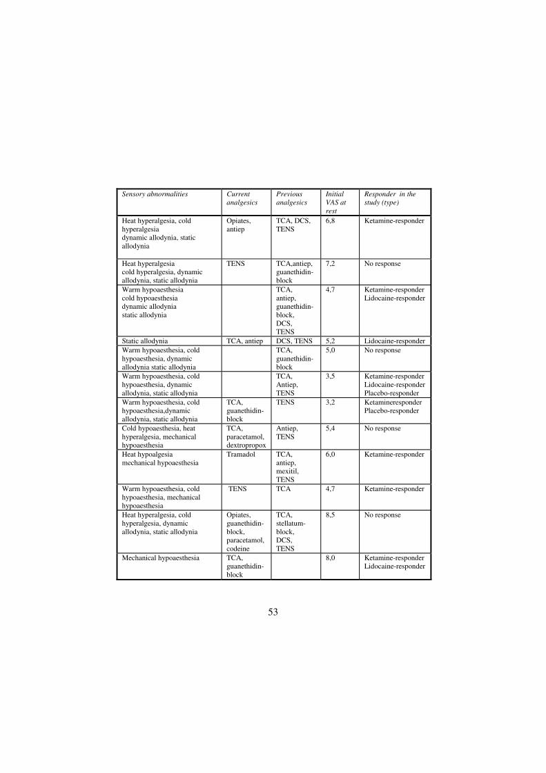
53
Sensory abnormalities Currentanalgesics
Previousanalgesics
InitialVAS atrest
Responder in thestudy (type)
Heat hyperalgesia, coldhyperalgesiadynamic allodynia, staticallodynia
Opiates,antiep
TCA, DCS,TENS
6,8 Ketamine-responder
Heat hyperalgesiacold hyperalgesia, dynamicallodynia, static allodynia
TENS TCA,antiep,guanethidin-block
7,2 No response
Warm hypoaesthesiacold hypoaesthesiadynamic allodyniastatic allodynia
TCA,antiep,guanethidin-block,DCS,TENS
4,7 Ketamine-responderLidocaine-responder
Static allodynia TCA, antiep DCS, TENS 5,2 Lidocaine-responderWarm hypoaesthesia, coldhypoaesthesia, dynamicallodynia static allodynia
TCA,guanethidin-block
5,0 No response
Warm hypoaesthesia, coldhypoaesthesia, dynamicallodynia, static allodynia
TCA,Antiep,TENS
3,5 Ketamine-responderLidocaine-responderPlacebo-responder
Warm hypoaesthesia, coldhypoaesthesia,dynamicallodynia, static allodynia
TCA,guanethidin-block
TENS 3,2 KetamineresponderPlacebo-responder
Cold hypoaesthesia, heathyperalgesia, mechanicalhypoaesthesia
TCA,paracetamol,dextropropox
Antiep,TENS
5,4 No response
Heat hypoalgesiamechanical hypoaesthesia
Tramadol TCA,antiep,mexitil,TENS
6,0 Ketamine-responder
Warm hypoaesthesia, coldhypoaesthesia, mechanicalhypoaesthesia
TENS TCA 4,7 Ketamine-responder
Heat hyperalgesia, coldhyperalgesia, dynamicallodynia, static allodynia
Opiates,guanethidin-block,paracetamol,codeine
TCA,stellatum-block,DCS,TENS
8,5 No response
Mechanical hypoaesthesia TCA,guanethidin-block
8,0 Ketamine-responderLidocaine-responder

54
Table 6. Patient characteristics in patients with central post-stroke pain (study III)
Patno
Age Sex Site of pain Etiology of pain Duration ofpain year
1 52 f Hemibody pain Thalamichemorrhage
1,5
2 76 f Hemibody pain Periventricularinfarctions
4
3 63 m Hemibody painexcept face, upperextremity mostsevere
Brainstem infarct 12
4 74 f Hemibody pain Brainstemhemorrhage
3
5 50 f Hemibody pain Midbrain infarct 3
6 72 f Upper extremity Unidentified 1
7 73 m Hemibody pain Subarachhemorrhage,temporaly
1
8 43 m Foot Intracerebralhemorrhage,frontal, temporal
3
9 59 m Hand and foot Unidentified 17
10 74 f Upper extremityand foot
Thalamic infarct 3

55
Quality ofpain
Non-sensoryneurologicalsigns
Current analgetics InitialVAS atrest
Responder
Burning,stabbing
Ataxia,oculomotordisorder
Amitriptyline,gabapentin
2,8 Morphineresponder
Stabbing,tearing
Paresis None 9,2 No response
Stabbing,aching
Paresis DextropropoxypheneTENS
2,1 Morphineresponder
Aching,sqeeezing,stabbing,throbbing,shooting
Paresis, ataxia Amitriptyline 3,4 Lidocaineresponder
Burning,aching
Paresis, ataxia,dysphasia
Dextropropoxyphene 7,7 No response
Burning,stabbing
Paresis None 5 No response
Aching Paresis,dysphasia,ataxia
Amitriptyline 1,4 No response
Burning Paresis,dysphasia,dysgnosia,dyspraxia
None 3,2 Morphineresponder
Burning,aching
Paresis None 6,3 No response
Burning,stabbing.aching
Ataxia Gabapentin,tramadol
5,9 No response

56
Table 7. Patient characteristics in patients with pain after spinal cord injury (studyIV).
Patno
Age Sex Levelofinjury
Durationof painyears
ASIAclass
Site if pain Description
1 35 M C 6-7 14 B Buttocks,lower leg,feet
Stinging,boiling
2 60 M C 3 4 C Belowinjurylevel
Burning,breaking
3 46 M Th 8-9 4 B Belowinjurylevel
Throbbing,squeezing,burning,stabbing,cutting
4 56 M Th 7-8 35 A Back,buttocks,thigh
Aching,stabbing,burning
5 30 M L 1 2 C Buttocks,lower legand foot
Burning
6 45 M C 5-6 2 B Lower leg,feet,hands,forearms
Squeezing,tightening
7 36 F Th 6 3 B Belowinjurylevel
Burning,bursting
8 51 M Th 5 8 B Buttocks,legs
Burning,squeezing
9 37 M C 3 2 C Buttocks,back
Burning,stabbing
10 53 M C 5-6 9 B Buttocks,trunk
Stabbing,cutting,burning
TCA= tricyclic antidepressants ASIA classification. The American Spinal InjuryAssociation (ASIA) Impairment Scale: A= complete. No sensory or motor functionpreserved in the sacral segments S4-S5; B= incomplete. Sensory but no motor

57
Current analgesics Initial VAS atrest
Responder
Paracetamol, codeine 4,2 Ketamine-responder
Paracetamol,dextropropoxifen
2,9 Lidocaine-responder
None 1,4 No response
Paracetamol 3,5 Ketamine-responder
None 5,7 Ketamine-responder
TCA, ketobemidon,paracetamol
7,2 No response
TCA, tramadol 9,1 No response
Tramadol 2,7 Ketamine-responder
TCA, baclofen 3,3 Ketamine-responder
None 8,2 No response
function preserved below the neurological level extending through the sacralsegments S4-S5; C= incomplete. Motor function preserved below the neurologicallevel with muscle grade less than 3 in the majority of key muscles; D= incomplete:Motor function preserved below the neurological level with muscle grade greaterthan or equal to 3 in the majority of key muscles (Ditunno 94).

58
Pain ratingThe initial mean VAS-score for continuous pain was:5.7 (range 3.2-8.5) in the group of patients with peripheral neuropathic pain(Table 5)4.6 (range 1.4-9.2) for the patients with CPSP (Table 6)4.8 (range 1.4-9.1) for the patients with pain after SCI (Table 7)
Individual valuesThe individual VAS scorings for each patient, treatment and measurementare presented in Fig. 9, 10 and 11.
Fig.9. (Next page) VAS scores in percent of the initial values for each patient withperipheral neuropathic pain, N=12, for the three treatments (ketamine -�-, lidocaine-�-, placebo -� -) at each point of measurement. The curves of the responders aremarked with colours, red for ketamine, green for lidocaine and blue for placeboresponders. Observe that the scale of the y-axis differs between patients.

59
pat 1
0 20 40 60 80 100 120 140 1600
20
40
60
80
100
120
140
pat 2
0 20 40 60 80 100 120 140 1600
20
40
60
80
100
120
140
160
pat 3
0 20 40 60 80 100 120 140 1600
20
40
60
80
100
120
pat 4
0 20 40 60 80 100 120 140 1600
20
40
60
80
100
120
140
pat 5
0 20 40 60 80 100 120 140 1600
20
40
60
80
100
120
140
pat 6
0 20 40 60 80 100 120 140 1600
20
40
60
80
100
120
pat 7
0 20 40 60 80 100 120 140 1600
50
100
150
200
250
300
pat 8
0 20 40 60 80 100 120 140 1600
20
40
60
80
100
120
pat 9
0 20 40 60 80 100 120 140 1600
20
40
60
80
100
120
pat 10
0 20 40 60 80 100 120 140 1600
20
40
60
80
100
120
pat 11
0 20 40 60 80 100 120 140 1600
20
40
60
80
100
120
140
pat 12
0 20 40 60 80 100 120 140 1600
20
40
60
80
100
120

60
Fig.10. VAS scores in percent of the initial values for each patient with CPSP,N=10, for the three treatments (morphine -�-, lidocaine -�-, placebo -� -) at eachpoint of measurement. The curves of the responders are marked with colours, purplefor morphine, green for lidocaine and blue for placebo responders. Observe that thescale of the y-axis differs between patients.
pat 1
0 20 40 60 80 100 120 140 1600
50
100
150
200
250
300
350
400
pat 2
0 20 40 60 80 100 120 140 1600
20
40
60
80
100
120
pat 3
0 20 40 60 80 100 120 140 1600
50
100
150
200
250
300
350
400
pat 4
0 20 40 60 80 100 120 140 1600
50
100
150
200
250
300
pat 5
0 20 40 60 80 100 120 140 1600
20
40
60
80
100
120
pat 6
0 20 40 60 80 100 120 140 1600
20
40
60
80
100
120
140
0 20 40 60 80 100 120 140 1600
200
400
600
800
1000
pat 7
0 20 40 60 80 100 120 140 1600
20
40
60
80
100
120
140
pat 8
0 20 40 60 80 100 120 140 1600
20
40
60
80
100
120
pat 9
0 20 40 60 80 100 120 140 1600
20
40
60
80
100
120
140
pat 10

61
Fig.11. VAS scores in percent of the initial values for each patient with SCI pain,N=10, for the three treatments (ketamine -�-, lidocaine -�-, placebo -� -) at eachpoint of measurement. The curves of the responders are marked with colours, red forketamine, green for lidocaine and blue for placebo responders. Observe that thescale of the y-axis differs between patients.
0 20 40 60 80 100 120 140 1600
20
40
60
80
100
120
140
pat 2
0 20 40 60 80 100 120 140 1600
50
100
150
200
250
300
pat 3
0 20 40 60 80 100 120 140 1600
50
100
150
200
250
300
350
pat 4
0 20 40 60 80 100 120 140 1600
20
40
60
80
100
120
pat 5
0 20 40 60 80 100 120 140 1600
20
40
60
80
100
120
pat 6
0 20 40 60 80 100 120 140 1600
20
40
60
80
100
120
pat 7
0 20 40 60 80 100 120 140 1600
20
40
60
80
100
120
140
160
180
0 20 40 60 80 100 120 140 1600
20
40
60
80
100
120
pat 9
0 20 40 60 80 100 120 140 1600
20
40
60
80
100
120
pat 10
pat 8
0 20 40 60 80 100 120 140 1600
20
40
60
80
100
120
140
pat 1

62
Mean valuesThe mean values for maximal pain reduction for the different drugs were asfollows:For ketamine the mean value was 55 % in the peripheral pain group and 38% in the group with pain after SCI.Morphine was given to the patients with CPSP and the mean value formaximal pain reduction was 14 %.For lidocaine the mean values were 34 % in the group with peripheral pain,22 % in the group of patients suffering CPSP and 10 % in the group ofpatients with pain after SCI.When placebo (saline) was given, the mean values were 22 % in theperipheral group, 6 % in the CPSP group and 3 % in the SCI group.The difference in VAS-reduction was significant between ketamine and
placebo in both the peripheral group (p = 0.009) and the SCI group (p =0.01). When lidocaine and morphine were given there were no significantdifferences in mean values compared to placebo (Fig.12).
Fig.12. Mean values for maximal pain reduction as percent of baseline VAS for thedifferent treatments ketamine, lidocaine, morphine and placebo in the three studies(mean SEM). Maximum pain relief with ketamine treatment versus placebo wassignificant in both the peripheral group (p=0.009) (study II) and the SCI group(p=0.01) (study IV).
ket II lid II pla II mo III lid III pla III ket IV lid IV pla IV-80
-60
-40
-20
0
20
Max
imal
pain
redu
ctio
n(%
)
Study II , PNP Study III , CPSP Study IV , SCI

63
Responders / non-respondersAccording to our definition of being a responder to drug-treatment (>50 %reduction in VAS-score), a significant difference in number of responderswas registered between ketamine and placebo both in the peripheral group (p= 0.025) and the SCI group (p = 0.025) (Fig. 13).
Fig.13. Number of responders to ketamine, lidocaine, morphine and placebo, in theperipheral group N=12, CPSP group N=10 and SCI group N=10. Ketamine versusplacebo showed a significant difference (p=0.025) in both the peripheral and the SCIgroup. Morphine showed a tendency toward significance compared to placebo(p=0.08).
There was no significant difference in number of responders betweenlidocaine treatment and placebo treatment in any group. The difference innumber of responders to drug treatment showed a tendency towardsignificance comparing morphine and placebo (p = 0.08) in the CPSP group.When numbers needed to treat (NNT) were calculated on the figures ofresponse, the results were as shown in table 8.
Table 8. NNT (95% CI)
Ketamine Lidocaine Morphine
Peripheral neuropathic pain 2,4 (1,5-32) 6 (2,1-�)
Central post-stroke pain 10 (2,5-�) 3,3 (1,6-�)
Pain after spinal cord injury 2 (1,4-8,5) 10 (2,5-�)
ketamine lidocaine placebo
No
pats
0
1
2
3
4
5
6
7
8
9
10
morphine lidocaine placebo0
1
2
3
4
5
6
7
8
9
10
ketamine lidocaine placebo0
1
2
3
4
5
6
7
8
9
10
11
12
p=0.025
p=0.32
SCI
p=0.08
p=0.32
CPSPp=0.025
p=0.32
PNP

64
Quantitative sensory testing with thermal stimulationIn all 32 patients, sensory abnormalities were registered either to thermal ormechanical stimuli within the painful region.
Peripheral neuropathic painIn the group of 12 patients with peripheral neuropathic pain there was aheterogeneous pattern in QST values for innocuous and noxioustemperatures. At baseline the following patterns were displayed for the 12individual patients:- Three patients showed normal thresholds for warm and cold detection butlower thresholds for heat and cold induced pain on the pain side compared tothe healthy side.-One patient had normal thresholds for warm and cold detection but higherthresholds for heat induced pain on the pain side than on the healthy side.-Two patients had higher thresholds for warm and cold detection on the painside than on the healthy side.-One patient had a higher threshold for cold detection on the pain side andhigher thresholds for heat induced pain on the pain side than on the healthyside.-Three patients had increased detection thresholds for warm and cold on boththe pain and the healthy side.- Two patients had normal thresholds.In table 9 the results are shown in another way
Table 9. Assessment of temperature thresholds in patients with peripheralneuropathic pain (n=12)
Normal Anaestethic Hypoaestethic Allodynia HyperpathiaWarm detection 7 0 5 0 0Cold detection 6 0 6 0 0Heat pain 7 0 2 3 0Cold pain 9 0 0 3 0
Central post-stroke painThe group of patients with CPSP also showed a heterogeneous pattern intheir sensibility for innocuous and noxious temperatures. Before treatment(baseline) the following were found: One patient had thresholds whichvaried without any discernible pattern, both on the pain side and on the otherside.

65
Warm detection: One patient could not detect any warmth, four had highthresholds and four had normal thresholds.Cold detection: Two patients could not detect any cold, two had highthresholds five had normal thresholds.Heat pain: One patient had no feeling of heat at all, one patient had heatallodynia, two had high thresholds and five had normal thresholds.Cold pain: Two patients had no sensation of cold pain, one had cold painallodynia, one had high thresholds and five had normal thresholds.In Table 10 the results are presented.
Table 10. Assessment of temperature thresholds in patients with central post-strokepain (one patient had thresholds which varied without any discernible pattern, theresults from the other nine are presented)
Normal Anaestethic Hypoaestethic Allodynia HyperpathiaWarmdetection
4 1 2 0 2
Cold detection 5 2 1 0 1Heat pain 5 1 2 1 0Cold pain 5 2 1 1 0
Pain after spinal cord injuryAmong the patients with pain after SCI six of ten patients did not perceivetemperature at all in the 5°C -52°C range at the painful denervated skin. Ofthe other four, two showed a pattern of hypoaesthesia, with higher thresholdsfor warmth, cold, heat pain and cold pain at painful denervated areas than atnormal skin area. Two showed normal detection thresholds for warmth andcold but lower thresholds for temperature induced pain; one for heat and onefor cold pain. The results are presented in Table 11.
Table 11. Assessment of temperature thresholds in patients with pain after spinalcord injury (n=10)
Normal Anaestethic Hypoaestethic Allodynia HyperpathiaWarm detection 2 6 2 0 0Cold detection 2 6 2 0 0Heat pain 1 6 2 1 0Cold pain 1 6 2 1 0
The results from thermal stimulation showed large inter-individualdifferences. No significant differences between responders and non-

66
responders could be seen in the results from thermal stimulation. The drugtreatment did not result in any significant changes.
Intra-individual differences of thermal thresholdsAs the patients were studied on three different occasions the baselineregistration was repeated three times. We found that the patients´ reaction tothe thermal stimulation at baseline varied for the same patient on differentoccasions. The mean range and SD for differences between the threebaseline measurements are presented in Table 12 and 13.When we assessed the temperature thresholds for innocuous and noxiousstimuli in ten healthy volunteers, on three different occasions, we alsoregistered a variability in thresholds. The variation was not as pronounced asin the patient groups. Notably the differences between measurements werelarger when tested on the legs compared to the arms, when detection wastested. The results are shown in Table 14.
Table 12. Intraindividual threshold differences at three separate baselinemeasurements at different occasions for 12 patients with peripheral neuropathic pain(mean range±SD).
Pain side Healthy sideWarmth threshold 3,0±3,7 2,3±2,1Cold threshold 4,1±6,8 2,7±2,6Heat pain threshold 4,5±4,0 4,8±2,6Cold pain threshold 6,0±5,7 6,3±5,8
Table 13. Intraindividual threshold differences at three separate baselinemeasurements at different occasions for 10 patients with central post-stroke pain(mean range±SD).
Pain side Healthy sideWarmth threshold 2,4±2,6 3,2±2,4Cold threshold 3,2±5,9 1,5±1,2Heat pain threshold 2,7±2,3 3,5±2,2Cold pain threshold 3,3±4,5 6,4±4,8

67
Table 14. Intraindividual threshold differences at three separate baselinemeasurements at different occasions for 10 healthy persons (mean range±SD).
Forearm CalfWarmth threshold 0,9±0,7 3,2±1,9Cold threshold 0,8±0,8 1,7±1,0Heat pain threshold 3,6±2,3 3,1±1,6Cold pain threshold 2,5±2,2 2,1±3,0
Sensibility to mechanical stimuliThe assessment of reactions to mechanical stimuli was done using strokewith a wisp of cotton for dynamic sensibility, von Frey filaments for staticsensibility, pin-prick (only CPSP), and vibration (only pain after SCI). Thereactions did not significantly differ between responders and non-respondersbefore infusion. All patients showed sensory abnormalities with hyper- and /or hypoesthesia. The pattern of sensory abnormalities found at baseline isseen in Table 15, 16 and 17. Changes induced by infusion were small andwithout a specific pattern.
Table 15. Assessment of mechanical sensibility in patients with peripheralneuropathic pain (n=12)
Normal Anaesthetic Hypoaesthetic Allodynia/Hyperalgesia
Hyperpathia
Dynamicsensibility
1 0 5 6 0
Staticsensibility
1 4 5 2 0
Table 16. Assessment of mechanical sensibility in patients with central post-strokepain (n=10)
Normal Anaesthetic Hypoaesthetic Allodynia/Hyperalgesia
Hyperpathia
Dynamicsensibility
2 0 4 0 4
Staticsensibility
2 0 5 0 3
Pin prick 1 0 2 3 4

68
Table 17. Assessment of mechanical sensibility in patients with pain after spinalcord injury (n=10)
Normal Anaesthetic Hypoaesthetic Allodynia/Hyperalgesia
Hyper-pathia
Dynamicsensibility
0 1 6 1 2
Staticsensibility
0 1 6 1 2
Vibration 0 2 8 0 0
Drug concentrationsIn Fig.14 (A, B, C, D, E, F) mean concentrations of ketamine, lidocaine andmorphine in the three different groups are shown. All drugs show a peakafter end of infusion (45 minutes) followed by a decrease until the end of theexperiment (150 minutes).The registered plasma concentrations were atlevels which indicated that clinically relevant doses had been given.

69
Fig.14. A, B, C, D, E, F. Plasma concentrations for the drugs in the studies. A and Bshow the peripheral group. C and D show the CPSP group. E and F show the SCIgroup (mean±SD).
0 15 30 45 60 75 90 105 120 135 150
keta
min
e(n
g/m
l)
0
50
100
150
200
250
300
0 15 30 45 60 75 90 105 120 135 150
lidoc
aine
(ug/
ml)
0,0
0,5
1,0
1,5
2,0
2,5
3,0
0 15 30 45 60 75 90 105 120 135 150
mor
phin
e(n
g/m
l)
0
10
20
30
40
50
0 15 30 45 60 75 90 105 120 135 150
lidoc
aine
(ug/
ml)
0,0
0,5
1,0
1,5
2,0
2,5
Time (min)
0 15 30 45 60 75 90 105 120 135 150
keta
min
e(n
g/m
l)
0
50
100
150
200
250
0 15 30 45 60 75 90 105 120 135 150
lidoc
aine
(ug/
ml)
0
1
2
3
4
A Study IIPNP
B Study IIPNP
C Study IIICPSP
D Study IIICPSP
E Study IVSCI
F Study IVSCI

70
Adverse effectsSide effects after infusion were common both after ketamine, lidocaine andmorphine infusions (Fig. 15 A, B, C). They were most frequent and mostintense after ketamine infusions. All patients with peripheral neuropathicpain perceived at least one side-effect from ketamine infusion and a sum of62 side-effects were registered among these 12 patients. Half of the patientsin this group had an unpleasant experience from ketamine and one patientstated that this was extremely unpleasant. In the group of patients with painafter SCI, ketamine produced side-effects in nine of 10 patients and a sum of39 side-effects were registered, somnolence and dizziness being the mostcommon.Also lidocaine infusions resulted in a substantial number of side-effects. Inthe peripheral group a total number of 29 side-effects were registered.Patients with CPSP reported 16, and patients with pain after SCI 13 side-effects. In the whole group somnolence appeared in 18 of 32 patients andperioral paresthesia in eight out of 32.Morphine induced side-effects in six of 10 patients (morphine was onlygiven to the CPSP group); the total number of registered side-effects was 21and somnolence, dizziness and nausea were the most frequent.Even placebo-infusions induced side effects. A total of 11 side effects wereregistered in the PNP group as well as in the CPSP group whereas only twowere registered in the SCI group.In none of the patients was there need to end the infusion because of adverseeffects, no specific treatment were given and we judged that the side effectsdid not disturb the evaluation of the patients.
Fig.15. Side effects after infusion of ketamine, lidocaine and placebo in theperipheral group A;(Study II).Side effects after infusion of morphine, lidocaine andplacebo in the CPSP group B;(Study III). Side effects after infusion of ketamine,lidocaine and placebo in the SCI group C; (Study IV).
Ketamine morphine lidocaine placebo

71
Number of patients
0 1 2 3 4 5 6 7 8 9 10 11 12
Somnolence
Dizziness
"Out of body sensation"
Changes in hearing
Changes in vision
Nausea
Itching
Unpleasant experience
Perioral paraesthesia
A Study IIPNP
Number of patients
0 1 2 3 4 5 6 7 8 9 10
Somnolence
Dizziness
"Out of body sensation"
Change in hearing
Change in vision
Nausea
Perioral paraesthesia
Unpleasant experience
C Study IVSCI
Somnolence
Dizziness
Changes in vision
Nausea
Itching
Unpleasant experience
Perioral paraesthesia
StudyIII
Number of patients
0 1 2 3 4 5 6 7 8 9 10
B Study IIICPSP

72
DISCUSSION
The main findings of this thesis were the following: The patients withperipheral neuropathic pain had a significantly impaired health-relatedquality of life. Ketamine yielded significant pain relief to patients withperipheral neuropathic pain and pain after spinal cord injury, although thepain relief was accompanied with considerable adverse effects. A smallgroup of patients with central pain after stroke responded with pain reliefwith morphine infusion: these results were however not significant.Lidocaine in the dose we gave did not give significant pain relief to any ofthe groups studied in this thesis.
Health related quality of life (study I)The limited clinical efficacy and tolerability of available treatment ofperipheral neuropathic pain leads to a substantial burden on the affectedpatients. The unrelieved pain affects physical as well as mental dimensionsof patients' lives and limits their ability to work. These aspects of patientwell-being can be measured using the instruments described in this study.Only patients whose primary diagnosis was PNP were included. Wehypothesize that the results observed are mainly related to continuous andevoked pain and side-effects from ongoing treatment.
Symptoms related to pain and side-effects of treatmentIn addition to the pain and ineffective treatment, patients were to a highextent troubled by difficulty in sleeping and concentrating and by lack ofenergy. Feelings of depression and anxiety were also frequently reported.Difficulty in concentrating, drowsiness, dry mouth, lack of appetite,dizziness and problems in urinating are known side-effects of the treatmentsused and were frequently reported as bothersome by the PNP patients. The

73
degree of causality between the distress from these symptoms and PNP oreach pain treatment cannot be ascertained from this study, since data are notavailable on the burden in the general population. The degree to whichpatients were bothered by gastrointestinal symptoms was similar to theresults from a general Swedish population study (Dimenäs 1993). This maybe because patients who had experienced these side effects had alreadydiscontinued the treatment which caused them; in addition, only few patientswere taking strong opioids.
Efficacy and tolerability of treatmentsDespite receiving treatment at specialized pain clinics, the PNP patients inthe study suffer a high intensity of pain, bothersome pain-related symptomsand side-effects from ongoing treatment. A large proportion of them triesvarious treatments, but often discontinues the treatment due to bothersomeside effects and inadequate pain relief. In this study patients reported painrelief from 40% of the previously tried treatments, but even those treatmentswere discontinued owing to insufficient effect and/or troublesome side-effects. These results are in accordance with a study evaluating the effect ofamitryptiline in patients with neuropathic pain following treatment of breastcancer (Kalso 1996). Although most patients in the amitryptiline groupexperienced adequate pain relief, and a reduction in the effect of pain ondaily life, only 20% of the patients were willing to continue the medicationowing to the side effects. A decreased pain intensity is thus not the onlypredictor of treatment success. Lower frequency of side-effects, improvedfunction, reduced distress and improved QoL should be viewed as animportant outcome in addition to reduced pain. Improving these componentswill also result in improved compliance.
Reduced work statusIn addition to patient suffering, the cost to society is high since a majority ofpatients (52% of those included) had a reduced employment status owing totheir PNP condition. These numbers are similar to the results from a Danishstudy of pain epidemiology and HRQoL in chronic non-malignant painpatients where 45% had disability pension (Becker 1997). Enabling PNPpatients to stay at and/or return to work could result in savings for society aswell as an improvement in patient well-being. When the patients were asked

74
to list in what respect their pain condition restricted them in doing thingsthey valued, a large proportion of them reported that they missed work mostof all. Different outdoor leisure activities were also frequently missed. Moreeffective treatments with fewer side effects would improve these aspects ofHRQoL.
HRQoLThe results clearly demonstrate that patients with PNP havemultidimensional impairment as a consequence of their pain. Scores fromSF-36 and NHP show that pain affects all dimensions of well-being, physicalas well as mental. Comparing scores with those reported for other seriousdiseases such as recent myocardial infarction, chronic heart failure andpoorly controlled diabetes with complications, confirms the severe burden ofPNP. The physical dimensions were comparable and the emotionaldimension scores were worse for PNP patients (McHorney 1993). Inaddition, the SF-36 scores in patients with PNP in all but one mentaldimension are as low as in patients with serious psychological conditions(endogenous depression) (McHorney 1993) (Fig 16). The results in thepresent study were comparable to the SF-36 scores in a study conducted byBecker on chronic non-malignant pain patients (Becker 97).Overall, the SF-36 was found to have sufficient reliability for measuring theHRQoL of patients with PNP as Chronbach's �was greater than or equal to0.70 for all dimensions. Patient scores were significantly lower than those ofthe Swedish general population, which indicates that the instrument can beused as an indicator of chronic pain states. Dimension scores also divergedin the expected direction when comparing working and non-workingsubgroups in the sample, with the dimensions physical function and bodilypain reaching a statistically significant difference. All dimensions of the SF-36 were significantly correlated with the patients' global health state ratings(rating scale). Item-same dimension and item-other dimension correlationindicated that the 35 items in the scales had captured the conceptshypothesized by the scale developers and translators (Ware 1993, Sullivan1995). The fact that the correlations were higher within the same dimensionthan with other dimensions lends credibility to the internal consistency anddiscriminant validity of the SF-36 in the PNP population (Meyer-Rosberg2001).NPH had a lower reliability, three of the six dimensions did not reach the

75
generally accepted standard of �� 0.70. Several other studies e.g. Badia(1994) and Essink-Bot (1996) in renal patient populations and Essink-Bot(1997) in migraine patients have all reported internal consistencyweaknesses in some of the NPH dimensions. The original authors of theNHP acknowledge that the instrument because of its binary response format(yes-no) taps only the extreme end of perceived health problems and thustends to yield many low scores (Hunt 1986). On the other hand NHP hasother advantages which might be useful for patients with PNP. The paindimension in NHP includes eight questions/items compared with only two inSF-36. Furthermore, NHP has a sleep dimension which is not included inSF-36. A sleep dimension is desirable as the great majority of patients (88%)reported they were bothered by difficulty in sleeping.
Fig.16. A comparison of scores from SF-36 between the groups; serious medicalcondition, psychiatric illness and our study population.
Although the scales contain comparable dimensions, in every comparisonexcept one (pain) the SF-36 had higher internal consistency estimates. TheSF-36 has the advantage of using a Likert-type scale scoring format whichallows patients to respond with a range of scaling choices.Both the SF-36 and the NHP have acceptable degrees of convergent andconstruct validity. However, we like others (VanderZee 1996, Bouchet
20
30
40
50
60
70
80
90Serious medical conditions (n=144)Psychiatric illness (n=153)Study population (n=126)
Physicalfunction
Rolephysical
Pain Generalhealth
Vitality Socialfunction
Roleemotional
Mentalhealth

76
2000) have concluded that the SF-36 has, overall, superior psychometricproperties. We suggest that further studies of the PNP population considerusing the SF-36 as the major HRQoL outcome measure. Additionalmeasurement of variables of importance to this population, such as sleep andpain intensity should also be included in any experimental treatment study.
Treatment studies II-IVWhen analysing the data in the treatment studies we decided to present meanand dichotomous data together, the former being the usual way ofpresentation but the latter offering certain advantages. Since the result ofpain reduction in neuropathic patients is usually neither continuous nornormally distributed, the mean has limitations. Moreover the dichotomousoutcome has the advantage of being more clinically informative since itprovides information about the percentage of patients likely to achieve aclinically important level of improvement (Moore 1996, Farrar 2000). Theanalgesic effect of ketamine was significant both using the mean values anddichotomous distribution. The treatment with lidocaine did not show anysignificant effect in the doses we used, either using mean or dichotomousvalues. When evaluating morphine the difference in proportion of respondersshowed a trend toward statistical significance between morphine and placebowhereas no difference was found looking at mean values.A patient with a more than 50% reduction of pain assessed by a VAS-scalewas defined as a responder in our study. This is quite a strict definition andone could argue that this will result in an underestimation of the efficacy ofthe treatment. Farrar claimed in a study in 2001 that 33% pain relief was agood cut-off point based on patient’s evaluations of a clinically importantanalgesic effect (Farrar 2001). If we had decided to classify patients with33% pain relief as responders, this would have resulted in only minorchanges in our present results. We chose the criterion >50% pain reductionbecause our aim was to show unequivocal results. As claimed by Moore andMcQuay 1996 it is a simple, convincing, clinical endpoint. Moreover, otherauthors using the dichotomous method to present their material in generaluse 50%. Our choice of 50% pain reduction thus facilitates comparison withother studies, e.g. using NNT calculations.

77
The analgesic effect of ketamine in patients with peripheralneuropathic pain and pain after spinal cord injury, studies II andIVClinical studies have demonstrated that systemic administration of theNMDA receptor antagonist ketamine in patients with peripheral neuropathicpain reduces spontaneous pain and hyperalgesia (Backonja 1994, Eide 1994,1995a, Max 1995, Felsby 1996, Leung 2001). Of these, the study conductedby Max is the only one concerning peripheral neuropathic pain of traumaticorigin. He investigated the analgesic effect in 7 patients, with a doseapproximately double compared to the one we used, and obtained responseaccording to our criteria 50% reduction, in 4 patients. His results are thuscomparable to ours.There are a limited number of studies on patients with central pain in the
literature. Backonja showed in 1994 in a study of patients with bothperipheral and central pain, that the two patients suffering central post-strokepain (CPSP) obtained an analgesic effect from ketamine.Yamamoto showed in an open, uncontrolled study that 11/23 patients withCPSP obtained pain reduction of more than 40% during ketamine treatment(Yamamoto 1997).The only study, to our knowledge, of ketamine in patients suffering painfollowing SCI was conducted by Eide in 1995 (Eide 1995b). He studied 9patients with central dysesthesia and evoked pain and observed an analgesiceffect on both spontaneous and evoked pain. The results in the present studyare in accordance with those of Eide who showed that seven out of ninepatients reported that ketamine most effectively reduced continuous pain,while 2 of 9 patients reported pain relief with placebo infusion. Our doseswere different, as Eide used approximately half the dose given in our studies.The ceiling analgesic effect of NMDA-receptor antagonists in clinicalstudies as well as in clinical practice is given by the dose-limiting sideeffects such as sedation, dizziness and visual distortions. Only one of thepatients in the present study did not note any adverse effects, and most of thepatients experienced quite disturbing side effects. Substantial side-effectswere registered, also in the study by Max (Max 1995), and it was noted thatmoderate-to-severe side effects preceded the analgesic effect and alsooutlasted the declining analgesic effect. Eide states that his treatment was notassociated with severe side effects, which can be due to the lower total doseand shorter infusion period; the serum concentrations in his study were alsolower than in these studies.

78
The side effects limit the clinical usefulness of the treatment as we have usedit. However, the high ratio of pain relief in this group of patients withintractable pain raises interest in other ways of administration, or thatdevelopment of new subtypes of NMDA-receptor antagonsists could widenthe therapeutic ratio.
The analgesic effect of lidocaine in patients with PNP, CPSP andpain after SCI, studies II-IVConcerning the therapeutic effect of lidocaine, there are studies showing aclear analgesic effect in patients with neuropathic pain of peripheral origin(Kastrup 1987, Rowbotham 1991, Galer 1996, Wallace 1996). This is incontrast with the moderate analgesic effect in our study including patientswith peripheral neuropathic pain of traumatic origin (4 responders of 12).Considerably fewer studies have been conducted on central pain states.Small open or single blinded trials have suggested that lidocaine is effectivein these pain patients (Backonja 1992, Edmonsson 1993). The onlyrandomised, controlled study of the effect of lidocaine in CPSP and SCIpatients was conducted by Attal (Attal 2000). She studied the effect ofintravenous lidocaine in 16 patients, 6 with CPSP and 10 with SCI pain.Spinal cord injuries included syringomyelia, post traumatic myelomalaciaand spondylosis with myelopathy. For the whole group, lidocainesignificantly decreased spontaneous ongoing pain when looking at meanVAS values. Brush-induced allodynia and static mechanical hyperalgesiawere also significantly decreased. Her results are in contrast with the resultsin the present study which showed negligible analgesia with lidocaine inboth patients with CPSP and SCI (one responder of 10 in each group). Oneexplanation for the divergent results could be the chosen dose; the total doseof Attal was twice as high as the one used in our study. However, looking atthe results as proportion of responders, only 9 of the 16 patients in Attal'sstudy had a decrease in VAS of more than 50% at the end of infusion,compared to 6 of 16 with placebo. Also in other lidocaine studies in patientswith peripheral neuropathic pain, higher doses were given (Rowbotham1991, Galer 1996, Wallace 1996) and one can speculate that the lowanalgesic effect in our study was because our dose was too low to obtain aneffect. However other studies using even lower doses than ours have showedan analgesic effect. Marchettini reported an analgesic effect with a dose of1.5 mg/kg (Marchettini 1992). Galer administered both 2mg/kg and 5mg/kg

79
to patients suffering peripheral neuropathic pain, and reported significantVAS reduction with both doses (Galer 1996). In a study of the analgesiceffect of lidocaine in patients with postherpetic neuropathic pain conductedby Baranowski in 1999, he investigated both 1 mg/kg and 5 mg/kg. Thatstudy showed a positive analgesic effect with both doses (Baranowski 1999).We want to emphasize that we believe that the chosen dose was prudent,given the toxicity of lidocaine and hence the risk for severe adverse effectsfrom the nervous and cardiovascular systems. Half of our 32 patientsreported side effects, 9 light-headedness and eight perioral paresthesia, thelatter of which we regard as a warning signal to other more severe sideeffects. We aimed to test effects with clinically applicable doses.
The analgesic effect of morphine in patients with CPSP, study IIIThe limited analgesic effect of morphine infusion was not unexpected. Otherstudies have shown similar results (Kupers 1991, Kalman 2002, Attal 2002).According to clinical experience (Gonzales 1995, Boivie 1999) only aminority of patients with CPSP responds positively to opioid treatment. Ifwe had given the infusion of morphine with individual dose titration there isa possibility that more patients would have responded to the treatment.However, in the study conducted by Attal where the dose of morphine wasindividually titrated, the effect on spontaneous pain was similar to ourresults. The plasma concentrations of morphine in our study were in therange of 23 to 52 ng/ml. This may be compared with concentrations thathave been found to reduce postoperative pain, namely a mean of 20 ng/ml(Dahlström 1982, Derbyshire 1985). Our doses are thus within the limitswhere an analgesic effect could be expected according to the literature. Thedose titration of effective drug may also make the blinding proceduredifficult and thereby also the interpretation of the results. Some studies of theresponsiveness of neuropathic pain to opioids have used much higher dosescompared to ours. Dellemjin used fentanyl given at a dose equivalent to 80mg of morphine and reported more than 50% responders in patients withdifferent types of neuropathic pain (Dellemjin 1997). Kalman administeredmorphine at a mean dose of 44 mg with an infusion rate of 1 mg / minute topatients with central pain due to multiple sclerosis with a response rate of4/14 (Kalman 2002). Both authors claimed that relief from pain was notobtained until high doses had been infused. In comparison, the mean dose of14 mg in the present study was considerably lower. However, long-term

80
administration of opioids has troublesome side-effects (McQuay 1992).Hence we focused on doses which could be of interest for daily use and inthat perspective even our doses could be considered to be in the upper limit.The three patients responding to morphine treatment in the present work hada clear response with marked pain relief. A reasonable conclusion from theevidence available seems to be that a few patients with central pain maybenefit from opioid analgesics and that it is important to evaluate the effectsin each individual. An interesting way to confirm the notion that ourresponders had a genuine analgesic effect would be to let them undergostudies using the N of 1 design (McQuay 1991).The total number of registered side effects was lower than expected giventhat most patients were old and that all had suffered a cerebral injury. Incontrast the group of patients with peripheral neuropathic pain, who wereconsiderably younger, experienced more frequent side effects both afterlidocaine and placebo infusion. In view of this, one can question ourdecision not to give these patients ketamine in fear of unmanageable adverseeffects.
General considerations of analgesic effectsThe patients recruited in this study all had severe neuropathic pain of longduration that had been resistant to other treatments. These factors mightdecrease the likelihood of treatment response. This category of patients isoften recruited for trials of neuropathic pain; the results could have beendifferent if we had recruited patients consecutively when they first appearedat the pain clinic. On the other hand, the categories of patients studied are inparticular need of treatment and several of the responders claimed that thistreatment was superior to any other they had previously experienced.Six of the patients with CPSP and SCI scored less than three on the VASbefore treatment. It can be argued that the pain of these patients was notsevere enough to be a substantial burden. However, these patients claimedthat their pain was severe, and they were therefore included in the studies.This is consistent with Boivie (Boivie 1999) who states that even patients inthis category who rate their pain as low may be severely affected.Pharmacological trials with this category of patients are few and with a smallnumber of subjects per trial. This is a general problem of studies on patientswith chronic neuropathic pain. A way to increase the efficiency of a clinicaltrial, thereby providing adequate power to use small samples, includes the

81
use of selection methods to create groups that are as homogeneous aspossible. In the study of peripheral neuropathic pain we chose patients withtraumatic origin to the nerve injury in an attempt to select a homogeneousgroup. On the other hand patients with peripheral neuropathic pain oftraumatic origin are probably a complex and heterogeneous group per sewith different types of fibre lesions, different magnitude of deafferentationand disinhibition, as well as different relations between peripheral andcentral mechanisms. In the study of patients with CPSP all patients with aclear diagnosis was included with no further selection. In the study ofpatients with pain after SCI we had a traumatic origin as a inclusioncriterion. Moreover we chose patients whose main concern was continuous,spontaneous pain below the level of injury, according to the taxonomyproposed by the SCI Pain Task Force of IASP (Sidall 2000a), even thoughsome of the patients also suffered from evoked pain and pain at the level ofinjury.When analysing effects of treatments of neuropathic pain, meta-analyses of anumber of comparable trials are important. An index that is being used withincreasing frequency in the pain literature is the number needed to treat(NNT) (Cook and Sacket 1995). In the pain literature NNT is typicallycalculated as the number of patients who report at least 50% pain relieffollowing an active treatment relative to the number who report at least 50%pain relief following a control treatment (Moore 1996). Generally NNTsbetween 2 and 5 are indicative of effective analgesic treatments (McQuay1999) and this was achieved with ketamine and morphine but not withlidocaine in this work.The long-time objective of studying intravenously administrated drugs is tofind drugs that can be given orally during long periods. Whether anintravenous pharmacological test may predict the long term effect of oraltreatment is an interesting issue that remains to be settled, and further studieswith long term follow-up are needed to evaluate this concept. Galer claimeda correlation for response to intravenous lidocaine and mexiletine treatment;three lidocaine responders claimed good pain relief with mexiletin, whileamong six poor lidocaine responders only two recieved good pain relief withmexiletine (Galer 1996). In the lidocaine trial by Attal (Attal 2000), 12patients received a lidocaine infusion followed by a 4-12 week oralmexiletine trial. Only 2 of 8 (25%) patients who responded with pain reliefto the lidocaine infusion went on to have a positive response to themexiletine trial. Furthermore one of 4 (25%) non responders to lidocaine

82
infusion went on to have a positive response to oral mexiletine. None ofthese patients was willing to be long-treated with mexiletine, essentiallybecause of the side effects. The six patients in our studies that achieved apain relief of more than 50% all tried mexiletine outside the studies and noneof them found the balance between pain relief and adverse effects goodenough to continue that treatment. Furthermore, in follow up studies of long-term opioid treatment by Attal and Dellemjin only 20% of the patients choseto continue treatment after one and two years, respectively (Attal 2002,Dellemjin 1998).
Diagnostic capacitySome authors claim the usefulness of ketamine, lidocaine and morphine tobe used in intravenous pharmacological pain analysis to separatenociceptive, neuropathic and idiopathic pain (Marchettini 1992, Sörensen1995). This is also a diagnostic routine in many clinics. In studies II, III andIV the response rate was 6/32 for lidocaine and 12/22 for ketamine. Hencelidocaine and ketamine treatment can hardly be used as diagnostic tools forneurophatic pain, as the sensitivity is too low. Moreover since both ketamineand lidocaine have an analgesic effect in nociceptive pain (Domino 1965,Cassuto 1985), the specificity is most certainly low as well. Because of thiswe question the usefulness of these drugs as pharmacological diagnostictools.
Sensory abnormalities at baselineWhen we assessed baseline data of somatosensory functions beforetreatment, we found a wide variety of sensory disturbances. One of ourhypotheses was that examination of patients with different sensoryabnormalities using tests of thermal thresholds and other sensory tests couldpredict response to treatment. This hypothesis could not be confirmed. Theperformed assessments can help to distinguish between different sensorydisturbances. One possible explanation for the limited value of predictioncould be that the available tests do not give sufficient information about thetype of nerve lesions. As mentioned above, patients with neuropathic painare probably a complex and heterogeneous group with different types offibre lesions, different magnitude of deafferentation and disinhibition, aswell as different relations between peripheral and central components. The

83
duration of pain is probably also of importance for the heterogeneity assynaptic plasticity may lead to permanent changes in nociceptive processing(Coderre 1993). It is likely that a large number of patients are needed toidentify specific patterns in such a heterogeneous group. However, thenumbers of patients in our studies were determined with regard to theprimary outcome variable, pain relief.In a study of neurological symptoms in 27 patients with CPSP, Boivie founda decreased sensibility to innocuous temperatures in all patients (Boivie1989). In his study, seventy percent could not differentiate temperaturesbetween zero and 50°C. Approximately half of these patients had normalthresholds to touch and vibration. A similar but somewhat less pronouncedtendency has been found in other studies (Vestergaard 1995, Bowsher 1998).These results are the basis for the hypothesis that CPSP is dependent oninjury to the spinothalamocortical pathways.The patients in our study showed a markedly varied pattern in temperaturesensibility. These results differ notably from the results of Boivie, Bowsherand Vestergaard. Only two of our ten patients showed severe disturbances intemperature sensibility. In respect to the sensibility to mechanically evokedpain using of pin-prick and touch using cotton wool, the results from ourstudy are in accordance with other studies. The majority of the patients hadan abnormal reaction; hypoesthesia, hyperalgesia as well as allodynia wereseen, similar to other studies.The reason for the differing results of our study as compared to thepreviously mentioned studies with respect to abnormality of temperaturethresholds is difficult to explain.One might speculate that our patients did not suffer from CPSP, but this isunlikely since they were carefully examined and diagnosed both by aneurologist and/or a neurosurgeon before referral, and by ananaesthesiologist at the pain clinic.The number of patients in our study is relatively small, precluding a closeranalysis of the differences between this study and the others mentioned.However the moderate impact on sensibility to temperature is noteworthy.Few studies of SCI pain have tested sensory function. Beric studied below-level pain in 13 patients with central dysesthesia syndrome (CDS) (Beric1988). Two patients in their study had no sensation at all below the level ofinjury, while 11 had incomplete preservation of sensation. Ten of 13patients did not perceive temperature sensation at all tested with QST.Vibratory sensibility and touch were reduced, but not absent in 10 and 8

84
patients respectively. He found a significant difference between thetemperature thresholds and those for vibration and touch. This is inaccordance to the results in our study. Beric interestingly stated that in alarge cohort of 243 SCI patients with and without altered sensation and pain,from which the 13 patients were drawn he found only 3 other patients,without CDS and with a similar combination of absent spinothalamicfunction and relatively preserved dorsal column function. This indicates ahigh incidence of pain in patients with this specific sensory profile.These sensory profiles are also in concordance with those found by Boivie inpatients with CPSP (Boivie 1989).We found a surprisingly high variability in the results from baselinemeasurements of sensory disturbances on different occasions in individualpatients. Similar findings have been made by others (Fagius 1981, Rommel2001). The investigation technique could play a role; however, themethodology was standardised as much as possible. All patients had beeninformed and educated in the same way and the experiments were allperformed by the same investigator. The greater part of the variabilityobserved in the present studies is probably due to central processing factors.Efferent pathways for facilitation and inhibition of afferent signals make itpossible to focus attention on a particular sensation and a particular site ofthe body (Bannister 1976). Factors altering this attention would have amarked influence on threshold levels. Thus, factors such as motivation,psychological and physiological stress, distraction, fatigue as well asexpectations have been found to affect the magnitude of the sensorythresholds both in the experimental situation with healthy subjects and inclinical practice (Lindblom 1974, Vallbo 1976, Dyck 1978). Patients canalso be biased toward showing abnormal results due to a conscious orunconscious wish to demonstrate more disability than they have (Dyck1998).
Sensory changes by treatmentWe also wanted to study if ketamine, lidocaine and morphine gave specificsensory effects such as normalisation of pathologic thermal thresholds ornormalisation of abnormal sensibility. No such effects were registered, noteven in responders.As the intraindividual results from thermotests showed relatively largevariation, also at T:0 and with placebo infusion, it was difficult to identify

85
other than major differences. No clinically relevant differences could bedetected.Normal values from temperature threshold measurements in a healthypopulation have shown variations both dependent on gender, age and site fortesting (Verdugo 1992, Meh 1994, Hilz 1999). This makes the interpretationof our results more difficult because of difference in gender, age and site fortesting (Table 5-7). On the other hand, we used the patients as their owncontrol as we repeated the tests and also tested the contralateral (healthy)side or painless area above the lesion (SCI).Few other studies have found changes in temperature thresholds aftertreatment, although Leung found an increase in both cold and cold-painthresholds after ketamine and alfentanil infusions in patients withneuropathic pain. In a study conducted by Jörum alfentanil but not ketaminereduced cold allodynia, by increasing cold-pain thresholds in patients withneuropathic pain. At the same time a marked reduction of hyperalgesia tocold was seen following both ketamine and alfentanil administration (Jörum2003).Nor did we observe any changes in mechanical sensibility after infusion.This contrasts with Attal's studies where reduced intensity of brushed-induced allodynia was found both with morphine and lidocaine infusions.Also Eide in his study of SCI pain observed that allodynia to brush and“wind-up-like-pain” was reduced by ketamine as well as alfentanil infusions.One explanation of this difference may be that the above studies measuredallodynia quantitatively with VAS while we only made a classification infour categories.
Future perspectivesThe narrow therapeutic ratio associated with the use of the clinicallyavailable NMDA-receptor antagonists may be overcome by the developmentof more selective NMDA-receptor antagonists, which modulate binding siteswithin the NMDA-complex, or have affinity at specific NMDA-receptorsubtypes.Recent studies have shown that there exist at least nine different sodiumchannels encoded by different genes. The abnormal sodium channel activityand changes in sodium channel expression under neuropathic conditionshave drawn much attention over the last several years (Waxman 1999). The

86
progress on studies of subtype-specific sodium channels and their link tocertain neuropathic pain symptoms may lead to the possibility to targethyperexcitable sensory neurons along the nociceptive pathway withoutproducing significant side effects.The heterogeneity of the pain states following peripheral or central nerveinjury may require an analgesic regimen that interacts at several selectivesites with the potential for acting synergistically in terms of efficacy but nottoxicity.The development of an effective pharmacological treatment and therebyimproving the quality of life of these patients would represent a fundamentalbreakthrough in clinical pain treatment.

87
CONCLUSIONS
The intense pain, limited efficacy and tolerability of available treatments, thelow overall rating of health, reduced work status and troublesome symptomsconstitute a substantial burden for patients with PNP. All dimensions ofHRQoL are significantly impaired� physical as well as mental� indicating amultidimensional burden of these patients.
The NMDA-antagonist ketamine yields substantial pain relief to patientswith peripheral neuropathic pain and patients with neuropathic pain belowthe level of SCI. The noted side effects limit the clinical usefulness of thetreatment. However, the high degree of pain relief in these usually refractorypain states raises interest in the development of NMDA-antagonists with awider therapeutic ratio.
Lidocaine, in the dose given in these studies did not give significant painrelief to patients with peripheral neuropathic pain, patients with CPSP andpatients with neuropathic pain below the level of SCI.
Morphine may represent a therapeutic alternative for some patients withCPSP, although only a small group of this category of patients seems torespond with analgesia.
Assessment of baseline somatosensory functions could not be used toidentify responders to treatment with either drug. Neither did ketamine,lidocaine or morphine cause any changes in thermal or mechanicalsensibility.

88
ACKNOWLEDGEMENTS
This study would never have been possible without the assistance,encouragement and support most generously provided by a great number ofpeople. The inspiring collaboration with all these dependable and positivepersons who have shown confidence in my work and shared with me theirknowledge and abilities in a variety of subjects, made this work worthwhile.I sincerely wish to thank you all.
This study was supported by grants from Astra Zeneca R&D, Södertälje,Sweden, Henning Larssons stipendie stiftelse, and the Swedish MedicalResearch Council (9077).

89
References
Albe-Fessard D, Lombard C. Use of an animal model to evaluate the origin of andprotection against deafferentation pain. In: Bonica JJ, ed. Advances in painresearch and therapy. New York: Raven Press, 1983: 691-700.
Andersen G, Vestergaard K, Ingeman-Nielsen M, Jensen TS. Incidence of centralpost-stroke pain. Pain 1995; 61: 187-193.
Arner S, Meyerson BA. Lack of analgesic effect of opioids on neuropathic andidiopathic forms of pain. Pain 1988; 33: 11-23.
Attal N, Gaude V, Brasseur L, Dupuy M, Guirimand F, Parker F, Bouhassira D.Intravenous lidocaine in central pain: a double-blind, placebo-controlled,psychophysical study. Neurology 2000; 54: 564-574.
Attal N, Guirimand F, Brasseur L, Gaude V, Chauvin M, Bouhassira D. Effects ofIV morphine in central pain: a randomized placebo-controlled study. Neurology2002; 58: 554-563.
Backonja M, Gombar K. Serum lidocaine levels following stellate ganglionsympathetic blocks and intravenous lidocaine injection. J Pain SymptomManage 1992; 7: 2-3.
Backonja M, Arndt G, Gombar KA, Check B, Zimmermann M. Response of chronicneuropathic pain syndromes to ketamine: a preliminary study. Pain 1994; 56:51-57.
Backonja M, Beydoun A, Edwards KR, Schwartz SL, Fonseca V, Hes M,LaMoreaux L, Garofalo E. Gabapentin for the symptomatic treatment of painfulneuropathy in patients with diabetes mellitus: a randomized controlled trial.JAMA 1998; 280: 1831-1836.
Badia X, Alonso J, Brosa M, Lock P. Reliability of the Spanish version of theNottingham Health Profile in patients with stable end-stage renal disease. SocSci Med 1994; 38: 153-158.

90
Bandler R. Columnar organization in the midbrain periaqueductal gray: modules foremotional expression? Trends Neurosci 1994; 17: 379-389.
Bannister LH. Sensory terminals of peripheral nerves. In: Landon DN, ed. Theperipheral nerve. London: Chapman and Hall, 1976: 396-463.
Baranowski AP, De Courcey J, Bonello E. A trial of intravenous lidocaine on thepain and allodynia of postherpetic neuralgia. J Pain Symptom Manage 1999;17: 429-433.
Becker N, Bondegaard Thomsen A, Olsen AK, Sjogren P, Bech P, Eriksen J. Painepidemiology and health related quality of life in chronic non-malignant painpatients referred to a Danish multidisciplinary pain center. Pain 1997; 73: 393-400.
Belfrage M, Segerdahl M, Arner S, Sollevi A. The safety and efficacy of intrathecaladenosine in patients with chronic neuropathic pain. Anesth Analg 1999; 89:136-142.
Bensen W, Weaver A, Espinoza L, Zhao WW, Riley W, Paperiello B, Recker DP.Efficacy and safety of valdecoxib in treating the signs and symptoms ofrheumatoid arthritis: a randomized, controlled comparison with placebo andnaproxen. Rheumatology (Oxford) 2002; 41: 1008-1016.
Beric A, Dimitrijevic MR, Lindblom U. Central dysesthesia syndrome in spinal cordinjury patients. Pain 1988; 34: 109-116.
Boivie J, Leijon G, Johansson I. Central post-stroke pain--a study of the mechanismsthrough analyses of the sensory abnormalities. Pain 1989; 37: 173-185.
Boivie J. Central Pain In: Wall P, Melzach R, eds. Textbook of Pain, 4th edition.New York: Churchill Livingstone, 1999: 879-914.
Bonica JJ, Loeser JD. History of pain concepts and therapies. In: Loeser JD, ed.Bonica´s mangement of pain. Philadelfia: Lippincott Williams & Wilkins, 2001:3-16.
Bouchet C, Guillemin F, Paul-Dauphin A, Briancon S. Selection of quality-of-lifemeasures for a prevention trial: a psychometric analysis. Control Clin Trials2000; 21: 30-43.
Bowsher D. Neurogenic pain syndromes and their management. Br Med Bull 1991;47: 644-666.
Bowsher D. Central pain: clinical and physiological characteristics. J NeurolNeurosurg Psychiatry 1996; 61: 62-69.

91
Bowsher D, Leijon G, Thuomas KA. Central poststroke pain: correlation of MRIwith clinical pain characteristics and sensory abnormalities. Neurology 1998;51: 1352-1358.
Carlsson AM. Assessment of chronic pain. I. Aspects of the reliability and validityof the visual analogue scale. Pain 1983; 16: 87-101.
Cassuto J, Wallin G, Hogstrom S, Faxen A, Rimback G. Inhibition of postoperativepain by continuous low-dose intravenous infusion of lidocaine. Anesth Analg1985; 64: 971-974.
Castro-Lopes JM, Tavares I, Coimbra A. GABA decreases in the spinal cord dorsalhorn after peripheral neurectomy. Brain Res 1993; 620: 287-291.
Castro-Lopes JM, Malcangio M, Pan BH, Bowery NG. Complex changes ofGABAA and GABAB receptor binding in the spinal cord dorsal horn followingperipheral inflammation or neurectomy. Brain Res 1995; 679: 289-297.
Chabal C, Russell LC, Burchiel KJ. The effect of intravenous lidocaine, tocainide,and mexiletine on spontaneously active fibers originating in rat sciaticneuromas. Pain 1989; 38: 333-338.
Chapman CR, Casey KL, Dubner R, Foley KM, Gracely RH, Reading AE. Painmeasurement: an overview. Pain 1985; 22: 1-31.
Chen Y, Devor M. Ectopic mechanosensitivity in injured sensory axons arises fromthe site of spontaneous electrogenesis. Eur J Pain 1998; 2: 165-178.
Claus D, Hilz MJ, Hummer I, Neundorfer B. Methods of measurement of thermalthresholds. Acta Neurol Scand 1987; 76: 288-296.
Claus D, Hilz MJ, Neundorfer B. Thermal discrimination thresholds: a comparisonof different methods. Acta Neurol Scand 1990; 81: 533-540.
Coderre TJ, Katz J, Vaccarino AL, Melzack R. Contribution of centralneuroplasticity to pathological pain: review of clinical and experimentalevidence. Pain 1993; 52: 259-285.
Cook RJ, Sackett DL. The number needed to treat: a clinically useful measure oftreatment effect. BMJ 1995; 310: 452-454.
Cotman CW. Excitatory amino acid neurotransmission: NMDA receptors and Hebb-type synaptic plasticity. Annu Rev Neurosci 1988; 11: 61-80.

92
Craig AD. Functional Antomy of Supraspinal Pain Processing with References tothe Central Pain Syndrome Pain 1999- an updated review. Seattle: IASP Press,1999:
Craig AD. New and old thoughts on the mechanisms of spinal cord injury pain In:Yezierski RP, Burchiel KJ, eds. Spinal cord injury pain: Assessment,mechanisms, management. Seattle: IASP Press, 2002: 237-261.
Dahlstrom B, Tamsen A, Paalzow L, Hartvig P. Patient-controlled analgesic therapy,Part IV: pharmacokinetics and analgesic plasma concentrations of morphine.Clin Pharmacokinet 1982; 7: 266-279.
Davidoff G, Roth E, Guarracini M, Sliwa J, Yarkony G. Function-limitingdysesthetic pain syndrome among traumatic spinal cord injury patients: a cross-sectional study. Pain 1987; 29: 39-48.
Davies SN, Lodge D. Evidence for involvement of N-methylaspartate receptors in'wind-up' of class 2 neurones in the dorsal horn of the rat. Brain Res 1987; 424:402-406.
Dejerine J Roussy G. La syndrome thalamique. Rev Neurol 1906; 14: 521-532.
Dellemijn PL, Vanneste JA. Randomised double-blind active-placebo-controlledcrossover trial of intravenous fentanyl in neuropathic pain. Lancet 1997; 349:753-758.
Dellemijn PL, van Duijn H, Vanneste JA. Prolonged treatment with transdermalfentanyl in neuropathic pain. J Pain Symptom Manage 1998; 16: 220-229.
Derbyshire DR, Bell A, Parry PA, Smith G. Morphine sulphate slow release.Comparison with i.m. morphine for postoperative analgesia. Br J Anaesth 1985;57: 858-865.
Devor M, Keller CH, Deerinck TJ, Levinson SR, Ellisman MH. Na+ channelaccumulation on axolemma of afferent endings in nerve end neuromas inApteronotus. Neurosci Lett 1989; 102: 149-154.
Devor M, Wall PD, Catalan N. Systemic lidocaine silences ectopic neuroma andDRG discharge without blocking nerve conduction. Pain 1992; 48: 261-268.
Devor M, Seltzer Z. Pathophysiology of damaged nerves in realtion to chronic painIn: Wall PD, Melzack R, eds. Textbook of 'Pain. New York: ChurchillLivingstone, 1999: 129-164.

93
Dickenson AH, Sullivan AF. Electrophysiological studies on the effects ofintrathecal morphine on nociceptive neurones in the rat dorsal horn. Pain 1986;24: 211-222.
Dickenson AH, Sullivan AF. Evidence for a role of the NMDA receptor in thefrequency dependent potentiation of deep rat dorsal horn nociceptive neuronesfollowing C fibre stimulation. Neuropharmacology 1987; 26: 1235-1238.
Dickenson AH. Where and how do opoids act? In: Gebhart G, Hammond D, JensenTS, eds. Proceedings at the 7th World Congress on Pain. Seattle: IASP Press,1994: 525-552.
Dimenas E, Glise H, Hallerback B, Hernqvist H, Svedlund J, Wiklund I. Quality oflife in patients with upper gastrointestinal symptoms. An improved evaluation oftreatment regimens? Scand J Gastroenterol 1993; 28: 681-687.
Ditunno Jr JF. The international standards booklet for neurological and functionalclassification of spinal cord injury. American Spinal Injury Association.Paraplegia 1994; 32: 70-80.
Domino EF, Chodoff SE, Corssen G. Pharmacologic effects of CI-581, a newdissociative anesthetic, in man. Clin Pharmacol Ther 1965; 6: 279-291.
Dostrovsky JO, Wells FEB, Tasker RR. Pain evoked by stimulation in humanthalamus In: Shigenaga Y, ed. International symposium on processingnociceptive information. Amsterdam: Elsevier, 1991: 115-120.
Dubner R, Ruda MA. Activity-dependent neuronal plasticity following tissue injuryand inflammation. Trends Neurosci 1992; 15: 96-103.
Dyck PJ, Zimmerman IR, O'Brien PC, Ness A, Caskey PE, Karnes J, Bushek W.Introduction of automated systems to evaluate touch-pressure, vibration, andthermal cutaneous sensation in man. Ann Neurol 1978; 4: 502-510.
Dyck PJ, O'Brien PC, Kosanke JL, Gillen DA, Karnes JL. A 4, 2, and 1 steppingalgorithm for quick and accurate estimation of cutaneous sensation threshold.Neurology 1993; 43: 1508-1512.
Dyck PJ, Dyck PJ, Kennedy WR, Kesserwani H, Melanson M, Ochoa J, Shy M,Stevens JC, Suarez GA, O'Brien PC. Limitations of quantitative sensory testingwhen patients are biased toward a bad outcome. Neurology 1998; 50: 1213.
Edmondson EA, Simpson Jr RK, Stubler DK, Beric A. Systemic lidocaine therapyfor poststroke pain. South Med J 1993; 86: 1093-1096.

94
Eide PK, Jorum E, Stubhaug A, Bremnes J, Breivik H. Relief of post-herpeticneuralgia with the N-methyl-D-aspartic acid receptor antagonist ketamine: adouble-blind, cross-over comparison with morphine and placebo. Pain 1994;58: 347-354.
Eide K, Stubhaug A, Oye I, Breivik H. Continuous subcutaneous administration ofthe N-methyl-D-aspartic acid (NMDA) receptor antagonist ketamine in thetreatment of post-herpetic neuralgia. Pain 1995a; 61: 221-228.
Eide PK, Stubhaug A, Stenehjem AE. Central dysesthesia pain after traumatic spinalcord injury is dependent on N-methyl-D-aspartate receptor activation.Neurosurgery 1995b; 37: 1080-1087.
Eide PK, Stubhaug A, Breivik H, Oye I. Reply to S.T. Meller: Ketamine: relief fromchronic pain through actions at the NMDA receptor. Pain 1997; 72: 289-291.
Ekblom A, Hansson P. Pain intensity measurements in patients with acute painreceiving afferent stimulation. J Neurol Neurosurg Psychiatry 1988; 51: 481-486.
Essink-Bot ML, Krabbe PF, Bonsel GJ, Aaronson NK. An empirical comparison offour generic health status measures. The Nottingham Health Profile, the MedicalOutcomes Study 36-item Short-Form Health Survey, the COOP/WONCAcharts, and the EuroQol instrument. Med Care 1997; 35: 522-537.
Essink-Bot ML, Krabbe PF, van Agt HM, Bonsel GJ. NHP or SIP--a comparativestudy in renal insufficiency associated anemia. Qual Life Res 1996; 5: 91-100.
Faden AI. A potential role for excitotoxins in the pathophysiology of spinal cordinjury. Ann Neurol 1988; 23: 623-626.
Fagius J, Wahren LK. Variability of sensory threshold determination in clinical use.J Neurol Sci 1981; 51: 11-27.
Farrar JT, Portenoy RK, Berlin JA, Kinman JL, Strom BL. Defining the clinicallyimportant difference in pain outcome measures. Pain 2000; 88: 287-294.
Farrar JT, Young Jr JP, LaMoreaux L, Werth JL, Poole RM. Clinical importance ofchanges in chronic pain intensity measured on an 11-point numerical pain ratingscale. Pain 2001; 94: 149-158.
Felsby S, Nielsen J, Arendt-Nielsen L, Jensen TS. NMDA receptor blockade inchronic neuropathic pain: a comparison of ketamine and magnesium chloride.Pain 1996; 64: 283-291.

95
Ferrante FM, Paggioli J, Cherukuri S, Arthur GR. The analgesic response tointravenous lidocaine in the treatment of neuropathic pain. Anesth Analg 1996;82: 91-97.
Fields HL. Can opiates relieve neuropathic pain? Pain 1988; 35: 365-367.
Fields H, Rowbotham M, Devor M. Excitability blockers: anticonvulsants and lowconcentration local anesthetics in the treatment of chronic pain. In: DickensonA, Benson J, eds. The Pharamcology of Pain, Handbook of ExperimentalPharmacology. Heidelberg: Springer, 1997: 93-116.
Fields H, Basbaum A. Central nervous system mechanisms of pain modulation In:Wall PD, Melzack R, eds. Textbook of pain, 4th edition. New York: ChurchillLivingstone, 1999a: 309-329.
Fields HL. Peripheral neuropathic pain: an approach to the management. In: WallPD, Melzack R, eds. Textbook of Pain, 4th edition. New York: ChurchillLivingstone, 1999b: 1523-1533.
Fields H, Hill RG. Neuropathic Pain: The Near and Far Horizon. In: Hansson P,Fields H, Hill R, Marchettini P, eds. Neuropathic Pain: Pathophysiology andTreatment. Progress in Pain Research and Management. Seattle: IASP Press,2001: 251-264.
Finnerup N, Johanesen I, Sindrup S, Bach F, Jensen TS. Pharmacological treatmentof spinal cord injury pain. In: Yezierski R, Burchiel KJ, eds. Spinal Cord InjuryPain: assessment, mechanisms, management. Progress in Pain Research andManagement. Seattle: IASP Press, 2002: 341-351.
Frater A, Costain D. Any better? Outcome measures in medical audit. BMJ 1992;304: 519-520.
Fruhstorfer H, Lindblom U, Schmidt WC. Method for quantitative estimation ofthermal thresholds in patients. J Neurol Neurosurg Psychiatry 1976; 39: 1071-1075.
Galer BS, Harle J, Rowbotham MC. Response to intravenous lidocaine infusionpredicts subsequent response to oral mexiletine: a prospective study. J PainSymptom Manage 1996; 12: 161-167.
Gonzales GR. Central pain: diagnosis and treatment strategies. Neurology 1995; 45:S11-6 discussion S35.
Govrin-Lippmann R, Devor M. Ongoing activity in severed nerves: source andvariation with time. Brain Res 1978; 159: 406-410.

96
Gracely RH. The Descriptor Differential Scale: applying psychophysical principlesto clinical pain assessment. Pain 1988; 35: 279-288.
Granit R, Skoglund CR. Facilitation, inhibition and the artificial synapse formed bythe cut end of a mammalian nerve. J Physiol (Lond) 1945; 103: 435-448.
Halling K. The Nottingham Health Profile � age and sex comparison in a Swedishcommunity. 1995. Manuscript perfomed at Department of Psychology,University of Göteborg.
Hansson P. Nociceptiv och neurogen smärta. Stockholm:Tryckeri AB Småland,1998.
Hansson P, Lacerenza M, Marchettini P. Aspect of clinical and experimentalneuropathic pain: The clinical perspective. In: Hansson P, Fields H, Hill R,Marchettini P, eds. Neuropathic pain: pathophysiology and treatment. Progres inpain research and management. Seattle: IASP Press, 2001: 1-18.
Hao JX, Yu YX, Seiger A, Wiesenfeld-Hallin Z. Systemic tocainide relievesmechanical hypersensitivity and normalizes the responses of hyperexcitabledorsal horn wide-dynamic-range neurons after transient spinal cord ischemia inrats. Exp Brain Res 1992; 91: 229-235.
Head H, Holmes G. Sensory disturbances from cerebral lesions. Brain 1911; 34:102-122.
Heft MW, Parker SR. An experimental basis for revising the graphic rating scale forpain. Pain 1984; 19: 153-161.
Herbert H. Organization of medullary adrenergic and noradrenergic projections tothe periaqueductal gray matter in the rat. J Comp Neurol 1992; 315: 34-52.
Hilz MJ, Stemper B, Axelrod FB, Kolodny EH, Neundorfer B. Quantitative thermalperception testing in adults. J Clin Neurophysiol 1999; 16: 462-471.
Hirota N. Met-enkephalin and morphine but not dynorphin inhibit noxious stimuli-induced release of substance P from rabbit dorsal horn in situ.Neuropharmacology 1985; 24: 567-570.
Hoffmann V, Coppejans H, Vercauteren M, Adriaensen H. Successful treatment ofpostherpetic neuralgia with oral ketamine. Clin J Pain 1994; 10: 240-242.
Hunt SM, McKenna SP, McEwen J, Backett EM, Williams J, Papp E. A quantitativeapproach to perceived health status: a validation study. J Epidemiol CommunityHealth 1980; 34: 281-286.

97
Hunt SM, McEwen J, McKenna S. Measuring Health Status. London: CroomHelm,1986.
Huskisson EC. Measurement of pain. Lancet 1974; 2: 1127-1131.
Huskisson EC. Visual analogue scale. In: Melzack R, ed. Pain measurement andassessement. New York: Raven, 1983: 33-37.
Hylden JL. Pharmacological characterization of substance P-induced nociception inmice: modulation by opioid and noradrenergic agonists at the spinal level. JPharmacol Exp Ther 1983; 226: 398-404.
Jensen MP, Karoly P, Braver S. The measurement of clinical pain intensity: acomparison of six methods. Pain 1986; 27: 117-126.
Jensen M, Karoly P. Self-repeat scales and procedures for assessing pain in adults.In: Turk D, Melzack R, eds. Handbook of pain assessment. New York: Guilford,1992: 135-151.
Jensen T. Mechanisms of neuropathic pain. In: Cambell JN, ed. Pain 1996: Anupdated review. Seattle: IASP Press, 1996: 77-88.
Joyce CR, Zutshi DW, Hrubes V, Mason RM. Comparison of fixed interval andvisual analogue scales for rating chronic pain. Eur J Clin Pharmacol 1975; 8:415-420.
Jörum E. Assessment of pain and sensory abnormalities in patients with spinal cordinjury. In: Yezierski R, Burchiel K, eds. Spinal cord injury pain: assessment,mechanisms, mangement. Progress in pain research and management. Seattle:IASP Press, 2002: 39-52.
Jörum E, Warncke T, Stubhaug A. Cold allodynia and hyperalgesia in neuropathicpain: the effect of N-methyl-D-aspartate (NMDA) receptor antagonist ketamine� a double-blind, cross-over comparison with alfentanil and placebo. Pain2003; 101: 229-235.
Kalman S, Osterberg A, Sorensen J, Boivie J, Bertler A. Morphine responsiveness ina group of well-defined multiple sclerosis patients: a study with i.v. morphine.Eur J Pain 2002; 6: 69-80.
Kalso E, Tasmuth T, Neuvonen PJ. Amitriptyline effectively relieves neuropathicpain following treatment of breast cancer. Pain 1996; 64: 293-302.

98
Kangrga I. Outflow of endogenous aspartate and glutamate from the rat spinal dorsalhorn in vitro by activation of low- and high-threshold primary afferent fibers.Modulation by mu-opioids. Brain Res 1991; 553: 347-352.
Karlsten R, Gordh T. How do drugs relieve neurogenic pain? Drugs Aging 1997;11: 398-412.
Kastrup J, Petersen P, Dejgard A, Angelo HR, Hilsted J. Intravenous lidocaineinfusion--a new treatment of chronic painful diabetic neuropathy? Pain 1987;28: 69-75.
Katz N. The impact of pain management on quality of life. J Pain Symptom Manage2002; 24: S38-S47.
Kingery WS. A critical review of controlled clinical trials for peripheral neuropathicpain and complex regional pain syndromes. Pain 1997; 73: 123-139.
Kremer E. Measurement of pain: patient preference does not confound painmeasurement. Pain 1981; 10: 241-248.
Kupers RC, Konings H, Adriaensen H, Gybels JM. Morphine differentially affectsthe sensory and affective pain ratings in neurogenic and idiopathic forms ofpain. Pain 1991; 47: 5-12.
Latham J, Davis BD. The socioeconomic impact of chronic pain. Disabil Rehabil1994; 16: 39-44.
Lenz FA, Tasker RR, Dostrovsky JO, Kwan HC, Gorecki J, Hirayama T, MurphyJT. Abnormal single-unit activity recorded in the somatosensory thalamus of aquadriplegic patient with central pain. Pain 1987; 31: 225-236.
Lenz FA, Kwan HC, Dostrovsky JO, Tasker RR. Characteristics of the burstingpattern of action potentials that occurs in the thalamus of patients with centralpain. Brain Res 1989; 496: 357-360.
Lenz FA. The thalamus and central pain syndromes: Human and animal studies. In:Casey KL, ed. Pain and central nervous system disease: The central painsyndromes. New York: Raven Press, 1991: 171-182.
Lenz FA, Kwan HC, Martin R, Tasker R, Richardson RT, Dostrovsky JO.Characteristics of somatotopic organization and spontaneous neuronal activityin the region of the thalamic principal sensory nucleus in patients with spinalcord transection. J Neurophysiol 1994; 72: 1570-1587.

99
Leung A, Wallace MS, Ridgeway B, Yaksh T. Concentration-effect relationship ofintravenous alfentanil and ketamine on peripheral neurosensory thresholds,allodynia and hyperalgesia of neuropathic pain. Pain 2001; 91: 177-187.
Levi R. The Stockholm spinal cord injury study: 1. Medical problems in a regionalSCI population. Paraplegia 1995; 33: 308-315.
Light AR, Perl ER. Peripheral sensory systems. In: Dyck PJ, Thomas PK, GriffinJW, Low PA, Poduslo JF, eds. Peripheral neuropathy, 3rd edition. Philadelphia:WB Saunders, 1993
Likert R. A technique for the measurement of attitudes. Archives of Psychology1932; 140: 1-55.
Lindblom U. Touch perception threshold in human glabrous skin in terms ofdisplacement amplitude on stimulation with single mechanical pulses. Brain Res1974; 82: 205-210.
Lindblom U, Verrillo RT. Sensory functions in chronic neuralgia. J NeurolNeurosurg Psychiatry 1979; 42: 422-435.
Lombard MC, Besson JM. Attempts to gauge the relative importance of pre- andpostsynaptic effects of morphine on the transmission of noxious messages in thedorsal horn of the rat spinal cord. Pain 1989; 37: 335-345.
Lukkarinen H, Hentinen M. Assessment of quality of life with the NottinghamHealth Profile among patients with coronary heart disease. J Adv Nurs 1997;26: 73-84.
Lundberg L, Johannesson M, Isacson D, Borgquist L. Health-state utilities in ageneral population in relation to age, gender and socioeconomic factors.European Journal of Public Health 1999; 9: 211-217.
MacDermott AB. NMDA-receptor activation increases cytoplasmic calciumconcentration in cultured spinal cord neurones. Nature 1986; 321: 519-522.
Marchettini P, Lacerenza M, Marangoni C, Pellegata G, Sotgiu ML, Smirne S.Lidocaine test in neuralgia. Pain 1992; 48: 377-382.
Mathisen LC, Skjelbred P, Skoglund LA, Oye I. Effect of ketamine, an NMDAreceptor inhibitor, in acute and chronic orofacial pain. Pain 1995; 61: 215-220.
Max MB, Byas-Smith MG, Gracely RH, Bennett GJ. Intravenous infusion of theNMDA antagonist, ketamine, in chronic posttraumatic pain with allodynia: adouble-blind comparison to alfentanil and placebo. Clin Neuropharmacol 1995;18: 360-368.

100
Mayer ML. Voltage-dependent block by Mg2+ of NMDA responses in spinal cordneurones. Nature 1984; 309: 261-263.
McEwen J, McKenna SP. Nottingham health profile. In: Spilker B, ed. Quality oflife and pharamcoeconomics in clinical trials (2nd ed). Philadelfia: Lippincot-Raven, 1996: 281-286.
McHorney CA, Ware Jr JE, Raczek AE. The MOS 36-Item Short-Form HealthSurvey (SF-36): II. Psychometric and clinical tests of validity in measuringphysical and mental health constructs. Med Care 1993; 31: 247-263.
McKenna F, Borenstein D, Wendt H, Wallemark C, Lefkowith JB, Geis GS.Celecoxib versus diclofenac in the management of osteoarthritis of the knee.Scand J Rheumatol 2001; 30: 11-18.
McLachlan EM, Janig W, Devor M, Michaelis M. Peripheral nerve injury triggersnoradrenergic sprouting within dorsal root ganglia. Nature 1993; 363: 543-546.
McQuay H. N of 1 Trials. In: Max M, Poretnoy R, Laska E, eds. Advances in painresearch. New York: Raven Press Ltd, 1991: 179-192.
McQuay HJ, Jadad AR, Carroll D, Faura C, Glynn CJ, Moore RA, Liu Y. Opioidsensitivity of chronic pain: a patient-controlled analgesia method. Anaesthesia1992; 47: 757-767.
McQuay HJ, Tramer M, Nye BA, Carroll D, Wiffen PJ, Moore RA. A systematicreview of antidepressants in neuropathic pain. Pain 1996; 68: 217-227.
McQuay HJ, Moore RA. Methods of therapeutic trials In: Wall PD, Melzack R, eds.Textbook of Pai, 4th edition. New York: Churchill Livingstone, 1999: 1125-1138.
Meh D, Denislic M. Quantitative assessment of thermal and pain sensitivity. JNeurol Sci 1994; 127: 164-169.
Meller ST. Production of endogenous nitric oxide and activation of solubleguanylate cyclase are required for N-methyl-D-aspartate-produced facilitation ofthe nociceptive tail-flick reflex. Eur J Pharmacol 1992; 214: 93-96.
Meller ST. Ketamine: relief from chronic pain through actions at the NMDAreceptor? Pain 1996; 68: 435-436.

101
Merskey H, Bogduk N. Classification of Chronic Pain: Description of Chronic PainSyndromes and Definitions of Pain Terms. Seattle:IASP Press, 1994.
Meyer RA, Raja SN, Campbell JN, Mackinnon SE, Dellon AL. Neural activityoriginating from a neuroma in the baboon. Brain Res 1985; 325: 255-260.
Meyer-Rosberg K. A comparison of the SF-36 and Nottingham Health Profile inpatients with chronic neuropathic pain. Eur J Pain 2001; 5: 391-403.
Mikkelsen S, Ilkjaer S, Brennum J, Borgbjerg FM, Dahl JB. The effect of naloxoneon ketamine-induced effects on hyperalgesia and ketamine-induced side effectsin humans. Anesthesiology 1999; 90: 1539-1545.
Mogil JS, Wilson SG, Bon K, Lee SE, Chung K, Raber P, Pieper JO, Hain HS,Belknap JK, Hubert L, Elmer GI, Chung JM, Devor M. Heritability ofnociception I: responses of 11 inbred mouse strains on 12 measures ofnociception. Pain 1999; 80: 67-82.
Monaghan DT. The excitatory amino acid receptors: their classes, pharmacology,and distinct properties in the function of the central nervous system. Annu RevPharmacol Toxicol 1989; 29: 365-402.
Moore A, McQuay H, Gavaghan D. Deriving dichotomous outcome measures fromcontinuous data in randomised controlled trials of analgesics. Pain 1996; 66:229-237.
Myles PS, Hunt JO, Fletcher H. Measuring health status (quality of recovery?) afteranesthesia and surgery. Anesth Analg 2001; 92: 281.
National board of health and wellfare (Sweden). National Guidelines for Stroke2000.
New PW. A survey of pain during rehabilitation after acute spinal cord injury.Spinal Cord 1997; 35: 658-663.
Newcombe RG. Interval estimation for the difference between independentproportions: Comparison of eleven methods. Stat Med 1998; 17: 873-890.
Nikolajsen L, Hansen CL, Nielsen J, Keller J, Arendt-Nielsen L, Jensen TS. Theeffect of ketamine on phantom pain: a central neuropathic disorder maintainedby peripheral input. Pain 1996; 67: 69-77.

102
Novakovic SD, Tzoumaka E, McGivern JG, Haraguchi M, Sangameswaran L,Gogas KR, Eglen RM, Hunter JC. Distribution of the tetrodotoxin-resistantsodium channel PN3 in rat sensory neurons in normal and neuropathicconditions. J Neurosci 1998; 18: 2174-2187.
Nyström B, Hagbarth KE. Microelectrode recordings from transected nerves inamputees with phantom limb pain. Neurosci Lett 1981; 27: 211-216.
Ochoa J, Torebjörk HE, Culp WJ, Schady W. Abnormal spontaneous activity insingle sensory nerve fibers in humans. Muscle Nerve 1982; 5: S74-S77.
Oye I. Effects of ketamine on sensory perception: evidence for a role of N-methyl-D-aspartate receptors. J Pharmacol Exp Ther 1992; 260: 1209-1213.
Patrick DL, Deyo RA. Generic and disease-specific measures in assessing healthstatus and quality of life. Med Care 1989; 27: S217-S232.
Persson J, Hasselström J, Wiklund B, Heller A, Svensson J-O, Gustavsson L. Theanalgesic effect of racemic ketamine in patients with chronic ischemic pain dueto lower extremity arteriosclerosis obliterans. Acta Anaesthiol Scand 1998; 42:750-758.
Pertovaara A, Wei H, Hamalainen MM. Lidocaine in the rostroventromedialmedulla and the periaqueductal gray attenuates allodynia in neuropathic rats.Neurosci Lett 1996; 218: 127-130.
Portenoy RK, Foley KM, Inturrisi CE. The nature of opioid responsiveness and itsimplications for neuropathic pain: new hypotheses derived from studies ofopioid infusions. Pain 1990; 43: 273-286.
Price DD, McGrath PA, Rafii A, Buckingham B. The validation of visual analoguescales as ratio scale measures for chronic and experimental pain. Pain 1983; 17:45-56.
Price DD, Harkins SW, Baker C. Sensory-affective relationships among differenttypes of clinical and experimental pain. Pain 1987; 28: 297-307.
Richards JS, Meredith RL, Nepomuceno C, Fine PR, Bennett G. Psycho-socialaspects of chronic pain in spinal cord injury. Pain 1980; 8: 355-366.
Roden D, Woosley R. Antiarrythmic drugs. In: Wood M WA, ed. Drugs andAnesthesia, Pharamcology for Anesthesiologists. Baltimore: William &Wilkins, 1990: 461-491.

103
Rommel O, Malin JP, Zenz M, Janig W. Quantitative sensory testing,neurophysiological and psychological examination in patients with complexregional pain syndrome and hemisensory deficits. Pain 2001; 93: 279-293.
Rowbotham MC, Reisner-Keller LA, Fields HL. Both intravenous lidocaine andmorphine reduce the pain of postherpetic neuralgia. Neurology 1991; 41: 1024-1028.
Rowbotham M, Harden N, Stacey B, Bernstein P, Magnus-Miller L. Gabapentin forthe treatment of postherpetic neuralgia: a randomized controlled trial. JAMA1998; 280: 1837-1842.
Rudy TE, Kerns RD, Turk DC. Chronic pain and depression: toward a cognitive-behavioral mediation model. Pain 1988; 35: 129-140.
Schipper H. Guidelines and caveats for quality of life measurement in clinicalpractice and research. Oncology (Huntingt) 1990; 4: 51-7 discussion 70.
Seltzer Z, Devor M. Ephaptic transmission in chronically damaged peripheralnerves. Neurology 1979; 29: 1061-1064.
Serpell MG. Gabapentin in neuropathic pain syndromes: a randomised, double-blind, placebo-controlled trial. Pain 2002; 99: 557-566.
Siddall PJ. Classification of pain following spinal cord injury. Spinal Cord 1997;35: 69-75.
Siddall PJ, Taylor DA, McClelland JM, Rutkowski SB, Cousins MJ. Pain report andthe relationship of pain to physical factors in the first 6 months following spinalcord injury. Pain 1999; 81: 187-197.
Siddall PJ, Yerzierski RP, Loeser JD. Pain following spinal cord injury: clinicalfeatures, prevalence and taxonomy IASP Newsletter. 2000a: 3-7.
Siddall PJ, Molloy AR, Walker S, Mather LE, Rutkowski SB, Cousins MJ. Theefficacy of intrathecal morphine and clonidine in the treatment of pain afterspinal cord injury. Anesth Analg 2000b; 91: 1493-1498.
Siddall PJ, McClelland JM, Rutkowski SB, Cousins MJ. A longitudinal study of theprevalence and characteristics of pain in the first 5 years following spinal cordinjury. Pain 2003; 103: 249-257.
Sindrup SH, Jensen TS. Efficacy of pharmacological treatments of neuropathic pain:an update and effect related to mechanism of drug action. Pain 1999; 83: 389-400.

104
Skevington SM. Investigating the relationship between pain and discomfort andquality of life, using the WHOQOL. Pain 1998; 76: 395-406.
Sörensen J, Bengtsson A, Backman E, Henriksson KG, Bengtsson M. Pain analysisin patients with fibromyalgia. Effects of intravenous morphine, lidocaine, andketamine. Scand J Rheumatol 1995; 24: 360-365.
Sotgiu ML, Lacerenza M, Marchettini P. Effect of systemic lidocaine on dorsal hornneuron hyperactivity following chronic peripheral nerve injury in rats.Somatosens Mot Res 1992; 9: 227-233.
Sriwatanakul K, Kelvie W, Lasagna L, Calimlim JF, Weis OF, Mehta G. Studieswith different types of visual analog scales for measurement of pain. ClinPharmacol Ther 1983; 34: 234-239.
Stein C. Peripheral opioid receptors mediating antinociception in inflammation.Evidence for involvement of mu, delta and kappa receptors. J Pharmacol ExpTher 1989; 248: 1269-1275.
Stewart AL, Greenfield S, Hays RD, Wells K, Rogers WH, Berry SD, McGlynn EA,Ware Jr JE. Functional status and well-being of patients with chronic conditions.Results from the Medical Outcomes Study. JAMA 1989; 262: 907-913.
Störmer S. Chronic pain/dysaesthesiae in spinal cord injury patients: results of amulticentre study. Spinal Cord 1997; 35: 446-455.
Sullivan M, Karlsson J, Ware Jr JE. The Swedish SF-36 Health Survey � I.Evaluation of data quality, scaling assumptions, reliability and construct validityacross general populations in Sweden. Soc Sci Med 1995; 41: 1349-1358.
Summers JD, Rapoff MA, Varghese G, Porter K, Palmer RE. Psychosocial factorsin chronic spinal cord injury pain. Pain 1991; 47: 183-189.
Sunderland S. The painful sequele of injuries to peripheral nerves. In: Nerves andnerve injuries. London: Churchill and Livingstone, 1978: 377-420.
Surkin J, Gilbert BJ, Harkey HL 3, Sniezek J, Currier M. Spinal cord injury inMississippi. Findings and evaluation, 1992-1994. Spine 2000; 25: 716-721.
Suzuki R, Dickenson AH. Neuropathic pain: nerves bursting with excitement.Neuroreport 2000; 11: R17-R21.
Tanelian DL, MacIver MB. Analgesic concentrations of lidocaine suppress tonic A-delta and C fiber discharges produced by acute injury. Anesthesiology 1991; 74:934-936.

105
Tarlov AR, Ware Jr JE, Greenfield S, Nelson EC, Perrin E, Zubkoff M. The MedicalOutcomes Study. An application of methods for monitoring the results ofmedical care. JAMA 1989; 262: 925-930.
Tasker RR. Identification of pain processing systems by electrical stimulation of thebrain. Hum Neurobiol 1982; 1: 261-272.
Testa MA, Anderson RB, Nackley JF, Hollenberg NK. Quality of life andantihypertensive therapy in men. A comparison of captopril with enalapril. TheQuality-of-Life Hypertension Study Group. N Engl J Med 1993; 328: 907-913.
Torebjörk HE, Ochoa JL, McCann FV. Paresthesiae: abnormal impulse generationin sensory nerve fibres in man. Acta Physiol Scand 1979; 105: 518-520.
Urban L, Thompson SW, Dray A. Modulation of spinal excitability: co-operationbetween neurokinin and excitatory amino acid neurotransmitters. TrendsNeurosci 1994; 17: 432-438.
Vallbo ÅB, Johanson R. Skin mechano receptors in the human hand � Neural andpsychophysical thresholds In: Zotterman Y, ed. Sensory function of the skin inprimates. Oxford: Pergamon Press, 1976: 185-199.
VanderZee KI, Sanderman R, Heyink J. A comparison of two multidimensionalmeasures of health status: the Nottingham Health Profile and the RAND 36-Item Health Survey 1.0. Qual Life Res 1996; 5: 165-174.
Verdugo R, Ochoa JL. Quantitative somatosensory thermotest. A key method forfunctional evaluation of small calibre afferent channels. Brain 1992; 115: 893-913.
Vestergaard K, Nielsen J, Andersen G, Ingeman-Nielsen M, Arendt-Nielsen L,Jensen TS. Sensory abnormalities in consecutive, unselected patients withcentral post-stroke pain. Pain 1995; 61: 177-186.
Wall PD, Devor M. The effect of peripheral nerve injury on dorsal root potentialsand on transmission of afferent signals into the spinal cord. Brain Res 1981;209: 95-111.
Wallace MS, Dyck JB, Rossi SS, Yaksh TL. Computer-controlled lidocaine infusionfor the evaluation of neuropathic pain after peripheral nerve injury. Pain 1996;66: 69-77.
Wallenstein SL, Heidrich G, Kaiko R, Houde RW. Clinical evaluation of mildanalgesics: the measurement of clinical pain. Br J Clin Pharmacol 1980; 10Suppl 2: 319S-327S.

106
Ware Jr JE. The MOS 36-item short-form health survey (SF-36). I. Conceptualframework and item selection. Med Care 1992; 30: 473-483.
Ware J, Snow KK, Kosinski M, Gandek B. SF-36 Health survey, manual andinterpretation guide. Boston: The health institute, 1993.
Ware Jr JE, Kosinski M, Bayliss MS, McHorney CA, Rogers WH, Raczek A.Comparison of methods for the scoring and statistical analysis of SF-36 healthprofile and summary measures: summary of results from the Medical OutcomesStudy. Med Care 1995; 33: AS264-AS279.
Watson CP, Babul N. Efficacy of oxycodone in neuropathic pain: a randomized trialin postherpetic neuralgia. Neurology 1998; 50: 1837-1841.
Waxman SG, Cummins TR, Dib-Hajj S, Fjell J, Black JA. Sodium channels,excitability of primary sensory neurons, and the molecular basis of pain. MuscleNerve 1999; 22: 1177-1187.
Wiesenfeld Z, Hallin RG. Influence of nerve lesions, strain differences andcontinuous cold stress on chronic pain behavior in rats. Physiol Behav 1981;27: 735-740.
Wiklund I, Romanus B, Hunt SM. Self-assessed disability in patients with arthrosisof the hip joint. Reliability of the Swedish version of the Nottingham HealthProfile. Int Disabil Stud 1988; 10: 159-163.
Wiklund I. The Nottingham Health Profile--a measure of health-related quality oflife. Scand J Prim Health Care Suppl 1990; 1: 15-18.
Woolf CJ, Wiesenfeld-Hallin Z. The systemic administration of local anaestheticsproduces a selective depression of C-afferent fibre evoked activity in the spinalcord. Pain 1985; 23: 361-374.
Woolf CJ, Thompson SW. The induction and maintenance of central sensitization isdependent on N-methyl-D-aspartic acid receptor activation; implications for thetreatment of post-injury pain hypersensitivity states. Pain 1991; 44: 293-299.
Woolf CJ, Shortland P, Coggeshall RE. Peripheral nerve injury triggers centralsprouting of myelinated afferents. Nature 1992; 355: 75-78.
Xu XJ, Puke MJ, Verge VM, Wiesenfeld-Hallin Z, Hughes J, Hokfelt T. Up-regulation of cholecystokinin in primary sensory neurons is associated withmorphine insensitivity in experimental neuropathic pain in the rat. Neurosci Lett1993; 152: 129-132.

107
Yaksh TL, Al-Rodhan NRF, Jenson TS. Sites of action of opiates in production ofanalgesia. In: Fields HL, Besson JM, eds. Progress in research. Amsterdam:Elsevier, 1988: 371-394.
Yamamoto T, Katayama Y, Hirayama T, Tsubokawa T. Pharmacologicalclassification of central post-stroke pain: comparison with the results of chronicmotor cortex stimulation therapy. Pain 1997; 72: 5-12.
Yarnitsky D, Ochoa JL. Warm and cold specific somatosensory systems.Psychophysical thresholds, reaction times and peripheral conduction velocities.Brain 1991; 114: 1819-1826.
Yarnitsky D, Sprecher E. Thermal testing: normative data and repeatability forvarious test algorithms. J Neurol Sci 1994; 125: 39-45.
Yarnitsky D. Quantitative sensory testing. Muscle Nerve 1997; 20: 198-204.
Ziegler EA, Magerl W, Meyer RA, Treede RD. Secondary hyperalgesia to punctatemechanical stimuli. Central sensitization to A-fibre nociceptor input. Brain1999; 122: 2245-2257.

Acta Universitatis UpsaliensisComprehensive Summaries of Uppsala Dissertations
from the Faculty of MedicineEditor: The Dean of the Faculty of Medicine
Distribution:Uppsala University Library
Box 510, SE-751 20 Uppsala, Swedenwww.uu.se, [email protected]
ISSN 0282-7476ISBN 91-554-5798-3
A doctoral dissertation from the Faculty of Medicine, Uppsala University,is usually a summary of a number of papers. A few copies of the completedissertation are kept at major Swedish research libraries, while the sum-mary alone is distributed internationally through the series Comprehen-sive Summaries of Uppsala Dissertations from the Faculty of Medicine.(Prior to October, 1985, the series was published under the title “Abstracts ofUppsala Dissertations from the Faculty of Medicine”.)