Embed Size (px)
Citation preview

Postgraduate Medical Journal (May 1982) 58, 268-273
Neurological manifestations of paranasal disease: problems in prognosis
I. R. LIVINGSTONEM.B., B.Ch., M.R.C.P.
K. HALLM.B., B.S., F.R.C.R.
W. J. K. CUMMING*M.D., F.R.C.P.
J. B. FOSTERM.D., F.R.C.P.
Regional Neurological Centre, Newcastle General Hospital, Westgate Road, Newcastle upon Tyne, NE4 6BE
SummaryEarly diagnosis of sphenoidal sinus mucocoeles pre-senting with visual loss is important as appropriatelytimed surgical intervention may improve the visualdeficit. Two cases of sphenoidal sinus mucocoelespresenting with visual symptoms are reported in whomthe diagnosis was only established at a late stage. Athird case thought on clinical and radiological groundsto have a sphenoidal sinus mucocoele causing visualsymptoms was found to have a frontal astrocyticglioma eroding through the ethmoidal sinus. Thesecase reports stress that the definitive diagnosis in casesof sphenoidal sinus mucocoele is often delayed andthat the pre-operative distinction between spheno-ethmoidal sinus mucocoeles and other lesions involvingthe spheno-ethmoidal region may not always bepossible.
IntroductionMucocoele of the sphenoid or posterior ethmoid
paranasal sinuses usually presents with ipsilateralorbital or frontal headache and with involvement ofone or more of the cranial nerves which pass throughthe orbital apex (Everberg, 1955; Nugent, Sprinkleand Bloor, 1970). Visual loss, which is the mostcommon neurological deficit resulting from theselesions, is usually slowly progressive but may be ofsudden onset (Everberg, 1955).The clinical presentations and radiological find-
ings of lesions involving the sphenoido-ethmoidalregion in 3 patients with impaired vision are reported,two of whom had sphenoid sinus mucocoeles andone a primary cerebral glioma.
Case reportsCase IThis young male initially presented at the age of
* Present address: Department of Neurology, ManchesterRoyal Infirmary, Manchester, England.
15 years with a 3-day history of painful loss of visionin the right eye. Clinical examination at that timerevealed a sluggish right afferent pupillary responseto light with normal appearance of the optic discs.Visual field examination showed a large right centralscotoma. A provisional diagnosis of retrobulbarneuritis was made and he was given oral prednisolonefor 3 weeks. Within 3 weeks of commencing treat-ment the pain subsided, and visual acuity returnedto normal with complete resolution of the scotoma.He remained well for 6 months when he againdeveloped painful loss of vision in the right eye witha right central scotoma. He was given a furthercourse of oral steroids again with complete recoverywithin 14 days at which stage his visual acuity,visual fields and pattern visual evoked responseswere normal. A skull radiograph revealed an asym-metrical expansion of the sella more marked on theright than the left with upward displacement of theright anterior clinoid process and impaired definitionof the inferior margin of the right optic foramen.Bilateral carotid angiography and air encephalo-ography were normal. Computerized tomography(CT) of the orbits showed no evidence of optic nerveor orbital pathology. The patient was discharged butwas re-admitted 2 months later with a further episodeof visual failure in the right eye. Visual acuity waslimited to hand movement only and visual field ex-amination again revealed a dense right centralscotoma to 200 with a 20 mm white target. His visualsymptoms and signs improved spontaneously. Pat-tern visual evoked responses performed at that timerevealed that the plO0 or major positive componentwas of normal latency on the left (120 msec) butdelayed (161 msec) on the right. Although the visualacuity in the right eye improved it did not return tonormal and remained at 6/18 on the right comparedto 6/6 on the left. He had two further episodes ofpainful impaired vision 14 and 16 months respec-tively after his initial presentation. Both these latter
0032-5473/82/0500-0268 $02.00 (©1982 The Fellowship of Postgraduate Medicine
copyright. on F
ebruary 5, 2020 by guest. Protected by
http://pmj.bm
j.com/
Postgrad M
ed J: first published as 10.1136/pgmj.58.679.268 on 1 M
ay 1982. Dow
nloaded from

Sphenoidal sinus mucocoeles 269
* -1-aS B'-lE-S: . . .!, - ....s '-- j-M-.-
FIG. 1. CT scan (Case 1) showing mucocoele in anterior partof sphenoidal sinus (black arrowheads). (Reproduced bypermission of Professor G. du Boulay, Lysholm Departmentof Radiology, The National Hospital, Queen Square,London.)
two episodes of visual failure were preceded by anupper respiratory tract infection. Despite furthercourses of steroid the best corrected visual acuity inthe right eye remained at 6/18. A repeat CT scanrevealed features of a large sphenoidal sinus muco-
i0 i.
4
. .4c,W 4b so 6
"A:.%dw, 0.0.-.
f.
FIG. 2. CT scan (Case 2) showing a right sphenoethmoidalmucocoele expanding the sinus into a right orbit (whitearrow) and destroying the right side of pituitary fossa (blackarrow).
coele (Fig. 1). Drainage of a 4 cm sphenoidal muco-coele was carried out through the right ethmoidalapproach. The patient made an eventual recoveryfrom surgery and has remained free from further
*1..
FIG. 3. Lateral skull radiograph (Case 3) showing destruction of planum sphenoidale and cribriform plate (black arrowheads)and evidence of raised intracranial pressure.
copyright. on F
ebruary 5, 2020 by guest. Protected by
http://pmj.bm
j.com/
Postgrad M
ed J: first published as 10.1136/pgmj.58.679.268 on 1 M
ay 1982. Dow
nloaded from

I. R. Livingstone et al.
attacks of painful visual loss, although the visualacuity of 6/18 in the right eye has not improved.
Case 2This 45-year-old male first presented at the age of
17 years to the ENT department when he underwenta drainage procedure of the ethmoid sinus. He re-mained well until the age of 28 years when he de-veloped the first of several episodes of painful visualloss in the right eye associated with mild right prop-tosis. At the age of 34 years he was found to haveright optic atrophy, a large right central scotoma anda 2 mm irreducible right proptosis. He was diagnosedas having complicated migraine. At the age of 44years he was admitted to hospital with a 3-weekhistory of an upper respiratory tract infection withincreasing headache associatedwith photophobiaandincreasing nausea and anorexia in the 10 days beforeadmission.
Clinical examination at that time revealed apyrexia of 38°C. He had bilaterally tender and swol-len parotid and lachrymal glands. He was mildlyphotophobic and had terminal neck stiffness. Therewas a divergent non-paralytic strabismus with a2 mm proptosis of the right eye. He had right opticatrophy and a right central scotoma. Visual acuitywas 6/36 in the right eye, and 6/6 in the left eye. Theremainder of the neurological examination wasnormal. General examination revealed mild aorticvalve incompetence but no signs of cardiac failureor bacterial endocarditis. Investigation showed:haemoglobin of 14-1 g/dl; white cell count 24 x 109/lpredominantly polymorphs. Pneumococcus wasgrown on four serial blood cultures and cerebro-spinal fluid (CSF) examination revealed a protein of0-6 g/l, glucose 4-2 mmol/l with a concomitant bloodsugar of 6'5 mmol/l. The CSF contained 11 poly-morphs and was sterile on culture. The appearanceof the skull radiographs was suggestive of a muco-coele in the sphenoid and posterior ethmoid region.There was destruction of the ethmoid region andgreater wing of the sphenoid on the right with erosionof the floor of the pituitary fossa and elevation of theplanum sphenoidale. CT scan showed a large non-enhancing spheno-ethmoidal mucocoele on the rightexpanding into the right orbit but with destruction ofthe right side of the pituitary fossa extending to in-volve the anterior clinoid process and lesser wing ofthe sphenoid (Fig. 2). There was also evident destruc-tion of the right ethmoid air cells. A diagnosis wasmade of septicaemia associated with a right spheno-ethmoidal pyocoele and treatment with intravenousampicillin and cloxacillin was commenced followedby intravenous penicillin G when the results of theculture became known. The infected contents of a6 cm right spheno-ethmoidal mucocoele were thendrained through the ethmoidal approach. The patient
made an uneventful recovery but with no furtherimprovement of his vision in the right eye.
Case 3A previously well 37-year-old female presented
with a 3-week history of bilateral visual failure. For14 days before admission she experienced intermit-tent flashing lights in both fields of vision.
FIG. 4. CT scans (Case 3) (unenhanced on left, enhanced onright) showing a large enhancing mass in the right frontal lobeextending into the ethmoidal and anterior sphenoidal regions.
Clinical neurological examination revealed anos-mia which she had not noticed previously. Thepupils were normally reactive to light and bilateralpapilloedema was noted. Visual acuity was 6/18right and 6/12 left, and the yisual fields showedbilateral central scotomata. Ocular movements were
270
copyright. on F
ebruary 5, 2020 by guest. Protected by
http://pmj.bm
j.com/
Postgrad M
ed J: first published as 10.1136/pgmj.58.679.268 on 1 M
ay 1982. Dow
nloaded from
Sphenoidal sinus mucocoeles
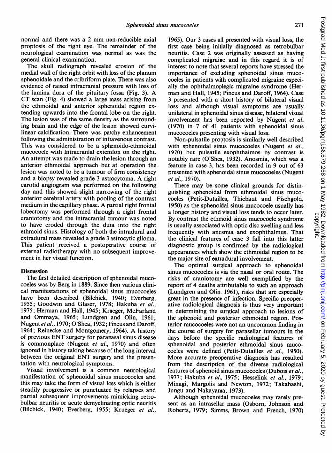
normal and there was a 2 mm non-reducible axialproptosis of the right eye. The remainder of theneuiological examination was normal as was thegeneral clinical examination.The skull radiograph revealed erosion of the
medial wall of the right orbit with loss of the planumsphenoidale and the cribriform plate. There was alsoevidence of raised intracranial pressure with loss ofthe lamina dura of the pituitary fossa (Fig. 3). ACT scan (Fig. 4) showed a large mass arising fromthe ethmoidal and anterior sphenoidal region ex-tending upwards into the frontal lobe on the right.The lesion was of the same density as the surround-ing brain and the edge of the lesion showed curvi-linear calcification. There was patchy enhancementfollowing the administration of intravenous contrast.This was considered to be a sphenoido-ethmoidalmucocoele with intracranial extension on the right.An attempt was made to drain the lesion through ananterior ethmoidal approach but at operation thelesion was noted to be a tumour of firm consistencyand a biopsy revealed grade 3 astrocytoma. A rightcarotid angiogram was performed on the followingday and this showed slight narrowing of the rightanterior cerebral artery with pooling of the contrastmedium in the capillary phase. A partial right frontallobectomy was performed through a right frontalcraniotomy and the intracranial tumour was notedto have eroded through the dura into the rightethmoid sinus. Histology of both the intradural andextradural mass revealed a grade 3 astrocytic glioma.This patient received a postoperative course ofexternal radiotherapy with no subsequent improve-ment in her visual function.
DiscussionThe first detailed description of sphenoidal muco-
coeles was by Berg in 1889. Since then various clini-cal manifestations of sphenoidal sinus mucocoeleshave been described (Bilchick, 1940; Everberg,1955; Goodwin and Glaser, 1978; Hakuba et al.,1975; Herman and Hall, 1945; Krueger, McFarlandand Ommaya, 1965; Lundgren and Olin, 1961;Nugent etal., 1970; O'Shea, 1932; Pincus and Daroff,1964; Reinecke and Montgomery, 1964). A historyof previous ENT surgery for paranasal sinus diseaseis commonplace (Nugent et al., 1970) and oftenignored in history taking because of the long intervalbetween the original ENT surgery and the presen-tation with neurological symptoms.
Visual involvement is a common neurologicalmanifestation of sphenoidal sinus mucocoeles andthis may take the form of visual loss which is eithersteadily progressive or punctuated by relapses andpartial subsequent improvements mimicking retro-bulbar neuritis or acute demyelinating optic neuritis(Bilchick, 1940; Everberg, 1955; Krueger et al.,
1965). Our 3 cases all presented with visual loss, thefirst case being initially diagnosed as retrobulbarneuritis. Case 2 was originally assessed as havingcomplicated migraine and in this regard it is ofinterest to note that several reports have stressed theimportance of excluding sphenoidal sinus muco-coeles in patients with complicated migraine especi-ally the ophthalmoplegic migraine syndrome (Her-man and Hall, 1945; Pincus and Daroff, 1964). Case3 presented with a short history of bilateral visualloss and although visual symptoms are usuallyunilateral in sphenoidal sinus disease, bilateral visualinvolvement has been reported by Nugent et al.(1970) in 7 of 41 patients with sphenoidal sinusmucocoeles presenting with visual loss.
Non-pulsatile proptosis is similarly well describedwith sphenoidal sinus mucocoeles (Nugent et al.,1970) but pulsatile exophthalmos by contrast isnotably rare (O'Shea, 1932). Anosmia, which was afeature in case 3, has been recorded in 9 out of 63presented with sphenoidal sinus mucocoeles (Nugentet al., 1970).
There may be some clinical grounds for distin-guishing sphenoidal from ethmoidal sinus muco-coeles (Petit-Dutailles, Thiebaut and Fischgold,1950) as the sphenoidal sinus mucocoele usually hasa longer history and visual loss tends to occur later.By contrast the ethmoid sinus mucocoele syndromeis usually associated with optic disc swelling and lessfrequently with anosmia and exophthalmus. Thatthe clinical features of case 3 fall into this latterdiagnostic group is confirmed by the radiologicalappearances which show the ethmoidal region to bethe major site of extradural involvement.The optimal surgical approach to sphenoidal
sinus mucocoeles is via the nasal or oral route. Therisks of craniotomy are well exemplified by thereport of 4 deaths attributable to such an approach(Lundgren and Olin, 1961), risks that are especiallygreat in the presence of infection. Specific preoper-ative radiological diagnosis is thus very importantin determining the surgical approach to lesions ofthe sphenoid and posterior ethmoidal region. Pos-terior mucocoeles were not an uncommon finding inthe course of surgery for parasellar tumours in thedays before the specific radiological features ofsphenoidal and posterior ethmoidal sinus muco-coeles were defined (Petit-Dutailles et al., 1950).More accurate preoperative diagnosis has resultedfrom the description of the diverse radiologicalfeatures of sphenoid sinus mucocoeles (Dubois et al.,1977; Hakuba et al., 1975; Hesselink et al., 1979;Minagi, Margolis and Newton, 1972; Takahashi,Jungu and Nakayama, 1973).Although sphenoidal mucocoeles may rarely pre-
sent as an intrasellar mass (Osborn, Johnson andRoberts, 1979; Simms, Brown and French, 1970)
271
copyright. on F
ebruary 5, 2020 by guest. Protected by
http://pmj.bm
j.com/
Postgrad M
ed J: first published as 10.1136/pgmj.58.679.268 on 1 M
ay 1982. Dow
nloaded from

272 I. R. Livingstone et al.
the distinction between lesions arising from thesphenoid sinus and pituitary tumours usually restson demonstrating the absence of sellar enlargementin sphenoid sinus lesions (Hakuba et al., 1975;Robertson et al., 1976), despite destruction of thefloor of the sella. Several reports have stressed theimportance of detailed tomography on several planesin defining the nature of lesions involving the eth-moid and sphenoid sinuses (Dubois et al., 1977;Minagi et al., 1972; Robertson et al., 1976; Takah-ashi et al., 1973). Opacification of the sinus andexpansion and thinning of the sinus walls has beenreported as a consistent finding with sphenoid sinusmucocoeles in one series of 14 cases (Robertsonet al., 1976). Axial CT scanning alone is not alwayssufficient for adequate evaluation of paranasal sinusmucocoeles (Hesselink et al., 1979) and needs to besupplemented with either coronal CT scanning orplain tomography. In a review of the CT scanappearance of 18 paranasal sinus mucocoeles(Hesselink et al., 1979), rim enhancement was notedto occur only in the presence of acute infection as, inthe absence of infection, the mucocoeles are usuallyof the same density as the brain.The erosion through the dura by a frontal astro-
cytic glioma into the ethmoidal in case 3 is unusual.Transdural extension of a glioma is a rare event asthe dura is resistant to penetration. Infiltration ofthe dura by glioblastomas and anaplastic astro-cytomas is usually confined to the inner dural layer(Russell and Rubinstein, 1971), Cases of transduralextension are, however, described. Rubinstein (1967)described infiltration of the sagittal venous sinus byan anaplastic astrocytoma. Russell and Rubinstein(1971) describe a case of a 7-year-old girl with asuperficial temporal glioblastoma with erosionthrough the great wing of the sphenoid and sellarturcica. Two further reports have described spon-taneous transdural extension of temporal lobeglioblastomas with invasion of the temporal bone(Nager, 1967; Sanerkin, 1962).The clinical features of our 3 reported cases sug-
gest that early diagnosis in cases of the sphenoidalsinus mucocoele syndrome presenting with visualsymptoms is still difficult in many instances and thatseveral radiological examinations may be requiredfor diagnosis. Furthermore, the preoperative differ-entiation of mucocoeles from other lesions in theregion of the ethmoid and sphenoidal sinuses in pati-ents presenting with the sphenoidal or posteriorethmoidal syndrome may not always be possible.
AcknowledgmentWe wish to thank Dr P. Hudgson for permission to report
Case 1.
ReferencesBERG, J. (1889) Bidrag till kannendomen om sjukdomarna:
nasans samt till laran om cerebro-spinal vatskas flytningur nasan. 2. Hydrops sinus sphenoidalis. Nordisk Medicin-historisk Aarsbok, 21, 5.
BILCHICK, E.B. (1940) Diseases of the sphenoid sinus withreport of a case of cyst of the sphenoid sinus. Archives ofOtolaryngology, 32, 1031.
DuBoIs, P.J., SCHULTZ, J.C., PERRIN, R.L. & DASTUR, K.J.(1977) Tomography in expansile lesions of the nasal andparanasal sinuses. Radiology, 125, 149.
EVERBERG, G. (1955) Retrobulbar neuritis due to cyst ofsphenoid sinus. Acta oto-laryngologica (Stockholm), 45,492.
GOODWIN, J.A. & GLASER, J.S. (1978) Chiasmal syndrome insphenoid sinus mucocele. Annals of Neurology, 4, 440.
HAKUBA, A., KATSUYAMA, J., MATSUOKA, Y., HONG SHIM, J.& NISHIMURA, S. (1975) Sphenoid sinus mucocoeles.Report of two cases. Journal of Neurosurgery, 43, 368.
HERMAN, K. & HALL, I.S. (1945) Sphenoidal mucocoele as acause of the 'opthalmoplegic migraine syndrome'. Trans-actions of the Ophthalmological Society of the UnitedKingdom, 64, 154.
HESSELINK, J.R., WEBER, A.L., NEW, P.F., DAVis, K.R.,ROBERSON, G.H. & TAVERAS, J.M. (1979) Evaluation ofmucocoeles of the paranasal sinuses with computedtomography. Radiology, 133, 397.
KRUEGER, T.P., MCFARLAND, J. & OMMAYA, A.K. (1965)Pyocoele of the sphenoid sinus. Journal of Neurosurgery.22, 616.
LUNDGREN, A. & OLIN, T. (1961) Mucopyocoele of thesphenoidal sinus or posterior ethmoidal cells with specialreference to the apex orbitae syndrome. Acta oto-laryn-gologica (Stockholm), 53, 61.
MINAGI, H., MARGOLIS, T. & NEWTON, T.H. (1972) Tomo-graphy in the diagnosis of sphenoid sinus mucocele.American Journal of Roentgenology, 115, 587.
NAGER, G.T. (1967) Gliomas involving the temporal bone.Clinical and pathological aspects. Laryngoscope (St Louis),77, 454.
NUGENT, G.R., SPRINKLE, P. & BLOOR, B.M. (1970) Sphenoidsinus mucocoeles. Journal of Neurosurgery, 32, 443.
OSBORN, A.G., JOHNSON, L. & ROBERTS, T.S. (1979) Sphe-noidal mucocoeles with intracranial extension. Journal ofCompuler Assisted Tomographv, 3, 335.
O'SHEA, H.V. (1932) Pulsating exophthalmos due to sphe-noidal sinus mucocoele. Lancet. i, 1253.
PETIT-DUTAILLEs, D., THIEBAUT, F. & FISCHGOLD, H. (1950)Contribution A 1'etude des compressions intracraniennesdes nerfs optiques par les abces ou les mucocoeles extra-durales d'origine sph6no6thmoidale. Revue Neurologique(Paris), 83, 325.
PINCUS, J.G. & DAROFF, R.B. (1964) Sphenoid sinus muco-celes. A curable cause of the ophthalmoplegic migrainesyndrome. Journal of the American Medical Association,187, 459.
REINECKE, R.D. & MONTGOMERY, W.W.M. (1964) Oculo-motor nerve palsy associated with mucocele of thesphenoid sinus. Archives of Ophthalmology, 71, 50.
ROBERTSON, G.H., PATTERSON, A.K., EL DEEB, M., MAISEL,R.H. & BONE, R.C. (1976) Sphenoethmoidal mucocele:Radiographic diagnosis. American Journal of Roentgeno-logy, 127, 595.
RUBINSTEIN, L.J. (1967) Development of extracranialmetastases from a malignant astrocytoma. Journal ofNeurosurgery, 26, 542.
RUSSELL, D.S., RUBINSTEIN, L.J. (1971) Pathology of tumoursof the nervous system. 3rd edn. Edward Arnold, London.
SANERKIN, N.G. (1962) Transdural spread of glioblastomamultiforme. Journal of Pathology, 84, 228.
copyright. on F
ebruary 5, 2020 by guest. Protected by
http://pmj.bm
j.com/
Postgrad M
ed J: first published as 10.1136/pgmj.58.679.268 on 1 M
ay 1982. Dow
nloaded from
Sphenoidal sinus mucocoeles 273
SIMMs, N.M., BROWN, W.E. & FRENCH, L.A. (1970) Muco-coele of the sphenoid sinus presenting as an intrasellarmass. Journal of NleurosurgerY, 32, 710.
TAKAHASHI, M., JINGU, K. & NAKAYAMA, T. (1973) Roent-genologic appearances of sphenoethmoidal mucocoele.Neuroradiology, 6, 45.
copyright. on F
ebruary 5, 2020 by guest. Protected by
http://pmj.bm
j.com/
Postgrad M
ed J: first published as 10.1136/pgmj.58.679.268 on 1 M
ay 1982. Dow
nloaded from