Embed Size (px)
Citation preview

Citation: Bissoon AK, Scipio JE and Harrynarine DK. Incidental Finding of Ethmoidal Osteoma on Cone Beam Computed Tomography. J Dent App. 2015;2(6): 240-242.
J Dent App - Volume 2 Issue 6 - 2015ISSN : 2381-9049 | www.austinpublishinggroup.com Bissoon et al. © All rights are reserved
Journal of Dental ApplicationsOpen Access
Abstract
Osteomas are slow- growing, benign bony tumours that occasionally occur in the paranasal sinuses. They consist of mature compact or cancellous bone. The aim of this report is to describe a case of incidental finding of this tumour in the ethmoidal sinus in a patient presenting with symptomatic florid cemento- osseous dysplasia of the mandible diagnosed radiographically. The importance of comprehensive evaluation of large field of view cone beam computed tomography (CBCT) scans is highlighted.
Keywords: Ethmoidal osteoma, incidental finding, cone beam computed tomography
IntroductionOsteomas are benign osteogenic neoplasms characterized by the
proliferation of compact or cancellous bone with sparse marrow. Maxillofacial osteomas most commonly occur in the mandible, followed by the paranasal sinuses of which the frontal sinus is the most frequent site followed by the ethmoid, maxillary and very rarely the sphenoid sinus [1,2]. These tumours are usually asymptomatic but may extend to surrounding structures such as the orbit and nasal cavity causing symptoms [3-7]. Many osteomas are discovered accidentally on sinus radiographs [8-10]. We report on a case of incidental ethmoidal osteoma diagnosed radiographically from cone beam computed tomography (CBCT).
Case ReportA 30- year- old female of Afro- Trinidadian descent presented to
the Maxillofacial Surgery Centre, Trinidad with palpation discomfort bilaterally in the first molar regions of the mandible. The associated teeth were non- carious and vital to electric pulp testing. Clinically expanded facial cortices were observed bilaterally in the mandibular premolar/ first molar regions. Dental panoramic radiography revealed multiple mixed radiolucent/radiopaque lesions in the mandibular premolar/molar regions bilaterally. The patient was subsequently referred for a cone beam computed tomography scan (CBCT) to further investigate the extent and effect of the mixed lesions on surrounding structures. A large field of view (FOV) CBCT scan was taken. A radiographic differential diagnosis of florid cemento- osseous dysplasia was given the mixed lesions in the mandible.
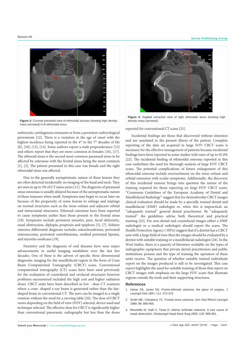
The radiologist reporting on the scan also observed a well- defined, non-enhancing, high density, circular mass 2.6 mm x 2.9 mm present in the right ethmoidal sinus arising from the medial wall. The mass was seen on the axial, coronal and sagittal sections (Figures 1- 3) through the ethmoid sinus. There was no effect on adjacent vital structures ascertained from the CBCT scan. The lesion appeared to be unrelated to the presenting complaint and a differential diagnosis
Case Report
Incidental Finding of Ethmoidal Osteoma on Cone Beam Computed TomographyBissoon AK1*, Scipio JE2 and Harrynarine DK3 1Department of Oral Diseases, University of the West Indies, Trinidad and Tobago, West Indies2Maxillofacial Surgery Centre, Woodbrook, Trinidad and Tobago, West Indies3Department of Oral Diseases, University of the West Indies, Trinidad and Tobago, West Indies
*Corresponding author: Bissoon AK, Department of Oral Diseases, University of the West Indies School of Dentistry, Eric Williams Medical Sciences Complex, Champs Fleurs, Trinidad and Tobago, West Indies, Tel: +1(868)752-2791; Fax: 645-3823, Email: [email protected]
Received: January 29, 2015; Accepted: April 08, 2015; Published: April 10, 2015
of ethmoidal osteoma was suggested given the radiographic features observed. Given the asymptomatic nature of the lesion the referring practitioner was advised to inform the patient of the presence of the mass and if any symptoms arise related to it consider re-imaging.
DiscussionOsteomas are slow- growing tumours of bone which can be
divided structurally into three types. These include those that are composed of compact bone known as ivory osteomas, those composed of cancellous bone and lastly those composed of a combination of compact and cancellous bone.
Although osteomas are the commonest benign neoplasms of the nose and paranasal sinuses, they are infrequent findings with reported incidences of sinus osteomas ranging from 0.01% to 0.43% [11]. These tumours have an obscure aetiology and the three proposed aetiological theories include developmental, traumatic and infectious causes. It has been hypothesized that osteomas arise either from
Figure 1: Axial extracted view of ethmoidal sinuses showing high density mass 2.6 mm x 2.9 mm (arrowed) in R ethmoidal sinus.
J Dent App 2(6): id1057 (2015) - Page - 0241
Bissoon AK Austin Publishing Group
Submit your Manuscript | www.austinpublishinggroup.com
embryonic cartilaginous remnants or from a persistent embryological periosteum [12]. There is a variation in the age of onset with the highest incidence being reported in the 4th to the 7th decades of life [8], [10], [13], [14]. Some authors report a male preponderance [15] and others report that they are more common in females [16], [17]. The ethmoid sinus is the second most common paranasal sinus to be affected by osteomas with the frontal sinus being the most common [1], [2]. The patient presented in this case was female and the right ethmoidal sinus was affected.
Due to the generally asymptomatic nature of these lesions they are often detected incidentally on imaging of the head and neck. They are seen in up to 3% of CT sinus series [11]. The diagnosis of paranasal sinus osteomas is usually delayed because of the asymptomatic nature of these tumours when small. Symptoms may begin to occur later on because of the propensity of some lesions to enlarge and impinge on normal structures such as the sinus ostium and adjacent orbital and intracranial structures. Ethmoid osteomas have been reported to cause symptoms earlier than those present in the frontal sinus [18]. Symptoms include persistent sinusitis, pain, facial deformity, nasal obstruction, diplopia, proptozis and epiphora [5], [7]. Solitary osteoma differential diagnosis includes osteochondroma, periosteal osteosarcoma, periosteal osteoblastoma, ossified periosteal lipoma, and myositis ossificans [19].
Dentistry and the diagnosis of oral diseases have seen major advancements in useful imaging modalities over the last few decades. One of these is the advent of specific three dimensional diagnostic imaging for the maxillofacial region in the form of Cone Beam Computerized Tomography (CBCT) scans. Conventional computerized tomography (CT) scans have been used previously for the evaluation of craniofacial and orofacial structures however problems encountered included the high cost and higher radiation doses. CBCT units have been described as low - dose CT scanners where a cone- shaped x-ray beam is generated rather than the fan- shaped beam in conventional CT. The jaws can be imaged in a single rotation without the need for a moving table [20]. The dose of CBCT varies depending on the field of view (FOV) selected, device used and technique selected. The effective dose for CBCT is significantly higher than conventional panoramic radiography but less than the doses
reported for conventional CT scans [21].
Incidental findings are those that discovered without intention and are unrelated to the present illness of the patient. Complete reporting of the data set acquired in large FOV CBCT scans is necessary for the effective management of patients because incidental findings have been reported in some studies with rates of up to 92.8% [22]. The incidental finding of ethmoidal osteoma reported in this case underlines the need for thorough analysis of large FOV CBCT scans. The potential complications of future enlargement of this ethmoidal osteoma include encroachment on the sinus ostium and orbital extension with ocular symptoms. Additionally, the discovery of this incidental tumour brings into question the nature of the training required for those reporting on large FOV CBCT scans. ‘‘Consensus Guidelines of the European Academy of Dental and Maxillofacial Radiology’’ suggest that for dentoalveolar CBCT images clinical evaluation should be made by a specially trained dental and maxillofacial (DMF) radiologist or, when this is impractical, an ‘‘adequately trained’’ general dental practitioner. By ‘‘adequately trained’’ the guidelines advise both theoretical and practical training [23]. For non-dental and craniofacial images either a DMF radiologist or a medical radiologist should report the scans. The Health Protection Agency ( HPA) suggest that if a dentist has a CBCT unit with a large field of view then the images should be evaluated by a dentist with suitable training or a maxillofacial radiologist [24]. In the West Indies, there is a paucity of literature available on the types of radiographic equipment that private dental practitioners and public institutions possess and the type of training the operators of these units receive. The question of whether suitably trained individuals report on the images produced is still to be investigated. This case report highlights the need for suitable training of those that report on CBCT images with emphasis on the large FOV scans that illustrate regions outside the teeth and their supporting structures.
References1. Hehar SS, Jones NS. Fronto-ethmoid osteoma: the place of surgery. J
Laryngol Otol 1997; 111: 372-375.
2. Smith ME, Calcaterra TC. Frontal sinus osteoma. Ann Otol Rhinol Laryngol 1989; 98: 896-900.
3. Mesolella M, Galli V, Testa D. Inferior turbinate osteoma: A rare cause of nasal obstruction. Otolaryngol Head Neck Surg 2005; 133: 989-991.
Figure 3: Sagittal extracted view of right ethmoidal sinus showing high density mass (arrowed).
Figure 2: Coronal extracted view of ethmoidal sinuses showing high density mass (arrowed) in R ethmoidal sinus.
J Dent App 2(6): id1057 (2015) - Page - 0242
Bissoon AK Austin Publishing Group
Submit your Manuscript | www.austinpublishinggroup.com
4. Lin C, Lin Y, Kang B. Middle turbinate osteoma presenting with ipsilateral facial pain, epiphora and nasal obstruction. Otolaryngol Head Neck Surg 2003; 128: 282-283.
5. Gillman GS, Lampe HB, Allen LH. Orbitoethmoid osteoma: Case report of an uncommon presentation of an uncommon tumour. Otolaryngol Head Neck Surg 1997; 117: 218-220.
6. Huang HM, Liu CM, Lin KN, Chen HT. Giant ethmoid osteoma with orbital extension, a nasoendoscopic approach using an intranasal drill. Laryngoscope 2001; 111: 430-432.
7. Osma U, Yaldiz M, Tekin M, Topcu I. Giant ethmoid osteoma with orbital extension presenting with epiphora. Rhinology. Rhinology 2003; 41: 122-124.
8. Atallah N, Jay MM. Osteomas of the paranasal sinuses. J Laryngol Otol 1981; 95: 291-304.
9. Koivunen P, Lopponen H, Fors AP, Jokinen K. The growth rate of osteomas of the paranasal sinuses. Clin Otolaryngol Allied Sci 1997; 22: 111-114.
10. Mansour AM, Salti H, Uwaydat S, Dakroub R, Bashshour Z. Ethmoid sinus osteoma presenting as epiphora and orbital cellulitis: case report and literature review. Surv Ophthalmol 1999; 43: 413-426.
11. Eller R, Sillers M. Common fibro-osseous lesions of the paranasal sinuses. Otolaryngol Clin North Am 2006; 39: 585-600.
12. Varboncoeur AP, Vanbelois HJ, Bowen LL. Osteoma of the maxillary sinus. J Oral Maxillofac Surg 1990; 48: 882-883.
13. Samy LL, Mostafa H. Osteoma of the nose and paranasal sinuses with a report of twenty one cases. J Laryngol Otol 1971; 85: 449-469.
14. Boysen M. Osteomas of the paranasal sinuses. J Otolaryngol 1978; 7: 366-370.
15. Kaplan I, Calderon S, Buchner A. Peripheral osteoma of the mandible: a study of 10 new cases and analysis of literature. J Oral Maxillo Fac Surg 1994; 52: 467-470.
16. Longo F, Califano L, De Maria G, Ciccarelli R. Solitary osteoma of the mandibular ramus: report of a case. J Oral Maxillofac Surg 2001; 59: 698-700.
17. Denia A, Perez F, Canalis RR, Graham MD. Extracanalicular osteoma of the temporal bone. Arch Otolaryngol 1979; 105: 706-709.
18. Menezes CA, Davidson TM. Endoscopic resection of a sphenoethmoid osteoma: A case report. Ear Nose Throat J 1994; 73: 598-600.
19. Greenspan A. Benign bone-forming lesions: osteoma,osteoid osteoma, and osteoblastoma. Clinical, imaging, pathologic, and differential considerations. Skeletal Radiol 1993; 22: 485-500.
20. Horner K, Drage N, Brettle D. 21st Century Imaging. Quintessence, 2008; London.
21. Ludlow JB, Davies-Ludlow LE, Brooks SL, Howerton WB. Dosimetry of 3 CBCT devices for oral and maxillofacial radiology:CB Mercuray, NewTom 3G and i-CAT. Dentomaxillofac Radiol 2006; 35: 219-226.
22. Cağlayan F, Tozoğlu U. Incidental findings in the maxillofacial region detected by cone beam CT. Diagn Interv Radiol 2012; 18:159-63.
23. Horner K, Islam M, Flygare L, Tsiklakis K, Whaites E. Basicprinciples for use of dental cone beam computed tomography:Consensus Guidelines of the European Academy of Dental and Maxillofacial Radiology. Dentomaxillofac Radiol 2009; 38: 187-195.
24. Holroyd JR, Gulson AD. The radiation protection implications of the use of cone-beam computed tomography [CBCT] in dentistry-what you need to know.
Citation: Bissoon AK, Scipio JE and Harrynarine DK. Incidental Finding of Ethmoidal Osteoma on Cone Beam Computed Tomography. J Dent App. 2015;2(6): 240-242.
J Dent App - Volume 2 Issue 6 - 2015ISSN : 2381-9049 | www.austinpublishinggroup.com Bissoon et al. © All rights are reserved