Embed Size (px)
Citation preview

Neurobiology of Disease
Involvement of the Thalamic Parafascicular Nucleus inMesial Temporal Lobe Epilepsy
Melanie Langlois,1 Pierre-Olivier Polack,3 Helene Bernard,1 Olivier David,2 Stephane Charpier,3 Antoine Depaulis,1
and Colin Deransart1
1Equipe 9, Dynamique des Reseaux Synchrones Epileptiques, and 2Equipe 5, Neuroimagerie Fonctionnelle et Metabolique, Grenoble Institut desNeurosciences, Institut National de la Sante et de la Recherche Medicale U 836-Université Joseph Fourier-Commisariat à l’Energie Atomique CentreHospitalier Universitaire, 38700 La Tronche, France, and 3Centre de Recherche de l’Institut du Cerveau et de la Moelle Épiniere, Universite Pierre et MarieCurie/Institut National de la Sante et de la Recherche Medicale, Unite Mixte de Recherche-S 975, Centre National de la Recherche Scientifique Unite Mixtede Recherche 7225, Hopital Pitie-Salpetriere, 75013 Paris, France
Mesial temporal lobe epilepsy (MTLE) is characterized by focal seizures, associated with hippocampal sclerosis, and often resistance toantiepileptic drugs. The parafascicular nucleus (PF) of the thalamus is involved in the generation of physiological oscillatory rhythms. Itreceives excitatory inputs from the cortex and inhibitory inputs from the basal ganglia, a system implicated in the control of epilepticseizures. The aim of this study was to examine the involvement of the PF in the occurrence of hippocampal paroxysmal discharges (HPDs)in a chronic animal model of MTLE in male mice. We recorded the local field potential (LFP) and the extracellular and intracellularactivity of hippocampal and PF neurons during spontaneous HPDs in vivo. The end of the HPDs was concomitant with a slow repolar-ization in hippocampal neurons leading to an electrical silence. In contrast, it was associated in the PF with a transient increase in thepower of the 10 –20 Hz band in LFPs and a depolarization of PF neurons resulting in a sustained firing. We tested the role of the PF in thecontrol of HPDs by single 130 Hz electrical stimulation of this nucleus and bilateral intra-PF injection of NMDA and GABAA antagonistand agonist. High-frequency PF stimulation interrupted ongoing HPDs at an intensity devoid of behavioral effects. NMDA antagonist andGABAA agonist suppressed hippocampal discharges in a dose-dependent way, whereas NMDA agonist and GABAA antagonist increasedHPDs. Altogether, these data suggest that the PF nucleus plays a role in the modulation of MTLE seizures.
IntroductionThe parafascicular nucleus (PF) of rodents, also called centrome-dian/parafascicular complex (CM/PF) in primates, belongs to theintralaminar system of the thalamus. Its input– output connec-tivity confers to this structure a key position to interfere with bothcortical and basal ganglia functions. Indeed, the PF receives affer-ent fibers from the somatosensory and motor cortices, reticularthalamic nucleus (nRT), zona incerta, entopeduncular nucleus,substantia nigra pars reticulata (SNr), mesencephalic reticularformation, and pedunculopontine nucleus and sends glutama-tergic projections to motor, somatosensory, and entorhinal cor-tices and to subcortical targets, such as the striatum, subthalamicnucleus, and SNr (Berendse and Groenewegen, 1991; Groenewegenand Berendse, 1994; Mouroux et al., 1995; Deschenes et al., 1996;Van der Werf et al., 2002; Smith et al., 2004). The PF participates
in cognitive (sleep wake cycle, reward mechanisms), sensory, andmotor processes (Pavlides et al., 1987; Van der Werf et al., 2002;Saade et al., 2007) as well as sensorimotor coordination (Liu et al.,2008). Recent clinical studies showed that deep brain stimulation(DBS) of the CM/PF improves behavioral and motor processes inpatients with traumatic brain injuries (Schiff et al., 2007) and hastherapeutic effects in severe generalized epileptic syndromes(Velasco et al., 2000, 2001, 2002, 2006). However, its effect onepilepsies such as the mesial temporal lobe epilepsy (MTLE) hasnever been tested (Cendes et al., 2002). MTLE is a focal epilepsysyndrome, with recurrent seizures mostly confined to the hip-pocampus and ipsilateral temporal lobe. Its is associated with aunilateral hippocampal sclerosis, characterized by an extensiveneuronal loss in the CA1 and CA3 regions and the hilus of thedentate gyrus, a reactive gliosis, and strong neuroplasticity(Engel, 1996; Wieser, 2004). Most patients with MTLE becomeresistant to antiepileptic drugs (Engel, 1996; Cendes et al., 2002),and surgical resection of the sclerotic temporal lobe has becomean alternative therapy (Ojemann, 1987; Chabardes et al., 2005).
Here, we test the hypothesis that the PF could control focalepileptic seizures, using a recently characterized mouse model ofMTLE. In this model, unilateral intrahippocampal injection ofkainic acid (KA) initially induces a nonconvulsive focal statusepilepticus followed by a latent period of approximately 2 weeks,during which recurrent spontaneous hippocampal paroxysmaldischarges (HPDs) and hippocampal sclerosis progressively de-
Received March 4, 2010; revised Sept. 24, 2010; accepted Sept. 30, 2010.This work was supported by the Institut National de la Sante et de la Recherche Medicale, the Agence Nationale
de la Recherche (ANR-06-NEURO-005-01), and the Region Rhone-Alpes (Cluster Handicap, Vieillissement, et Neu-rosciences). We thank Dr. Isabelle Guillemain for critical reading of this manuscript and Anne-Marie Godeheu forhistological processing. M.L. received a PhD fellowship from the Ministere Francais de la Recherche.
Correspondence should be addressed to Colin Deransart, Equipe 9, Dynamique des Reseaux Synchrones Epilep-tiques, Centre de recherche Inserm U 836-UJF-CEA-CHU, Grenoble Institut des Neurosciences, Universite JosephFourier, Batiment Edmond J. Safra, Domaine de la Merci, 38700 La Tronche Cedex, France. E-mail: [email protected].
DOI:10.1523/JNEUROSCI.1109-10.2010Copyright © 2010 the authors 0270-6474/10/3016523-13$15.00/0
The Journal of Neuroscience, December 8, 2010 • 30(49):16523–16535 • 16523

velop (Suzuki et al., 1995; Riban et al., 2002). Hippocampal sei-zures sporadically spread to the cerebral cortex and are resistantto most antiepileptic drugs (Riban et al., 2002). Altogether, thismurine model reproduces most of electroclinical, histological,and pharmacological features of MTLE. To examine the corre-lated changes occurring in the networks and neurons of the PFand hippocampus during MLTE, we performed in this model invivo local field potential (LFP) recordings as well as extracellularand intracellular recordings from the hippocampus and PF nu-cleus. The putative remote control of hippocampal seizures bythe PF nucleus was further investigated by testing the effects of130 Hz stimulations and pharmacological manipulations of thisnucleus on the occurrence of hippocampal paroxysms.
Materials and MethodsAnimals. Experiments were performed on 63 8-week-old C57BL/6 malemice (Janvier) weighing 26 –35 g, housed in individual cages with foodand water ad libitum, and kept in a 12 h light/dark cycle. All animalexperimentations were performed in accordance with the rules of theEuropean Committee Council Directive of November 24, 1986 (86/609/EEC), and all procedures were approved by the local department of theveterinarian services for the use and care of animals (agreement #380612)as well as by the ethical committees of our institutes. All efforts weremade to minimize animal suffering and reduce the number of animalsused in each series of experiments.
Freely moving experiments. Mice were stereotaxically injected undergeneral anesthesia (4% chloral hydrate in 0.9% NaCl; 10 ml/kg, i.p.) with50 nl of a 20 mM solution of KA (Sigma) in 0.9% NaCl (i.e., 1 nmol) intothe right dorsal hippocampus [anteroposterior (AP), �1.9; mediolateral(ML), �1.5; dorsoventral (DV), �2 mm] with bregma as the reference(Paxinos and Franklin, 2001), using a stainless-steel cannula [outer di-ameter (o.d.), 0.28 mm; Cortat SA] connected to a 0.5 �l microsyringe(Hamilton) via PE20 tubing filled with distilled water. Each injection wasperformed over 1 min using a micropump (CMA/100; Carnegie Medi-cin) as in previous studies (Riban et al., 2002; Heinrich et al., 2006). At theend of the injection, the cannula was left in place for an additional 1 minperiod to limit reflux along the cannula track.
After intrahippocampal injection, all mice were implanted with(1) two monopolar surface electrodes placed over the left and right fron-toparietal cortex for pharmacological experiments or a bipolar electrodewithin the right frontal sensorimotor cortex (AP, �1.4; ML, �1.6; DV,�2 mm from bregma) for signal analysis; (2) a monopolar electrodeplaced over the cerebellum (reference electrode); and (3) a bipolar elec-trode inserted into the injected hippocampus. The electrodes were madeof stainless-steel wire isolated by polyester (diameter, 0.125 mm;FE245840; Goodfellow). They were inserted in the skull above the cortexand the cerebellum. The bipolar electrode was formed of two twistedpolyester insulated stainless-steel wires. It was aimed at the right hip-pocampus with the same coordinates as for the injection site.
For recordings or stimulations within the PF, mice were implantedbilaterally with bipolar electrodes (AP, �2.3; ML, � 0.6; DV, �3.7 mmfrom bregma). For local drug applications, they were implanted withstainless-steel injection guide cannulae (o.d., 0.40 mm; inner diameter,0.3 mm; Phymep; AP, �2.3; ML, �0.6; DV, �2.6 mm from bregma).These guide cannulae were positioned 1.1 mm above the target areas.Stainless-steel stylets (o.d., 0.27 mm; Cortat) were inserted into the guidecannulae to keep their patency. All of the electrodes were soldered to afemale microconnector (BLR150Z; Fischer Elektronik) fixed to the skullby cyanolytic glue and dental acrylic cement. Animals were allowed aweek for recovery after surgery.
In vivo extracellular and intracellular recording experiments. Extracel-lular (single-unit) and intracellular recordings were performed in vivofrom 10 adult (12–16 weeks old) C57BL/6 male mice, 4 weeks after theyreceived an intrahippocampal injection of KA. Animals were initiallyanesthetized with chloral hydrate (4% chloral hydrate in 9% NaCl; 10ml/kg, i.p.). A cannula was then inserted into the trachea, and the animalwas placed in a stereotaxic frame. Wounds and pressure points were
repeatedly (every 2 h) infiltrated with lidocaine (2%). Once the surgicalprocedures had been completed (see below), mice were maintained in anarcotized and sedated state by injections of fentanyl (3 �g � kg �1, i.p.;Janssen-Cilag) and haloperidol (600 �g � kg �1, i.p.; Janssen-Cilag) re-peated every 20 –30 min (Charpier et al. 1999; Slaght et al., 2002). Toobtain long-lasting stable intracellular recordings, mice were immobi-lized with D-tubocurarine (0.2 mg, i.p., every hour; Sigma) and artificiallyventilated. The degree of anesthesia was assessed by continuously mon-itoring the LFPs and heart rate, and additional doses of fentanyl, halo-peridol, and D-tubocurarine were administrated at the slightest changetoward an awaked pattern. Body temperature was maintained (36.5–37.5°C) with a homeothermic blanket. At the end of the experiments,animals received an overdose of sodium pentobarbital (60 mg/kg, i.p.;Dolethal; Centravet).
Electrophysiological recordings and analysis. For standard LFP record-ings, animals were placed in Plexiglas boxes (14 � 14 � 24.5 cm) forhabituation at least 1 h before recordings. LFPs were recorded in awakefreely moving animals using a digital acquisition system (Coherence3NT; Deltamed) with a sampling rate of 256 Hz and analog bandpassfiltering between 1 and 90 Hz. Each animal was recorded at regular in-tervals (three times/week) for 1 month after KA injection to monitor theprogressive development of HPDs (i.e., epileptogenesis) and their stabi-lization and recurrence (i.e., chronic phase) as described previously(Riban et al., 2002). Only animals showing reproducible and recurrentHPDs were used in this study.
For the quantification of seizure reproducibility, the number, mean,and cumulative duration of hippocampal discharges were measured ev-ery week during three 20 min periods and then averaged. For local drugapplications, the number, mean, and cumulative duration of hippocam-pal discharges were measured during three and six 20 min periods beforeand after local drug application, respectively. For PF electrical stimula-tion experiments, animals were recorded for 1 h before the stimulationsessions. During the recording and stimulation sessions, the mice werecontinuously watched to detect changes in their behavior.
Mice were directly connected to a miniature headstage preamplifier(MPA8I; Multi Channel Systems), and signals were amplified, filtered(2000�, bandpass 1 Hz to 5 kHz; FA32I; Multi Channel Systems), andstored to hard disk (16 bit analog-to-digital converter; 2 kHz samplingfrequency; CED Power1401; Cambridge Electronic Design) using theSpike2 software.
Time–frequency analysis of 36 HPDs and concomitant right PF andsensorimotor cortex activity collected in six freely moving mice was per-formed using an in-house developed toolbox for dynamical analysis ofintracerebral LFP. The amplitude (square root of power) of oscillatoryactivity between 1 and 60 Hz was obtained using standard time–fre-quency analysis based on the Morlet wavelet transform (Le Van Quyen etal., 2001). The first and last spikes of each discharge were used to defineits onset and termination, respectively. The time window of analysis ofeach discharge was defined to contain 6 s before HPDs onset and afterHPDs termination. For each frequency, the amplitude was computed onseven periods length sliding time window, providing an effectivefrequency-specific time resolution. Time–frequency sampling of thetime–frequency plane was 50 ms/1 Hz. Time–frequency data were nor-malized using the standard procedure: for each frequency, the mean ofthe baseline was subtracted to the data and then demeaned data weredivided by the standard deviation of the baseline. Baseline was defined asthe 4 s preceding each HPD. Finally, the typical time–frequency patternof recorded HPDs was defined as the median value over HPDs of thenormalized time–frequency charts computed as such. Before medianaveraging, time–frequency charts were resampled linearly on an arbitrarytime scale to normalize HPD duration.
Neurons from the hippocampus, presumably located within the CA1region, were recorded within 1 mm of the LFP electrode at the followingcoordinates: 2.3 mm posterior to the bregma, 0.6 mm lateral to the mid-line, and 1.5–2 mm under the cortical surface. Intracellular and extracel-lular recordings of thalamic neurons were made from the same region ofthe PF nucleus ipsilateral to the injected hippocampus at the followingstereotaxic coordinates: 2.3 mm posterior to the bregma, 0.6 mm lateralto the midline, and 2.75–3.75 mm ventral to the brain surface. The re-
16524 • J. Neurosci., December 8, 2010 • 30(49):16523–16535 Langlois et al. • Parafascicular Control of Temporal Lobe Epilepsy

corded thalamic cells were subsequently confirmed as PF neurons afterexamination of their morphology and their localization (see below). Inall experiments, the intracellular or single-unit extracellular recordingswere simultaneously performed with the corresponding ipsilateral hip-pocampal LFP.
Intracellular recordings were obtained with glass micropipettes filledwith 2 M potassium acetate (50 –70 M�), and the reference electrode wasplaced in a muscle at the opposite side of the head. For single-unit extra-cellular recordings and juxtacellular labeling (see below), glass electrodeswere filled with 0.5 M NaCl and 1.7% neurobiotin (10 –20 M�; VectorLaboratories).
Intracellular recordings were obtained under current-clamp condi-tions using the active bridge mode of an Axoclamp-2B amplifier (Molec-ular Devices). Data were digitized with a sampling rate of 10 kHz(intracellular and extracellular signals) or 1 kHz (LFP) for off-line anal-ysis. Measurements of apparent membrane input resistance and timeconstant were based on the linear electrical cable theory applied to anidealized isopotential neuron (Rall, 1969). Membrane input resistancewas assessed by measurement of the mean (n � 10) membrane potentialchange at the end of hyperpolarizing current pulses of �0.4 nA (200 msduration, applied every 1.25 s), and the membrane time constant was thetime taken for the membrane potential to reach 63% of its final value.Average membrane potential of hippocampal and thalamic neurons wasdetermined from the average membrane potential of all interictal periods.When a tip potential was recorded after termination of the intracellularrecording, the membrane potential values were corrected accordingly.The amplitude of action potentials was calculated as the potential differ-ence between their voltage threshold, measured as the membrane poten-tial at which the dV/dt exceeded 10 V s �1 (Mahon et al., 2003), and thepeak of the spike waveform. Numerical values are given as means � SDunless stated otherwise.
Cumulative histograms of the binned action potential discharge ofextracellularly recorded neurons were performed by first encoding intime the position of the peak of LFP spikes and action potentials in twoseparate channels using the memory buffer function and then using the“stimulus histogram” function of the Spike 2 software (CED Software;Cambridge Electronic Design).
Morphological identification of recorded PF neurons. Extracellularly re-corded neurons were labeled by juxtacellular injection of neurobiotin(Pinault, 1996). Briefly, positive current pulses (1– 8 nA; 200 ms) wereapplied at a frequency of 2.5 Hz through the bridge circuit of the ampli-fier. The current was slowly increased while the electrode was advancedtoward the neuron in 1 �m steps (LSS-1000 Inchworm motor position-ing system; Burleigh Instruments) until the cell discharge was driven bythe injected current. Current pulses were applied for a 10 –30 min periodto obtain a reliable labeling of neuronal processes. At 1–2 h after theinjection, the animal received a lethal dose of pentobarbital and wasperfused via the ascending aorta with 200 ml of 0.3% glutaraldehyde and4% paraformaldehyde in phosphate buffer (PB), 0.1 M, pH 7.4. Brainswere postfixed for 2 h in the same fixative solution without glutaralde-hyde and then immersed in 30% sucrose PB at 4°C until sectioning.Frozen sections of fixed brains were cut at 50 –70 �m in the frontal planeand serially collected in PB. After several rinses in PB, neurobiotin wasrevealed by incubation of the sections in the avidin– biotin peroxidasecomplex (1:100; Vector Laboratories) in PB containing 0.3% TritonX-100 for at least 12 h at 4°C. Incubated sections where washed in PB(two times for 10 min) before immersion in a solution containing 0.005%3,3�-diaminobenzidine tetrahydrochloride (Sigma), 0.4% nickel-ammo-nium sulfate, and 0.0006% H2O2. After several washes in PB, sections weremounted on gelatin-coated slides, counterstained with safarin, and dehy-drated through alcohol to xylene for light microscopic examination. Thelocation of labeled neurons within the PF thalamic nucleus was confirmedusing the atlas of Paxinos and Franklin (2001).
Intracerebral electrical stimulations. All stimulations were performedwith a Grass S88H stimulator (Grass Technologies) delivering squarecurrent pulses of constant current. Before starting each stimulation ses-sion, a period of 1 h was allowed for habituation of the animals to the testcage. Thereafter, the animals, equipped with bipolar intra-PF electrodes,received a 5 s electrical stimulation applied within the first 2 s of an HPD
onset. The parameters used (frequency, 130 Hz; monophasic mode,pulse width, 60 �s) were determined according to previous stimulationstudies of other structures (e.g., SNr, subthalamic nucleus) showing sup-pressive effects on epileptic seizures (Vercueil et al., 1998; Velísek et al.,2002; Deransart and Depaulis, 2004; Feddersen et al., 2007). Additionaltrials were made with 20 Hz stimulations. The stimulation intensity wasprogressively increased from 5 to 150 �A by 5 �A steps, with at least a 1min interval between two stimulations, until the observation of (1) HPDinterruption, defined as the return to baseline of hippocampus activitywithin 2 s after stimulation onset for at least 15 s (antiepileptic thresh-old), (2) antiepileptic threshold associated with a slight head and/ortrunk straightening without paw repositioning (behavioral threshold),or (3) behavioral threshold associated with paw repositioning and/orclonus of the limbs or contralateral whole body turning around the axis(motor threshold) (Deransart and Depaulis, 2004). These changes inbehavior were measured by continuous observation of the animal by theexperimenter. Such thresholds were determined for different modes ofstimulation (referential vs bipolar; unilateral vs bilateral).
Intraparafascicular drug applications. D-(E)-2-amino-4-methyl-5-phos-phono-3-pentanoic acid (CGP40116; Ciba Geigy), NMDA (Sigma), musci-mol (5-aminomethyl-3-hydroxyisoxazole; Sigma), and picrotoxin (Sigma)were dissolved in NaCl 0.9%, aliquoted, and kept frozen at �80°C.
After a 60 min reference period of LFPs recording, animals were gentlyhandled and received within 1 min a bilateral injection of (1) 2 or 4 pmol(10 or 20 �M) of CGP40116, a competitive inhibitor of NMDA glutama-tergic receptors; (2) 17.5 or 35 pmol of muscimol, a GABAA receptorsagonist; (3) 5 or 10 pmol of NMDA, the glutamatergic NMDA receptorsagonist; or (4) 2.5 or 5 pmol of picrotoxin, a GABAA receptors antago-nist, in a volume of 200 nl per side. These compounds and the doses usedwere determined according to previous studies in other animal models ofepilepsy (Deransart et al., 1996, 1998, 1999; Nail-Boucherie et al., 2005).Since HPD generally occurs when animals are in a state of quiet wakeful-ness, we used only low doses of drugs to avoid behavioral side effects, likeawakening, that may have artificially decreased the occurrence of HPDs.Injection cannulae (o.d., 0.28 mm) were connected to a 10 �l Hamiltonmicrosyringe moved by a micropump (CMA/100; Carnegie Medicin).Each animal received the drug and the vehicle (NaCl 0.9%) according toa Latin-square design with at least 48 h between two injections. Eachmouse was thus used as its own control and was injected a maximum offour times. LFP activity was then recorded for 120 min. The animals werecontinuously watched to detect any behavioral effects induced by thedrug injection.
Effects of pharmacological injections on spontaneous LFPs recordedin the cortex and hippocampus were evaluated with the amplitude spec-trum of the fast Fourier transform computed on 6 s time windows ofinterictal activity taken every 20 min before and after injection.
Histology. After completion of each freely moving experiment, theanimals were killed with an overdose of pentobarbital (60 mg/kg, i.p.;Dolethal; Centravet) and their brains were removed, frozen, and cut in 20�m coronal sections. These sections were stained with cresyl violet andeach recording, stimulation, or injection site was localized with referenceto the atlas of Paxinos and Franklin (2001). Only data from animals with(1) correct location of the hippocampal electrode, (2) correct histologicalfeature in the injected hippocampus (Riban et al., 2002), and (3) injec-tion or stimulation sites located within the boundaries of the PF werekept in statistical analyses.
Statistics. For the follow-up of HPD stability, data were expressed asmean � SEM of cumulative duration, mean duration, and number ofHPDs per 20 min period. The data collected at different weeks after KAinjection were compared using a nonparametric ANOVA for relatedsamples (Friedman test). Paired comparisons between two time pointswere done using a nonparametric test for related samples (Wilcoxon test)(Siegel, 1956).
For deep brain stimulation experiments, thresholds were expressed asmean � SEM and were compared using the nonparametric tests ofKruskal–Wallis and Mann–Whitney when independent groups wereconcerned (localization study, comparisons between modes of stimula-tion) or the nonparametric tests of Friedman and Wilcoxon for related
Langlois et al. • Parafascicular Control of Temporal Lobe Epilepsy J. Neurosci., December 8, 2010 • 30(49):16523–16535 • 16525
samples (comparisons between parameters ofstimulations, comparison between periods).
For local drug application experiments, datawere expressed as mean � SEM of percentageof cumulative duration, mean duration, andnumber of HPDs per 20 min period, comparedwith vehicle condition (NaCl, 100%). Withineach period, the means between test condi-tions were compared using a nonparametricANOVA (Friedman test). Then, paired com-parisons versus control conditions (vehicleonly) were performed using a nonparametrictest for related samples (Wilcoxon test).
Statistical analysis was performed with Sig-maStat 3.0 (SPSS). The significance level for allstatistical analysis was set at p � 0.05.
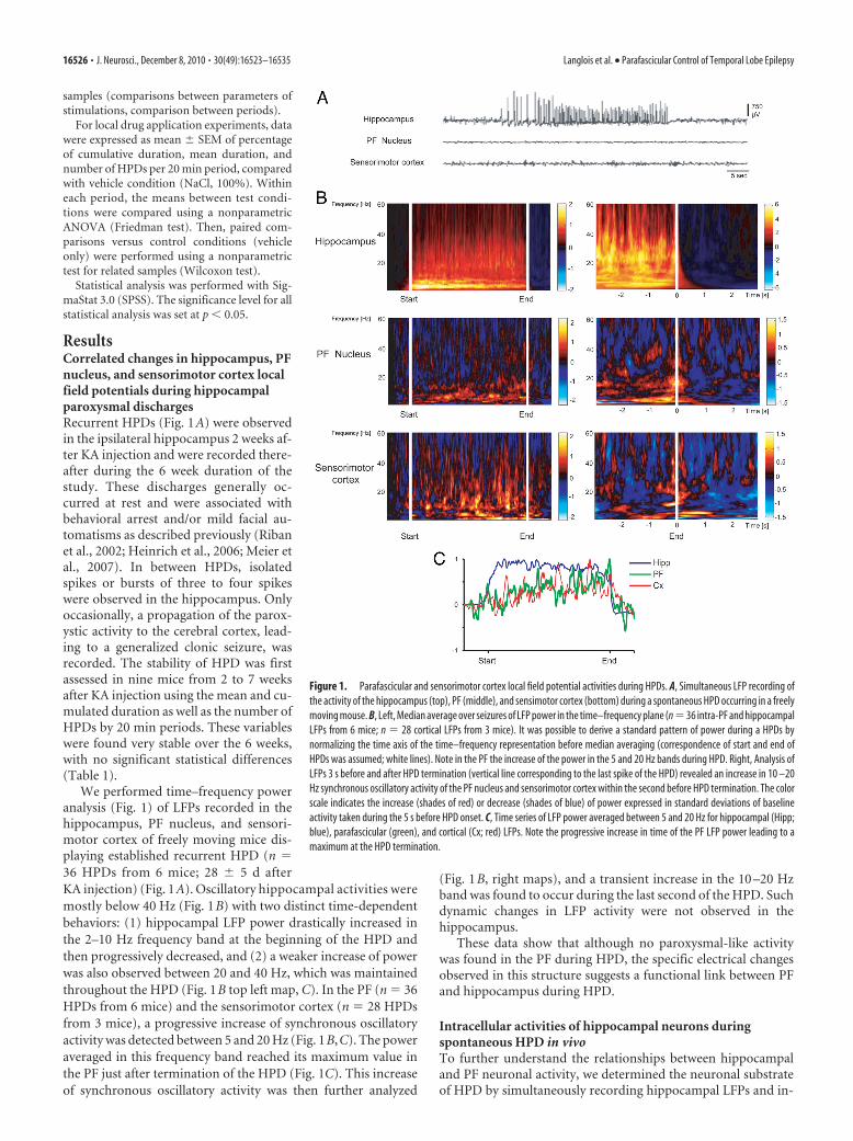
ResultsCorrelated changes in hippocampus, PFnucleus, and sensorimotor cortex localfield potentials during hippocampalparoxysmal dischargesRecurrent HPDs (Fig. 1A) were observedin the ipsilateral hippocampus 2 weeks af-ter KA injection and were recorded there-after during the 6 week duration of thestudy. These discharges generally oc-curred at rest and were associated withbehavioral arrest and/or mild facial au-tomatisms as described previously (Ribanet al., 2002; Heinrich et al., 2006; Meier etal., 2007). In between HPDs, isolatedspikes or bursts of three to four spikeswere observed in the hippocampus. Onlyoccasionally, a propagation of the parox-ystic activity to the cerebral cortex, lead-ing to a generalized clonic seizure, wasrecorded. The stability of HPD was firstassessed in nine mice from 2 to 7 weeksafter KA injection using the mean and cu-mulated duration as well as the number ofHPDs by 20 min periods. These variableswere found very stable over the 6 weeks,with no significant statistical differences(Table 1).
We performed time–frequency poweranalysis (Fig. 1) of LFPs recorded in thehippocampus, PF nucleus, and sensori-motor cortex of freely moving mice dis-playing established recurrent HPD (n �36 HPDs from 6 mice; 28 � 5 d afterKA injection) (Fig. 1A). Oscillatory hippocampal activities weremostly below 40 Hz (Fig. 1B) with two distinct time-dependentbehaviors: (1) hippocampal LFP power drastically increased inthe 2–10 Hz frequency band at the beginning of the HPD andthen progressively decreased, and (2) a weaker increase of powerwas also observed between 20 and 40 Hz, which was maintainedthroughout the HPD (Fig. 1B top left map, C). In the PF (n � 36HPDs from 6 mice) and the sensorimotor cortex (n � 28 HPDsfrom 3 mice), a progressive increase of synchronous oscillatoryactivity was detected between 5 and 20 Hz (Fig. 1B,C). The poweraveraged in this frequency band reached its maximum value inthe PF just after termination of the HPD (Fig. 1C). This increaseof synchronous oscillatory activity was then further analyzed
(Fig. 1B, right maps), and a transient increase in the 10 –20 Hzband was found to occur during the last second of the HPD. Suchdynamic changes in LFP activity were not observed in thehippocampus.
These data show that although no paroxysmal-like activitywas found in the PF during HPD, the specific electrical changesobserved in this structure suggests a functional link between PFand hippocampus during HPD.
Intracellular activities of hippocampal neurons duringspontaneous HPD in vivoTo further understand the relationships between hippocampaland PF neuronal activity, we determined the neuronal substrateof HPD by simultaneously recording hippocampal LFPs and in-
Figure 1. Parafascicular and sensorimotor cortex local field potential activities during HPDs. A, Simultaneous LFP recording ofthe activity of the hippocampus (top), PF (middle), and sensimotor cortex (bottom) during a spontaneous HPD occurring in a freelymoving mouse. B, Left, Median average over seizures of LFP power in the time–frequency plane (n�36 intra-PF and hippocampalLFPs from 6 mice; n � 28 cortical LFPs from 3 mice). It was possible to derive a standard pattern of power during a HPDs bynormalizing the time axis of the time–frequency representation before median averaging (correspondence of start and end ofHPDs was assumed; white lines). Note in the PF the increase of the power in the 5 and 20 Hz bands during HPD. Right, Analysis ofLFPs 3 s before and after HPD termination (vertical line corresponding to the last spike of the HPD) revealed an increase in 10 –20Hz synchronous oscillatory activity of the PF nucleus and sensorimotor cortex within the second before HPD termination. The colorscale indicates the increase (shades of red) or decrease (shades of blue) of power expressed in standard deviations of baselineactivity taken during the 5 s before HPD onset. C, Time series of LFP power averaged between 5 and 20 Hz for hippocampal (Hipp;blue), parafascicular (green), and cortical (Cx; red) LFPs. Note the progressive increase in time of the PF LFP power leading to amaximum at the HPD termination.
16526 • J. Neurosci., December 8, 2010 • 30(49):16523–16535 Langlois et al. • Parafascicular Control of Temporal Lobe Epilepsy

tracellular activities. Recorded hippocampal neurons were lo-cated in the CA1 region of the epileptic hippocampus in thevicinity (1 mm) of the KA injection site. These neurons had elec-trical membrane properties and intrinsic firing patterns similarto those described previously from CA1 pyramidal neurons re-corded from normal rats in vivo (Henze and Buzsaki, 2001) andKA-treated mice in vitro (Le Duigou et al. 2008). Their actionpotential had an amplitude of 63.5 � 4.5 mV (n � 8 cells), aduration of 1.3 � 0.5 ms (n � 8 cells), and a voltage threshold of�51.6 � 7.1 mV (n � 8 cells), and was followed by a high-amplitude afterhyperpolarization (10.6 � 7.4 mV; n � 8 cells)(Fig. 2A). Membrane input resistance and membrane time con-stant were 31.1 � 14.8 M� and 3.9 � 2.4 ms, respectively (n � 8cells) (Fig. 2A). In response to injection of suprathreshold cur-rent pulses, hippocampal cells discharged repetitively with a tonicand regular firing pattern (Fig. 2A).
In absence of paroxysmal activity in the corresponding LFP,hippocampal neurons were highly polarized, with a mean mem-
brane potential of �78.1 � 9.6 mV (n � 8 cells), and displayedsmall-amplitude bursts of depolarizing postsynaptic potentials(dPSPs) that remained, in the vast majority of the cells (seven ofeight), subthreshold for action potential generation (Fig. 2B1,arrow). The remaining neuron showed a moderate backgroundspontaneous firing of 1.76 Hz.
The occurrence of interictal-like activities, which consisted ofsingle or short clusters (two to five paroxysmal events) of high-voltage sharp waves as described previously (Riban et al., 2002),was coincident in hippocampal cells with an increase in bothduration and amplitude of the grouped dPSPs (Fig. 2B1, crossedarrow). A progressive temporal summation (lasting 0.5–1 s) ofthese interictal excitatory synaptic events (Fig. 2B2, arrow) couldeventually lead to a sustained depolarization (up to 33.7 � 9.2mV; n � 126 interictal events from 8 cells) generating bursts ofaction potentials (13.4 � 5.3 action potentials; n � 126 interictalevents from 8 cells) (Fig. 2B2). These paroxysmal depolarizing shiftsslowly decayed within 3.3 � 1.5 s (n � 126 interictal events, 8 cells).
Table 1. Stability of HPDs
Time after KA injection (weeks)
2 3 4 5 6 7
Number of HPDs 24 � 2 21 � 2 22 � 2 19 � 1 21 � 1 19 � 1Cumulated duration of HPDs (s) 407 � 43 347 � 40 325 � 27 349 � 28 369 � 26 380 � 34Mean duration of HPDs (s) 17 � 1 16 � 2 17 � 1 19 � 1 18 � 1 21 � 2
Averaged number of, cumulated, and mean durations by 20 min periods of HPD recorded in nine mice from 2 to 7 weeks after KA injection.
Figure 2. Intracellular activity of hippocampal neurons from the KA-injected mouse in vivo. A, Voltage responses (top traces) of a neuron located in the CA1 region of the epileptic hippocampusto hyperpolarizing (average of 5 successive trials) and depolarizing (single-response) current pulses (bottom traces). B, Intracellular correlate (bottom traces) of LFP (top traces) interictal-likeactivities. B1, Subthreshold events. In between the interictal spikes, the hippocampal (Hipp.) neuron displayed bursts of dPSPs (arrow), which were amplified (crossed arrow) when coincident withparoxysmal LFP. B2, Suprathreshold events. Brief clusters of LFP spikes could be correlated with neuronal bursting, which were generated by large-amplitude depolarization gradually constructedby temporal summation of excitatory synaptic events (inset; calibration: 30 mV, 20 ms). C, D, Intracellular activity (bottom trace) of pyramidal neurons associated with LFP seizure activity (top trace).Note the abrupt depolarization at the onset of seizure (arrows) and the sustained neuronal depolarization throughout the local paroxysm. The inset in C, is the enlargement of the initial neuronalparoxysmal shift indicated by the arrow. Arrowheads indicate the mean interictal membrane potential. Records shown in A–C and D are from two separate experiments.
Langlois et al. • Parafascicular Control of Temporal Lobe Epilepsy J. Neurosci., December 8, 2010 • 30(49):16523–16535 • 16527

HPDs were characterized in the LFP bythe repetition of high-voltage biphasicsharp waves (44.0 � 14.4 waves; n � 58HPDs from 8 cells) (Fig. 2C,D) lasting16.9 � 7.6 s and having an interval fre-quency (2.9 � 1.3 Hz) similar to that cal-culated from behaving KA-treated mice(see Table 1). Contrasting with the inter-ictal discharges, the start of HPDs wasconcomitant with an abrupt (Fig. 2C, in-set) and large (33.6 � 8.2 mV; n � 58HPDs from 8 cells) depolarization, arisingfrom a relatively quiescent membrane po-tential and causing a burst of action po-tentials (Fig. 2C,D). This initial ictalneuronal event was followed by a sus-tained membrane depolarization of19.2 � 6.9 mV (n � 58 HPDs from 8cells), which was maintained throughoutthe HPDs (Fig. 2C,D). This prolonged de-polarization was mostly crowned by re-peated sharp depolarizing potentials,which occurred from the decay phase ofthe preceding one and generated a clusterof action potentials (4.0 � 1.8 action po-tentials; n � 58 epileptic HPDs from 8cells). At the end of the HPDs, hippocampalneurons slowly repolarized, within 6.3 �3.6 s (n � 58 HPDs from 8 cells), towardtheir interictal membrane potential.
These findings provide, to our knowl-edge, the first in vivo intracellular descrip-tion of the interictal and ictal eventsoccurring in the hippocampus of a mousemodel of MTLE.
Intracellular activity of PF neuronsduring HPDTo investigate the functional coupling between hippocampusand thalamic PF nucleus during HPD, we performed in vivo in-tracellular recordings of PF neurons simultaneously with LFPactivities in the ipsilateral CA1 region. The five recorded neurons,from five different mice, had an average membrane resistance of29.7 � 9.3 M� and a membrane time constant of 10.5 � 3.7 ms(Fig. 3A). Their action potentials had an amplitude of 58.1 � 6.8mV and a duration of 0.9 � 0.3 ms, and were followed by anafterhyperpolarization of large amplitude (9.2 � 4.4 mV) (Fig.3A). When applied from the resting potential, suprathresholdcurrent pulses elicited sustained tonic firing (Fig. 3A1). More-over, the injection of negative current pulses was immediatelyfollowed by a postanodal excitation, likely caused by a low-threshold calcium potential (Fig. 3A2, top) generating a burst ofaction potentials (Fig. 3A1,A2), and/or induced a sag potential (Fig.3A2, bottom), likely caused by a hyperpolarization-activated inwardcationic current. Altogether, these electrical membrane propertieswere consistent with those classically described in thalamocorticalcells (Jahnsen and Llinas, 1984).
During interictal periods, the intracellular activity of PF neu-rons was characterized by a sustained barrage of high-frequency, small-amplitude, depolarizing synaptic potentialscorresponding to a mean membrane potential of �58.5 � 3.8mV (n � 161 interictal periods from 5 neurons). This back-
ground synaptic activity caused an irregular discharge of ac-tion potentials, with a mean frequency of 7.9 � 7.0 Hz (n � 5neurons) (Fig. 3 B, C).
The intracellular activity of PF neurons (n � 3) was clearlyaffected by the occurrence of HPDs. The onset of hippocampalparoxysms was correlated with a slight hyperpolarization of PFneurons (�1.1 � 0.5 mV; n � 27 from three cells) and a transientdecrease in the mean firing rate by 30.5 � 10.5% (n � 27 fromthree cells) (Fig. 3B,C), which could lead to a complete interrup-tion in cell discharge. A striking finding was the robust increase inPF neurons firing that coincided with the termination of HPDs(Fig. 3B,C). This sustained excitation had a duration of 3.0 �1.8 s (n � 27 from three cells) and resulted in a mean firing rate of17.7 � 9.5 Hz (Fig. 3B,C). Similar exacerbated activities in PFneurons could be also observed immediately after interictal dis-charges in the hippocampal LFPs (result not shown). Continuousinjection of negative DC currents (n � 3 cells, three animals)revealed that these discharges in PF neurons at the end of theseizure were attributable to a sustained membrane depolarizationsculpted by the temporal summation of high-frequency dPSPs(Fig. 3D).
These data confirm and extend the correlated changes foundin the thalamic LFPs as a function of the different phases of theHPDs, and further indicate a mirror-like firing activity in hip-pocampal and thalamic cells.
Figure 3. Intracellular activity of PF thalamic neurons is disrupted by hippocampal seizures in vivo. A, Electrophysiologicalproperties of recorded PF thalamic neurons. A1, Voltage responses (top traces) of a PF thalamic neuron to hyperpolarizing (averagepotential from 10 successive trials) and depolarizing (single-response) current pulses (bottom traces). Note the tonic firing inducedby the positive current and the postinhibitory excitatory rebound evidenced by the averaging of action potentials (arrow). A2, Inthese two other PF cells, the current-induced hyperpolarization was clearly followed by a robust postanodal excitation, reminiscentof a low-threshold calcium potential, crowned by a burst of sodium spikes. A sag potential, likely caused by the hyperpolarization-activated inward cationic current, could also detected in some cells (bottom, arrow). B, C, Disruption of the spontaneous intracel-lular activity of PF neurons (bottom trace) during paroxysmal activity in the ipsilateral hippocampal (Hipp.) LFP (top trace). B, Thethalamic cell was slightly hyperpolarized during the epileptic episode, and its firing was transiently interrupted. A prolonged trainof action potentials promptly followed the HPD. C, In this other neuron, the firing rate was decreased during the HPD and thendramatically augmented at the termination of hippocampal paroxysms (dashed boxes). D, The excitatory postictal rebound in PFneurons resulted from summed dPSPs. At the end of the HPD (top), the thalamic neuron, which was hyperpolarized by DC currentinjection (�1.0 nA; middle trace), displayed a sustained membrane depolarization. As shown by the expansion of the recordsegment (bottom) indicated by the asterisk, the excitatory rebound resulted from the temporal summation of individual dPSPs(oblique lines). Arrowheads indicate the mean interictal membrane potential.
16528 • J. Neurosci., December 8, 2010 • 30(49):16523–16535 Langlois et al. • Parafascicular Control of Temporal Lobe Epilepsy

Extracellular activity of PF neurons during HPDsTo further extend the statistical analysis of the functional rela-tionships between hippocampus and PF nucleus during HPDs,we then recorded the spontaneous activity of PF neurons in KA-treated mice by extracellular recordings of 30 single units fromseven mice. Recorded cells, which were morphologically identi-fied by juxtacellular injection of neurobiotin (see Materials andMethods), were located within the PF nucleus (Fig. 4A1). We
could discriminate two populations of PFcells according to their distinctive pat-terns of electrical activity which were af-fected, or not, by the occurrence of HPDs.However, all PF neurons showed an ar-rhythmic spontaneous spike activity(2.9 � 0.53 Hz) during the interictal peri-ods (Fig. 4A2,A3).
The first subpopulation of PF cells(Fig. 4A2) (n � 19), the activity of whichwas changed during HPDs, had a meanfiring frequency during interictal periodsof 3.04 � 0.77 Hz (from 0.1 to 9.48 Hz).During interictal clusters of isolatedspikes (interictal discharges) (Fig. 4B1,B2)and HPDs (Fig. 4B3,B4), the firing rate ofthis set of thalamic neurons decreased,reaching the mean values of 2.4 � 0.6 Hz(range, 0 – 4.89 Hz; n � 375 interictal clus-ters of isolated spikes) and 1.44 � 0.28 Hz(range, 0 – 4.2 Hz; n � 131 HPDs), respec-tively. The transition between interictalclusters of isolated spikes or HPDs andpostictal periods was characterized inthese PF cells by a drastic increase in theirfiring rate (Fig. 4B2,B4), the end of hip-pocampal paroxysms being concomitantwith an abrupt and transient excitation ofthese neurons. Before this increase of fir-ing rate, the mean discharge frequency ofPF thalamic cells during hippocampal par-oxysmal events was 2.5 � 0.26 Hz, whereasthe rebound of excitation had a mean fre-quency of 11 � 0.68 Hz and lasted 2.02 �0.12 s (n � 102 interictal clusters of isolatedspikes and HPDs, 19 cells). This increase inthe firing rate of PF cells concomitant withthe end of hippocampal events was statisti-cally significant ( p � 0.001; Mann–Whit-ney rank sum test).
The second category of PF cells (n � 11),which were also located within the bound-aries of the PF nucleus, did not show anysignificant change in their firing rate be-tween ictal and interictal periods (interictalmean frequency, 2 � 0.63 Hz; range, 0.084–6.58 Hz; mean frequency during clusters ofisolated spikes, 2.53 � 0.8 Hz; range, 0.054–8.88 Hz; mean frequency during HPDs, 3 �0.85 Hz; range, 0.86–7.34 Hz; n � 11 cells).
We found no correlation between thefiring pattern and the morphology of thePF neurons.
Finally, 13 neurons located outside thePF (e.g., ventral posteromedial, centrolat-
eral, or posterior thalamic nuclei) were recorded in the sameconditions, and their firing patterns did not show any detectablecorrelation with hippocampal LFP and were not modified bythe occurrence of HPDs (interictal mean frequency, 3.25 �1.15 Hz; range, 0.031–13.6 Hz; mean frequency during clus-ters of isolated spikes, 3.83 � 1.3 Hz; range, 0.67–15.1 Hz;mean frequency during HPDs, 2.75 � 0.85 Hz; range, 0.62–9.4Hz; n � 13 cells).
Figure 4. Extracellular recordings from PF neurons during HPDs. A1, Microphotograph of a PF neuron labeled by juxtacellular injectionof neurobiotin. The cell body (filled circle in the inset, schematic coronal plane drawing) was located in the central region of the lateral partof the PF nucleus. Anteriority from the interaural line was 1.62 mm. Fr, Fasciculus retroflexus; Po, posterior thalamic nucleus; VPM, ventralposteromedial thalamic nucleus. Scale bar, 100 �m. A2–A4, Single-spike activity of thalamic neurons (bottom traces) during paroxysmalactivity in the ipsilateral hippocampus (top traces). A2, Disruption of the spontaneous extracellular activity of the PF neuron shown in A1
during the HPD (between the dashed vertical lines) and representative of the first subpopulation of PF cells. A3, Extracellular activity ofanother PF neuron affected by the HPD occurrence and representative of the second subpopulation of PF cells. No change in their firing ratewas observed. A4, Simultaneous recording of two centrolateral thalamic nucleus (CL) neurons during HPD. Note that the activity of thesetwo cells is not affected by the HPD. B1–B4, Mean cumulative histograms of binned action potential discharge of 19 PF neurons aligned tohippocampal epileptic events (5 s before and after the start of short clusters of isolated spikes (B1, B3) and HPDs (B2, B4). Note slightdecrease of the activity of PF neurons after the beginning of hippocampal clusters of isolated spikes (B1) or HPDs (B3). Contrarily, the meanfiringrateofPFneurons increased500msbeforetheendofclusterof isolatedspikes(B2)andHPDs(B4)andlastedapproximately1.5safterthe end of hippocampal paroxysms.
Langlois et al. • Parafascicular Control of Temporal Lobe Epilepsy J. Neurosci., December 8, 2010 • 30(49):16523–16535 • 16529

Altogether, our intracellular and extra-cellular recordings from the PF demon-strate that the firing pattern of most of PFneurons is drastically modified by the oc-currence of HPDs.
Suppressive effect of high-frequencystimulation of the PFnucleusTo test the hypothesis of a role of the PF inthe control of HPDs, we then examinedthe effects of high-frequency stimulation(HFS) of this structure. Such stimulationsare now commonly used for DBS in hu-man patients and are suggested to result inan overall suppression of the local neuro-nal activity (McIntyre et al., 2004), al-though this effect is very much debated(Gradinaru et al., 2009). In this experi-ment, we tested single ipsilateral, con-tralateral, or bilateral 5 s stimulations ofthe PF using either monophasic and bi-polar modes (frequency, 130 Hz; pulsewidth, 60 �s) in 18 mice. In a few mice,the effects of lower frequency (20 Hz)were also tested to mimic the behaviorof PF neurons observed at terminationof HPDs.
Ipsilateral stimulationsIn nine animals with both electrode tipslocated within the ipsilateral PF nucleus,bipolar stimulations interrupted the on-going HPDs at a mean threshold of 25 �1.9 �A. The antiepileptic and behavioralthresholds were significantly lower thanthe motor threshold (73 and 65% de-creases, respectively; p � 0.004; Wilcoxontest) (Fig. 5A). A slight but not significantdifference was observed between antiepi-leptic and behavioral thresholds ( p �0.25; Wilcoxon test). Furthermore, time–frequency analysis revealed that ipsilateral130 Hz stimulations of the PF at the anti-epileptic threshold suppressed all oscilla-tory activities between 0 and 20 Hz at the hippocampal andcortical levels (Fig. 5C).
When a referential mode was used (monopolar stimulationwith one PF electrode as the cathode and the reference elec-trode over the cerebellum as the anode), the thresholds were slightlyincreased (6%), although no significant differences were observed(results not shown).
To investigate the specificity of the PF as a target for deep brainstimulation, we compared the thresholds for ipsilateral bipolarstimulations between sites located within the ipsilateral PF (insites, n � 9) and adjacent regions (out sites, n � 6; namely, theposterior thalamic nuclei group, the ventral anterior pretectalnucleus, the dorsal anterior pretectal nucleus, and the periven-tricular fiber system) (Fig. 5D). The thresholds for antiepileptic,behavioral, and motor effects were significantly lower when stim-ulations were applied within the PF region ( p � 0.013, 0.016, and0.013, respectively; Mann–Whitney test) (Fig. 5B,D). The possi-bility to interrupt HPDs by stimulations of adjacent regions,
although with higher current intensities, could account forspreading phenomenons as well as the involvement of other pu-tative neuronal networks. Similarly to intracerebral pharmaco-logical manipulations, such a gradient effect could also beobserved when stimulations were applied to other structures(Feddersen et al., 2007).
Then we tested the effect of 20 Hz stimulations, which corre-sponds to the mean firing rate of PF neurons during the reboundof excitation observed at the end of HPDs. In two mice equippedwith bipolar electrodes in the ipsilateral PF, 20 Hz stimulation didnot interrupt ongoing HPDs, whatever the amount of currentapplied (up to 150 �A). However, transient interruptions ofHPDs were observed, which lasted the duration of the stimula-tion (results not shown). This suggests that 20 Hz stimulation ofthe PF interferes with HPDs without terminating them.
Contralateral and bilateral stimulationsContralateral stimulations of the PF also interrupted ongoingHPDs (n � 9 mice) with a mean antiepileptic threshold of 35 �
Figure 5. Effects of 130 Hz stimulation of the PF nucleus on HPDs. A, Intensity thresholds according to stimulation mode:ipsilateral, contralateral, or bilateral stimulations (n � 9, 9, and 4 mice respectively; *p � 0.05, Wilcoxon test, compared withipsilateral stimulation values; #p � 0.05, Wilcoxon test, compared with antiepileptic threshold values). B, Site specificity ofipsilateral 130 Hz stimulation. Intensity thresholds according to electrode tip localization, inside versus outside the boundaries ofthe PF nucleus (n � 9 mice inside; n � 6 mice outside; *p � 0.05, Mann–Whitney test, compared with thresholds obtained withstimulations applied within the PF nucleus). C, Time–frequency chart of power averaged over hippocampal and cortical derivationsand over 24 hippocampal HPDs interrupted by 130 Hz stimulation of the PF nucleus. Before averaging, the power was frequencynormalized according to interictal activity. The color scale indicates the increase (shades of red) or decrease (shades of blue) ofpower expressed in standard deviations of the interictal activity. D, Mouse’s brain coronal sections showing histological recon-struction of the electrode tip localization inside the PF nucleus (black dots) and outside the boundaries of the PF nucleus (opentriangles). IA, Interaural; ml, medial lemniscus; fr, fasciculus retroflexus.
16530 • J. Neurosci., December 8, 2010 • 30(49):16523–16535 Langlois et al. • Parafascicular Control of Temporal Lobe Epilepsy

3.2 �A and higher behavioral (�10%; p � 0.25) and motorthresholds (�95%; p � 0.004; Wilcoxon test). All of these thresh-olds were significantly higher than for ipsilateral stimulations(antiepileptic, p � 0.03; behavioral, p � 0.006; motor, p � 0.001;Mann–Whitney test) (Fig. 5A). When bilateral stimulations wereapplied (n � 4 mice), bipolar 130 Hz stimulation interruptedHPDs with threshold values that were similar to ipsilateral stim-ulations ( p � 0.12) and significantly lower than contralateralstimulations ( p � 0.017) (Fig. 5A).
Effects of PF glutamatergic neurotransmission modulationon HPDsThe PF receives strong glutamatergic inputs mainly arising fromthe sensorimotor cortex. To determine the potential role of thisglutamatergic neurotransmission on the PF, the effects of (1)blockade and (2) potentiation of NMDA-mediated glutamater-gic neurotransmission on the occurrence of HPDs were exam-ined by local application of CGP40116 (n � 8 mice) or NMDA(n � 9 mice), respectively.
Bilateral intra-PF injection of CGP40116 (2 and 4 pmol perside) significantly suppressed the number of HPDs (data notshown) and their cumulated duration for up to 100 min at thehighest dose (Fig. 6A), compared with the vehicle condition.During the first 40 min after injection, the number and cumu-lated duration of HPDs were decreased by 95%, compared withvehicle (NaCl injection, 100%) (Fig. 6A). Ictal activity then re-turned to baseline (Fig. 6A, reference periods Ref1, Ref2, andRef3) within 2 h. Moreover, the latency for the reoccurrence ofHPDs after drug injection was significantly increased at 2 or 4pmol per side in a dose-dependent way (Fig. 6B). At either dose,the interictal activity in the hippocampus or in the cortex during the0–20 and 20–40 min periods after injection was not altered as indi-cated by the stability of the shape of the LFP amplitude spectrumbetween before and after injection periods (supplemental Fig. S1A,available at www.jneurosci.org as supplemental material).
In addition, the suppressive effects on HPDs were observedonly when injections were performed inside the PF (Fig. 6D,black dots, tips of injection cannulae). In three additional animalsinjected outside the PF (namely, the dorsal anterior pretectalnucleus, deep mesencephalic nucleus, and ventral tegmentalarea), no significant effects on HPD suppression or reoccurrencelatency (16 � 4 min outside the PF vs 48 � 6 min inside the PF at4 pmol/side) were observed (Fig. 6B,D, open triangles).
Finally, at the doses used, no behavioral effects were observed:mice remained quiet in their test cage and explored their envi-ronment or groomed. In two mice injected with a higher dose (8pmol/side), suppression of HPDs was observed, along with pros-trations beginning within the first minutes after the injection andlasting for up to 60 min (results not shown).
By contrast, bilateral intra-PF injection of NMDA signifi-cantly increased by 100 and 202%, respectively, the number (datanot shown) and cumulative duration of HPDs during the first 20min after injection of the lowest dose (5 pmol/side) comparedwith vehicle injection (NaCl, 100%) (Fig. 6A). This worseningeffect was mostly over (19 and 36%, respectively) 40 min afterinjection and back to the baseline level, at 60 min after injection(Fig. 6A). No effects on the mean duration of HPDs were ob-served (data not shown). At this dose, no behavioral or motorside effects were observed at any time. At the highest dose ofNMDA (10 pmol/side), behavioral side effects such as sniffing orexploring behaviors that clearly interfered with the occurrence ofHPDs were observed. Nonetheless, the latency of reoccurrence ofthe first HPDs after drug injection significantly decreased afterinjection of both 5 and 10 pmol per side, compared with NaCl(Fig. 6C). However, no generalizations of the HPDs by spreadingto the cortex were ever observed.
In five additional animals injected outside the boundaries of thePF (namely, the ventromedial thalamic nucleus, mediodorsal tha-lamic nucleus, dorsal anterior pretectal nucleus, superior cerebellarpeduncle, and posterior thalamic nuclei group), no significant ef-
Figure 6. Effects of bilateral intra-PF injections of CGP40116 and NMDA on HPDs. A, Percentage (mean and SEM) of cumulated duration of HPDs per 20 min period before [reference (Ref)] and after intra-PFmicroinjections of CGP40116 (4 pmol/side; n�8; *p�0.05; Wilcoxon test) or NMDA (5 pmol/side; n�9; *p�0.05; Wilcoxon test) compared to vehicle (NaCl). B, Reoccurrence latency of HPDs after vehicle(NaCl), CGP40116 (2 and 4 pmol/side) intra-PF microinjections, and CGP40116 (4 pmol/side) injected outside the boundaries of the PF nucleus (OUT; n � 8; *p � 0.05; Wilcoxon test). C, Reoccurrence latencyof HPDs after vehicle (NaCl), NMDA (5 and 10 pmol/side) intra-PF microinjections, and NMDA (5 pmol/side) injected outside the boundaries of the PF nucleus (OUT; n�9; *p�0.05; Wilcoxon test). D, Exampleofmouse’sbraincoronalsectionsshowinghistologicalreconstructionofthemicroinjectionsitesatwhichbilateral intra-PFinjectionsofCGP40116(2and4pmol/side)resultedinasuppressionofHPDs(blackdots,tips of the injection cannulae) and sites at which the same doses of CGP40116 were without effects on HPDs occurrence (open triangles). IA, Interaural; ml, medial lemniscus; fr, fasciculus retroflexus.
Langlois et al. • Parafascicular Control of Temporal Lobe Epilepsy J. Neurosci., December 8, 2010 • 30(49):16523–16535 • 16531

fects on HPD suppression or reoccurrence latency (14 � 3 minoutside vs 4 � 1 min inside at 5 pmol/side) were observed (Fig. 6C).
Effects of PF GABAergic neurotransmission modulationon HPDsTo determine the potential role of GABAergic neurotransmis-sion from the basal ganglia structures (mainly the SNr) on thePF neurons, the effects of (1) potentiation and (2) blockade ofGABAA-mediated neurotransmission were examined by localapplications of muscimol (n � 8) or picrotoxin (n � 8),respectively.
Bilateral intra-PF injection of muscimol (17.5 and 35 pmol perside) significantly suppressed the number of HPDs (data not shown)and their cumulated duration for up to 80 min at the highest dose,compared with vehicle injection (Fig. 7A). At the lowest dose, bilat-eral injection of muscimol induced a significant suppression in thenumber of HPDs for 40 min and a significant decrease in cumulatedduration for 20 min after injection, compared with vehicle injection(data not shown). At the highest dose, the number and cumulatedduration of HPDs decreased by 90% during the first 20 min periodafter injection, compared with the vehicle condition (NaCl, 100%)(Fig. 7A). It then progressively returned to baseline within 2 h. More-over, the latency for the reoccurrence of HPDs after drug injectionincreased significantly at 17.5 or 35 pmol per side in a dose-dependent way (Fig. 7B). At either dose, the interictal activity in thehippocampus or the cortex during the 0–20 and 20–40 min periodsafter injection was not altered, as indicated by the stability of theshape of the LFP amplitude spectrum between preinjection andpostinjection periods (supplemental Fig. S1B, available at www.jneurosci.org as supplemental material).
In addition, the suppressive effects were observed only wheninjections were performed within the PF (Fig. 7B,D, black dots,tips of injection cannulae). In seven additional animals injected
outside the PF (namely, in the superior cerebellar peduncle, ven-tromedial thalamic nucleus, posterior hypothalamic area, periac-queductal gray, lateral hypothalamic area, deep mesencephalicnucleus, and ventral tegmental area), no significant effects onHPD suppression or reoccurrence latency (15 � 2 min outsidethe PF vs 50 � 6 inside the PF at 35 pmol/side) were observed(Fig. 7B,D, open triangles).
Finally, at the doses used, no behavioral effects were observed.In two mice injected with a higher dose of muscimol (70 pmol/side), behavioral and motor side effects such as forelimb stereo-typies in upright position and rotations were observed, beginningwithin the first minutes after the injection and lasting for up to 60min. In this case, LFP recordings were contaminated by move-ment artifacts.
Bilateral intra-PF injection of picrotoxin induced a significantincrease in the number and cumulative duration of HPDs duringthe first hour after injection at the lowest dose (2.5 pmol/side)(Fig. 7A). The cumulated durations of HPDs were worsened by107, 195, and 52% at 20, 40, and 60 min, respectively, comparedwith vehicle (NaCl, 100%) (Fig. 7A). The latency for the reoccur-rence of HPDs after drug injection significantly increased at 2.5pmol/side (Fig. 7C). At this dose, neither behavioral nor motorside effects were observed. At the dose of 5 pmol/side, picrotoxinalso induced an aggravation in both the number and cumulatedduration of HPDs (35 and 42%, respectively, between 40 and 60min after injection) lasting up to 60 min. However, at this dose,motor side effects that interfered with the occurrence of HPDswere noticed, but no generalizations of the HPDs by spreading tothe cortex were ever observed. In four additional animals injectedoutside the boundaries of the PF (namely, in the lateral habenularnucleus, ethmoid nucleus, dorsal anterior pretectal nucleus, sub-commissural nucleus), no significant effects on HPD suppression
Figure 7. Effects of bilateral intra-PF injections of muscimol and picrotoxin on HPDs. A, Percentage (mean and SEM) of cumulated duration of HPDs per 20 min period, before [reference (Ref)] andafter intra-PF microinjections of muscimol (35 pmol/side; n � 8; *p � 0.05; Wilcoxon test) or picrotoxin (2.5 pmol/side; n � 8; *p � 0.05; Wilcoxon test) compared to vehicle (NaCl). B,Reoccurrence latency of HPDs after vehicle (NaCl), muscimol (17.5 and 35 pmol/side) intra-PF microinjections, and muscimol (35 pmol/side) injected outside the boundaries of the PF nucleus (OUT;n � 8; *p � 0.05; Wilcoxon test). C, Reoccurrence latency of HPDs after vehicle (NaCl), picrotoxin (2.5 and 5 pmol/side) intra-PF microinjections, and picrotoxin (2.5 pmol/side) injected outside theboundaries of the PF nucleus (OUT; n � 8; *p � 0.05; Wilcoxon test). D, Example of mouse’s brain coronal sections showing histological reconstruction of the microinjection sites at which bilateralintra-PF injections of muscimol (17.5 and 35 pmol/side) resulted in a suppression of HPDs (black dots, tips of the injection cannulae) and sites at which the same doses of muscimol were withouteffects on HPDs occurrence (open triangles). IA, Interaural; ml, medial lemniscus; fr, fasciculus retroflexus.
16532 • J. Neurosci., December 8, 2010 • 30(49):16523–16535 Langlois et al. • Parafascicular Control of Temporal Lobe Epilepsy
or reoccurrence latency (10 � 2 min outside the PF vs 4 � 1 insidethe PF at 2.5 pmol/side) were observed (Fig. 7C).
DiscussionWe showed here that (1) PF neurons are hyperpolarized duringHPDs and fire synchronously just before the end of the HPDs, (2)HFS of this structure interrupts ongoing HPDs, and (3) pharma-cological inhibition or activation of the PF neurons respectivelysuppresses or aggravates HPDs. These data suggest that the PFplays a role in the control of focal hippocampal seizures in MTLE.
Hippocampal neuronal activity during HPDsThis study provides the first description of the intracellular activ-ity of hippocampal neurons during spontaneous HPDs in vivo.Recorded neurons in the CA1 region could be either pyramidalneurons or granule cells. Indeed, very few CA1, CA3, and hilarneurons are still present 2–3 weeks after KA injection, whereasgranule cells are dispersed in the dorsal hippocampus (Riban etal., 2002). However, the interictal firing profile of hippocampalcells closely resembles that observed in vitro in identified pyrami-dal neurons from a similar mouse model (Le Duigou et al., 2008).It is also consistent with the “paroxysmal depolarization shift”described in vitro from other animal models and human tissue(Tancredi et al., 1990; Smith et al., 1998; Huberfeld et al., 2008).The intracellular events underlying the spontaneous occurrenceof HPDs are composed of a sustained membrane depolarizationon which rhythmic depolarizations crowned by action potentialsare superimposed. Although this exacerbated neuronal activityshares some features with that found in vivo in pyramidal cellsduring cortical seizures, additional investigations will be requiredto determine the specific synaptic and intrinsic mechanisms un-derlying the intracellular paroxysmal activity in hippocampal cell(Giaretta et al., 1987; Steriade et al., 1998).
How are PF neurons influenced during HPDs?LFP and intracellular recordings in the PF indicate that althoughPF neurons do not actively initiate HPDs, their activity is drasti-cally modulated by these events. The amplitude of the hyperpo-larization of PF neurons during HPDs did not change when themembrane potential was maintained at different levels of polar-ization by DC injection and could not be reversed in polaritywhen it was hold at hyperpolarized potential (less than �80 mV).This may result from a disfacilitation of glutamatergic inputs orfrom a shunting inhibition of PF neurons originating from thenRT or the SNr, which receives fibers from the hippocampus andtargets the distal dendrites of PF neurons (Tsumori et al., 2002).
The brief suprathreshold depolarization of PF neurons justbefore the termination of HPDs results from a barrage of depo-larizing postsynaptic potentials, possibly amplified by the de-crease in the SNr cells activity (Deransart et al., 2003; Paz et al.,2007). Indeed, suppression of HPDs was observed in MTLE miceafter intranigral injection of muscimol (Deransart and Depaulis,2004). The reduced activity of inhibitory nigral projections ontoPF cells might resume the hyperpolarization of the thalamic neu-rons and facilitate the dendritic summation and propagation oftheir excitatory postsynaptic potentials.
Is the PF involved in the interruption of HPDs?The short change in activity observed in a majority of PF neuronswithin the last second of HPDs strongly suggests the involvementof this structure in the active termination of seizures. This sup-ports the general concept that subcortical circuits play a role inthe modulation of epileptic seizures (Deransart and Depaulis,
2002). The involvement of the PF in a subcortical circuit modu-lating seizures is confirmed by the suppression of HPDs observedafter HFS or pharmacological manipulations. Because of thesmall number of structures injected or stimulated outside of thePF in each group, we cannot provide statistical analysis, but it ispossible that hippocampal activity after injections or stimula-tions in the PF may be different from that in outside structures.Data of the midline thalamus and the mediodorsal and reuniensnuclei support this hypothesis suggesting their participation inthe initiation and spread of limbic seizures (Cassidy and Gale,1998; Bertram et al., 2001, 2008).
Our data suggest that HPD suppression follows drug infusionsthat may hyperpolarize PF neurons (muscimol or CGP40116),whereas they are aggravated by drugs (picrotoxin, NMDA) thatlikely result in depolarization. However, the local injection of mus-cimol may also facilitate synchronization of PF neurons and there-fore facilitate the regular occurrence of bursts of action potentials(Liu et al., 1992; Cope et al., 2005). The frequency of such burstscould be critical in HPD suppression, as 130 Hz stimulations wereeffective but 20 Hz were not. Data dealing with the precise effect oflocally applied drugs on the PF cells’ activity, and hence on fibers ofpassage, are missing from the available literature. Further experi-ments coupling local drug application with intracellular recordingsthus remain necessary to precisely understand how our electrophys-iological data fit with our stimulation and pharmacological data.
Which circuits mediate the modulatory effects of the PFover HPDs?No monosynaptic projection from the PF to the hippocampushas been confirmed in recent studies (Wyss et al., 1979; Van derWerf et al., 2002). Therefore, the increased activity in PF neurons,as well as the suppressive or worsening effects on HPDs, verylikely involves the cerebral cortex or subcortical structures as thebasal ganglia or the nRT. Our data suggest that the PF exerts acontrol over the hippocampus via a modulation of cortical syn-chronization. Indeed, time–frequency analysis of LFPs record-ings revealed that synchronous oscillatory activity in the 10 –20Hz frequency band also increased in the sensorimotor cortexduring the last second of HPDs. These data are consistent with therole of the PF in the modulation of many physiological oscillatoryactivities that could interfere with the pathological synchronousoscillations in the hippocampus (Glenn and Steriade, 1982; Dahland Winson, 1986; Pavlides et al., 1987; Steriade et al., 1991;Seidenbecher and Pape, 2001; Lacey et al., 2007). Connectionsfrom the PF nucleus to the entorhinal cortex have been describedand are likely to be involved in these modulatory effects(Berendse and Groenewegen, 1991). The fact that HFS of the PFinterrupts ongoing HPDs with a greater efficacy when appliedipsilaterally to the injected hippocampus is in agreement withanatomical studies describing projections of the PF as mainlyipsilateral (Vercelli et al., 2003). However, cross talk between thetwo PF nuclei through the nRT has also been described (Raos andBentivoglio, 1993; Kolmac and Mitrofanis, 1997). This could ex-plain the interruption of HPDs, although less effective, whenstimulation was applied on the contralateral side, and why bilat-eral stimulations were not more efficient to stop HPDs.
The control of the PF over the HPDs might also be mediatedthrough the basal ganglia (McHaffie et al., 2005). The PF couldmediate its antiepileptic effects through its diffuse projections tothe striatum and the subthalamic nucleus by impinging on theactivity of the SNr, a structure modulating seizures in MTLE mice(Deransart and Depaulis, 2004). In a genetic model of absenceepilepsy, striatal output neurons were shown to be silent during
Langlois et al. • Parafascicular Control of Temporal Lobe Epilepsy J. Neurosci., December 8, 2010 • 30(49):16523–16535 • 16533

the paroxysmal activities and to exhibit a rebound firing at theend of the ictal discharges (Slaght et al., 2004). The transientinterruption of firing, similar to that of PF neurons in our model,presumably decreases the synaptic inhibition on SNr neuronsand modifies the balance between synaptic inhibition and excita-tion, tending to reinforce the actions of excitatory synaptic inputsarising from the subthalamic nucleus (Kita, 1994). Whether thetransient burst of action potentials observed in the PF at the endof HPD triggers striatal neurons or is triggered by nigral ones willrequire further electrophysiological studies.
The PF nucleus: a potential therapeutic target for focalseizures?Both drug applications and HFS in the PF reduced the occurrenceof HPDs without affecting their LFP pattern, mean duration orbackground EEG activity and could be obtained without behav-ioral side effects. This suggests that such manipulations act on theability of the hippocampus to generate HPDs rather than onchanging their EEG characteristics. Our pharmacological dataalso suggest that the antiepileptic effects of 130 Hz stimulation ofthe PF are rather associated with neuronal inhibition, in agree-ment with several studies, although this question still remainsdebated (Urbano et al., 2002; McIntyre et al., 2004). However,imposing a frequency that strongly differs from that observedintrinsically during HPDs may also explain the antiepileptic ef-fects observed (Garcia et al., 2005; Hammond et al., 2008).
We also showed that the modulation of seizures is specific tothe PF. It may therefore constitute an interesting target for ther-apeutic DBS in patients with drug-resistant seizures. This is inagreement with several clinical studies where HFS of the CM/PFreduced and even abolished generalized seizures in patients withLennox–Gastaut syndrome, an effect thought to be related to theactivation of ipsilateral desynchronizing thalamocortical projec-tions (Velasco et al., 2000, 2006). Our present study thus providesnew hypothesis to explain the antiepileptic effects of DBS appliedto the CM/PF in human.
Altogether, our data suggest that the PF can influence thegeneration of seizures in the hippocampus. The synchronizedoscillations and the increased firing rate of PF neurons at the endof HPDs require further investigations to understand its role inthe termination of seizures.
ReferencesBerendse HW, Groenewegen HJ (1991) Restricted cortical termination
fields of the midline and intralaminar thalamic nuclei in the rat. Neuro-science 42:73–102.
Bertram EH, Mangan PS, Zhang DX, Scott CA, Williamson JM (2001) Themidline thalamus: alterations and a potential role in limbic epilepsy. Epi-lepsia 42:967–978.
Bertram EH, Zhang D, Williamson JM (2008) Multiple roles of midlinedorsal thalamic nuclei in induction and spread of limbic seizures. Epilep-sia 49:256 –268.
Cassidy RM, Gale K (1998) Mediodorsal thalamus plays a critical role in thedevelopment of limbic motor seizures. J Neurosci 18:9002–9008.
Cendes F, Kahane P, Brodie M, Andermann F (2002) The mesio-temporallobe epilepsy syndrome. In: Epileptic syndromes in infancy, childhoodand adolescence (Roger J, Bureau M, Dravet C, Genton P, Tassinari CA,Wolf P, eds), pp 513–530. Eastleigh, UK: John Libbey.
Chabardes S, Kahane P, Minotti L, Tassi L, Grand S, Hoffmann D, BenabidAL (2005) The temporopolar cortex plays a pivotal role in temporal lobeseizures. Brain 128:1818 –1831.
Charpier S, Leresche N, Deniau JM, Mahon S, Hughes SW, Crunelli V (1999)On the putative contribution of GABA(B) receptors to the electricalevents occurring during spontaneous spike and wave discharges. Neuro-pharmacology 38:1699 –1706.
Cope DW, Hughes SW, Crunelli V (2005) GABAA receptor-mediated tonicinhibition in thalamic neurons. J Neurosci 25:11553–11563.
Dahl D, Winson J (1986) Influence of neurons of the parafascicular regionon neuronal transmission from perforant pathway through dentate gyrus.Brain Research 377:391–396.
Deransart C, Depaulis A (2002) The control of seizures by the basal ganglia?A review of experimental data. Epileptic Disord 4 [Suppl 3]:S61–S72.
Deransart C, Depaulis A (2004) Le concept de controle nigral des epilepsiess’applique-t-il aux epilepsies partielles pharmacoresistantes? Epilepsies16:75– 82.
Deransart C, Marescaux C, Depaulis A (1996) Involvement of nigral gluta-matergic inputs in the control of seizures in a genetic model of absenceepilepsy in the rat. Neuroscience 71:721–728.
Deransart C, Le BT, Marescaux C, Depaulis A (1998) Role of thesubthalamo-nigral input in the control of amygdala-kindled seizures inthe rat. Brain Res 807:78 – 83.
Deransart C, Riban V, Le BT, Hechler V, Marescaux C, Depaulis A (1999)Evidence for the involvement of the pallidum in the modulation of sei-zures in a genetic model of absence epilepsy in the rat. Neurosci Lett265:131–134.
Deransart C, Hellwig B, Heupel-Reuter M, Leger JF, Heck D, Lucking CH(2003) Single-unit analysis of substantia nigra pars reticulata neurons infreely behaving rats with genetic absence epilepsy. Epilepsia 44:1513–1520.
Deschenes M, Bourassa J, Doan VD, Parent A (1996) A single-cell study ofthe axonal projections arising from the posterior intralaminar thalamicnuclei in the rat. Eur J Neurosci 8:329 –343.
Engel J Jr (1996) Introduction to temporal lobe epilepsy. Epilepsy Res26:141–150.
Feddersen B, Vercueil L, Noachtar S, David O, Depaulis A, Deransart C(2007) Controlling seizures is not controlling epilepsy: a parametricstudy of deep brain stimulation for epilepsy. Neurobiol Dis 27:292–300.
Garcia L, D’Alessandro G, Fernagut PO, Bioulac B, Hammond C (2005)Impact of high- frequency stimulation parameters on the pattern of dis-charge of subthalamic neurons. J Neurophysiol 94:3662–3669.
Giaretta D, Avoli M, Gloor P (1987) Intracellular recordings in pericruciateneurons during spike and wave discharges of feline generalized penicillinepilepsy. Brain Res 405:68 –79.
Glenn LL, Steriade M (1982) Discharge rate and excitability of corticallyprojecting intralaminar thalamic neurons during waking and sleep states.J Neurosci 2:1387–1404.
Gradinaru V, Mogri M, Thompson KR, Henderson JM, Deisseroth K (2009)Optical deconstruction of parkinsonian neural circuitry. Science 324:354 –359.
Groenewegen HJ, Berendse HW (1994) The specificity of the “nonspecific”midline and intralaminar thalamic nuclei. Trends Neurosci 17:52–57.
Hammond C, Ammari R, Bioulac B, Garcia L (2008) Latest view on themechanism of action of deep brain stimulation. Mov Disord23:2111–2121.
Heinrich C, Nitta N, Flubacher A, Muller M, Fahrner A, Kirsch M, Freiman T,Suzuki F, Depaulis A, Frotscher M, Haas CA (2006) Reelin deficiencyand displacement of mature neurons, but not neurogenesis, underlie theformation of granule cell dispersion in the epileptic hippocampus. J Neu-rosci 26:4701– 4713.
Henze DA, Buzsaki G (2001) Action potential threshold of hippocampalpyramidal cells in vivo is increased by recent spiking activity. Neuro-science 105:121–130.
Huberfeld G, Clemenceau S, Cohen I, Pallud J, Wittner L, Navarro V, BaulacM, Miles R (2008) Epileptiform activities generated in vitro by humantemporal lobe tissue. Neurochirurgie 54:148 –158.
Jahnsen H, Llinas R (1984) Electrophysiological properties of guinea-pigthalamic neurones: an in vitro study. J Physiol 349:205–226.
Kita H (1994) Parvalbumin-immunopositive neurons in rat globus palli-dus: a light and electron microscopic study. Brain Res 657:31– 41.
Kolmac CI, Mitrofanis J (1997) Organisation of the reticular thalamic pro-jection to the intralaminar and midline nuclei in rats. J Comp Neurol377:165–178.
Lacey CJ, Bolam JP, Magill PJ (2007) Novel and distinct operational princi-ples of intralaminar thalamic neurons and their striatal projections.J Neurosci 27:4374 – 4384.
Le Duigou C, Bouilleret V, Miles R (2008) Epileptiform activities in slices of
16534 • J. Neurosci., December 8, 2010 • 30(49):16523–16535 Langlois et al. • Parafascicular Control of Temporal Lobe Epilepsy

hippocampus from mice after intra-hippocampal injection of kainic acid.J Physiol 586:4891– 4904.
Le Van Quyen M, Foucher J, Lachaux J, Rodriguez E, Lutz A, Martinerie J,Varela FJ (2001) Comparison of Hilbert transform and wavelet methodsfor the analysis of neuronal synchrony. J Neurosci Methods 111:83–98.
Liu C, Wang Y, Smallwood PM, Nathans J (2008) An essential role for Friz-zled5 in neuronal survival in the parafascicular nucleus of the thalamus.J Neurosci 28:5641–5653.
Liu Z, Vergnes M, Depaulis A, Marescaux C (1992) Involvement of in-trathalamic GABAB neurotransmission in the control of absence seizuresin the rat. Neuroscience 48:87–93.
Mahon S, Casassus G, Mulle C, Charpier S (2003) Spike-dependent intrinsicplasticity increases firing probability in rat striatal neurons in vivo.J Physiol 550:947–959.
McHaffie JG, Stanford TR, Stein BE, Coizet V, Redgrave P (2005) Subcorti-cal loops through the basal ganglia. Trends Neurosci 28:401– 407.
McIntyre CC, Savasta M, Kerkerian-Le Goff L, Vitek JL (2004) Uncoveringthe mechanism(s) of action of deep brain stimulation: activation, inhibi-tion, or both. Clin Neurophysiol 115:1239 –1248.
Meier R, Haussler U, Aertsen A, Deransart C, Depaulis A, Egert U (2007)Short-term changes in bilateral hippocampal coherence precede epilepti-form events. NeuroImage 38:138 –149.
Mouroux M, Hassani OK, Feger J (1995) Electrophysiological study of theexcitatory parafascicular projection to the subthalamic nucleus and evi-dence for ipsi- and contralateral controls. Neuroscience 67:399 – 407.
Nail-Boucherie K, Le-Pham BT, Gobaille S, Maitre M, Aunis D, Depaulis A(2005) Evidence for a role of the parafascicular nucleus of the thalamus inthe control of epileptic seizures by the superior colliculus. Epilepsia46:141–145.
Ojemann GA (1987) Surgical therapy for medically intractable epilepsy.J Neurosurg 66:489 – 499.
Pavlides C, Aoki C, Chen JS, Bailey WH, Winson J (1987) Differential glu-cose utilization in the parafascicular region during slow-wave sleep, thestill-alert state and locomotion. Brain Res 423:399 – 402.
Paxinos G, Franklin KB (2001) The mouse brain in stereotaxic coordinates,Ed 2. Academic, New York.
Paz JT, Chavez M, Saillet S, Deniau JM, Charpier S (2007) Activity of ventralmedial thalamic neurons during absence seizures and modulation of cor-tical paroxysms by the nigrothalamic pathway. J Neurosci 27:929 –941.
Pinault D (1996) A novel single-cell staining procedure performed in vivounder electrophysiological control: morpho-functional features of juxta-cellularly labeled thalamic cells and other central neurons with biocytin orNeurobiotin. J Neurosci Methods 65:113–136.
Rall W (1969) Time constants and electrotonic length of membrane cylin-ders and neurons. Biophys J 9:1483–1508.
Raos V, Bentivoglio M (1993) Crosstalk between the two sides of the thala-mus through the reticular nucleus: a retrograde and anterograde tracingstudy in the rat. J Comp Neurol 332:145–154.
Riban V, Bouilleret V, Pham-Le BT, Fritschy JM, Marescaux C, Depaulis A(2002) Evolution of hippocampal epileptic activity during the develop-ment of hippocampal sclerosis in a mouse model of temporal lobe epi-lepsy. Neuroscience 112:101–111.
Saade NE, Al Amin H, Abdel Baki S, Chalouhi S, Jabbur SJ, Atweh SF (2007)Reversible attenuation of neuropathic-like manifestations in rats by le-sions or local blocks of the intralaminar or the medial thalamic nuclei. ExpNeurol 204:205–219.
Schiff ND, Giacino JT, Kalmar K, Victor JD, Baker K, Gerber M, Fritz B,Eisenberg B, Biondi T, O’Connor J, Kobylarz EJ, Farris S, Machado A,McCagg C, Plum F, Fins JJ, Rezai AR (2007) Behavioral improvementswith thalamic stimulation after severe traumatic brain injury. Nature448:600 – 604.
Seidenbecher T, Pape HC (2001) Contribution of intralaminar thalamicnuclei to spike-and-wave-discharges during spontaneous seizures in agenetic rat model of absence epilepsy. Eur J Neurosci 13:1537–1546.
Siegel S (1956) Nonparametric statistics for the behavioral sciences. Tokyo:McGraw-Hill Kogakusha.
Slaght SJ, Leresche N, Deniau JM, Crunelli V, Charpier S (2002) Activity of
thalamic reticular neurons during spontaneous genetically determinedspike and wave discharges. J Neurosci 22:2323–2334.
Slaght SJ, Paz T, Chavez M, Deniau JM, Mahon S, Charpier S (2004) On theactivity of the corticostriatal networks during spike-and-wave dischargesin a genetic model of absence epilepsy. J Neurosci 24:6816 – 6825.
Smith KL, Lee CL, Swann JW (1998) Local circuit abnormalities in chroni-cally epileptic rats after intrahippocampal tetanus toxin injection in in-fancy. J Neurophysiol 79:106 –116.
Smith Y, Raju DV, Pare JF, Sidibe M (2004) The thalamostriatal system: ahighly specific network of the basal ganglia circuitry. Trends Neurosci27:520 –527.
Steriade M, Dossi RC, Nunez A (1991) Network modulation of a slow in-trinsic oscillation of cat thalamocortical neurons implicated in sleep deltawaves: cortically induced synchronization and brainstem cholinergic sup-pression. J Neurosci 11:3200 –3217.
Steriade M, Amzica F, Neckelmann D, Timofeev I (1998) Spike-wave com-plexes and fast components of cortically generated seizures. II. Extra- andintracellular patterns. J Neurophysiol 80:1456 –1479.
Suzuki F, Junier MP, Guilhem D, Sørensen JC, Onteniente B (1995) Mor-phogenetic effect of kainate on adult hippocampal neurons associatedwith a prolonged expression of brain-derived neurotrophic factor. Neu-roscience 64:665– 674.
Tancredi V, Hwa GG, Zona C, Brancati A, Avoli M (1990) Low magnesiumepileptogenesis in the rat hippocampal slice: electrophysiological andpharmacological features. Brain Res 511:280 –290.
Tsumori T, Yokota S, Ono K, Yasui Y (2002) Synaptic organization ofGABAergic projections from the substantia nigra pars reticulata and thereticular thalamic nucleus to the parafascicular thalamic nucleus in therat. Brain Res 957:231–241.
Urbano FJ, Leznik E, Llinas RR (2002) Cortical activation patterns evokedby afferent axons stimuli at different frequencies: an in vitro voltage-sensitive dye imaging study. Thalamus Rel Syst 1:371–378.
Van der Werf YD, Witter MP, Groenewegen HJ (2002) The intralaminarand midline nuclei of the thalamus. Anatomical and functional evidencefor participation in processes of arousal and awareness. Brain Res Rev39:107–140.
Velasco AL, Velasco F, Jimenez F, Velasco M, Castro G, Carrillo-Ruiz JD,Fanghanel G, Boleaga B (2006) Neuromodulation of the centromedianthalamic nuclei in the treatment of generalized seizures and the improve-ment of the quality of life in patients with Lennox-Gastaut syndrome.Epilepsia 47:1203–1212.
Velasco M, Velasco F, Velasco AL, Jimenez F, Brito F, Marquez I (2000)Acute and chronic electrical stimulation of the centromedian thalamicnucleus: modulation of reticulo-cortical systems and predictor factors forgeneralized seizure control. Arch Med Res 31:304 –315.
Velasco M, Velasco F, Velasco AL (2001) Centromedian thalamic and hip-pocampal electrical stimulation for the control of intractable epilepticseizures. Clin Neurophysiol 18:1–15.
Velasco M, Eugenia-Díaz-de Leon A, Marquez I, Brito F, Carrillo-Ruiz JD,Velasco AL, Velasco F (2002) Temporo-spatial correlations betweenscalp and centromedian thalamic EEG activities of stage II slow wave sleepin patients with generalized seizures of the cryptogenic Lennox-Gastautsyndrome. Clin Neurophysiol 113:25–32.
Velísek L, Velískova J, Moshe SL (2002) Electrical stimulation of substantianigra pars reticulata is anticonvulsant in adult and young male rats. ExpNeurol 173:145–152.
Vercelli A, Marini G, Tredici G (2003) Anatomical organization of the tel-encephalic connections of the parafascicular nucleus in adult and devel-oping rats. Eur J Neurosci 18:275–289.
Vercueil L, Benazzouz A, Deransart C, Bressand K, Marescaux C, Depaulis A,Benabid AL (1998) High-frequency stimulation of the subthalamic nu-cleus suppresses absence seizures in the rat: comparison with neurotoxiclesions. Epilepsy Res 31:39 – 46.
Wieser HG (2004) ILAE Commission Report. Mesial temporal lobe epilepsywith hippocampal sclerosis. Epilepsia 45:695–714.
Wyss JM, Swanson LW, Cowan WM (1979) A study of subcortical afferentsto the hippocampal formation in the rat. Neuroscience 4:463– 476.
Langlois et al. • Parafascicular Control of Temporal Lobe Epilepsy J. Neurosci., December 8, 2010 • 30(49):16523–16535 • 16535