Embed Size (px)
Citation preview
Copyright © Texas Education Agency, 2012. All rights reserved.
Nervous System
Course Anatomy and Physiology Unit VIII Nervous System Essential Question What are the tissues and systems of the human body? TEKS 130.206 (c) 1A,1B 2E,2F,2H 10A,10B,10C, 10D Prior Student Learning Understanding of the cell Estimated time 7-10 hours
Rationale The nervous system is the most highly organized system in the human body. It is a fast, complex communication system that regulates thoughts, emotions, movement, impressions, reasoning, learning, memory and choices. Objectives Upon completion of this lesson, the student will be able to:
• Define and distinguish terms pertaining to the nervous system • Differentiate between the major organs of the nervous system • Investigate the parts and functions of the brain • Analyze diseases and disorders of the nervous system
Engage How does the nervous system work? Key Points
Part I
I. Cells of the Nervous System—Neurons A. unusual shape
B. nerve fibers extend from cell body (soma) 1. dendrites – branched fibers that receive neural impulses 2. axons – transmits neural messages from cell body
towards another neuron II. Two principal divisions of the nervous system:
A. Central Nervous System (CNS) 1. brain and spinal cord
a. serves as control center for entire organism b. integrates incoming information and determines
appropriate responses B. Peripheral Nervous System (PNS)
1. made up of nerves outside the CNS -- acts as communication lines to and from CNS
2. made up of sense organs—eyes, ears, taste buds, olfactory receptors, and touch receptors
C. Understanding of mental disorders most often involves understanding of the structure and function of the CNS
III. Main Divisions and Structures of Brain A. Cerebral Hemispheres
1. right and left halves of largest part of forebrain
Copyright © Texas Education Agency, 2012. All rights reserved.
a. each side is organized to receive sensory information, mostly from contralateral (opposite) side of body & control muscles mostly on contralateral side
2. connected by two bundles of axons known as the corpus callosum
B. Cerebral Cortex 1. thin outer surface of the forebrain 2. largely made of cell bodies which are gray (thus—gray
matter) 3. most highly evolved portion of brain
C. Cerebrum – forebrain; the largest, most anterior part of human brain
1. controls motor activities and interprets sensation D. Lobes of the Cerebrum
1. Frontal Lobe a. motor cortex – controls skeletal muscles b. Broca’s speech area – formation of words c. responsible for personality d. damage to frontal lobe can cause changes in
personality 2. Temporal Lobe
a. auditory area b. language memory and speech capacity c. damage can result in aphasia (partial or total inability
to produce and understand speech as a result of brain damage caused by injury or disease)
3. Parietal Lobe a. sensory association areas – impulses from skin
such as pain and temperature are interpreted -- area for estimation of distances, sizes, and shapes
4. Occipital Lobe a. primary visual area b. trauma can result in blindness c. lesions can cause visual hallucinations
E. Structures Under the Cerebral Cortex—Diencephalon 1. Thalamus
a. relay station for all sensory information (except smell)
2. Hypothalamus a. helps maintain homeostasis (body temperature,
body fluid, hormone secretion) b. dysfunction can cause appetite, sleep, body
temperature regulation problems 3. Pineal Gland – small gland posterior to thalamus a. secretes the hormone melatonin
b. contributes to regulation of sleep-activity cycles
Copyright © Texas Education Agency, 2012. All rights reserved.
4. Pituitary Gland – attached to the hypothalamus a. “master gland” b. its secretions control timing and amount of hormone
secretions by other endocrine glands (thyroid, adrenal glands, ovaries, testes, etc.)
F. Cerebellum – highly convoluted structure in the hindbrain 1. responsible for coordination of movements
a. makes movements smooth b. helps maintain muscle tone c. helps maintain equilibrium
G. Pons (bridge) 1. forms bulge on anterior surface of brain stem 2. the link (bridge) that connects various parts of the brain
H. Medulla Oblongata 1. most posterior portion of brain stem 2. vital centers of medulla:
a. cardiac center – controls heart rate b. vasomotor center – helps regulate blood pressure c. respiratory center – initiates and regulates breathing d. center for reflex actions (vomiting, sneezing,
coughing, and swallowing)
IV. The Ventricles and Cerebrospinal Fluid (CSF) A. Ventricles: four fluid-filled cavities within the brain B. Cerebrospinal fluid (CSF): clear, colorless fluid
1. CSF is contained within system of cavities called ventricles
2. CSF is produced mainly by structure called choroid plexus located in the ventricles
C. Functions of Ventricles & CSF 1. Protection: “buffers” the brain—acts to cushion blows to
head and lessen impact 2. Buoyancy: since brain is immersed in fluid, net weight of
brain is reduced—therefore, pressure at base of brain is reduced
3. Excretion of waste products: one-way flow from CSF to blood takes potentially harmful metabolites, drugs, and other substances away from brain
4. Endocrine medium for brain: CSF serves to transport hormones to other areas of brain
V. Spinal Cord
A. Controls many reflex activities of body B. Transmits information back and forth from nerves of PNS to
brain
Copyright © Texas Education Agency, 2012. All rights reserved.
Part II I. Neurons – nerve cells that make up the brain and peripheral nerve
A. Communicate with each other at synapses 1. synapse is a functional (not physical) contact between
two neurons B. About 100 billion neurons in human brain
1. each neuron has about 10,000 synaptic contacts with other neurons
II. Parts of a Neuron A. Cell body or soma (soma – from Greek root meaning body)
1. contains nucleus (DNA) of the cell 2. soma constitutes “receiving” surface of the neuron (along
with dendrites) 3. if a soma is damaged, a neuron will not recover
B. Dendrites (term comes from Greek root work meaning tree) 1. multiple branches come off the soma 2. branches receive nerve impulses from other neurons 3. dendrite branching is influenced by environment during
development, both pre and post natal a. the more branches, the more receiving sites for a
neuron b. dendrites are few and sparsely branched in certain
conditions such as Downs Syndrome and Fetal Alcohol Syndrome
c. lab animals who have received stimulation as infants show more dendritic branching
C. Axon (term comes from Greek word meaning axis) 1. single fiber that is thicker and longer than dendrites 2. axon may have many branches at its end 3. axons may be very short (1 micron) to very long (1 meter)
depending on their destinations in the nervous system 4. damaged neuron may show sprouting of new terminals to
fill in spaces vacated by damaged axons 5. mature neurons may not have an axon
a. can only convey information to adjacent neurons 6. axon terminals in brain continuously reorganized
themselves over life span a. learning and memory may be represented by these
reorganizations D. Myelin sheath
1. the lipid and protein sheath surrounding the axon 2. purpose is to insulate neuron 3. the more heavily mylinated the neuron, the faster the
electrical impulse can travel down the axon to other neurons
Copyright © Texas Education Agency, 2012. All rights reserved.
4. Multiple Sclerosis (MS): condition where myelin of brain and spinal cord degenerate a. nerve impulses unable to travel smoothly and
efficiently
III. The Synapse A. a dynamic region between neurons consisting of:
1. axon terminal (carries electrical impulses away from soma)
2. synaptic cleft (a space between terminal axon and receiving neuron)
3. dendrite (or adjacent neuron body) Remember, both soma and dendrites constitute the receiving surface of a neuron
IV. The Chemical Messengers: Neurotransmitters A. chemicals (hormones) that are made in soma and stored in small
synaptic vesicles (“packages”) at the tip to the axon B. as electrical impulse travels from soma to axon,
neurotransmitters are released into synapse C. neurotransmitters stick to receptor proteins in neighboring
dendrite and trigger nerve impulse that travels down dendrite, across soma, down axon, etc.
D. our behavior is consequence of millions of cells “talking” to each other via these electrochemical processes
V. Inactivation of Neurotransmitters A. Action of neurotransmitters can be stopped by four different
mechanisms: 1. diffusion – neurotransmitter drifts away out of synaptic
cleft where it can no longer act on a receptor 2. enzyme deactivation – specific enzyme changes
structure of neurotransmitter so it is not recognized by receptor
3. glia cells – astrocytes remove neurotransmitters from synaptic cleft
4. reuptake – whole neurotransmitter molecule is taken back into axon terminal that released it
a. this is a common way that action of neurotransmitters norepinephrine, dopamine, and serotonin are stopped
VI. Some of the Better Known Neurotransmitters A. Acetycholine (Ach)
1. contributes to movement, learning, memory processes, and REM sleep
Copyright © Texas Education Agency, 2012. All rights reserved.
2. only transmitter between motor neurons and voluntary muscles
3. EXCESS: muscle paralysis or convulsions, sometimes death
4. DEFICIT: memory impairment (Alzheimer’s disease) B. Dopamine (DA)
1. used by neurons that control voluntary movements 2. also used by neurons that are important for learning,
attention, thought, and emotion 3. EXCESS: irrational thought, delusion, and/or
hallucinations (Schizophrenia) 4. DEFICIT: tremors, muscular rigidity (Parkinson’s
disease) C. Serotonin and Norepinephrine
1. serotonin plays prominent role in regulation of mood, sleep, impulsivity, aggression, and appetite
2. norepinephrine plays role in eating, sleep, and mood 3. lower level of activity in serotonin and norepinephrine is
related to depression 4. DEFICIT in serotonin may lead to increased aggressive
behavior and suicide 5. some antidepressant drugs act to block reuptake of
serotonin or norepinephrine D. Gamma-Aminobutyric Acid (GABA)
1. appears to have inhibitory effects at synapses 2. contributes to regulation of anxiety 3. lower levels of activity related to anxiety 4. antianxiety drugs (tranquilizers such as Valium) facilitate
GABA synapses and thereby reduce anxiety 5. abnormality in GABA neurons may cause epilepsy
E. Endorphins (“endogenous morphine”) 1. opiate-like substances produced in the body 2. provide relief from pain and produce feelings of pleasure
and well-being 3. drugs such as opium, morphine, and heroin bind with
receptors for endorphins 4. endorphins may explain “runners-high” experienced by
long-distance runners
Part III I. Peripheral Nervous System
A. Cranial Nerves (12 pairs: “On Old Olympus’ Towering Top, A Finn and German Grew Some Hops”, “Some Say Marry Money But My Brother Says, Bad Business, Marry Money”)
1. Olfactory: I, sensory, smell 2. Optic: II, sensory, vision
Copyright © Texas Education Agency, 2012. All rights reserved.
3. Oculomotor: III, motor, eye movement and pupil 4. Trochlear: IV, motor, eye movement, peripheral vision 5. Trigeminal: V, both, ophthalmic maxillary, mandibular
(sensory); face and mouth (chewing) touch and pain (motor)
6. Abducens: VI, motor, abducts eye 7. Facial Nerve: VII, both, facial expression, taste (salvia),
tongue movement, secretion of tears 8. Vestibulocochlear: VIII, sensory, hearing and balance 9. Glossopharyngeal: IX, both, tongue, throat, swallowing
senses carotid BP 10. Vagus: X, both, organ sense (thoracic and abdominal)
inhibitor, senses aortic blood pressure, slows heart rate stimulates digestive organs and taste
11. Accessory: XI, motor, spinal accessory, shoulder and head movement (controls trapezius & sternocleidomastoid; controls swallowing movements)
12. Hypoglossal: XII, motor, tongue and throat movement B. Spinal Nerves: 31 pairs of mixed nerves attached to spinal cord
by ventral and dorsal roots 1. 8 cervical (pass through intervertebral foramina), 12
thoracic, 5 lumbar (exit cord at 1st lumbar vertebra, but do not exit spinal canal until reaching their intervertebral foramina so gives cord a “cauda equina” look), 5 sacral, 1 coccygeal
2. Each nerve forms several large branches + rami, which subdivide into four complex networks called plexuses (cervical, brachial, lumbar, sacral)
3. Dermatome: mapping of skin surface of nerve Intervention
II. Special Senses A. Sense of Taste
1. Chemoreceptors respond to chemicals in aqueous solution
2. Taste: gustation 3. Taste buds: sensory receptor organs for taste; primarily
on tongue papillae 4. Primary sensations: sweet, salty, sour, bitter 5. Sensitivity
a. Tip of tongue: sweet and salty b. Sides of tongue: sour c. Back of tongue: bitter
6. Thresholds a. Bitter: minute amounts b. Sour: less sensitive

Copyright © Texas Education Agency, 2012. All rights reserved.
c. Sweet and salty: least sensitive 7. Anterior 2/3 of tongue sensory stimulation travels by the
facial nerve to the parietal lobe of the cerebral cortex for interpretation and appreciation of what is being tasted.
8. Posterior 1/3 of the tongue sensory stimulation travels by the glossopharyngeal nerve to the medulla oblongata and then to the parietal lobe of the cerebral cortex for interpretation
9. 80% of taste is actually smell 10. Other influences: thermoreceptors, mechanoreceptors,
nocioreceptors (temperature and texture enhance or distract from taste i.e. chili peppers stimulate pain receptors)
B. Sense of Smell 1. Specialized neurons with olfactory cilia in upper nasal
cavity 2. Stimulated by gas molecules or chemicals 3. Sniffing draws air forcefully up into the nose 4. Sensory cells live for an average of 30 days 5. Sensory cells affected by a variety of factors: age,
nutrition, hormones, drugs, therapeutic radiation 6. Stimulated and send impulses by the olfactory nerve to
the cerebral cortex for interpretation 7. Smell memory is long-lasting and stimulation by similar
smells can trigger memory of events that occurred long ago
8. Olfactory receptors easily fatigued - adaptation occurs a. Process of conforming to the environment after
continuous stimulation of constant intensity b. These changes in awareness of odors allow us to
continue to function at an optimum level. 9. 7 primary odors: floral, musky, camphor, peppermint,
ethereal, pungent (stinging), putrid (rotten) 10. Homeostatic imbalances
a. Anosmia: without smell; some genetic causes include head injuries that tear olfactory nerves, after effects of nasal cavity inflammation (cold, allergy, smoking), physical destruction of nasal cavity due to polyps, aging, zinc deficiency
b. Uncinate fits: olfactory hallucinations,; epileptic auras (transient uncinate fits)
C. Sense of Vision 1. Anatomy
a. Eyebrows: physical protection of eyes; short, coarse hairs
b. Eyelids (palpebrae): physically protect the eye
Copyright © Texas Education Agency, 2012. All rights reserved.
and prevent the cornea from drying via blink reflex; medial and lateral canthi (angle of eye); caruncle (fleshy elevation of medial canthus which contains sebaceous and sweat glands to produce “Sandman’s eye-sand”)
c. Eyelashes: hairs with glands at the base for lubrication; inflammation = sty
d. Meibomian glands: secrete a lipid tear film spread by blinking; reduces evaporation of tear film, prevents tear film from running down face, gives an even spread over the eyeball; inflammation = chalazion
e. Lacrimal glands: secrete aqueous tear film containing globulins and lysozyme; supplies nourishment to the cornea and helps to provide antimicrobial activity; nasolacrimal duct (empties into nasal cavity; excess tears= tearing, nasal secretions; secretions decrease with age
f. Conjunctiva: membrane that lines the eyelid; secretes a mucous tear component that helps reduce surface tension; it accumulates at the medial canthus (corner angle) as “sleep”; inflammation = pinkeye
g. Extrinsic eye muscles: annular ring (tendinous ring from which originate the rectus muscles); rectus muscles (superior, inferior, lateral, medial move eye in direction of name); oblique muscles (superior, inferior move eye in vertical plane when eye is turned medially by rectus muscle); diplopia = double vision when movements not perfectly coordinated and can’t focus both eyes; strabismus = congenital weakness causing cross-eyed appearance (deviant eye becomes functionally blind)
h. Sclera: outermost white covering of the eyeball; anchor site for muscles
i. Cornea: the transparent front of the sclera; it has no blood vessels but richly supplied with sensory nerves; depends on tear film for nutrition, O2, and removal of waste; window for light to enter; extraordinary capacity for regeneration; transplantation without rejection due to avascular nature
j. Choroid: highly vascular middle layer of eye; dark membrane on posterior wall inside eye; provides nutrients to all tunics; pigment absorbs light to
Copyright © Texas Education Agency, 2012. All rights reserved.
prevent scatter and reflection internally k. Ciliary body: encircles lens l. Anterior chamber: between cornea and iris filled
with aqueous humor that supplies nutrients to cornea; helps maintain ocular shape; constantly being formed and excess drains through canal of Schlemm to the bloodstream
m. Iris: visible colored part of the eye; muscles control pupil size which regulates the amount of light entering the lens; sympathetic = dilation, parasympathetic = constriction
n. Pupil: round central opening of iris; allows light to enter
o. Lens: transparent spherical structure suspended by suspensory ligaments between the iris and the vitreous humor; being a convex lens - 1/3 of the refractive power of the eye; accommodation = as objects are brought closer to the eye, the ciliary muscles contract and make the lens more convex, increasing its refractive power;
p. Vitreous humor: secreted by the retinal cells and makes up the posterior chamber; maintains the shape of the eye and positions the retina against the choroid and transmits light
q. Retina: innermost pigment layer of the eye where the rods and cones (visual receptors) are located; absorbs light and recycles visual pigments
r. Fovea: focus point for light rays for best visual acuity; composed mostly of cone cells
s. Optic disc: the “blind spot” where neurons exit the eyeball as the optic nerve
D. Sense of sight 1. Light waves bent first by the cornea, the eye’s fixed
outer lens; bending of the light rays = refraction. Iris, whose pigment gives an eye its color, contracts in bright light and expands in dim light to regulate the amount of light entering the pupil. Ciliary muscles around the inner crystalline lens flex to focus the image precisely on the retina, a thin sheet of nerve tissue
a. Light floods the retina and activates photoreceptors, called rods and cones (due to their shape) Cones specialize in bright light and are concentrated in a central patch of the retina called the fovea; cones provide acute central vision, rich with color. Color-blind rods enable us to see in dim light. Signals from the rods and
Copyright © Texas Education Agency, 2012. All rights reserved.
cones are sent to the cerebral cortex by the optic nerve; as much as 1/3 of the cortex is devoted to visual processing. Sight mediates and validates the other senses
b. At the optic chiasm, the nerve splits, distributing input from each eye to relay stations in the thalamus; this circuitry enables us to see with one eye if necessary; different neurons transmit data about motion, color, fine detail, and depth perception
c. Visual area of the temporal cortex identifies and recognizes the object. An area of the parietal cortex locates the object in relation to space
d. Visual acuity: clearness/sharpness of visual perception recorded as 2 numbers
(1) First number represents the distance in feet between the subject and the test chart (Snellen Chart)
(2) Second number represents the number of feet a person with normal acuity would stand to see clearly
(3) 20-20 is considered normal acuity (4) 20-100 means a person can see objects at
20 feet that a person with normal vision can see at 100 feet
(5) Visual acuity worse than 20-200 after correction is considered legally blind
e. Homeostatic imbalances (1) Myopia: nearsighted; focus falls short of
the retina; far objects are blurred; radial keratotomy can correct or improve this condition
(2) Hyperopia: farsightedness; focus falls behind the retina; close objects are blurred
(3) Astigmatism: cornea is not spherical, focused image is distorted
(4) Color blindness: congenital lack of one or more types of cones (red, green, blue); sex-linked
E. Sense of Hearing 1. Anatomy of external ear
a. Auricle (Pinna): the flap that funnels sound waves; helix = rim; lobule = earlobe
b. External auditory meatus: opening to the auditory canal, lined with cerumen/wax glands
c. External auditory canal: short, narrow chamber
Copyright © Texas Education Agency, 2012. All rights reserved.
extends from auricle to tympanic membrane d. Tympanic membrane: the eardrum that stretches
across the canal and vibrates in response to sound waves; transmits them to the middle ear
2. Anatomy of the middle ear: tiny cavity in the temporal bone
a. Auditory ossicles: 3 bones that vibrate to transmit sound waves to the inner ear
(1) Malleus: hammer shaped, handle is attached to the tympanic membrane
(2) Incus: anvil shaped (3) Stapes: stirrup shaped
b. Oval/vestibular window: opens to internal ear c. Round/Cochlear window: covered by membrane,
opens to internal ear d. Pharyngotympanic/auditory/Eustachian tube:
connects middle ear to pharynx; helps to equalize pressure so eardrum will vibrate; children’s tubes are more horizontal - otitis media (myringotomy = lancing of eardrum to relieve pressure - insertion of tubes for drainage of fluid/pus)
e. Mastoid sinuses: air spaces in the temporal bone that drain into the middle ear
3. Anatomy of the inner ear: labyrinth, located in the hollowed out portion of the temporal bone
a. Vestibule and semicircular canals: involved in equilibrium. Maculae found in the utricle and saccule of the vestibule provides information related to head position. Crista ampullaris in semicircular canals respond to angular/rotational movements of head; tiny otoliths detect changes due to position and stimulate a reflex to restore normal body position
b. Cochlea: snail like part of the inner ear for hearing; surrounded by perilymph and filled with endolymph fluid
(1) Upper section is scala vestibuli (2) Lower section is scala tympani (3) Reissner’s membrane = floor of cochlea (4) Basilar membrane = floor of cochlea (5) Organ of Corti: receptor organ for hearing -
8th cranial nerve; sense organ that rests on the basilar membrane consisting of hair cells; sensory dendrites are wrapped around the base of the hair cells, they transmit impulses to the axons that form
Copyright © Texas Education Agency, 2012. All rights reserved.
the auditory nerve (acoustic nerve) 4. Physiology of hearing
a. Sound waves caught by auricle, channeled through the auditory canal and strike against the tympanic membrane causing it to vibrate
b. Vibrations move the malleus, incus, and stapes against the oval window
c. Pressure is exerted inward into the perilymph of the scala vestibuli
d. Ripple starts in the perilymph that is transmitted through the vestibular membrane to endolymph inside the organ of Corti
e. Endolymph ripple causes basilar membrane to bulge up in response to sound wave vibrations; the higher the upward bulge, the more cilia are bent, the more cells are stimulated on the basilar membrane
f. Stimulated cells transmit nerve impulses along the auditory nerve
g. Impulses travel to the auditory cerebral cortex -- interpreted as sound
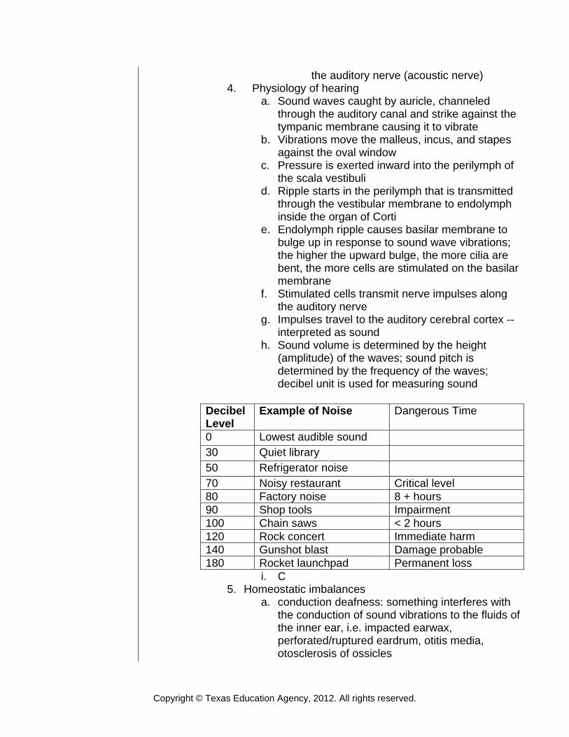
h. Sound volume is determined by the height (amplitude) of the waves; sound pitch is determined by the frequency of the waves; decibel unit is used for measuring sound
i. C
5. Homeostatic imbalances a. conduction deafness: something interferes with
the conduction of sound vibrations to the fluids of the inner ear, i.e. impacted earwax, perforated/ruptured eardrum, otitis media, otosclerosis of ossicles
Decibel Level
Example of Noise Dangerous Time
0 Lowest audible sound 30 Quiet library 50 Refrigerator noise 70 Noisy restaurant Critical level 80 Factory noise 8 + hours 90 Shop tools Impairment 100 Chain saws < 2 hours 120 Rock concert Immediate harm 140 Gunshot blast Damage probable 180 Rocket launchpad Permanent loss
Copyright © Texas Education Agency, 2012. All rights reserved.
b. Sensorineural deafness: damage to neural structures at any point from cochlear hair cells to auditory cortical cells; can be gradual loss of receptor cells, exposure to single loud noise, degeneration of cochlear nerve, cerebral infarcts, tumors; treatment can be cochlear implants
c. Tinnitus: ringing or clicking sound in the ears in the absence of auditory stimuli; can be 1st symptom of cochlear nerve degeneration or inflammation of middle/inner ear or side effect of some medications, i.e. aspirin
d. Meniere’s Syndrome: labyrinth disorder that affects the semicircular canals and cochlea; transient but repeated attacks of severe vertigo
e. Presbycusis: loss of the ability to hear high pitched sounds; becoming common in young people due to noise
F. Sense of Touch, Heat, Cold, Pain 1. Sensory receptors make it possible or the body to
respond to environmental stimuli 2. Receptors respond to a stimulus and convert the
stimulus to a nerve impulse 3. Nerve impulses travel by afferent sensory neurons to the
brain for interpretation 4. Touch: mechanoreceptors/exteroceptors; located on the
body surfaces; respond to touch, stretch, and pressure a. Meissner’s corpuscles: in fingertips, lips, and
hairless body parts for fine touch b. Pacinian corpuscles: in skin, joints, and genitals
for deep pressure and stretch c. Krause’s end bulbs: in eyelids, lips, and genitals
for light touch d. Ruffini’s corpuscles: found in skin for continuous
touch 5. Heat/cold: thermoreceptors 6. Pain: nocioceptors; free nerve endings for pain, tickle,
itch; noci = pain, injury
III. Disorders of the Nervous System A. Shingles: herpes zoster viral infection, causes inflammatory
vesicles along peripheral nerves B. Neuralgia: sudden, sharp, severe, stabbing pain along a nerve
pathway C. Neuritis: inflammation of nerve; causes pain, muscular atrophy,
hypersensitivity, paresthesia D. Bell’s palsy: unilateral facial paralysis, sudden onset, viral
Copyright © Texas Education Agency, 2012. All rights reserved.
inflammation of trigeminal nerve E. Poliomyelitis: viral infection or gray matter of spinal cord;
permanent paralysis or weakness F. Encephalitis: viral inflammation of brain tissue, causes fever,
lethargy, weakness, nuchal rigidity, and opisthotonos, coma, death
G. Meningitis: bacterial/viral inflammation of meninges; causes headache, fever, sore throat, back and neck pain, loss of mental alertness
H. Meningiocele: congenital hernia in which meninges protrude through opening in spinal cord
I. Epilepsy: idiopathic recurring and excessive electrical discharge from neurons causing seizure activity (grand mal, petit mal)
J. Hydrocephalus: increased accumulation of CSF within the ventricles, causes cranium to enlarge unless treated with a shunt to remove excess fluid
K. Parkinson’s disease: tremors, uncontrolled shaking, related to decreased amounts of dopamine
L. Huntington’s chorea: progressive dementia with bizarre involuntary movements; genetic
M. Dysmetria: inability to fix the range of movement in muscle activity
N. Cerebral palsy: congenital brain disorder/damage causing damage to motor neurons, flaccid or spastic paralysis
O. Multiple sclerosis: autoimmunity destruction of oligodendrocytes leading to demyelination with progressive muscular weakness
P. Muscular dystrophy: genetic defect in muscle metabolism; causes progressive atrophy
Q. Myasthenia gravis: disease characterized by muscular weakness; possibly due to decreased amounts of acetylcholine at the muscle effector sites
R. Alzheimer’s disease: dementia producing lesions in the cerebral cortex
S. Anencephalic: infants born without frontal cerebrum; congenital, possibly related to toxins; may be related to Folic Acid deficiency in mother.
Activity
I. Write definitions on backside of Brain Function Flashcards II. See the Cranial Nerve Lesson for additional information and activities
dealing with the cranial nerves III. View video The Brain Body Connection IV. Dissect and identify structures of a sheep brain (teams of two) V. Complete the Nervous System Worksheet
Copyright © Texas Education Agency, 2012. All rights reserved.
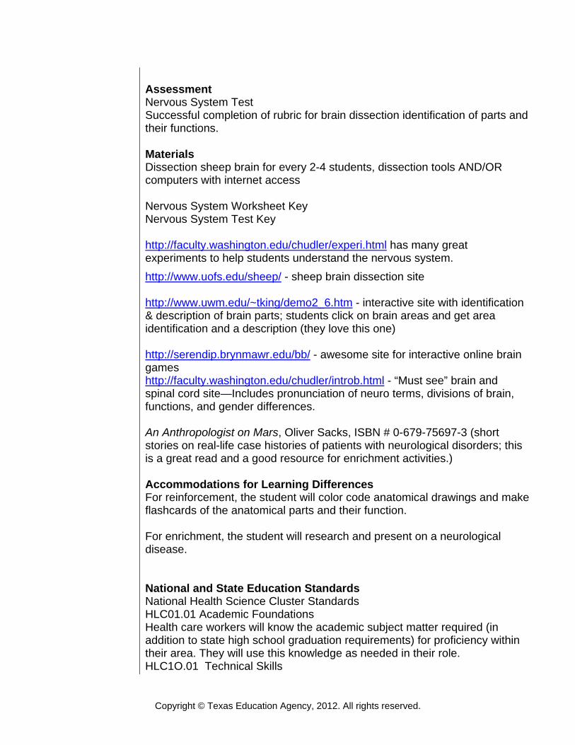
Assessment Nervous System Test Successful completion of rubric for brain dissection identification of parts and their functions. Materials Dissection sheep brain for every 2-4 students, dissection tools AND/OR computers with internet access Nervous System Worksheet Key Nervous System Test Key http://faculty.washington.edu/chudler/experi.html has many great experiments to help students understand the nervous system. http://www.uofs.edu/sheep/ - sheep brain dissection site http://www.uwm.edu/~tking/demo2_6.htm - interactive site with identification & description of brain parts; students click on brain areas and get area identification and a description (they love this one) http://serendip.brynmawr.edu/bb/ - awesome site for interactive online brain games http://faculty.washington.edu/chudler/introb.html - “Must see” brain and spinal cord site—Includes pronunciation of neuro terms, divisions of brain, functions, and gender differences. An Anthropologist on Mars, Oliver Sacks, ISBN # 0-679-75697-3 (short stories on real-life case histories of patients with neurological disorders; this is a great read and a good resource for enrichment activities.) Accommodations for Learning Differences For reinforcement, the student will color code anatomical drawings and make flashcards of the anatomical parts and their function. For enrichment, the student will research and present on a neurological disease. National and State Education Standards National Health Science Cluster Standards HLC01.01 Academic Foundations Health care workers will know the academic subject matter required (in addition to state high school graduation requirements) for proficiency within their area. They will use this knowledge as needed in their role. HLC1O.01 Technical Skills
Copyright © Texas Education Agency, 2012. All rights reserved.
Health Care Workers will apply technical skills required for all career specialties. They will demonstrate skills and knowledge as appropriate. TEKS 130.206 (c)(1)(A) demonstrate safe practices during laboratory and field investigations; 130.206 (c)(1)(B) demonstrate an understanding of the use and conservation of resources and the proper disposal or recycling of materials; 130.206 (c)(2)(E) plan and implement descriptive, comparative, and experimental investigations, including asking questions, formulating testable hypotheses, and selecting equipment and technology; 130.206 (c)(2)(F) collect and organize qualitative and quantitative data and make measurements with accuracy and precision using tools such as calculators, spreadsheet software, data-collecting probes, computers, standard laboratory glassware, microscopes, various prepared slides, stereoscopes, metric rulers, electronic balances, hand lenses, Celsius thermometers, hot plates, lab notebooks or journals, timing devices, Petri dishes, lab incubators, dissection equipment, meter sticks, and models, diagrams, or samples of biological specimens or structures; 130.206 (c)(2)(H) communicate valid conclusions supported by the data through methods such as lab reports, labeled drawings, graphic organizers, journals, summaries, oral reports, and technology-based reports; 130.206 (c)(4)(A) analyze the chemical reactions that provide energy for the body; 130.206 (c)(4)(B) evaluate the means, including the structure and function of the digestive system, by which energy is processed and stored within the body; 130.206 (c)(4)(C) analyze the effects of energy deficiencies in malabsorption disorders such as diabetes, hypothyroidism, and Crohn's disease; 130.206 (c)(4)(D) analyze the effects of energy excess in disorders such as obesity as it relates to cardiovascular and musculoskeletal systems; 130.206 (c)(10)(A) analyze the relationships between the anatomical structures and physiological functions of systems, including the integumentary, nervous, skeletal, musculoskeletal, cardiovascular, respiratory, gastrointestinal, endocrine, and reproductive; 130.206 (c)(10)(B) evaluate the cause and effect of disease, trauma, and congenital defects on the structure and function of cells, tissues, organs, and systems; 130.206 (c)(10)(C) research technological advances and limitations in the treatment of system disorders; and 130.206 (c)(10)(D) examine characteristics of the aging process on body systems. Texas College and Career Readiness Standards English Language Arts II. B. Understand new vocabulary and concepts and use them accurately in
Copyright © Texas Education Agency, 2012. All rights reserved.
reading, writing, and speaking. III. B. Develop effective speaking styles for both group and one-on-one situations. IV. A. Apply listening skills as an individual, and as a member of a group in a variety of settings. IV. B. 2. Listen actively and effectively in one-on-one communication situations. Science 1.A.1. Utilize skepticism, logic, and professional ethics in science. 1.A.2. Use creativity and insight to recognize and describe patterns in natural phenomena. 1.A.3. Formulate appropriate questions to test understanding of a natural phenomenon. 1.A.4. Relay on reproducible observations of empirical evidence when constructing analyzing, and evaluating explanations of natural events and processes. 1.E.2. Use essential vocabulary of the discipline being studied. 3.A.1. Use correct applications of writing practices in scientific communication.
Copyright © Texas Education Agency, 2012. All rights reserved.
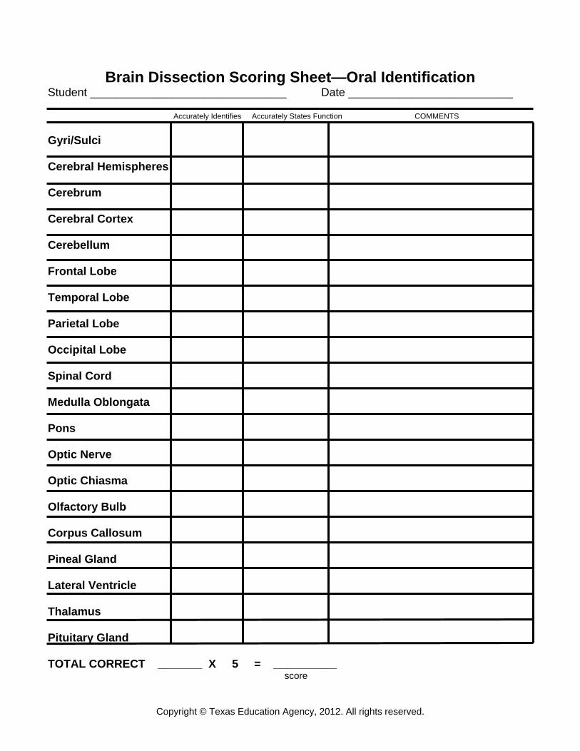
Brain Dissection Scoring Sheet—Oral Identification Student _______________________________ Date __________________________ Accurately Identifies Accurately States Function COMMENTS Gyri/Sulci Cerebral Hemispheres Cerebrum Cerebral Cortex Cerebellum Frontal Lobe Temporal Lobe Parietal Lobe Occipital Lobe Spinal Cord Medulla Oblongata Pons Optic Nerve Optic Chiasma Olfactory Bulb Corpus Callosum Pineal Gland Lateral Ventricle Thalamus Pituitary Gland TOTAL CORRECT _______ X 5 = __________ score
Copyright © Texas Education Agency, 2012. All rights reserved.
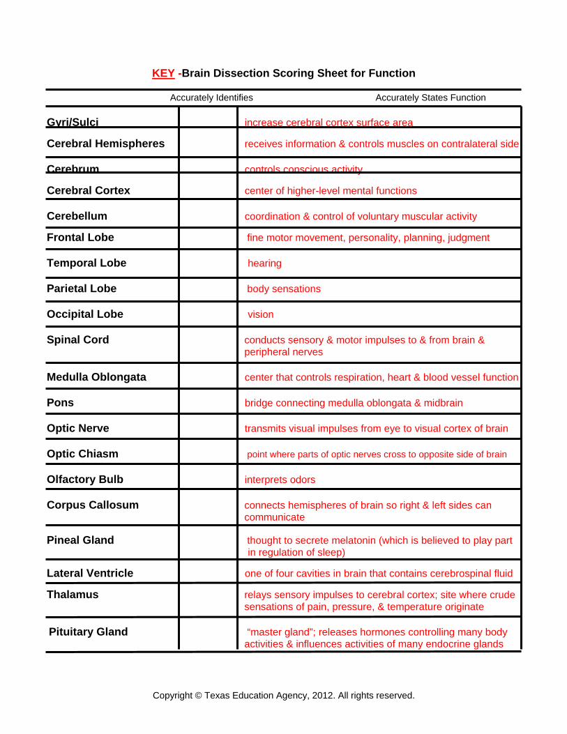
KEY -Brain Dissection Scoring Sheet for Function
Accurately Identifies Accurately States Function Gyri/Sulci increase cerebral cortex surface area Cerebral Hemispheres receives information & controls muscles on contralateral side
Cerebrum controls conscious activity Cerebral Cortex center of higher-level mental functions Cerebellum coordination & control of voluntary muscular activity Frontal Lobe fine motor movement, personality, planning, judgment Temporal Lobe hearing Parietal Lobe body sensations Occipital Lobe vision Spinal Cord conducts sensory & motor impulses to & from brain &
peripheral nerves Medulla Oblongata center that controls respiration, heart & blood vessel function Pons bridge connecting medulla oblongata & midbrain Optic Nerve transmits visual impulses from eye to visual cortex of brain Optic Chiasm point where parts of optic nerves cross to opposite side of brain Olfactory Bulb interprets odors Corpus Callosum connects hemispheres of brain so right & left sides can
communicate Pineal Gland thought to secrete melatonin (which is believed to play part in regulation of sleep) Lateral Ventricle one of four cavities in brain that contains cerebrospinal fluid Thalamus relays sensory impulses to cerebral cortex; site where crude
sensations of pain, pressure, & temperature originate
Pituitary Gland “master gland”; releases hormones controlling many body
activities & influences activities of many endocrine glands

Copyright © Texas Education Agency, 2012. All rights reserved.
Flash Cards (Have students write functions on back of cards. Students can work in groups of five and test in a “circle”. One student selects a card and holds it up for person to his/her left. That person must answer and then hold a card for the next person to his left. Students should go around the circle several times.)
CNS is comprised of: PNS made up of:
Lay term for neurons Description of dendrites
Description of axons Function & location of medulla
Location & function of pons Where is CSF produced? What are ventricles? Name the 2 parts of
diencephalon.
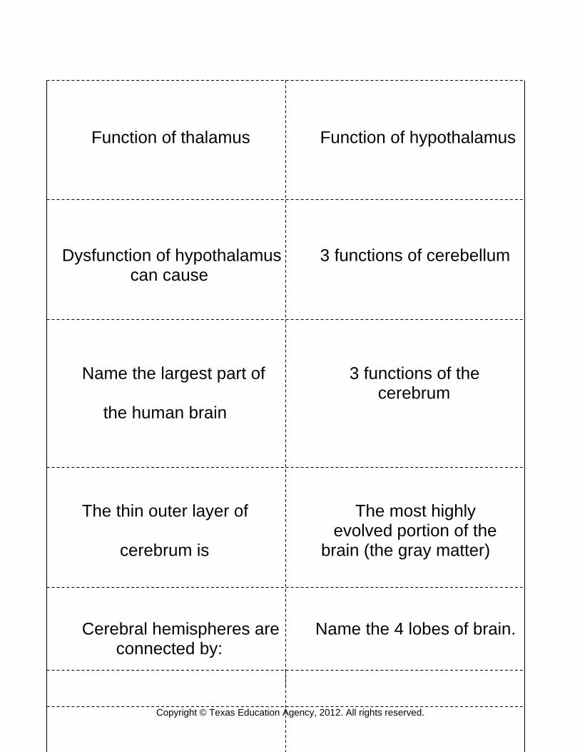
Copyright © Texas Education Agency, 2012. All rights reserved.
Function of thalamus Function of hypothalamus Dysfunction of hypothalamus 3 functions of cerebellum
can cause
Name the largest part of 3 functions of the cerebrum
the human brain
The thin outer layer of The most highly evolved portion of the cerebrum is brain (the gray matter)
Cerebral hemispheres are Name the 4 lobes of brain. connected by:
Copyright © Texas Education Agency, 2012. All rights reserved.
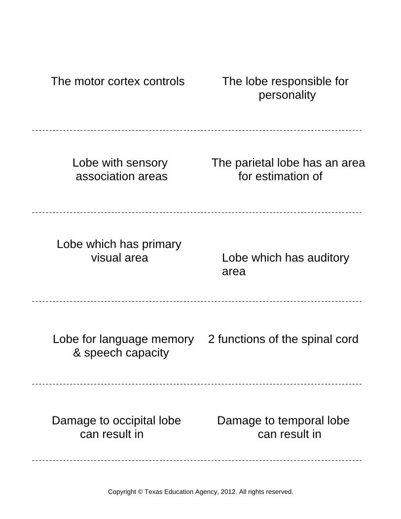
The motor cortex controls The lobe responsible for personality
Lobe with sensory The parietal lobe has an area association areas for estimation of
Lobe which has primary visual area Lobe which has auditory
area
Lobe for language memory 2 functions of the spinal cord
& speech capacity
Damage to occipital lobe Damage to temporal lobe can result in can result in

Copyright © Texas Education Agency, 2012. All rights reserved.
NAME: DATE:
NERVOUS SYSTEM TEST 1. The nervous system exhibits all of these functions EXCEPT: a. monitoring change b. integrating impulses c. storing calcium d. effecting responses 2. Ciliated CNS neuroglia that play an active role in moving the CSF are: a. ependymal cells b. Schwann cells c. oligodendrocytes d. astrocytes 3. The Sheath of Schwann is also called: a. axolemma b. neurilemma c. white matter d. myelin sheath 4. An excitatory neurotransmitter secreted by motor neurons innervating skeletal muscle is: a. cholinesterase (AChE) b. norepinephrine c. acetylcholine (ACh) d. gamma aminobutyric acid 5. Which of the following is NOT a structural feature of a neuron? a. synaptic cleft b. Nissl body c. dendrites d. axon 6. The part of the neuron that conducts impulses away from its cell body is called: a. dendrite b. axon c. neurilemma d. Schwann cell
Copyright © Texas Education Agency, 2012. All rights reserved.
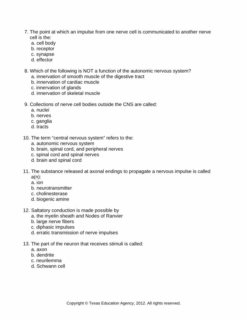
7. The point at which an impulse from one nerve cell is communicated to another nerve cell is the: a. cell body b. receptor c. synapse d. effector 8. Which of the following is NOT a function of the autonomic nervous system? a. innervation of smooth muscle of the digestive tract b. innervation of cardiac muscle c. innervation of glands d. innervation of skeletal muscle 9. Collections of nerve cell bodies outside the CNS are called: a. nuclei b. nerves c. ganglia d. tracts 10. The term “central nervous system” refers to the: a. autonomic nervous system b. brain, spinal cord, and peripheral nerves c. spinal cord and spinal nerves d. brain and spinal cord 11. The substance released at axonal endings to propagate a nervous impulse is called a(n): a. ion b. neurotransmitter c. cholinesterase d. biogenic amine 12. Saltatory conduction is made possible by a. the myelin sheath and Nodes of Ranvier b. large nerve fibers c. diphasic impulses d. erratic transmission of nerve impulses 13. The part of the neuron that receives stimuli is called: a. axon b. dendrite c. neurilemma d. Schwann cell
Copyright © Texas Education Agency, 2012. All rights reserved.
14. Place the following parts of the reflex arc in proper sequence: a. effector-motor neuron-integration center-sensory neuron-receptor b. receptor-motor neuron-integration center-sensory neuron-effector c. receptor-sensory neuron-integration center-motor neuron-effector d. effector-sensory neuron-integration center-motor neuron-receptor 15. The sympathetic and parasympathetic are subdivisions of the: a. central nervous system b. voluntary nervous system c. autonomic nervous system d. somatic nervous system 16. Neuroglia that controls the chemical environment around neurons by buffering potassium and recapturing neurotransmitters are: a. astrocytes b. oligodendrocytes c. microglia d. Schwann cells 17. Schwann cells are functionally similar to: a. ependymal cells b. microglia c. oligodendrocytes d. astrocytes 18. Reflexes are rapid, automatic responses to stimuli. a. True b. False 19. A motor neuron carries stimuli from the central nervous system to the effector. a. True b. False 20. Myelination of the nerve fibers in the CNS is the job of the oligodendrocytes. a. True b. False 21. Neurons do NOT undergo mitosis in the adult. a. True b. False 22. Afferent neurons transmit impulses from the periphery to the CNS. a. True b. False
Copyright © Texas Education Agency, 2012. All rights reserved.
23. Which of the following is NOT a characteristic of neurons? a. extreme longevity b. amitotic c. stimulation d. high metabolic rate 24. Neurons that transmit impulses from sensory receptors in the skin or internal organs toward the CNS are called: a. receptor neurons b. axons c. sensory neurons d. motor neurons 25. Ohm’s Law states: a. an action potential either happens completely or not at all b. depolarization becomes self-generating at threshold c. current varies directly with voltage and inversely with resistance d. neurons communicate through neurotransmitters 26. The degeneration and hardening of the myelin sheath is known as: a. saltatory conduction b. multiple sclerosis c. synapses d. depolarization Matching: Match the correct location to the correct portion of the brain. A. Frontal C. Occipital B. Parietal D. Temporal 27. Auditory area 28. Primary sensory cortex 29. Reasoning 30. Motor speech area 31. Learning 32. Seat of intelligence and abstract reasoning 33. Visual areas 34. Willpower 35. Taste (gustatory) area
Copyright © Texas Education Agency, 2012. All rights reserved.
Matching: A. Dura Mater D. Subarachnoid space B. Pia mater E. Ventricles C. Arachnoid
36. The innermost layer of the meninges; delicate, contains many blood vessels 37. The web-like, spidery middle meningeal layer 38. Normally, the CSF flows freely from the ventricle; then into the _______. Matching: A. Arachnoid villi D. Meningitis B. Central canal E. Hydrocephalus C. Choroid plexus 39. The CSF helps protect the brain and spinal cord against shock. It is filtered into the ventricles through the _____________. 40. The CSF is returned to the blood in the venous sinuses through projections called ___________________. 41. Any obstruction to the normal flow of CSF within the brain may give rise to a condition called ________________. 42. Inflammation of the brain coverings that may be due to pathogenic bacteria. . Multiple Choice 43. The brain stem consists of the: a. cerebrum, pons, midbrain, and medulla b. midbrain, medulla, pons c. pons, medulla, cerebellum, and midbrain d. midbrain only 44. The vital centers for control of heart rate, respiration, and blood pressure are located in: a. the pons b. the medulla c. the midbrain d. the cerebrum
Copyright © Texas Education Agency, 2012. All rights reserved.
45. Which of the following would you not find in normal cerebrospinal fluid? a. glucose b. red blood cells c. potassium d. protein 46. Mr. J. H. was injured in an accident that completely severed his spinal cord at the level of T-12. You would expect to find all of the following EXCEPT: a. paralysis of the lower extremities b. loss of sensation below the level of injury c. slurred speech d. perspiration in the affected area 47. The rough leathery meningeal layer is the: a. dura mater b. subarachnoid c. arachnoid d. pia mater 48. The CSF: a. is secreted by the arachnoid villi b. enters the four ventricles after filling and circulating through the subarachnoid space c. is secreted mostly by the ependymal cells lining the brain ventricles d. is formed mostly by the choroid plexuses 49. The hypothalamus: a. is the thermostat of the body since it regulates temperature b. contains the feeding and hunger centers c. contains neurons sensitive to the solute concentration of the blood d. all of the above are correct 50. The blood-brain barrier is effective against: a. metabolic waste such as urea b. nutrients such as glucose c. alcohol d. anesthetics 51. An electroencephalogram: a. is a record of the total body electrical activity b. is a record of the electrical activity of the heart c. can only detect normal electrical activity d. is a record of the electrical activity of the brain
Copyright © Texas Education Agency, 2012. All rights reserved.
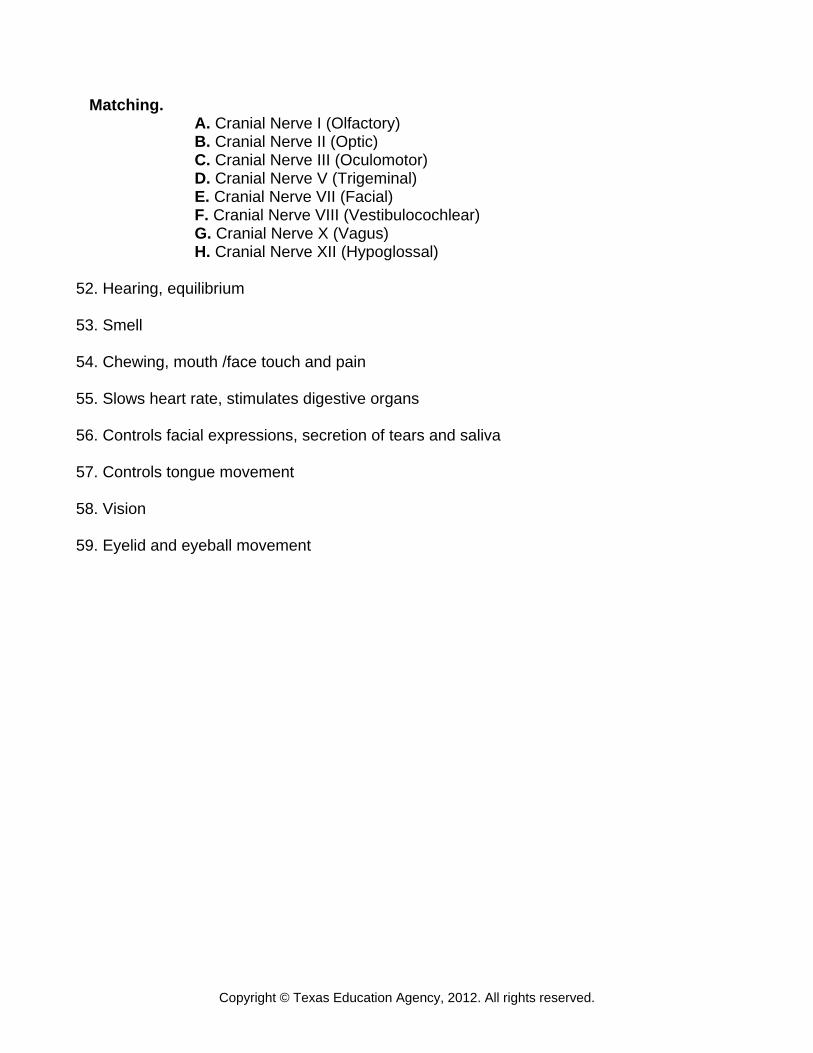
Matching. A. Cranial Nerve I (Olfactory) B. Cranial Nerve II (Optic) C. Cranial Nerve III (Oculomotor) D. Cranial Nerve V (Trigeminal) E. Cranial Nerve VII (Facial) F. Cranial Nerve VIII (Vestibulocochlear) G. Cranial Nerve X (Vagus) H. Cranial Nerve XII (Hypoglossal) 52. Hearing, equilibrium 53. Smell 54. Chewing, mouth /face touch and pain 55. Slows heart rate, stimulates digestive organs 56. Controls facial expressions, secretion of tears and saliva 57. Controls tongue movement 58. Vision 59. Eyelid and eyeball movement
Copyright © Texas Education Agency, 2012. All rights reserved.
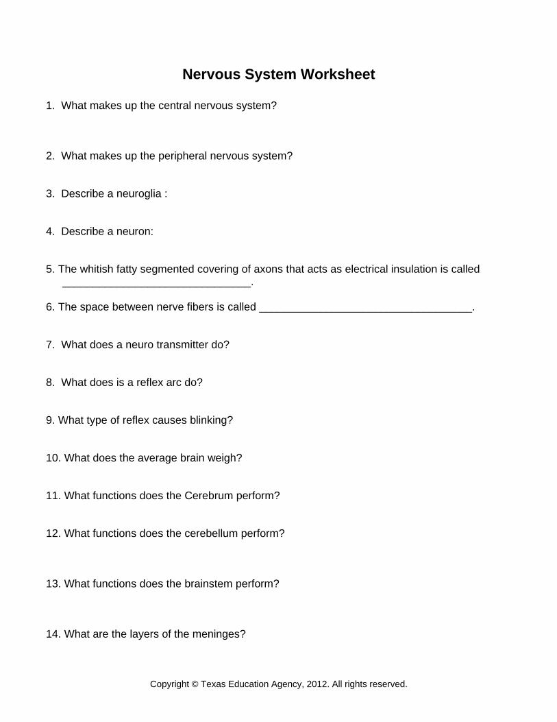
Nervous System Worksheet
1. What makes up the central nervous system? 2. What makes up the peripheral nervous system? 3. Describe a neuroglia : 4. Describe a neuron: 5. The whitish fatty segmented covering of axons that acts as electrical insulation is called
_______________________________. 6. The space between nerve fibers is called ___________________________________. 7. What does a neuro transmitter do? 8. What does is a reflex arc do? 9. What type of reflex causes blinking? 10. What does the average brain weigh? 11. What functions does the Cerebrum perform? 12. What functions does the cerebellum perform? 13. What functions does the brainstem perform? 14. What are the layers of the meninges?
Copyright © Texas Education Agency, 2012. All rights reserved.
15. What does cerebral spinal fluid do? 16. List the 12 cranial nerves: 17. How many spinal nerves are there and where are they located? 18. What viral infection of the nervous system causes unilateral facial paralysis? 19. What idiopathic disorder causes recurring and excessive electrical discharge? 20. What nervous system disorder causes tremors; uncontrolled shaking related to a decrease
of dopamine? 21. What nervous system disorder occurs when lesions form on the cerebral cortex causing
dementia? 22. What nervous system disorder is also known as an autoimmune disease?
Copyright © Texas Education Agency, 2012. All rights reserved.
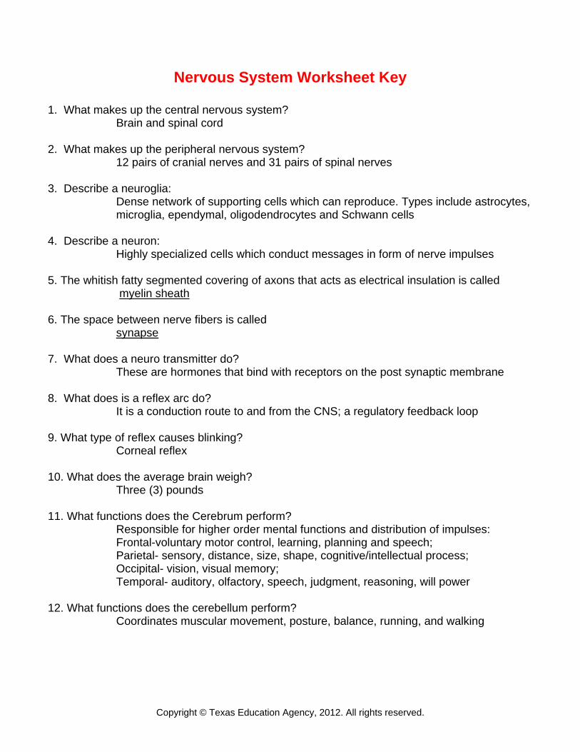
Nervous System Worksheet Key
1. What makes up the central nervous system? Brain and spinal cord
2. What makes up the peripheral nervous system?
12 pairs of cranial nerves and 31 pairs of spinal nerves 3. Describe a neuroglia:
Dense network of supporting cells which can reproduce. Types include astrocytes, microglia, ependymal, oligodendrocytes and Schwann cells
4. Describe a neuron:
Highly specialized cells which conduct messages in form of nerve impulses 5. The whitish fatty segmented covering of axons that acts as electrical insulation is called
myelin sheath
6. The space between nerve fibers is called synapse 7. What does a neuro transmitter do?
These are hormones that bind with receptors on the post synaptic membrane 8. What does is a reflex arc do?
It is a conduction route to and from the CNS; a regulatory feedback loop 9. What type of reflex causes blinking?
Corneal reflex 10. What does the average brain weigh?
Three (3) pounds 11. What functions does the Cerebrum perform? Responsible for higher order mental functions and distribution of impulses:
Frontal-voluntary motor control, learning, planning and speech; Parietal- sensory, distance, size, shape, cognitive/intellectual process; Occipital- vision, visual memory; Temporal- auditory, olfactory, speech, judgment, reasoning, will power
12. What functions does the cerebellum perform?
Coordinates muscular movement, posture, balance, running, and walking
Copyright © Texas Education Agency, 2012. All rights reserved.
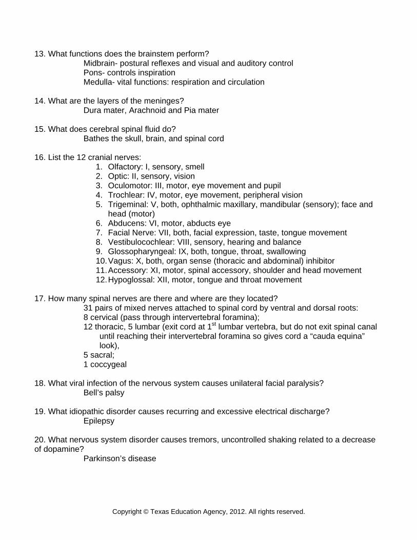
13. What functions does the brainstem perform? Midbrain- postural reflexes and visual and auditory control Pons- controls inspiration Medulla- vital functions: respiration and circulation
14. What are the layers of the meninges?
Dura mater, Arachnoid and Pia mater 15. What does cerebral spinal fluid do?
Bathes the skull, brain, and spinal cord 16. List the 12 cranial nerves:
1. Olfactory: I, sensory, smell 2. Optic: II, sensory, vision 3. Oculomotor: III, motor, eye movement and pupil 4. Trochlear: IV, motor, eye movement, peripheral vision 5. Trigeminal: V, both, ophthalmic maxillary, mandibular (sensory); face and
head (motor) 6. Abducens: VI, motor, abducts eye 7. Facial Nerve: VII, both, facial expression, taste, tongue movement 8. Vestibulocochlear: VIII, sensory, hearing and balance 9. Glossopharyngeal: IX, both, tongue, throat, swallowing 10. Vagus: X, both, organ sense (thoracic and abdominal) inhibitor 11. Accessory: XI, motor, spinal accessory, shoulder and head movement 12. Hypoglossal: XII, motor, tongue and throat movement
17. How many spinal nerves are there and where are they located?
31 pairs of mixed nerves attached to spinal cord by ventral and dorsal roots: 8 cervical (pass through intervertebral foramina); 12 thoracic, 5 lumbar (exit cord at 1st lumbar vertebra, but do not exit spinal canal until reaching their intervertebral foramina so gives cord a “cauda equina” look), 5 sacral; 1 coccygeal
18. What viral infection of the nervous system causes unilateral facial paralysis?
Bell’s palsy 19. What idiopathic disorder causes recurring and excessive electrical discharge?
Epilepsy 20. What nervous system disorder causes tremors, uncontrolled shaking related to a decrease of dopamine?
Parkinson’s disease
Copyright © Texas Education Agency, 2012. All rights reserved.
21. What nervous system disorder occurs when lesions form on the cerebral cortex causing dementia?
Alzheimer’s 22. What nervous system disorder is also known as an autoimmune disease?
Multiple sclerosis
Copyright © Texas Education Agency, 2012. All rights reserved.
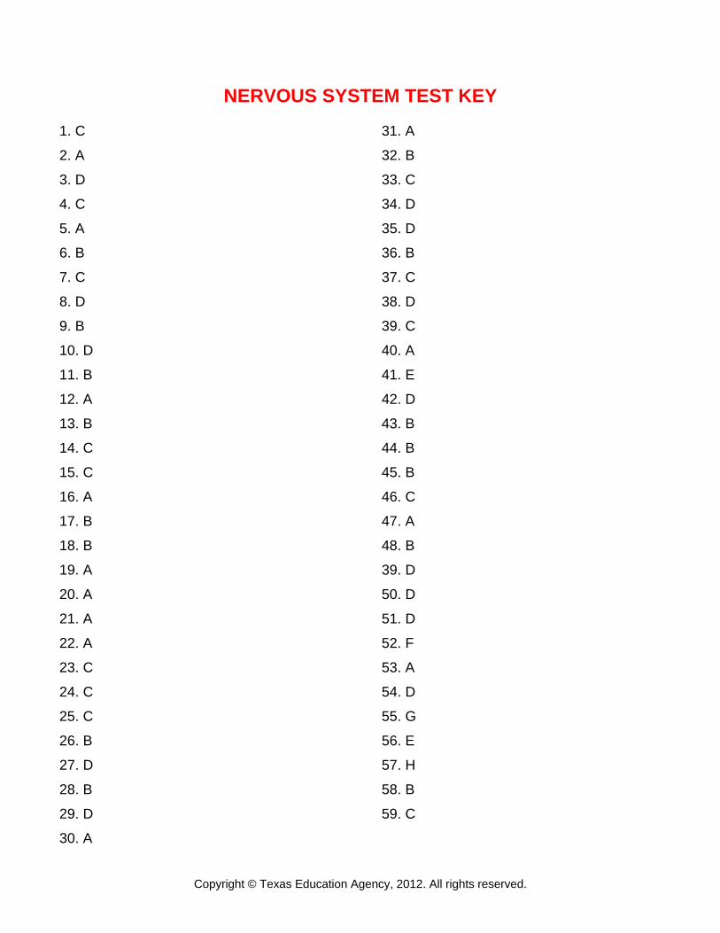
NERVOUS SYSTEM TEST KEY 1. C
2. A
3. D
4. C
5. A
6. B
7. C
8. D
9. B
10. D
11. B
12. A
13. B
14. C
15. C
16. A
17. B
18. B
19. A
20. A
21. A
22. A
23. C
24. C
25. C
26. B
27. D
28. B
29. D
30. A
31. A
32. B
33. C
34. D
35. D
36. B
37. C
38. D
39. C
40. A
41. E
42. D
43. B
44. B
45. B
46. C
47. A
48. B
39. D
50. D
51. D
52. F
53. A
54. D
55. G
56. E
57. H
58. B
59. C