Embed Size (px)
Citation preview

Nervous systemReflexes and Senses
Physiology Lab-4
Wrood Slaim, MScDepartment of Pharmacology and Toxicology
University of Al-Mustansyria2017-2018
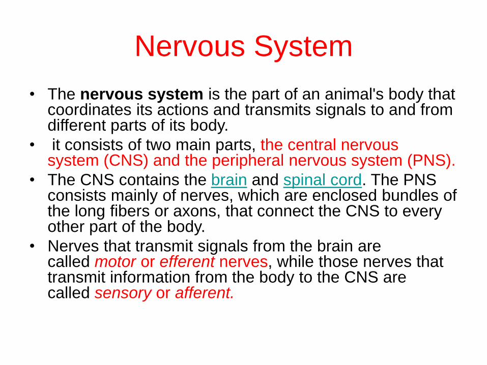
• The nervous system is the part of an animal's body that coordinates its actions and transmits signals to and from different parts of its body.
• it consists of two main parts, the central nervous system (CNS) and the peripheral nervous system (PNS).
• The CNS contains the brain and spinal cord. The PNS consists mainly of nerves, which are enclosed bundles of the long fibers or axons, that connect the CNS to every other part of the body.
• Nerves that transmit signals from the brain are called motor or efferent nerves, while those nerves that transmit information from the body to the CNS are called sensory or afferent.
Nervous System
Reflexes
• We are always under the influence of external stimuli from our surrounding external environment , and we react with these stimuli to perform a response .
• Our sensory receptors detect changes in the surrounding environment and transmit them to the central nervous system , where they would be interpreted and processed to form a motor order, that will be transported to an effector organ to perform a response.
• The mentioned process is called neural reflex. So neural reflex is a rapid involuntary , and repeatable response to a stimulus.
• Types of reflexes : Reflexes could be : spinal vs. cranial , congenital vs. acquired , mono-synaptic vs. poly-synaptic, somatic or visceral( autonomic).
• .

There are five parts to a reflex arc:1. The receptor detects a stimulus.
2. The sensory (afferent) neuron sends an electrical signal to the CNS.
3. The integration center consists of one or more synapses in the CNS, and processes the information.
4. The motor (efferent) neuron sends an electrical signal from the CNS to the effector.
5. The effector, which may be muscle tissue or a gland, responds appropriately.
Reflex arc: a pathway in which signals travel over many
synapses on their way back to the muscle.
Figure 1: the flexor reflex

Reflex Arc
There are two types of reflex arc:
1. autonomic reflex arc (affecting cardiac
,smooth muscles and glands).
2. somatic reflex arc (affecting skeletal
muscles).

Autonomic Reflexes:
• Autonomic reflexes (or visceral) reflexes are mediated through the autonomic nervous system, and we are not usually aware of them.
• These reflexes activate smooth muscles, cardiac muscle, and the glands of the body, and they regulate body functions such as digestion, elimination, blood pressure, salivation, and sweating
• In general these reflexes contain the same basic components as somatic reflexes but a key difference is that autonomic reflexes have the ability to both stimulate or inhibit the smooth muscle/gland.
• Examples of Autonomic Reflexes:
1. Pupillary-pupil constricts on both sides when shine a light into the eye
2. Accomodation-focus on distant object then near object-pupil constricts on both sides.
Somatic Reflexes
• Somatic reflexes include all those reflexes that involve stimulation of skeletal muscles by the somatic division
of the nervous system.
• Some require only spinal cord activity; others require brain involvement as well.
• there are many different types of somatic reflexes including withdrawal reflexes and stretch reflexes and tendon reflexes.
• Stretch reflexes: are important postural reflexes, normally acting to maintain posture, balance, and locomotion
• Examples of Somatic reflexes :
Corneal- blink reflex (respond to stimulus)
Pateller: knee jerk (tap patellar ligament-stretches quads)

Purpose of the Reflexes tests:
• Reflex testing is an important diagnostic
tool for assessing the general health of the
nervous system. Distorted, exaggerated
or absent reflexes may indicate pathology.
If the spinal cord is damaged, reflex tests
can help pinpoint the level of damage
Activity 1: Somatic Reflex :The Patellar
Reflex
• The patellar (or knee-jerk) reflex is called a
stretch reflex because it is initiated by
tapping a tendon, which stretches the
muscle
• Stretch reflexes generally act to maintain
posture, balance and locomotion.
• it asses the L2- L4 segments of the spinal
cord.
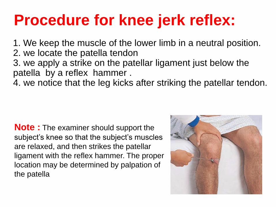
Procedure for knee jerk reflex:
1. We keep the muscle of the lower limb in a neutral position.2. we locate the patella tendon3. we apply a strike on the patellar ligament just below the patella by a reflex hammer .4. we notice that the leg kicks after striking the patellar tendon.
Note : The examiner should support the
subject’s knee so that the subject’s muscles
are relaxed, and then strikes the patellar
ligament with the reflex hammer. The proper
location may be determined by palpation of
the patella

Figure: The patellar (knee-jerk) reflex—a specific example of a stretch reflex.
Purpose of testing:
• After the tap of a hammer, the leg is normally extended once and comes to rest.
• The absence or decrease of this reflex is problematic, and known as Westphal's sign.
• This reflex may be diminished or absent in lower motor neuron lesions and during sleep.
• On the other hand, multiple oscillation of the leg (pendular reflex) following the tap may be a sign of a cerebellarl disease. Exaggerated (brisk) deep tendon reflexes such as this can be found in upper motor neuron lesions, hyperthyroidism, anxiety or nervousness.
• The test itself assesses the nervous tissue between and including the L2 and L4 segments of the spinal cord.
Activity 2: Crossed-Extensor Reflex
• The crossed-extensor reflex is more complex than the stretch
reflex. It consists of a flexor, or withdrawal, reflex followed by
extension of the opposite limb.
• This reflex is quite obvious when, for example, a stranger suddenly
and strongly grips one’s arm. The immediate response is to withdraw
the grabbed arm and push the intruder away with the other arm.
• The reflex is more difficult to demonstrate in a laboratory because it is
anticipated, and under these conditions the extensor part of the reflex
may be inhibited.

Activity 3: Autonomic Reflexes
:Pupillary Reflexes test
• The pupillary light reflex (PLR) is a reflex that controls the diameter of the pupil, in response to the intensity of light that falls on the retinal ganglion cells of the eye, thereby assisting in adaptation to various levels of lightness/darkness.
• A greater intensity of light causes the pupil to constrict (miosis)(allowing less light in), whereas a lower intensity of light causes the pupil to dilate (mydriasis, expansion) (allowing more light in). Thus, the pupillary light reflex regulates the intensity of light entering the eye
• We will test the pupillary light reflex and the consensual reflex. In both, the retina of the eye is the receptor, the optic nerve holds the afferent fibers, the oculomotor nerve contains the efferent fibers, and the smooth muscle of the iris is the effector organ.

Clinical signification
• Under normal conditions, the pupils of both eyes
respond identically to a light stimulus, regardless
of which eye is being stimulated. Light entering
one eye produces a constriction of the pupil of
that eye, the direct response, as well as a
constriction of the pupil of the un stimulated eye,
the consensual response.
• For example, if light is shone into right eye only,
right pupil constriction is a direct pupillary light
reflex, and simultaneous left pupil constriction is a
consensual pupillary light reflex. Therefore, light
shone into one eye causes ipsilateral direct
pupillary light reflex and contralateral consensual
pupillary light reflex
• Comparing these two responses in both eyes is
helpful in locating a lesion
Purpose of testing:
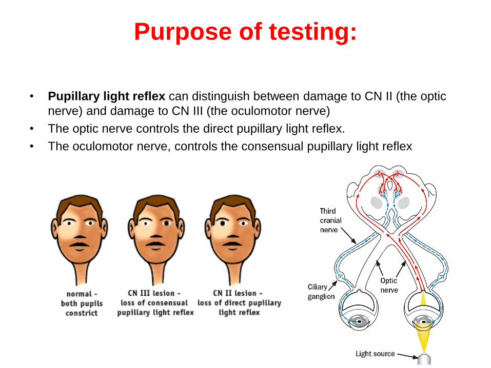
• Pupillary light reflex can distinguish between damage to CN II (the optic
nerve) and damage to CN III (the oculomotor nerve)
• The optic nerve controls the direct pupillary light reflex.
• The oculomotor nerve, controls the consensual pupillary light reflex
Sensory Receptor Physiology
• Sensory receptors act as transducers, changing
environmental stimuli into nerve impulses that are relayed to
the CNS.
• Sensation (awareness of the stimulus) and perception
(interpretation of the meaning of the stimulus) occur in the
brain.
• Four qualities of cutaneous sensations have traditionally
been recognized: tactile (touch), heat, cold, and pain.

Activity 4:Touch receptor :Two-point
Discrimination Test
• The minimum distance in millimetres at which the two points of the
calipers are felt separately is noted.
• In general, areas that have the greatest density of tactile receptors have
a heightened ability to “feel.” These areas correspond to areas that
receive the greatest motor innervation; thus they are also typically areas
of fine motor control.
• On the basis of this information, which areas of the body do you predict
will have the greatest density of touch receptors?
• The distance between the points, at which the subject loses the ability
to feel two points is the diameter of the receptive field. The two point
discrimination threshold is less than 5 mm at the finger tips and is about
40 mm at the thigh

Procedure
1. Using calipers, test the ability of the subject to differentiate two distinct sensations when the skin is touched simultaneously at two points
2. Start with the points right together, then gradually increase the distance apart. Record the distance at which the subject first reports feeling two distinct points of contact with the skin (the two-point threshold).
3. Find the shortest distance of caliper spikes at which the subject still senses two touches.
4. Repeat this measurement and Test the areas of the body as listed in the chart.
Q/ Of the tested areas of the body, which ones seem to have the greatest density of receptors (smallest two-point threshold)?

Adaptation of Sensory Receptors
• The number of impulses transmitted by sensory receptors often
changes both with the intensity of the stimulus and with the length
of time the stimulus is applied.
• In many cases, when a stimulus is applied for a prolonged period,
the rate of receptor discharge slows and conscious awareness of the
stimulus declines or is lost until some type of stimulus change
occurs.This phenomenon is referred to as adaptation.
• Some receptors adapt rapidly, such as certain types of touch
receptors, and others, such a pain receptors, may not adapt
at all.
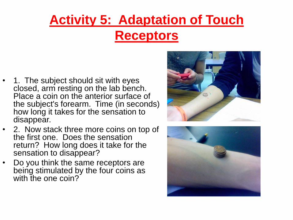
Activity 5: Adaptation of Touch
Receptors
• 1. The subject should sit with eyes closed, arm resting on the lab bench. Place a coin on the anterior surface of the subject's forearm. Time (in seconds) how long it takes for the sensation to disappear.
• 2. Now stack three more coins on top of the first one. Does the sensation return? How long does it take for the sensation to disappear?
• Do you think the same receptors are being stimulated by the four coins as with the one coin?