Embed Size (px)
Citation preview
NEOPLASIA-------
(MALIGNANT TUMORS)
PRACTICAL 6
Foundation Block
Pathology Dept, KSU

1- SQUAMOUS CELL CARCINOMA OF
THE SKIN
Foundation Block
Pathology Dept, KSU
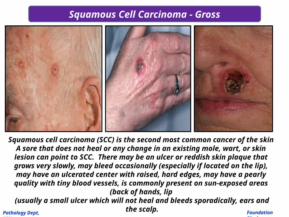
Squamous Cell Carcinoma - Gross
Squamous cell carcinoma (SCC) is the second most common cancer of the skin A sore that does not heal or any change in an existing mole, wart, or skin lesion can point to SCC. There may be an ulcer or reddish skin plaque that grows very slowly, may
bleed occasionally (especially if located on the lip), may have an ulcerated center with raised, hard edges, may have a pearly quality with tiny blood vessels, is commonly present on sun-
exposed areas (back of hands, lip (usually a small ulcer which will not heal and bleeds
sporadically, ears and the scalp.Foundation Block
Pathology Dept, KSU

Squamous Cell Carcinoma - Histopathology
The normal squamous epithelium at the right merges into the squamous
cell carcinoma at the left, which is infiltrating downward. The neoplastic squamous cells are still similar to the normal
squamous cells, but are less orderly
Sq Cell Carcinoma
Foundation Block
Pathology Dept, KSU

At high magnification, this squamous cell carcinoma demonstrates enough differentiation to tell that the cells are
of squamous origin. The cells are pink and polygonal in shape with intercellular bridges.
The neoplastic cells show pleomorphism, with hyperchromatic nuclei.
A mitotic figure is present near the center
Squamous Cell Carcinoma - HPF
Foundation Block
Pathology Dept, KSU

The dermis is infiltrated by masses of well differentiated neoplastic squamous cells separated by fibrous tissue stroma
with chronic inflammatory cells. Tumour cells show pleomorphism, hyperchromatism and many mitotic figures .
Pinkish laminated keratin pearls (epithelial cell nests) are
present in the center of some cell masses
Squamous Cell Carcinoma - HPF
Foundation Block
Pathology Dept, KSU

Here is a moderately differentiated squamous cell carcinoma in which some, but not all, of the neoplastic
cells in nests have pink cytoplasmic keratin
Squamous Cell Carcinoma - Histopathology
Foundation Block
Pathology Dept, KSU

A mitotic figure is seen here in the center, surrounded by cells of a poorly differentiated squamous cell
carcinoma, with pleomorphic cells that have minimal pink keratinization in their cytoplasm. In general, mitoses are more likely to be seen in malignant
neoplasms
Squamous Cell Carcinoma - HPF
Foundation Block
Pathology Dept, KSU

2- ADENOCARCINOMA OF THE LARGE
INTESTINE
Foundation Block
Pathology Dept, KSU

Colon showing polypoid mass with haemorrhagic area on its surface. Because of this , occult blood in the faeces
can be detected on lab examination of the patient which is at used to confirm and support our histopathological
diagniosis.
Adenocarcinoma of the Colon
Foundation Block
Pathology Dept, KSU

This cancer is more exophytic in its growth pattern. Thus, one of the complications of a carcinoma is
obstruction (usually partial).
Adenocarcinoma of the Colon
Foundation Block
Pathology Dept, KSU

Adenocarcinoma of the Colon, arising from villous
adenoma
Tumor consists of crowded irregular malignant acini separated by
thin fibrovascular stroma. Colonic adenocarcinoma arising from villous adenoma .
Foundation Block
Pathology Dept, KSU

The acini are lined by one or several layers of neoplastic cells with papillary projection showing pleomorphism,
hyperchromatism and few mitoses.Sometimes, the patient with 100’s polyps in their lumen
(Familial polyposis coli) with APS gene mutation are more prone to develop Adenocarcinoma of the colon.
Adenocarcinoma of the Colon - LPF

Here is an adenocarcinoma in which the glands are much larger
and filled with necrotic debris.
Adenocarcinoma of the Colon - LPF
Foundation Block
Pathology Dept, KSU

At high magnification, the neoplastic glands of adenocarcinoma have crowded nuclei with
hyperchromatism and pleomorphism. No normal goblet cells are seen
Adenocarcinoma of the Colon - HPF
Foundation Block
Pathology Dept, KSU

3- LEIOMYOSARCOMA
Foundation Block
Pathology Dept, KSU

Cut surface of this leiomyosarcoma showing ill defined pale and soft large fleshy mass with
hemorrhage and necrosis.
Leiomyosarcoma
Foundation Block
Pathology Dept, KSU

It is a large mass obstructing the small bowel lumen.Pale firm and partly haemorrhagic cut surface with focal necrosis.
.
Leiomyosarcoma of Small Intestine
Foundation Block
Pathology Dept, KSU

Marked atypia and cellularity with multiple mitoses present. Classic
features of leiomyosarcoma including cigar shaped nuclei and arrangement of cells in fascicles are seen.
Leiomyosarcoma – HPF Microscopy
Foundation Block
Pathology Dept, KSU

Leiomyosarcoma of the Uterus - HPF
Foundation Block
Pathology Dept, KSU
Multinucleation / large bizarre giant cells.
Mitotic figures
cigar shaped nuclei and arrangement of cells in fascicles.

Immunohistochemistry is performed for confirmation of the diagnosis of this sarcoma.
The neoplastic cells are positive for actin and desmin stain confirming cell of origin as Smooth muscle cell or fiber .
Hence we can make the final histopathological diagnosis of leiomyosarcoma.

THE END