Embed Size (px)
Citation preview

NeoplasiaLecture 1
Abdulmalik Alsheikh, M.D, FRCPCDr. Maha Arafah, MBBS, KSFP
Foundation block 2014Pathology
Definition and Nomenculature

Neoplasia
• Cancer is one of the second leading causes of death worldwide.
• Emotional and physical suffering by the patient.
• Different mortality rate …..– Some are curable such as Hodgkin lymphoma – Others are fatal such as pancreatic
adenocarcinoma

NeoplasiaUpon completion of these lectures, the student should:• Define a neoplasm. Contrast neoplastic growth with hyperplasia, metaplasia, and dysplasia.• Know the basic principles of the nomenclature of benign and malignant processes.• Define and use in the proper context:
– Adenoma.– Papilloma.– Polyp.– Cystadenoma.– Carcinoma.– Adenocarcinoma.
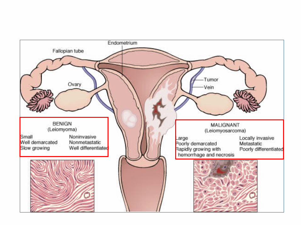
• Compare and contrast benign and malignant tumors with respect to:– demarcation from surrounding tissue (capsule, local invasiveness)– rate of growth– degree of differentiation (Explain the meaning of differentiation).– distant spread (metastases).
• Describe the morphologic changes associated with poorly differentiated tumors; define and understand the usage of the terms anaplasia, pleomorphism, nuclear atypia, abnormal mitoses and tumor giant cells.
- Sarcoma.- Teratoma.
-Blastoma. -Hamartoma.

Neoplasia
• Neoplasia = new growth• Neoplasm = tumor• Tumor = swelling• The study of tumors = Oncology
– Oncos = tumor + ology = study of

Neoplasia
• Definition:– is an abnormal mass of tissue, – the growth of which is uncoordinated with that of normal
tissues, – and that persists in the same excessive manner after the
cessation of the stimulus which evoked the change“– With the loss of responsiveness to normal growth controls
– Different from hyperplasia, metaplasia and dysplasia.

Neoplasia Classification
Benign
Will remain localizedCannot spread to distant
sitesGenerally can be locally
excisedPatient generally survives
Malignant
Can invade and destroy adjacent structure
Can spread to distant sitesCause death (if not treated)
Benign – malignant

Neoplasia
• All tumors have two basic components:– Parechyma: made up of neoplastic cells– Stroma: made up of non-neoplastic, host-
derived connective tissue and blood vessels
The parenchyma:Determines the biological behavior of the tumorFrom which the tumor derives its name
The stroma:Carries the blood supplyProvides support for the growth of the parenchyma


Nomenclature of benign and malignant neoplasm
Benign tumors

Neoplasia
• Nomenclature– Benign tumors:
• prefix + suffix• Type of cell + (-oma)
Neoplasia– Benign tumor arising in fibrous tissue: Fibro + oma = Fibroma– Benign tumor arising in fatty tissue: Lipo + oma = lipoma– Benign tumor arising in cartilage
chondro + oma = chondroma– Benign tumor arising in smooth muscle
Leiomyo + oma = leiomyoma– Benign tumor arising in skeletal muscle
Rhabdomyo + oma = rhabdomyoma

Neoplasia
• epithelial benign tumors are classified on the basis of :– The cell of origin – Microscopic pattern– Macroscopic pattern

Neoplasia
– Adenoma : benign epithelial neoplasms producing gland pattern….OR … derived from glands but not necessarily exhibiting gland pattern
– Papilloma : benign epithelial neoplasms growing on any surface that produce microscopic or macroscopic finger-like pattern

Adenoma
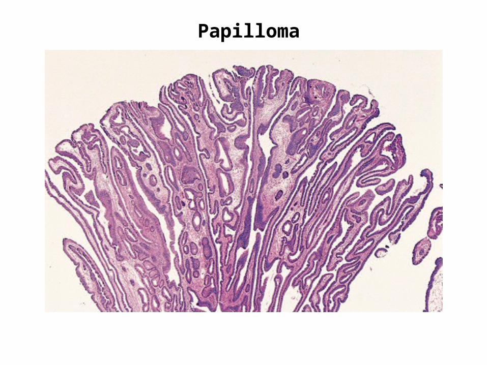
Papilloma
Neoplasia
• Polyp : a mass that projects above a mucosal surface to form a macroscopically visible structure.
e.g. - colonic polyp - nasal polyp

Polyp

Neoplasia
• Examples of benign epithelial neoplasms : – Respiratory airways: Bronchial adenoma– Renal epithelium: Renal tubular adenoma– Liver cell : Liver cell adenoma– Squamous epithelium: squamous papilloma

Cystadenoma

Malignant tumor

Neoplasia
• Malignant tumors:– Malignant tumor arising in mesenchymal tissue:
SARCOMA• From fibrous tissue: Fibrosarcoma• From bone : Osteosarcoma• From cartilage : chondrosarcoma

Osteosarcoma

Neoplasia
• Malignant tumors arising from epithelial origin: CARCINOMA– Squamous cell carcinoma– Renal cell adenocarcinoma– Cholangiocarcinoma

Carcinomas arising from any epithelium of the body that exhibit squamous differentiation are termed squamous cell carcinoma.
Normal squamous epithlium
Squamous cell carcinoma

Nomenclatureother descriptive terms may be added such as:
Papillary Cystadenocarcinoma of the Ovary

Nomenclatureother descriptive terms may be added such as:
Papillary Cystadenocarcinoma of the Ovary

NeoplasiaExceptions
• Melanoma ( skin )• Mesothelioma (mesothelium )• Seminoma ( testis )• Lymphoma ( lymphoid tissue )
See table 5-1 page 164 ( Robbin’s )

Nomenclature of benign and malignant neoplasm
• Based on the biological behavior : – Benign and malignant
• Based on the cell of origin : – One neoplastic cell type : lipoma, adenocarcinoma– More than one neoplastic cell type : fibroadenoma– More than one neoplastic cell type derived from more
than one germ-cell layer: teratoma– Derived from embryonic tissue: blastoma (could be
benign e.g. osteoblastoma, or malignant e.g. neuroblastoma)
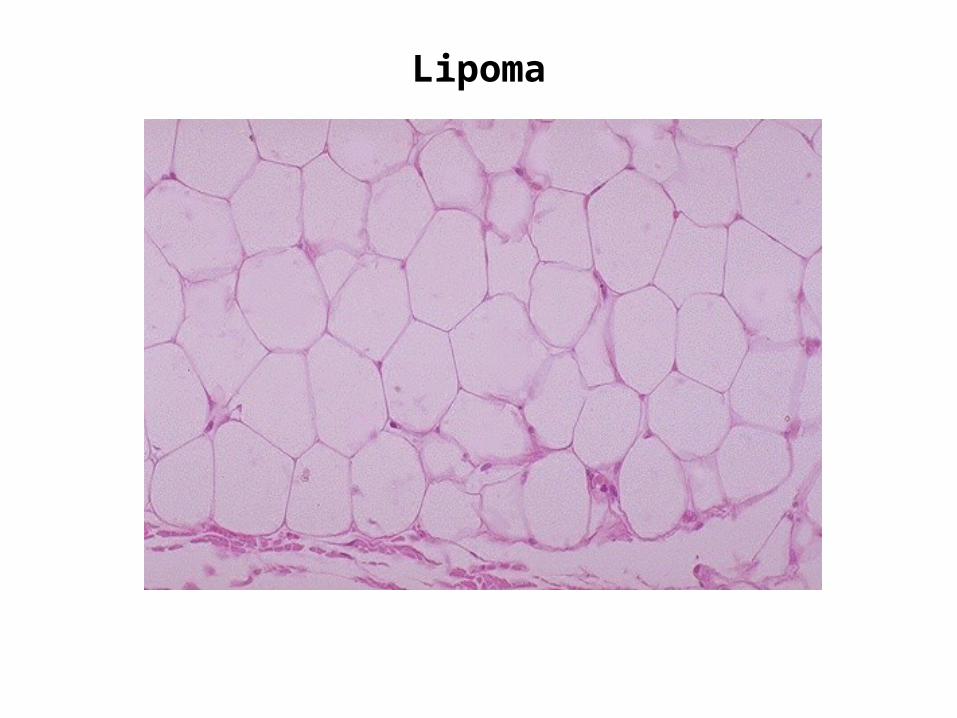
Lipoma
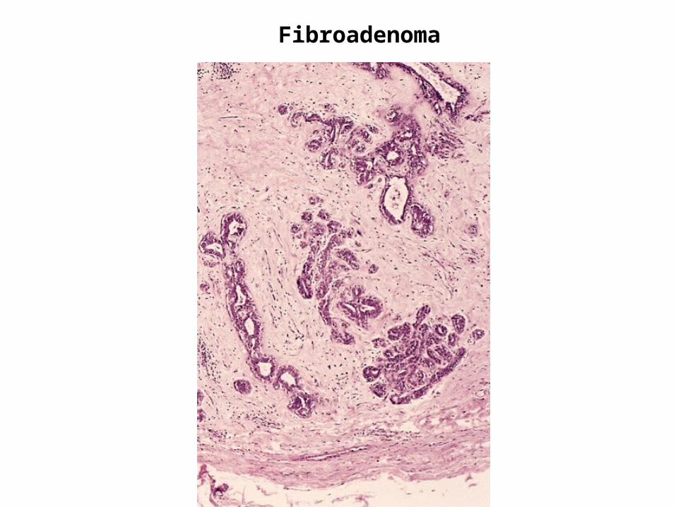
Fibroadenoma
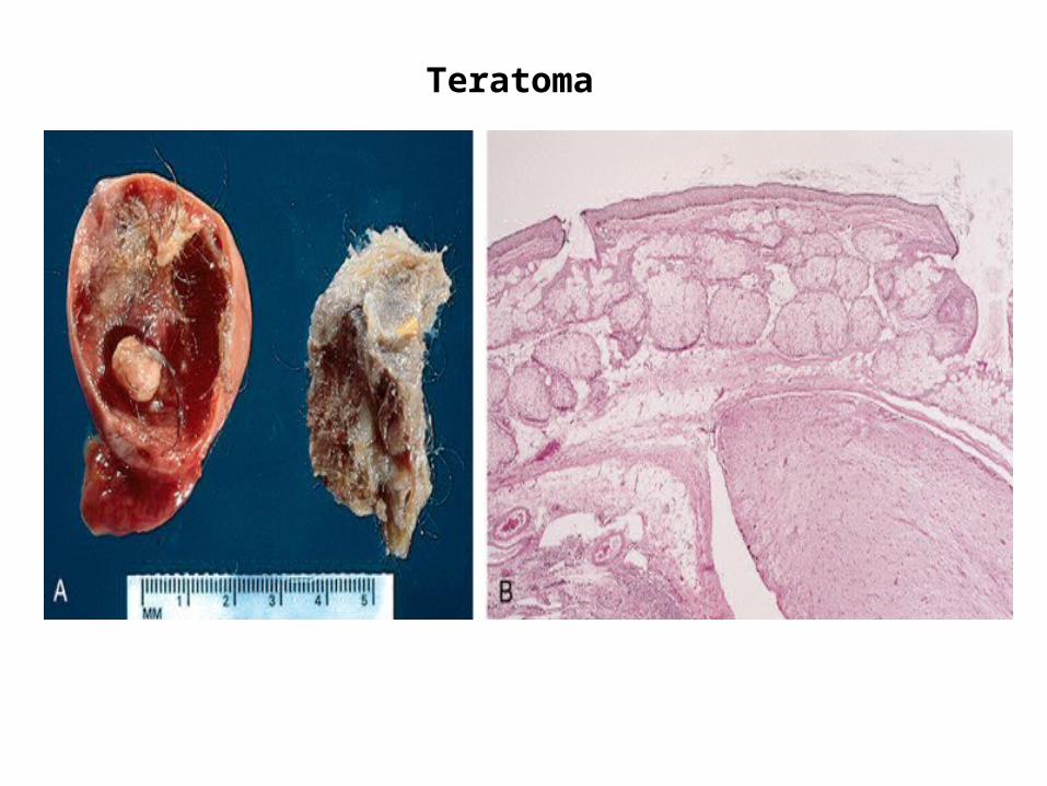
Teratoma

Neoplasia
• Teratoma:– Teratoma contains recognizable mature or
immature cells or tissues representative of more than one germ-cell layer and some times all three.
– Teratomas originate from totipotential cells such as those normally present in the ovary and testis.
Neoplasia
• Such cells have the capacity to differentiate into any of the cell types found in the adult body. So they may give rise to neoplasms that mimic bone, epithelium, muscle, fat, nerve and other tissues.
• Most common sites are: ovary & testis

Neoplasia
• If all the components parts are well differentiated, it is a benign (mature) teratoma.
• If less well differentiated, it is an immature (malignant) teratoma.

Neoplasia nomenclature- historic eponyms – “first described by…”
Malignant lymphoma (HL) of B Lymphocyte cell origin
Hodgkin’s disease
NHL – B Lymphocyte cell in children (jaw and GIT)
Burkitt tumor
Bone tumor: Primitive neuroectodermal tumor (PNET)
Ewing tumor
Kidney tumor - clear cell adenocarcinoma Grawitz tumor
Malignant tumor derived from vascular epithelium (AIDS)
Kaposi sarcoma
Ovarian tumor derived from Brenner cells Brenner tumor
Skin tumor derived from Merkel cell Merkel tumor
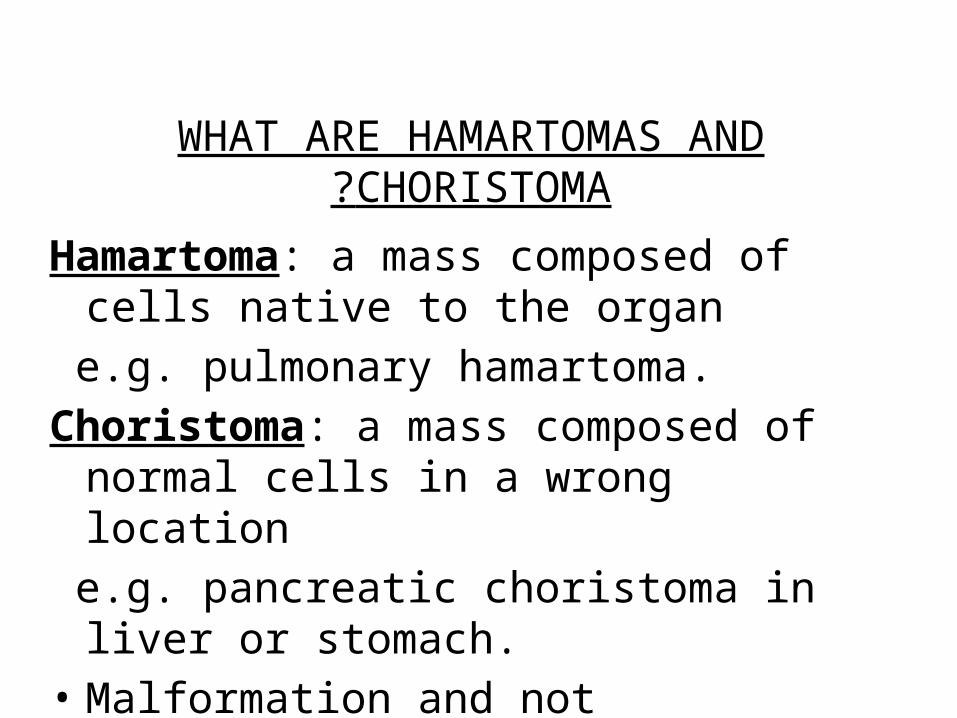
WHAT ARE HAMARTOMAS AND CHORISTOMA?
Hamartoma: a mass composed of cells native to the organ
e.g. pulmonary hamartoma.Choristoma: a mass composed of normal cells
in a wrong location e.g. pancreatic choristoma in liver or
stomach.• Malformation and not neoplasm.

Pulmonary Hamartoma

Pancreatic choristoma in gall bladder

Neoplasia
Hamartoma and Choristoma
• They are distinguished from neoplasms by the fact that they do not exhibit continued growth. they are group of tumor-like tissue masses which may be confused with neoplasms
Characteristics of benign and malignant neoplasms
•Differentiation and anaplasia•Rate of growth•Local invasion
•Metastasis
Characteristics of benign and malignant neoplasms
1. Differentiation and anaplasia
•Differentiation means : the extent to which the parenchymal cells of the tumor resemble
their normal counterparts morphologically and functionally

•well differentiated = closely resemble their normal counterparts
•Moderately differentiated•Poorly differentiated•Undifferentiated ( Anaplasia )
Characteristics of benign and malignant neoplasms
1. Differentiation and anaplasia

•Benign tumors = well differentiated•Malignant tumors =
well differentiated -----> anaplastic
Characteristics of benign and malignant neoplasms
1. Differentiation and anaplasia

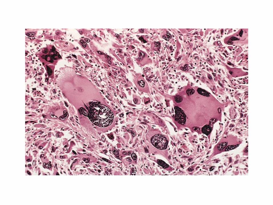

•In the histological examination of a tumor you should look for:
–Pleomorphism : variation in size–High nuclear/ cytoplasm ratio ( N/C ratio)–Hyperchrmasia ( dark cell )–Mitosis ….?abnormal one
Characteristics of benign and malignant neoplasms
1. Differentiation and anaplasia


Neoplasia
•In the histological examination of a tumor you should look for:
–Pleomorphism : variation in size–High nuclear/ cytoplasm ratio ( N/C ratio)–Hyperchrmasia ( dark cell )–Mitosis ….?abnormal one

anaplasia
• Anaplastic cells: undifferentiated cells• Lose their resemblance to the normal cells
from which they have arisen whatever their tissue of origin
• Loss of function


Dysplasia
–Definiton: a loss in the uniformity of the individual cells and a loss in their architectural orientation.
–Non-neoplastic–Occurs mainly in the epithelia–Dysplastic cells shows a degree of : pleomorphism,
hyperchrmasia, increased mitosis and loss of polarity.

Neoplasia
•Dysplasia does not mean cancer•Dyplasia does not necessarily progress to
cancer•Dysplasia may be reversible•If dysplastic changes involve the entire
thickness of the epithelium it is called :
CARCINOMA IN-SITU
Dysplasia

Dysplasia

Neoplasia
•Carcinoma in-situ–Definition: an intraepithelial malignancy in which
abnormal cells involve the entire thickness of the epithelium without penetration of the basement
membrane.
–Applicable only to epithelial neoplasms .


Dysplasia
• Increased rate of multiplication.
• Disordered maturation.
• Nuclear abnormality– Increased N/C ratio– Irregular nuclear membrane– Increased chromatin content
• Cytoplasmic abnormalities due to failure of normal maturation
Dysplasia: Features
Dysplasia
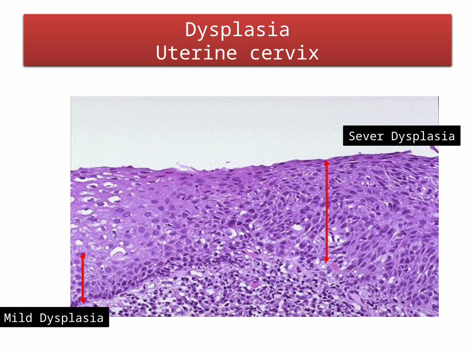
Mild Dysplasia
Sever Dysplasia
DysplasiaUterine cervix
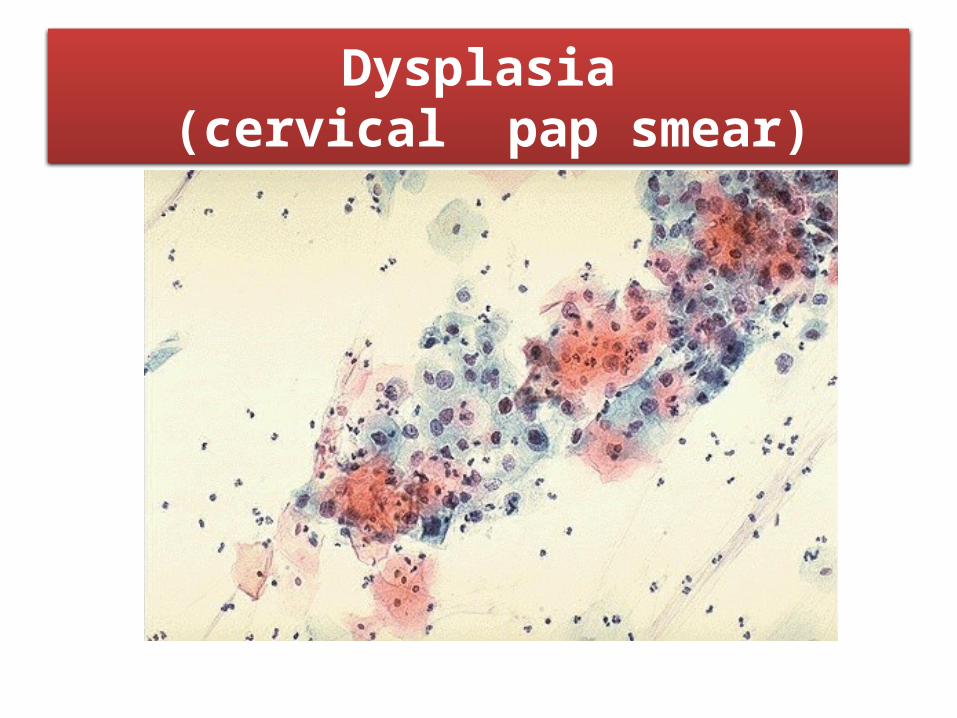
Dysplasia (cervical pap smear)
Dysplasia
•Clinical significance:–It is a premalignant condition.–The risk of invasive cancer varies with :
grade of dysplasia (mild, moderate, sever) duration of dysplasia site of dysplasia
Dysplasia

Dysplasia
•Differences between dysplasia and cancer.
lack of invasiveness Reversibility
Dysplasia
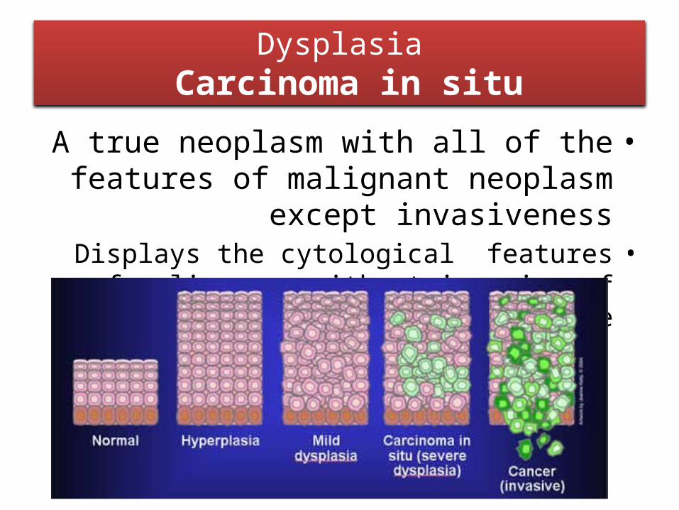
•A true neoplasm with all of the features of malignant neoplasm except invasiveness
•Displays the cytological features of malignancy without invasion of the basement membrane.
Dysplasia Carcinoma in situ
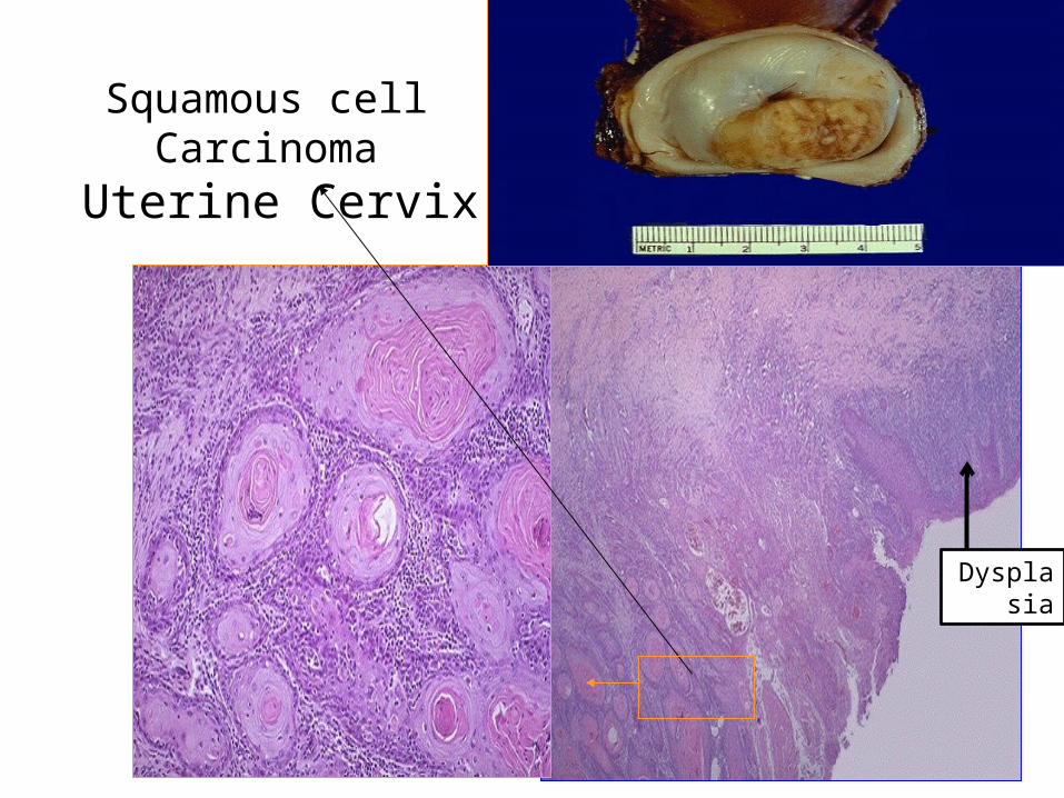
Squamous cell Carcinoma Uterine Cervix
Dysplasia

Thank you


