Embed Size (px)
Citation preview

Proc. Natl. Acad. Sci. USAVol. 93, pp. 11354-11358, October 1996Colloquium Paper
This paper was presented at a colloquium entitled "Genetic Engineering of Viruses and of Virus Vectors," organized byBernard Roizman and Peter Pakse (Co-chairs), held June 9-11, 1996, at the National Academy of Sciences in Irvine, CA.
Negative-strand RNA viruses: Genetic engineeringand applicationsPETER PALESE*, HONGYONG ZHENG, OTHMAR G. ENGELHARDT, STEPHAN PLESCHKA, AND ADOLFO GARCIA-SASTREDepartment of Microbiology, Mount Sinai School of Medicine, 1 Gustave L. Levy Place, New York, NY 10029
ABSTRACT The negative-strand RNA viruses are a broadgroup of animal viruses that comprise several importanthuman pathogens, including influenza, measles, mumps, ra-bies, respiratory syncytial, Ebola, and hantaviruses. Thedevelopment of new strategies to genetically manipulate thegenomes of negative-strand RNA viruses has provided us withnew tools to study the structure-function relationships of theviral components and their contributions to the pathogenicityof these viruses. It is also now possible to envision rationalapproaches-based on genetic engineering techniques-todesign live attenuated vaccines against some of these viralagents. In addition, the use of different negative-strand RNAviruses as vectors to efficiently express foreign polypeptideshas also become feasible, and these novel vectors have poten-tial applications in disease prevention as well as in genetherapy.
DNA-Containing Viruses
Among animal viruses, DNA-containing viruses were the firstto become amenable to genetic engineering techniques. Thisbreakthrough was achieved for simian virus 40 when a clonedcDNA copy was transfected into cells, resulting in the forma-tion of infectious virus (see Table 1). Transfected mutatedcDNA molecules gave rise to defined mutant viruses (1). Asecond methodology involving the use of homologous recom-bination allowed, for the first time, the rescue of large DNA-containing viruses such as herpes viruses (2). In this approach,intact herpes viral DNA as well as cloned DNA flanked by viralsequences was transfected into cells. Homologous recombina-tion between the cloned DNA and the wild-type genome canoccur, and novel viruses can be selected under appropriateconditions. For example, recombinants with DNA fragmentscontaining a viral thymidine kinase gene can be selected inappropriate cell lines and media, and viruses lacking a thymi-dine kinase can be isolated in the presence of nucleosideanalogs (e.g., Ara T). This general technique allows the suc-cessful construction of viral variants of herpes viruses, andsimilar procedures have been developed for pox viruses (3, 4)and other DNA-containing viruses including adenoviruses (5)and parvoviruses (6). Finally, strategies have been developedto generate infectious as well as mutant viruses by transfectingcosmids containing overlapping portions of large viral ge-nomes. Viruses arise via recombination between the cosmids.This system was successfully used to rescue infectious herpessimplex 1 viruses (7), cytomegaloviruses (8) and Epstein-Barrviruses (9) from their respective cosmids.
Positive-Strand RNA Viruses
RNA-containing viruses belong to a variety of families withdiverse replication strategies. Unique among the RNA virusesare the retroviruses, whose replication involves a double-stranded DNA phase, making these viruses an easy target forgenetic manipulation. Transfection of full-length cDNA mol-ecules leads to the establishment of replicating virus particlesand integration of the viral genetic information into the hostgenome (10). The engineering of retroviral genomes hasbecome one of the most successful genetic approaches inmodern virology and is central to the study both of viral geneexpression and of protein structure-function analysis. In ad-dition, retrovirus constructs are among the most widely usedvectors for gene transfer and gene therapy (11).Most of the other positive-strand RNA viruses are also
amenable to genetic engineering approaches (Table 1). In thecase of the small and medium sized positive-strand RNAviruses, full-length genomic RNA has been shown to beinfectious when transfected into cells. Plus-strand RNA servesas mRNA for the synthesis of viral proteins as well as templatefor viral RNA replication. Thus, transfection of cloned DNAof poliovirus RNA (or of cDNA-derived RNA) into permissivecells results in the formation of infectious virus particles (12).
Remarkably successful have been studies using Sindbis virusesand Semliki forest virus (13, 14). The cDNA-derived RNAs ofthese positive-strand RNA viruses can be used to efficientlyrescue infectious viruses, thus allowing an extensive analysis of thepromoter elements of the viral RNAs as well as structure-function studies of the viral proteins. Furthermore, these viruseshave received increased attention because of their potential forexpressing copious amounts of heterologous genes via recombi-nant constructs. Up to 108 molecules of heterologous protein percell have been expressed using these systems.t
Introduction of cDNA-Derived RNA into a Negative-StrandRNA Virus (Influenza Virus)
The life cycle of negative-strand RNA viruses differs from thatof the other RNA viruses in many ways. Specifically, thegenomic RNA of negative-strand RNA viruses is not infec-tious, and infectious virus particles must also deliver their ownRNA-dependent RNA polymerase into the infected cell tostart the first round of virus-specific mRNA synthesis.
Thus, approaches different from those used for positive-strand RNA viruses had to be developed to allow the rescue of
Abbreviations: RNP, ribonucleoprotein; HA, hemagglutinin; NA,neuraminidase, VSV, vesicular stomatitis virus.*To whom reprint requests should be addressed. e-mail: [email protected].
tBelli, B. A., Polo, J. M., Driver, D. A., Latham, E., Banks, T. A.,Chang, S. M. W. & Dubensky, T. W., Jr., National Academy ofSciences Colloquium on Genetic Engineering of Viruses and of VirusVectors, June 9-11, 1996, Irvine, CA, no. 1. (abstr.).
11354
The publication costs of this article were defrayed in part by page chargepayment. This article must therefore be hereby marked "advertisement" inaccordance with 18 U.S.C. §1734 solely to indicate this fact.

Proc. Natl. Acad. Sci. USA 93 (1996) 11355
Table 1. Genetic engineering of animal viruses
Type of genome
dsDNA
ssDNAssRNA
Prototype viruses StrategiesSimian virus 40, herpes, Transfection of cDNA; homologous
adenovirus, poxvirus recombination using cloned DNA and intactviral DNA or helper viruses; transfection ofcosmids containing viral genes
Adeno-associated virus (AAV) Transfection of plasmids containing AAV genesRetrovirus Transfection of infectious cDNA
Plus-sense RNAPicornavirus, Semliki forest Transfection of cDNA-derived infectious RNA
virus, Sindbis virus
Minus-sense RNAInfluenza virus, rhabdovirus,
parainfluenza virus,bunyavirus
dsRNA
ds, Double stranded; ss, single stranded.
Transfection of reconstituted ribonucleoproteinin the presence of helper virus; rescue ofvirus from cDNA clones transcribed in vitroor in vivo in the presence of helper virus orof viral polymerase proteins expressedintracellularly in trans
genetically engineered viruses of these virus families (Table 1).Site-specifically altered influenza viruses were first obtainedby reconstituting in vitro a biologically active ribonucleoproteincomplex (made of synthetic RNA and purified nucleoproteinand polymerase proteins) and then transfecting the complexinto helper virus-infected cells (Fig. 1) (15). The helper virusprovides in trans the viral proteins required for amplificationof the synthetic RNP complex. Subsequent reassortment of thesynthetic gene and helper virus-derived RNA segments, fol-lowed by selection for the reassortant (transfectant) virus,allows the introduction of site-specific changes into the ge-nome of influenza viruses (16). Selection of the transfectantvirus can be achieved by choosing host range or temperature-sensitive mutants as helper viruses. Alternatively, antibodypreparations specific for the viral surface proteins can be usedto select against the helper virus or for these novel viralconstructs. Following such protocols, six of the eight genes[PB2, hemagglutinin (HA), neuraminidase (NA), NP, M andNS] of influenza A viruses and the HA of an influenza B virushave now successfully been altered by genetic engineeringmethods (17-22).
Plasmid-Based Reverse Genetics Systemfor Influenza Virus
A method was recently developed to reconstitute a biologicallyactive influenza virus RNP complex within a cell rather thanin vitro. This alternative approach avoids the need to purifyviral proteins and to transfect an RNA-protein complex intocells; instead, this method involves the transfection of plas-mids. The first plasmid contains a human polymerase I pro-moter and a hepatitis delta virus-derived ribozyme sequencewhich flank the synthetic influenza virus gene. The polymeraseI-driven plasmid is cotransfected into human cells with poly-merase II-responsive plasmids expressing in trans the viralPB1, PB2, PA, and NP proteins. Such a system involving theuse of five plasmids allows the amplification and expression ofa synthetic influenza virus gene and takes advantage of theconvenience of plasmid transfections as compared with RNPtransfections (23). Using this approach, it was possible torescue a synthetic NA gene into a recombinant influenza Avirus. A synthetic HA gene has also been rescued by this noveltechnique (Fig. 2) (A.G.-S., unpublished results). It should benoted, however, that this plasmid-based reverse genetics sys-tem still relies on the presence of a helper virus which providesthe genetic backbone into which the plasmid-derived gene canbe introduced.
Chimeric Influenza Viruses Expressing ForeignEpitopes or Polypeptides
The development of methods to rescue synthetic RNAs intothe genomes of influenza viruses allowed the construction ofchimeric viruses expressing a variety of foreign epitopes.Specifically, epitopes derived from HIV, plasmodia, or lym-phocytic choriomeningitis virus proteins were successfullyexpressed in either the HA or the NA of different influenzaviruses (16, 24). Such constructs were shown to induce a potentB-cell and/or T-cell response against the foreign epitope inexperimental animal systems. Specifically, Li et al. (25) gen-
RESCUE OF INFECTIOUS INFLUENZA VIRUSES
\
I
C=== RNP
SELECTION
- TRANSFECTANT\- / VIRUS
FIG. 1. A reverse genetics system for the rescue of infectiousinfluenza viruses containing cDNA-derived RNA. The method allowsthe substitution of one of the eight genomic RNA segments of the virusby a synthetic RNA. A biologically active viral ribonucleoproteincomplex (RNP) is made in vitro by mixing cDNA-derived RNA withpurified viral nucleoprotein and polymerase proteins. The RNPs aretransfected into cells which have been previously infected with aninfluenza helper virus. Using a selection method, viruses containingthe genetically engineered RNP (transfectant viruses) can be isolated.
Colloquium Paper: Palese et al.

11356 Colloquium Paper: Palese et al.
PLASMID-BASED REVERSE GENETICSSYSTEM FOR INFLUENZA VIRUS
qp l
iJ7
polI X HA RZI
HELPER VIRUS
TRANSFECTANT VIRUSFIG. 2. A plasmid-based reverse genetics system for the rescue of
infectious influenza viruses containing a genetically engineered seg-ment. Cells are transfected with four plasmids that are able to expressthe viral NP and polymerase (PB2, PB1, and PA) proteins from acellular polymerase II-responsive promoter (pol II). An additionalplasmid which contains, for example, the HA open reading frameflanked by the 5' and 3' noncoding regions of the viral RNA segment(black boxes) is cotransfected. The HA plasmid is able to express anHA-specific viral RNA by transcription from a polymerase I-respon-sive promoter (pol I) followed by the ribozyme (RZ)-mediatedcleavage of the transcript. The HA-specific RNA segment is intracel-lularly complexed with the NP and polymerase proteins to form RNPsthat can be rescued into a transfectant virus if the cells are also infectedwith an influenza helper virus. Selection of the transfectant viruses canbe performed by using neutralizing antibodies against the HA proteinof the helper virus.
erated a recombinant influenza virus that expressed a CD8+T-cell epitope derived from the circumsporozoite (CS) proteinof Plasmodium yoelii in its HA. Mice immunized with thistransfectant virus made a vigorous cytotoxic T lymphocyteresponse against this epitope (25). By boosting mice with a
recombinant vaccinia virus expressing the CS protein, it waspossible to achieve protective immunity (60%) against chal-lenge with live P. yoelii sporozoites. Additional protective im-mune responses were generated by immunizing mice with trans-fectants expressing B-cell-specific epitopes located in the repeatregion of the CS protein of P. yoelii. Up to 80% of immunizedmice were immune to challenge with one hundred P. yoeliisporozoites (26).
Foreign epitopes can be inserted into several sites on the HAmolecule of influenza viruses, and most conveniently into thestalk region of the NA. In fact, stretches of more than 80foreign amino acids have been successfully inserted into thestalk region of the NA (27, 28) (S. Itamura, personal commu-nication). Although some of these constructs show interestingbiological properties, this approach of epitope grafting has itslimitations in terms of the size and the nature of the epitopethat can be expressed (since the chimeric protein may affectthe viability of the recombinant virus).A generic approach to the expression of foreign proteins is
the construction of bicistronic genes which can be packagedinto infectious particles. The foreign gene can replace the openreading frame of one of the influenza virus genes and therespective influenza virus protein is then translated from aninternal ribosome entry site (IRES element) on the geneticallyengineered gene. Alternatively, the foreign protein can be trans-lated from an internal IRES sequence. Expression of severalforeign polypeptides was achieved in this way (16, 29). However,many constructs did not result in viable viruses (unpublishedresults). Attempts are currently being made to identify the factorswhich determine the limitations of this approach.
The second method for the expression of foreign proteinstakes advantage of autoproteolytic elements placed within afusion protein. For example, a virus was constructed thatexpresses a fusion protein consisting of the full-length chlor-amphenicol acetyltransferase (CAT) protein, the 2A proteaseof foot and mouth disease virus, and the viral NA (30). Thisvirus was stably passaged and expressed copious amounts ofCAT protein in infected cells. However, in all cases of thefusion protein constructs, the foreign protein contains a 16-amino acid extension derived from the 2A protease which mayalter the biological properties of the foreign protein.
Rescue of Infectious Rabies Virus from cDNA
Like the segmented negative-strand RNA viruses, the Monon-egavirales group packages its own RNA-dependent RNApolymerase into virus particles to initiate viral RNA synthesis.Thus, naked RNA alone is unable to drive the replication cycle.Several approaches were taken to rescue model and full-lengthRNAs. First, a Sendai virus-like RNA transcript was amplifiedand expressed by transfecting the naked model RNA intoSendai virus-infected cells (31). This experiment suggests thatcomplementation in trans by the viral polymerase complex isrequired for the amplification and expression of the viralRNA-like reporter gene. Subsequently, in a remarkable study,Schnell et al. (32) succeeded in constructing a plasmid thatexpresses a full-length rabies virus RNA transcript from a T7RNA polymerase promoter. The plasmid DNA containing thisviral insert was transfected into cells infected with a recom-binant vaccinia virus expressing the T7 polymerase. Threeother plasmids expressing the rabies virus N, P and L proteinswere also cotransfected into these cells. In this recombinantvaccinia virus-driven system, the presence of the viral poly-merase complex and of a full-length viral RNA (in plus sense)led to the formation of recombinant rabies virus.
This system has been elegantly exploited to study thepromoter elements of rabies virus RNA and to elucidate theinteraction of this interesting virus with cells (33). Surprisingly,cells infected with a mutant lacking the virus' only glycoprotein(G) were still able to bud from the cell surface, albeit at a30-fold lower efficiency (34). This experiment revealed that thesurface protein G exhibits an intrinsic exocytotic activity. Thesystem was further developed to show that a hybrid G/HIV-1glycoprotein was able to form pseudotypes with the "G-less"particle, thus changing the host range by restricting infectionto CD4+ cells. This experiment clearly demonstrates thatgenetic engineering can redirect the host range and celltropism of rabies viruses. This should prove helpful for thedevelopment of novel vaccines as well as for gene therapy.
Rescue of Other Nonsegmented Negative-StrandRNA Viruses
An effective DNA transfection system has also been developedfor another rhabdovirus, vesicular stomatitis virus (VSV) (35,36) (Fig. 3). Again, the polymerase complex (N, P, and Lproteins) was expressed in cells from plasmids transcribed bya T7 RNA polymerase-containing vaccinia virus recombinant.Recombinant VSVs expressing an additional transcriptionalunit were rescued and high-expression levels of heterologousproteins were achieved (37). In a dramatic experiment, theauthors were able to construct a recombinant VSV expressingthe CD4 protein. This protein was packaged at levels of up to30% of the G protein itself, and the recombinant particle hadan 18% greater length than wild-type virus due to the extragene. These results illustrate that VSV is an effective vector toexpress foreign proteins at high levels, and that the virus istolerant to the insertion of novel transcriptional units. Reversegenetics systems have also been developed for paramyxovi-ruses. In the case of measles virus, a cell line constitutively
|POI III PBI
|POI III PB2
IPol II| PA
lpol Ill I
Proc. Natl. Acad. Sci. USA 93 (1996)
Proc. Natl. Acad. Sci. USA 93 (1996) 11357
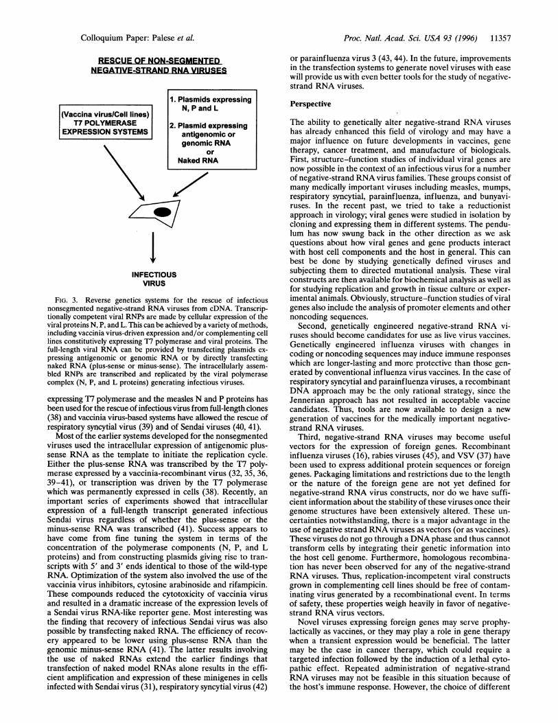
RESCUE OF NON-SEGMENTEDNEGATIVE-STRAND RNA VIRUSES
(Vaccina virus/Cell lines)T7 POLYMERASE
EXPRESSION SYSTEMS
'IINFECTIOUS
VIRUS
FIG. 3. Reverse genetics systems for the rescue of infectiousnonsegmented negative-strand RNA viruses from cDNA. Transcrip-tionally competent viral RNPs are made by cellular expression of theviral proteins N, P, and L. This can be achieved by a variety of methods,including vaccinia virus-driven expression and/or complementing celllines constitutively expressing T7 polymerase and viral proteins. Thefull-length viral RNA can be provided by transfecting plasmids ex-pressing antigenomic or genomic RNA or by directly transfectingnaked RNA (plus-sense or minus-sense). The intracellularly assem-bled RNPs are transcribed and replicated by the viral polymerasecomplex (N, P, and L proteins) generating infectious viruses.
expressing T7 polymerase and the measles N and P proteins hasbeen used for the rescue of infectious virus from full-length clones(38) and vaccinia virus-based systems have allowed the rescue ofrespiratory syncytial virus (39) and of Sendai viruses (40, 41).Most of the earlier systems developed for the nonsegmented
viruses used the intracellular expression of antigenomic plus-sense RNA as the template to initiate the replication cycle.Either the plus-sense RNA was transcribed by the T7 poly-merase expressed by a vaccinia-recombinant virus (32, 35, 36,39-41), or transcription was driven by the T7 polymerasewhich was permanently expressed in cells (38). Recently, animportant series of experiments showed that intracellularexpression of a full-length transcript generated infectiousSendai virus regardless of whether the plus-sense or theminus-sense RNA was transcribed (41). Success appears tohave come from fine tuning the system in terms of theconcentration of the polymerase components (N, P, and Lproteins) and from constructing plasmids giving rise to tran-scripts with 5' and 3' ends identical to those of the wild-typeRNA. Optimization of the system also involved the use of thevaccinia virus inhibitors, cytosine arabinoside and rifampicin.These compounds reduced the cytotoxicity of vaccinia virusand resulted in a dramatic increase of the expression levels ofa Sendai virus RNA-like reporter gene. Most interesting wasthe finding that recovery of infectious Sendai virus was alsopossible by transfecting naked RNA. The efficiency of recov-ery appeared to be lower using plus-sense RNA than thegenomic minus-sense RNA (41). The latter results involvingthe use of naked RNAs extend the earlier findings thattransfection of naked model RNAs alone results in the effi-cient amplification and expression of these minigenes in cellsinfected with Sendai virus (31), respiratory syncytial virus (42)
or parainfluenza virus 3 (43, 44). In the future, improvementsin the transfection systems to generate novel viruses with easewill provide us with even better tools for the study of negative-strand RNA viruses.
Perspective
The ability to genetically alter negative-strand RNA viruseshas already enhanced this field of virology and may have amajor influence on future developments in vaccines, genetherapy, cancer treatment, and manufacture of biologicals.First, structure-function studies of individual viral genes arenow possible in the context of an infectious virus for a numberof negative-strand RNA virus families. These groups consist ofmany medically important viruses including measles, mumps,respiratory syncytial, parainfluenza, influenza, and bunyavi-ruses. In the recent past, we tried to take a reductionistapproach in virology; viral genes were studied in isolation bycloning and expressing them in different systems. The pendu-lum has now swung back in the other direction as we askquestions about how viral genes and gene products interactwith host cell components and the host in general. This canbest be done by studying genetically defined viruses andsubjecting them to directed mutational analysis. These viralconstructs are then available for biochemical analysis as well asfor studying replication and growth in tissue culture or exper-imental animals. Obviously, structure-function studies of viralgenes also include the analysis of promoter elements and othernoncoding sequences.
Second, genetically engineered negative-strand RNA vi-ruses should become candidates for use as live virus vaccines.Genetically engineered influenza viruses with changes incoding or noncoding sequences may induce immune responseswhich are longer-lasting and more protective than those gen-erated by conventional influenza virus vaccines. In the case ofrespiratory syncytial and parainfluenza viruses, a recombinantDNA approach may be the only rational strategy, since theJennerian approach has not resulted in acceptable vaccinecandidates. Thus, tools are now available to design a newgeneration of vaccines for the medically important negative-strand RNA viruses.
Third, negative-strand RNA viruses may become usefulvectors for the expression of foreign genes. Recombinantinfluenza viruses (16), rabies viruses (45), and VSV (37) havebeen used to express additional protein sequences or foreigngenes. Packaging limitations and restrictions due to the lengthor the nature of the foreign gene are not yet defined fornegative-strand RNA virus constructs, nor do we have suffi-cient information about the stability of these viruses once theirgenome structures have been extensively altered. These un-certainties notwithstanding, there is a major advantage in theuse of negative strand RNA viruses as vectors (or as vaccines).These viruses do not go through a DNA phase and thus cannottransform cells by integrating their genetic information intothe host cell genome. Furthermore, homologous recombina-tion has never been observed for any of the negative-strandRNA viruses. Thus, replication-incompetent viral constructsgrown in complementing cell lines should be free of contam-inating virus generated by a recombinational event. In termsof safety, these properties weigh heavily in favor of negative-strand RNA virus vectors.
Novel viruses expressing foreign genes may serve prophy-lactically as vaccines, or they may play a role in gene therapywhen a transient expression would be beneficial. The lattermay be the case in cancer therapy, which could require atargeted infection followed by the induction of a lethal cyto-pathic effect. Repeated administration of negative-strandRNA viruses may not be feasible in this situation because ofthe host's immune response. However, the choice of different
1. Plasmids expressingN, P and L
2. Plasmid expressingantigenomic orgenomic RNA
orNaked RNA
Colloquium Paper: Palese et aL
1

11358 Colloquium Paper: Palese et al.
antigenic variants (as is possible with influenza viruses) mayovercome this limitation.
Finally, the highly efficient expression of viral and foreignproteins via negative-strand RNA virus vectors may haveadditional biotechnological applications. It is possible thatdefective RNA constructs could be used for genetic immuni-zation. This form of vaccination would resemble DNA vacci-nation (46) in that the defective particle would go throughmany replication rounds and persist without spreading toneighboring cells. Such RNA replicons may have interestingbiological properties since the efficiency of infection should becomparable to that of whole viruses. Also, replication com-petent viral vectors may help in the manufacture of largequantities of biological reagents, since the quantities expressedby negative-sense RNA viruses can be high. It is also possiblethat purification of expressed proteins could be made easier ifthey were incorporated into extracellular virus particles.The solutions to many of the issues discussed here will
depend on the continuing success of basic science and thedevelopment of novel strategies to study viruses. Our horizonsmust expand and include the analysis not only of the viruses butalso of their interactions with the host cell. Only by continuingto study these fundamental processes may we hope to reap thebenefits offered to us by these new opportunities.
Work done in this laboratory was supported by National Institutesof Health grants to P.P.
1. Goff, S. P. & Berg, P. (1976) Cell 9, 695-705.2. Post, L. E. & Roizman, B. R. (1981) Cell 25, 227-232.3. Panicalli, D. & Paoletti, E. (1982) Proc. Natl. Acad. Sci. USA 79,
4927-4931.4. Mackett, M., Smith, G. L. & Moss, B. (1982) Proc. Natl. Acad.
Sci. USA 79, 7415-7419.5. Jones, N. & Shenk, T. (1978) Cell 13, 181-188.6. Samulski, R. J., Chang, L. & Shenk, T. (1989) J. Virol. 63,
3822-3828.7. Cunningham, C. & Davison, A. J. (1993) Virology 197, 116-124.8. Kemble, G., Duke, G., Winter, R., Spaete, R. & Mocarski, E. S.
(1996) J. Virol. 70, 2044-2048.9. Cohen, J. I., Wang, F., Mannick, J. & Kieff, E. (1989) Proc. Natl.
Acad. Sci. USA 86, 9558-9562.10. Wei, C.-M., Gibson, M., Spear, P. G. & Scolnick, E. M. (1981)
J. Virol. 39, 935-944.11. Mulligan, R. C. (1993) Science 260, 926-932.12. Racaniello, V. R. & Baltimore, D. (1981) Science 214, 916-918.13. Rice, C. M., Levis, R., Strauss, J. H. & Huang, H. V. (1987)
J. Virol. 61, 3809-3819.14. Liljestrom, P., Lusa, S., Huylebroeck, D. & Garoff, H. (1991)
J. Virol. 65, 4107-4113.15. Enami, M., Luytjes, W., Krystal, M. & Palese, P. (1990) Proc.
Natl. Acad. Sci. USA 87, 3802-3805.16. Garcia-Sastre, A. & Palese, P. (1995) Biologicals 23, 171-178.
17. Subbarao, E. K., Kawaoka, Y. & Murphy, B. R. (1993) J. Virol.67, 7223-7228.
18. Enami, M. & Palese, P. (1991) J. Virol. 65, 2711-2713.19. Li, S., Xu, M. & Coelingh, K. (1995) Virus Res. 37, 153-161.20. Yasuda, J., Bucher, D. J. & Ishihama, A. (1994) J. Virol. 68,
8141-8146.21. Castrucci, M. R. & Kawaoka, Y. (1995) J. Virol. 69, 2725-2728.22. Barclay, W. S. & Palese, P. (1995) J. Virol. 69, 1275-1279.23. Pleschka, S., Jaskunas, S. R., Engelhardt, 0. G., Zurcher, T.,
Palese, P. & Garcia-Sastre, A. (1996) J. Virol. 70, 4188-4192.24. Castrucci, M. R., Hou, S., Doherty, P. C. & Kawaoka, Y. (1994)
J. Virol. 68, 3486-3490.25. Li, S., Rodrigues, M., Rodriguez, D., Rodriguez, J. R., Esteban,
M., Palese, P., Nussenzweig, R. S. & Zavala, F. (1993) Proc. Natl.Acad. Sci. USA 90, 5214-5218.
26. Rodrigues, M., Li, S., Murata, K., Rodriguez, D., Rodriguez,J. R., Bacik, I., Bennick, J. R., Yewdell, J. W., Garcia-Sastre, A.,Nussenzweig, R. S., Esteban, M., Palese, P. & Zavala, F. (1994)J. Immunol. 153, 4636-4648.
27. Castrucci, M. R. & Kawaoka, Y. (1993) J. Virol. 67, 759-764.28. Luo, G., Chang, J. & Palese, P. (1993) Virus Res. 29, 141-153.29. Garcia-Sastre, A., Muster, T., Barclay, W. S., Percy, N. & Palese,
P. (1994) J. Virol. 68, 6254-6261.30. Percy, N., Barclay, W. S., Garcia-Sastre, A. & Palese, P. (1994)
J. Virol. 68, 4486-4492.31. Park, K. H., Huang, T., Correia, F. & Krystal, M. (1991) Proc.
Natl. Acad. Sci. USA 88, 5537-5541.32. Schnell, M. J., Mebatsion, T. & Conzelmann, K.-K. (1994)EMBO
J. 13, 4195-4203.33. Mebatsion, T. & Conzelmann, K.-K. (1996) Proc. Natl. Acad. Sci.
USA 93, 11366-11370.34. Mebatsion, T., Konig, M. & Conzelmann, K.-K. (1996) Cell 84,
941-951.35. Lawson, N. D., Stillman, E. A., Whitt, M. A. & Rose, J. K. (1995)
Proc. Natl. Acad. Sci. USA 92, 4477-4481.36. Whelan, S. P. J., Ball, L. A., Barr, J. N. & Wertz, G. T. W. (1995)
Proc. Natl. Acad. Sci. USA 92, 8388-8392.37. Schnell, M. J., Buonocore, L., Kretzschmar, E., Johnson, E. &
Rose, J. K. (1996) Proc. Natl. Acad. Sci. USA 93, 11359-11365.38. Radecke, F., Spielhofer, P., Schneider, H., Kaelin, K., Huber, M.,
Dotsch, C., Christiansen, G. & Billeter, M. A. (1995)EMBO J. 14,5773-5784.
39. Collins, P. L., Hill, M. G., Camargo, E., Grosfeld, H., Chanock,R. M. & Murphy, B. R. (1995) Proc. Natl. Acad. Sci. USA 92,11563-11567.
40. Garcin, D., Pelet, T., Calain, P., Roux, L., Curran, J. & Kola-kofsky, D. (1995) EMBO J. 14, 6087-6094.
41. Kato, A., Sakai, Y., Shioda, T., Kondo, T., Nakanishi, M. &Nagai, Y. (1996) Genes Cells 1, 569-579.
42. Collins, P. L., Mink, M. A. & Stec, D. S. (1991) Proc. Natl. Acad.Sci. USA 88, 9663-9667.
43. De, B. P. & Banerjee, A. K. (1993) Virology 196, 344-348.44. Dimock, K. & Collins, P. L. (1993) J. Virol. 67, 2772-2778.45. Conzelmann, K.-K. (1996) J. Gen. Virol. 77, 381-389.46. McClements, W. L., Armstrong, M. E., Keys, R. D. & Liu, M. A.
(1996) Proc. Natl. Acad. Sci. USA 93, 11414-11420.
Proc. Natl. Acad. Sci. USA 93 (1996)


![and Lymphatic Filariasis - pdfs.semanticscholar.org filevirus (SLEV), Sindbis virus, Rift Valley fever virus (RVFV) and lymphatic filariasis (LF) [1, 2]. Cx. pipiens is the most widely](https://img.dokumen.tips/doc/110x75/5cd0d37788c993bc268bd53c/and-lymphatic-filariasis-pdfs-slev-sindbis-virus-rift-valley-fever-virus.jpg)