Embed Size (px)
Citation preview

Nebraska Public Health Laboratory
2008 CLSI M100-S18 update
Paul D. Fey, Ph.D. Associate Professor/Associate DirectorJosh Rowland, M.T. (ASCP) State Training Coordinator

Agenda
• Discuss 2008 M100-S18 major changes– Organism specific
changes
– KPC carbapenemase
• Other AST issues and questions from discussion group

Issue 1-New Appendices
• Appendix A -ESBL screening
• Appendix B -Staphylococcus aureus susceptibility testing issues
• Appendix C -Coagulase-negative Staphylococcus susceptibility testing issues
• Appendix D -Screening for high-level Aminoglycoside resistance within the enterococci.

Tables 1 and 1A-Antibiotic reporting tables
• Starting pages 24 (Disk diffusion) and 86 (MIC). Tables 1 and 1A.
• Changes Table 1: – Tobramycin moved from group B (selective reporting) to group A
(primary) in Enterobacteriaceae and Pseudomonas aeruginosa.– Ticarcillin moved from group A to group B in Enterobacteriaceae
and Pseudomonas aeruginosa.– Macrolides, Clindamycin, Trimethoprim/sulfa. moved to group A
under Staphylococcus. – Note that Daptomycin is MIC only-do not use disk diffusion disks– Acinetobacter spp. Ampicillin-sulbactam, ciprofloxacin,
levofloxacin and gentamicin, tobramycin moved to group A.

Tables 1 and 1A-Antibiotic reporting tables
• Starting pages 24 (Disk diffusion) and 86 (MIC). Tables 1 and 1A.
• Changes table 1A– Note that “Streptococcus spp. other than
Streptococcus pneumoniae” column has been split into two columns: 1) Streptococcus spp. Beta-hemolytic group (Groups A, B, C, and G) and 2) Streptococcus spp. Viridans group (Small colony forming -hemolytic colonies [ S. anginosus] are included in this group).
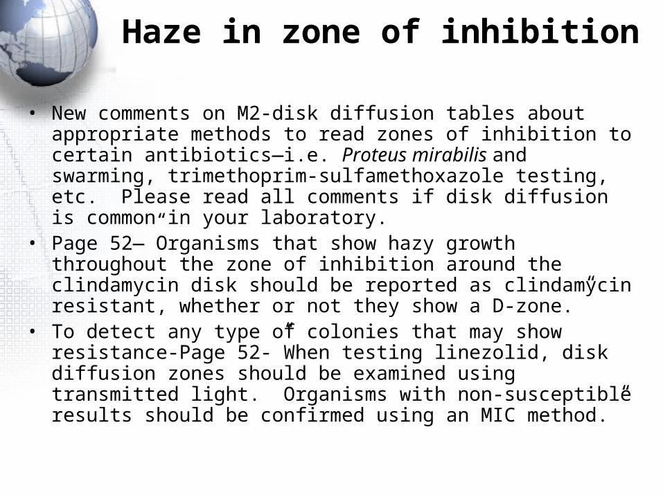
Haze in zone of inhibition
• New comments on M2-disk diffusion tables about appropriate methods to read zones of inhibition to certain antibiotics—i.e. Proteus mirabilis and swarming, trimethoprim-sulfamethoxazole testing, etc. Please read all comments if disk diffusion is common in your laboratory.
• Page 52—”Organisms that show hazy growth throughout the zone of inhibition around the clindamycin disk should be reported as clindamycin resistant, whether or not they show a D-zone.”
• To detect any type of colonies that may show resistance-Page 52-”When testing linezolid, disk diffusion zones should be examined using transmitted light. Organisms with non-susceptible results should be confirmed using an MIC method.”

Streptococcus pneumoniae
• If you are performing disk diffusion to detect resistance in Streptococcus pneumoniae, read page 66.– “Penicillin MICs should be determined for those
isolates with oxacillin zone diamters < 19 mm, because zones of < 19 mm occur with penicillin-resistant, intermediate, or certain susceptible strains based upon the meningitis and oral penicillin V interpretive criteria given in M7, Table 2G. Isolates should not be reported as penicillin-resistant or intermediate based solely on an oxacillin-zone < 19 mm.”

Streptococcus pneumoniae
• Significant reporting changes regarding penicillin– Interpretive standards for parenteral penicillin non-
meningitis, meningitis, and oral penicillin non-meningitis.
– On meningitis isolates, only report meningitis interpretive standards.
– On non-meningitis isolates (e.g. pneumonia or blood), report both meningitis and non-meningitis interpretive standards.

Streptococcus pneumoniae isolated from blood-penicillin MIC of 1
• Ceftriaxone (meningitis)- <0.5 S
• Ceftriaxone (non-meningitis)- <0.5 S
• Erythromycin->1 R
• Levofloxacin- <0.5 S
• Penicillin (meningitis)-1 R
• Penicillin (non-meningitis)-1 S
• Penicillin oral (non-meningitis)-1 I
• Vancomycin-0.5 S
• Based on therapy category, different interpretive standards.
• Multiple comment suggestions in the CLSI
• These changes must reflected in your antibiogram.

Staphylococcus aureus changes
• Page 111. Cefoxitin MIC test to detect methicillin resistance—only reliable for S. aureus and S. lugdunensis. – “The results of cefoxitin MIC tests can be used to
predict the presence of mecA-mediated resistance in S. aureus and S. lugdunensis. Isolates for which cefoxitin MICs are > 8g/ml should be reported as resistant. Isolates for which the cefoxitin MICs are < 4g/ml should be reported as oxacillin susceptible.”

Staphylococcus aureus changes
• Page 114-Erythromycin-resistant and clidamycin-susceptible isolates. New approved way to detect inducible resistance to clindamycin. Well containing 4 g/ml erythromycin and 0.5 g/ml clindamycin—growth in the well indicates resistance to clindamycin.

KPC carbapenemase
• Enzyme capable of hydrolyzing carbapenems (imipenem, ertapenem, meropenem, doripenem).
• Isolated primarily on the east coast of US in ICUs.• Klebsiella pneumoniae and other Enterobacteriaceae. • KPC not easily detected using commercial susceptibility
systems—some isolates have an MIC of 2 to the carbapenems (still susceptible)
• KPC enzymes hydrolyze expanded-spectrum cephalosporins as well—therefore, all Enterobacteriaceae isolates that have an ESBL phenotype should be screened for a carbapenemase.
KPC carbapenemase
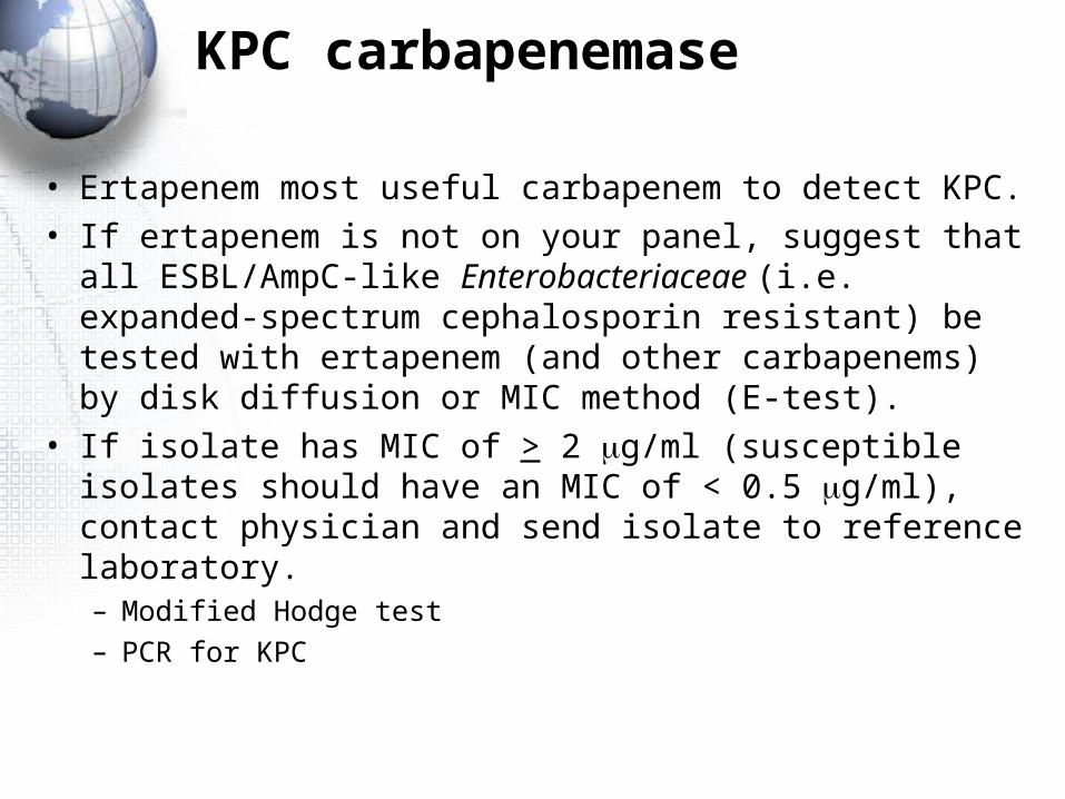
• Ertapenem most useful carbapenem to detect KPC.• If ertapenem is not on your panel, suggest that all
ESBL/AmpC-like Enterobacteriaceae (i.e. expanded-spectrum cephalosporin resistant) be tested with ertapenem (and other carbapenems) by disk diffusion or MIC method (E-test).
• If isolate has MIC of > 2 g/ml (susceptible isolates should have an MIC of < 0.5 g/ml), contact physician and send isolate to reference laboratory. – Modified Hodge test– PCR for KPC