Embed Size (px)
Citation preview

Natural history and prognostic factorsin localized prostate cancer
Avh_Ove Andre�n_K160308.indd 1 08-04-17 15.42.16

To Ulrika, Erik, Ina och Nils
”It’s a job that’s never started
that takes the longest to fi nish.”
J. R. R. Tolkien
Avh_Ove Andre�n_K160308.indd 2 08-04-17 15.42.17

Örebro Studies in Medicine 15
Ove Andrén
Natural history and prognostic factors
in localized prostate cancer
Avh_Ove Andre�n_K160308.indd 3 08-04-17 15.42.17

© Ove Andrén 2008
Title: Natural history and prognostic factorsin localized prostate cancer
Publisher: Örebro University 2008www.publications.oru.se
Editor: Heinz [email protected]
Printer: Intellecta DocuSys, V Frölunda 04/2008
issn 1642-4063isbn 978-91-7668-592-1
Avh_Ove Andre�n_K160308.indd 4 08-04-17 15.42.18

ABSTRACT
The natural history of localized prostate cancer is not fully understood. In most patients the tumor will never progress to a lethal disease, while a subset of patients will ultimately die of the disease. Effi cient tools to separate indolent from lethal disease is currently lacking which means that many patients will be offered treat-ment without any benefi t, but still be at risk of experiencing treatment related side effects.
The aims of these studies were to get more insight into the natural history of untreated localized prostate cancer, to assess the prognostic value of established clinical parameters such as Gleason score, nuclear grade and tumor volume and, moreover, some new prognostic markers Ki-67, AMACR and MUC-1. We also aimed to study time trends in the detection of incidental tumors in Sweden.
Patients with localized disease (n=223) and no initial treatment were followed for 21 years. Most patients had a favorable outcome. However, a subset of patients developed lethal disease even beyond 15 years of follow-up and these patients defi ne the group that may benefi t most from treatment with curative intent. Patients with poorly differentiated tumors experienced a 9 time higher risk of dying in prostate cancer.
The studies on prognostic markers are based on a cohort of patients (n=253) with incidental prostate cancer detected by transurethral resection for presumed benign hyperplasia. All patients were left without initial treatment. Gleason grade, nuclear grade and tumor volume turned all out to be independent prognostic factors. MUC-1, AMACR and Ki-67 also carried prognostic information. However, after adjustment for Gleason grade, nuclear grade and tumor volume only MUC-1 and AMACR remained as statistically signifi cant prognostic factors. When tested for sensitivity and specifi city they all failed and, consequently, they seem to be of less value in daily practice for cancelling an individual patient regarding the choice of treatment.
Time trends in incidental prostate tumors in Sweden were analyzed in a cohort of patients with prostate tumors detected by transurethral resection (TUR-P). Through linkage of the national registration number (NRN) with several registers, e.g. the Swedish Cancer Registry, the National Inpatient registry and the Cause of Death Registry we identifi ed, during the period 1970 through 2003, in total 23288 patients with incidental prostate cancer, who constituted the study group. As comparison group we choose all patients diagnosed with prostate cancer between 1970–2003 excluding those with incidental cancer, in total 112204 patients. Our result confi rms earlier fi ndings that there has been a dramatic change over time in incidence of incidental prostate cancers in Sweden, which parallels the introduction of prostate specifi c antigen. We also found that the cumulative incidence of prostate cancer death is high in the incidental group, opposing earlier fi ndings that incidental tumours are a non-lethal disease.
Keywords: Prostate cancer, Natural History, Survival, Prognostic factor, Tumor grade, TUR-P, Incidental, MUC-1, AMACR, Ki67, Tumor volume.
Avh_Ove Andre�n_K160308.indd 5 08-04-17 15.42.18

Avh_Ove Andre�n_K160308.indd 6 08-04-17 15.42.19

CONTENTS
LIST OF PAPERS ....................................................................................9
ABBREVIATIONS ...................................................................................10
INTRODUCTION ....................................................................................11
PROSTATE CANCER EPIDEMIOLOGY IN SWEDEN ......................11
PSA SCREENING .................................................................................12
TREATMENT OF LOCALIZED PROSTATE CANCER ....................13
PROSTATE CANCER DETECTED BY TRANSURETHRAL
RESECTION (TUR-P) ..........................................................................14
PROGNOSTIC AND PREDICTIVE FACTORS ................................... 15
PROGNOSTIC FACTORS USED TODAY ........................................... 15
Histological grade .................................................................................. 15
Local tumor stage ..................................................................................16
Prostate specifi c antigen (PSA) ...............................................................16
Age at diagnosis .....................................................................................16
Co-morbidity .........................................................................................17
NEW PROGNOSTIC MARKERS ........................................................17
Tumor proliferation markers ..................................................................17
AMACR ................................................................................................17
MUC-1 ..................................................................................................17
AIMS ......................................................................................................19
MATERIAL AND METHODS ..................................................................21
Paper I ....................................................................................................21
Paper II–IV ............................................................................................24
Paper V ..................................................................................................25
STATISTICAL METHODS ...................................................................27
Paper I ....................................................................................................27
Paper II–IV ............................................................................................27
Paper III .................................................................................................27
Paper V ..................................................................................................28
Avh_Ove Andre�n_K160308.indd 7 08-04-17 15.42.19

RESULTS ...............................................................................................29
Paper I ....................................................................................................29
Paper II ..................................................................................................37
Paper III .................................................................................................40
Paper IV .................................................................................................44
Paper V ..................................................................................................50
DISCUSSION . ........................................................................................5
Paper I ....................................................................................................5
Paper II .................................................................................................. 56
Paper III .................................................................................................
Paper IV .................................................................................................
Paper V ..................................................................................................6
CONCLUSIONS ...................................................................................
SUMMARY AND FUTURE ASPECTS ................................................
ACKNOWLEDGMENTS .....................................................................
67
REFERENCES ......................................................................................
69
Avh_Ove Andre�n_K160308.indd 8 08-04-17 15.42.19
5
5
59
162
65
17

9
LIST OF PAPERS
The thesis is based on the following papers, which will be referred to by their Roman numerals:
I. Jan-Erik Johansson, Ove Andrén, Swen-Olof Andersson, Paul W Dickman, Lars Holmberg, Anders Magnuson, Hans-Olov Adami. Natural history of early, localized prostate cancer. JAMA. 2004 Jun 9;291(22):2713-2719
II. Ove Andrén, Katja Fall, Lennart Franzén, Swen-Olof Andersson, Jan-Erik Johansson, Mark Rubin. How well does the Gleason score predict prostate cancer death? – A 20-year follow-up of a population-based cohort in Sweden. J Urol. 2006 Apr;175(4):1337-1340.
III. Mark Rubin MA, Bismar TA, Andrén O, Lorelei A Mucci, Shen R, Kim R, Ghosh D, Wei JT, Chinnaiyan AM. Adami HO, Kantoff PW, Johansson J-E. Decreased alpha-methylacyl CoA racemase expression in localized prostate cancer is associated with an increased rate of biochemical recurrence and cancer-specific death. Cancer Epidemiol
Biomarkers Prev. 2005 Jun; 14(6):1424-1432.
IV. Ove Andrén, Katja Fall, Sven-Olof Andersson, Mark A Rubin, Tarek A Bismar, Mats Karlsson, Jan-Erik Johansson, Lorelei A Mucci. MUC-1 gene is associated with prostate cancer death: – A 20 year follow-up of a population-based study in Sweden.Br J Cancer. 2007 Sep 17;97(6):730-734.
V. Ove Andrén, Hans Garmo, Lorelei Mucci, Swen-Olof Andersson, Jan-Erik Johansson and Katja Fall. Time trends and survival in men with incidental prostate cancer: -aregister-based study comprising 23,288 men diagnosed between 1970–2003 in Sweden,Submitted.
Avh_Ove Andre�n_K160308.indd 9 08-04-17 15.42.20
99

10
ABBREVIATIONS
ASAP atypical small acinar proliferationAMACR alpha methylacyl CoA racemaseBR Björn RisbergBRFS biochemical recurrence free survivalcDNA complementary deoxyribonucleic acidCI confidence intervalCT computer tomographyELISA enzyme linked immunosorbent assayHR hazard ratioICD international classification of diseasesHDR hospital discharge registerKi67 cell proliferation markerNRN national registration numberMIB-1 mindbomb homolog 1MR Mark RubinMRI magnetic resonance imagingMUC-1 mucin 1, anti-adhesion molecule OAE open adenoma enucleationPCa prostate cancerPSA prostate specific antigenROC receiver operating characteristicRT-PCR real time p chain reactionSD standard deviationSPCG Scandinavian prostate cancer groupTNM tumor nodule metastasisTUR-P transurethral resection of the prostateUSÖ university hospital of ÖrebroWHO world health organization
Avh_Ove Andre�n_K160308.indd 10 08-04-17 15.42.21
1010

11
INTRODUCTION
The appropriate management of localized prostate cancer is still unclear. Even though some tumors will progress to metastatic disease, most patients will not experience advanced disease nor need treatment. Indeed, several studies on the natural history of the localized disease, reveal that the vast majority of patients will not die of their disease1-3. Despite this knowledge, the number of patients treated for localized disease with curative intent has increased dramatically over the previous decades4. As a result, in current practice, prostate cancer patients are over treated and experience the consequences of treatment without any benefit5, 6.
Therefore, one of the most important areas in prostate cancer research today involves finding tools that distinguish indolent disease from aggressive disease. The prognostic factors used today are PSA, tumor stage7, grade and volume. Although the classification of tumor stage is dependent on the examiner, it has some prognostic value and is still a cornerstone in clinical practice. While tumor volume is a strong prognostic marker8, 9, it is hard to estimate for most tumors in the diagnostic setting10, 11. For a long time tumor grade, which was nuclear grade during the era of fine needle aspiration12, was the dominant grading system in Sweden. In the United States, where core biopsies were introduced earlier, the Gleason grading system13 has been the governing grading system and is the one supported by the World Health Organization(WHO). Even though these four factors are strong prognostic markers10, 14, they are not strong enough to predict the outcome of individual patients. New and more reliable prognostic markers are needed in determining the course of the disease and in deciding whether or not to treat the patient. Several studies to find new and more reliable markers11, 15,
16 have been performed, but no marker has proven to be a better predictor than tumor stage and grade.
PROSTATE CANCER EPIDEMIOLOGY IN SWEDEN
The incidence rate of prostate cancer has increased dramatically during the last twenty years (Fig. 1)4, 17, 18. The main reason for this is believed to be an increase in diagnostic activity. This view is partly supported by the fact that the incidence of T1c tumors, detected by elevated PSA, has seen the greatest increase. There have been smaller changes in incidence of clinically detected tumors, except for a decline in metastatic disease at diagnosis4. There are surprisingly large geographic differences in prostate cancer incidence; two geographically close counties can report large differences in prostate cancer incidence4, 19. The median age at diagnosis has decreased from 74 to 694. The number of patients treated with curative intent has increased dramatically during last 15 years4, 20. Even though we diagnose and treat more patients with localized tumors the number of men that die of prostate cancer has remained fairly unchanged.4
Avh_Ove Andre�n_K160308.indd 11 08-04-17 15.42.22
1111

12
PSA SCREENING
Prostate specific antigen (PSA) is a serine protease produced exclusively by the prostate. All prostatic diseases, including prostate cancer, may increase the serum levels of PSA. Increased levels of PSA in serum may be indicative of prostate cancer21. This knowledge has dramatically changed the management of prostate cancer, and increased PSA testing is the main factor for the enormous increase in prostate cancer incidence in Sweden over the past decades 4, 17, 18. PSA has been proposed as a screening tool for prostate cancer22, and two large clinical trials are ongoing to explore the effect of PSA screening; however, no results have been reported.
Avh_Ove Andre�n_K160308.indd 12 08-04-17 15.42.23
1212

13
Fig 1. Change in prostate cancer incidence and prostate cancer mortality in Sweden 1970–2006
0
50
100
150
200
250
300
1972
1975
1978
1981
1984
1987
1990
1993
1996
1999
2002
2005
Inc
ide
ns
pe
r1
00
00
0
Prostate cancer incidence
Prostate cancer mortality
Avh_Ove Andre�n_K160308.indd 13 08-04-17 15.42.23
1313

14
TREATMENT OF LOCALIZED PROSTATE CANCER
In the early eighties, patients with localized prostate cancer were usually left without initial treatment. The common opinion was that prostate cancer was an “old man’s” disease for which treatment was withheld until symptoms appeared. The treatment was mainly hormone manipulation, which dates back to late 1800, but which was first really understood in the forties by the work of Huggins23.
Since the early nineties there has been a dramatic shift in the treatment of localized disease4.In 2005, 5347 patients were treated with curative intent in Sweden as compared with 1069 patients in 1998. Curative treatment consists today of surgery or radiation therapy. The first surgical attempt to cure a patient from prostate cancer also dates back to the late 1800s, but it was not until the 1980s that the technique started to develop. In 1990, Dr. Patrick Walsh described in detail the procedure still used today24. This technique improved surgical outcome and quality of life, and has also been shown to improve cancer and overall survival compared to watchful waiting5. In addition, the technique of radiotherapy has improved during the last decade and today external multi-field techniques as well as brachytherapy are used. One reason behind the increase in number of patients treated with curative intent is the introduction of prostate specific antigen (PSA) in 198721. PSA screening has made it possible to find prostate cancer at an earlier stage, where the possibility to cure the patient is better. At the same time, PSA screening has led to considerable over diagnosis of cancers that otherwise would not have come to clinical attention25.
PROSTATE CANCER DETECTED BY TRANSURETHRAL RESECTION (TUR-P)
The clinical significance of incidental prostate cancer, detected by transurethral resection (TUR-P) or by open adenoma enucleation (OAE) for assumed benign hyperplasia, has been a matter of debate for years. One main question has been if these tumors represent a biological domain different from those detected by rectal palpation or by PSA tests. A common opinion has been that these tumors are harmless and could be left without immediate treatment, especially those with volume tumors less than 5% of the total amount of resected tissue (T1a) according to TNM classification 199226. Recent studies27-29 have shown that with increasing tumor volume, greater than 5% of total resected tissue (T1b), the course becomes increasingly unfavorable, comparable to palpable T2 tumors.
The resected tissue originates mainly from the transition zone of the prostate 30, 31. Tumors from this part of the prostate have been described as low grade with low potential of malignancy, and therefore are not believed to need treatment2, 32, 33. On the other hand, 8–37%of all incidental tumours34 ultimately will progress. Bostwick et al.34 suggested that TUR-Pdetected tumors may not exclusively have their origin in the transition zone, but instead might represent peripheral zone tumors that have grown into the transition zone. He further suggested that there may be a significant overlap between T1b, T1c and T2 tumors and that it is merely the location of the tumor in the prostate and the ability of the urologist to find it that ultimately determines the T-stage.Before the PSA era, the urologist had to rely mainly on rectal palpation and serum acid phosphates to evaluate if voiding symptoms were due to a benign condition or prostate cancer. A great proportion of these men with voiding symptoms were subject to TUR-P or
Avh_Ove Andre�n_K160308.indd 14 08-04-17 15.42.24
1414

15
OAE. Happaniemi et al. 35 showed that the introduction of TUR-P in the late seventies dramatically increased the number of incidental prostate cancer. The introduction of PSA revolutionized the diagnosis of prostate cancer and the management of men seeking urologists due to voiding symptoms. With the introduction of the PSA test, the frequency of incidental tumors decreased significantly (Kien et al.) 36. Currently in Sweden incidentally detected prostate tumors constitute around 10 % of all diagnosed PCa4.
PROGNOSTIC AND PREDICTIVE FACTORS
In the work of Humphrey, prognosis is defined as 37 “the prediction of future behavior of established malignancy, either in absence of or after application of therapy.” Factors used to predict prognosis fall into two groups38. Factors that provide information on the expected behavior of an untreated tumor are called prognostic factors, while those that provide information about the outcome of active therapy are called treatment-predictive. Factors that could improve the identification of tumors that will progress, or respond favorably to a certain therapy would facilitate choosing between treatment options. New prognostic factors mayalso increase the understanding of the pathogenesis of prostate cancer, and could identify new subgroups of tumors.
In the clinical practice we use prognostic factors on the group and the individual level. At the group level we have a larger cohort of patients whom we, for example, try to stratify into different risk groups. This perspective is often used in trials and in formulating general treatment guidelines. At the individual level, the physician is counseling one patient in making a decision on how to best treat his disease. In this latter situation, the patient is probably less interested to learn that he has a two times greater risk of progression, than in confidently knowing whether or not his tumor will progress to a lethal disease. Given this situation neither the patient nor the doctor is likely willing to take any risks. As a result, most patients opt for curative treatment even though very few of them will really benefit from the treatment5. To assess the value of a prognostic factor we need to calculate its sensitivity and specificity. The sensitivity is the probability that the disease will progress if the marker is positive. Specificity on the other hand is the probability that the prognostic factor is negative if the disease does not progress. The ultimate prognostic factor will have both a high sensitivity and a high specificity, but in certain occasions it may be enough to only have a high specificity or a high sensitivity.
PROGNOSTIC FACTORS USED TODAY
The prognostic factors that are used today for localized prostate cancer have not changed during recent years. The most used clinical prognostic factors are still tumor grade, local tumor stage, serum PSA, age and co-morbidity.
Histological grade
During the 20th century more then 40 different grading systems for prostate cancer were proposed37. Before the introduction of transrectal ultrasound and core biopsies in the late eighties, fine needle aspiration was the main tool used for diagnosis in Sweden. During this
Avh_Ove Andre�n_K160308.indd 15 08-04-17 15.42.25
1515

16
era nuclear grade was the grading system used in Sweden12. This is a three-tier grading system based on the morphology of the nuclei. Today tumor differentiation is based on the Gleason grading system39. This system grades the morphologic growth pattern of the tumor and divides them into five categories. The Gleason score is then constructed by adding the value of the most common pattern with the second most common, giving a Gleason score between 2 and 10. This grading system has proven to be a strong prognostic factor for both biochemical recurrence as well as prostate cancer death37.
Local tumor stage
The anatomic local spread of the tumor has been shown to be a strong prognostic factor in many types of cancer40. In prostate cancer this is judged by digital rectal examination7. This method is flawed by several limitations. Variability between observers, as well as the inaccuracy in determining capsule penetration, are the most important limitations, both of which are well documented41. Utilizing more objective staging modalities such as ultrasound, CT or MRI has so far not proven to enhance the accuracy of staging.
Prostate specific antigen (PSA)
Prostate specific antigen (PSA) is a glycoprotein characterized by Wang et al. in 197942. It is almost exclusively produced in the prostate gland. After secretion into the excretory ducts it rapidly forms complexes with alfa-1-antichymotrypsin and to a lesser extent with alfa-2-macroglobulin. Some 10–30% of the circulating PSA remains uncomplexed, so called “free” PSA. The main function of PSA is proteolysis of the semen coagulum to facilitate sperm motility43. Increased levels of PSA may be suggestive of prostate cancer. However, like all diseases affecting the prostate, benign prostatic hyperplasia may give rise to PSA levels44.Studies have also shown a strong correlation with patient age45, tumor volume and prognosis46. The sensitivity and specificity of PSA is low. However, several refinements of PSA screening have been proposed to enhance the predictability of PSA. The free PSA/total PSA ratio has shown to increase the specificity47. The PSA change (PSA velocity) before, as well as after diagnosis has also been suggested to carry prognostic information48. Despite these refinements, PSA screening has limitations for diagnosing of prostate cancer49.However, PSA has still proven to be a useful marker for monitoring the clinical course of the disease.
Age at diagnosis
Younger patients have not been shown to have a more aggressive disease50. However, they do have a greater risk of dying from prostate cancer as compared to older patients.51 This is mainly due to the fact that they have a longer life expectancy and a lower risk of dying of other causes. Thus, age has an impact on disease specific mortality in this slow growing disease. The impact of age is also seen in the results from the SPCG 4 trial5 where patients younger then 65 years seem to have the greatest advantage of surgery.
Avh_Ove Andre�n_K160308.indd 16 08-04-17 15.42.26
1616

17
Co-morbidity
Several studies have shown that co-morbidity is a strong prognostic factor in prostate cancer52. This is parallel to the discussion about age at diagnosis. A person with less co-morbidity has longer life expectancy and concurrently a higher risk that the prostate cancer will develop to a more advanced disease.
NEW PROGNOSTIC MARKERS
A number of new prognostic factors, including morphometric, immunophenotypic and genotypic have been discovered and put forward during the last decades15, 16. To prove the utility of these new markers they have to be tested in prospective, randomized, controlled clinical trials. Moreover they need to provide additive and independent value in multivariate analysis, beyond established prognostic factors, such as pathologic stage and Gleason score. So far none of these proposed new prognostic factors has fulfilled these criteria.
Tumor proliferation markers
There is convincing evidence that disturbance in the cell cycle machinery may contribute to the uncontrolled growth characteristic for tumor cells53. Immunohistochemical analysis using MIB-1/Ki67 antibody is very useful for measuring cell cycle progression in human tissue54.Proliferation rate, assessed by Ki-67 immunoreactivity, have further been associated with biochemical recurrence and prostate cancer death55-58.
AMACR
�-Methylacyl CoA racemase (AMACR) is a biomarker that was identified by both differential display and expression array analysis as a gene abundantly expressed in prostate cancer relative to benign prostate epithelium 59-61. In a meta-analysis of 4 cDNA expression array data sets, AMACR was one of the genes most consistently over expressed in prostate cancer62. AMACR is a peroxisomal and mitochondrial enzyme that plays an important role in bile acid biosynthesis and �-oxidation of branched-chain fatty acids through the interconversion of (R)- and (S)-2-methyl-branched-chain fatty acyl-CoA fragments63. Our group initially reported that AMACR expression is consistently lower both at the transcriptional (cDNA expression arrays and RT-PCR) and at the protein level (Western blot analysis and immunohistochemistry) in metastatic prostate cancer compared to localized prostate cancer 59, 64. More recently, a fluorescent-based measurement of AMACR in tissue samples confirmed these observations 65.
MUC-1
The mucin family of anti-adhesion molecules have been implicated in the biological behavior and progression of several types of cancer66, 67. The mucin, MUC-1, which is expressed at the apical cell surface of many normal secretory epithelial cells68, contains an extra cellular
Avh_Ove Andre�n_K160308.indd 17 08-04-17 15.42.27
1717

18
domain that extends above most other cell membrane-associated proteins69, 70. As such, MUC-1 has been suggested to prevent adhesion and to promote development of metastatic disease.In prostate cancer, over-expression of MUC-1 in tissue has been correlated with both higher Gleason grade and advanced tumor stage71. One study has, furthermore, suggested that MUC-1 expression may predict prostate cancer recurrence after prostatectomy72, although these results have been challenged by others73, 74. The disparate findings may in part be explained by the use of PSA-recurrence as a measure of outcome, since biochemical failure does not necessarily herald prostate cancer death75 .
Avh_Ove Andre�n_K160308.indd 18 08-04-17 15.42.28
1818

19
AIMS
The specific aims of the study were:
to study the natural history of prostate cancer, and evaluate the prognostic value of clinical parameters, in a population based cohort of patients with localized prostate cancer with an observation time of 20-years (paper I).
to study the prognostic value of Gleason score, nuclear grade, tumor extent and tumor proliferation using Ki67 (mib-1) as a proliferation marker among a cohort of 253 patients with untreated prostate cancer detected through TUR-P and with long follow up (paper II).
to study the prognostic value of AMACR expression in prostate tumors, and compare prostate cancer death and biochemical failure as endpoints (paper III).
to study the prognostic value of MUC-1 expression in prostate tumors in a cohort of 253 patients with untreated prostate cancer detected through TUR-P and with long follow up (paper IV).
to study the changes in the incidence and survival of TUR-P detected tumors in Sweden over time (paper V).
Avh_Ove Andre�n_K160308.indd 19 08-04-17 15.42.29
1919

Avh_Ove Andre�n_K160308.indd 20 08-04-17 15.42.30

21
MATERIAL AND METHODS
Paper I
The subjects comprised a population-based cohort of patients with early, initially untreated prostate cancer has previously been described in detail76. The TNM system of 1978 77 and the World Health Organization78 classification of malignant diseases were used. At the time of diagnosis, all patients underwent a clinical examination, excretory urography, chest radio-graphy, bone scan, skeletal radiography (if needed) and had routine blood samples taken. The nodal status was not known in any of the patients.
From March 1977 through February 1984, a total of 654 new cases of prostate cancer were diagnosed. Patients were given no initial treatment if the tumor growth was localized to the prostate gland. (T0-2) and no distant metastases were present (306 patients). The following restrictions were applied, however, among those with palpable tumors (T1-2). From March 1977 through February 1979, only patients with a highly differentiated tumor (grade I) were included in the untreated group. From March 1979 through the end of the recruitment period, patients under 75 years of age at diagnosis and with moderately or poorly differentiated tumors (grades II–III) were randomly allocated to receive local radiation (10 patients) or no treatment, and only the latter group was included in this cohort study. Patients older than 75 years of age were not treated and included in the study.
Among the 227 eligible patients, 4 (2 percent) were given initial treatment and had to be excluded from the analyses. The distribution of the study group of 223 patients by age, stage and grade at the time of diagnosis is shown in table 1. The mean age at diagnosis was 72 years (range 41–91 years). Altogether 106 (48 percent) were detected by histopathological examinations of specimens obtained at operations for suspected benign prostatic hyperplasia whilst the remaining 117 patients had a palpable clinical disease localized to the prostate gland. About 2/3 of patients had highly differentiated tumors whilst only 9 (4 percent) had a poorly differentiated tumor (Table 1).
All 223 patients were followed up from diagnosis until death or the end of the observation period (September 1st, 2001). No patient was lost to follow-up. Clinical examination, laboratory tests, and bone scans were carried out every six months during the first two years after diagnosis and subsequently once a year during the first 10 years of observation and thereafter at least once every second year. Those in whom the cancer progressed to sympto-matic disease were treated with exogenous estrogens or orchidectomy.
Local progression was defined as tumor growth through the prostate capsule (T3) as judged by digital rectal examination. Development of distant metastasis (M1) was classified as generalization.
During the six first years of follow-up, all patients who were still alive and consented had a new fine-needle biopsy every other year. We obtained such biopsies from altogether 178 of the 223 (80 percent) patients. Although this procedure has lower than 100 percent sensitivity, notably for impalpable tumors, remaining cancer growth was confirmed cytological in the
Avh_Ove Andre�n_K160308.indd 21 08-04-17 15.42.30
2121

22
majority of patients. Altogether 31 out of 178 (17 percent) patients showed evidence of dedifferentiation.
The medical records of all deceased patients were reviewed. In most instances, the cause of death was obvious on clinical grounds alone. Prostate cancer was recorded as the underlying cause of death, a contributory cause of death, or unrelated to death as described in detail in a previous report 76. If treatment of the prostate cancer was related to death prostate cancer was recorded as a contributory cause. As a validation, we compared our own classification of causes of death with those recorded in the Swedish Death Register. There was agreement in 90 percent of the patients and no evidence of systematic over or under ascertainment of prostate cancer as cause of death in our data.
Avh_Ove Andre�n_K160308.indd 22 08-04-17 15.42.31
2222

23
Table 1. Characteristics of 223 patients with early prostate cancer (T0-2, Nx, M0) who received no initial treatment, according to age, tumor stage and grade at time of diagnosis in 1977-1984. The number of patients with progression of the tumor manifested by local growth (T3) or distant metastases (M1) and the number and causes of death are shown.
Category Total number
T3 M1 Total (%)
Prostatic cancer (%)
Other cause1)
Age, years
-61 13 6 4 6 (46) 3 (23) 3 61-70 86 40 22 46 (53) 19 (22) 59
71-80 96 26 13 29 (30) 12 (12) 79
81- 28 8 0 8 (29) 1 (4) 27
Tumor stage2)
T01 72 14 11 17 (24) 10 (14) 55
T0d 34 14 8 17 (50) 8 (24) 26
T1-2 117 52 20 55 (47) 17 (15) 87
Grade3)
I 148 42 18 45 (30) 14 (9) 118
II 66 35 16 38 (58) 16 (24) 46
III 9 3 5 6 (67) 5 (56) 4
Total (%) 223 80 (36) 39 (17) 89 (40) 35 (16) 168 (75)
1) Three patients died of cardiovascular disease during treatment with estrogens.2) T0 = clinically occult, incidental;
T0 l =T0 pT loc; cancer <25% of the total specimen.T0d = T0pT diff; cancer >25% of the total specimen.
T1-2 = confined to prostate gland.T1 = nodule surrounded by normal prostatic tissue.T2 = large nodule or multiple nodules.T3 = localized to periprostatic area.This represents TNM classification from 1978 5. In the classification from 2000 T1 is considered no evidence of clinical disease and T2 is palpable disease.
3) I = highly, II = moderately, III = poorly differentiated.This refers to the World Health Organization classification of malignant disease 6. It could not directly be compared to Gleason grade and score but in some reports 1 grade I corresponds to Gleason score 2-4, grad II to Gleason 5-7 and grade III to Gleason score 8-10.
Avh_Ove Andre�n_K160308.indd 23 08-04-17 15.42.31
2323

24
Paper II–IV
The University Hospital in Örebro (USÖ) has a defined catchment area of about 190, 000 inhabitants. All patients with voiding symptoms and suspicion of prostate cancer were referred to the urological department at the University Hospital in Örebro. From March 1977through September 1991, a total of 1,230 patients were diagnosed with prostate cancer in the area. Of these, 252 patients were diagnosed through transurethral resection of the prostate (TUR-P) or transvesical adenoma enucleation(OAE). Histopathological material was missing in three patients and in nine patients there were no cancer found when reviewed by the pathologist. Thus, 240 subjects were included in the analyses, although tissue was only available for tissue micro array in 190 patients. Only patients without skeletal metastases at diagnosis were included in this study. The baseline evaluation at diagnosis included physical examination, chest radiography, bone scan and skeletal radiography (if needed). For staging and grading of the tumors the TNM-classification from 199226 were used. No initial treatment was offered to these patients with incidental prostate cancer. If the tumors progressed to symptomatic disease the patients were treated hormonally. The patients were followed until death from prostate cancer, or censored at time of other death or until end of observation period in September 2003. No patient was lost to follow-up. The mean follow-up period was 108 months (range 1 to 280 months). Follow up and cause of death was defined as in paper one.
In the analysis of AMACR we also used a cohort of patients with localized disease. This cohort consisted of 204 patients, who underwent radical retro pubic prostatectomy as a primary therapy. Clinical data regarding this cohort has been separately reported79, 80, and a summary of the patient demographics is presented in table 1. Disease progression was defined as a serum PSA increase to 0.2 ng./ml, or above after radical prostatectomy
Tumor specimens were fixed in buffered formalin and embedded in paraffin and the resected tissue chips were randomly divided and placed in 6 paraffin blocks if enough material, otherwise in as many as possible. Examination and Gleason grading39 was performed by one pathologist (MR). Grading according to WHO classification12 was also preformed by one pathologist (BR). We further assessed tumor volume by calculation of the ratio between the number of chips with cancer and the total number of chips (hereafter referred to as the ratio)8.The tissue micro arrays were assembled using the manual tissue arrayer (Beecher Instrument, Silver Spring, MD) as previously described81. A 4 �m section was cut and stained with standard biotin-avidin complex immunohistochemistry with MIB antibodies to evaluate Ki67 and (p504s, Zeta Co., Ca.) to evaluate AMACR and for MUC-1 (Mucin 1(VU4H5): sc 73–13) was used. Semi-automated, quantitative immunohistochemistry was undertaken using the Chromavision system, and protein intensity was measured on a scale from 0–255. In the AMACR study also a manual evaluation of the staining was preformed, by two study pathologists using a categorical scoring method ranging from negative to strong staining intensity as previously reported59.
Avh_Ove Andre�n_K160308.indd 24 08-04-17 15.42.33
2424

25
Paper V
In this study, we utilized nationwide data from several swedish health care registers. The use of the national registration number (NRN), a unique 10 digit identification number assigned to all Swedish residents, enables record linkages between the registers. There was almost no private institutional care available in Sweden during the study period, so essentially all men were referred to and treated at the main hospital in their county of residence and, therefore, the study can be considered population-based.
The National Inpatient Register
For primarily administrative purposes the National Board of Health and Welfare started to compile data for the National Inpatient Register in 1964 to capture hospitalizations. Registration increased over the years and was 60% in 1969, 85% in 1983, and in 1987 the coverage reached 100%. This register includes data on the NRN, discharge diagnoses (a maximum of 8 including up to 6 surgical procedures) for each hospitalization, and date of discharge. The 7th revision of the International Classification of Diseases (ICD-7) was used for coding diagnoses through 1968, the 8th revision (ICD-8) between 1969 and 1986, the 9th
revision (ICD-9) between 1987 and 1996, and the 10th revision (ICD-10) thereafter. Surgical procedures were coded according to the Swedish Classification of Operations and Major Procedures.
The Swedish Cancer Register and Cause of Death Register
The Swedish Cancer Registry captures all incident cancers in the population, including PCa. Mandatory reporting by clinicians and pathologists has contributed to the thoroughness of the Swedish Cancer Register and the coverage has exceeded 97% since 198482. The Cause of Death register was established in 1952, and the registries has captured virtually all deaths in Sweden since 1961. The most significant variables in this register are the main and contributory causes of death, date of death, notes on section, sex, age, and birthplace. Cancer diagnoses and causes of death are coded according to the International Classification of diseases and causes of deaths (ICD 7-10).
The Register of Population Changes
This register is based on the official Swedish Census data and includes all residents alive and living in Sweden at the end of each year.
Methods
We identified all men (N=76,778) in the Swedish Inpatient Register who had been discharged after TURP or OAE between 1970 and 2003. By linking the records of these men to the Cancer Registry, we identified all men who were incidentally diagnosed with PCa by TURP or OAE. The definition of incidentally detected PCa required the following criteria to be fulfilled: (1) the date of the PCa diagnosis had to follow the date of first TURP/OA admission, (2) the date of the PCa diagnosis had to be set within 14 days from discharge, (3) the diagnosis of PCa had to be histopathologically verified, and (4) the hospital stay had to be less than 60 days. A diagnosis of high-grade intraprostatic neoplasia (PIN) was not classified as PCa. In total 23,288 men with incidentally diagnosed PCa was identified.
Avh_Ove Andre�n_K160308.indd 25 08-04-17 15.42.34
2525

26
As a comparison group, we chose all men clinically diagnosed with PCa in the Swedish Cancer Register during the same time period and from the same counties. All men with PCa in the comparison group (N=112,204) were diagnosed on clinical grounds alone (i.e. by tissue biopsies or fine needle biopsies/cytology). By linking the records of these men to the records of those discharged for TURP/OAE we were able to separate out men with histologically confirmed PCa before surgery and men diagnosed later than 14 days after the TURP/OAE. We further excluded all autopsy detected PCa. In total, the study population, including those with both incidentally and clinically diagnosed PCa, comprised 135,492 men.
Avh_Ove Andre�n_K160308.indd 26 08-04-17 15.42.35
2626

27
STATISTICAL METHODS
Paper I
We estimated various measures of patient survival using the actuarial (life-table) method83.Cause-specific survival was estimated by considering only deaths due to prostate cancer as events of interest (deaths due to other causes were considered censored), observed survival by considering deaths due to any cause as events, and progression-free survival by considering progression as the event of interest. Relative survival was estimated, defined as the ratio of observed survival to the expected survival of a comparable group from the general population assumed to be free of prostate cancer. We estimated expected survival using the Hakulinen method84 based on Swedish population life tables stratified by age, gender, and calendar time. Prostate cancer-specific mortality rates was also calculated (deaths per 1,000 person years at risk) and associated 95 percent confidence intervals85. We estimated Poisson regression models85 to study the association between prostate cancer mortality and time since diagnosis while adjusting for age at diagnosis, stage, and grade. Relative survival was estimated using software developed at the Finnish Cancer Registry86. All other analyses were performed using State (Stata Corporation, College Station, Texas). All reported p-values are two-sided.
Paper II and IV
We used Cox regression models to examine the risk of dying in prostate cancer as a function of the various prognostic markers. The values of Ki-67 intensity were divided into quintiles based on the distribution among the patients, and the lowest quintile was used as the reference.
MUC-1 intensity was defined normal as the mean intensity in benign prostate tissue ± 0.25.standard deviations. Individuals whose MUC-1 tumor expression was within the normal range represented the reference group. Individuals whose tumor expression was above or below normal were so classified.
Relative hazard ratios were calculated before and after adjustment for all included variables. We tested for linear trend by constructing ordinal variables through assigning consecutive integers to consecutive levels of categorized variables. The Pearson correlation test and the chi-square test were applied to formally assess correlation.
Paper III
AMACR intensity readings were obtained for each of the TMA slides separately and were then normalized within each array before combining the data for analysis. Cut points for the AMACR intensity scores, were determined by determining the cut point that best differentiated PSA biochemical failure in the 204 patients from the surgical series. Kaplan-Meier estimates of the survival probabilities were computed and p-values from log-rank tests were obtained to assess the discriminative power of the dichotomized marker. The optimal cut point of dichotomization was then chosen to minimize the log-rank test p-values. A similar
Avh_Ove Andre�n_K160308.indd 27 08-04-17 15.42.35
2727

28
process was repeated for the Örebro watchful waiting cohort (n=240 cases) using cancer specific death as the endpoint.
We tested the optimal cut point derived using the surrogate endpoint (PSA failure) on the watchful waiting cohort to determine if it would predict a true endpoint (prostate cancer specific death). The cut point was then tested using prostate cancer specific death as the endpoint on the surgical series to see if it would predict PSA biochemical failure. Cox Hazards regression analysis was used to develop multivariate models taking into account other clinical parameters.
Investigators were blinded to all clinical outcome data. Statistical analysis for the surgical series and watchful waiting series were performed at the Statistical centers in Michigan and Sweden, respectively.
Paper V
The age-standardized incidence was calculated using the age distribution of the Swedish population on January 1, 2000; data were obtained from Statistics Sweden87. We calculated cumulative incidence curves to illustrate the risk of prostate cancer death while accounting for the competing risk of death from other causes88. All statistical calculations were performed using the statistical program package R89.
Avh_Ove Andre�n_K160308.indd 28 08-04-17 15.42.36
2828

29
RESULTS
Paper I
During a mean observation period of 21 years, 89 patients (40 percent) experienced progression of disease and of these 39 (17 percent of the entire cohort) developed generalized disease. A total of 203 patients (91 percent of the entire cohort) died during follow-up with prostate cancer considered the cause of death in 35 (16 percent of the entire cohort) (Table 1). Among patients who were 70 years or younger at diagnosis, 22 percent died from prostate cancer during follow-up whilst this proportion decreased markedly at higher ages. The proportion of patients dying from prostate cancer was strikingly similar among those with non-palpable (T0) tumors detected at transurethral resection (17 percent) and those with a palpable tumor (15 percent). In contrast, poor differentiation was a strong predictor of prostate cancer specific death (Table 1).
Although based on small numbers, the progression and mortality rates remained fairly constant during the first three 5-year periods following diagnosis (Table 2). Averaged over the first 15 years, the rate of progression to metastatic disease was 18 per 1000 person years (95 percent confidence interval: 13–25) and the prostate cancer mortality rate was 15 per 1000 person-years (95 percent confidence interval: 10–21). In contrast, an approximately three fold higher rate was found both for progression and death during follow-up beyond 15 years (Table 2). This increase was almost significant for progression (p=0.06) and statistically significant for death (p=0.01). Table 3 shows various measures of survival after 15 and 20 years of follow-up, respectively. During this 5-year period, the progression free survival decreased from 45.0 to 36.0 percent. The low and rapidly decreasing observed survival reflects chiefly the impact of causes of death other than prostate cancer. Most notably, however, we found a substantial decline by about 25 percentage points both in the relative and in the cause-specific-survival rate during the 5 years of follow-up. Figure 2 further illustrates how a gradual decline in relative and cause specific-survival seemingly dropped more rapidly after approximately 16 years of follow-up. This change seemed to affect tumors regardless of initial stage (Figure 3) and also to affect both tumors that were initially highly and moderately differentiated (Figure 4). The gloomy outlook among patients with poorly differentiated tumors became manifested already within the first 5 years of follow-up.
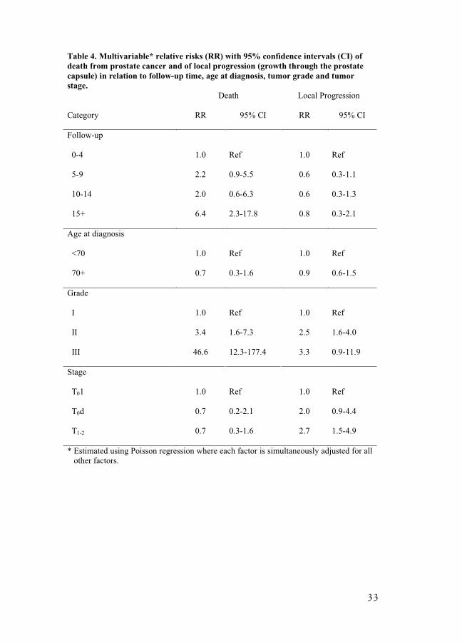
Prostate cancer mortality was slightly higher among patients diagnosed at age 70 or younger than among those diagnosed at older ages (Table 2). Strikingly similar mortality rates were found among patients who had localized non-palpable cancer compared with those who had a cancer in stage T1 or T2. In contrast, the mortality rate was 70 percent higher among men with a non-palpable diffuse cancer. With regard to differentiation, the mortality rate increased drastically from highly to poorly differentiated tumors (Table 2). Multivariable Poisson regression models were fitted in order to quantify the independent effects of follow-up time, age at diagnosis, grade and stage (Table 4). Our analyses showed a significant, about six fold, higher mortality rate after 15 years of follow-up compared with the first five years. The strong prognostic impact of grade, notably of poorly differentiated tumors, was also confirmed. In contrast, neither age at diagnosis nor stage of disease was significantly associated with risk of death due to prostate cancer. A separate model was fitted in which the event of interest was local progression. In this analysis we disregarded if and when regional and/or distant progression/metastases were ascertained. Except for age at diagnosis, the pattern for local
Avh_Ove Andre�n_K160308.indd 29 08-04-17 15.42.37
2929

30
progression was strikingly different from that of death due to prostate cancer (Table 4). Hence, the risk of local progression did not increase over follow-up time, and the association with grade was weak. Moreover, compared with T0l tumors, growth beyond the prostate capsule was two or three times more likely in patients with T0d and T1-2 tumors, respectively.
Avh_Ove Andre�n_K160308.indd 30 08-04-17 15.42.38
3030

31
Table 2. Rates per 1000 person-years with 95 percent confidence interval (CI) for progression to metastatic disease and death due to prostate cancer by years of follow-up, age, stage and grade at diagnosis
Progression Prostate cancer deathCategory Events Rate 95% CI Events Rate 95% CI
Follow-up 1)
0-4 18 20 13 - 32 11 12 7 - 225-9 9 15 8 - 30 11 18 10 - 3310-14 5 16 7 - 38 5 15 6 - 3615+ 7 41 20 - 87 8 44 22 - 88
Age, yrs- 70 26 24 16 - 35 22 19 13 - 29
71 - 13 15 9 - 26 13 15 8 – 25
Stage T01T0d
T1-2
11820
173119
9 - 30 15 - 62 12 - 30
10 817
152816
8 - 2814 - 5710 - 25
Grade III
III
18165
1329242
8 - 21 18 - 47101 - 581
1416 5
10 27194
6 - 1717 - 4481 - 466
Total 39 20 14-27 35 17 12-24
1) Numbers of patients alive after 5, 10 and 15 years of follow-up were 150, 91 and 49 respectively.
Avh_Ove Andre�n_K160308.indd 31 08-04-17 15.42.39
3131

32
Tab
le 3. Th
e 15 and
20 year progression
-free survival (P
FS
), observed
survival (O
S), relative su
rvival (RS
) and
cause-sp
ecific su
rvival (CS
) rates with
95% con
fiden
ce intervals (w
ithin
brack
ets), by stage an
d grad
e at diagn
osis.P
FS
OS
RS
CS
Category
15-year20-year
15-year20-year
15-year20-year
15-year20-year
Stage
T0 1
57.752.5
22.27.5
82.156.1
81.157.9
(38.9 – 76.5)(32.7 – 72.3)
(12.4 – 32,0)(-0.1 – 15.0)
(45.9 – 118.4)(0.6 – 112.8)
(66.5 – 95.6)(28.3 – 87.5)
T0 d
35.917.9
14.70
63.60
69.746.4
(15.1 – 56.5)(-9.5 – 45.3)
(2.6 – 26.8)
(11.1 – 116.1)
(50.1 – 89.2(6.3 – 86.5)
T1-2
38.433.1
23.110.3
75.261.5
80.356.9
(26.8 – 50.1)(20.9 – 45.4)
(15.3 – 30.9)(4.2 – 16.3)
(49.8 – 100.6)(25.2 – 97.9)
(69.8– 90.8)
(34.9 – 78.9)
Grade
I56.0
46.024.3
9.783.7
63.488.9
71.8(44.4 – 67.6)
(30.9 – 61.0)(17.2 – 31.4)
(4.3 – 15.1)(59.4 – 108.0)
(28.1 – 98.8)(81.4 – 96.3)
(54.9 – 88.7) II
28.824.3
18.23.5
64.723.4
64.522.1
(15.2 – 42.3)(10.2 – 38.4)
(8.7 – 27.7)(-1.8 – 8.7)
(30.9 – 98.4)(-12.0 – 58.7)
(47.2 – 81.8)(-7.5 – 51.7)
III
All
15.6*(-12.8 – 43.9)
45.036.0
0
21.5
07.5
0
75.9
0
49.9
28.6*(5.6 – 62.7)
78.754.5
Patients
(35.7 – 54.3)(24.2 – 47.9)
(16.0– 27.0)
(3.5 – 11.4)(56.5 – 95.3)
(23.4 – 76.4)(70.8 – 86.7)
(37.6 – 71.4)
Fo
otn
ote
: * afte
r 7 y
ear.
Avh_Ove Andre�n_K160308.indd 32 08-04-17 15.42.40
3232
33
Table 4. Multivariable* relative risks (RR) with 95% confidence intervals (CI) of death from prostate cancer and of local progression (growth through the prostate capsule) in relation to follow-up time, age at diagnosis, tumor grade and tumor stage.
Death Local Progression
Category RR 95% CI RR 95% CI
Follow-up
0-4 1.0 Ref 1.0 Ref
5-9 2.2 0.9-5.5 0.6 0.3-1.1
10-14 2.0 0.6-6.3 0.6 0.3-1.3
15+ 6.4 2.3-17.8 0.8 0.3-2.1
Age at diagnosis
<70 1.0 Ref 1.0 Ref
70+ 0.7 0.3-1.6 0.9 0.6-1.5
Grade
I 1.0 Ref 1.0 Ref
II 3.4 1.6-7.3 2.5 1.6-4.0
III 46.6 12.3-177.4 3.3 0.9-11.9
Stage
T01 1.0 Ref 1.0 Ref
T0d 0.7 0.2-2.1 2.0 0.9-4.4
T1-2 0.7 0.3-1.6 2.7 1.5-4.9
* Estimated using Poisson regression where each factor is simultaneously adjusted for all other factors.
Avh_Ove Andre�n_K160308.indd 33 08-04-17 15.42.41
3333

34
Figure 2. Observed survival (OS), cause-specific
survival (CS), and relative survival (RS) in a cohort of
223 patients with prostate cancer.
Avh_Ove Andre�n_K160308.indd 34 08-04-17 15.42.42
3434

35
Figure 3. Cause-specific survival by stage of disease at diagnosis:
T0 loc, T0 diff and T1+T2.
Avh_Ove Andre�n_K160308.indd 35 08-04-17 15.42.43
3535

36
Figure 4. Cause-specific survival by tumor grade at time of diagnosis:
Grade I, highly
Avh_Ove Andre�n_K160308.indd 36 08-04-17 15.42.43
3636

37
Paper II
The baseline characteristics of the 240 patients and distribution according to tumor stage, grade of differentiation, Gleason score, Ki67 expression and tumor volume are shown in table 5. Of the 240 patients 118 (49.2%) patients were in stage T1a and 122 (50.8%) in stage T1b. The mean age at diagnosis was 73 years. Eighty-four percent (n=167) of the cases had either Gleason score 6 or 7 and only 4% (n=11) had a Gleason score below 6. 175 patients (74%) had nuclear grade 1. By the end of the observation period in September 2003, a total of 207 of the 240 patients (86%) (Table 5) had died. Prostate cancer was a more common cause of death in patients 70 years of age and younger in comparison with those over 70 years of age.
The age-adjusted risk of dying in prostate cancer with respect to grade of differentiation, Ki67, Gleason score and tumor volume is shown in table 2 (Table 6). The risks tended to rise with increasing Gleason score, nuclear grade and tumor volume (p for trend<0.05). There was a 9-fold statistically significant increased risk for those with the highest Gleason score (8–9) compared to those with the lowest (4–6). Among patients with Gleason score 7 a Gleason 3 predominant pattern (3+4) was not significantly different from a Gleason 4 predominant pattern (4+3) (data not shown). Nuclear grade 3 tumors hade an almost eleven fold higher risk to die in prostate cancer compared with tumors with nuclear grade 1. After adjustment, cases with T1b tumors (ratio > 5%) were 2.5 times more likely to die of their disease than cases with T1a tumors (HR=2.5; 95% CI, 1.1–5.7).
In the multivariate model Gleason score, nuclear grade and tumor volume remained statistically significant independent prognostic markers of prostate cancer death. Ki67 was highly correlated with Gleason grade, and the trend of increasing mortality rates with rising values of Ki67, that was observed in the univariate analysis, disappeared after adjustment for the other variables.
The sensitivity and specificity of the Gleason grading system at different cut-off points is presented in table 7. Setting the cut-off value to �7 resulted in a sensitivity of 0.64 and a specificity of 0.66. As the cut-off was increased by one step, the corresponding values fell to 0.33 and increased to 0.92, respectively. Moreover, the positive predictive value of Gleason �7 was 0.29 and the negative predictive value 0.89. With a cut-off of 8, the corresponding values were 0.48 and 0.87, respectively.
Avh_Ove Andre�n_K160308.indd 37 08-04-17 15.42.44
3737

38
Table 5. Characteristics of 240 patients with incidental prostate cancer (T1a-b, Nx, M0) who received no initial treatment, according to age, nuclear grade, Gleason score, tumor volume and Ki67 expression at time of diagnosis.
TotalN
Prostate cancer deaths
Deaths by
other cause
Alive MeanSurvival(Month)
MinimumSurvival(Month)
MaximumSurvival(Month)
Age < 70 80 16 42 22 138 16 280 > 70 160 26 123 11 90 1 272
Nuclear-grade* 1 175 20 124 31 98 1 280 2 45 13 31 1 94 4 229 3 16 8 7 1 59 1 158
Gleason Score 4 4 - 2 2 132 56 167 5 7 3 3 1 122 5 235 6 136 12 99 25 118 1 284 7 64 13 46 5 96 1 230 8 26 13 13 0 73 4 229 9 3 1 2 0 49 17 85
Ratio chips with cancer (%)** �5 118 9 87 22 102 1 237 6-25 92 20 61 11 97 1 280 26-50 13 6 7 0 85 17 153 >50 17 7 10 0 54 4 143
Ki67 (mean) Q1 (101) 40 6 24 10 123 6 280 Q2 (112) 36 5 26 5 105 10 273 Q3 (117) 39 10 26 3 94 1 228 Q4 (122) 37 7 26 4 96 4 212 Q5 (129) 38 9 25 4 98 8 211*Only 236 patients where graded according to nuclear grade.** T1a has a ratio � 5% T1b has a ratio > 5%
Avh_Ove Andre�n_K160308.indd 38 08-04-17 15.42.45
3838

39
Table 6. Hazard Ratio (HR) and 95% Confidence Interval (CI) of death from prostate cancer in relation to age, nuclear grade, Gleason score, tumour volume and Ki67 expression.
HRa 95 % CI HRb 95% CIAge <70 1.0 1.0 >70 1.5 0.8-2.8 1.4 0.6-3.2
Nuclear grade I 1.0 1.0 II 3.5 1.7-7.1 1.4 0.5-3.5 III 10.7 4.5-25.4 3.4 1.1-10.7p ( trend) 0.03
Gleason score 4-6 1.0 1.0 7 2.5 1.2-5.3 1.5 0.5-4.2 8-9 9.5 4.5-20.2 3.3 1.0-11.6p ( trend) 0.04
Ratio chips with cancer (%) �5 1.0 1.0 6-25 3.0 1.3-6.6 1.9 0.6-5.8 25-50 10.3 3.5-30.5 3.6 0.8-15.7 50 16.8 6.0-47.5 5.2 1.2-22.8p(trend) <0.01
Ki67 Q1 (medelvärde för varje Q) 1.0 1.0 Q2 1.2 0.4-4.2 1.2 0.3-4.0 Q3 2.6 0.9-7.6 1.4 0.4-4.9 Q4 1.9 0.6-6.0 0.9 0.2-3.2 Q5p(trend)
2.5 0.9-7.4 1.3 0.4-4.20.86
aage-adjusted badjusted for all other variables
Table 7. Sensitivity, specificity, positive predictive value (PPV) and negative predictive value (NPV) of Gleason grading in predicting prostate cancer death.
Gleason Score* Sensitivity Specificity PPV NPV
� 6 0.92 0.04 0.17 0.73
� 7 0.64 0.66 0.29 0.89
� 8 0.33 0.92 0.48 0.87
*cut-off point
Avh_Ove Andre�n_K160308.indd 39 08-04-17 15.42.46
3939
40
Paper III
AMACR protein expression was evaluated manually by the study pathologist and graded on a 4-tiered scale, demonstrated a significant difference in intensity between prostate cancer (mean score=3.14/4) and benign prostate epithelium (mean score=1.3/4). Using these categorical scores for AMACR, no significant associations between AMACR intensity and biochemical failure were observed in the surgical series consistent with previous observations59.
Using the semi outomated analysis, in the surgical series using biochemical failure as the endpoint, lower AMACR intensity was associated with a worse outcome (Tables 8 and 9). We established a dichotomous cut point for AMACR intensity of –1.11 SD (i.e. samples with minimum AMACR intensity of –1.11 SD below that of mean), wherein, 37,5% of patients with AMACR intensity scores below the cut point progressed as compared to 14,5% ofpatients with AMACR intensity scores above the cut point (P=0.0002). This univariate association can be visually appreciated by Kaplan-Meier analysis (Figure 5). Using this AMACR cut point in multivariate analysis, patients with AMACR expression levels below the threshold were at a significantly higher risk of developing PSA recurrence (HR=2.12; 95% CI 1.04–4.32) after adjusting for pre-operative PSA, Gleason score and surgical margin status.
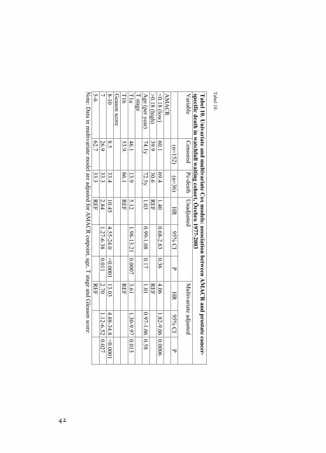
Doing the same procedure in the watchfull waiting cohort using prostate cancer death as endpoint. We determined an optimal cutpoint of 0.18 SD (HR=4.06; 95% CI 1.82–9.06). As demonstrated in table 10 and figure 8, a significant cut point was identified in multivariable analysis that takes patient age, Gleason score, and tumor stage into account. Although this cut point is different from the one derived using biochemical failure, we also observe that lower AMACR intensity is associated with worse outcome, in this case cancer specific death.
Using the surgical cutpoint –1.1SD, AMACR intensity did not predict prostate cancer specific survival. In contrast, there was evidence that AMACR levels below the cutpoint determined by prostate cancer death was significantly associated with time to biochemical failure at the univariate level as demonstrated by the Kaplan-Meier analysis (Figure 6). On multivariate analysis, AMACR expression, Gleason score, surgical margin status, and pre-treatment PSA were all independently associated with PSA biochemical failure (HR= 3.67; 95% CI 1.25–10.78).
Avh_Ove Andre�n_K160308.indd 40 08-04-17 15.42.48
4040

41
Table 8. Univariate Cox Regression Analysis for Associations between AMACR intensity and Prostate Specific Biochemical FailureFactor P-value Hazard Ratio 95% CI for
HRAMACR(min>-1.11 vs. <=-1.11) 0.0003 0.34 0.19 0.60
Gleason Score �7 vs. <7) 0.0003 4.36 1.96 9.73
SM (Positive vs. Negative) <.0001 3.61 2.26 5.76
Path stage <.0001 3.01 2.09 4.34
ln(PSA) <.0001 2.41 1.72 3.38
Tumor Size (>2 vs.�2 cm) <.0001 3.49 1.91 6.37
DRE (Positive vs. Negative) 0.03 1.90 1.07 3.36
Gland Weight 0.94 1.00 0.99 1.02
Age 0.61 1.01 0.97 1.05
EPE <.0001 2.79 1.97 3.94
Table 9. Multivariate Cox model: Independent associations of AMACR,Gleason score, PSA and positive margin with PSA Recurrence after Radical Prostatectomy
Parameter P-valueHazard Ratio
95% CI for HR
AMACR(min>-1.11 vs. <=-1.11) 0.001 0.36 0.20 0.66
Gleason(>=7 vs. <=6) 0.04 2.35 1.02 5.40
SM (Positive vs. Negative) 0.001 2.78 1.53 5.03
ln(PSA) 0.001 1.89 1.31 2.74
Avh_Ove Andre�n_K160308.indd 41 08-04-17 15.42.49
4141
42
Tabel 10.
Note: D
ata in multivariate m
odel are adjusted for AM
AC
R cutpoint, age, T
stage and Gleason score
Tab
el 10. Un
ivariate and
mu
ltivariate Cox m
odels: association
betw
een A
MA
CR
and
prostate can
cer-sp
ecific death
in w
atchfu
ll waitin
g cohort, Ö
rebro 1977-2003
Variable
Censored
Pc-death
Unadjusted
Multivariate adjusted
(n=152)
(n=36)
HR
95% C
IP
HR
95% C
IP
AM
AC
R<
0.18 (low)
60.169.4
1.400.68-2.83
0.364.06
1.82-9.060.0006
>0.18 (high)
39.930.6
RE
FR
EF
Age (per year)
74.1y72.5y
1.030.99-1.08
0.171.01
0.97-1.060.58
T stage
T1a
46.113.9
5.121.98-13.21
0.00073.61
1.30-9.970.013
T1b
53.9.86.1
RE
FR
EF
Geason score
8-108.5
33.410.45
4.55-24.0<
0.000113.03
4.88-34.8<
0.00017
26.933.3
2.841.27-6.38
0.0112.70
1.12-6.520.027
5-662.7
33.3R
EF
RE
F
Avh_Ove Andre�n_K160308.indd 42 08-04-17 15.42.50
4242

43
Figur 5.
Fig 6.
Avh_Ove Andre�n_K160308.indd 43 08-04-17 15.42.51
4343

44
Paper IV
The baseline characteristics of the 195 patients are presented in table 11. Mean MUC-1expression for all 195 patients was 107.3 (range 95–179, SD 10.2). Of the 43 patients that had a MUC-1 intensity close to normal tissue (102.5–106) (Fig. 7 A-B) three (7%) died of prostate cancer, compared with 34 (23%) of the 152 patients that deviated from the normal MUC-1intensity (Fig. 7 C-D). As illustrated in table 12, there was no correlation between Gleason score and MUC-1 intensity (p-value 0.8), while there was a tendency to correlation between tumor extent and MUC-1 (p-value 0.08). The age-adjusted risk of dying of prostate cancer with respect to MUC-1, Gleason score and tumor extent is presented in table 12. The risk of dying of prostate cancer was four times higher among those with a higher [HR 3.9 (95% CI, 1.1–14)] or lower [HR 3.8 (95%CI, 1.1–13)] MUC-1 expression than among those with a MUC-1expression within the normal range (Fig 8). After adjusting for tumor extent and Gleason score, the effect of MUC-1 was even stronger [HR 5.1(95%CI, 1.4–18)] and [HR 4.5 (95%CI, 1.3–15)], respectively indicating that MUC-1 predicts prostate cancer death independently of clinical parameters (Fig 9).
We further cross-classified participants on MUC-1 and Gleason score. The group with Gleason score � 7 and MUC-1 lower or higher then normal had a 17 [HR 17.1 (95%CI, 2.3–128)] times higher risk of prostate cancer death compared with tumors with Gleason score < 7 and normal MUC-1 intensity (Table 14). We further assessed the ability of MUC-1 expression (deviating from the normal range) to correctly classify prostate cancer cases as indolent or lethal (defined as progressing to metastases and/or death). The sensitivity for MUC-1 as predictor of lethal prostate cancer was 0.91 while the specificity was 0.25 (Table 15). When combined with information on Gleason score, the specificity increased to 0.75 but the sensitivity decreased to 0.56.
Avh_Ove Andre�n_K160308.indd 44 08-04-17 15.42.53
4444

45
Table 11. Characteristics of 195 patients with incidental prostate cancer (T1a-b, Nx,M0) who received no initial treatment, according to age, Gleason score, tumor extent, and MUC-1expression at time of diagnosis 1977-1991
N Prostate cancer deaths
Deaths by other
causes
Alive Meansurvival
Minimumsurvival
Maximumsurvival
Age <70 56 13 27 16 138 16 280 > 70 139 24 105 10 90 1 272Gleason score 4 3 - 1 2 132 56 167 5 7 3 3 1 122 5 235 6 107 10 77 20 118 1 284 7 53 12 38 3 96 1 230 8 22 11 11 0 73 4 229 9 3 1 2 0 49 17 85Percentage of chips with cancer <5% 80 6 58 16 102 1 237 6-25% 85 18 57 10 97 1 280 26-50% 13 6 7 0 85 17 153 >50% 17 7 10 0 54 4 143MUC-1 Normal 43 3 35 5 118 4 284 Low 68 16 38 14 108 1 280 High 84 18 59 7 100 1 238
Table 12. Correlation between MUC-1 intensity expression and age, Gleason score and tumor volume
MUC-1 intensityFactor Normal
N (%)Low
N (%)High
N (%)p-value
Age<70 10 (23) 25 (37) 21 (25)Age>70 33 (77) 43 (63) 63 (75)
0.188
Gleason 4-6 25 (58) 41 (60) 51(61)Gleason 7 13 (30) 20 (29) 20 (24)Gleason 8-9 5 (12) 7 (10) 13(16)
0.826
Percent chips< 5% 15 (35) 23 (34) 42 (50)Percent chips 5-25% 23 (54) 33 (49) 29 (35)Percent chips 25-50% 0 (0) 5 (7) 8 (10)Percent chips >50% 5 (12) 7 (10) 5 (6)
0.085
Avh_Ove Andre�n_K160308.indd 45 08-04-17 15.42.54
4545

46
Table 13. Hazard Ratio (HR) and 95% Confidence Interval (CI) for prostate cancer death in relation to protein expression of MUC-1 in tumor tissue from patients with localized prostate cancer.
Na Prostate cancer deaths
Crude HR (95% CI)
HRb (95% CI)
MUC-1 (intensity) Normal (102.5-106) 43 3 1.0 1.0 Low (<102.5) 68 16 3.9 (1.1-14) 5.1 (1.4-18) High (>105.5) 84 18 3.8 (1.1-13) 4.5 (1.3-15)a A total of 195 patients were assayed for MUC-1. b Adjusted for age, Gleason score, and tumor extent.
Table 14. Hazard ratio (95% CI) of prostate cancer death associated with MUC-intensity and Gleason score, cross classified.
Normal MUC-intensity Aberrant MUC-intensityPC death/ Total N
HR (95% CI)PC death/ Total N
HR (95% CI)Gleason < 7 1/25
Ref.12/92
3.8 (0.5-29)Gleason � 7 2/18
3.8(0.3-43)22/60
17.1 (2.3-128)
Table 15. Sensitivity, specificity, positive predictive value (PPV) and negative predictive value (NPV) of Gleason grade and MUC-1 intensity in predicting prostate cancer death.Gleason Score and MUC-1intensity
Sensitivity Specificity PPV NPV
Aberrant MUC-1 intensity 0.91 0.25 0.22 0.93
Gleason >6 and Aberrant MUC-1 intensity
0.59 0.76 0.37 0.89
Avh_Ove Andre�n_K160308.indd 46 08-04-17 15.42.55
4646

47
Figure 7. Tissue micro array analysis of MUC-1 immunohistochemistry:
selected images of TMA cores representing normal, high, and low MUC-1
intensity
Fig A, B. Normal MUC-1 intensity
Fig C, D. High (C) and Low (D) MUC-1 intensity
A B
C D
Avh_Ove Andre�n_K160308.indd 47 08-04-17 15.42.56
4747

48
Figure 8.
Avh_Ove Andre�n_K160308.indd 48 08-04-17 15.42.59
4848

49
Avh_Ove Andre�n_K160308.indd 49 08-04-17 15.42.59
Figure 9
4949


50
Paper V
Background data obtained through computerized registry linkage are shown in table 16. Most patients with incidental PCa are older patients, about 50% are 75 years or older (mean age 74.4) and somewhat older than patients in the comparison group, i.e. men diagnosed on clinical grounds alone. The mean age at incidental PC diagnosis increased slightly over time from 72.5 in 1970–79 to 75.2 years in the last calendar period 1999–2003. The age standardized incidence of incidental PCa peaked in the early nineties (Fig 10) and successively declined during subsequent years, reaching 12% during the period of 1999–2003. We observed a marked change in hospitalization time over calendar time. Initially, in the early seventies, when the transurethral technique was introduced, the hospitalization time was comparably long, lasting about 2–3 weeks. After a continuously and dramatic decrease the hospitalization time leveled off at 4–5 days during the last 5-year period. During the entire study period, 1970–2003, the age standardized PCa incidence rate raised continuously and increased almost 15 times from 1973 to the mid eighties. The upward trend appeared to be driven by the increased frequency of TUR-P, but the incidence rate rose continuously even after the TUR-P frequency had leveled off.
At the end of 2003, 6300 patients (27.1%) with incidental PCa had died due to PCa and a majority of them (50,9%) had died of intercurrent causes, while 5138 patients (22,1%) were still alive. A somewhat higher death rate due to PCa was found in the comparison group consisting of clinical detected PCa (33.2%). The age standardized cumulative incidence of PCa death is demonstrated in fig 11. The corresponding curves by time period and by mode of detection are shown in fig 12. We observed a clear decreasing trend over time after diagnosis, but the prostate cancer specific mortality decreased both according to mode of detection and calendar time. The mortality was higher among those with incidentally detected tumors compared to those in the comparison group. However, it should be noted that although the prostate cancer specific mortality among those with incidental PCa leveled off by time, it was still 30% after 15 years of follow up after diagnosis.
Avh_Ove Andre�n_K160308.indd 50 08-04-17 15.43.00
5151

49
Figur 10. Age-standardized prostate cancer incidence and relation to history of TUR-P, in Sweden between 1970 and 2003.
1970 1975 1980 1985 1990 1995 2000 2005
Year
0
50
100
150
200
250
Age
sta
ndar
dize
d in
cide
nce
of
pro
stat
e ca
ncer
(pe
r 10
0 00
0 m
en)
TURP-detected
Not TURP detected
All PC
Avh_Ove Andre�n_K160308.indd 49 08-04-17 15.42.59
5052

Tabel 16. Characteristics of 23288 prostate cancer patients diagnosed through or hospitalized for TUR-P between 1970 and 2003 in Sweden
TURP-hospitalisations
TURP-diagnosed patients PC-comparisons
N % N % N %
Age
<55 708 (0.9) 123 (0.5) 2462 (2.2)
55-64 8931 (11.6) 2157 (9.3) 17740 (15.8)
65-74 30854 (40.2) 9085 (39.) 43060 (38.4)
75-84 31163 (40.6) 10114 (43.4) 40769 (36.3)
85+ 5122 (6.7) 1809 (7.8) 8173 (7.3)
Years
1970-79 12324 (16.1) 2627 (11.3) 13045 (11.6)
1980-86 20793 (27.1) 6693 (28.7) 17116 (15.3)
1987-90 14409 (18.8) 4961 (21.3) 13311 (11.9)
1991-93 10106 (13.2) 3349 (14.4) 12480 (11.1)
1994-98 9790 (12.8) 2795 (12.0) 20270 (18.1)
1999-2003 9356 (12.2) 2863 (12.3) 35982 (32.1)
Hospitalisation time
0-3 days 17978 (23.4) 5680 (24.4) NA NA
4-7 days 34758 (45.3) 10997 (47.2) NA NA
8-14 days 14964 (19.5) 4175 (17.9) NA NA
2-4 weeks 6655 (8.7) 1844 (7.9) NA NA
4+ weeks 2423 (3.2) 592 (2.5) NA NA
Nr TURP-treatments
0 NA NA 0 (0.0) 82389 (73.4)
1 NA NA 18007 (77.3) 23689 (21.1)
2 NA NA 4118 (17.7) 4719 (4.2)
3 NA NA 864 (3.7) 1062 (0.9)
4+ NA NA 299 (1.3) 345 (0.3)
Status at end of follow up 20031231
Alive NA NA 5138 (22.1) 39527 (35.2)
Dead from other causes NA NA 11850 (50.9) 35469 (31.6)
PC-death NA NA 6300 (27.1) 37208 (33.2)
Follow up
Time in years , mean (sd) NA NA 6.1 (4.7) 4.3 (4.0)
Avh_Ove Andre�n_K160308.indd 51 08-04-17 15.43.01
53

Figur 11. Cumulative incidence of prostate cancer deaths in Sweden between 1970 and 2003, in relation to history of TUR-P.
0 2 4 6 8 10 12 14 16
Time (years)
0
0.1
0.2
0.3
0.4
0.5
Cum
ulat
ive
inci
denc
e o
f pr
ost
ate
can
cer
dea
thTURP-detected ( n = 23288 )
Not TURP-detected ( n = 112204 )
Avh_Ove Andre�n_K160308.indd 52 08-04-17 15.43.02
54

53
DISCUSSION
Paper I
Although our cohort of patients with early stage, initially untreated, prostate cancer has been previously followed-up in great detail during on average 15 years 76, the additional six years included in this analysis revealed an unexpected change in prognostic outlook; the cause-specific survival rate dropped by almost 25 percentage points reflecting an approximate threefold increase in prostate cancer mortality rate compared to the first 15 years of follow-up. This change occurred consistently across stage and grade except for poorly differentiated cancers in which excess mortality becomes manifest already during early follow-up. We were unable to conceive any bias that could have spuriously generated these recent findings. Indeed, the internal validity of our population-based study should be high because we achieved complete follow-up and used standardized procedures for clinical examination, ascertainment of disease progression and classification of death. Moreover, the slight difference between cause-specific and relative survival estimates was largely consistent over time. This argues against any shifting criteria for classification of cause of death since the relative survival rate reflects excess mortality (compared to mortality in the general population) and is thus unaffected by any subjective judgment. Hence, chance is the only realistic alternative to a real deterioration in prognosis after long-term follow-up and the level of statistical significance argues against this explanation.
If our data reflect a real phenomenon, they would imply that the probability of progression from localized and indolent to metastatic mortal disease increases markedly after long-term follow-up. This progression is not restricted to cancers diagnosed due to clinical symptoms, but includes also tumors detected incidentally at transurethral resection due to presumed benign prostatic hyperplasia. Our survival data – supported by biopsies during follow-up –would further imply that these latter lesions are either incompletely removed or multifocal with malignant clones left at a transurethral resection. Contrary to emerging views 90, 91 our data also suggest that metastases may arise as a consequence of late mutations rather than being determined already by the early mechanisms of malignant transformation. According to a rival interpretation, the phenomenon we observed reflects transformation of new, more aggressive cancer clones rather than progression of those initially detected. Empirical testing of these complementary, but not mutually exclusive, theories seems difficult. It may be difficult to validate our survival data in any new cohort study of watchful waiting since aggressive treatment of localized prostate cancer has become more routine now than 25 years ago when we started to assemble our cohort 92-95. Indeed, it has been estimated that about 60,000 men undergo radical prostatectomy yearly in the United States alone and the number performed annually in England increased nearly 20-fold between 1991 and 1999 96. This development towards treatment with a curative intent may further accelerate following recent documentation that radical prostatectomy reduces prostate cancer mortality by about 50 percent 97. Hence, support for our findings has to be found chiefly in existing studies of watchful waiting. Other such cohorts are, however, few and small, none of them are population-based and prospective, 98-101 and virtually no follow-up data are available beyond 15 years after diagnosis. Within these constraints, experience during the first 10 years after diagnosis is strikingly similar in existing cohorts of patients with early stage prostate cancer left without initial treatment with a favorable course of the disease for men with highly or moderately differentiated tumors 3.
Avh_Ove Andre�n_K160308.indd 53 08-04-17 15.43.09
54 55

54
From a public health perspective, implications of late progression from early stage to mortal disease may not be dramatic because without PSA testing, average age at diagnosis of prostate cancer is so high that competing causes of death predominate (Table 1). Although it is well established 1, 50 that an excess death rate continues long-term in population-based cohorts of prostate cancer patients, these data do not enable distinction of deaths generated by patients initially diagnosed with localized disease. In our entire cohort, 25 out of 223 (11 percent) patients died from prostate cancer within 15 years of diagnosis, and an additional 10 during subsequent follow-up until a time when only 9 percent of all patients in the cohort were still alive and therefore at risk of progression. Assuming that radical prostatectomy prevents approximately 50 percent of prostate cancer deaths 97, about 18 patients in our entire cohort (8 percent) – that is 0.5 x 35 – would have experienced a survival benefit whilst the remaining 205 would not. However, among elderly men, reducing the risk of death from prostate cancer by a certain amount may have limited impact on their overall survival.
Our data may be important for counseling and clinical management of individual patients.First, postponement of death is not the only treatment objective since local progression may create substantial suffering. Indeed, many of our patients experienced symptomatic local growth without generalized disease (Table 1) requiring treatment with estrogens or orchidectomy. Obviously, radical prostatectomy is a major procedure with substantial side-effects, chiefly impotence and incontinence102, 103. Because these complications are surprisingly well tolerated 6 many patients may prefer a radical prostatectomy even if prolonged survival is an uncertain consequence. Our data may be particularly relevant to otherwise healthy men diagnosed with prostate cancer at an early age. If such patients are in their 60s or younger, disease progression occurring after 15 or more years may be a real concern arguing for early local treatment with a curative intent. In patients with a PSA detected cancer 22, such counseling is, however, complicated by the fact that a lead time that cannot be individually determined, has to be added to the approximately 15 years that may precede more rapid tumor progression. One important and complicated question is how the findings of this study relate to the current era when many patients are detected by means of PSA testing. The results are directly relevant for patients with clinical disease diagnosed before the PSA era and also to preclinical disease detected at transurethral resection for presumed benign prostatic hyperplasia. Indeed, as shown in Table 4, these two categories of patients experienced similar risk of dying from prostate cancer. The natural history we have described, reflect also what would happen among PSA detected cancer if the lead-time couldbe accommodated and the patients were left without early therapeutic intervention. However, a substantial proportion of PSA diagnosed cancers represents over detection of sub clinical disease. These cancers would never have surfaced clinically during the subject’s lifetime –either because they are indolent or because death occurs from competing causes before clinical manifestation of the malignancy. By definition, these cancers do not generate any mortality.
In conclusion, our data indicate that the probability of progression to a more aggressive and lethal phenotype may increase after long-term follow-up of prostate cancers that are diagnosed at an early stage and initially left without treatment. These findings provide no information on the possible benefit of early PSA testing followed by aggressive treatment in men with sub clinical disease. However, they argue for early radical treatment of patients with clinical disease and a long-life expectancy. Not only would such surgical intervention potentially prevent deaths; it would also convey prevention from disability caused by local tumor growth.
Avh_Ove Andre�n_K160308.indd 54 08-04-17 15.43.10
5556

55
Paper II
In this population-based cohort of incidentally discovered localized prostate cancer cases (T1a-b, Nx, M0) left without initial treatment, our main focus was evaluation of clinical determinants of prostate cancer death. Deriving from a follow-up of more than 20 years, our results confirm the findings of earlier studies that Gleason score is a predictor of prostate cancer death 32, 98, 104, 105. However, the predictive value of the Gleason grading is noticeably low.
The study cohort is well characterized, carefully followed during an extended period of time and no patients have been lost to follow up. Validation of the classification of causes of death against the Swedish Death Register showed a correlation exceeding 90%, in our previous study76. The population-based study design with inclusion of all consecutive cases with newly diagnosed, incidentally discovered, prostate cancer from a strictly defined catchment area, further alleviates concerns of selection bias. Our results should therefore be both valid and generalizable.
Like earlier longitudinal cohort studies using prostate cancer death as outcome we found Gleason and nuclear grade to be a determinant of prostate cancer death98, 104-106. There are today many articles like the one of Roehl et al106 that uses biochemical recurrence-free survival (bRFS) as end point. Using bRFS as surrogate endpoint for prostate cancer specific death has been debated107, 108, and as long as there is no clear evidence that there is a good correlation between bRFS and prostate cancer specific death it is difficult to compare studies with different end points. However, the accuracy of using the Gleason grading system to correctly classify patients according to their risk of dying in prostate cancer was rather poor. As a consequence, the positive predictive value of the Gleason grading system is expected to be low. In these data, even a high set cut-point (Gleason score �8) could only forecast a 50% probability of dying in prostate cancer. Moreover, it appears that the evaluation according tothe Gleason system has shifted towards intermediary values over time and in Sweden, more than 70% of the localized tumors are presently graded as Gleason 6 or 7 4 (Fig.13). In the present data more than 80% were diagnosed with Gleason 6 or 7. A grading system that categorizes a majority of the cases into one group with highly variable outcomes is obviously insufficient. In view of the increasing diagnostic activities in the Western world and the resulting over-treatment of indolent prostate cancers, the need for other or additional prognostic markers becomes even more evident.
The previous findings 55, 58, 109 of Ki67 as a prognostic marker for prostate cancer outcome could not be confirmed in these data. Possible explanations may be the comparably long follow-up time of the present study or the absence of more advanced tumors. In another follow-up study of tumors detected through transurethral resection (TUR-P), Ki-67 was found to be a prognostic marker of prostate cancer death, but this study included patients with more advanced disease (T1-T3, M0–M1)58. Increased Ki-67 expression may precede the onset of more advanced disease, such as local infiltration and metastases, which may explain the difference between the study results. Comparisons with studies using PSA-recurrence as outcome are furthermore difficult55, 109, because of limitations with surrogate end-points and the potential effects of surgical intervention.
Furthermore, the present data confirm the previous findings of tumor volume at diagnosis as an important predictor of outcome 8, 110. Yet, in clinical practice the value of tumor volume is limited since it is difficult to assess other than through TUR-P.
Avh_Ove Andre�n_K160308.indd 55 08-04-17 15.43.11
56 57

56
In summary, these data draw attention to the apparent limitations of the most frequently used prognostic tools and highlight the need for improvement in this field. Considering the increasing numbers of early detected prostate cancers and the consequent over-treatment of many benign tumors, further efforts in finding other, or complementary, indicators of prostate cancer outcome are warranted.
Fig 13. Gleasonscore in localized tumours (T0-T2,Nx,M0) in patients diagnosed in Sweden 1996 and 2002. Data from the Swedish Nationel Prostate Cancer register.
Gleasonscore 1996 och 2002 in localized tumours
0
5
10
15
20
25
30
35
40
45
50
1996 2002
Year
Pe
rce
nta
ge
2 3 4
5 6 7
8 9 10
Avh_Ove Andre�n_K160308.indd 56 08-04-17 15.43.12
5758

57
Paper III
This is the first demonstration that AMACR protein expression levels are associated with long-term clinical outcome. Our original observations suggested that AMACR expression decreases with prostate cancer progression59, 64. However, standard immunohistochemical evaluation failed to identify clinically significant associations between AMACR expression and biochemical failure 59, 111, 112. Using a more sensitive technique to measure AMACR protein expression, we were able to identify an AMACR cut point that was associated with a three times greater chance of biochemical recurrence and a three to seven greater risk of developing cancer specific death. These observations were independent of other conventional clinical and pathologic parameters in multivariable analysis.
The paradigm for developing prostate cancer biomarkers has focused on using PSA biochemical failure as the surrogate endpoint for the development of metastatic disease and cancer specific death. Studies based on surrogate endpoints are inherently less reliable than studies with clinical endpoints such as cancer specific death113. PSA failure is considered a surrogate for the progression of metastatic disease and prostate cancer specific death114, 115. Indeed, studies by Pound et al. support the potential role of PSA failure as a clinically relevant surrogate endpoint115. For example, of 304 men who develop a PSA failure following radical prostatectomy for clinically localized disease, 103 (34%) developed metastatic disease with a mean actuarial time to metastases of 8 years following the initial PSA elevation. The Pound study also demonstrated that time from initial diagnosis to PSA failure and PSA doubling time predicted metastatic disease. These finding suggest that biochemical failure per se is not a complete surrogate for clinical outcomes, in particular death, and should take into account PSA kinetics as well116. Additional support that biochemical failure may not be an optimal endpoint was recently reported by D’Amico et al.117. They evaluated surrogate endpoints for prostate cancer-specific mortality in two multi-institutional databases of over 8669 patients with prostate cancer treated with surgery (5918 men) or radiation (2751 men) The post-treatment PSA doubling time was statistically significantly associated with time to prostate cancer-specific mortality and with time to all-cause mortality.
As the current study demonstrates, the optimal definition of biochemical failure is still controversial and may be critical for the development of prostate cancer biomarkers. We observed that the AMACR cutpoints differed depending on whether it was derived from the PSA failure or cancer specific death. Biochemical failure may be due to other causes unrelated to the biology of prostate cancer such as local recurrence due to positive surgical margins. Moreover, given the slow progression of prostate cancer, even eight years may not be sufficient to identify all of the patients who will die from prostate cancer. Therefore censoring underestimates the number of men who will experience prostate cancer progression. The work from Pound et al. and D’Amico et al. would suggest that the surrogate endpoint needs further consideration.
The use of prostate cancer specific death, to develop a biomarker test is likely a more valid strategy. In the current study, using cancer specific death to determine the AMACR cutoff yielded a different cutoff from that derived from the surrogate endpoint. Using this revised cutoff on the patients treated for clinically localized disease, AMACR expression independently predicted PSA relapse even accounting for pre-treatment PSA, surgical margin status, and Gleason score. The high-risk group included 81% of the patients in this surgical cohort suggesting that using PSA recurrence as the surrogate endpoint, we are missing some patients who would have progressed. This finding is not what one would anticipate if
Avh_Ove Andre�n_K160308.indd 57 08-04-17 15.43.14
58 59

58
biochemical failure also included “false positives” due to other cases such as surgical technique. If these data are confirmed then risk of progression given sufficient time is not being adequately measured using one elevation of PSA following treatment. The most recent review of the entire Örebro watchful waiting series identified a significant increase in prostate cancer specific mortality beyond 15 years of follow up118. This would also further support the need for better prognostic markers. In the randomized Swedish study, survival benefits were observed for surgery over watchful waiting but the absolute reduction in prostate cancer mortality was small 97. Longer follow up might demonstrate even greater benefits from localized therapy give the significant percentage of men who developed metastatic disease over the 8 years of follow up97.
This study also highlights one of the technical limitations of prior biomarker work. Most studies to date, including our own work, have used manual evaluation dividing immunostaining results into a small number of categories. Although reasonable repro-ducibility may be achieved using a 4-tiered (i.e., negative, weak, moderate, or strong), 3-tiered or dichotomous scale, the range of expression is compressed and the ability of the pathologist to distinguish, for example, various shades of moderate staining intensity is not possible. The current study illustrates how critical this might be in the development of prostate cancer biomarkers. Several studies on AMACR failed to identify an association with decreased expression and prostate cancer progression 59, 60, 64, 111, 112. Our initial observations suggested that AMACR expression decreased with cancer progression as both at the transcriptional level and protein level AMACR was lower in metastatic prostate samples 59, 64. However, using manual standard pathology review, no clinical associations were discovered. More recent work using a fluorescence-based approach confirmed lower AMACR expression in metastatic samples. This finding suggested that a quantitative method might be required to identify a critical cutoff to distinguish clinically localized tumors with subtle differences in AMACR expression levels 65. The current study confirmed this hypothesis. No significant cutoff could be determined using a standard evaluation of immunohistochemical markers by the pathologist. In contrast, the use of a continuous scale of protein measurement allowed us to identify a critical cutoff. This cutoff could then be translated into a specific test using an intensity level that was not appreciated despite numerous studies on AMACR to date.
The use of this semi-automated quantitative method for biomarker analysis is promising. However, the clinical application of using such systems still in its early phases, and the results need to be validated and tested in different cohorts and standardization needs to be worked out. Specifically, can we determine a cut point that would be useful to apply to prostate needle biopsies in the clinical assessment of men with clinically localized prostate cancer? One future approach would be to evaluate prostate needle biopsies for AMACR staining intensity and determine if this information added to currently employed clinical nomograms. As recently described by Kattan et al., one could explore to see if the addition of the biomarker, in this case AMACR intensity, improves the ROC area under the curve as compared to a traditional nomogram 119, 120.
The current study now extends the potential utility of AMACR as prostate cancer biomarker. The AMACR (p504s) antibody is gaining wide acceptance in clinical practices as an adjuvant tool in the work up diagnostically challenging prostate lesions referred to as atypical small
acinar proliferation (ASAP) or atypical suspicious for cancer59, 60, 64, 111, 112, 121-126.
Pathologists can now use AMACR (p504s) in combination with a basal cell marker to help make a more definitive diagnosis. We also recently observed a measurable humoral response directed against the AMACR protein127. Using an ELISA assay, this response could be
Avh_Ove Andre�n_K160308.indd 58 08-04-17 15.43.15
5960

59
measured in serum samples from men with known prostate cancer but not age-matched controls. This early work suggests that AMACR expression, despite the fact that it is not a secreted protein, may potentially become a clinically useful serum test. A second tissue based assay can also detect enzymatic activity in prostate needle biopsy samples and may represent another means of determining AMACR protein expression in a more reproducibly 128.
In summary, we demonstrated for the first time the AMACR expression is associated with prostate cancer progression after examining tumors from over 440 men diagnosed with clinically localized prostate cancer. Selection of the optimal test cut point came from analysis using prostate specific death and not a surrogate endpoint, suggesting that the definition of the surrogate endpoints, such a PSA biochemical failure need to be further evaluated. Future work will need to determine if this AMACR tissue test can be applied to prostate needle biopsies in a prospective manner to predict the risk of recurrence.
Paper IV
In this population-based cohort of men with localized prostate cancer cases (T1a-b, Nx, M0), we found that MUC-1 expression at diagnosis was a predictor of prostate cancer death. Individuals with dysregulated (either over- or under) MUC-1 expression had a 4–5-fold increased risk of dying of prostate cancer, independent of clinical parameters. We further examined the accuracy of using MUC-1 as a test for prostate cancer progression and found that using MUC-1 alone resulted in a reasonable sensitivity but poor specificity. The addition of information on Gleason Score improved the specificity of the MUC-1 biomarker, but at the cost of a lower sensitivity. Still, these data suggest that MUC-1 may be promising to include in a panel of molecular markers to distinguish aggressive disease from indolent at diagnosis.
These data are in line with the accumulating evidence of the role of MUC-1 in the cell- cell interaction. On one hand, over-expression of MUC-1 has been shown to increase the metastatic potential of the cancer cells129-131. On the other, Kontani et al.132 suggested that the loss of MUC-1 expression or modulation of its antigenicity might cause cancer cells to be unresponsive to the effect of cytotoxic T-lymphocytes. These competing mechanisms provide possible explanations of our findings that both over and under expression of MUC-1 increase the risk of prostate cancer progression.
In this study, we could not confirm the findings of Kirchenbaum et al.71 of a correlation between MUC-1 expression and tumor differentiation. Differences in the assessment of MUC-1 expression may explain the diverging results however; while we evaluated staining intensity, Kirchenbaum et al examined staining patterns (apical and diffuse). Our data furthermore confirm the results of Lapointe et al.72 demonstrating that MUC-1 is an independent prognostic marker that adds prognostic information over and above known risk factors of grade and stage.
The strengths of the study include the long-term and complete follow-up of more than 20 years. A unique feature is that the cohort includes a sizeable number of prostate cancer deaths, which allowed for us to evaluate an important clinical outcome. The fact that the cases had not received any curative treatment further enabled us to explore the effect of dysregulated MUC-1 expression in the natural course of the disease. We employed high-density tissue micro arrays, which represent an efficient approach for immunohistochemical analysis that also reduces potential batch-to-batch variation in staining. Moreover, the Chromavision
Avh_Ove Andre�n_K160308.indd 59 08-04-17 15.43.16
60 61

60
system provided an automated assessment of protein intensity on a continuous scale. Our cases were diagnosed prior to introduction of PSA screening in the population, and thus PSA levels at diagnosis were not available. While controlling for PSA levels could attenuate the association between MUC-1 and prostate cancer death, it is unlikely that it would fully explain the relationship.
In summary, these data show that MUC-1 is an independent prognostic marker for prostate cancer death. Furthermore, although based on small numbers, our data suggest that MUC-1expression together with Gleason grade provide substantial information to distinguish prostate cancer outcomes.
As the accuracy of MUC-1 alone or together with the Gleason Score in predicting prostate cancer progression was low, we conclude that its use as a single biomarker in clinical decision making is limited. Yet, we believe that alterations in MUC-1 expression may be useful as part of a composite set of biomarkers in accurately predicting prostate cancer outcome.
Paper V
In this large, population-based study of incidental PCa we observed changes over time in the incidence of incidental PCa in Sweden. We also found significant prostate-specific mortality among men with PCa diagnosed by TURP or by other modes of detection. These data do not fit earlier descriptions of incidental PCa as a non-lethal disease32, 133.
The introduction of TURP in the late 1970s increased not only the incidence of incidental tumours as earlier described 35, but also the overall incidence of prostate cancer. TURP specimens mainly derive from the transition zone of the prostate 30, 31. Tumours originating from this part of the prostate have been described as low grade with low potential of malignancy, and therefore not considered in need of treatment2, 32, 33. On the other hand, 8-37% of all incidental tumours will ultimately progress34. Chun et al134 demonstrated in a study of radical prostatectomy specimens that there is no difference in time to biochemical failure between transition zone tumours and tumours that derive from the peripheral zone. Bostwick et al34 suggested that TURP detected tumours do not exclusively have their origin in the transition zone, but may instead represent peripheral zone tumours that have grown into the transition zone. Thus, there may be a significant overlap between T1b, T1c, and T2 tumours. In the end, it might be the tumor location within the prostate gland and the chance the urologist has of finding it that ultimately determines the T stage.
Before the PSA era, the clinical decision of whether or not presentation with voiding symptoms was indicative of a benign condition or PCa was mainly based on rectal palpation and measurements of acid phosphates. A substantial proportion of men with voiding symptoms were treated by TURP or OAE. Our data confirm earlier observations of a declining incidence of incidental tumours after the introduction of PSA screening36. In Sweden, incidental PCa currently constitutes around 10% of all diagnosed PCa4. We further observed that the cumulative incidence of PCa mortality seemed to decrease over time; this may either describe an increase in lead time after the introduction of PSA, or that tumours detected with TURP today represent a different biological domain then earlier incidental tumours.
Avh_Ove Andre�n_K160308.indd 60 08-04-17 15.43.17
6162

61
This large, register-based study with essentially complete follow-up allowed a comprehensive examination of incidence and mortality trends in Sweden over the last three decades. Since health care is free to all Swedish residents, selection forces associated with the TURP procedure should be minimal. A limitation, shared by all register-based studies, though, is the quality of available data. The accuracy of the official causes of death data, with regard to prostate cancer, should however be high135.
Most studies of cancer mortality among incidental tumours are based on patients diagnosed during the 1970s and 1980s. Our study indicates that those findings are not representative of incidental tumours of today. Indeed, the incidental tumors of earlier decades are more likely to represent today’s T1c and T2 tumours. Our data support the thesis that the T-classification system describes availability of tools for the urologist rather than the biology of the tumour34.Thus, new knowledge is needed to characterize incidental tumours detected today.
Avh_Ove Andre�n_K160308.indd 61 08-04-17 15.43.18
62 63


63
CONCLUSIONS
Paper I
Although most prostate cancers diagnosed at an early stage have an indolent course, local tumor progression and aggressive metastatic disease may develop in the long term follow up. These findings would argue for early radical treatment of clinical disease, notably among patients with an estimated life expectancy exceeding 15 years. Tumor grade is a prognostic factor, where poorly differentiated tumors experienced a 9 time higher risk of dying in prostate cancer.
Paper II
Although a high Gleason score, tumor volume and high nuclear grade are determinans of prostate cancer death, their positive predictive value are relatively low. Further efforts in finding other, or complementary, indicators of prostate cancer outcome are thus needed. The proliferation marker Ki67 is closely related to Gleason grade and do not add prognostic information.
Paper III
AMACR expression is associated with prostate cancer progression. It also highlights the fact that surrogate endpoint, such a PSA biochemical failure is doubtful, and need to be further evaluated.
Paper IV
MUC-1 is an independent prognostic marker for prostate cancer death. The usefulness of MUC-1 in clinical decision-making is limited, at least when used as a single determinant of prostate cancer progression.
Paper V
There has been a dramatic change over time in the incidence of incidental prostate cancers in Sweden, which parallels the introduction of prostate specific antigen. We also found that the cumulative incidence of prostate cancer death is high in the incidental group, opposing that incidental tumours are a non-lethal disease. Further we found that the cumulative incidence of prostate cancer death decreased over time.
Avh_Ove Andre�n_K160308.indd 63 08-04-17 15.43.19
6365

Avh_Ove Andre�n_K160308.indd 64 08-04-17 15.43.20

65
SUMMARY AND FUTURE ASPECTS
The result of our study on the natural history of localized prostate cancer with high to intermediate grade of differentiation indicates that in most cases, the course of the disease is favorable and most men die with and not from their prostate cancer. However, it seems that asubset of tumors still progress and become lethal as long as 15 years after diagnosis, including a proportion of T1a or low grade tumors. Although our study does not provide any explanation behind these remarkable phenomena, one might speculate that it could reflect a transformation of new, more aggressive cancer clones or progression of those initially detected.
Our studies further show that tumor characteristics, such as tumor volume, nuclear grade and Gleason score are all strong and independent prognostic factors. Regarding Gleason grading we observe a shift in grading towards Gleason grades 6 and 7 over time. Indeed, now more than 70% of all prostate tumors are judged to fall into these two groups, which obviously reduces the predictive value of this grading system in current clinical practice.
We also found that Ki-67, MUC-1 and AMACR expression in tumors harbors prognostic information. Both MUC-1 and AMACR, but not Ki-67, were found to have independent prognostic value when other clinical parameters such as stage, grade and tumor volume were taken into account. However, possibly the most important lesson from the studies of these markers is that risk estimates for a specific tumor marker alone is not sufficient to assess its clinical value. More important is to analyze its accuracy in terms of sensitivity and specificity, preferably as the area under the curve of a Receiver Operating Characteristic (ROC). This is especially important if we want to use a prognostic or predictive tumor marker in daily practice, where the patient demands information regarding the prognosis and we have to council him regarding the best form of treatment.
The results from our study on trends over time indicate that many of the tumors we classified as T1A and T1B in the eighties resemble those we today classify as T1C. Since most studies on tumors detected by TUR-P were performed during the seventies and eighties they cannot be viewed as biologically similar to those we currently diagnose by TUR-P. More studies regarding T1 A-C tumors are needed to clarify their biological behavior. During the last decade an enormous body of work has emerged on potential prognostic factors15, 16, 136, 137. So far none has shown to be superior to those we already use in clinical practice today. However, there is intense research ongoing in this field.
Why have none of the proposed tumor markers turned out to be useful in daily practice? One simple reason is that when it comes to sensitivity and specificity they have all failed. Moreover, as our knowledge about molecular genetics and the complexity of cellular mechanism have increased enormously during recent years, it does not seem reasonable to hope for a single tumor marker that would predict the course of an individual tumor as there appear to be a large number of important pathways cooperating in networks. Consequently, we hope to identify sets of genes that carry prognostic and predictive information. We have used this strategy in a large collaboration group and identified a 9-138 and a 12-gene model (in press) where we included the strongest prognostic genes, but so far the results have been discouraging. In fact, none of these models were superior to the Gleason grading alone in predicting outcome.
Avh_Ove Andre�n_K160308.indd 65 08-04-17 15.43.20
6567

66
A new approach in the search for new genes carrying prognostic information has been the utilization of microarray16 analysis in defining gene expression profiles co-segregating with poor clinical outcome. In breast cancer, for example, a 70-gene model has been found to adequately identify those women that will benefit from adjuvant treatment after surgery139, 140.In a collaborative work we have used this approach in a large cohort of patients with prostate cancer with extended follow-up and with disease specific death as endpoint. So far the results have been discouraging. This may have several explanations. First, prostate cancer is a multifocal and heterogeneous disease in a majority of cases (> 80%),141-145 which confers sampling difficulties, in identifying the index tumor, which ultimately will define the course. Second, due to the genetic instability and mutations occurring in the tumor over time, the hypothesis that there is a genetic profile of an index tumor identified at diagnosis that can predict the outcome might be wrong. This would imply that the prognostic information is not available at diagnosis. If so, this would be discouraging for the effort to find new prognostic markers. Nonetheless, there is an urgent need for more knowledge in this field. It is quiet clear that this knowledge only can be obtained within the context of large collaborative groups of multidisciplinary nature consisting of urologists, pathologists, bio-informatics, statisticians, genetics, and molecular genetics. Moreover, these types of studies require large, well-characterized cohorts of molecular genetics among patients with long follow-up.
Avh_Ove Andre�n_K160308.indd 66 08-04-17 15.43.21
6668

67
ACKNOWLEDGMENTS
I wish to express sincere gratitude to everyone involved in these investigations.
I would like to extend special thanks to:
Jan-Erik Johansson, for being a trusting tutor, guiding me in to the field of prostate cancer research, and always being there when needed.
Swen-Olof Andersson, my co-author and tutor who have spent a lot of time revising my manuscripts. Also for thought full discussions and for being a wonderful travel companion.
Anders Kristoffersson, for long and stimulating lunch talks, and introducing me in to the field of urology.
Katja Fall, for introducing me in to the field of epidemiology, and for long and fruit full conversations during our strolls in Boston and always guiding me home.
Mark Rubin, for introducing me in to the field of modern lab technology, and for teaching me prostate pathology during our endless reviews of materials.
Lorelei Mucci, for all the linguistic help with my manuscript and for being such an enthusiastic and helpful collaborator.
My fellow doctorial student Gunnel Andersson, for fruit full discussions and learning me all about collars.
Mats, Aleksandra and Gisela at the pathology department, for good advice in the field of pathology and excellent support with all my requests for blocks and slides.
All my colleagues at the Department of Urology, Örebro University Hospital for your patience and help with our clinical work while I was preoccupied with this thesis.
Irene, for excellent secretarial service.
Lennart Franzén, for introducing me in to the world of pathology
Hans Garmo, for invaluable statistical help and fruit full collaboration.
Bibbi och Göran, for having the best accommodation in Örebro.
All my research colleagues in Boston for fun and inspiring collaborations
All my research colleagues at Mark Rubins lab at Cornell University for fun and inspiring collaborations
All my colleagues in Norrbotten for their support and patience with my research during my time in Norrbotten.
Avh_Ove Andre�n_K160308.indd 67 08-04-17 15.43.22
6769

68
Erik, Ina and Nils, my children, for love and tolerance and for steadily reminding me that life is more than urology.
Ulrika my dearest wife for patience, love and support
The studies were supported by grants from:
Örebro County Council Research CommitteeNorrbotten County Council Research CommitteeNyckelfondenSwedish Cancer SocietyNationell institute of health research foundation.
Avh_Ove Andre�n_K160308.indd 68 08-04-17 15.43.22
6870

69
REFERENCES
1. Adolfsson, J., Oksanen, H., Salo, J. O. et al.: Localized prostate cancer and 30 years of follow-up in a population-based setting. Prostate Cancer Prostatic Dis, 3: 37, 2000
2. Albertsen, P. C., Hanley, J. A., Fine, J.: 20-year outcomes following conservative management of clinically localized prostate cancer. JAMA, 293: 2095, 2005
3. Chodak, G. W., Thisted, R. A., Gerber, G. S. et al.: Results of conservative management of clinically localized prostate cancer. N Engl J Med, 330: 242, 1994
4. Regionalt Onkologiskt Centrum ROC: Prostatacancer. Primärregistrering 1997-2001: Regionalt Onkologiskt Centrum, vol. 2004, 2003
5. Bill-Axelson, A., Holmberg, L., Ruutu, M. et al.: Radical prostatectomy versus watchful waiting in early prostate cancer. N Engl J Med, 352: 1977, 2005
6. Steineck, G., Helgesen, F., Adolfsson, J. et al.: Quality of life after radical prostatectomy or watchful waiting. N Engl J Med, 347: 790, 2002
7. Hendrick, M. J.: WHO International histological classification of tumors of domestic animals. Washington: Armed Forces Institute of Pathology, p. 64, 1998
8. Humphrey, P., Vollmer, R. T.: The ratio of prostate chips with cancer: a new measure of tumor extent and its relationship to grade and prognosis. Hum Pathol, 19: 411, 1988
9. Ackerman, D. A., Barry, J. M., Wicklund, R. A. et al.: Analysis of risk factors associated with prostate cancer extension to the surgical margin and pelvic node metastasis at radical prostatectomy. J Urol, 150: 1845, 1993
10. Bostwick, D. G., Grignon, D. J., Hammond, M. E. et al.: Prognostic factors in prostate cancer. College of American Pathologists Consensus Statement 1999. Arch Pathol Lab Med, 124: 995, 2000
11. Montironi, R., Mazzucchelli, R., Scarpelli, M. et al.: Prostate carcinoma I: prognostic factors in radical prostatectomy specimens and pelvic lymph nodes. BJU Int, 97: 485, 2006
12. Mostofi, F. K., Sesterhenn, I., Sobin, L. H.: Histological typing of prostate tumours. Geneva,, pp. 26 , [16] pl.-bl., 1980
13. Gleason, D. F.: Histologic grade, clinical stage, and patient age in prostate cancer. NCI Monogr: 15, 1988
14. Partin, A. W., Kattan, M. W., Subong, E. N. et al.: Combination of prostate-specific antigen, clinical stage, and Gleason score to predict pathological stage of localized prostate cancer. A multi-institutional update. JAMA, 277: 1445, 1997
15. Chakravarti, A., Zhai, G. G.: Molecular and genetic prognostic factors of prostate cancer. World J Urol, 21: 265, 2003
16. Quinn, D. I., Henshall, S. M., Sutherland, R. L.: Molecular markers of prostate cancer outcome. Eur J Cancer, 41: 858, 2005
17. Sandblom, G., Mattsson, E., Nilsson, J. et al.: Prostate cancer registration in four Swedish regions 1996-- differences in incidence, age structure and management. Scand J Urol Nephrol, 33: 306, 1999
18. Varenhorst, E., Garmo, H., Holmberg, L. et al.: The National Prostate Cancer Register in Sweden 1998-2002: trends in incidence, treatment and survival. Scand J Urol Nephrol, 39: 117, 2005
19. Stattin, P., Johansson, R., Lodnert, R. et al.: Geographical variation in incidence of prostate cancer in Sweden. Scand J Urol Nephrol, 39: 372, 2005
Avh_Ove Andre�n_K160308.indd 69 08-04-17 15.43.23
6971

70
20. Varenhorst, E., Garmo, H., Holmberg, L. et al.: [Prostatic cancer in Sweden 1998-2003. Drastic change of the disease panorama]. Lakartidningen, 103: 285, 2006
21. Stamey, T. A., Yang, N., Hay, A. R. et al.: Prostate-specific antigen as a serum marker for adenocarcinoma of the prostate. N Engl J Med, 317: 909, 1987
22. Catalona, W. J., Smith, D. S., Ratliff, T. L. et al.: Measurement of prostate-specific antigen in serum as a screening test for prostate cancer. N Engl J Med, 324: 1156, 1991
23. Huggins, C., Hodges, C. V.: Studies on prostatic cancer: I. The effect of castration, of estrogen and of androgen injection on serum phosphatases in metastatic carcinoma of the prostate. 1941. J Urol, 168: 9, 2002
24. Walsh, P. C., Quinlan, D. M., Morton, R. A. et al.: Radical retropubic prostatectomy. Improved anastomosis and urinary continence. Urol Clin North Am, 17: 679, 1990
25. Etzioni, R., Penson, D. F., Legler, J. M. et al.: Overdiagnosis due to prostate-specific antigen screening: lessons from U.S. prostate cancer incidence trends. J Natl Cancer Inst, 94: 981, 2002
26. Hermanek, P., Sobin, L. H., International Union against Cancer: TNM classification of malignant tumours, 4. ed. Berlin ; New York: Springer-Vlg., p. 217, 1992
27. Cuzick, J., Fisher, G., Kattan, M. W. et al.: Long-term outcome among men with conservatively treated localised prostate cancer. Br J Cancer, 95: 1186, 2006
28. Robinson, D., Aus, G., Bak, J. et al.: Long-term follow-up of conservatively managed incidental carcinoma of the prostate: a multivariate analysis of prognostic factors. Scand J Urol Nephrol, 41: 103, 2007
29. Andren, O., Fall, K., Franzen, L. et al.: How well does the Gleason score predict prostate cancer death? A 20-year followup of a population based cohort in sweden. J Urol, 175: 1337, 2006
30. McNeal, J. E., Price, H. M., Redwine, E. A. et al.: Stage A versus stage B adenocarcinoma of the prostate: morphological comparison and biological significance. J Urol, 139: 61, 1988
31. McNeal, J. E., Redwine, E. A., Freiha, F. S. et al.: Zonal distribution of prostatic adenocarcinoma. Correlation with histologic pattern and direction of spread. Am JSurg Pathol, 12: 897, 1988
32. Cantrell, B. B., DeKlerk, D. P., Eggleston, J. C. et al.: Pathological factors that influence prognosis in stage A prostatic cancer: the influence of extent versus grade. J Urol, 125: 516, 1981
33. Epstein, J. I., Carmichael, M. J., Partin, A. W. et al.: Small high grade adenocarcinoma of the prostate in radical prostatectomy specimens performed for nonpalpable disease: pathogenetic and clinical implications. J Urol, 151: 1587, 1994
34. Bostwick, D. G.: The pathology of incidental carcinoma. Cancer Surv, 23: 7, 199535. Haapiainen, R.: Latent (pTO) prostatic carcinoma--a retrospective study of frequency
and natural history. Ann Chir Gynaecol, 75: 172, 198636. Mai, K. T., Isotalo, P. A., Green, J. et al.: Incidental prostatic adenocarcinomas and
putative premalignant lesions in TURP specimens collected before and after the introduction of prostrate-specific antigen screening. Arch Pathol Lab Med, 124: 1454, 2000
37. Humphrey, P. A.: Gleason grading and prognostic factors in carcinoma of the prostate. Mod Pathol, 17: 292, 2004
38. Hayes, D. F., Bast, R. C., Desch, C. E. et al.: Tumor marker utility grading system: a framework to evaluate clinical utility of tumor markers. J Natl Cancer Inst, 88: 1456, 1996
Avh_Ove Andre�n_K160308.indd 70 08-04-17 15.43.24
7072

71
39. Gleason, D. F.: Classification of prostatic carcinomas. Cancer Chemother Rep, 50:125, 1966
40. Fielding, L. P., Fenoglio-Preiser, C. M., Freedman, L. S.: The future of prognostic factors in outcome prediction for patients with cancer. Cancer, 70: 2367, 1992
41. Hamdy, F. C.: Prognostic and predictive factors in prostate cancer. Cancer Treat Rev, 27: 143, 2001
42. Wang, M. C., Valenzuela, L. A., Murphy, G. P. et al.: Purification of a human prostate specific antigen. Invest Urol, 17: 159, 1979
43. Balk, S. P., Ko, Y. J., Bubley, G. J.: Biology of prostate-specific antigen. J Clin Oncol, 21: 383, 2003
44. Carter, H. B.: Assessing risk: does this patient have prostate cancer? J Natl Cancer Inst, 98: 506, 2006
45. Oesterling, J. E., Jacobsen, S. J., Chute, C. G. et al.: Serum prostate-specific antigen in a community-based population of healthy men. Establishment of age-specific reference ranges. JAMA, 270: 860, 1993
46. Freedland, S. J., Hotaling, J. M., Fitzsimons, N. J. et al.: PSA in the New Millennium: A Powerful Predictor of Prostate Cancer Prognosis and Radical Prostatectomy Outcomes - Results from the SEARCH Database. Eur Urol, 2007
47. Catalona, W. J., Smith, D. S., Ornstein, D. K.: Prostate cancer detection in men with serum PSA concentrations of 2.6 to 4.0 ng/mL and benign prostate examination. Enhancement of specificity with free PSA measurements. JAMA, 277: 1452, 1997
48. D'Amico, A. V., Chen, M. H., Roehl, K. A. et al.: Preoperative PSA velocity and the risk of death from prostate cancer after radical prostatectomy. N Engl J Med, 351:125, 2004
49. Fall, K., Garmo, H., Andren, O. et al.: Prostate-specific antigen levels as a predictor of lethal prostate cancer. J Natl Cancer Inst, 99: 526, 2007
50. Helgesen, F., Holmberg, L., Johansson, J. E. et al.: Trends in prostate cancer survival in Sweden, 1960 through 1988: evidence of increasing diagnosis of nonlethal tumors. J Natl Cancer Inst, 88: 1216, 1996
51. Gronberg, H., Damber, J. E., Jonsson, H. et al.: Patient age as a prognostic factor in prostate cancer. J Urol, 152: 892, 1994
52. Albertsen, P. C., Fryback, D. G., Storer, B. E. et al.: The impact of co-morbidity on life expectancy among men with localized prostate cancer. J Urol, 156: 127, 1996
53. Sherr, C. J.: Cancer cell cycles. Science, 274: 1672, 199654. Scalzo, D. A., Kallakury, B. V., Gaddipati, R. V. et al.: Cell proliferation rate by MIB-
1 immunohistochemistry predicts postradiation recurrence in prostatic adenocarcinomas. Am J Clin Pathol, 109: 163, 1998
55. Bubendorf, L., Sauter, G., Moch, H. et al.: Ki67 labelling index: an independent predictor of progression in prostate cancer treated by radical prostatectomy. J Pathol, 178: 437, 1996
56. Bubendorf, L., Tapia, C., Gasser, T. C. et al.: Ki67 labeling index in core needle biopsies independently predicts tumor-specific survival in prostate cancer. Hum Pathol, 29: 949, 1998
57. Khoo, V. S., Pollack, A., Cowen, D. et al.: Relationship of Ki-67 labeling index to DNA-ploidy, S-phase fraction, and outcome in prostate cancer treated with radiotherapy. Prostate, 41: 166, 1999
58. Stattin, P., Damber, J. E., Karlberg, L. et al.: Cell proliferation assessed by Ki-67immunoreactivity on formalin fixed tissues is a predictive factor for survival in prostate cancer. J Urol, 157: 219, 1997
Avh_Ove Andre�n_K160308.indd 71 08-04-17 15.43.26
7173

72
59. Rubin, M. A., Zhou, M., Dhanasekaran, S. M. et al.: alpha-Methylacyl coenzyme A racemase as a tissue biomarker for prostate cancer. JAMA, 287: 1662, 2002
60. Luo, J., Zha, S., Gage, W. R. et al.: alpha-Methylacyl-CoA Racemase: A New Molecular Marker for Prostate Cancer. Cancer Res, 62: 2220, 2002
61. Xu, J., Stolk, J. A., Zhang, X. et al.: Identification of differentially expressed genes in human prostate cancer using subtraction and microarray. Cancer Res, 60: 1677, 2000
62. Rhodes, D. R., Barrette, T. R., Rubin, M. A. et al.: Meta-analysis of microarrays: interstudy validation of gene expression profiles reveals pathway dysregulation in prostate cancer. Cancer Res, 62: 4427, 2002
63. Ferdinandusse, S., Denis, S., L, I. J. et al.: Subcellular localization and physiological role of alpha-methylacyl-CoA racemase. J Lipid Res, 41: 1890, 2000
64. Kuefer, R., Varambally, S., Zhou, M. et al.: alpha-Methylacyl-CoA racemase: expression levels of this novel cancer biomarker depend on tumor differentiation. Am J Pathol, 161: 841, 2002
65. Rubin, M. A., Zerkowski, M. P., Camp, R. L. et al.: Quantitative Determination of Expression of the Prostate Cancer Protein alpha-Methylacyl-CoA Racemase Using Automated Quantitative Analysis (AQUA): A Novel Paradigm for Automated and Continuous Biomarker Measurements. Am J Pathol, 164: 831, 2004
66. Gendler, S. J., Spicer, A. P.: Epithelial mucin genes. Annu Rev Physiol, 57: 607, 199567. Bresalier, R. S., Niv, Y., Byrd, J. C. et al.: Mucin production by human colonic
carcinoma cells correlates with their metastatic potential in animal models of colon cancer metastasis. J Clin Invest, 87: 1037, 1991
68. Ho, S. B., Niehans, G. A., Lyftogt, C. et al.: Heterogeneity of mucin gene expression in normal and neoplastic tissues. Cancer Res, 53: 641, 1993
69. Hilkens, J., Vos, H. L., Wesseling, J. et al.: Is episialin/MUC1 involved in breast cancer progression? Cancer Lett, 90: 27, 1995
70. Hilkens, J., Ligtenberg, M. J., Vos, H. L. et al.: Cell membrane-associated mucins and their adhesion-modulating property. Trends Biochem Sci, 17: 359, 1992
71. Kirschenbaum, A., Itzkowitz, S. H., Wang, J. P. et al.: MUC1 Expression in Prostate Carcinoma: Correlation with Grade and Stage. Mol Urol, 3: 163, 1999
72. Lapointe, J., Li, C., Higgins, J. P. et al.: Gene expression profiling identifies clinically relevant subtypes of prostate cancer. Proc Natl Acad Sci U S A, 101: 811, 2004
73. O'Connor J, C., Julian, J., Lim, S. D. et al.: MUC1 expression in human prostate cancer cell lines and primary tumors. Prostate Cancer Prostatic Dis, 2004
74. Zellweger, T., Ninck, C., Bloch, M. et al.: Expression patterns of potential therapeutic targets in prostate cancer. Int J Cancer, 113: 619, 2005
75. Jhaveri, F. M., Zippe, C. D., Klein, E. A. et al.: Biochemical failure does not predict overall survival after radical prostatectomy for localized prostate cancer: 10-year results. Urology, 54: 884, 1999
76. Johansson, J. E., Holmberg, L., Johansson, S. et al.: Fifteen-year survival in prostate cancer. A prospective, population-based study in Sweden. Jama, 277: 467, 1997
77. Cancer, I. U. A.: TNM Classification of Malignant Tumors. Revised 3rd ed., 197878. Organization, W. H.: International Classification of Tumors. Geneva, Switzerland,
198079. Nelson, C. P., Rubin, M. A., Strawderman, M. et al.: Preoperative parameters for
predicting early prostate cancer recurrence after radical prostatectomy. Urology, 59:740, 2002
80. Nelson, C. P., Dunn, R. L., Wei, J. T. et al.: Contemporary preoperative parameters predict cancer-free survival after radical prostatectomy: a tool to facilitate treatment decisions. Urol Oncol, 21: 213, 2003
Avh_Ove Andre�n_K160308.indd 72 08-04-17 15.43.27
7274

73
81. Perrone, E. E., Theoharis, C., Mucci, N. R. et al.: Tissue microarray assessment of prostate cancer tumor proliferation in African- American and white men. J Natl Cancer Inst, 92: 937, 2000
82. Mattsson, B., Wallgren, A.: Completeness of the Swedish Cancer Register. Non-notified cancer cases recorded on death certificates in 1978. Acta Radiol Oncol, 23:305, 1984
83. Cutler, S. J., Ederer, F.: Maximum utilization of the life table method in analyzing survival. J Chronic Dis, 8: 699, 1958
84. Hakulinen, T.: On long-term relative survival rates. J Chronic Dis, 30: 431, 197785. Clayton D, H. M.: Statistical Models in Epidemiology-. Oxford, England: Oxford
University Press, 199386. Hakulinen, T., Abeywickrama, K. H.: A computer program package for relative
survival analysis. Comput Programs Biomed, 19: 197, 198587. Statistiska Centralbyrån (Statistics Sweden): Statistical database. Stockholm: SCB,
vol. 200888. Kalbfleisch, J. D., Prentice, R. L.: The statistical analysis of failure time data, 2nd ed.
Hoboken, N.J: Wiley-Interscience, 200289. Ihaka, R., Gentleman, R.: R: a language for data analysis and graphics. Journal of
Computational and Graphical Statistics, 5: 299, 199690. Ramaswamy, S., Ross, K. N., Lander, E. S. et al.: A molecular signature of metastasis
in primary solid tumors. Nat Genet, 33: 49, 200391. Bernards, R., Weinberg, R. A.: A progression puzzle. Nature, 418: 823, 200292. Walsh, P. C., Lepor, H.: The role of radical prostatectomy in the management of
prostatic cancer. Cancer, 60: 526, 198793. Gerber, G. S., Thisted, R. A., Scardino, P. T. et al.: Results of radical prostatectomy in
men with clinically localized prostate cancer. Jama, 276: 615, 199694. Blasko, J. C., Grimm, P. D., Sylsvester, J. E. et al.: The role of external beam
radiotherapy with I-125/Pd-103 brachytherapy for prostate carcinoma. Radiother Oncol, 57: 273, 2000
95. Hanks, G. E., Hanlon, A. L., Pinover, W. H. et al.: Dose selection for prostate cancer patients based on dose comparison and dose response studies. Int J Radiat Oncol Biol Phys, 46: 823, 2000
96. Oliver, S. E., Donovan, J. L., Peters, T. J. et al.: Recent trends in the use of radical prostatectomy in England: the epidemiology of diffusion. BJU Int, 91: 331, 2003
97. Holmberg, L., Bill-Axelson, A., Helgesen, F. et al.: A randomized trial comparing radical prostatectomy with watchful waiting in early prostate cancer. N Engl J Med, 347: 781, 2002
98. Albertsen, P. C., Fryback, D. G., Storer, B. E. et al.: Long-term survival among men with conservatively treated localized prostate cancer. Jama, 274: 626, 1995
99. Aus, G., Hugosson, J., Norlen, L.: Long-term survival and mortality in prostate cancer treated with noncurative intent. J Urol, 154: 460, 1995
100. Borre, M., Nerstrom, B., Overgaard, J.: The natural history of prostate carcinoma based on a Danish population treated with no intent to cure. Cancer, 80: 917, 1997
101. Adolfsson, J., Steineck, G., Hedlund, P. O.: Deferred treatment of clinically localized low-grade prostate cancer: actual 10-year and projected 15-year follow-up of the Karolinska series. Urology, 50: 722, 1997
102. Litwin, M. S., Hays, R. D., Fink, A. et al.: Quality-of-life outcomes in men treated for localized prostate cancer. Jama, 273: 129, 1995
Avh_Ove Andre�n_K160308.indd 73 08-04-17 15.43.28
7375

74
103. Bishoff, J. T., Motley, G., Optenberg, S. A. et al.: Incidence of fecal and urinary incontinence following radical perineal and retropubic prostatectomy in a national population. J Urol, 160: 454, 1998
104. Egevad, L., Granfors, T., Karlberg, L. et al.: Prognostic value of the Gleason score in prostate cancer. BJU Int, 89: 538, 2002
105. Partin, A. W., Walsh, A. C., Pitcock, R. V. et al.: A comparison of nuclear morphometry and Gleason grade as a predictor of prognosis in stage A2 prostate cancer: a critical analysis. J Urol, 142: 1254, 1989
106. Roehl, K. A., Han, M., Ramos, C. G. et al.: Cancer progression and survival rates following anatomical radical retropubic prostatectomy in 3,478 consecutive patients: long-term results. J Urol, 172: 910, 2004
107. Sandler, H. M., DeSilvio, M. L.: Surrogate end points for prostate cancer: what is prostate-specific antigen telling us? J Natl Cancer Inst, 95: 1352, 2003
108. Kwan, W., Pickles, T.: In regard to Kupelian et al.: impact of biochemical failure on overall survival after radiation therapy for localized prostate cancer in the PSA era. IJROBP 2002;52:704-711. Int J Radiat Oncol Biol Phys, 54: 1577, 2002
109. Cowen, D., Troncoso, P., Khoo, V. S. et al.: Ki-67 staining is an independent correlate of biochemical failure in prostate cancer treated with radiotherapy. Clin Cancer Res, 8: 1148, 2002
110. Lowe, B. A., Listrom, M. B.: Incidental carcinoma of the prostate: an analysis of the predictors of progression. J Urol, 140: 1340, 1988
111. Beach, R., Gown, A. M., De Peralta-Venturina, M. N. et al.: P504S immunohistochemical detection in 405 prostatic specimens including 376 18-gauge needle biopsies. Am J Surg Pathol, 26: 1588, 2002
112. Jiang, Z., Woda, B. A., Rock, K. L. et al.: P504S: a new molecular marker for the detection of prostate carcinoma. Am J Surg Pathol, 25: 1397, 2001
113. Schatzkin, A., Gail, M.: The promise and peril of surrogate end points in cancer research. Nat Rev Cancer, 2: 19, 2002
114. Stamey, T. A., McNeal, J. E., Yemoto, C. M. et al.: Biological determinants of cancer progression in men with prostate cancer. Jama, 281: 1395, 1999
115. Pound, C. R., Partin, A. W., Eisenberger, M. A. et al.: Natural history of progression after PSA elevation following radical prostatectomy. Jama, 281: 1591, 1999
116. Swindle, P. W., Kattan, M. W., Scardino, P. T.: Markers and meaning of primary treatment failure. Urol Clin North Am, 30: 377, 2003
117. D'Amico, A. V., Moul, J. W., Carroll, P. R. et al.: Surrogate end point for prostate cancer-specific mortality after radical prostatectomy or radiation therapy. J Natl Cancer Inst, 95: 1376, 2003
118. Johansson, J. E., Andren, O., Andersson, S. O. et al.: Natural history of early, localized prostate cancer. JAMA, 291: 2713, 2004
119. Kattan, M. W., Shariat, S. F., Andrews, B. et al.: The addition of interleukin-6 soluble receptor and transforming growth factor beta1 improves a preoperative nomogram for predicting biochemical progression in patients with clinically localized prostate cancer. J Clin Oncol, 21: 3573, 2003
120. Kattan, M. W.: Judging new markers by their ability to improve predictive accuracy. J Natl Cancer Inst, 95: 634, 2003
121. Epstein, J. I.: Diagnosis and reporting of limited adenocarcinoma of the prostate on needle biopsy. Mod Pathol, 2004
122. Zhou, M., Jiang, Z., Epstein, J. I.: Expression and diagnostic utility of alpha-methylacyl-CoA-racemase (P504S) in foamy gland and pseudohyperplastic prostate cancer. Am J Surg Pathol, 27: 772, 2003
Avh_Ove Andre�n_K160308.indd 74 08-04-17 15.43.29
7476

75
123. Magi-Galluzzi, C., Luo, J., Isaacs, W. B. et al.: Alpha-methylacyl-CoA racemase: a variably sensitive immunohistochemical marker for the diagnosis of small prostate cancer foci on needle biopsy. Am J Surg Pathol, 27: 1128, 2003
124. Kunju, L. P., Rubin, M. A., Chinnaiyan, A. M. et al.: Diagnostic usefulness of monoclonal antibody P504S in the workup of atypical prostatic glandular proliferations. Am J Clin Pathol, 120: 737, 2003
125. Yang, X. J., Wu, C. L., Woda, B. A. et al.: Expression of alpha-Methylacyl-CoA racemase (P504S) in atypical adenomatous hyperplasia of the prostate. Am J Surg Pathol, 26: 921, 2002
126. Jiang, Z., Wu, C. L., Woda, B. A. et al.: P504S/alpha-methylacyl-CoA racemase: a useful marker for diagnosis of small foci of prostatic carcinoma on needle biopsy. Am J Surg Pathol, 26: 1169, 2002
127. Sreekumar, A., Laxman, B., Rhodes, D. R. et al.: Humoral immune response to alpha-methylacyl-CoA racemase and prostate cancer. J Natl Cancer Inst, 96: 834, 2004
128. Kumar-Sinha, C., Shah, R. B., Laxman, B. et al.: Elevated alpha-methylacyl-CoA racemase enzymatic activity in prostate cancer. Am J Pathol, 164: 787, 2004
129. Schut, I. C., Waterfall, P. M., Ross, M. et al.: MUC1 expression, splice variant and short form transcription (MUC1/Z, MUC1/Y) in prostate cell lines and tissue. BJU Int, 91: 278, 2003
130. Huang, D. M., Guh, J. H., Chueh, S. C. et al.: Modulation of anti-adhesion molecule MUC-1 is associated with arctiin-induced growth inhibition in PC-3 cells. Prostate, 59: 260, 2004
131. Evangelou, A., Letarte, M., Marks, A. et al.: Androgen modulation of adhesion and antiadhesion molecules in PC-3 prostate cancer cells expressing androgen receptor. Endocrinology, 143: 3897, 2002
132. Kontani, K., Taguchi, O., Narita, T. et al.: Modulation of MUC1 mucin as an escape mechanism of breast cancer cells from autologous cytotoxic T-lymphocytes. Br J Cancer, 84: 1258, 2001
133. Epstein, J. I., Paull, G., Eggleston, J. C. et al.: Prognosis of untreated stage A1 prostatic carcinoma: a study of 94 cases with extended followup. J Urol, 136: 837, 1986
134. Chun, F. K., Briganti, A., Jeldres, C. et al.: Zonal origin of localized prostate cancer does not affect the rate of biochemical recurrence after radical prostatectomy. Eur Urol, 51: 949, 2007
135. Fall, K., Strömberg, F., Rosell, J. et al.: Reliability of death certificates in prostate cancer patients. Scand J Urol Nephrol, in press 2008
136. Kumar-Sinha, C., Chinnaiyan, A. M.: Molecular markers to identify patients at risk for recurrence after primary treatment for prostate cancer. Urology, 1: 19, 2003
137. Verhagen, P. C., Tilanus, M. G., de Weger, R. A. et al.: Prognostic factors in localised prostate cancer with emphasis on the application of molecular techniques. Eur Urol, 41: 363, 2002
138. Mucci, L. A., Pawitan, Y., Demichelis, F. et al.: Nine-gene molecular signature is not associated with prostate cancer death in a watchful waiting cohort. Cancer Epidemiol Biomarkers Prev, 17: 249, 2008
139. Nuyten, D. S., Kreike, B., Hart, A. A. et al.: Predicting a local recurrence after breast-conserving therapy by gene expression profiling. Breast Cancer Res, 8: R62, 2006
140. Nuyten, D. S., van de Vijver, M. J.: Gene expression signatures to predict the development of metastasis in breast cancer. Breast Dis, 26: 149, 2006
Avh_Ove Andre�n_K160308.indd 75 08-04-17 15.43.30
7577

76
141. Bostwick, D. G., Shan, A., Qian, J. et al.: Independent origin of multiple foci of prostatic intraepithelial neoplasia: comparison with matched foci of prostate carcinoma. Cancer, 83: 1995, 1998
142. Cheng, L., Song, S. Y., Pretlow, T. G. et al.: Evidence of independent origin of multiple tumors from patients with prostate cancer. J Natl Cancer Inst, 90: 233, 1998
143. Greene, D. R., Wheeler, T. M., Egawa, S. et al.: A comparison of the morphological features of cancer arising in the transition zone and in the peripheral zone of the prostate. J Urol, 146: 1069, 1991
144. Macintosh, C. A., Stower, M., Reid, N. et al.: Precise microdissection of human prostate cancers reveals genotypic heterogeneity. Cancer Res, 58: 23, 1998
145. Ruijter, E. T., Miller, G. J., van de Kaa, C. A. et al.: Molecular analysis of multifocal prostate cancer lesions. J Pathol, 188: 271, 1999
Avh_Ove Andre�n_K160308.indd 76 08-04-17 15.43.31
7678

Publikationer i serien Örebro Studies in Medicine
1. Bergemalm, Per-Olof (2004). Audiologic and cognitive long-term sequelae from closed head injury.
2. Jansson, Kjell (2004). Intraperitoneal Microdialysis.Technique and Results.
3. Windahl, Torgny (2004). Clinical aspects of laser treatment of lichen sclerosus and squamous cell carcinoma of the penis.
4. Carlsson, Per-Inge (2004). Hearing impairment and deafness.Genetic and environmental factors – interactions – consequences.A clinical audiological approach.
5. Wågsäter, Dick (2005). CXCL16 and CD137 in Atherosclerosis.
6. Jatta, Ken (2006). Infl ammation in Atherosclerosis.
7. Dreifaldt, Ann Charlotte (2006). Epidemiological Aspects onMalignant Diseases in Childhood.
8. Jurstrand, Margaretha (2006). Detection of Chlamydia trachomatis and Mycoplasma genitalium by genetic and serological methods.
9. Norén, Torbjörn (2006). Clostridium diffi cile, epidemiology and antibiotic resistance.
10. Anderzén Carlsson, Agneta (2007). Children with Cancer – Focusing on their Fear and on how their Fear is Handled.
11. Ocaya, Pauline (2007). Retinoid metabolism and signalling invascular smooth muscle cells.
12. Nilsson, Andreas (2008). Physical activity assessed by accelerometry in children.
13. Eliasson, Henrik (2008). Tularemia – epidemiological, clinical and diagnostic aspects.
14. Walldén, Jakob (2008). The infl uence of opioids on gastric function: experimental and clinical studies.
15. Andrén, Ove (2008). Natural history and prognostic factors inlocalized prostate cancer.
Avh_Ove Andre�n_K160308.indd 134 08-04-17 15.44.09

Avh_Ove Andre�n_K160308.indd 133 08-04-17 15.44.08