Embed Size (px)
Citation preview

NATIONAL TOXICOLOGY PROGRAM
EXECUTIVE SUMMARY OF SAFETY AND TOXICITY INFORMATION
FUMONISIN
CAS Number 116355-83-0
Fumonisin B1
Fumonisin B2
Fumonisin B3
Fumonisin B4
Fumonisin A1
Fumonisin A2
January 6, 1992
Submitted to:
NATIONAL TOXICOLOGY PROGRAM
Submitted by:
Arthur D. Little, Inc.
Board of Scientific Counselors Draft Report

TABLE OF CONTENTS
I. NOMINATION HISTORY AND REVIEW
A. Nomination History
1. Source 2. Date 3. Recommendations 4. Priority 5. Rationale/Remarks
B. Chemical Evaluation Committee Review
1. Date of Review 2. Recommendation 3. Priority 4. NTP Chemical Selection Principle(s) 5. Rationale/Remarks
C. Board of Scientific Counselors Review D. Executive Committee Review
II. CHEMICAL AND PHYSICAL DATA
A. Chemical Identifiers B. Synonyms and Trade Names C. Chemical and Physical Properties
III. PRODUCTION/USE
A. Production
1.Manufacturing Process 2.Producers and Importers 3.Volume 4.Technical Product Composition
B.Use
IV. EXPOSURE/REGULATORY STATUS
A. Consumer Exposure Table 1: Means of Fumonisin Levels Determined in Commercial Corn-Based Foodstuffs from Canada, Egypt, Peru, South Africa, and the United States B. Occupational Exposure C. Environmental Occurrence
Environmental Occurrence 1 Environmental Occurrence 2 Environmental Occurrence 3

Environmental Occurrence 4 Environmental Occurrence 5 Environmental Occurrence 6 Environmental Occurrence 7 Environmental Occurrence 8
D. Regulatory Status E. Exposure Recommendations
V. TOXICOLOGICAL EFFECTS
A. Chemical Disposition
1. Human Data 2. Animal Data
B. Acute
1. Human Data 2. Animal Data oral, rat
C. Prechronic
1. Human Data 2. Animal Data oral, rat
Table 2: Summary of the Dosing Regimen, Dietary Fumonisin Concentrations, and Associated Clinical, Serum, Chemical, and Histopathologic Findings
Table 3: Summary of Liver Weight Data
oral, horse oral, horse oral, horse oral, swine oral/intravenous, swine
Table 4: Body Weights of Swine Fed Corn Screenings
oral/intravenous, swine intravenous, horse
Table 5: Summary of the Prechronic Effects of Exposure to Dietary Fumonisins in Animals
Table 6: Summary of the Prechronic Effects of Intravenous and Oral Administration of Fumonisins in Animals
D. Chronic/Carcinogenicity
1. Human Data/Case Reports 2. Animal Data

oral, rat oral, rat oral, rat
Table 7: Pathological Changes in the Liver of Rats Fed a Diet Containing 50 mg/kg of FB1
E. Reproductive Effects and Teratogenicity
1. Human Data 2. Animal Data
F. Genetic Toxicology
1. Human Data 2. Prokaryotic Data 3. Eukaryotic Data
G. Other Toxicological Effects
1. Immunotoxicity
in vitro, chicken
2. Neurotoxicity 3. Biochemical Toxicology
in vitro, rat hepatocytes and rat liver microsomes in vitro, frog
4. Cytotoxicity
in vitro, human epithelial/baby hamster kidney cells in vitro, rat hepatocytes
H. Federal Research Progress
Occurrence of Mycotoxins and the Implications to Animal and Human Health
Fumonisins and Other Mycotoxins Produced by Fusarium Monilforme
Chemical Isolation and Toxicologic Characterization of Fumonisins
Reduction of Mycotoxin Hazards Through Assessment of Their Toxicological Properties
Occurrence, Biosynthesis, and Regulation of Toxic Secondary Metabolites in Fungal-Infected Plants
Occurrence of Mycotoxins and the Implications to Animals and Human Health
Molds and Mycotoxin Hazards in Foods, Feeds, and the Environment

VI. STRUCTURE ACTIVITY RELATIONSHIPS
VII. REFERENCES
APPENDIX I, ON-LINE DATA BASES SEARCHED
OVERVIEW
Nomination History: Fumonisin was nominated by the Food and Drug Administration (FDA) in September, 1991, as the FDA's Fiscal Year 1991 priority chemical nomination for National Toxicology Program (NTP) carcinogenicity testing. This nomination was based on the potential adverse effects of fumonisins on humans consuming contaminated corn products or animals that have been exposed to fumonisins. The FDA also noted that the fumonisins are mycotoxins produced by certain strains of Fusarium moniliforme, which is one of the most commonly occurring fungi on corn and other agricultural products, and that this fungus has been associated with equine leukoencephalomalacia (ELEM), porcine pulmonary edema (PPE), and human esophageal cancer. FDA reported that Fumonisin B1 has been identified as the etiologic agent responsible for ELEM and PPE and that Fumonisin B1 has been found to be a tumor promoter and an inducer of primary hepatocarcinomas and cholangiocarcinomas in rats.
The Chemical Evaluation Committee (CEC) recommended (October 18, 1991) that fumonisins be studied for carcinogenicity with a high priority. The Committee indicated that NTP should coordinate its testing efforts with those of the FDA and the United States Department of Agriculture (USDA). In addition, the CEC concluded that the decision as to whether the test material should be pure fumonisin B1, or a culture material consisting of a mixture of fumonisins, should be made by the NTP Toxicology Design Review Committee.
Chemical and Physical Properties: Fumonisins have been dissolved in water, saline with ethanol (5%), and methanol for toxicological evaluation. However, this compound is unstable in methanol when stored for prolonged periods of time. No other data were found on the physical and chemical properties of fumonisins.
Production/Uses/Exposure: Fumonisins are not produced commercially; these mycotoxins are isolated and purified from Fusarium moniliforme by foreign and domestic laboratories for research purposes (at purities ranging from >90-98%). Fumonisin is a natural feed contaminant that can be ingested by consumers of corn-based products. In addition to this type of direct exposure, consumers may also be indirectly exposed to fumonisins from the consumption of animals that have ingested contaminated feed. In many cases, fumonisin B1 and B2 have been found at high levels in corn and feed samples contaminated with F. moniliforme that were obtained from areas with high incidences of esophageal cancer and outbreaks of PPE and ELEM.
Toxicological Effects:
Human: No data were found on the chemical disposition, acute, prechronic, chronic/carcinogenic, reproductive, or teratogenic effects of fumonisins in humans. However, several reports indicate that ingestion of Fusarium moniliforme-contaminated grains containing fumonisin B1 and B2 by humans is linked to relatively high incidences of human esophageal cancer.

Animal: Prechronic exposure to dietary fumonisins (B1 and B2) induced equine leukoencephalomalacia (ELEM), porcine pulmonary edema (PPE), and equine and porcine liver disease. Dietary fumonisin exposure also caused pancreatic lesions in swine. In swine, intravenous administration of fumonisin B1(for 4-9 days) caused hepatotoxicity and PPE, while in horses, ELEM and liver damage developed after both intravenous (for 9 days) and oral administration (for 29-33 days). Rats exposed to dietary fumonisin B1 and B2 for four weeks had increased liver enzyme levels, hepatosis, and decreased body weight.
In a short-term cancer promotion-initiation bioassay, dietary fumonisin (0.1%) induced the formation of gamma-glutamyl-transpeptidase-positive (GGT+) foci in diethylnitrosamine-initiated and noninitiated rats, indicating that the compound is a tumor promotor. Necropsy of these animals revealed severe chronic hepatitis, and liver and kidney lesions. Similar lesions were seen in rats given daily oral doses of fumonisin B1 in dimethyl sulfoxide (DMSO). In two rats dosed for three days with a high concentration of fumonisin B1 (0.95 g/10 ml DMSO), pulmonary edema and myocardial necrosis were also seen. Chronic exposure (26 months) to dietary fumonisin B1 (50 mg/kg) was hepatotoxic and hepatocarcinogenic to rats, causing cholangiofibrosis and cirrhosis, and inducing the formation of cholangio and hepatocellular carcinomas.
Fumonisin B1 has been found to inhibit sphingolipid biosynthesis in rat hepatocytes and rat liver microsomes, and has been found to be toxic to chicken peritoneal macrophages, the chicken macrophage cell line MQ-NCSU, human epithelial cells, rat hepatoma cells, and MDCK dog kidney epithelial cells.
No data were found on the chemical disposition, reproductive, or teratogenic effects of fumonisins in animals.
The U.S. Department of Agriculture is currently sponsoring numerous ongoing federal research programs on fumonisin mycotoxins.
Genetic Toxicology: Fumonisins were not found to be mutagenic, and did not cause unscheduled DNA synthesis (organism not reported).
Structure Activity Relationships: No data were found on the carcinogencity of structurally related compounds.
I. NOMINATION HISTORY AND REVIEW
A. Nomination History
1. Source: Food and Drug Administration [FDA, 1991]
2. Date: September, 1991
3. Recommendations: Carcinogenicity
4. Priority: FDA's Fiscal Year 1991 priority chemical nomination for NTP carcinogenicity testing
5. Rationale/Remarks:

·Center for Veterinary Medicine, FDA, is concerned about adverse effects of fumonisins on animal health and potential toxic effects of fumonisin residues on humans consuming animals exposed to fumonisins.
·Center for Food Safety and Applied Nutrition, FDA, is concerned about potential adverse effects of fumonisin contaminated corn products consumed directly by humans.
·Fumonisins (B1, B2, B3, B4, A1 and A2) are mycotoxins produced by certain strains of Fusarium moniliforme, which is one of the most commonly occurring fungi on U.S. agricultural products, especially corn.
·Three diseases [equine leukoencephalomalacia (ELEM), porcine pulmonary edema (PPE), and human esophageal cancer] have been associated with consumption of foods and feeds produced from corn contaminated with fusarium moniliforme.
·Fumonisin B1 has been identified as the etiologic agent responsible for ELEM and PPE caused by the consumption of foods and feeds prepared from corn.
·Recent studies inidcated that Fumonisin B1 is a tumor promoter in rats, and induces primary hepatocarcinomas and cholangiocarcinomas in rats.
B. Chemical Evaluation Committee Review
1. Date of Review: October 18, 1991
2. Recommendation: Carcinogenicity
3. Priority: High
4. NTP Chemical Selection Principle(s): 1, 8
5. Rationale/Remarks:
·Potential for human and animal exposure.
·FDA's concern about adverse effect of fumonisins, in particular, fumonisin B1, on the health of animals and humans consuming fumonisin contaminated food products.
·Fumonisins are produced by Fusarium moniliforme, which is associated with human esophageal cancers, equine leukoencephalomalacia (ELEM), and porcine pulmonary edema (PPE).
·Fumonisin B1 identified as the etiologic agent for ELEM and PPE.
·Fumonisin B1 implicated as a tumor promoter and hepatocarcinogen in rats.
·Decision as to whether the test material should be pure fumonisin B1 or a culture material consisting of a mixture of fumonisins should be made by NTP toxicology design review committee.
·NTP should coordinate its testing efforts with those of FDA and USDA.

C. Board of Scientific Counselors Review
1. Date of Review:
2. Recommendations:
3. Priority:
4. Rationale/Remarks:
D. Executive Committee Review
1. Date of Review:
2. Decision:
II.CHEMICAL AND PHYSICAL DATA
A. Chemical Identifiers

FUMONISIN - revised 10/6/04
- - - - -
- Molecular Formula Molecular Weight CAS # RTECS #
- - - - -
Fumonisin C34H59NO15 722 12654-17-9 Not listed
Fumonisin B1 C34H59NO15 722 116355-83-0 Not listed
Fumonisin B2 C34H59NO14 706 116355-84-1 Not listed
Fumonisin B3 C34H59NO14 706 Not listed Not listed
Fumonisin B4 C34H59NO13 691 Not listed Not listed
Fumonisin A1 C36H61NO16 763 117415-48-2 Not listed
Fumonisin A2 C36H61NO15 747 117415-47-1 Not listed
B. Synonyms and Trade Names
Synonyms:
Fumonisin B1: 1,2,3-propanetricarboxylic acid, 1, 1'-(1-(12-amino-4,9,11-trihydroxy-2-methyltridecyl)-2-(1-methylpentyl)-1,2,-ethanediyl) ester (9CI)
Fumonisin B2: 1,2,3-propanetricarboxylic acid, 1, 1'-(1-(12-amino-9,11-dihydroxy-2-methyltridecyl)-2-(1-methylpentyl)-1,2,-ethanediyl) ester (9CI)
Fumonisin A1 1,2,3-propanetricarboxylic acid, 1, 1'-(1-(12-(acetylamino)-4,9,11-trihydroxy-2-methyltridecyl)-2-(1-methylpentyl)-1,2,-ethanediyl) ester (9CI)
Fumonisin A2: 1,2,3-propanetricarboxylic acid, 1, 1'-(1-(12-(acetylamino)-9,11-dihydroxy-2-methyltridecyl)-2-(1-methylpentyl)-1,2,-ethanediyl) ester (9CI)
Trade Names: None
C. Chemical and Physical Properties
Description: No data were available.
Melting Point: No data were available.
Boiling Point: No data were available.
Density/Specific Gravity: No data were available.

Refractive Index: No data were available.
Solubility in Water: Fumonisin B1 has been dissolved in water [Kellerman et al., 1991], and saline containing 5% ethanol [Harrison et al., 1991] for toxicological evaluation.
Solubility in other Solvents: Fumonisin B1 has been dissolved in methanol [Gelderblom et al., 1991] for toxicological evaluation.
Log Octanol/Water
Partition Coefficient: No data were available.
Reactive Chemical Hazards: Fumonisin B1 is unstable in methanol when stored for extended periods of time [Wilson et al., 1991].
Flammability Hazards: No data were available.
III. PRODUCTION/USE
A. Production
1. Manufacturing Process
Fumonisins are not manufactured commercially. They are metabolites of various isolates of the fungus Fusarium. The proposed biosynthetic pathway procedes by means of the condensation of alanine with linoloyl-CoA and subsequent methylation, hydroxylation, and esterification to form fumonisins [Plattner et al., as reported in FDA, 1991]. Fumonisins A1 and A2 are generally felt to be artifacts that arise during the clean-up and extraction process of culture material of Fusarium moniliforme [NTP, 1992].
2. Producers and Importers
Fumonisins are not produced or imported commercially in the United States or abroad. However, these mycotoxins are isolated and purified from the fungus Fusarium monilifome by foreign and domestic laboratories for research purposes.
3. Volume
No production data on fumonisins were found.
4. Technical Product Composition
Fumonisins are not available commercially; however, in studies examining the toxicological effects of these compounds, fumonisin B1 has been isolated from the fungus Fusarium moniliforme (MRC 826) at a purity of >90% [Gelderblom et al., 1991], 92% [Marasas et al., 1988], >92% [Sydenham et al., 1990b], 95% [Vesonder et al., 1990], 95-98% [Kellerman et al., 1990], and 98%. Fumonisin B2 was also isolated from F. moniliforme at a purity of 98% [Harrison et al., 1990].
B. Use
No data were found.
IV. EXPOSURE/REGULATORY STATUS
A. Consumer Exposure
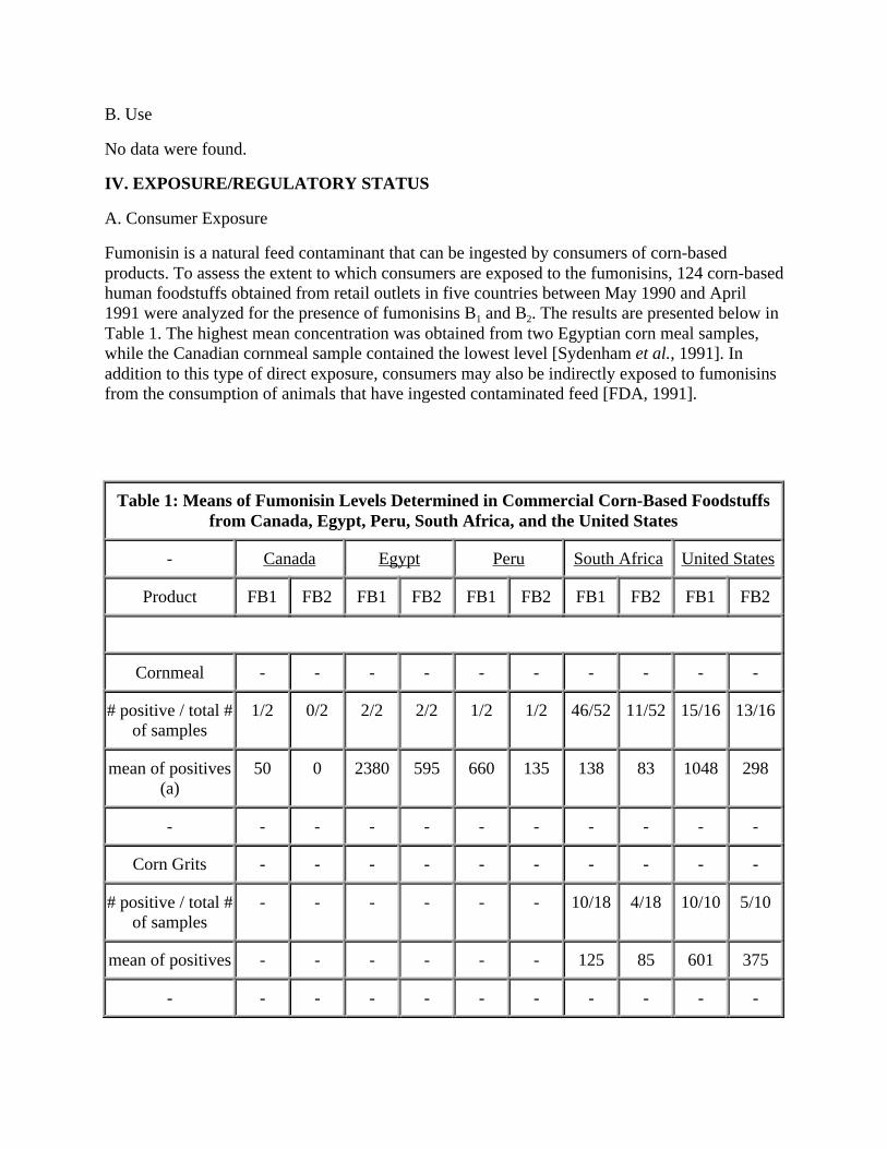
Fumonisin is a natural feed contaminant that can be ingested by consumers of corn-based products. To assess the extent to which consumers are exposed to the fumonisins, 124 corn-based human foodstuffs obtained from retail outlets in five countries between May 1990 and April 1991 were analyzed for the presence of fumonisins B1 and B2. The results are presented below in Table 1. The highest mean concentration was obtained from two Egyptian corn meal samples, while the Canadian cornmeal sample contained the lowest level [Sydenham et al., 1991]. In addition to this type of direct exposure, consumers may also be indirectly exposed to fumonisins from the consumption of animals that have ingested contaminated feed [FDA, 1991].
Table 1: Means of Fumonisin Levels Determined in Commercial Corn-Based Foodstuffs from Canada, Egypt, Peru, South Africa, and the United States
- Canada Egypt Peru South Africa United States
Product FB1 FB2 FB1 FB2 FB1 FB2 FB1 FB2 FB1 FB2
Cornmeal - - - - - - - - - -
# positive / total # of samples
1/2 0/2 2/2 2/2 1/2 1/2 46/52 11/52 15/16 13/16
mean of positives (a)
50 0 2380 595 660 135 138 83 1048 298
- - - - - - - - - - -
Corn Grits - - - - - - - - - -
# positive / total # of samples
- - - - - - 10/18 4/18 10/10 5/10
mean of positives - - - - - - 125 85 601 375
- - - - - - - - - - -

___________
Cornflakes - - - - - - - - - -
# positive / total # of samples
- - - - - - 0/3 0/3 0/2 0/2
mean of positives - - - - - - 0 0 0 0
- - - - - - - - - - -
Alkali treated (b) - - - - - - - - - -
# positive / total # of samples
- - - - 0/2 0/2 - - 1/3 0/3
mean of positives - - - - 0 0 - - 55 0
- - - - - - - - - - -
Miscellaneous - - - - - - - - - -
# positive / total # of samples
- - - - - - 2/8 0/8 4/4 3/4
mean of positives - - - - - - 84 0 409 148
(a) Fumonisin levels, ng/g (b) Peruvian samples were corn kernals; the U.S. samples were tortilla preparations
Reference: Sydenham et al., 1991
B. Occupational Exposure
No data were found on occupational exposure to fumonisins.
C. Environmental Occurrence
Fumonisins are produced by Fusarium moniliforme, one of the most common fungal contaminants of agricultural products, especially corn [Sydenham et al., 1990a; Thiel et al., 1991]. The following studies describe the detection and quantitative measurement of fumonisin species occurring naturally in the environment.
·Ninety-eight samples of feeds associated with 44 cases of equine leukoencephalomalacia (ELEM), and 83 samples of feed associated with 42 cases of porcine pulmonary edema syndrome (PPE) were analyzed for fumonisin B1. In addition, 51 feed samples not associated with either disease were also analyzed. The samples were obtained from various locations in the United States, and their composition varied considerably (corn, corn screening, pelleted feed,

etc.). Feeds associated with ELEM contained concentrations of fumonisin B1 ranging from < 1 µg/g - 126 µg/g, with 75% of the cases having at least 1 sample above 10 µg/g. Feeds associated with PPE contained concentrations of fumonisin B1 ranging from < 1µg/g - 330 µg/g, with 71% of the cases having at least 1 sample greater than 10 µg/g. (The authors point out that the 10 µg/g level was used as a point of reference and is not assumed to be a toxic or a safe level.) The concentrations of fumonisin B1 in nonproblem feeds were always less than 9 µg/g, with 94% of the samples containing less than 6 µg/g [Ross et al., 1991].
·A sample of home grown corn taken from the Transkei (southern Africa) in 1978 was analyzed for the presence of fumonisins. Fumonisin B1 was measured in subsamples of healthy corn kernels, moldy corn kernels and Fusarium-infected corn kernels at concentrations of <10, 44 and 83 µg/g, respectively [Sydenham et al., 1990a].
·During 1985, moldy and healthy corn samples were collected from two areas of the Transkei, southern Africa; in the southwest area of Kentani, which has a high rate of human esophageal cancer, and in the northeast region of Bizana, which has a low rate. The corn samples were analyzed for the presence of several Fusarium mycotoxins, including fumonisins B1 (FB1) and B2
(FB2). Both fumonisin species were detected in all samples of moldy corn. In the northeast area, the mean concentrations of FB1 and FB2 were 6.5 ± 5.3 and 2.5 ± 2.2 µg/g, respectively. In the southwest area, the mean concentrations of FB1 and FB2 were 23.9 ± 14.6 and 7.6 ± 4.6 µg/g, respectively. The fumonisin levels determined in the healthy corn samples taken from the Bizana were low, with a mean concentration of FB1 and FB2 of 0.06 ± 0.2 and <0.05 ± 0.05 µg/g, respectively (only 3 of the 12 samples analyzed were positive for fumonisins). In Kentani, however, the mean concentrations of FB1 and FB2 were 1.6 ± 2.1 and 0.5 ± 0.7 µg/g, respectively, and were recorded in 100% and 83% of the samples, respectively [Sydenham et al., 1990b].
·Twenty-two feed and corn samples associated with equine leukoencephalomalacia were screened for the presence of fumonisin B1. In at least 6 of 18 positive samples, fumonisin B1 was found at concentrations greater than 100 ppm [Vesonder et al., 1990].
The following studies examine the production of fumonisins in culture by strains of Fusarium obtained from different origins and from different substrates.
·Ninety strains of Fusarium moniliforme were tested for the ability to produce fumonisins in culture. The strains were selected to represent a wide range of substrates and geographic areas. In most cultures, three fumonisin homologs were detected; fumonisins B1, B2, and a newly characterized isomer of B2. Of the three homologs present in culture, B1 accounted for 70-95% of the total fumonisins measured. Also, there was considerable variation in the amounts of fumonisins produced by strains from different sources, but less variation among strains from the same source. Fumonisin B1 was produced at intermediate (50-500 ppm) or high levels (>500 ppm) by 95% of the strains from corn-based feed associated with equine leukoencephalomalacia (from Georgia, Indiana, Mississippi, North Carolina, and Pennsylvania) by all of the strains from good quality corn used in poultry feed (from Maryland, Virginia, and Pennsylvania), and by all of the strains from corn silks in Iowa. In contrast, all but one of the ten strains from Nepal were low-level producers (trace to 49 ppm) of fumonisin B1. Fusarium strains from millet and sorghum grain from Africa, from a corn-based laboratory rat diet from the United States, and from sorghum from Australia were primarily low- and intermediate-level producers (a few

strains were high-level producers). Finally, 85% of the strains from mycotic keratitis, ulcers, and various types of unspecified cancer in humans were intermediate- or high-level producers of fumonisin B1. The authors concluded that, although there is some variation in the amounts of fumonisins produced, the potential for production in natural substrates and agricultural commodities exists in strains from a variety of substrates and geographic areas [Nelson et al., 1991].
·Cultures of both Fusarium moniliforme and Fusarium proliferatum were examined for their potential to produce fumonisins B1 (FB1) and B2 (FB2). Nine feed samples (comprised primarily of corn and/or corn screenings) were obtained from farms in southeastern Iowa; two samples were associated with equine leukoencephalomalacia (ELEM), five were associated with porcine pulmonary edema syndrome (PPE), and two were not associated with any animal health problems. F. moniloforme was isolated from all samples, and F. proliferatum was isolated from one ELEM sample, one PPE sample, and one "nonproblem" sample; the isolates were cultured on autoclaved corn, and after one month the culture materials (CMs) were analyzed. The nine F. moniloforme CMs had concentrations of FB1 and FB2 ranging from 960-2350 and 120-320 µg/g, respectively. The three F. proliferatum CMs had levels of FB1 and FB2 that ranged from 1670-2790 and 150-320 µg/g, respectively. The authors point out that the high levels of fumonisins by isolates from both problem and nonproblem feeds suggests potential for fumonisin contamination in any feed containing F. moniliforme and/or F. proliferatum [Ross et al., 1990].
·Ten commercially prepared feeds and four corn samples were obtained from various places in the southeastern United States between 1983 and 1986. All fourteen samples were taken from feeds given to horses prior to the development of leukoencephalomalacia (LEM). Each sample was analyzed for fumonisin B1 (FB1) and B2 (FB2). In addition, FB1 and FB2 were measured in corn cultures of 10 isolates of F. moniliforme taken from feed associated with LEM and tested for toxicity on ducklings (the ten most toxic cultures were selected). All ten of the corn cultures produced both FB1 and FB2. The concentration of FB1 ranged from 160-3800 µg/g, while the concentration of FB2 ranged from 20-950 µg/g. The level of FB1 was 80-96% of the total fumonisin concentration (180-4690 µg/g). All 14 feed samples examined contained both FB1
(1.3-27.0 µg/g) and FB2 (0.1-12.6 µg/g), with FB1 comprising 53-93% of the total fumonisin concentration in the samples (1.4- 39.6 µg/g) [Thiel et al., 1991a].
·Forty toxic Fusarium isolates, representing 27 taxa in 9 of the 12 sections of Fusarium, as well as two recently described species not yet classified into sections, were examined for their potential to produce fumonisins B1 (FB1) and B2 (FB2). The isolates were from several different origins and were taken from several different substrates. With the exception of one isolate of F. nygamai, fumonisin production was restricted to isolates of F. moniliforme (7) and F. proliferatum (4), in the section Liseola. The seven F. moniliforme isolates were from corn from a high-risk area of human esophageal cancer in the Transkei, southern Africa; six produced both FB1 (180-7100 µg/g) and FB2 (40-3000 µg/g) in cultures on corn, while one produced only FB1
(105 µg/g). The highest producer of both FB1 and FB2 was F. moniliforme MRC 826, the strain from which the fumonisins were originally isolated and characterized. The four isolates of F. proliferatum were either from sorghum in South Africa or from corn in the United States and Sierra Leone; all four produced both FB1 and FB2 in cultures on corn at concentrations ranging from 20-660 µg/g and 65-450 µg/g, respectively. The F. nygamai isolate was from soil in South Africa and produced 605 µg/g of FB1 and 530 µg/g of FB2. According to the authors, this is the

first report of fumonisin production by F. nygamai [Thiel et al., 1991b].
D. Regulatory Status
·OSHA has not established a permissible exposure limit (PEL) for fumonisins.
·An Interagency Working Group has been established to address the problems created by fumonisins. The Interagency Working Group includes representatives from USDA/ARS, FDA/CFSAN, USDA/APHIS, and FDA/CVM [FDA, 1991].
E. Exposure Recommendations
·ACGIH has not recommended a threshold limit value (TLV) for fumonisins.
·NIOSH has not recommended an exposure limit (REL) for fumonisins.
V. TOXICOLOGICAL EFFECTS
Fumonisins are metabolites of Fusarium moniliforme, which has been linked with several diseases in humans and animals, including equine leukoencephalomalacia (ELEM), human esophageal cancer, and porcine pulmonary edema syndrome (PPE) [Ross et al., 1990; Voss et al., 1989]. In many cases, fumonisin B1 and B2 have been found at high levels in corn and feed samples contaminated with F. moniliforme that were obtained from areas with high incidences of esophageal cancer and outbreaks of PPE and ELEM (see section IV.C).
Although the databases were searched for toxicological information pertaining to the fumonisins A1, A2, B1, B2, B3 and B4, most of the investigations found in the published literature focus on the toxic effects fumonisin B1, which is the major fumonisin produced in nature [Sydenham et al., 1991].
A. Chemical Disposition
1. Human Data
No data were found.
2. Animal Data
No data were found.
B. Acute
1. Human Data
No data were found.
2. Animal Data
·oral, rat
It was reported in an abstract that cultures of Fusarium moniliforme fed to rats of unspecified

strain and sex, killed the animals in less than 24 hours. However, when pure fumonisin B1 (21 mg/rat), a metabolite of F. moniliforme, was administered to rats by stomach intubation, no toxic effects were observed. No other data were reported [Mirocha et al., 1990].
C. Prechronic
1. Human Data
No data were found.
2. Animal Data
A summary of the Prechronic effects of Fumonisin in animals is presented at the end of this section in Tables 5 and 6; Table 5 describes the results of dietary fumonisin exposure, and Table 6 reports on the effects following intravenous and oral administration.
·oral, rat
To examine the relationship of dietary fumonisin concentration to hepatotoxicity, male Sprague Dawley rats were fed diets containing extracts of Fusarium moniliform (strain MRC 826) culture material (CM) and/or the extracted CM residues. Two experiments were conducted; one to assess the hepatotoxicity of chloroform/methanol (1:1) CM extractions and the CM residue after chloroform/methanol extraction, and the second to assess the hepatotoxicity of aqueous (using distilled-deionized water) CM extracts and the CM residue after water extraction. Control corn was also extracted in a similar manner for incorporation into the solvent control diets. Each experiment consisted of 4 groups of 5 animals; a solvent control group that was fed a diet containing the extract and the residue of control corn, a group fed the CM extract, a group fed the CM residue after extraction, and a group fed both the CM extract and residue (see Table 2 below). The amount of extract or residue per kilogram of the formulated test and solvent control diets was equivalent to 200 g of CM or control corn, respectively. In addition, positive and negative control groups (5 rats/group) were fed diets containing unextracted CM or unextracted control corn, respectively.
CM extracts and residues were analyzed for fumonisins B1 and B2 by hydrolysis followed by gas chromatography/mass spectroscopy, and by thin layer chromatography. Throughout each experiment, animals were observed daily for clinical signs, and body weight and food consumption were measured weekly. After two and four weeks, blood samples were taken for the determination of serum aminotransferase (ALT), aspartate aminotransferase (AST), alkaline phosphatase (AP), and bilirubin levels. At the end of the study (week 4), all animals were sacrificed and necropsied. Unless otherwise specified, statistical significance was judged at the level P<0.05. A summary of the dosing regimen, dietary fumonisin concentrations, and associated clinical, serum chemical, and histopathologic findings are presented below in Table 2.
All animals survived until the end of the study, and the behavior and appearance of animals in each group were similar. No significant differences were found in body weights, food consumption, relative liver weights, and histology of the liver between the solvent control groups and the negative control group. Throughout the study, animals fed the CM residue after chloroform/methanol extraction, the aqueous CM extract, or the unextracted CM (positive control) had significantly lower body weights than animals in the solvent or negative control

groups (specific weights not reported). In each of these three groups, significantly decreased weight gains were found during weeks 1 and 2 only (data not reported). No significant differences in body weight were found between the groups fed the chloroform/methanol CM extract or the aqueous CM residue and their respective controls. When compared to the solvent controls, food consumption was significantly decreased in animals fed the chloroform/methanol CM extract plus extracted CM residue (during weeks 1-2 only), in animals fed the aqueous CM extract (during weeks 2-3 only), and animals fed the aqueous CM extract plus extracted CM residue (during weeks 2-4 only). Throughout the study (weeks 1-4), food consumption in the positive control group was significantly decreased compared to that in the solvent and negative control groups. After 2 and 4 weeks, serum ALT, AST, and AP activities were significantly increased in groups fed the CM residue after chloroform/methanol extraction, the chloroform/methanol CM extract plus extracted CM residue, the aqueous CM extract, the aqueous CM extract plus extracted CM residue, or the unextracted CM (positive control) compared to their respective solvent and negative controls.
Gross necropsy revealed that absolute and relative liver weights were significantly decreased in the groups fed the CM residue after chloroform/methanol extraction, the aqueous CM extract, and the unextracted CM compared to their respective control groups (see Table 3 below). No gross liver lesions were found. However, histological examination revealed liver lesions in 4-5 animals fed CM residue after chloroform/methanol extraction, chloroform/methanol CM extract plus extracted CM residue, aqueous CM extract, aqueous CM extract plus extracted CM residue, and the unextracted CM. These liver lesions were typically characterized by minimal to mild bile duct proliferation and hepatocellular hyperplasia. Other findings included hepatocellular degeneration and necrosis, apoptosis, pyknotic nuclei, mitotic figures, minimal fibrosis, and scant acute inflammatory infiltrates.
Fumonisins B1 and B2 were detected in all CM extracts and residues after extraction, and the highest fumonisin concentrations were present in those diets associated with toxic effects. There were no detectable fumonisins in the negative or solvent controls. The authors of this study point out that because the test diets were formulated with extracts and residues, the presence of other compounds in these materials having additive or synergistic effects cannot be dismissed; however, they feel that the data show a positive correlation between fumonisin concentration of the test diets and hepatotoxicity [Voss et al., 1990].

Table 2: Summary of the Dosing Regimen, Dietary Fumonisin Concentrations, and Associated Clinical, Serum, Chemical and Histopathologic Findings
Dietary Fumonisin
Concentration (ppm)2
Group and Treatment1 B1 B2 Total Findings
Chloroform / methanol extraction
1) E + R of Corn
ND ND ND None
2) E of CM 22 33 55 None
3) R of CM 117 99 216 Decreased body weight; increased ALT, AST and AP; hepatosis
4) E + R of CM
139 132 271 Decreased body weight and food consumption; increased ALT, AST and AP; hepatosis
Water extraction
5) E + R of Corn
ND ND ND None
6) E of CM 93 82 175 Decreased body weight and food consumption; increased ALT, AST, and AP; hepatosis
7) R of CM 18 65 83 None
8) E + R of CM
111 147 258 Decreased body weight and food consumption; increased ALT, AST, and AP; hepatosis
Positive and negative controls

______________
9) Unextracted CM
139 131 270 Decreased body weight and food consumption; increased ALT, AST, and AP; hepatosis
10) Unextracted corn
ND ND ND None
1E = extract; R = residue after extraction; Corn = control corn; CM = culture material. Materials were added to basal feed at concentrations equivalent to 200 g CM per kg of formulated diet. 2Calculated dietary concentrations of fumonisin B1 and B2 based upon GC/MS analysis of CM extracts, CM residues and control corn; ND = none detected
Reference: Voss et al., 1990
Table 3: Summary of Liver Weight Data
Liver Weight
Relative
Group and Treatment1 Body Weight (g)2 Absolute (g) (% B.Wt.)
Chloroform / methanol extraction
1) E + R of Corn 324 (16.4)b 11.5 (1.44)b 3.6 (0.27)c
2) E of CM 315 (19.1)b 11.4 (2.00)b 3.6 (0.45)b
3) R of CM 289 (23.0)c 8.3 (0.79)c 2.9 (0.12)c
4) E + R of CM 287 (3.4)c 8.5 (0.74)c 3.0 (0.23)c
Water extraction
5) E + R of Corn 329 (17.8)b 11.3 (0.59)b 3.4 (0.12)b
6) E of CM 289 (15.8)c 8.8 (0.88)c 3.0 (0.16)c
7) R of CM 316 (7.9)b 11.0 (0.75)b 3.5 (0.24)b
8) E + R of CM 280 (9.7)c 8.0 (0.86)c 2.8 (0.23)c

______________
Positive and negative controls
9) Unextracted CM 262 (21.4)c 7.4 (0.54)c 2.8 (0.13)c
10) Unextracted corn 317 (16.5)b 10.2 (0.97)b 3.2 (0.20)b
1E = extract; R = residue after extraction; Corn = control corn; CM = culture material. Materials were added to basal feed at concentrations equivalent to 200 g CM per kg of formulated diet. 2Numbers in parantheses represent standard deviations; Groups with different letters (b or c) are significantly different; P<0.05.
Reference: Voss et al., 1990
·oral, horse
Leukoencephalomalacia (LEM) was induced in two horses (unspecified strain) by the oral administration of fumonisin B1 (FB1). In a pilot trial, a filly received 59.5 mg/kg of a 50% preparation of FB1, administered in 21 doses of 1.25-4 mg/kg over 33 days (the other 50% was inorganic matter that co-eluted during purification). In the second experiment, a colt received 44.3 mg/kg of 95% pure FB1 in 20 doses of 1-4 mg/kg in 29 days. The FB1 used in both experiments was isolated from corn cultures of Fusarium moniliforme MRC 826. The horses were closely observed, and serum samples were collected periodically for the determination of aspartate transaminase (AST), gamma glutamyl transferase (GGT), lactate dehydrogenase (LD), and total bilirubin. When dosing was complete, the animals were sacrificed and necropsied.
In the filly, clinical signs became apparent on days 22-27 and consisted of apathy, changes in temperament, lack of coordination, walking into objects, and paralysis of the lips and tongue. However, the filly improved progressively and by day 28 had apparently recovered. The colt exhibited clinical signs from days 24-26 and again from days 31-33. The symptoms consisted of apathy, docility, tremors, pawing motions, bumping into objects, inability to eat or drink, and soporiferousness. Chemical analyses of serum samples showed that the filly had elevated AST activity between days 22-31 (maximum of 365 U/l on day 23), while the colt had elevated GGT activity between days 20-33 (maximum of 52 U/l on day 33).
Gross necropsy of the filly revealed a sunken area (2 cm in diameter) in the lateral part of the anterior frontal lobe of the left cerebral hemisphere. There was slightly more cerebrospinal fluid in this area, and the fluid was tinged yellowish-brown. In addition, the white matter on the cut section of this focus was softer than normal and reddish-brown. Microscopic examination of the lesion revealed necrosis of the white matter, numerous macrophages, aggregates of mineralization, and small hemorrhages. At the periphery of the necrotic area, the blood vessels showed hypertrophy and hyperplasia of endothelial cells, fibrinoid changes of their cell walls, and perivascular mononuclear cell infiltration. The white matter close to the focal lesion had mild status spongiosis and mild to moderate proliferation of astrocytes. No other lesions were evident in other tissues, except for diffuse cloudy swelling and hydropic degeneration of hepatocytes.

Necropsy of the colt showed swelling of the cerebral hemisphere and flattening of the gyri. A yellowish-brown focus was seen in the subcortical white matter of the left dorsal frontal lobe, and extended posteriorly to the occipital lobe. A smaller, gelatinous focus was found in the white matter of the right occipital lobe. In addition, the kidneys were moderately swollen and appeared grayish-yellow. No other macroscopic lesions were seen in any tissues. Microscopic examination of the lesions revealed rarefaction of the neuropil, partial loss of cellular detail of the white matter, swelling and proliferation of the astrocytes, infiltration of macrophages, and swelling of the axons. As seen in the filly, the blood vessels around the foci had hyperplasia and hypertrophy of endothelial cells, as well as perivascular edema. The white and grey matter of the rest of the left side of the brain showed moderate edema, and the right side showed only a mild edema. Evaluation of the proximal convoluted tubules in the kidneys revealed cloudy swelling and hydropic degeneration.
The lesions seen in both horses are characteristic of LEM, and the authors concluded that these results unequivocally prove that fumonisin B1 can induce LEM in horses [Kellerman et al., 1990].
oral, horse
During the fall of 1989, 18 of 66 purebred Arabian horses at a breeding/training stable in Arizona became ill over a 7-day period with equine leukoencephalomalacia (ELEM). Of the 18 horses affected, 14 died from the condition and 4 partially recovered, but were mildly affected with impaired vision and deviated lips and noses. All of the animals had been fed a diet containing a substantial amount of white corn screenings (1:1 with sweet feed) for 26 days. The animals also received 0.2 kg/day of a protein supplement and free choice of alfalfa or grass hay. Gross examination of the two batches of screenings used in the feed did not reveal any obvious mold, and both batches contained cob parts, damaged kernels, and undamaged kernels. Necropsies were performed on 10 animals, and tissues were collected for histological examination. In addition, several feed samples (corn screenings, sweet feed, protein supplement, alfalfa pellets) were collected and chemically analyzed for the presence of fumonisin B1 (FB1) and B2 (FB2).
Concentrations of FB1 in the single subsample from batch one of the corn screenings and the two subsamples from batch two were 37, 58, and 122 ppm, respectively. The respective levels of FB2
in these samples were 2, 11, and 23 ppm. Subsamples of the protein supplement, alfalfa pellets, and sweet feed contained little if any FB1 (<5 ppm). A subsample from batch two was then separated into undamaged kernels, damaged kernels, and cob parts, and the levels of fumonisins were measured in each component. In damaged kernels and cob parts, the concentrations of FB1
were 148 and 144 ppm, respectively, and the levels of FB2 were 41 and 31 ppm, respectively. In the sample of undamaged kernels the levels of FB1 and FB2 were less than 5 ppm.
Gross examination of all horses necropsied showed focal to diffuse unilateral areas of liquefactive necrosis in areas of the cerebral white matter. In some animals, portions of the cerebrum disintegrated when removed from the cranial vault. Also, hemorrhagic foci were often present in the brain stem. Histopathological findings included rarefied white matter with pyknotic nuclei and eosinophilic cytoplasm. Tissue structures were unidentifiable in some sections, while other sections often had hemorrhagic foci located in a distinct perivascular pattern. Microscopic lesions were present mostly in the cerebrum, but were also observed in the

brain stem. The authors of this study using information on diet, animal weights, and feeding practices, estimated the total FB1 dosage for 13 of the 14 horses that died during the outbreak of ELEM; the doses ranged from 0.6-2.1 mg/kg/day. This was the first definitive report on ELEM and associated fumonisin concentrations [Wilson et al., 1990].
·oral, horse
An abstract of an unpublished paper presented at the Fumonisin Symposium held in Raleigh, North Carolina (April 24-25, 1991) describes the results of a study done to determine the minimal dose of contaminated corn screenings needed to reproduce equine leukoencephalomalacia (ELEM) in ponies. Groups of 4-5 ponies were fed formulated diets containing naturally contaminated corn screenings with fumonisin B1 concentrations of 8, 22, or 44 ppm. Two of the ponies fed 44 ppm fumonisin B1 died of moderate to severe liver disease and mild encephalopathy. The remaining two ponies in this group died of classic ELEM. Only one pony in the 22 ppm dose group died of ELEM; nine days prior to death, this animals developed elevated liver enzyme levels. The other three ponies fed 22 ppm fumonisin B1 showed mild behavioral problems, but did not have acute signs of ELEM or elevated liver enzyme levels. In the group given feed containing 8 ppm fumonisin B1, one pony showed behavioral changes, but no significant gross lesions were found upon necropsy. The ponies fed 8 ppm did show minor, nonspecific lesions in the liver, kidney and brain stem. The authors of this abstract concluded that further evaluation of diets at 8 ppm fumonisin B1 are needed [Wilson et al., 1991, as reported in FDA, 1991].
·oral, swine
An abstract of an unpublished paper presented at the Fumonisin Symposium held in Raleigh, North Carolina on April 24-25, 1991 reports that pigs fed naturally contaminated corn screenings containing 166 ppm fumonisin B1 and 48 ppm fumonisin B2 developed pulmonary edema, pancreatic lesions, and liver damage. Respiratory problems that were observed were not, according to the authors, due to cardiac injury since cardiac dysfunction was not seen. Elevated serum cholesterol and liver enzyme levels were seen in pigs with lung injury. However, a progressive increase in these levels was also observed in pigs that did not die of pulmonary edema. Electron microscopy of tissue sections revealed that hepatocytes, pulmonary type II epithelial cells, and pancreatic acinar cells had intracellular membrane degeneration and plasma membrane changes. According to the authors of this abstract, these findings suggest that cell membranes might be an early target of fumonisins. In addition, Kupffer cells and intravascular macrophages contained myelin figures, suggesting that these cells might also be involved in pathogenesis. The authors speculated that fumonisins induce abnormalities in membrane lipid turnover activated processes in the affected cells, which culminate in pulmonary edema [Haschek et al., 1991, as reported in FDA, 1991].
·oral/intravenous, swine
On 2 southwest Georgia farms, pulmonary edema and hydrothorax were observed in mature swine that died approximately 5 days after consuming corn screenings. An experimental feeding study was conducted in conjunction with a fumonisin injection study to investigate the possible relationship between the deaths and the presence of fumonisins, toxic metabolites of the fungus Fusarium moniliforme. Corn screenings from each farm (1.5 kg samples) were analyzed for the

presence of fumonisins, and preliminary data indicated that the concentrations of FB1 in Feed A and Feed B were 105 mg/kg and 155 mg/kg, respectively. No data were reported on the concentrations of FB2.
For the feeding study, two groups (Group A or Group B) of 3 swine were fed corn screenings collected from each farm (Feed A or Feed B) for 28 days; a control pig was fed a commercially available grower ration. All pigs were weighed on days 0, 14, and 28, and were observed twice daily for any clinical abnormalities. For the injection study, fumonisins B1 and B2 (FB1 and FB2; 98% pure) were dissolved in saline with 5% ethanol and injected into swine according to the following dosing regimes: swine 1 received 4 daily injections of 0.4 mg FB1/kg body weight; swine 2 was given 7 daily injections 0.174 mg FB1/kg; a third pig received 5 daily injections of 0.3 mg FB2/kg; and a fourth pig was injected for 7 days with 1.0 ml of saline with 5% ethanol (solvent control). Swine that died during the feeding study or as a result of the injections were necropsied. All other animals were sacrificed and necropsied at the end of the study. Tissue samples were taken from each animal for histological evaluation.
Animals in the feeding study were unable to maintain body weight; the data are reported below in Table 4. On the seventh day of the feeding study, one pig in Group B (fed Feed B) was found dead, and a second, severely dyspneic pig, was euthanized. Necropsy of these animals revealed marked pulmonary edema and hydrothorax. The remaining pig in Group B was sacrificed and necropsied on day 28, and no signs of pulmonary edema were found. In Group A, a severely anorectic pig was euthanized on day 14, and the other two animals were sacrificed at the end of the study. Necropsy showed that none of these animals had developed pulmonary edema or hydrothorax. In the second part of the study, the pig injected with 0.4 mg FB1/kg/day died on day 5, after receiving a total of 11.3 mg of FB1. Necropsy of this animal revealed lesions similar to field cases and other experimental cases of pulmonary edema. The other two animals (one receiving a total of 8.65 mg FB1; the other receiving a total of 10 mg FB2) survived until the end of the study, and were sacrificed and necropsied 24 hours after their last injection. Neither of these animals had developed pulmonary edema.
The pathological abnormalities found in the animals that developed pulmonary edema after feeding or after injection were essentially the same. The trachea and bronchi contained a clear, foamy liquid, and a golden-yellow liquid filled the thoracic cavities. Interlobular edema was marked, and was most pronounced in the hilus area. Lobular atelectasis was also seen. Microscopically, the alveoli contained only a few cells (mostly macrophages), and had focal to diffuse areas of alveolar septal congestions with capillary thromboses (indicating thrombostasis). In addition, pancreatic lesions were present in all pigs with pulmonary edema/hydrothorax, and consisted of focal to massive necrosis, acinar cell dissociation, and rounded individual acinar cells. In pigs from the feeding study, liver changes were also found; these changes were characterized by centrolobular and random hepatocellular cytoplasmic vacuolar change, hepatocellular cytomegaly, disorganized hepatic cords, and early pirolobular fibrosis. No pulmonary, pancreatic, or liver pathology was noted in the control pig from either study.
The authors of this study conclude that FB1 affects the pancreas and the lungs, and produces distinct lesions that should not be confused with other conditions that induce pulmonary and/or thoracic effusion. Also, they state that since only swine in the feeding study developed liver lesions, the damage may have been related to nutrient availability, and additional research should

_____________
be conducted to determine the hepatotoxicity of FB1 [Harrison et al., 1990].
Table 4: Body Weights of Swine Fed Corn Screenings
Body Weight (kg)
Pig number Day 0 Day 14 Day 28
Group A
34 18.2 15.4 12.2
35 22.2 18.2 16.8
36 15.0 12.7 *
Group B
37 24.0 21.8 18.2
38 18.6 * *
39 19.1 * *
Control
40 15.4 20.5 31.8
*Deceased or removed from study.
Reference: Harrison et al., 1990
·oral/intravenous, swine
As described in an abstract, the hepatotoxicity of fumonisin B1 was examined in female crossbred swine. In the first part of the study, two pigs were given daily intravenous injections of FB1 (70% pure); one pig received 7.9 mg/kg/day for 9 days and the other received 4.5 mg/kg/day for 4 days (for a total of 72 and 77 mg, respectively). A third control pig was given daily intravenous injections of saline. Clinical signs and gross lesions were not observed in any of the three pigs. However, necropsy revealed that the hepatic lobules were disorganized with scattered hepatocyte necrosis and mitosis. In the second part of the study, corn screenings contaminated with FB1 (222 ppm) were fed to three pigs, and uncontaminated corn was fed to two control pigs.

All three pigs fed contaminated corn developed respiratory distress within 3-5 days; one was killed on day 4 and one was found dead on day 6. These animals had severe pulmonary interstitial edema, pleural effusion, and individual pancreatic acinar necrosis. Clinical signs in the third pig regressed, and the animal was sacrificed on day 15. The two control pigs were sacrificed on days 4 and 15. Pigs fed FB1 had liver lesions identical to pigs given FB1
intravenously, and in both groups, liver enzymes were elevated. The authors concluded that this mycotoxin, given orally and intravenously, is hepatotoxic to pigs [Ness et al., 1991].
·intravenous, horse
Fumonisin B1 (FB1) was extracted and purified from the culture material of Fusarium moniliforme MRC 826; the culture material contained approximately 1 g/kg of FB1. A mare (unspecified strain) was given seven intravenous injections of 0.125 mg FB1/kg body wight/day on days 0-4, 7, and 9. Serum samples were taken periodically for the determination of aspartate transaminase (AST), gamma glutamyl transferase (GGT), lactate dehydrogenase (LD), and total bilirubin. The horse was sacrificed on day 10 and necropsied.
Clinical signs became apparent on day 8 and consisted of transient nervousness followed by apathy, reluctance to move, loss of coordination, inability to eat, paralysis of the lower lip and tongue, watery, green discharge from the nostrils, and dyspnea. The horse fell down in a convulsive seizure and was euthanasized (day 10). Chemical analysis of serum samples revealed mild elevations of the AST (229 U/l) and GGT (222 U/l) levels on days 8-10. Gross necropsy of the animal revealed severe edema of the brain, and grayish-brown foci (5 mm in diameter) in the medulla oblongata. Other lesions that were noted were congestion and edema of the diaphragmatic lobe of the left lung, mild perirenal edema, and petechiae in the mucosa, and a mild edema of the submucosa in the cecum. Microscopic examination of the medulla oblongata revealed distinct areas of severe necrosis of the gray and white matter that were characterized by rarefaction of the neuropil, necrosis of neurons and glial cells, swelling of glial cells and axons, infiltration by neutrophils and macrophages, and small perivascular hemorrhages. The white and gray matter around these necrotic areas showed evidence of severe edema. Other abnormalities included congestion of the spinal cord, mild edematous changes in the gray matter of the lumbar region, mild nephrosis and edema of the submucosa in the large intestine, and mild congestion and edema of the lungs. No other significant changes were seen in the other tissues examined. The authors stated that these changes represented early, bilaterally distributed leukoencephalomalacia in the brain stem. They also concluded that fumonisin B1, produced by F. moniliforme, causes equine leukoencephalomalacia [Marasas et al., 1988].

Table 5: Summary of the Prechronic Effects of Exposure to Dietary
Fumonisins in Animals
Concentration of
Fumonisin in Duration
Species Strain/Sex feed samples1 (days) Effect Reference
Rat SD/male 0-22 ppm FB1 / 0-65 ppm FB2
28 None Voss et al., 1990
- - 93-139 ppm FB1 / 82-147 ppm FB2
28 decreased body weight; increased liver enzyme levels; hepatosis
Voss et al., 1990
Swine Crossbred /female
222 ppm FB1 3-15 pulmonary edema; pancreatic necrosis; liver lesions; increased liver enzyme levels
Ness et al., 1991
Swine NS/NS2 105 mg FB1 /
kg feed 28 decreased body weight Harrison
et al., 1990
- - 155 mg FB1 / kg feed
28 decreased body weight; pulmonary edema / hydrothorax
Harrison et al., 1990
Swine NS/NS 166 ppm FB1 / 48 ppm FB2
NS pulmonary edema; pancreatic lesions; liver damage; increased liver enzyme levels
Haschek et al., 1991
Horse Arabian/N S
37-122 ppm FB13 / 2-23 ppm FB2
26 leukoencephalomalacia Wilson et al., 1990
Horse NS/NS 44 ppm FB1 NS leukoencephalomalacia; liver disease Wilson et al., 1991
(pony) - 22 ppm FB1 NS leukoencephalomalacia (1/4 animals); increased liver enzyme levels.
Wilson et al., 1991

- - 8 ppm FB1 NS None Wilson et al., 1991
1FB1 and FB2 = fumonisin B1 and B2, respectively 2NS = Not specified 3The authors estimated that the daily dose of fumonisin B1 was 0.6-2.1 mg/kg
Table 6: Summary of the Prechronic Effects of Intravenous and
Oral Administration of Fumonisins in Animals
- - -Dose of
Study Duration
- -
- - - fumonisin1 (days) / No. of
- -
Route Species Strain/Sex (mg/kg/dose) doses Effects Reference
Oral Horse NS2/Female 1.25-4 (FB1) 33/21 leukoencephalomalacia; elevated liver enzyme levels
Kellerman et al., 1990
- - - - - - -
Oral Horse NS/male 1-4 (FB1) 29/20 leukoencephalomalacia; elevated liver enzyme levels
Kellerman et al., 1990
- - - - - - -
Intraven ous
Swine NS/NS 0.4 (FB1) NS/4 pulmonary edema Harrison et al., 1990
- - NS/NS 0.174 (FB1) NS/7 None reported Harrison et al., 1990
- - NS/NS 0.3 (FB2) NS/5 None reported Harrison et al., 1990
- - - - - - -
Intraven ous
Swine Crossbreed 7.9 (FB1) 9/9 elevated liver enzyme levels; hepatoxicity
Ness et al., 1991

/female
- - - 4.5 (FB1) 4/4 elevated liver enzyme levels; hepatoxicity
Ness et al., 1991
- - - - - - -
Intraven Horse NS/Male 0.125 (FB1) 9/7 Leukoencephalomalacia; Marasas et ous elevated liver enzyme al., 1988
levels
1FB1 and FB2 = fumonisin B1 and B2, respectively 2NS = Not Specified
D. Chronic/Carcinogenicity
1. Human Data/Case Reports
Although there is no data directly linking fumonisins to cases of human cancer, several reports indicate that ingestion of Fusarium moniliforme contaminated grains by humans is linked to relatively high incidences of human esophageal cancer. In South Africa, human esophageal cancer has a high occurrence in the southwestern districts of the Transkei (Kentani), while in the northeastern region (Bizana) the rate is low. Corn is the main dietary staple in both areas; however corn in the southwestern districts contains a higher percentage of F. moniliforme. During 1985, moldy and healthy corn samples were collected from the two areas, and were analyzed for the presence of several Fusarium mycotoxins, including fumonisins B1 (FB1) and B2
(FB2). Both fumonisin species were detected in all samples of moldy corn. In the northeast area, the mean concentrations of FB1 and FB2 were 6.5 ± 5.3 and 2.5 ± 2.2 µg/g, respectively. In the area with high rates of esophageal cancer, the levels were significantly higher (P<0.01), with mean concentrations of FB1 and FB2 of 23.9 ±14.6 and 7.6 ± 4.6 µg/g, respectively. The fumonisin levels determined in the healthy corn samples taken from the Bizana were low, with a mean concentration of FB1 and FB2 of 0.06 ± 0.2 and <0.05 ± 0.05 µg/g, respectively (only 3 of the 12 samples analyzed were positive for fumonisins). In Kentani, however, the mean concentrations of FB1 and FB2 were significantly (P<0.001) higher (1.6 ± 2.1 and 0.5 ± 0.7 µg/g, respectively), and were recorded in 100% and 83% of the samples, respectively [Sydenham et al., 1990b].
In addition, both fumonisin B1 and fumonisin B2 were detected in commercial corn-based samples obtained from Charleston, South Carolina in 1989. This city has one of the highest incidences and mortality rates of esophogeal cancer in the United States. The mean levels of fumonisin B1 (detected in 7/7 samples) and B2 (detected in 6/7 samples) were 635 and 182 ng/g, respectively. Even though these levels were lower than those determined in home-grown corn samples from the Transkei, the authors stated that the presence of these mycotoxins in commercial foodstuffs is cause for further investigation of the role of fumonisins in the etiology of esophogeal cancer [Sydenham et al., 1991].

_______________________
2. Animal Data
·oral, rat
A short-term cancer promotion-initiation bioassay was used as a monitoring system to isolate cancer-promoting compounds from cultures of Fusarium moniliforme MRC 826. Fractions isolated from culture material were screened for cancer-promoting activity by incorporating them in rat mash (at a concentration of 5%) and feeding them to groups of 5 male BDIX rats that had been initiated with a 200 mg/kg intraperitoneal dose of diethylnitrosamine (DEN). The feeding period lasted 4 weeks. All rats were killed after the 4-week promotion treatment, and their livers were analyzed histologically for gamma-glutamyl-transpeptidase-positive (GGT+) foci; the induction of GGT was used as the indicator of cancer-promoting activity. In the first part of the study, culture material was successively extracted with ethyl acetate and aqueous methanol (CH3OH-H2O; 3:1). Two samples of culture material, both extracts, and the remaining residues were tested for cancer-promoting activity. In addition, two control groups were included; one initiated with DEN and given feed without culture material, and one receiving treated feed without initiation (given dimethyl sulfoxide (DMSO) instead of DEN). In the second part of the study, the aqueous methanol extract was successively partitioned and fractionated to purify the cancer-promoting compounds; fractions obtained at each step were tested for cancer-promoting activity as described above.
Exposure to diets containing 5% of culture material for 4 weeks significantly induced (P<0.001) the formation of GGT+ foci in DEN-initiated rats. Induction of the foci was not seen in either control group. Following extraction, the bulk of the cancer-promoting activity was recovered in the aqueous methanol extract. When this extract was dried and partitioned between CH3OH-H2O (1:3) and CHCl3, all of the cancer-promoting activity (induction of GGT+ foci) was again found in the aqueous phase, and none was detected in the CHCl3 phase. This CH3OH-H2O fraction was chomatographed on an Amberlite XAD-2 column, and the column was successively eluted with H2O, CH3OH-H2O (1:3 and 1:1), and CH3OH. Although the majority of the fraction eluted from the column with the CH3OH-H2O eluent, the cancer-promoting compound(s) were eluted with CH3OH. The CH3OH eluate was further purified on Amberlite XAD-2; the column was successively eluted with CH3OH-H2O (3:1) and CH3OH. This time the active compound was recovered with the CH3OH-H2O, and it was fractionated on a Sephadex LH-20 column; fractions obtained from this column were tested for cancer-promoting activity. Two compounds induced GGT+ foci; they were purified on a C18 reverse-phase column, chemically characterized and given the names fumonisin B1 and B2. Fumonisin B1 was the main compound obtained (10 times more than fumonisin B2), with approximately 2 g purified from 1 kg of the cultured corn in later bulk extractions. The purity of fumonisin B1 obtained from these later extractions (determined by high performance liquid chromatography) was 92% [Gelderblom et al., 1988].
In the study described above, the cancer-promoting activity and toxicity of fumonisin B1 (FB1) was tested in male BD IX rats. The cancer-promoting activity was tested using the same 4-week promotion-initiation bioassay described above for the culture material of Fusarium moniliforme, except FB1 was incorporated into the diet at a concentration of only 0.1%. To examine the toxicity of the compound, four rats were given daily oral doses of 0.95 g FB1/10 ml of dimethyl sulfoxide (DMSO). However, since three of the four rats died within 3 days, another experiment

was run in which four rats were given 12 daily doses of 0.19 g FB1/10 ml DMSO, followed by 9 daily doses of 0.28 g FB1/10 ml DMSO. Rats were sacrificed after 21 (toxicity study) or 33 days (cancer-promotion study), and examined histologically. In addition, rats given dietary FB1 were weighed twice weekly.
A dietary level of 0.1% FB1 "markedly" induced the formation of GGT+ foci in both DEN-initiated rats and the noninitiated (DMSO control) rats; however, induction was significantly higher (P<0.005) in the initiated group than in the control group. Both of these groups also had a reduction in weight gain in the first week of treatment. By the end of the 4-week feeding period, the mean body weights of the rats treated with FB1 (initiated and noninitiated) were significantly lower (P<0.0001) than those of the nontreated rats (exact weights not reported).
Necropsy of rats that died after 3 days of dosing with FB1 revealed toxic hepatitis, characterized by single-cell necrosis with mild fatty changes, hydropic degeneration, and hyaline droplet degeneration. Also, Kupffer cells were increased and enlarged, and a few hepatocellular nuclei were enlarged. Less severe lesions occurred in some of the other organs. These included fatty changes and scant necrosis in the proximal convoluted tubules of the kidney, and lymphoid necrosis in the Peyer's patches and scattered focal epithelial necrosis in the mucosa of the stomach. In addition, two of the rats had severe, disseminated acute myocardial necrosis and severe pulmonary edema. Similar chronic toxic hepatitis was seen in the rats killed after 21 days of oral dosing with FB1 and rats killed after 33 days of receiving dietary FB1. In the latter group, the changes in the liver were more advanced and caused distortion of the lobular structure. These animals also developed hyperplastic nodules containing hepatocytes with vesicular nuclei, foamy cytoplasm, and mitotic figures, and kidney lesions similar to those seen in the rats that died after three days of oral dosing. No lesions occurred in the liver or the kidneys of rats in the control groups.
The authors of this study concluded that fumonisin B1 is a complete carcinogen, and might be responsible for the hepatocarcinogenicity of F. moniliforme MRC 826. They also state that the subacute pathological changes in the liver of rats caused by FB1 were similar to those caused by culture material of F. moniliforme, indicating this fumonisin may also be responsible for the toxicity of the fungus in rats [Gelderblom et al., 1988].
·oral, rat
As described in an abstract, the carcinogenic potential of an alcohol:water (1:1) extract of Fusarium moniliforme (FUSX), containing 20 ppm fumonisin B1 (FB1) was examined in female F344/N rats. Five groups of 6 animals were fed a semipurified diet, with or without FUSX and with or without a 30 mg/kg oral dose of diethylnitrosamine (DEN) as an initiation agent. The dosing scheme was as follows: group 1 received the control diet (diet without FUSX) for 13 weeks; group 2 also received the control diet for 13 weeks, but was given the dose of DEN after one week; group 3 was given the FUSX diet for 13 weeks; group 4 was given the FUSX diet for 1 week, the dose of DEN, and then the control diet for 12 weeks; and group 5 received the FUSX diet for 13 weeks, with the dose of DEN after week 1. To assess the early stages of carcinogenesis, placental glutathione S-transferase-positive (PGST[+]) hepatocytes were counted in 5 frozen hepatic sections/rat using immuohistochemistry. The results show that groups 4 and 5 had significantly more (P<0.05) PGST[+] hepatocytes than the other three groups; animals in both these groups were fed diets containing FUSX and were given the dose of DEN. The authors

of this study concluded that FUSX had significant co-initiating activity, and F. moniliforme may pose a co-carcinogenic risk even during short-term, low-level exposure [Lebepe and Hendrich, 1991].
·oral, rat
The toxic and carcinogenic effects of the Fusarium moniliforme metabolite, fumonisin B1 (FB1), were examined in 50 male BD IX rats. For over 26 months, a group of 25 animals was fed a semi-purified corn-based diet containing 50 mg/kg of pure (not < 90%) FB1, isolated from a culture material of F. moniliforme strain MRC 826. The FB1 was dissolved in methanol and evaporated into a subsample of the diet, which was then mixed into the diet to obtain the desired fumonisin concentration. A control group of 25 animals received the same diet without FB1, and with an equal volume of methanol. Five rats from each group were sacrificed at 6, 12, 20 and 26 months. When rats in the experimental group died during the course of the study, an equal number of rats from the control group were killed. During the experiment, the rats were observed daily for clinical signs and weighed weekly. After sacrifice or death, the animals were necropsied, and the organs were examined macroscopically for abnormalities; the liver was also examined histologically. In addition, blood was collected from each animal, and the serum samples were analyzed for alanine aminotransferase (ALT), aspartate aminotransferase (AST), alkaline phosphatase (ALP), gamma-glutamyltranspeptidase (GGT), bilirubin, total protein, globulin, albumin, cholesterol, urea, and creatinine.
Both control and experimental animals became obese after 20 months; however, from 12 months onward, the weight gain of the control group was significantly more (P<0.01) than that seen in the FB1 group (see Table 7 below). Five rats from the FB1 group died, mainly from pneumonia, between months 18 and 24, while the survival rate of the control group was 96%. No other clinical signs were reported. Analysis of the serum samples showed that there was a "marked increase" in levels of AST, GGT, ALP, creatinine and bilirubin in the FB1-treated rats killed at 20 and 26 months (data not reported). However, only the levels of AST, GST and bilirubin were significantly higher in the experimental group compared to controls (P values not reported). In addition, the albumin:globulin ratio was significantly reduced (P<0.005) in the experimental group at 26 months compared to controls. This reduction was due to an increase in serum globulin levels (data not reported). Serum cholesterol levels were significantly higher (P<0.005) in the FB1-treated animals killed at 20 months, but not in the groups killed at 26 months. No differences were seen in the total protein content, and no results were reported on urea.
Pathological changes are summarized below in Table 7. All FB1-treated animals that died or were killed from 18 months onward (n=15) suffered from a macro- and micronodular cirrhosis, had large expansive nodules of cholangiofibrosis at the hilus of the liver, and had a multitude of hepatic regenerative nodules. The changes that would evolve into cirrhosis and cholangiofibrosis were present in the livers of FB1 rats killed after only 6 months; these changes consisted of scattered areas of fibrosis, bile duct hyperplasia, and lobular distortion. In treated rats killed from 12 months onward, the liver was distorted and had fatty changes, necrosis, hemorrhage, and irregular blood supply. Fully developed regenerative nodules and cholangiofibrosis were present in the liver of treated rats as early as 6 months. However, both types of lesions increased in severity and size, and changed histologically as the study progressed. By the terminal stages of the study, some of the regenerative nodules manifested cellular characteristics of preneoplastic

changes, and a few transformed into hepatocellular carcinoma. Cholangiofibrosis, from 18 months onward, was characterized by irregular, duct-like structures with an epithelial lining that contained necrotic cells and lacked mitotic figures. The large amounts of cellular debris and mucus produced by the epithelium caused distention and rupture of the tubules. According to the authors, some of the larger lesions (up to 3 cm) may have progressed to cholangiocarcinoma. Ten of the 15 rats in the experimental group that were killed or died after 18 months, developed primary hepatocellular carcinoma with varying histological differentiation. Four of these animals also developed metastases of the heart, lungs, or kidneys. No neoplastic lesions were seen in any of the control animals during the course of the experiment.
Animals from both the experimental and the control groups consistently developed lesions in the kidneys and the lungs. A majority of the rats (specific numbers not reported) that died or were killed after 18 months had a mild to moderately severe incidental interstitial pneumonia and lymphocytic bronchitis that did not differ significantly between the two groups. The lesions in the kidneys, which consisted of focal to diffuse interstitial lymphocytic nephritis and mild membranoproliferative glomerulonephritis, did not differ significantly between the experimental and control group up to 20 months. However, chronic lesions (interstitial nephritis) occurred in the kidneys of FB1-treated rats killed at 26 months. The authors point out that no lesions were observed in the esophagus, heart, or forestomach of treated rats, which is contrary to previous findings when F. moniliforme was fed to rats (see Gelderblom et al., 1988 above). They concluded that FB1 is responsible for the hepatocarcinogenic and the hepatotoxic, but not all the toxic effects induced by culture material of F. moniliforme MRC 826 in rats [Gelderblom et al., 1991].
Table 7: Pathological Changes in the Liver of Rats Fed a Diet
Containing 50 mg/kg of FB1
Duration Body weight Liver weight Pathological changes2
(months) gain (g)1 (% of body
wt)1 RN CF Cirrh. HCC3
6 - - - - - -
Treated 330.2±14.5a ND4 5/5 4/5 0/5 0/5
Control 381.6±25.4a ND 0/5 0/5 0/5 0/5
12 - - - - - -
Treated 353.0±18.4a ND 5/5 5/5 0/5 0/5
Control 434.-±60.6b ND 0/5 0/5 0/5 0/5

_________________
20 - - - - - -
Treated 404.2±24.5a 4.2±0.3a 5/5 5/5 5/5 3/5
Control 482.2±53.7b 2.6±0.3B 0/5 0/5 0/5 0/5
26 - - - - - -
Treated 454.8±88.8a 8.6±3.4a 5/5 5/5 5/5 4/5
Control 618.4±56.8B 2.3±0.2B 0/5 0/5 0/5 0/5
18-255 - - - - - -
Treated (died) ND ND 5/5 5/5 5/5 3/5
Control ND ND 0/5 0/5 0/5 0/5
(killed)
1Means in a column followed by the same letter do not differ significantly (P<0.05). If the letters differ but the cases do not, then P<0.01; if the letters and cases differ, then P<0.001. 2RN = regenerative nodules; CF = cholangiofibrosis; Cirrh. = cirrhosis; HCC = hepatocellular carcinoma. 3Lung, heart and/or kidney metastases - 4/15 of the FB1-treated rats that were killed or died between 18-26 months. 4ND = not determined 5Survival rate: controls, 96%; treated, 80%
Reference: Gelderblom et al., 1991
E. Reproductive Effects and Teratogenicity
1. Human Data
No data were found.
2. Animal Data
No data were found.
F. Genetic Toxicology
1. Human Data
No data were found.

2. Prokaryotic Data
·In an abstract of an unpublished study presented at the Fumonisin Symposium held in Raleigh, North Carolina on April 24-25, 199, it was stated that fumonisins (B's) are not mutagenic and had no effect on unscheduled DNA synthesis. No other data were reported [Gelderblom et al., 1991, as reported in FDA, 1991].
3. Eukaryotic Data
No data were found.
G. Other Toxicological Effects
1. Immunotoxicity
·in vitro, chicken
As described in an abstract, chicken peritoneal macrophages (PM) and a chicken macrophage cell line MQ-NCSU were exposed to fumonisin B1 at concentrations of 0.5, 5.0, 10.0, 20.0, 40.0, or 100.0 µg/ml for 2 and 4 hours. The three lowest concentrations of FB1 caused significant (P value not reported) cytotoxicity in PM after 2 and 4 hours of exposure. Morphological changes in these cells included cytoplasmic blebing and nuclear disintegration. After 4 hours of exposure to 20, 40, and 100 µg/ml of FB1, significant depression was seen in the phagocytic potential of PM. Exposure to FB1 alone and after stimulation with lipopolysaccharide induced cytolytic factor secretion by MQ-NCSU cells. According to the authors, these findings, and the fact that FB1 is a metabolite of Fusarium moniloforme, imply that FB1 might be a cause of the immunosuppression widely observed in poultry production [Qureshi and Hagler, 1991].
2. Neurotoxicity
No data were found.
3. Biochemical Toxicology
·in vitro, rat hepatocytes and rat liver microsomes
The potential for fumonisins to inhibit de novo sphingolipid biosynthesis by disrupting the metabolism of sphingosine (base backbone of sphingolipids) was examined in cultures of hepatocytes obtained from male Sprague-Dawley rats and in rat liver microsomes. First, the effects of fumonisin B1 and B2 on the incorporation of [14C]serine (serene palmitoyltransferase is a biochemical intermediate to sphingosine) into [14C]sphingosine were examined with respect to time of incubation and concentration of fumonisin. Cell cultures were incubated for 2 hours with 1 µM fumonisin B1, for 2 or 16 hours with 1 µM fumonisin B1 and [14C] serine (25 mCi/mmol), or for 16 hours with 1 µM fumonisin B1, followed by a 2-hour incubation with [14C] serine. Control cultures were incubated without fumonisin B1.
Fumonisin B1 caused almost complete inhibition of [14C]sphingosine formation by the hepatocytes. Similar inhibition occurred when [14C]serine and fumonisin B1 were added together for 2 or 16 hours, and when the cells were incubated for 16 hours with fumonisin B1 before the addition of [14C]serine. The IC50 for this inhibition was approximately 0.1 µM for fumonisin B1.

Fumonisin B2 produced a comparable degree of inhibition (IC50 not reported). In contrast, incubation with fumonisin B1 increased the amount of the biosynthetic intermediate sphinganine in the cultures; hepatocytes incubated with 1µM fumonisin B1 for 4 days, had a 110-fold increase in sphinganine. According to the authors, this suggests that fumonisins inhibit the conversion of [14C]sphinganine to N-acyl-[14C]sphinganine; a step that may precede the introduction of the 4,5-trans double bond of sphingosine.
To demonstrate the inhibition at this step of the sphingosine metabolic pathway, the authors conducted in vitro assays of sphingosine N-acyltransferase (reported to acylate both sphinganine and sphingosine) using rat liver microsomes, and followed the conversion of [3H]sphingosine to [3H]ceramide by rat hepatocytes. Results from these tests showed that at 0.1 µM of fumonisin B1
caused 50% inhibition in sphingosine N-acyltransferase activity. Also, when rat hepatocytes were incubated with 1 µM fumonisin B1 and 1 µCi of [3H]sphingosine for one hour, the conversion of [3H]sphingosine to ceramides was significantly inhibited (P<0.05) compared that of untreated control cultures, with an IC50 of 0.1 µM. The authors concluded that these results provide identification of a biochemical target for the action of fumonisins and imply that inhibition of de novo sphingolipid biosynthesis in vitro may underlie the hepatotoxicity and hepatocarcinogenicity of this mycotoxin in vivo [Wang et al., 1991].
·in vitro, frog
The effects of fumonisin on transmembrane potentials and currents of the frog (Rana esculenta) heart muscle (fine atrial trabeculae) were studied using the double sucrose gap technique. Fumonisin (280 µM) shortened the duration of the plateau and the repolarization phase of the action potential and, under voltage clamp conditions, inbited the calcium current by 44%. The block occurred without alteration of either the kinetic parameters or the apparent reversal potential of the current, suggesting that fumonisin blocked the maximal calcium conductance. Fumonisin mainly reduced the phasic component of the peak tension. The time to peak tension was unchanged, while its relaxation phase was accelerated at positive membrane potentials. This finding suggests that sodium-calcium exchange was accelerated by fumonisin. Dose-response curves for the calcium current and the peak tension indicated half inhibition at about 100 µM fumonisin and a stochiometric parameter of 1.7. The authors state that this may indicate that more than one toxin molecule interacted with the calcium channel. The authors also conclude that these data suggest that the effect of fumonisin on the calcium current and the tension might account for cardiac failure reported in cases of Fusarium moniliforme-induced animal intoxication [Sauviet et al., 1991].
4. Cytotoxicity
·in vitro, human epithelial/baby hamster kidney cells
Human epithelial (Hep-2) and baby hamster kidney (BHK) cells were used to determine the toxicity of fumonisin B1. Cell cultures were incubated for 24 hours with 1, 2, 3, or 4 µl of fumonisin extract (2 mg) dissolved in methanol (1 ml). These volumes were added to 0.2 ml of cell culture media and resulted in fumonisin concentrations of 5, 10, 15, and 20 µg/ml. A known toxin standard (T-2) and a methanol control were also included. Cytotoxicity was evaluated by staining (Giemsa) for cell viability. The results of this assay showed that BHK cells were not sensitive to fumonisin B1; none of the concentrations tested caused significant cell death (data

and P values not reported). However, fumonisin B1 did effect Hep-2 cells, with even the lowest concentration decreasing cell viability [La Grenade, 1990].
·in vitro, rat hepatocytes
In a study examining the cytotoxicity of aqueous and organic extracts of 10 phytopathogenic isolates of Fusarium moniliforme, the possible correlation between the toxic effects and the ability of the isolates to produce fumonisin B1 was investigated. Cultures of rat hepatocytes obtained from male Sprague-Dawley rats were exposed for two hours to aliquots of the organic extracts (dissolved in dimethyl sulfoxide) or aqueous extracts (dissolved in water) at doses equivalent to 50, 100, or 200 mg of freeze dried culture material. The fumonisin B1
concentrations were determined in each of the 10 culture materials; the mycotoxin was produced by 8 of the 10 isolates at concentrations ranging from 3-1090 µg/g dry weight. Cells were exposed for two hours to purified fumonisin B1 (isolated from F. moniliforme MRC 826 culture material) at concentrations ranging from 10-7 to 10-2 M. Cytotoxicity was determined by the measurement of [3H] valine uptake and lactate dehydrogenase (LDH) release. Valine incorporation was expressed as a percentage of the values determined in control cells dosed with only solvent, and LDH release was expressed as a percentage of the release measured after the lysis of control cells.
The results of LDH analysis show that aqueous extracts of the cultures caused no hepatocyte lethality at the doses used in this study (did not cause an LDH release comparable to control cultures). However, organic extracts were more toxic at the higher dose levels (100 and 200 mg equivalents/ml), and the extract of the isolate RRC 415 was the most potent. Despite the absence of significant cell death, the uptake of valine by hepatocytes was reduced or completely blocked by aqueous extracts of the culture material when compared to that of control cultures (treated with solvent only). Organic extracts had less effect on valine incorporation, with the observed reductions coinciding with cell death. Fumonisin B1 did not cause a release of LDH and only partially inhibited valine uptake at the highest dose tested (10-2 M). The authors point out that the aqueous extracts of cultures that produced little or no fumonisin B1 had severe effects on the ability of hepatocytes to incorporate valine; therefore, fumonisin B1 was not responsible for the cytotoxicity seen in these cultures [Norred et al., 1991].
· According to an abstract of a study by Shier, fumonisins B1 and B2 were toxic to 7/9 rat hepatoma cell lines, with approximate LD50 values for the most sensitive cell line (H4TG) of 5 and 2 µg/ml, respectively. Of 15 other mammalian cell lines examined, only MDCK dog kidney epithelial cells were sensitive to these mycotoxins; the LD50 values for both fumonisin B1 and B2
in this cell line were 3 µg/ml [Shier, 1990].
H. Federal Research In Progress
The U. S. Department of Agriculture (USDA) is currently sponsoring numerous on-going federal research programs on fumonisin mycotoxins. The objectives of those research programs pertaining specifically to animal and human toxicity have been summarized below. Following each summary, the performing organization, the project number, and the research contract dates have been listed [Federal Research in Progress Database, 1991].

·Occurrence of Mycotoxins and the Implications to Animal and Human Health Determine the effect of fumonisin B1 on immunocompetent cells in vitro; determine the toxic effects of fumonisin B1 during a 90-day subchronic exposure in rats; determine the systemic and local gut level immunologic effects of fumonisin B1 [University of Idaho, Project No. IDA00995, July 1991-June 1994].
·Fumonisins and Other Mycotoxins Produced by Fusarium Moniliforme
Establish the toxicity of mycotoxins produced in F. moniliforme infected corn fed to swine and the residue levels of mycotoxins, especially the fumonisins in corn and animal tissues. Intermediate objectives include isolation of fumonisin B1 and B2 in quantities suitable for feeding studies to demonstrate the toxicity of these mycotoxins in rats and swine. Develop improved analytical methods for determination of fumonisins and other F. moniliforme mycotoxins and mycotoxin metabolites in corn and animal tissues. Survey the mycotoxins and mycotoxin metabolites in corn and animal tissues. Survey the mycotoxin profile of contaminated corn used as swine in feed [Iowa State University, Project No. IOW02955, June, 1990-June 1995].
·Chemical Isolation and Toxicologic Characterization of Fumonisins
Develop and validate analytical procedures for detection of fumonisins in grain. Characterize the potential for hepatic, neurologic, and reproductive effects of fumonisin toxicosis in horses and swine. Attempt to detect toxicokinetic characteristics of fumonisins in food animals [Iowa State University, Project Number IOWV-410-2390, January, 1990-September, 1992].
·Reduction of Mycotoxin Hazards Through Assessment of Their Toxicological Properties
Assess the toxic properties of mycotoxins from fungal species such as the Fusaria, Aspergilli and Penicillia in order to develop strategies to alleviate the hazards associated with mycotoxin contamination of crops including corn and wheat. Study the interaction of mycotoxins with critical biochemical/metabolic pathways to delineate those toxins with carcinogenic, mutagenic, or other toxic properties [Agricultural Research Service, Athens, Georgia, Project No. 6612-42000-014-000, March 1991-March 1996].
·Occurrence, Biosynthesis and Regulation of Toxic Secondary Metabolites in Fungal-Infected Plants
Determine structures of the fumonisin mycotoxins from Fusarium moniliforme, produce sufficient quantities for collaborative studies of analytical methodology and cellular toxicity, and explore methodology to inactivate the biological activity of these toxins [Northern Regional Research Center, Project No. 3620-42000-006-00D, July 1987- June 1992].
·Occurrence of Mycotoxins and the Implications to Animals and Human Health
Identify in corn, small grains, and forages new fungal metabolites and/or new mycotoxin combinations associated with deleterious biological effects on animals and characterize the fungi, and environmental conditions affecting their production, with special emphasis on the fumonisins. Develop new methods and improve existing methods for detection, identification, and quantification of specific mycotoxins and their metabolites in feeds and food. Improve

methods for controlling formation and detoxification of mycotoxins in feeds and goods and develop recommendations for utilization or disposal of contaminated feeds [Georgia Coastal Plain Experimental Station, Tifton Georgia, Project No., GE000122, January, 1990-March, 1995].
·Molds and Mycotoxin Hazards in Foods, Feeds, and the Environment
Study the incidence of Fusarium graminearum and Fusarium moniliforme and their toxins in grain and grain products; develop needed methods for detection, enumeration, and quantification of Fusarium species and their toxins in grains and grain products. Evaluate the potential toxicity of Fusarium species isolated from grains and grain products; study the fate of Fusarium molds and mycotoxins in several types of food processes and processes food products; study the levels of molds and mycotoxins in grain dusts; evaluate mold and mycotoxin problems in foods and feeds as they arise due to mold invasion [University of Nebraska, Project No. NEB-16-056, October, 1990-September, 1995].
VI. STRUCTURE ACTIVITY RELATIONSHIPS
No data were found on the carcinogenicity of structurally related compounds.
VII. REFERENCES
Federal Research in Progress, Database available from Dialog Information Services, searched November, 1991.
Food and Drug Administration (FDA), Letter from Dr. William T. Allaben, Associate Director for Scinentific Coordination (HFT-30), FDA Liaison to the NTP/Chairman, FDA Chemical Selection Working Group to Dr. Victor Fung, Chemical Selection Coordinator/NTP, September 13, 1991.
Gelderblom, W.C.A., Jaskiewicz, K., Marasas, W.F.O., Thiel, P.G., Horak, R.M., Vleggaar, R., and Kriek, N.P.J., "Fumonisins - Novel Mycotoxins with Cancer-Promoting Activity Produced by Fusarium moniliforme." Applied and Environmental Microbiology. Vol., 54, No. 7 (1988), pp. 1806-1811.
Gelderblom, W.C.A., Kriek, N.P.J., Marasas, W.F.O., and Thiel, P.G., "Toxicity and Carcinogenicity of the Fusarium moniliforme Metabolite, Fumonisin B1, in Rats." Carcinogenesis. Vol. 12, No. 7 (1991), pp. 1247-1251.
Gelderblom, W.C.A., Marasas, W.F.O., Vleggaar, R., Thiel, P.G., and Cawood, M.E., Fumonisins: Isolation, Chemical Characterization and Toxicological Effects. Abstract presented at the Fumonisin Symposium, Raleigh, North Carolina, April 24-25, 1991.
Harrison, L.R., Colvin, B.M., Greene, J.T., Newman, L.E., and Cole, J.R., "Pulmonary Edema and Hydrothorax in Swine Produced by Fumonisin B1, a Toxic Metabolite of Fusarium moniliforme." Journal of Veterinary Diagnostic Investigation. Vol. 2 (1990), 217-221.
Haschek, W.M., Beasley, V.R., Ness, D.K., Motelin, G., Parker, H.M., Vesonder, R.F., Peterson, R., Bane, D.P., Harlin, K.S., and Hall, W.F., Pathology, Ultrastructure, amd Cell Specific Effects

of Fumonisin Containing Materials in Swine. Abstract presented at the Fumonisin Symposium, Raleigh, North Carolina, April 24-25, 1991.
Kellerman, T.S., Marasas, W.F.O., Thiel, P.G., Gelderblom, W.C.A., Cawood, M., and Coetzer, J.A.W., "Leukoencephalomalacia in Two Horses Induced by Oral Dosing of Fumonisin B1." Onderstepoort Journal of Veterinary Research. Vol. 57 (1990), pp. 269-275.
La Grenade, C.E.F., Studies on Fusarium Moniliforme: Causal Agent of Equine Leucoencephalomalacia. Dissertation submitted to the Faculty of the Graduate School of the University of Maryland in partial fulfillment of the requirements for the degree of Doctor of Plilosophy, U.M.I. Dissertation Information Service, 1991, pp. 40-43, 49-51.
Lebepe, S. and Hendrich, S., "Fusarium moniliforme Extract Fed Before a Single Dose of Diethylnitrosamine Increases the Numbers of Placental Glutathione S-Transferase Positive Hepatocytes in Rat Liver." FASEB Journal. Vol. 5, No. 4 (1991), p. A485.
Marasas, W.F.O., Kellerman, T.S., Gelderblom, W.C.A., Coetzer, J.A.W., Thiel, P.G., and Van der Lugt, J.J., "Leukoencephalomalacia in a Horse Induced by Fumonisin B1 Isolated from Fusarium moniliforme." Onderstepoort Journal of Veterinary Research. Vol. 55 (1988), pp. 197-203.
Mirocha, C.J., Gilchrist, D.G., Martensen, A., Abbas, H.K., Plasencia, J., and Vesonder, R.F., "Production of Toxins with Equivalent Host Specific Activity, Fumonisin and AAL, by Nonpathogens of Tomato." Phytopathology. Vol. 80, No. 10 (1990), p. 1004.
National Toxicology Program (NTP), Meeting of the NTP Fumonisin Study Group, January 8, 1992, National Institute of Environmental Health Sciences/Research Triangle Park.
Nelson, P.E., Plattner, R.D., Shackelford, D.D., and Desjardins, A.E., "Production of Fumonisins by Fusarium moniliforme Strains from Various Substrates and Geographic Areas." Applied and Environmental Microbiology. Vol. 57, No. 8 (1991), pp. 2410-2412.
Ness, D.K., Motelin, G., Vesonder, R., Bane, D.P., Hall, W.F., Harlin, K.S., Beasley, V.R., and Haschek, W.M., Fumonisin B1: A Novel Pulmonary and Hepatotoxic Mycotoxin of Swine. Thirtieth Annual Meeting of the Society of Toxicology, Dallas, Texas. February 25-March 1, 1991.
Norred, W.P., Bacon, C.W., Plattner, R.D., and Vesonder, R.F., "Differential Cytotoxicity and Mycotoxin Content Among Isolates of Fusarium moniliforme." Mycopathologia. Vol. 115 (1991), pp. 37-43.
Plattner, R.D., Desjardins, A., and Shackelford, D., Chemical Methodology for Fumonisins and Related Compounds. Abstract presented at the Fumonisin Symposium, Raleigh, North Carolina, April 24-25, 1991.
Qureshi, M.A. and Hagler, W.M., "Effect of Fumonisin-B1 Exposure on Chicken Macrophage Functions." Poultry Science. Vol. 70, Suppl. 1 (1991), p. 96.
Ross, P.F., Nelson, P.E., Richard, J.L., Osweiler, G.D., Rice, L.G., Plattner, R.D., and Wilson, T.M. "Production of Fumonisins by Fusarium moniliforme and Fusarium proliferatum Isolates

Associated with Equine Leukoencephalomalacia and a Pulmonary Edema Syndrome in Swine." Applied and Environmental Microbiology. Vol. 56, No. 10 (1990), pp. 3225-3226.
Ross, P.F., Rice, L.G., Plattner, R.D., Osweiler, G.D., Wilson, T.M., Owens, D.L., Nelson, H.A., and Richard, J.L., "Concentrations of Fumonisin B1 in Feeds Associated with Animal Health Problems." Mycopathologia. Vol 114 (1991), pp. 129-135.
Sauviat, M., Laurent, D., Kohler, F., and Pellegrin, F., "Fumonisin, a Toxin from the Fungus Fusarium moniliforme Sheld, Blocks Both the Calcium Current and the Mechanical Activity in Frog Atrial Muscle. Toxicon. Vol. 29, No. 8 (1991), pp. 1025-1031.
Shier, W.T., Abbas, H.K., and Mirocha, C.J., "Toxicity of the Mycotoxins Fumonisins B1 and B2
and Alternaria alternata f sp. lycopersici (AAL) Toxin in Cultured Mammalian Cell Lines." Phytopathology. Vol. 80, No. 10 (1990), p. 1052.
Sydenham, E.W., Gelderblom, W.C.A., Thiel, P.G., and Marasas, W.F.O., "Evidence for the Natural Occurrence of Fumonisin B1, a Mycotoxin Produced by Fusarium moniliforme, in Corn." Journal of Agriculture and Food Chemistry. Vol. 38 (1990a), pp. 285-290.
Sydenham, E.W., Thiel, P.G., Marasas, W.F.O., Shephard, G.S., Van Schalkwyk, D.J., and Koch, K.R., "Natural Occurrence of Some Fusarium Mycotoxins in Corn from Low and High Esophageal Cancer Prevalence Areas of the Transkei, Southern Africa." Journal of Agriculture and Food Chemistry. Vol. 38 (1990b), pp. 1900-1903.
Sydenham, E.W., Shephard, G.S., Thiel, P.G., Marasas, F.O., and Stockenstrom, S., "Fumonisin Contamination of Commercial Corn-Based Human Foodstuffs." Journal of Agriculture and Food Chemistry. Vol. 39 (1991), pp. 2014-2018.
Thiel, P.G., Shephard, G.S., Sydenham, E.W., Marasas, W.F.O., Nelson, P.E., and Wilson, T.M., "Levels of Fumonisins B1 and B2 in Feeds Associated with Confirmed Cases of Equine Leukoencephalomalacia." Journal of Agriculture and Food Chemistry. Vol. 39 (1991a), pp. 109-111.
Thiel, P.G., Marasas, W.F.O., Sydenham, E.W., Shephard, G.S., Gelderblom, W.C.A., Nieuwenhuis, J.J., "Survey of Fumonisin Production by Fusarium Species." Applied and Environmental Microbiology. Vol. 57, No. 4 (1991b), pp. 1089-1093.
Vesonder, R.F., Peterson, R.E., Haliburton, J., and Jursinic, P.A.,"Fumonisin Isolation and Purification from Cultured Corn, and its Determination in Feed and Corn Samples." Phytopathology. Vol. 80, No. 10 (1990), p. 1052.
Voss, K.A., Norred, W.P., Plattner, R.D., and Bacon, C.W., "Hepatotoxicity and Renal Toxicity in Rats of Corn Samples Associated with Field Cases of Equine Leukoencephalomalacia." Food Chemistry and Toxicology. Vol. 27, No. 2 (1989), pp. 89-96.
Voss, K.A., Plattner, R.D., Bacon, C.W., and Norred, W.P., "Comparative Studies of Hepatotoxicity and Fumonisin B1 and B2 Content of Water and Chloroform/Methanol Extracts of Fusarium moniliforme Strain MRC 826 Culture Material." Mycopathologia. Vol. 112 (1990), pp. 81-92.

Wang, E., Norred, W.P., Bacon, C.W., Riley, R.T., and Merrill, A.H., "Inhibition of Sphingolipid Biosynthesis by Fumonisins." The Journal of Biological Chemistry. Vol. 266, No. 22 (1991), pp. 14486-14490.
Wilson, T.M., Ross, P.F., Rice, L.G., Osweiler, G.D., Nelson, H.A., Owens, D.L., Plattner, R.D., Reggiardo, C., Noon, T.H., and Pickrell, J.W. "Fumonisin B1 Levels Associated with an Epizootic of Equine Leukoencephalomalacia. Journal of Veterinary Diagnostic Investigation. Vol. 2 (1990), pp. 213-216.
Wilson, T.M, Ross, P.F., and Nelson, P.E., "Fumonisin Mycotoxins and Equine Leukoencephalomalacia." JAVMA. Vol. 198, No. 7 (1991), p. 1104.
Wilson, T.M, Ross, P.F., Osweiler, G.D., and Nelson, H.A, ELEM and its Relationship to Fumonisins in Feeds. Abstract presented at the Fumonisin Symposium, Raleigh, North Carolina, April 24-25, 1991.
APPENDIX I. ON-LINE DATABASES SEARCHED
- DATE OF SEARCH
TIME PERIOD
BRS: - -
HZDB November, 1991 Current
- - -
TSCAPP: - -
TSCATS November, 1991 -
Ohmtads November, 1991 -
- - -
DIALOG: - -
Agribusiness USA November, 1991 -
Agricola November, 1991 1970-1991
Agris International November, 1991 1974-1991
Analytical Abstracts November, 1991 -
Biobusiness November, 1991 -

Biosis Previews November, 1991 1969-1991
CAB Abstracts November, 1991 1972-1991
Cancerlit November, 1991 1963-1991
Chicago Tribune November, 1991 -
Conference Papers November, 1991 -
CRIS USDA November, 1991 -
Current Biotechnol. Abst. November, 1991 -
Current Contents November, 1991 -
Diogenes November, 1991 -
Dissertation Abstracts November, 1991 -
Embase November, 1991 1974-1991
Environmental Bibliography November, 1991 1974-1991
Fed. Res. in progress November, 1991 -
Foods Adlibra November, 1991 1974-1991
FSTA November, 1991 1969-1991
F & S Index November, 1991 -
Life Sciences Collection November, 1991 1978-1991
Medline November, 1991 1966-1991
Pascal November, 1991 -
PTS Prompt November, 1991 1972-1991
Scisearch November, 1991 -
St. Louis Dispatch November, 1991 -
Trade and Industry ASAP November, 1991 1983-1991
Toxline November, 1991 -1991

- - -
MEAD: - -
Nexis/Lexis-BNA ENV November, 1991 -
- - -
NLM: - -
Chemid November, 1991 -
Chemline November, 1991 -
HSDB November, 1991 -
IRIS November, 1991 -
RTECS November, 1991 -
Toxline 65 November, 1991 1965-1980
Toxline November, 1991 1981-1991
Toxlit November, 1991 1981-1990
Toxlit 65 November, 1991 1965-1980
Eticback November, 1991 1950-1989
Emicback November, 1991 1950-1991
- - -
STN: - -
Registry November, 1991 -
CA November, 1991 1967-1991
CApreviews November, 1991 updated weekly
Chemlist November, 1991 1979-1991
Beilstein November, 1991 -
CSCHEM November, 1991 -

APPENDIX II. SAFETY INFORMATION
·HANDLING AND STORAGE
XXX is stable under normal laboratory conditions.
·EMERGENCY FIRST AID PROCEDURES
Eye: First check the victim for contact lenses and remove if present. Flush victim's eyes with water or normal saline solution for 20 to 30 minutes while simultaneously calling a hospital or poison control center. Do not put any ointments, oils, or medication in the victim's eyes without specific instructions from a physician. Immediately transport the victim to a hospital even if no symptoms (such as redness or irritation) develop.
Skin: IMMEDIATELY flood affected skin with water while removing and isolating all contaminated clothing. Gently was affected skin areas thoroughly with soap and water. If symptoms such as inflammation or irritation develop, IMMEDIATELY call a physician or go to a hospital for treatment.
Inhalation: IMMEDIATELY leave the contaminated area and take deep breaths of fresh air. If symptoms (such as wheezing, coughing, shortness of breath, or burning in the mouth, throat, or chest) develop, call a physician and be prepared to transport the victim to a hospital.
Provide proper respiratory protection to rescuers entering an unknown atmosphere. Whenever possible, Self-Contained Breathing Apparatus (SCBA) should be used.
Ingestion: If the victim is conscious and not convulsing, give 1 or 2 glasses of water to dilute the chemical and IMMEDIATELY call a hospital or poison control center. Be prepared to transport the victim to a hospital if advised by a physician.
If the victim is convulsing or unconscious, do not give anything by mouth, ensure that the victim's airway is open and lay the victim on his/her side with the head lower than the body. DO NOT INDUCE VOMITING. IMMEDIATELY TRANSPORT THE VICTIM TO A HOSPITAL.
·PROTECTIVE EQUIPMENT
Eye: Safety glasses
Gloves: Two pairs of dissimilar protective gloves shall be worn when handling the neat chemical, otherwise one pair. When contact with this chemical has been known to occur, change gloves immediately.
Clothing: Minimally, a disposable laboratory suit (e.g. Tyvek ®) shall be worn, as specified in the most current NTP Statement of Work or the NTP Health and Safety Minimum Requirements.
Respiratory A NIOSH-approved chemical cartridge respirator with an
Protection: organic vapor and high-efficiency particulate filter cartridge.

·EXTINGUISHANT
Dry chemical, carbon dioxide or halon extinguisher.
·MONITORING PROCEDURES
There is no NIOSH analytical method reported in the NIOSH Manual of Analytical Methods for XXX.
· SPILLS AND LEAKAGE
Persons not wearing the appropriate protective equipment and clothing shall be restricted from areas of spills until cleanup has been completed. When exposure to unknown concentrations may occur, air-purifying respirators may not be used. Chemical cartridge respirators with organic vapor cartridges may not be used when airborne concentrations exceed 1000 ppm.
If XXX is spilled the following steps shall be taken:
1. In order to prevent dust formation, use moistened paper towels to clean up a solid spill. Avoid dry sweeping.
2. If a liquid solution is spilled, use vermiculite, sodium bicarbonate, sand, or paper towels to contain and absorb the spill.
3. Clean the spill area with dilute alcohol (approximately 60-70%) followed by a strong soap and warm water washing.
4. Dispose of all absorbed material as hazardous waste.
· DECONTAMINATION OF LABORATORY EQUIPMENT
TDMS Terminal: Whenever feasible, a protective covering (e.g.,plastic wrap) shall be placed over the keyboard when in use.
General Equipment: Before removing general laboratory equipment (i.e., lab carts, portable hoods and balances) from animal dosing rooms and/or chemical preparation areas, a decontamination process shall be conducted in addition to routine housekeeping procedures.
· WASTE MANAGEMENT AND DISPOSAL PROCEDURES
Waste Management: If an inhalation study is to be conducted, all exhaust air from the inhalation chamber must be cleaned with appropriate air cleaning devices unless the laboratory has informed local and state air pollution regulatory agencies of both the laboratory's operating practices and the potential hazards of the chemical's in use. Compliance with all federal, state and local air pollution laws and regulations is required. A specific air cleaning system design must consider the specific conditions of the laboratory (eg., air flow rates and volumes, mixing of exhaust streams, size of inhalation chamber, etc.) and the dosing regimen selected. Air cleaning systems designs must be described by the laboratory and approved by the NTP Office of Laboratory Health and Safety.

Waste Disposal: Securely package and label, in double bags, all waste material. All potentially contaminated material (i.e., carcasses, bedding, disposable cages, labware) shall be disposed of by incineration in a manner consistent with federal (EPA), state, and local regulations or disposed of in a licensed hazardous waste landfill.