Embed Size (px)
Citation preview
High Fumonisin Content in Maize: Search for Source of
Infection and Biological Function
Dissertation
to obtain the Ph. D. degree
in the International Ph.D. Program for Agricultural Sciences in Göttingen (IPAG)
at the Faculty of Agricultural Sciences,
Georg-August-University Göttingen, Germany
Presented by
Raana Dastjerdi
Born in Ghazvin, Iran
Göttingen, March 2014

D7
1. Name of supervisor: Prof. Dr. Petr Karlovsky
2. Name of co-supervisor: Dr. Horst-Henning Steinmann
Date of dissertation: May 8, 2014

Life is not empty
Kindness, apples, faith
Aye,!
While there are peonies one can live on…
In my heart , there is something like a blaze of light, like a morning dream
And so restless am I that I feel like running
To the far end of the plains, up to the mountain top
A voice keeps calling me from afar………
“Sohrab Sepehri” , Iran (1928-1980)
“I dedicate this thesis to my parents”
Contents
I
Contents
Chapter 1 ....................................................................................................................................... 1
General Background ..................................................................................................................... 1
Introduction ................................................................................................................................. 1
Epidemiology of Maize Fusarium Diseases ............................................................................... 2
Source of Fusarium Inoculum .................................................................................................... 3
Weed Plants as Symptomless Alternative Hosts ......................................................................... 4
Identification and Characterization of Fusarium Species ........................................................... 6
Detection and Quantification of Fungal Biomass ....................................................................... 8
Role of Mycotoxins in Fusarium Diseases ................................................................................. 9
Objectives of the Study ............................................................................................................. 12
Literature Cited ............................................................................................................................. 13
Chapter 2 ..................................................................................................................................... 21
Real-time PCR (qPCR) for Simultaneous Quantification of Maize Pathogens: Fusarium
avenaceum, F. culmorum, F. equiseti, F. graminearum, F. poae, F. proliferatum,
F. subglutinans, F. tricinctum, and F. verticillioides in 4-µl Reactions ................................... 21
Abstract ..................................................................................................................................... 21
Introduction ............................................................................................................................... 22
Material and Methods................................................................................................................ 24
Fungal Isolates and DNA Isolation ....................................................................................... 24
Preparation of DNA Standards for qPCR .............................................................................. 24
Selection of Primers and Evaluation of their Specificity ...................................................... 27
Real-time PCR (qPCR) .......................................................................................................... 27
Determination of qPCR Sensitivity and Efficiency ............................................................... 28
Contents
II
Results ....................................................................................................................................... 28
Selection and Specificity of Primers ..................................................................................... 28
Optimization of a Protocol for Simultaneous Quantification of Nine Fusarium spp. in a
Single Microplate .................................................................................................................. 29
Optimization of the qPCR Assays for Individual Fusarium Species .................................... 31
F. avenaceum ..................................................................................................................... 31
F. culmorum ....................................................................................................................... 32
F. equiseti ........................................................................................................................... 32
F. graminearum ................................................................................................................. 32
F. poae ............................................................................................................................... 32
F. proliferatum ................................................................................................................... 33
F. subglutinans ................................................................................................................... 33
F. tricinctum ....................................................................................................................... 33
F. verticillioides ................................................................................................................. 33
Discussion ................................................................................................................................. 37
Author,s Contributions .................................................................................................................. 41
Literature Cited ............................................................................................................................. 41
Supplemental Table 1 ................................................................................................................... 45
Chapter 3 ..................................................................................................................................... 53
Colonization of Weed Species with Fusarium spp. in Maize Fields ....................................... 53
Abstract ..................................................................................................................................... 53
Introduction ............................................................................................................................... 54
Material and Methods................................................................................................................ 57
Collection and Processing of Weed Plants from Maize Fields ............................................. 57
Detection of Fusarium spp. in Weeds by Real-time PCR ..................................................... 58
Contents
III
Fungal Isolation and Morphological Identification of Fusarium spp. ................................... 58
Fusarium DNA Sequencing and Taxonomic Analysis ......................................................... 59
Inoculation Studies ................................................................................................................ 60
Mycotoxin Analysis ............................................................................................................... 61
Data Processing and Statistical Analysis ............................................................................... 62
Results ....................................................................................................................................... 63
Fungal Isolation and Identification ........................................................................................ 67
Fusarium DNA Sequencing and Phylogenetic Analysis ....................................................... 69
Greenhouse Experiments ....................................................................................................... 73
Determination of Mycotoxins Content .................................................................................. 74
Discussion ................................................................................................................................. 76
Acknowledgements ....................................................................................................................... 81
Authors' Contributions .................................................................................................................. 81
Literature Cited ............................................................................................................................. 82
Supplemental Table 1 ................................................................................................................... 89
Supplemental Table 2 ................................................................................................................... 90
Chapter 4 ..................................................................................................................................... 96
Aggressiveness of Fusarium verticillioides Strains Differing in Ability to Produce
Fumonisin in Maize, Sorghum, Rice, and Beetroot Seedlings ................................................ 96
Abstract ..................................................................................................................................... 96
Introduction ............................................................................................................................... 97
Material and Methods.............................................................................................................. 100
Fungal Strains and Inoculum Preparation ........................................................................... 100
Greenhouse Tests ................................................................................................................. 100
DNA Extraction and Determination of Matrix Effects ....................................................... 102
Contents
IV
Molecular Quantification of Fungal DNA........................................................................... 102
Data Processing and Statistical Analysis ............................................................................. 103
Results ..................................................................................................................................... 103
Discussion ............................................................................................................................... 105
Acknowledgements ..................................................................................................................... 110
Literature Cited ........................................................................................................................... 111
Supplemental Fig. 1. ................................................................................................................... 116
Chapter 5 ................................................................................................................................... 117
General Discussion .................................................................................................................... 117
Literature Cited ........................................................................................................................... 124
Summary .................................................................................................................................... 129
Acknowledgements ................................................................................................................... 131
Curriculum Vitae ....................................................................................................................... 133
Publications ............................................................................................................................... 134
Chapter 1: General Background
1
Chapter 1
General Background
Introduction
Maize (Zea mays) is one of the most important cereal crops in the world, and is widely
grown in diverse climatic and ecological conditions. Worldwide production of maize is 690
million tons, of which 273 million tons (around 40%) is grown by the United States (FAO,
2013). China and Brazil, with productions of 208 and 71 million tons respectively, are the next
largest producers in the world. Maize plays an important role in the diet of both humans and
animals. Therefore, the growing demand from a rising human population has been causing a
steady increase in the global production of maize (Shiferaw et al., 2011; Nuss & Tanumihardjo,
2010).
Fungi in the genus Fusarium are well known as economically important maize
pathogens. They cause ear and kernel rot, stalk rot and seedling blight, with subsequent yield
reductions of 10 to 30%, while the quality of the products is also influenced (Logrieco et al.,
2002; Gilbertson et al., 1985; Pintos Varela et al., 2013). Among the large number of Fusarium
spp. reported from maize infected tissues, relatively few are considered to be of major
significance. The main Fusarium pathogens associated with maize ear rot are divided into two
groups. The members of the Gibberella fujikorui species complex, including Fusarium
verticillioides (Sacc.) Nirenberg, F. proliferatum (Matsushima) Nirenberg, F. subglutinans
(Wollenweb. & Reinking) P. E. Nelson, T. A. Toussoun & Marasas and F. temperatum are
responsible for Fusarium ear rot or pink fusariosis. These species are representatives of Liseola
section. Recently, F. temperatum has been separated from F. subglutinans (Scauflaire et al.,
2011) and shown to be a pathogen of seedling blight and stalk rot of maize in Spain (Pintos
Varela et al., 2013). The second group contains species of the Discolour section, mainly
F. graminearum Schwabe and F.culmorum (W. G. Smith) Sacc, which cause red fusariosis or
Gibberella ear rot (Munkvold, 2003). Although these Fusarium species are of the greatest
overall significance, other toxigenic Fusarium spp. including F. avenaceum, F. poae, F. equiseti,
F. tricinctum, F. cerealis, F. sporotrichioides and F. semitectum also contribute to the infection
of maize ear tissue in particular situations. F. oxysporum has been found to be a less common
Chapter 1: General Background
2
species in maize and other cereal crops. F. venenatum is another species that sporadically has
been isolated from maize tissues (Logrieco et al., 2002; Kosiak et al., 2003; Yli-Mattila, 2010).
The distribution and prevalence of different Fusarium spp. largely depends on environmental
conditions and agricultural practices (Arino & Bullermann, 1994). Simultaneous occurrence of
several species of Fusarium on maize kernels can affect the final level of infection and
mycotoxin contamination (Munkvold, 2003).
Maize Fusarium diseases have received more attention because of their association with
kernel mycotoxin contamination. Mycotoxins are secondary metabolites produced by fungi such
as Fusarium spp. that are capable of contaminating food and feed products. Occurrence of
mycotoxins in maize had always been a serious concern for both human and animal consumers.
Low doses of mycotoxins could also be dangerous when consumed over prolonged periods of
time (Pestka & Smolinski, 2005). The main Fusarium mycotoxins, including trichothecenes,
fumonisins, zearalenone, enniatins and moniliformin are usually reported from infected maize
tissues (Glenn, 2007; Placinta et al., 1999). They are formed in rotted stalks (zearalenone,
zearalenols) (Bottalico et al., 1985), infected leaves (nivalenol) and in entire plants (zearalenone)
(reviewed in Logrieco et al., 2002). Many countries have established threshold values for
Fusarium mycotoxin contamination in cereals and products and samples exceeding this limit are
not allowed to be marketed (D'Mello et al., 1999).
Epidemiology of Maize Fusarium Diseases
F. verticillioides is the predominant Fusarium pathogen, known as causal agent of maize
pink ear rot. It is a heterothallic species and forms fruiting structures less readily than
homothallic ones. Therefore, it usually produces thickened hyphae to be able to survive the
absence of host plants during the off season. Although sexual reproduction is considered
important for the genetic recombination of the fungi, in epidemiology it does not play a major
role. For this heterothallic Fusarium spp., formation of asexual spores on plant residues is
considered as the main source of inoculum. Infection of the silk channel by airborne conidia
leads to symptomless kernel infection. The primarily infectious propagules, thus, are
microconidia, although dispersion of macroconidia by wind has been reported as well
(Munkvold, 2003).
Chapter 1: General Background
3
In contrast, reports show F. graminearum isolates (teleomorph: Gibberella zeae
(Schweinitz) Petch, which are a causal agent of both head blight (scab) of wheat and red ear rot
of maize, are able to produce brownish perithecia in nature (Parry et al., 1995). In Europe, this
species (F. graminearum, lineage 7 sensu stricto) has been replacing the closely related
F. culmorum (Yli-Mattila, 2010). The perithecia are formed on the surface of crop debris and the
ascospores start the primary infection. The optimal temperature for releasing the ascospores is
around 16°C (Tschantz et al., 1976). Reports show that the perithecial drying during the day
followed by high relative humidity at night can stimulate discharging of spores, but ascospore
release will be inhibited under heavy rainfall conditions (Paulitz, 1996). In addition to sexual
spores as the principal inoculum source of fungus, asexual spores originating from sporodochia
also are important for the infection of maize plants (Paul et al., 2004). Dispersal of ascospores is
usually by air (Paulitz et al., 1999); while macroconidia are disseminated by water splashes or
wind (Parry et al., 1995). No secondary infection by the fungus is demonstrated (Fernando et al.,
1997). Since F. graminearum is primarily a monocyclic disease, the role of primary inoculum in
disease epidemics is outstanding.
Source of Fusarium Inoculum
Fusarium species that invade cereals are able to survive saprophytically on crop residues
(Parry et al., 1995). This residue in or on the soil where the crop is grown or in nearby fields, is
considered to provide the primary source of inoculum for the infection of the plants during the
growing season (Munkvold, 2003). Different reports show that the maize plant debris could be
the principal source of inoculum for both maize and wheat crops when they are grown in rotation
(Seaman, 1982; Clear & Abramson, 1986; Teich & Nelson, 1984). Further studies in Europe and
North America confirmed that, compared to other debris, maize residues are more effective
inoculum contributing to the incidence of fusarium head blight of wheat crop (Dill-Macky &
Jones, 2000; Schaafsma et al., 2001). In Uruguayan production systems, however, maize debris
contributes a lower propotion of inoculum production for F. graminearum epidemics (Pereyra &
Dill-Macky, 2008). Maize debris provides a long-term reservoir of inoculum, particularly if they
form a surface residue (Cotten & Munkvold, 1998). Re-colonization of surface residue by
airborne inoculum leads to long-term survival of fungus, production of new spores, and
subsequently infection of new maize plants. In this case, survival of the fungus is affected by the
Chapter 1: General Background
4
size and residue depth (Cotten & Munkvold, 1998). Infected debris generates different types of
infectious propagules including sexual or asexual spores (Munkvold, 2003).
In addition to wheat, barley and maize debris, some weed species and wild plants have
been demonstrated as inoculum source of F. graminearum (Jenkinson & Parry, 1994); while lack
of perithecia production by G. zeae on sunflower residues indicated this substrate could not
contribute to the primary inoculum of the fungus (Pereyra & Dill-Macky, 2008). On the other
hand, the potential inoculum of Fusarium spp. in the soil is affected by the previous crop and the
system of soil preparation, as well. Therefore, organic farming systems have indicated a
significantly lower level of inoculum in the soil compared to the fields under integrated
management (Meier et al., 2001).
Most of the Fusarium species in maize such as F. verticillioides, F. graminearum and
F. subglutinans are seedborne pathogens. Therefore, seeds are another source of inoculum in the
fields (reviewed in Munkvold, 2003). Symptomless systemic infection of maize by
F. verticillioides has been reported. The fungus transmits from infected seeds to the upper parts
of plants (Munkvold et al., 1997; Munkvold & Carlton, 1997). Furthermore, Desjardins et al.
(1998) also demonstrated symptomless systemic infection of maize kernels by spores originating
from the root rhizosphere.
Weed Plants as Symptomless Alternative Hosts
Although weed plants provide suitable habitat conditions such as high humidity for the
development of plant diseases, the role of weeds as hosts is probably more complex. For many
years, wild plants, weeds as well as cultivated plants have been considered as bridges between
seasons or between crops for the survival of fungal pathogens over the periods. These bridging
hosts contribute to epidemics of plant diseases either as a source for production of additional
inoculum or as a harbor for resting propagules of pathogens. Therefore, the pathogens will be
able to remain active between seasons. On the other hand, with the presence of weeds and wild
plants the possibility of potential danger of minor pathogens should not be ignored; even though
the main cultivated crop has escaped from those (Dinoor, 1974).
Chapter 1: General Background
5
The involvement of weed plants in epidemics of plant pathogens is a delicate subject. It is
claimed that in fields under suitable agricultural management, the causal organism may be
present and contribute to the buildup of inoculum, but disease epidemics would not occur. The
transition from a minor or moderate level of disease to a major problem will only happen when
farmers do not employ suitable cultural practices such as tillage, residue management or rotation.
In such situations, wild plants may be contaminated by the pathogen and the development of
disease will occur in wild hosts (Dinoor, 1974). Weed plants and natural vegetation are thought
to be a reservoir of Fusarium pathogens over the winter (Jenkinson & Parry, 1994). The first
report regarding weeds as Fusarium harbor indicated recovery of Fusarium spp. from 19 species
of cereals and grasses as well as 24 species of common weeds (Gordon, 1959). In 1960, for the
first time the importance of weed plants as a source of fungal inoculum was emphasized (Garrett,
1960). Garrett proposed that weed roots infected by soil-borne pathogens could directly act as a
source of inoculum for the roots of susceptible plants. Viable and compatible inoculum is
necessary to establish a destructive epidemic (Dinoor, 1974). It is believed the role of weed
plants in the survival of Fusarium species such as F. poae which have no saprophytic growth on
debris and no resting spore for overwintering is significantly important (Jenkinson & Parry,
1994).
Alternative hosts have been generally considered as the bridges between crops that
support the fungal inoculum source. They include any hosts such as grasses or broad-leaved
weeds in addition to the main host. Alternative hosts harbor the pathogens during the off season,
when the economically main host is absent and, then, serve as bridges in the main growing
season (Dinoor, 1974; Parry et al., 1995). Therefore, a fungal pathogen may have an entirely
different adaptation to a range of hosts that would be distinctly unrelated. Jenkinson & Parry,
(1994) demonstrated some new weed hosts for F. avenaceum, F. culmorum, F. graminearum,
F. poae, and F. sambucinum. Studies in Southern Manitoba indicated some wild grasses such as
Bromus intermis, Calamagrostis canadensis, and Agropyron trachycaulum were colonized more
easily than others as symptomless carriers by Fusarium spp. (Inch & Gilbert, 2003). The
gramineous weeds of Digitaria sanguinalis, Setaria spp., Lolium multiflorum, and Cynodon
dactylon as well as wheat, barley and maize debris have also been cited as the sources of
F. graminearum inoculum; while white clover and birdsfoot trefoil (the perennial pastures in
Chapter 1: General Background
6
Uruguay) did not contribute to G. zeae inoculum (Pereyra & Dill-Macky, 2008). Several weed
species have been found to host 14 different Fusarium spp. in Croatian fields (Postic et al.,
2012). Most of the studies on weeds as alternative hosts for fungal pathogens have concentrated
on F. oxysporum (Katan, 1971; McDonald & Leach, 1976; Haware & Nene, 1982; Helbig &
Carroll, 1984; Altinok, 2013).
Studies have mostly revealed the symptomless colonization of weeds by different
pathogenic fungi (Roy, 1982; Cerkauskas et al., 1983; Helbig & Carrol, 1984; Roy et al., 1994;
Jenkinson & Parry, 1994; Postic et al., 2012; Altinok, 2013). Katan, (1971) reported that the
contribution of a symptomless carrier for inoculum production of Fusarium tomato wilt is 1-4%
of the propagules developed on a susceptible host. The exhibition of no symptoms by weed
plants infected with Fusarium species has been remained a contentious issue. Some reports have
claimed that only less aggressive strains can invade weeds and produce a symptomless infection
(Helbig & Carroll, 1984). Lack of strong adaptation of Fusarium on alternative hosts showed
that the passage through an alternative host causes the reduction of pathogenic fitness
(aggressiveness); but increases overall fungal reproduction (saprophytic behavior). As a result of
transition through an alternative host, a conversion of pathogen behavior may be occurred and
colonization of primary host would be improved (Akinsanmi et al., 2007). Successful infection
of weed plant tissues and lack of diseases symptoms on them would suggest that the
contamination of weeds by Fusarium spp. may be endophytic (Jenkinson & Parry., 1994). The
endophytic growth of F. culmorum, F. graminearum as well as Microdochium nivale
(syn. F. nivale) has been demonstrated (Sieber at al., 1988). All these studies conclude that
effective weed management can be a useful approach for the reduction of inoculum of Fusarium
diseases in maize fields (reviewed in Fandohan et al., 2003).
Identification and Characterization of Fusarium Species
The current estimated number of described Fusarium species is between 70 and 500
(Leslie & Summerell, 2011), and the number is expected to increase in the coming years as more
are discovered. The most common and widely applied method for identification of Fusarium
species relies on morphological and other phenotypic traits. Typical criteria employed for
traditional identification are the presence or absence of microconidia and chlamydospores, the
Chapter 1: General Background
7
size and shape of micro- and macroconidia, morphological characters of conidiogenous cells as
well as colony morphology and growth rate (Leslie & Summerell, 2006). However, the
application of morphological characters alone for identification and differentiation of similar
species may not be sufficient. The problem is clearly obvious when very closely related
Fusarium species, such as members of the F. avenaceum/F. acuminatum/F. tricinctum species
complex, are studied. Furthermore, description of species boundaries and subsequently detection
of inter- and intra-specific variability by such classical methods is difficult or in some cases
probably impossible (Harrow et al., 2010). Fortunately, alternative procedures have been
introduced in recent years, in which DNA sequence analysis is used to evaluate and characterize
the Fusarium species status. Performing these techniques as alternative or complementary
approaches has reduced some of the disadvantages of conventional diagnostic methods.
As mentioned, the molecular techniques generally use the DNA sequences data to
support classical phenotypic identification and to increase knowledge of the taxonomy of
Fusarium. They provide the opportunity to distinguish unknown isolates and clarify the
phylogenetic structure within closely related species. In addition, the DNA sequences of
single-copy nuclear genes have successfully been used to determine the evolutionary history of
secondary metabolites, and many toxin profiles have been mapped to the Fusarium species
(O'Donnell et al., 1998b; Kristensen et al., 2005; Marín et al., 2012; Gräfenhan et al., 2013). In
comparison with phenotypic variations, DNA sequence variations are more numerous and
undergo fewer changes in culture collections (Leslie & Summerell, 2011). Several gene
sequences have successfully been used to differentiate species in the genus of Fusarium
including the ß-tubulin gene, the calmodulin gene (O'Donnell et al., 1998a; Yli-Mattila et al.,
2002; Steenkamp et al., 2002), and the translation elongation factor 1-alpha (TEF-1α) gene
sequence (O'Donnell et al., 1998b, 2000; Knutsen et al., 2004; Harrow et al., 2010; Marín et al.,
2012; Gräfenhan et al., 2013).
The partial TEF-1α gene is taxonomically most informative sequence in the Fusarium
genus. It is constantly single-copy in Fusarium and, due to its high sequence polymorphism
among closely related species, has been a useful genetic marker. It was, therefore, used as a
single-locus identification tool to create the Fusarium-ID v.1.0 database (Geiser et al., 2004). The
ef1 and ef2 primer pairs, which can amplify a 700 bp region of TEF, were first designed to
Chapter 1: General Background
8
evaluate the relationships within F. oxysporum species complex (O'Donnell et al., 1998b).
Currently, these primers are used for a wide variety of filamentous ascomycetes. In some cases,
the phylogenetically distinct species may show slight differences in the TEF-1α gene, which are
not sufficient for their differentiation by Fusarium-ID database. Classification should, therefore,
be done cautiously and extra sequence data from other gene markers is necessary to establish the
phylogenetic relationships within such groups. According to previous studies, for example,
differentiation between F. avenaceum and F. arthrosporioides strains is possible only when the
combined ATP Citrate Lyase (acl1) and TEF-1α gene sequences are employed (Gräfenhan et al.,
2013). One possible explanation would be the presence of a fair to poor representation of such
species in the database (Geiser et al., 2004).
Detection and Quantification of Fungal Biomass
For many years, DNA-based methods, particularly real-time PCR (qPCR) have
developed as potentially more reliable techniques for identification, detection and quantification
of plant pathogens as well as studying plant systemic infections (McCartney et al., 2003). The
qPCR assay allows the quantification of unknown samples, which means to determine the
number of copies of the target gene present in a sample. Since monitoring of PCR products is
possible either by fluorescent DNA-intercalating dye (such as SYBR Green I) or
sequence-specific probe-based assays (Wittwer et al., 1997), measurement of the intensity of
fluorescent signals during the exponential step of DNA amplification will lead to DNA
quantification. For this purpose, standard curve needs to be constructed. It can be generated by
running the qPCR for a serial dilution of pure genomic DNA of fungus. The average amount of
threshold values (Ct) should, then, be plotted against the logarithmic scale of starting DNA
quantity (SQ). Afterwards, the initial number of copies of the target gene in an unknown sample
is measured by interpolating its Ct value to the standard curve equation (McCartney et al., 2004).
Quantitative and species-specific determination of Fusarium spp. biomass in plant tissues
is essential in disease etiology and epidemiology research, as well as in resistance breeding. Both
absolute and relative qPCR assays have been used with success for detection and quantification
of the pathogens such as F. solani f.sp. glycines in soybean that are slow growing fungi with
variable phenotypic characteristics (Gao et al., 2004). Since several studies have shown a
Chapter 1: General Background
9
positive correlation between the fungal biomass and mycotoxin content (Waalwijk et al., 2004;
Schnerr et al., 2002; Yli-Mattila et al., 2008; Fredlund et al., 2010), it is supposed that the qPCR
can be used as a fast and cost-effective means to assess the risk of grain contamination. Although
mycotoxin profile analysis should not be displaced by the qPCR procedure, it can be employed
as a high throughput and low cost method in quarantine posts where batches and cargoes are
initially inspected for the risk of mycotoxin contamination. This primitive process can help
sorting the contaminated plant materials that likely exceed the legal thresholds, so they can
undergo further chemical analysis. Simultaneous quantification of different target DNA in a
single qPCR with small reaction volumes will make the assessments more applicable, faster and
cost-effective.
Role of Mycotoxins in Fusarium Diseases
Possible involvement of toxins or other certain pathogen-produced molecules in plant
diseases has always been of great interest for plant pathologists. Pathogenesis as a qualitative
term has simply been defined as the ability of pathogen to cause disease; while virulence is a
quantitative term which describes the amount or extent of disease caused. The economic or
scientific importance of a virulence factor may be identical or even higher than the pathogenicity
factor. Virulence factors should thus be regarded as significant as pathogenicity factors (Yoder,
1980). Fungal toxins may play a role in pathogenicity, virulence or no role in plant disease. To
evaluate toxins as factors in pathogenesis, commonly used criteria include: i) Host specificity,
ii) Present in infected plant, iii) Production at a key step in disease development, iv) Induction of
typical disease symptoms (Yoder, 1980). A common and practical approach to find the toxin
function is elimination of toxin from the biological system, leaving the rest of process the same
and, then, monitoring how disease will be changed. For this purpose, several ways such as using
metabolic inhibitors have been introduced (Yoder, 1980). For many years, genetic analysis was
employed to evaluate toxins as pathogenicity factors by application of the toxin-producing
strains against the natural variants that are unable to produce toxins. Such strains have different
genetic backgrounds. They undoubtedly differ in many traits other than toxin production that
may affect the virulence of the pathogen. Therefore, the results of research on this topic have
been presented cautiously. In recent years, genetic manipulation via recombination or mutational
Chapter 1: General Background
10
analysis has generated identical amended strains differing only in a gene that confers toxin
production (Desjardins et al., 2002).
Many of the Fusarium species are well known to be aggressive plant pathogens. Since
the mycotoxins produced by these species are phytotoxic, it is speculated that Fusarium
mycotoxins should have a role in the pathogenicity of fungus (Yang et al., 1996). Among the
most important Fusarium associated with maize, most researches have been focused on species
that are known as producers of trichothecenes and fumonisins. This is due primarily to their
contribution in the human and animal food chain.
All trichothecene-producing Fusarium species are principal pathogens that can infect a
range of the plant species and cause destructive diseases. Wet weather at harvesting time and
high humidity during storage can increase the trichothecene level in maize and wheat kernels
(Desjardins et al., 1993). Trichothecenes have been known to be the virulence factors in some
Fusarium spp. pathogens (Yoder, 1980). They can damage the protein synthesis in plants and/or
suppress or delay the plant defense reaction (Harris et al., 1999). According to the high
correlation between levels of trichothecene production, sexual fertility and the original isolation
of the strain from diseased plant materials in G. pulicaris (anamorph: F. sambucinum), it is
suggested that trichothecene production may be implicated in both pathogenicity and fertility
(Beremand et al., 1991). Other reports show trichothecenes are not necessary for fungal growth
in vitro. The growth rate and morphology of non-trichothecene producing strains were not
distinguishable from those of the progenitor strains (Hohn & Desjardins, 1992). Since toxin
production in vitro is affected by the physical environment and culture compounds, therefore,
virulence may not be correlated with amount of toxin produced in culture (Yoder, 1980).
Trichothecenes act as virulence factors in some Fusarium spp. pathogens. They are able
to produce different disease symptoms such as necrosis, chlorosis and wilting in a variety of
plant species and affect the amount or extent of disease (Yoder, 1980; Desjardins et al., 1993). It
has been shown that trichothecenes are host non-specific and different eukaryotic organisms such
as plants are influenced through exposure even to the low concentrations of toxin (Desjardins
et al., 1993). Although trichothecenes have been found in some Fusarium infected plant tissues
(Snijders & Perkowski, 1990; Desjardins et al., 1989; Desjardins & Plattner, 1989), detection of
Chapter 1: General Background
11
toxin in the infected tissues, however, have not always been successful (Bean et al., 1984).
Toxins may fail to establish in infected tissues, although the infection may have resulted from
toxin action. In this case, the plant enzymes may be responsible for toxin inactivation or it is not
detectable due to the complex plant matrix effects (Yoder, 1980; Mitchell, 1984). The results of
disruption of the Tox5 gene in G. pulicaris, responsible for trichothecene synthase, suggested the
role of trichothecenes in virulence may be different from one plant species to another. It has been
clearly observed to be the virulence factor of F. sporotrichioides and G. pulicaris on parsnip
roots, while infection of potato tubers by G. pulicaris is independent of trichothecene (reviewed
in Desjardins et al., 1993). Similarly, although trichothecenes contribute to the virulence of
F. graminearum to cause fusarium head blight on wheat (Desjardins et al., 1996), they are not
essential for the infection of maize tissue. Trichothecenes may act as a virulence factor to
enhance the spread of the fungus on maize plants (Harris et al., 1999).
The results for finding the importance of naphthazarin production on the virulence of
F. solani var. martii (teleomorph: Nectria haematococca) indicated that this toxic compound
would not be a significant virulence factor for the infection of pea plants (Holenstein & Defago,
1983). Regarding the function of secondary metabolites in the infection process of F. avenaceum
on potato tuber tissues, although the reports have suggested some additional pathogenicity
factors, they obviously show the contribution of the enniatin toxin to the virulence of the
pathogen (Herrmann et al., 1996).
Fumonisins as polyketide mycotoxins are produced by number of Fusarium spp. such as
F. verticillioides. Toxicity of fumonisins to plants and field animals has been clearly
demonstrated (Lamprecht et al., 1994); but there are controversial reports regarding the potential
function of fumonisins in virulence on maize. In latest reports, fumonisin bioavailability to maize
roots has been linked to the reduction in stalk weight and root mass, while the number of leaf
lesions increased (Williams et al., 2006). These findings have supported the importance of
fumonisins in plant pathogenesis. Recently studies have presented that the expression of foliar
maize diseases is associated with fumonisin production, and this toxin can contribute to all
aspects of F. verticillioides maize seedling diseases (Williams et al., 2007; Glenn et al., 2008).
According to the field studies using F. verticillioides strains carrying gene disruptions, fumonisin
production is not necessary for the fungus to cause maize ear rot (Desjardins et al., 2002). If
Chapter 1: General Background
12
fumonisins have no role in maize ear rot, they may be important in other ecological aspects of
F. verticillioides. Furthermore, they may distribute to enhance the fungal virulence on plant
species other than maize (Proctor et al., 2002).
Objectives of the Study
Develop a qPCR protocol for simultaneous quantification of the DNA of nine Fusarium
species occurring in maize at a reaction volume of 4 µl. Furthermore, specific optimal conditions
for each species should be established to maximize the performance of the analysis when a single
pathogen is studied.
The second objective was to investigate the role of weeds in survival of Fusarium
pathogens and assess their ability to produce mycotoxins. Motivation for this studey was an
observation of rare occurrence of unusually high amounts of fumonisins and fungal colonization
in well-controlled field trials, which was difficult to explain (Nutz & Karlovsky, unpublished).
There is a hypothesis that particular species of weed plants that host Fusarium spp. increase the
infection pressure locally and account for the high levels of Fusarium mycotoxins in organic
maize fields. The project should address the following questions: Do the weed plants play a role
as alternative hosts for Fusarium species pathogenic to maize? Can they provide a significant
source of inoculum for maize plants and thus increase mycotoxin accumulation?
In the third part of this study, the aim was to identify differences between the
aggressiveness of F. verticillioides strains, differring in the production of fumonisins, towards
maize, sorghum, rice and beetroot seedlings in vitro. These differences would define a biological
function for fumonisins in the virulence of F. verticillioides for maize and other hosts. We
hypothesize that fumonisin synthesis originated on hosts other than maize and in plant tissues
other than silks/cobs.
Chapter 1: General Background
13
Literature Cited
Akinsanmi, O. A., Chakraborty, S., Backhouse, D., and Simpfendorfer, S. 2007. Passage
through alternative hosts changes the fitness of Fusarium graminearum and Fusarium
pseudograminearum. Environ. Microbiol. 9:512-520.
Altinok, H. H. 2013. Fusarium species isolated from common weeds in eggplant fields
and symptomless hosts of Fusarium oxysporum f. sp. melongenae in Turkey. J.
Phytopathol. 161:335-340.
Arino, A., and Bullerman, L. B. 1994. Fungal colonization of corn grown in Nebraska in
relation to year, genotype and growing conditions. J. Food Protect. 57:1084-1087.
Bean, G. A., Fernando, T., Jaris, B. B., and Bruton, B. 1984. The isolation and
identification of trichothecene metabolites from a plant pathogenic strain of Myrothecium
roridum. J. Nat. Prod. 47:727-728.
Beremand, M. N., Desjardins, A. E., Hohn, T. M., and VanMiddlesworth, F. L. 1991.
Survey of Fusarium sambucinum (Gibberella pulicaris) for mating type, trichothecene
production, and other selected traits. Phytopathology 81:1452-1458.
Bottalico, A., Visconti, A., Logrieco, A., Solfrizzo, M., and Mirocha, C. J. 1985.
Occurrence of zearalenols (diastereomeric mixture) in corn stalk rot and their production
by associated Fusarium species. Appl. Environ. Microb. 49:547-551.
Cerkauskas, R. F., Dhingra, O. D., Sinclair, G. B., and Asmus, G. 1983. Amaranthus
spinosus, Leonotis nepetaefolia and Leonurus sibiricus: new hosts of Phomopsis spp. in
Brazil. Plant Dis. 67:821-824.
Clear, R. M., and Abramson, D. 1986. Occurrence of fusarium head blight and
deoxynivalenol in two samples of Manitoba wheat in 1984. Can. Plant Dis. Surv. 66:9-11.
Cotten, T. K., and Munkvold, G. P. 1998. Survival of Fusarium moniliforme,
F. proliferatum and F. subglutinans in maize stalk residue. Phytopathology 88:550-555.
Desjardins, A. E., Munkvold, G. P., Plattner, R. D., and Proctor, R. H. 2002. FUM1- A
gene required for fumonisin biosynthesis but not for maize ear rot and ear infection by
Gibberella moniliformis in field tests. Mol. Plant-Microbe Interact. 15:1157-1164.
Desjardins, A. E., Plattner, R. D., Lu, M., and Claflin, L. E. 1998. Distribution of
fumonisins in maize ears infected with strains of Fusarium moniliforme that differ in
fumonisin production. Plant Dis. 82:953-958.
Chapter 1: General Background
14
Desjardins, A. E., Proctor, R. H., Bai, G., McCormick, S. P., Shaner, G., Buechley, G.,
and Hohn, T. M. 1996. Reduced virulence of trichothecene-nonproducing mutants of
Gibberella zeae in wheat field tests. Mol. Plant-Microbe Interact. 9:775-781.
Desjardins, A. E., Hohn, T. M., and McCormick, S. P. 1993. Trichothecene biosynthesis
in Fusarium species: chemistry, genetics, and significance. Microbiol. Rev. 57:595-604.
Desjardins, A. E., Plattner, R. D. 1989. Trichothecene toxin production by strains of
Gibberella pulicaris (Fusarium sambucinum) in liquid culture and in potato tubers. J.
Agric. Food Chem. 37:388-392.
Desjardins, A. E., Spencer, G. F., and Plattner, R. D. 1989. Tolerance and metabolism of
furanocoumarins by the phytopathogenic fungus Gibberella pulicaris (Fusarium
sambucinum. Phytochemistry 28:2963-2969.
Dill-Macky, R., and Jones, R. K. 2000. The effect of previous crop residues and tillage on
fusarium head blight of wheat. Plant Dis. 84:71-76.
Dinoor, A. 1974. Role of wild and cultivated plants in the epidemiology of plant disease
in Israel. Ann. Rev. Phytopathol. 12:413-436.
D'Mello, J. P. F., Placinta, C. M., and Macdonald. A. M. C. 1999. Fusarium mycotoxins:
a review of global implications for animal health, welfare and productivity. Anim. Feed
Sci. Tech. 80:183-205.
Fandohan, P., Hell, K., Marasas, W. F. O., and Wingfield, M. J. 2003. Infection of maize
by Fusarium species and contamination with fumonisin in Africa. Afr. J. Biotechnol.
2:570-579.
Fernando, W. G. D., Paulitz, T. C., Seaman, W. L., Dutilleul, P., and Miller, J. D. 1997.
Head blight gradients caused by Gibberella zeae from area sources of inoculum in wheat
field plots. Phytopathology 87:414-421.
Fredlund, E., Gidlund, A., Pettersson, H., Olsen, M., and Börjesson, T. 2010. Real-time
PCR detection of Fusarium species in Swedish oats and correlation to T-2 and HT-2
toxin content. World Mycotoxin Journal 3:77-88.
Gao, X., Jackson, T. A., Lambert, K. N., Li, S., Hartman, G. L., and Niblack, T. L. 2004.
Detection and quantification of Fusarium solani f.sp. glycines in Soybean roots with
real-time quantitative polymerase chain reaction. Plant Dis. 88:1372-1380.
Chapter 1: General Background
15
Garrett, S. D. 1960. Inoculum Potential in Plant Pathology. An Advanced Treatise. Vol. 3
(ed. J. G. Horsfall & A. E. Dismond), p. 23-56. Academic press: New York.
Geiser, D. M., Jime´nez-Gasco, M. D. M., Kang, S., Makalowska, I., Veeraraghavan, N.,
Ward, T. J., Zhang, N., Kuldau, G. A., and O'Donnell, K. 2004. FUSARIUM-ID v. 1.0: A
DNA sequence database for identifying Fusarium. Eur. J. Plant Pathol. 110:473-479.
Gilbertson, R. L., Brown, W. M. J. R., and Ruppel, E. G. 1985. Prevalence and virulence
of Fusarium spp. associated with stalk rot of corn in Colorado. Plant Dis. 69:1065-1068.
Glenn, A. E., Zitomer, N. C., Zimeri, A. M., Williams, L. D., Riley, R. T., and Proctor, R.
H. 2008. Transformation-mediated complementation of a FUM gene cluster deletion in
Fusarium verticillioides restores both fumonisin production and pathogenicity. Mol.
Plant-Microbe Interact. 21:87-97.
Glenn, A. E. 2007. Mycotoxigenic Fusarium species in animal feed. Anim. Feed Sci.
Tech. 137:213-240.
Gordon, W. L. 1959. The occurrence of Fusarium species in Canada. VI. Taxonomic and
geographic distribution of Fusarium species on plants, insects and fungi. Can. J. Botany
37:257-290.
Gräfenhan, T., Patrick, S. K., Roscoe, M., Trelka, R., Gaba, D., Chan, J. M., McKendry,
T., Clear, R. M., and Tittlemier, S. A. 2013. Fusarium damage in cereal grains from
western Canada. Phylogenetic analysis of moniliformin-producing Fusarium species and
their natural occurrence in mycotoxin-contaminated. J. Agric. Food Chem. 61:5425-5437.
Harris, L. J., Desjardins, A. E., Plattner, R. D., Nicholson, P., Butler, G., Young, J. C.,
Weston, G., Proctor, R. H., and Hohn, T. M. 1999. Possible role of trichothecene
mycotoxins in virulence of Fusarium graminearum on maize. Plant Dis. 83:954-960.
Harrow, S. A., Farrokhi-Nejad, R., Pitman, A. R., Scott, I. A. W., Bentley, A., Hide, C.,
and Cromey, M. G. 2010. Characterization of New Zealand Fusarium populations using a
polyphasic approach differentiates the Fusarium avenaceum/ F. acuminatum/
F. tricinctum species complex in cereal and grassland systems. Fungal Biol. 114:293-311.
Haware, M. P., and Nene, Y. L. 1982. Symptomless carriers of the chickpea wilt
Fusarium. Plant Dis. 66:250-251.
Helbig, J. B., and Carroll, A. B. 1984. Dicotyledonous weeds as a source of Fusarium
oxysporum pathogenic on soybean. Plant Dis. 68:694-696.
Chapter 1: General Background
16
Herrmann, M., Zocher, R., and Haese, A. 1996. Effect of disruption of the enniatin
synthetase gene on the virulence of Fusarium avenaceum. Mol. Plant-Microbe Interact.
9:226-232.
Hohn, T. M., and Desjardins, A. E. 1992. Isolation and gene disruption of the Tox5 gene
incoding trichodiene synthase in Gibberella pulicaris. Mol. Plant-Microbe Interact.
5:249-256.
Holenstein, J., and Defago, G. 1983. Inheritance of naphthazarin production and
pathogenicity to pea in Nectria haematococca. J. Exp. Bot. 34:927-935.
Inch, S., and Gilbert, J. 2003. The incidence of Fusarium species recovered from
inflorescences of wild grasses in southern Manitoba. Can. J. Plant Pathol. 25:379-383.
Jenkinson, P., and Parry, D. W. 1994. Isolation of Fusarium species from common
broad-leaved weeds and their pathogenicity to winter wheat. Mycol. Res. 98:776-780.
Katan, J. 1971. Symptomless carriers of the tomato Fusarium wilt pathogen.
Phytopathology 61:1213-1217.
Knutsen, A. K., Torp, M., and Holst-Jensen, A. 2004. Phylogenetic analyses of the
Fusarium poae, Fusarium sporotrichioides and Fusarium langsethiae species complex
based on partial sequences of the translation elongation factor-1 alpha gene. Int. J. Food
Microbiolo. 95:287-295.
Kosiak, B., Torp, M., Skjerve, E., and Thrane, U. 2003. The prevalence and distribution
of Fusarium species in Norwegian cereals: a survey. Acta Agr. Scand. (Section B- Soil
and Plant Science) 53:168-176.
Kristensen, R., Torp, M., Kosiak, B., and Holst-Jensen, A. 2005. Phylogeny and
toxigenic potential is correlated in Fusarium species as revealed by partial translation
elongation factor 1 alpha gene sequences. Mycol. Res. 109:173-186.
Lamprecht, S. C., Marasas, W. F. O., Alberts, J. F., Cawood, M. E., Gelderblom, W. C.
A., Shephard, G. S., Thiel, P. G., and Calitz, F. J. 1994. Phytotoxicity of fumonisins and
TA-toxin to corn and tomato. Phytopathology 84:383-391.
Leslie, J. F., and Summerell, B. A. 2011. In search of new Fusarium species. Plant
Breeding and Seed Science 63:93-101.
Leslie, J. F., and Summerell, B. A. 2006. The Fusarium Laboratory Manual. Blackwell
Publishing. 388 pp.
Chapter 1: General Background
17
Logrieco, A., Mule, G., Moretti, A., and Bottalico, A. 2002. Toxigenic Fusarium species
and mycotoxins associated with maize ear rot in Europe. Eur. J. Plant Pathol. 108:
597-609.
MacDonald, J. D., and Leach, L. D. 1976. Evidence for an expanded host range of
Fusarium oxysporum f. sp. betae. Phytopathology 66:822-827.
Marín, P., Moretti, A., Ritieni, A., Jurado, M., Vázquez, C., and González-Jaén, M. T.
2012. Phylogenetic analyses and toxigenic profiles of Fusarium equiseti and Fusarium
acuminatum isolated from cereals from Southern Europe. Food Microbiol. 31:229-237.
McCartney, H. A., Foster, S. J., Fraaije, B. A., and Ward, E. 2003. Molecular diagnostics
for fungal plant pathogens. Pest Manag. Sci. 59:129-142.
Meier, A., Birzele, B., Oerke, E. C., Steiner, U., Krämer, J., and Dehne, H. W. 2001.
Significance of different inoculum sources for the Fusarium infection of wheat ears.
Mycotoxin Research 17:71-75.
Mitchell, R. E. 1984. The relevance of non-host-specific toxins in the expression of
virulence by pathogens. Annu. Rev. Phytopathol. 22:215-245.
Munkvold, G. P. 2003. Epidemiology of Fusarium diseases and their mycotoxins in
maize ears. Eur. J. Plant Pathol. 109:705-713.
Munkvold, G. P., and Carlton, W. M. 1997. Influence of inoculation method on systemic
Fusarium moniliforme infection of maize plants grown from infected seeds. Plant Dis.
81:211-216.
Munkvold, G. P., McGee, D. C., and Carlton, W. M. 1997. Importance of different
pathways for maize kernel infection by Fusarium moniliforme. Phytopathology 87:
209-217.
Nuss, E. T., and Tanumihardjo, S. A. 2010. Maize: A paramount staple crop in the
context of global nutrition. Comprehensive Reviews in Food Science and Food Safety
9:417-436.
O'Donnell, K., Kistler, H. C., Tacke, B. K., and Casper, H. H. 2000. Gene genealogies
reveal global phylogeographic structure and reproductive isolation among lineages of
Fusarium graminearum, the fungus causing wheat scab. P. Natl. Acad. Sci. USA.
97:7905-7910.
Chapter 1: General Background
18
O'Donnell, K., Cigelnik, E., and Nirenberg, H. I. 1998a. Molecular systematics and
phylogeography of the Gibberella fujikuroi species complex. Mycologia 90:465-493.
O'Donnell, K., Kistler, H. C., Cigelnik, E., and Ploetz, R. C. 1998b. Multiple
evolutionary origins of the fungus causing Panama disease of banana: Concordant
evidence from nuclear and mitochondrial gene genealogies. Proceedings of the National
Academy of Sciences of the United States of America 95:2044-2049.
Parry, D. W., Jenkinson, P., and McLeod, L. 1995. Fusarium ear blight (scab) in small
grain cereals- A review. Plant Pathol. 44:207-238.
Paul, P. A., El-Allaf, S. M., Lipps, P. E., and Madden, L. V. 2004. Rain splash dispersal
of Gibberella zeae within wheat canopies in Ohio. Phytopathology 94:1342-1349.
Paulitz, T., Dutilleul, P., Yamasaki, S. H., Fernando, W. D. G., and Seaman, W. L. 1999.
A generalized two-dimensional Gaussian model of disease foci of head blight of wheat
caused by Gibberella zeae. Phytopathology 89:74-83.
Paulitz, T. 1996. Diurnal release of ascospores by Gibberella zeae in inoculated wheat
plots. Plant Dis. 80:674-678.
Pereyra, S. A., and Dill-Macky, R. 2008. Colonization of the residues of diverse plant
species by Gibberella zeae and their contribution to fusarium head blight inoculum. Plant
Dis. 92:800-807.
Pestka, J. J., and Smolinski, A. T. 2005. Deoxynivalenol: toxicology and potential effects
on humans. J. Toxicol. Environ. Health. B Crit. Rev. 8:39-69.
Pintos Varela, C., Aguín Casal, O., Chaves Padin, M., Ferreiroa Martinez, V., Sainz
Oses, M. J., Scauflaire, J., Munaut, F., Bande Castro, M. J., and Mansilla Vázquez, J. P.
2013. First report of Fusarium temperatum causing seedling blight and stalk rot on maize
in Spain. Plant Dis. (Abstr.) 97:1252.
Placinta, C. M., D'Mello, J. P. F., and Macdonald, A. M. C. 1999. A review of worldwide
contamination of cereal grains and animal feed with Fusarium mycotoxins. Anim. Feed
Sci. Technol. 78:21-37.
Postic, J., Cosic, J., Vrandecic, K., Jurkovic, D., Saleh, A. A., and Leslie, J. F. 2012.
Diversity of Fusarium species isolated from weeds and plant debris in Croatia. J.
Phytopathol. 160:76-81.
Chapter 1: General Background
19
Proctor, R. H., Desjardins, A. E., McCormick, S. P., Plattner, R. D., Alexander, N. J., and
Brown, D. W. 2002. Genetic analysis of the role of trichothecene and fumonisin
mycotoxins in the virulence of Fusarium. Eur. J. Plant Pathol. 108: 691-698.
Roy, K. W. 1982. Cercospora kikuchii and other pigmented species: cultural and
reproductive characteristics and pathogenicity to soybean. Can. J. Plant Pathol. 4:
226-232.
Roy, K. W., Miller, W. A., and McLean, K. S. 1994. Survey of pathogenic genera on
fungi on foliage of weeds in Mississippi. Can. J. Plant Pathol. 16:25-29.
Scauflaire, J., Gourgue, M., and Munaut, F. 2011. Fusarium temperatum sp. nov. from
maize, an emergent species closely related to F. subglutinans. Mycologia 103:586-597.
Schaafsma, A. W., Tamburic-Ilinic, L., Miller, J. D., and Hooker, D. C. 2001. Agronomic
considerations for reducing deoxynivalenol in wheat grain. Can. J. Plant Pathol. 23:
279-285.
Schnerr, H., Vogel, R. F., and Niessen, L. 2002. Correlation between DNA of
trichothecene-producing Fusarium species and deoxynivalenol concentrations in
wheat-samples. Lett. Appl. Microbiol. 35:121–125.
Seaman, W. L. 1982. Epidemiology and control of mycotoxigenic Fusaria on cereal
grains. Can. J. Plant Pathol. 4:187-190.
Shiferaw, B., Prasanna, B. M., Hellin, J., and Baenziger, M. 2011. Crops that feed the
world 6. Past successes and future challenges to the role played by maize in global food
security. Food Sec. 3:307–327.
Sieber. T., Riesen, T. K., Müller, E., and Fried, P. M. 1988. Endophytic fungi in four
winter wheat cultivars (Triticum aestivum L.) differing in resistance against
Stagonospora nodorum (Berk.) Cast. & Germ.=Septoria nodorum (Berk.) Berk. J.
Phytopathol. 122:289-306.
Snijders, C. H. A., and Perkowski, J. 1990. Effects of head blight caused by Fusanum
culmorum on toxin content and weight of wheat kernels. Phytopathology 80:566-570.
Steenkamp, E. T., Wingfield, B. D., Desjardins, A. E., Marasas, W. F. O., and Wingfield,
M. J. 2002. Cryptic speciation in Fusarium subglutinans. Mycologia 94:1032-1043.
Teich, A. H., and Nelson, k. 1984. Survey of fusarium head blight and possible effects of
cultural practices in wheat fields in Lambton County in 1983. Can. Plant Dis. Surv. 64:
11-13.

Chapter 1: General Background
20
Tschanz, A. T., Horst, R. K., and Nelson, P. E. 1976. The effect of environment on sexual
reproduction of Gibberella zeae. Mycologia 68:327-340.
Waalwijk, C., van der Heide, R., de Vries, I., van der Lee, T., Schoen, C., Costrel-de
Corainville, G., Häuser-Hahn, I., Kastelein, P., Köhl, J., Lonnet, P., Demarquet, T., and
Kema, G. H. J. 2004. Quantitative detection of Fusarium species in wheat using TaqMan.
Eur. J. Plant Pathol. 110:481-494.
Williams, L. D., Glenn, A. E., Zimeri, A. M., Bacon, C. W., Smith, M. A., and Riley, R.
T. 2007. Fumonisin disruption of ceramide biosynthesis in maize roots and the effects on
plant development and Fusarium verticillioides-induced seedling disease. J. Agric. Food
Chem. 55:2937-2946.
Williams, L. D., Glenn, A. E., Bacon, C. W., Smith, M. A., and Riley, R. T. 2006.
Fumonisin production and bioavailability to maize seedlings grown from seeds
inoculated with Fusarium verticillioides and grown in natural soils. J. Agric. Food Chem.
54:5694-5700.
Wittwer, C. T., Herrmann, M. G., Moss, A. A., and Rasmussen, R. P. 1997. Continuous
fluorescence monitoring of rapid cycle DNA amplification. BioTechniques 22:130-138.
Yang, G., Rose, M. S., Turgeon, B. G., and Yoder, O. C. 1996. A Polyketide synthase is
required for funga1 virulence and production of the polyketide T-Toxin. The Plant Cell
8:2139-2150.
Yli-Mattila, T. 2010. Ecology and evolution of toxigenic Fusarium species in cereals in
Northern Europe and Asia. J. Plant Pathol. 92:7-18.
Yli-Mattila, T., Paavanen-Huhtala, S., Jestoi, M., Parikka, P., Hietaniemi, V., Gagkaeva,
T., Sarlin, T., Haikara, A., Laaksonen, S., and Rizzo, A. 2008. Real-time PCR detection
and quantification of Fusarium poae, F. graminearum, F. sporotrichioides and
F. langsethiae in cereal grains in Finland and Russia. Archives of Phytopathology and
Plant Protection 41:243-260.
Yli-Mattila, T., Paavaeen-Huhtala, S., Bulat, S. A., Alekhina, I. A., and Nirenberg, H. I.
2002. Molecular, morphological and phylogenetic analysis of the Fusarium avenaceum/
F. arthrosporioides/ F. tricinctum species complex- a polyphasic approach. Mycol. Res.
106:655-669.
Yoder, O. C. 1980. Toxins in pathogenesis. Ann. Re. Phytopathol. 18:103-129.

Chapter 2: Developing Fusarium qPCR Protocol
21
Chapter 2
Real-time PCR (qPCR) for Simultaneous Quantification of Maize Pathogens:
Fusarium avenaceum, F. culmorum, F. equiseti, F. graminearum, F. poae,
F. proliferatum, F. subglutinans, F. tricinctum, and F. verticillioides in 4-µl Reactions
Raana Dastjerdi, Eva-Maria Becker, Petr Karlovsky
Molecular Phytopathology and Mycotoxin Research, Georg-August-University Göttingen, Grisebachstr. 6, 37077
Göttingen, Germany
Corresponding author: P. Karlovsky; E-mail address: [email protected]
Abstract
Fusarium avenaceum, F. culmorum, F. equiseti, F. graminearum, F. poae,
F. proliferatum, F. subglutinans, F. tricinctum, and F. verticillioides are mycotoxin producing
pathogens of maize and small-grain cereals. We developed a species-specific real-time PCR
(qPCR) assay for simultaneous quantification of genomic DNA of these nine Fusarium species
in plant tissues in 384-well microplates in a total volume of 4 µl. The reactions are set by
combining 1 µl sample DNA with 3 µl master mix containing SYBR Green; the wells are sealed
with mineral oil instead of adhesive foil to prevent concentration changes due to the evaporation.
The thermocycler program was optimized to allow for simultaneous quantification of all nine
Fusarium species in the same microplate. The sensitivity of method ranged from 0.05-1.52 pg
DNA per well and repeatability ranged from 0.81% to 1.71% RSD (relative standard deviation).
The PCR efficiency of 92.15% on the average was achieved. The assay was used for the analysis
of several thousands field samples of maize grain, wheat grain and whole plants. It can easily be
extended to simultaneous, low-cost quantification of further pathogens with a throughput of over
a thousand assays per day and thermocycler.
Additional keywords: qPCR, 384-well microplate, Fusarium avenaceum, Fusarium culmorum,
Fusarium equiseti, Fusarium graminearum, Fusarium poae, Fusarium proliferatum, Fusarium
subglutinans, Fusarium tricinctum, Fusarium verticillioides
Chapter 2: Developing Fusarium qPCR Protocol
22
Introduction
Real-time PCR (qPCR) has become the standard method for species-specific
quantification of fungal biomass in plant tissues. The qPCR assays for major plant pathogens and
decay fungi have been established and used extensively in the last decade. The majority of qPCR
assays carried out in research laboratories and plant diagnostic services relied on real-time
thermocyclers in 96-well format; both SYBR Green-based detection of PCR products and
hybridization probes (e.g., TaqMan) were extensively used. In most published qPCR assays,
thermocycler programs were optimized for each assay separately, making it necessary to carry
out a separate thermocycler run for each template. The growing need for multiple qPCR assays
and the availability of thermocyclers with 384-well blocks entailed the development of common
thermocycler programs shared by several assays to be carried out simultaneously in the same
microplate. 384-well blocks increased the throughput, permitting the analysis of over a thousand
samples per day with a single-block machine. Special instrumentation allows reducing the
reaction volume to several nanoliters (Brenan & Morrison, 2005; Dahl et al., 2007). Most
laboratories, however, still work with standard thermocyclers and set their qPCR assays in 15 to
25 µl even when using 384-well thermocyclers.
Simultaneous quantification of several targets in a single qPCR, designated multiplexing,
is not possible with low-cost assays based on intercalating dyes such as SYBR Green II. Several
targets that occur in a mutually exclusive fashion can theoretically be quantified in a single
reaction with SYBR Green II detection but only semi-quantitative data can be obtained for
samples containing more than one target (Brandfass & Karlovsky, 2006). Detection based on
doubly labeled hybridization probes such as TaqMan allows limited multiplexing (up to four
simultaneous assays). The comparatively high costs of doubly-labeled probes, however, as well
as high demands on qPCR optimization in a multiplex set up and competition among templates
have limited the use of the method. Small reaction volumes and large well densities of new
thermocycler blocks dwarfed the advantages of multiplexing as compared to a set of parallel
assays with intercalating dyes. We envision a new paradigm in which growing number of qPCR
assays will be adapted to few shared thermocycler programs, allowing for simultaneous
quantification of different targets in the same microplate.
Chapter 2: Developing Fusarium qPCR Protocol
23
Genus Fusarium comprises economically important pathogens of crop plants, most of
which are known to produce mycotoxins (Moretti, 2009). Maize (Zea mays L.) is a host of
several Fusarium species that cause ear and kernel rot, stalk rot and seedling blight (Logrieco
et al., 2002; Gilbertson et al., 1985; Pintos Varela et al., 2013). Some Fusarium species colonize
maize without visible symptoms and can therefore be regarded as endophytes (Gelderblom et al.,
1988). Major Fusarium pathogens reported to cause ear rot of maize can be divided into two
groups. The first group contains members of Gibberella fujikuroi species complex, Fusarium
verticillioides (Sacc.) Nirenberg, F. proliferatum (Matsushima) Nirenberg, F. subglutinans
(Wollenweb. & Reinking) P. E. Nelson, T. A. Toussoun & Marasas and F. temperatum, the latter
of which has recently been separated from F. subglutinans (Scauflaire et al., 2011). The second
group consists of species of the Discolour section, most importantly F. graminearum Schwabe
and F.culmorum (W. G. Smith) Sacc, which are responsible for red ear rot (Gibberella ear rot).
A range of further Fusarium species have been reported to infect maize ears and cause tissue
damage and mycotoxin accumulation, including F. avenaceum, F. poae, F. equiseti, and
F. tricinctum. While the role of G. fujikuroi species complex and section Discolour in the
accumulation of mycotoxins in maize grains is established, the relative importance of further
species is the subject of ongoing research.
Quantitative and species-specific determination of Fusarium spp. biomass in plant tissue
is indispensable in research on disease etiology and epidemiology as well as in resistance
breeding. Because of positive correlation between fungal biomass and mycotoxin content in
plant tissue (Waalwijk et al., 2004; Schnerr et al., 2002; Yli-Mattila et al., 2008; Fredlund et al.,
2010), qPCR can be used as a fast and cost-effective means to assess the risk of grain
contamination. In contrast to mycotoxin analysis, qPCR can be carried out with a high
throughput and low costs. The assessment of mycotoxin risk based on fungal biomass does not
replace mycotoxin analysis; but it may help identifying batches or cargoes in risk of exceeding
legal thresholds, tagging them for chemical analysis of mycotoxin content or for exclusion from
human consumption in growing areas where mycotoxin analysis is not available or bears
prohibitive costs.
In this work we developed a protocol for the simultaneous quantification of DNA of nine
Fusarium spp. occurring in maize by qPCR with SYBR Green detection in 384-well plates with
Chapter 2: Developing Fusarium qPCR Protocol
24
a reaction volume of 4 µl. Furthermore, specific optimal conditions for each species were
established to maximize performance of the analysis when a single pathogen is studied.
Material and Methods
Fungal Isolates and DNA Isolation
Fungal strains used in this study are listed in Table 1. The cultures were grown on potato
dextrose broth (PDB) (Roth, Karlsruhe, Germany) in Erlenmeyer flasks in darkness at 25°C for 7
to 10 days. Mycelium was harvested by filtration onto sterile paper disks, frozen in -70°C,
freeze-dried and stored at room temperature till extracting genomic DNA. Forty milligrams of
lyophilized mycelium were ground in 2 ml Eppendorf tubes with round bottom containing 4-5
wolfram carbide spheres (diameter 3 mm, Retsch, Haan, Germany) in a reciprocal mill (Mixer
Mill MM 200,Retsch, Haan, Germany). Genomic DNA was extracted by a CTAB method
(Brandfass & Karlovsky, 2008), further purified by phenol extraction, precipitated and dissolved
in TE buffer. The DNA quality was checked and concentration determined using electrophoresis
in 0.8% (w/v) agarose gels (Cambrex, Rockland, ME, USA).
Preparation of DNA Standards for qPCR
DNA quantification was carried out by densitometry of DNA bands after electrophoretic
separation; because calculation of DNA concentration from absorbance of the solution in UV
light is error-prone (Wilfinger et al., 1997). For this purpose, a range of dilutions of genomic
DNA was separated electrophoretically on 1.2% (w/v) agarose gels prepared in TAE buffer
(40 mM Tris, 1 mM EDTA, pH set to 8.5 with acetic acid, Riedel-de Haen, Hanover, Germany)
along with a dilution series of lambda phage DNA of known concentration (methylated, from
Escherichia coli host strain W3110). The electrophoresis was carried out at 4 V cm-1
for 60 min.
After staining with ethidium bromide (0.5 µg ml-1
) and destaining in demineralized water, DNA
bands were visualized in UV light using a CCD camera (Vilber Lourmat, Marne La Vallee,
France). The electrophoretic bands within the linear range of the densitometry were used for
quantification using Multi Analyst-Software (BioRad, Hercules, CA, USA). Standards for qPCR
were prepared in a range of 0.056 pg µl-1
to 1111 pg µl-1
by consecutive 1:3 dilutions, starting
with the highest standard, in sterile distilled water.

Chapter 2: Developing Fusarium qPCR Protocol
25
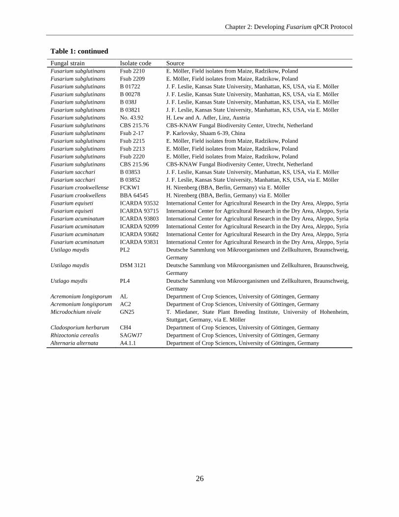
Table 1. Fungal isolates used in this research
Table 1: continued
Fungal strain Isolate code Source
Fusarium graminearum DSM 62722 Deutsch Sammlung fon Mikroorganismen und Zellkulturen, Braaunschweig,
Germany
Fusarium graminearum DSM 67638 Deutsch Sammlung fon Mikroorganismen und Zellkulturen, Braaunschweig,
Germany
Fusarium graminearum BBA 62048 H. Nirenberg (BBA, Berlin, Germany) via E. Möller
Fusarium graminearum Fg 5 H. Nirenberg (BBA, Berlin, Germany) via E. Möller
Fusarium graminearum Fg 71 T. Miedaner, University of Hohenheim, Germany
Fusarium graminearum Fg 210. 1 wt Plant Pathological Strain Collection of the University of Göttingen,
Germany
Fusarium proliferatum FPRO 1 A. Szecsi, Budapest, Hungary via E. Möller
Fusarium proliferatum FPRO 2 A. Szecsi, Budapest, Hungary via E. Möller
Fusarium proliferatum FPRO 4 A. Szecsi, Budapest, Hungary via E. Möller
Fusarium proliferatum FPRO 5 A. Szecsi, Budapest, Hungary via E. Möller
Fusarium proliferatum FPRO 6 A. Szecsi, Budapest, Hungary via E. Möller
Fusarium proliferatum FPRO 7 A. Szecsi, Budapest, Hungary via E. Möller
Fusarium proliferatum FPRO 8 A. Szecsi, Budapest, Hungary via E. Möller
Fusarium proliferatum FPRO 9 A. Szecsi, Budapest, Hungary, via E. Möller
Fusarium proliferatum FPRO 11 A. Szecsi, Budapest, Hungar,y via E. Möller
Fusarium proliferatum FPRO 12 A. Szecsi, Budapest, Hungary, via E. Möller
Fusarium avenaceum Fa 95 E. Möller, University of Hohenheim, Germany
Fusarium avenaceum Fa 23 Department of Crop Sciences, University of Göttingen, Germany
Fusarium avenaceum Fa 5-2 Department of Crop Sciences, University of Göttingen, Germany
Fusarium avenaceum DSM 62161 Deutsche Sammlung von Mikroorganismen und Zellkulturen, Braunschweig,
Germany
Fusarium culmorum Fc 15 T. Miedaner, State Plant Breeding Institute, University of Hohenheim,
Stuttgart, Germany, via E. Möller
Fusarium culmorum Fc 2 H. Nirenberg (BBA, Berlin, Germany) via E. Möller
Fusarium culmorum Fc 22 T. Miedaner, State Plant Breeding Institute, University of Hohenheim,
Stuttgart, Germany, via E. Möller
Fusarium culmorum FCH 69 Department of Crop Sciences, University of Göttingen, Germany
Fusarium tricinctum FT 2 Department of Crop Sciences, University of Göttingen, Germany
Fusarium tricinctum FT 3 Department of Crop Sciences, University of Göttingen, Germany
Fusarium verticillioides A00102 J. F. Leslie, Kansas State University, Manhattan, KS, USA, via E. Möller
Fusarium verticillioides 1.34 Mykothek FAP (W.Winter), via E. Möller
Fusarium verticillioides FV 234/1 P, Battilani, Faculty of Agriculture, UniversitaCattolica del SacroCoure,
Piacenza, Italy, via T. Miedaner
Fusarium verticillioides FM 8114 Fusarium Research Center, Pennsylvania State University, USA
Fusarium verticillioides FV Ita 1 A. Prodi, University of Bologna, Italy
Fusarium oxysporum Fo 125 Department of Crop Sciences, University of Göttingen, Germany
Fusarium oxysporum Foxy 121 Department of Crop Sciences, University of Göttingen, Germany
Fusarium oxysporum Foxy 436 Department of Crop Sciences, University of Göttingen, Germany
Fusarium oxysporum Foxy 119 Department of Crop Sciences, University of Göttingen, Germany
Fusarium oxysporum Foxy 6 Department of Crop Sciences, University of Göttingen, Germany
Fusarium oxysporum Foxy 2 Mykothek FAP (W.Winter), via E. Möller
Fusarium poae Fpoae 517 Department of Crop Sciences, University of Göttingen, Germany
Fusarium poae Fpoae 369 Department of Crop Sciences, University of Göttingen, Germany
Fusarium poae FP 2 T. Miedaner, State Plant Breeding Institute, University of Hohenheim,
Stuttgart, Germany, via E. Möller
Fusarium poae DSM 62376 Deutsche Sammlung von Mikroorganismen und Zellkulturen, Braunschweig,
Germany
Chapter 2: Developing Fusarium qPCR Protocol
26
Table 1: continued
Fungal strain Isolate code Source
Fusarium subglutinans Fsub 2210 E. Möller, Field isolates from Maize, Radzikow, Poland
Fusarium subglutinans Fsub 2209 E. Möller, Field isolates from Maize, Radzikow, Poland
Fusarium subglutinans B 01722 J. F. Leslie, Kansas State University, Manhattan, KS, USA, via E. Möller
Fusarium subglutinans B 00278 J. F. Leslie, Kansas State University, Manhattan, KS, USA, via E. Möller
Fusarium subglutinans B 038J J. F. Leslie, Kansas State University, Manhattan, KS, USA, via E. Möller
Fusarium subglutinans B 03821 J. F. Leslie, Kansas State University, Manhattan, KS, USA, via E. Möller
Fusarium subglutinans No. 43.92 H. Lew and A. Adler, Linz, Austria
Fusarium subglutinans CBS 215.76 CBS-KNAW Fungal Biodiversity Center, Utrecht, Netherland
Fusarium subglutinans Fsub 2-17 P. Karlovsky, Shaam 6-39, China
Fusarium subglutinans Fsub 2215 E. Möller, Field isolates from Maize, Radzikow, Poland
Fusarium subglutinans Fsub 2213 E. Möller, Field isolates from Maize, Radzikow, Poland
Fusarium subglutinans Fsub 2220 E. Möller, Field isolates from Maize, Radzikow, Poland
Fusarium subglutinans CBS 215.96 CBS-KNAW Fungal Biodiversity Center, Utrecht, Netherland
Fusarium sacchari B 03853 J. F. Leslie, Kansas State University, Manhattan, KS, USA, via E. Möller
Fusarium sacchari B 03852 J. F. Leslie, Kansas State University, Manhattan, KS, USA, via E. Möller
Fusarium crookwellense FCKW1 H. Nirenberg (BBA, Berlin, Germany) via E. Möller
Fusarium crookwellens BBA 64545 H. Nirenberg (BBA, Berlin, Germany) via E. Möller
Fusarium equiseti ICARDA 93532 International Center for Agricultural Research in the Dry Area, Aleppo, Syria
Fusarium equiseti ICARDA 93715 International Center for Agricultural Research in the Dry Area, Aleppo, Syria
Fusarium acuminatum ICARDA 93803 International Center for Agricultural Research in the Dry Area, Aleppo, Syria
Fusarium acuminatum ICARDA 92099 International Center for Agricultural Research in the Dry Area, Aleppo, Syria
Fusarium acuminatum ICARDA 93682 International Center for Agricultural Research in the Dry Area, Aleppo, Syria
Fusarium acuminatum ICARDA 93831 International Center for Agricultural Research in the Dry Area, Aleppo, Syria
Ustilago maydis PL2 Deutsche Sammlung von Mikroorganismen und Zellkulturen, Braunschweig,
Germany
Ustilago maydis DSM 3121 Deutsche Sammlung von Mikroorganismen und Zellkulturen, Braunschweig,
Germany
Ustlago maydis PL4 Deutsche Sammlung von Mikroorganismen und Zellkulturen, Braunschweig,
Germany
Acremonium longisporum AL Department of Crop Sciences, University of Göttingen, Germany
Acremonium longisporum AC2 Department of Crop Sciences, University of Göttingen, Germany
Microdochium nivale GN25 T. Miedaner, State Plant Breeding Institute, University of Hohenheim,
Stuttgart, Germany, via E. Möller
Cladosporium herbarum CH4 Department of Crop Sciences, University of Göttingen, Germany
Rhizoctonia cerealis SAGWJ7 Department of Crop Sciences, University of Göttingen, Germany
Alternaria alternata A4.1.1 Department of Crop Sciences, University of Göttingen, Germany
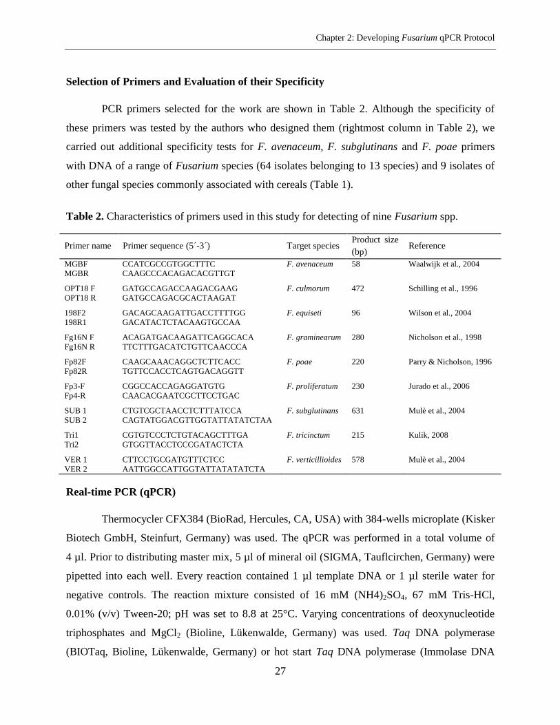
Chapter 2: Developing Fusarium qPCR Protocol
27
Selection of Primers and Evaluation of their Specificity
PCR primers selected for the work are shown in Table 2. Although the specificity of
these primers was tested by the authors who designed them (rightmost column in Table 2), we
carried out additional specificity tests for F. avenaceum, F. subglutinans and F. poae primers
with DNA of a range of Fusarium species (64 isolates belonging to 13 species) and 9 isolates of
other fungal species commonly associated with cereals (Table 1).
Table 2. Characteristics of primers used in this study for detecting of nine Fusarium spp.
Primer name Primer sequence (5´-3´) Target species Product size
(bp) Reference
MGBF CCATCGCCGTGGCTTTC F. avenaceum 58 Waalwijk et al., 2004
MGBR CAAGCCCACAGACACGTTGT
OPT18 F GATGCCAGACCAAGACGAAG F. culmorum 472 Schilling et al., 1996
OPT18 R GATGCCAGACGCACTAAGAT
198F2 GACAGCAAGATTGACCTTTTGG F. equiseti 96 Wilson et al., 2004 198R1 GACATACTCTACAAGTGCCAA
Fg16N F ACAGATGACAAGATTCAGGCACA F. graminearum 280 Nicholson et al., 1998
Fg16N R TTCTTTGACATCTGTTCAACCCA
Fp82F CAAGCAAACAGGCTCTTCACC F. poae 220 Parry & Nicholson, 1996
Fp82R TGTTCCACCTCAGTGACAGGTT
Fp3-F CGGCCACCAGAGGATGTG F. proliferatum 230 Jurado et al., 2006
Fp4-R CAACACGAATCGCTTCCTGAC
SUB 1 CTGTCGCTAACCTCTTTATCCA F. subglutinans 631 Mulè et al., 2004
SUB 2 CAGTATGGACGTTGGTATTATATCTAA
Tri1 CGTGTCCCTCTGTACAGCTTTGA F. tricinctum 215 Kulik, 2008 Tri2 GTGGTTACCTCCCGATACTCTA
VER 1 CTTCCTGCGATGTTTCTCC F. verticillioides 578 Mulè et al., 2004
VER 2 AATTGGCCATTGGTATTATATATCTA
Real-time PCR (qPCR)
Thermocycler CFX384 (BioRad, Hercules, CA, USA) with 384-wells microplate (Kisker
Biotech GmbH, Steinfurt, Germany) was used. The qPCR was performed in a total volume of
4 µl. Prior to distributing master mix, 5 µl of mineral oil (SIGMA, Tauflcirchen, Germany) were
pipetted into each well. Every reaction contained 1 µl template DNA or 1 µl sterile water for
negative controls. The reaction mixture consisted of 16 mM (NH4)2SO4, 67 mM Tris-HCl,
0.01% (v/v) Tween-20; pH was set to 8.8 at 25°C. Varying concentrations of deoxynucleotide
triphosphates and MgCl2 (Bioline, Lükenwalde, Germany) was used. Taq DNA polymerase
(BIOTaq, Bioline, Lükenwalde, Germany) or hot start Taq DNA polymerase (Immolase DNA
Chapter 2: Developing Fusarium qPCR Protocol
28
Pol., Lükenwalde, Germany) was employed in the activity 0.1 u/reaction. 0.1x SYBR Green I
(Invitrogen, Karlsruhe, Germany) and primers at a concentration of 0.3 µM were used.
Fluorescent data were obtained during the annealing phase to construct a melting curve at the
end of each assay. The qPCR was completed by carrying out a melting curve analysis according
to the following protocol: PCR products were denatured at 95°C for 1 min, annealed at 55°C for
1 min and gradually heated from 55°C to 95°C at a rate of 0.05°C/s while the fluorescence was
continuously recorded.
Determination of qPCR Sensitivity and Efficiency
Sensitivity was defined as the lowest amount of standard DNA (see Preparation of
DNA standards for qPCR) that generated PCR products with the expected melting temperature
in at least 7 out of 8 replicates. Each standard DNA was run in at least eight replicates on three
different days (Supplemental Table 1). The PCR efficiency was calculated in a common way
(Nutz et al., 2011).
Results
Selection and Specificity of Primers
The primers used in this work were designed and tested previously for species-specific
detection of Fusarium spp. (Table 2). Apart from specificity tests carried out by the researchers
who designed them, we conducted additional specificity tests for three primer pairs of
F. subglutinans (Nicolaisen et al., 2009; Möller et al., 1999; Mulè et al., 2004), two primer pairs
of F. avenaceum (Turner et al., 1998; Waalwijk et al., 2004) and one primer pair of F. poae
(Parry & Nicholson, 1996). We carried out these tests under conditions recommended in
publications in which the primers were described but reduced the reaction volume to 10 µl
(F. subglutinans) or 4 µl (F. avenaceum, F. poae).
In our thermocycler, primer pair Fsub565/Fsub622A described by Nicolaisen et al.
(2009) as specific for F. subglutinans generated products with different melting temperatures for
different F. subglutinans strains; moreover, DNA of the strains F. graminearum Fg 71 and
F. verticillioides Fv Ita 1 as template were amplified, too. The primer pair 61-2 F/61-2 R (Möller
et al., 1999) turned out not to be specific for F. subglutinans because it generated products with
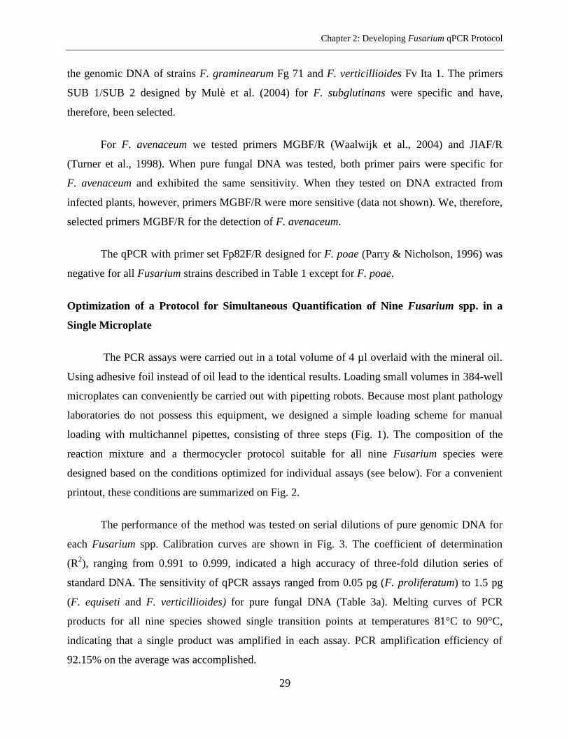
Chapter 2: Developing Fusarium qPCR Protocol
29
the genomic DNA of strains F. graminearum Fg 71 and F. verticillioides Fv Ita 1. The primers
SUB 1/SUB 2 designed by Mulè et al. (2004) for F. subglutinans were specific and have,
therefore, been selected.
For F. avenaceum we tested primers MGBF/R (Waalwijk et al., 2004) and JIAF/R
(Turner et al., 1998). When pure fungal DNA was tested, both primer pairs were specific for
F. avenaceum and exhibited the same sensitivity. When they tested on DNA extracted from
infected plants, however, primers MGBF/R were more sensitive (data not shown). We, therefore,
selected primers MGBF/R for the detection of F. avenaceum.
The qPCR with primer set Fp82F/R designed for F. poae (Parry & Nicholson, 1996) was
negative for all Fusarium strains described in Table 1 except for F. poae.
Optimization of a Protocol for Simultaneous Quantification of Nine Fusarium spp. in a
Single Microplate
The PCR assays were carried out in a total volume of 4 µl overlaid with the mineral oil.
Using adhesive foil instead of oil lead to the identical results. Loading small volumes in 384-well
microplates can conveniently be carried out with pipetting robots. Because most plant pathology
laboratories do not possess this equipment, we designed a simple loading scheme for manual
loading with multichannel pipettes, consisting of three steps (Fig. 1). The composition of the
reaction mixture and a thermocycler protocol suitable for all nine Fusarium species were
designed based on the conditions optimized for individual assays (see below). For a convenient
printout, these conditions are summarized on Fig. 2.
The performance of the method was tested on serial dilutions of pure genomic DNA for
each Fusarium spp. Calibration curves are shown in Fig. 3. The coefficient of determination
(R2), ranging from 0.991 to 0.999, indicated a high accuracy of three-fold dilution series of
standard DNA. The sensitivity of qPCR assays ranged from 0.05 pg (F. proliferatum) to 1.5 pg
(F. equiseti and F. verticillioides) for pure fungal DNA (Table 3a). Melting curves of PCR
products for all nine species showed single transition points at temperatures 81°C to 90°C,
indicating that a single product was amplified in each assay. PCR amplification efficiency of
92.15% on the average was accomplished.
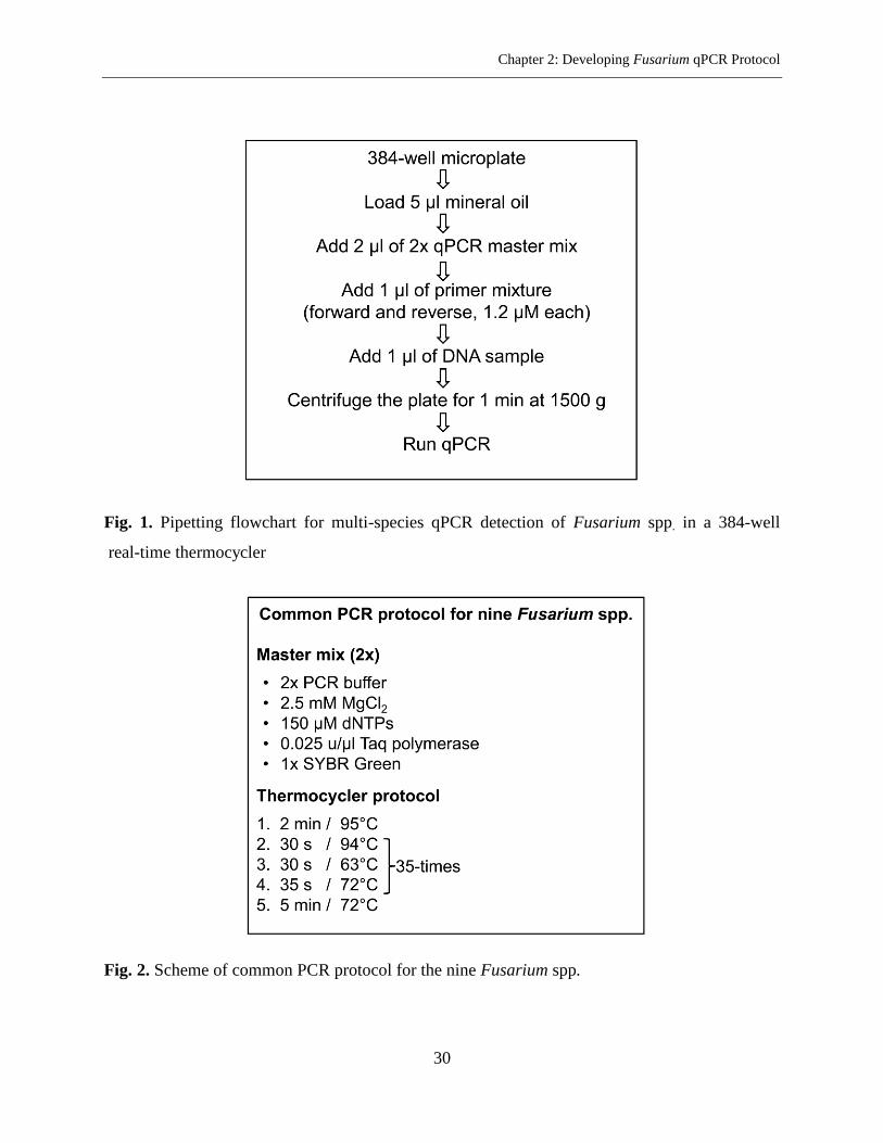
Chapter 2: Developing Fusarium qPCR Protocol
30
Fig. 1. Pipetting flowchart for multi-species qPCR detection of Fusarium spp. in a 384-well
real-time thermocycler
Fig. 2. Scheme of common PCR protocol for the nine Fusarium spp.
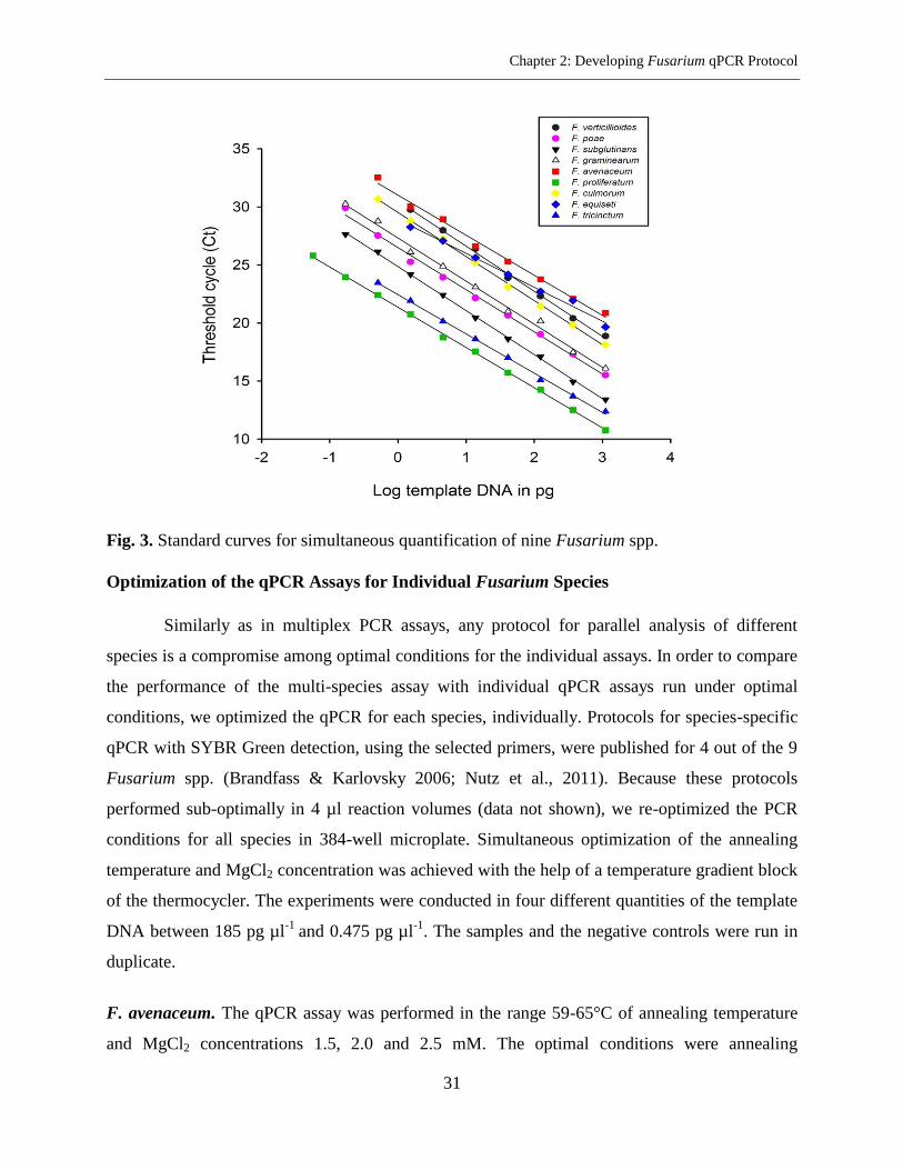
Chapter 2: Developing Fusarium qPCR Protocol
31
Fig. 3. Standard curves for simultaneous quantification of nine Fusarium spp.
Optimization of the qPCR Assays for Individual Fusarium Species
Similarly as in multiplex PCR assays, any protocol for parallel analysis of different
species is a compromise among optimal conditions for the individual assays. In order to compare
the performance of the multi-species assay with individual qPCR assays run under optimal
conditions, we optimized the qPCR for each species, individually. Protocols for species-specific
qPCR with SYBR Green detection, using the selected primers, were published for 4 out of the 9
Fusarium spp. (Brandfass & Karlovsky 2006; Nutz et al., 2011). Because these protocols
performed sub-optimally in 4 µl reaction volumes (data not shown), we re-optimized the PCR
conditions for all species in 384-well microplate. Simultaneous optimization of the annealing
temperature and MgCl2 concentration was achieved with the help of a temperature gradient block
of the thermocycler. The experiments were conducted in four different quantities of the template
DNA between 185 pg µl-1
and 0.475 pg µl-1
. The samples and the negative controls were run in
duplicate.
F. avenaceum. The qPCR assay was performed in the range 59-65°C of annealing temperature
and MgCl2 concentrations 1.5, 2.0 and 2.5 mM. The optimal conditions were annealing
Chapter 2: Developing Fusarium qPCR Protocol
32
temperature of 63°C and 2.5 mM MgCl2. The threshold values (Ct) indicated a delicate
fluctuation for annealing temperatures 60-63°C and 2.0-2.5 mM MgCl2 for standard DNA
amounts higher than 10 pg. Therefore, considering the lowest amount of Ct in different quantities
of template DNA, the suitable qPCR conditions were selected. The annealing time was lowered
from the recommended time of 60 s (Waalwijk et al., 2004) to 25 s. Considering the length of
PCR amplicon, the best time for elongation step was 10 s.
F. culmorum. Performance of annealing gradient qPCR assay between 58-66°C and 2.5, 3.0, 3.5
mM of Mg++
concentrations indicated that temperature of 62°C and 2.5 mM MgCl2 can produce
suitable Ct values in all DNA quantities, in contrast to the annealing temperature of 64°C and 4
mM of Mg++
concentration which are suggested by Brandfass & Karlovsky (2008). The time for
annealing and extension steps was 40 s and 45 s, respectively.
F. equiseti. The qPCR assay was performed in range 58.2-66.8°C of annealing temperature and
MgCl2 concentrations 1.5, 2.0 and 2.5 mM. Initial experiments showed that 63°C of annealing
temperature and 2.5 mM MgCl2 would be the optimal qPCR conditions. But there was unspecific
peak especially in low concentrations of DNA. Therefore the PCR condition was improved by
using a high quality Taq DNA polymerase. In this case the optimal annealing temperature was
increased to 65°C for 20 s. The time for extension step was 20 s.
F. graminearum. Annealing temperature in the range 59.0-66.5°C and Mg++
concentrations 2.0,
2.5 and 3.0 mM were tested. The optimal conditions were annealing temperature of 61°C and
2.5 mM MgCl2, similar to Brandfass & Karlovsky (2008) who found 64°C and 2.5 mM MgCl2 to
be optimal for qPCR with the same primers in a reaction volume of 25 µl. We found no
difference in the threshold cycle for annealing temperatures 60.0-62.5°C and 2.0–3.0 mM MgCl2
for standard DNA amounts higher than 40 pg. The time for annealing and elongation steps were
reduced from 45 s recommended by Brandass & Karlovsky (2008) to 30 s.
F. poae. Testing annealing temperature in the range 60-65°C and 1.5, 2.0 and 2.5 mM Mg++
concentration indicated that the qPCR could be operated in a wide range of annealing
temperature and MgCl2. The amount of Ct value did not show a big fluctuation in different
conditions. Therefore, the annealing temperature of 62.5°C and 2 mM of MgCl2 were selected as
Chapter 2: Developing Fusarium qPCR Protocol
33
the best qPCR conditions. Parry & Nicholson (1996) had suggested the annealing temperature of
60°C for the primer set Fp82 F/R. Time for annealing and extension steps was optimized on 30 s
and 35 s, respectively.
F. proliferatum. The qPCR assay was carried out in the range 59-65°C of annealing temperature
and 2.0, 2.5, 3.0 mM of Mg++
concentration. The optimal conditions were annealing temperature
of 64°C and 2 mM MgCl2, similar to Brandfass & Karlovsky (2008). There was no difference in
threshold cycles for the annealing temperatures 60-65°C and 2.0-2.5 mM MgCl2 while standard
DNA amounts were less than 20 pg. The best time for annealing and extension steps was 30 s
and 35 s, respectively.
F. subglutinans. Annealing temperature in the range 54-68°C and Mg++
concentration
2.0-4.5 mM were tested. An annealing temperature of 65°C yielded specific products, in contrast
to the annealing temperature of 56°C suggested by the primer designers (Mulè et al., 2004). The
optimal concentration for MgCl2 was 3.0 mM. We found no difference in Ct values for 64-66°C
and 2.5-3.5 mM MgCl2. Considering the lowest Ct value and specificity for primer annealing, the
best qPCR conditions were selected. Desired time for annealing and extension steps was 30 s and
40 s, respectively.
F. tricinctum. Performance of annealing gradient qPCR assay between 58.2-66.8°C and 2.5, 3.0,
3.5 mM of Mg++
concentrations indicated that temperature of 65°C and 2.5 mM MgCl2 can
produce suitable Ct values in all DNA quantities. The annealing conditions were similar to Kulik
(2008); but the time for extension step was reduced of 55 s (Kulik, 2008) to 25 s.
F. verticillioides. Annealing temperature in the range 56-65°C and Mg++
concentrations 2.0, 2.5
and 3.0 mM were evaluated. The optimal conditions were annealing temperature of 62.5°C and
2.5 mM MgCl2. There was no difference in Ct values for annealing temperatures 60.0-62.5°C
and 2.5-3.0 mM MgCl2 for the standard DNA amounts higher than 10 pg, but the best qPCR
conditions were selected considering the suitable conditions for lower DNA quantities. The time
for annealing and elongation steps were lowered from the recommended time of 50 s and 60 s
(Nutz et al., 2011) to 30 s and 40 s, respectively.
Chapter 2: Developing Fusarium qPCR Protocol
34
The optimal qPCR conditions were tested for the overall performance of assay by using
the data generated from a serial dilution of pure genomic DNA for each Fusarium spp.,
individually. The average amount of Ct values was graphed versus the logarithm of the
correspondent starting DNA quantity (SQ) to construct the standard curves. Comparing of R2
indicated the accuracy of the dilution series and the overall assay sensitivity. The coefficient of
determination ranged from 0.990 to 0.999. Typical calibration curves generated for Fusarium
spp. DNA are given in Fig. 4. The sensitivity of qPCR assays was estimated to be 0.05 pg for
pure DNA of F. proliferatum, 0.17 pg for F. avenaceum, F. graminearum, F. poae,
F. subglutinans and 0.50 pg for F. culmorum, F. equiseti, F. tricinctum and F. verticillioides in a
background free of plant tissue and other contamination. The average repeatability varied from
0.84% RSD (relative standard deviation) for F. verticillioides to 2.00% RSD for F. tricinctum
(Table 3b). The main point in this protocol was the least possible number of samples which are
included in each qPCR assay. Considering amount of Taq DNA polymerase, the master mix
would be prepared at least for 50 samples to decrease the pipetting error.
As it is presented above, the qPCR optimization tests indicated flexibility in some cases.
In such conditions, qPCR assay could be operated in a wide range of annealing temperature and
MgCl2 concentrations. Nevertheless, concerning low Ct values and specificity of primer
annealing, the best conditions were selected for each Fusarium spp. Optimized qPCR conditions
for each Fusarium spp. are summarized in Table 3b. The performance of new optimized assays
for nine species of Fusarium in 4 µl volumes was gratified and low concentrations of genomic
DNA were also detectable by qPCR assay without creating unspecific peak.
The results also show that the difference between qPCR efficiency in single species and
multi-species detection of Fusarium spp. is not high and the Ct values did not change by more
than approximately 1 Ct. Furthermore, we compared PCR sealing film and the mineral oil as a
coating layer for qPCR microplate. There was no difference in Ct values and efficiency of assay.
Therefore, we would suggest using the mineral oil instead of other sealing film.
Chapter 2: Developing Fusarium qPCR Protocol
35
Table 3a. PCR conditions and performance parameters of multi-species qPCR assay
Table 3b. PCR conditions and performance parameters of single species qPCR assays
1. Sensitivity ("Sens."): The lowest standard in a 3-fold dilution series starting with 1111 pg that was amplified in at
least 7 out of 8 replicates
2. Average percent of relative standard deviation ("Ave %RSD") for all detectable qPCR standards
3. Melting temperature ("Tm") determined with accuracy of 0.5 °C
Fusarium spp.
MgCl2
(mM)
dNTPs
(µM)
Ta
(°C)
Ann. time
(s)
Ext. time
(s)
PCR
Efficiency
(%)
Sens.1
(pg)
Ct for 1
pg
Ave of
%RSD2
Tm3
(°C)
F. avenaceum 94.8 0.508 31.01 1.57 81
F. culmorum 83.4 0.508 29.52 1.71 87
F. equiseti 120.1 1.524 28.91 1.00 86
F. graminearum 85.9 0.169 27.31 1.40 82
F. poae 2.5 150 63 30 35 88.5 0.169 26.49 1.69 83
F. proliferatum 94.4 0.056 21.35 1.58 89
F. subglutinans 83.5 0.169 24.85 1.50 88
F. tricinctum 97.6 0.508 22.44 0.81 90
F. verticillioides 81.0 1.524 30.05 1.62 88
Fusarium spp.
MgCl2
(mM)
dNTPs
(µM)
Ta
(°C)
Ann. time
(s)
Ext. time
(s)
PCR Efficiency
(%)
Sens.1
(pg)
Ct for 1
pg
Ave of
%RSD2
Tm3
(°C)
F. avenaceum 2.5 150 63.0 25 10 98.19 0.169 30.15 1.42 81
F. culmorum 2.5 150 62.0 40 45 94.28 0.508 28.27 1.11 87
F. equiseti 2.5 150 65 20 10 110.1 0.508 29.18 0.96 86
F. graminearum 2.5 150 61.0 30 30 100.3 0.169 28.09 1.79 82
F. poae 2.0 100 62.5 30 35 99.83 0.169 25.93 1.47 83
F. proliferayum 2.0 150 64.0 30 35 98.64 0.056 21.53 1.56 89
F. subglutinans 3.0 100 65.0 30 40 97.75 0.169 24.49 1.56 88
F. tricinctum 2.5 150 65 25 30 106.0 0.508 21.62 2.00 90
F. verticillioides 2.5 100 62.5 30 40 96.37 0.508 29.40 0.84 88
Chapter 2: Developing Fusarium qPCR Protocol
36
Fig. 4. Standard curves for Fusarium spp. qPCR assays
Chapter 2: Developing Fusarium qPCR Protocol
37
Discussion
Cereals are contaminated with a wide range of Fusarium spp., most of which produce
mycotoxins (Jurado et al., 2005). Quantitative determination of fungal biomass in plant tissue is
invaluable in studies of plant diseases. Furthermore, quantitative data on fungal biomass can be
used for the prediction of mycotoxin content; the biomass of a Fusarium species and the
concentration of its mycotoxin in the infected tissue often tightly correlate (Waalwijk et al.,
2004; Schnerr et al., 2002; Yli-Mattila et al., 2008). Therefore, quantitative diagnosis of
pathogenic Fusaria in plant tissues is an essential and powerful tool in both research and
diagnostic laboratories. Sensitive and fast diagnostic methods are also important in assaying crop
propagules for quarantine pathogens (Waalwijk et al., 2004). Since the range of Fusarium
species associated with cereals is quite extensive, to facilitate fusarium diseases investigation
while testing some samples for more than one species of Fusarium, we developed a
multi-species qPCR assay in 4 µl volume reactions by using a 384-well thermocycler. The qPCR
assays for all nine species, studied in this work, can be carried out in the same microplate,
because they utilize the same thermocycle profile. Multi-species qPCR protocol is particularly
useful when user have a few samples, but wants to check them for more than one Fusarium spp.
In this case, we will likely have less labor intensive if we can run the samples in 384-well qPCR
plate and the time of qPCR analysis could be reduced intensely to a single day. Simultaneous
quantification approach allows maximum amount of data to be generated from each qPCR assay.
Nine species of Fusarium evaluated in this study, cause important diseases on maize and small
grain cereals. The qPCR optimization tests which quantified Fusarium spp. individually, had
revealed that some of the qPCR variables can work in a wide range of PCR conditions.
Therefore, concerning the low Ct values and specificity of primer annealing, the most suitable
conditions were selected. On the other hand, in our experiments, assessment of common domains
of qPCR variables helped us for designing a multi-species qPCR assay. Eventually, owing to
applicable common range in qPCR variables we concluded on specific PCR profiling for the nine
species of Fusarium.
Sensitivity and robustness of qPCR assay are known as the crucial factors. Standard
performance parameters of analytical methods are the limit of detection (LOD) and limit of
quantification (LOQ). Standard methods for the determination of LOD and LOQ, based on the
Chapter 2: Developing Fusarium qPCR Protocol
38
noise of the background are not suitable for qPCR. Several authors suggested alternative
definitions of LOD and LOQ in qPCR (Bustin et al., 2009; Mattarucchi et al., 2005; Vaerman
et al., 2004; Gao et al., 2004); but none of these procedures was accepted by the community. The
use of receiver operating curve analysis is the most recent suggestion for the determination of
LOQ in qPCR (Nutz et al., 2011); but the method requires the analysis of large field data sets
which was not available for our assays. We, therefore, used a simple sensitivity parameter
defined as the lowest amount of template DNA that generated products with the expected
melting temperature in at least seven out of eight replicates. The sensitivity determined in this
way depends on DNA quantities of the standards; because we used a 3-fold dilution series,
sensitivity value of S specifies a confidence interval for LOD as follows: S > LOD > S/3.
According this definition, the real-time PCR assay was sensitive with consistent detection of
0.05 pg µl-1
of standard DNA for F. proliferatum, 0.17 pg µl-1
for F. graminearum, F. poae,
F. subglutinans, 0.50 pg for F. avenaceum, F. culmorum, F. tricinctum and 1.52 pg µl-1
for
F. equiseti and F. verticillioides. Lack of adequate sensitivity for quantifying of F. equiseti and
F. verticillioides in multi-species qPCR protocol indicates the importance of using the single
species detection method in the precise quantification researches of these two species of
Fusarium.
The previous published qPCR conditions for F. culmorum, F. graminearum,
F. proliferatum and F. verticillioides (Brandfass & Karlovsky, 2006; Nutz et al., 2011) for 25 µl
volumes were not fit when used in 4 µl reactions. In most cases, especially in low concentrations
of DNA, a second peak prior to specific melting point made disorders for the qPCR dissociation
curve. This wide shape peaks would be generated from primer-dimmers or unspecific
amplification products and should be discarded after enhancing of qPCR conditions. Therefore,
for all of those species, we re-optimized the qPCR conditions to find the best options for running
a single species qPCR. The main changes for the thermal cycling parameters were involved
annealing and elongation steps. Considering threshold cycle and specificity of primer annealing,
the optimal concentration of PCR reagents was selected for the nine species of Fusarium.
The single species qPCR protocol showed high sensitivity for F. proliferatum (0.05 pg
DNA µl-1
), F. avenaceum, F. graminearum, F.poae, F. subglutinans (0.17 pg DNA µl-1
) and it
was 0.50 pg DNA µl-1
for F. culmorum, F. equiseti, F. tricinctum and F. verticillioides. Further
Chapter 2: Developing Fusarium qPCR Protocol
39
experiments indicated a constant higher sensitivity for F. proliferatum (2 fg DNA µl-1
) while the
average of the Ct value was 29.8 (data not shown). Using the hot start Taq DNA polymerase
could enhance the amplification of F. equiseti strains. Due to the results, the correspondence
among single-species and multi-species qPCR assays demonstrated in this work, was optimal
and the Ct values did not change by more than approximately 1 Ct in two reported protocols. The
DNA samples of plant tissues often need to be diluted prior to PCR; because of the presence of
different inhibitors in DNA extracts which can affect the target amplification. The PCR
inhibitors are particularly known to attend in the DNA prepared from senescing plant tissues
(Turner et al., 1998) and also the DNA extracts from the soil or plant roots (Van de Graaf et al.,
2003; Gao et al., 2004).
The Ct value for 1 pg DNA µl-1
was between 24-30 cycles for all nine Fusarium spp.
except F. proliferatum and F. tricinctum, which have a threshold cycle of 21 for this amount of
the pure genomic DNA. The reason is that the primers used for these two Fusarium species were
derived from the IGS (Intergenic Spacer) rDNA region which is a multi-copy domain in the
genome and fungal detection is more sensitive for multi-copy IGS-based PCR assay (Jurado
et al., 2006).
The set of species-specific primers used in this work allowed amplifying the considering
specific fragments by qPCR assay. Some of the primer pairs indicated specificity problems and
excluded from the further experiments. For F. subglutinans, Primer Fsub565/Fsub622A
(Nicolaisen et al., 2009) produced non-specific PCR products for F. subglutinans DNA samples,
Fg 71 and Fv Ita l isolates. Furthermore, primer 61-2 F/61-2 R published by Möller et al. (1999),
generated specific melting temperature for some of the reference DNA. Zheng & Ploetz (2002),
demonstrated primer pairs 61-2 F/61-2 R were not useful for identifying the F. subglutinans-like
isolates such as F. sacchari and F. circinatum. As a result in our work the primer pair SUB
1/SUB 2, designed based on the calmodulin partial gene (Mulè et al., 2004) considered as the
species-specific primer in real-time PCR for F. subglutinans. There was no cross-reaction with
other Fusarium spp. and the fungal DNA tested. Therefore, F. subglutinans optimization was
continued with SUB 1/SUB 2 primers. Regarding to F. avenaceum, Turner et al. (1998),
designed primer JIAF/R based on the nuclear 5.8s rDNA ITS sequence and demonstrated to be
specific for F. avenaceum with no cross reactivity with F. tricinctum or any other wheat

Chapter 2: Developing Fusarium qPCR Protocol
40
pathogens. Furthermore, primer MGBF/R which is a set of novel oligonucleotides including
“Minor Groove Binder” (MGB) ligands has been designed by Waalwijk et al. (2004). These
scientists produced this primer based on the sequencing of a 920 bp amplified DNA fragment
generated by Fa F/R primer. The primer Fa F/R was reported to be specific to F. avenaceum
(Doohan et al., 1998). In our experiments both of these primers (JIAF/R and avenaceum
MGBF/R) produced specific peak only in F. avenaceum wells; but more sensitivity of primer
MGBF/R with field samples led to its selection as the specific primer for F. avenaceum. Higher
specificity of MGB ligands primers compared to the classical primers is for the nature of ligands
(Afonia et al., 2002). Primer set Fp82F/R for F. poae was created from clone T161 by Parry and
Nicholson (1996). They had presented this primer as a monomorphic marker which is able to
detect F. poae in wheat samples; but another primer pair designed for F. poae (Fp8F/R) had
shown polymorphic bands and, therefore, did not develop further. In our experiments primer
Fp82F/R, amplified the target DNA while no amplification occurred in the reference fungal
DNA nor for the negative control. Consequently, the ability of primer pairs SUB 1/SUB 2,
MGBF/R and Fp82F/R was confirmed to detect corresponding Fusarium species in the extracts
from maize plant tissue samples by qPCR. Primer pair 198F2/198R1 designed by Wilson et al.
(2004) for F. equiseti was tested against isolates of the target species and a variety of other
Fusarium species and other fungi associated with diseases of cereals (Wilson et al., 2004;
Nicholson et al., 2004). Kulik (2008) evaluated the specificity of Tri1/Tri2 primers on genomic
DNA extracted from 56 isolates representing 20 Fusarium species and also 12 F. tricinctum
isolates. He found unexpected amplicons which were amplified from one isolate of
F. acuminatum and one from F. nurragi. Further sequencing experiments indicated 100%
identity of F. acuminatum isolate to F. tricinctum while F. nurragi had 99% of similarity to CBS
261.51 (F. tricinctum).
In conclusion it is believed that the real-time PCR protocols described in this study are
specific, reliable and sensitive for singly detection and quantification of nine Fusarium spp.
genomic DNA. Furthermore, the established multi-species qPCR method is also robust, precise,
cost effective, quick and applicable for monitoring and quantifying of nine Fusarium spp. These
protocols are widely used in our department for detection and quantification of Fusarium spp. in
cereal crops including maize, wheat, sorghum, rice, and different other plants such as weeds
Chapter 2: Developing Fusarium qPCR Protocol
41
which naturally or artificially have single or multiple infection of Fusarium spp. It is anticipated
that the reported protocols would be applicable for other crops, as well.
Author,s Contributions
In this work, the specificity test for F. subglutinans primer sets was performed by
Dr. Eva-Maria Becker.
Literature Cited
Afonia, I. A., Reed, M. W., Lusby. E., Shishkina. I. G., and Belousov, Y. S. 2002. Minor
Groove Binder-conjugated DNA probes for quantitative DNA detection by
hybridization-triggered fluorescence. Biotechniques 32:940-949.
Brandfass, C., and Karlovsky, P. 2008. Upscaled CTAB-based DNA extraction and
real-time PCR assays for Fusarium culmorum and F. graminearum DNA in plant material
with reduced sampling error. Int. J. Mol. Sci. 9:2306-2321.
Brandfass, C., and Karlovsky, P. 2006. Simultaneous detection of Fusarium culmorum
and F. graminearum in plant material by duplex PCR with melting curve analysis. BMC
Microbiol. 6 (4). Available from: http://www.biomedcentral.com/1471-2180/6/4 : URL.
DOI.
Brenan, C., and Morrison, T. 2005. High throughput, nanoliter quantitative PCR. Drug
Discovery Today: Technologies 2:247-253.
Bustin, S. A., Benes, V.,Garson, J. A., Hellemans, J., Huggett, J., Kubista, M., Mueller,
R., Nolan, T., Pfaffl, M. W., Shipley, G. L., Vandesompele, J., and Wittwer, C. T. 2009.
The MIQE guidelines: minimum information for publication of quantitative real-time
PCR experiments. Clin. Chem. 55:611-622.
Dahl, A., Sultan, M., Jung, A., Schwartz, R., Lange, M., Steinwand, M., Livak, K. J.,
Lehrach, H., and Nyarsik, L. 2007. Quantitative PCR based expression analysis on a
nanoliter scale using polymer nano-well chips. Biomed. Microdevices 9:307-314.
Doohan, F. M., Parry, D. W., Jenkinson, P., and Nicholson, P. 1998. The use of species-
specific PCR-based assays to analyse Fusarium ear blight of wheat. Plant Pathol. 47:
197-205.
Chapter 2: Developing Fusarium qPCR Protocol
42
Fredlund, E., Gidlund, A., Pettersson, H., Olsen, M., and Börjesson, T. 2010. Real-time
PCR detection of Fusarium species in Swedish oats and correlation to T-2 and HT-2
toxin content. World Mycotoxin Journal 3:77-88.
Gao, X., Jackson, T. A., Lambert, K. N., Li, S., Hartman, G. L., and Niblack, T. L. 2004.
Detection and quantification of Fusarium solani f.sp. glycines in soybean roots with
real-time quantitative polymerase chain reaction. Plant Dis. 88:1372-1380.
Gelderblom, W. C. A., Jaskiewicz, K., Marasas, W. F. O., Thiel, P. G., Horak, R. M.,
Vleggaar, R., and Kriek, N. P. J. 1988. Fumonisins-novel mycotoxins with
cancer-promoting activity produced by Fusarium moniliforme. Appl. Environ. Microb.
54:1806–1811.
Gilbertson, R. L., Brown, W. M. Jr., and Ruppel, E. G. 1985. Prevalence and virulence of
Fusarium spp. associated with stalk rot of corn in Colorado. Plant Dis. 69:1065-1068.
Jurado, M., Vazquez, C., Patino, B., and González-Jaén, M. T. 2005. PCR detection
assays for the trichothecene-producing species Fusarium graminearum, Fusarium
culmorum, Fusarium poae, Fusarium equiseti and Fusarium sporotrichioides. Syst. Appl.
Microbiol. 28:562-568.
Jurado, M., Vazquez, C., Marin, S., Sanchis, V., and González-Jaén, M. T. 2006.
PCR-based strategy contamination with mycotoxicogenic Fusarium species in maize.
Syst. Appl. Microbiol. 29:681-689.
Kulik, T. 2008. Detection of Fusarium tricinctum from cereal grain using PCR assay. J.
Appl. Genet. 49:305-311.
Logrieco, A., Mulè, G., Moretti, A., and Bottalico, A. 2002. Toxigenic Fusarium species
and mycotoxins associated with maize ear rot in Europe. Eur. J. Plant Pathol. 108:
597-609.
Mattarucchi, E., Marsoni, M., Binelli, G., Passi, A., Curto, F. L., Pasquali, F., and Porta,
G. 2005. Different real time PCR approaches for the fine quantification of SNP’s alleles
in DNA pools: assays development, characterization and pre-validation. J. Biochem. Mol.
Biol. 38:555-562.
Moretti, A. N. 2009. Taxonomy of Fusarium genus, a continous fight between lumpers
and splitters. Proc. Nat. Sci, Matica Srpska Novi Sad. 117:7-13.
Chapter 2: Developing Fusarium qPCR Protocol
43
Mulè, G., Susca, A., Stea, G., and Moretti, A. 2004. A species-specific PCR assay based
on the calmodulin partial gene for identification of Fusarium verticillioides,
F. proliferatum and F. subglutinans. Eur. J. Plant Pathol. 110:495–502.
Möller, E. M., Chełkowski, J., and Geiger, H. H. 1999. Species-specific PCR assays for
the fungal pathogens Fusarium moniliforme and Fusarium subglutinans and their
application to diagnose maize ear rot disease. J. Phytopathol. 147:497–508.
Nicholson, P., Simpson, D. R., Weston, G., Rezanoor, H. N., Lees, A. K., Parry, D. W.,
and Joyce, D. 1998. Detection and quantification of Fusarium culmorum and Fusarium
graminearum in cereals using PCR assays. Physiol. Mol. Plant Pathol. 53:17–37.
Nicholson, P., Simpson, D. R., Wilson, A. H., Chandler, E., and Thomsett, M. 2004.
Detection and differentiation of trichothecene and enniatin-producing Fusarium species
on small-grain cereals. Eur. J. Plant Pathol. 110:503-514.
Nicolaisen, M., Supronienė, S., Nielsen, L. K., Lazzaro, I., Spliid, N. H., and Justesen, A.
F. 2009. Real-time PCR for quantification of eleven individual Fusarium species in
cereals. J. Microbial. Methods. 76:234–240.
Nutz, S., Döll, K., and Karlovsky, P. 2011. Determination of the LOQ in real-time PCR
by receiver operating characteristic curve analysis: application to qPCR assays for
Fusarium verticillioides and F. proliferatum. Anal. Bioanal. Chem. 401:717-726.
Parry, D. W., and Nicholson, P. 1996. Development of a PCR assay to detect Fusarium
poae in wheat. Plant Pathol. 45:383-391.
Pintos Varela, C., Aguín Casal, O., Chaves Padin, M., Ferreiroa Martinez, V., Sainz Oses,
M. J., Scauflaire, J., Munaut, F., Bande Castro, M. J., and Mansilla Vázquez, J. P. 2013.
First report of Fusarium temperatum causing seedling blight and stalk rot on maize in
Spain. Plant Dis. (Abstr.) 97:1252.
Scauflaire, J., Gourgue, M., and Munaut, F. 2011. Fusarium temperatum sp. nov. from
maize, an emergent species closely related to F. subglutinans. Mycologia 103:586-597.
Schilling, A. G., Möller, E. M., and Geiger, H. H. 1996. Polymerase chain reaction-based
assays for species-specific detection of Fusarium culmorum, F. graminearum, and
F. avenaceum. Phytopathology 86:515-522.
Schnerr, H., Vogel, R. F., and Niessen, L. 2002. Correlation between DNA of
trichothecene-producing Fusarium species and deoxynivalenol concentrations in
wheat-samples. Lett. Appl. Microbiol. 35:121–125.
Chapter 2: Developing Fusarium qPCR Protocol
44
Turner, A. S., Lees, A. K., Rezanoor, H. N., and Nicholson, P. 1998. Refinement of
PCR-detection of Fusarium avenaceum and evidence from DNA marker studies for
phonetic relatedness to Fusarium tricinctum. Plant Pathol. 47:278-288.
Vaerman, J. L., Saussoy, P., and Ingargiola, I. 2004. Evaluation of real-time PCR data. J.
Biol. Regul. Homeost. Agents. 18:212-214.
Van de Graaf, P., Lees, A. K., Cullen, D. W., and Duncan, J. M. 2003. Detection and
quantification of Spongospora subterranea in soil, water and plant tissue samples using
real-time PCR. Eur. J. Plant Pathol. 109:589–597.
Waalwijk, C., van der Heide, R., de Vries, I., van der Lee, T., Schoen, C., Costrel-de
Corainville, G., Häuser-Hahn, I., Kastelein, P., Köhl, J., Lonnet, P., Demarquet, T., and
Kema, G. H. J. 2004. Quantitative detection of Fusarium species in wheat using TaqMan.
Eur. J. Plant Pathol. 110:481–494.
Wilfinger, W. W., Mackey, K., and Chomczynski, P. 1997. Effect of pH and ionic
strength on the spectrophotometric assessment of nucleic acid purity. Biotechniques
22:474–481.
Wilson, A., Simpson, D., Chandler, E., Jennings, P., and Nicholson, P. 2004.
Development of PCR assays for the detection and differentiation of F. sporotrichoides
and F. longsethiae. FEMS Microbiol. Lett. 233:69-76.
Yli-Mattila, T., Paavanen-Huhtala, S., Jestoi, M., Parikka, P., Hietaniemi, V., Gagkaeva,
T., Sarlin, T., Haikara, A., Laaksonen, S., and Rizzo, A. 2008. Real-time PCR detection
and quantification of Fusarium poae, F. graminearum, F. sporotrichioides and
F. langsethiae in cereal grains in Finland and Russia. Archives of Phytopathology and
Plant Protection 41:243–260.
Zheng, Q., and Ploetz, R. 2002. Genetic diversity in the mango malformation pathogen
and development of a PCR assay. Plant Pathol. 51:208-216.

Chapter 2: Developing Fusarium qPCR Protocol
45
Supplemental Table 1. Standard curves and determination of LOQ of qPCR for the nine
Fusarium species with a common thermocycler program
Supplemental Table 1: continued
F. avenaceum
F. culmorum
F. equiseti
DNA (pg) Ct Tm (°C)1
DNA (pg) Ct Tm (°C)
DNA (pg) Ct Tm (°C)
1111 18.99 82
1111 17.42 87
1111 19.54 86.5
1111 19.05 82
1111 17.73 87.5
1111 20.04 86.5
1111 19.34 82
1111 18.15 87
1111 19.77 86.5
370.37 21.57 81.5
370.37 18.99 87
370.37 21.10 86.5
370.37 21.39 82
370.37 19.26 87
370.37 21.50 86.5
370.37 21.25 82
370.37 19.38 87
370.37 21.20 86.5
123.45 22.60 82
123.45 21.07 87
123.45 22.98 86.5
123.45 23.37 81.5
123.45 21.17 87
123.45 23.16 86.5
123.45 Failed
123.45 21.08 87
123.45 22.87 86.5
41.15 23.28 82
41.15 22.44 87
41.15 24.75 86.5
41.15 25.38 81.5
41.15 22.77 87
41.15 24.42 86.5
41.15 25.11 81.5
41.15 22.68 87
41.15 24.25 86.5
13.71 26.74 81.5
13.71 24.54 87
13.71 26.44 86
13.71 26.33 82
13.71 24.13 87
13.71 25.75 86
13.71 25.82 82
13.71 24.32 87
13.71 Failed
4.57 28.33 81.5
4.57 25.69 87
4.57 25.89 86
4.57 28.12 81.5
4.57 26.68 87
4.57 Failed
4.57 28.15 82
4.57 26.02 87
4.57 27.63 86
1.52 29.37 81.5
1.52 27.29 87
1.52 28.15 86
1.52 29.94 81.5
1.52 27.51 87
1.52 27.30 86
1.52 29.26 81.5
1.52 Failed
1.52 27.72 86
0.508 31.41 81.5
0.508 30.45 87
0.508 29.89 86
0.508 31.29 81.5
0.508 28.96 87
0.508 30.01 86
0.508 31.79 82
0.508 28.35 87
0.508 Failed
0.169 32.02 81.5
1111 17.89 87
1111 19.51 86.5
0.169 33.30 81.5
1111 18.18 87
1111 19.63 86.5
0.169 32.61 81.5
370.37 20.08 87
1111 19.65 86.5
1111 18.97 82
370.37 19.68 87.5
370.37 21.05 86.5
1111 19.73 82
123.45 21.23 87
370.37 21.42 86.5
370.37 21.62 82
123.45 21.97 87
370.37 21.30 86.5
370.37 21.26 82
41.15 23.01 87
123.45 22.96 86.5
123.45 23.15 82
41.15 22.84 87
123.45 23.10 86.5
123.45 22.91 82
13.71 24.91 87
123.45 22.66 86.5
41.15 24.80 82
13.71 24.98 87.5
41.15 24.28 86.5
41.15 24.36 82
4.57 26.24 87.5
41.15 24.44 86.5
13.71 26.64 81.5
4.57 26.13 87
41.15 24.19 86
13.71 26.35 82
1.52 27.41 87
13.71 26.44 86
4.57 29.40 81.5
1.52 26.91 87.5
13.71 25.75 86
4.57 28.07 81.5
0.508 29.31 87.5
13.71 25.10 86
1.52 29.97 81
0.508 Failed
4.57 27.03 86
1.52 29.10 81.5
1111 17.49 87
4.57 26.89 86
0.508 31.85 81.5
1111 17.98 87
4.57 26.47 86
0.508 31.35 81.5
1111 18.14 87
1.52 28.48 86
0.169 32.34 81.5
370.37 19.06 87
1.52 28.59 86
0.169 Failed
370.37 19.46 87
1.52 28.03 86
1111 19.24 82
370.37 19.48 87
0.508 30.43 86
1111 20.03 82
123.45 21.13 87.5
0.508 29.85 86
1111 19.96 82
123.45 21.37 87.5
0.508 30.37 86.5
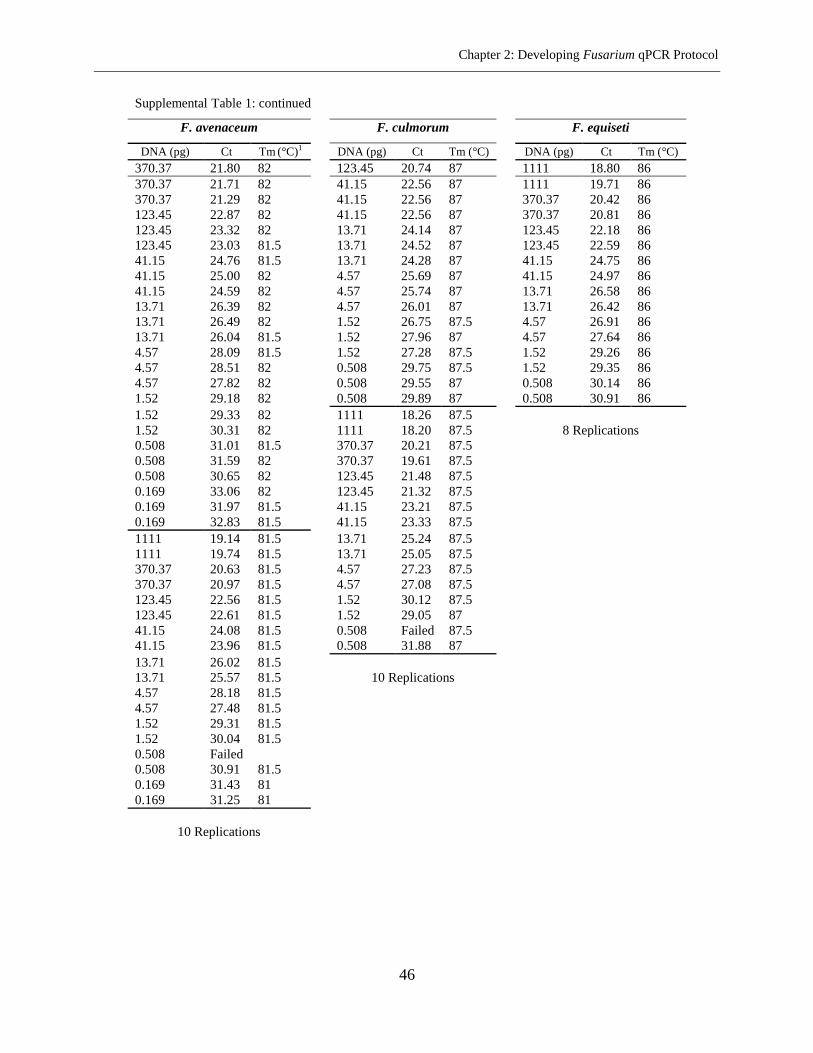
Chapter 2: Developing Fusarium qPCR Protocol
46
Supplemental Table 1: continued
F. avenaceum
F. culmorum
F. equiseti
DNA (pg) Ct Tm (°C)1
DNA (pg) Ct Tm (°C)
DNA (pg) Ct Tm (°C)
370.37 21.80 82
123.45 20.74 87
1111 18.80 86
370.37 21.71 82
41.15 22.56 87
1111 19.71 86
370.37 21.29 82
41.15 22.56 87
370.37 20.42 86
123.45 22.87 82
41.15 22.56 87
370.37 20.81 86
123.45 23.32 82
13.71 24.14 87
123.45 22.18 86
123.45 23.03 81.5
13.71 24.52 87
123.45 22.59 86
41.15 24.76 81.5
13.71 24.28 87
41.15 24.75 86
41.15 25.00 82
4.57 25.69 87
41.15 24.97 86
41.15 24.59 82
4.57 25.74 87
13.71 26.58 86
13.71 26.39 82
4.57 26.01 87
13.71 26.42 86
13.71 26.49 82
1.52 26.75 87.5
4.57 26.91 86
13.71 26.04 81.5
1.52 27.96 87
4.57 27.64 86
4.57 28.09 81.5
1.52 27.28 87.5
1.52 29.26 86
4.57 28.51 82
0.508 29.75 87.5
1.52 29.35 86
4.57 27.82 82
0.508 29.55 87
0.508 30.14 86
1.52 29.18 82
0.508 29.89 87
0.508 30.91 86
1.52 29.33 82
1111 18.26 87.5
1.52 30.31 82
1111 18.20 87.5
8 Replications
0.508 31.01 81.5
370.37 20.21 87.5
0.508 31.59 82
370.37 19.61 87.5
0.508 30.65 82
123.45 21.48 87.5
0.169 33.06 82
123.45 21.32 87.5
0.169 31.97 81.5
41.15 23.21 87.5
0.169 32.83 81.5
41.15 23.33 87.5
1111 19.14 81.5
13.71 25.24 87.5
1111 19.74 81.5
13.71 25.05 87.5
370.37 20.63 81.5
4.57 27.23 87.5
370.37 20.97 81.5
4.57 27.08 87.5
123.45 22.56 81.5
1.52 30.12 87.5
123.45 22.61 81.5
1.52 29.05 87
41.15 24.08 81.5
0.508 Failed 87.5
41.15 23.96 81.5
0.508 31.88 87
13.71 26.02 81.5
13.71 25.57 81.5
10 Replications
4.57 28.18 81.5
4.57 27.48 81.5
1.52 29.31 81.5
1.52 30.04 81.5
0.508 Failed
0.508 30.91 81.5
0.169 31.43 81
0.169 31.25 81
10 Replications
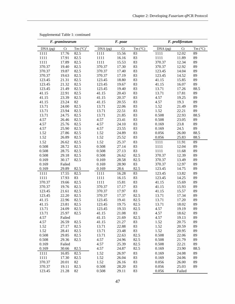
Chapter 2: Developing Fusarium qPCR Protocol
47
Supplemental Table 1: continued
F. graminearum
F. poae
F. proliferatum
DNA (pg) Ct Tm (°C)1
DNA (pg) Ct Tm (°C)
DNA (pg) Ct Tm (°C)
1111 17.76 82.5
1111 15.56 83
1111 12.02 89
1111 17.91 82.5
1111 16.16 83
1111 11.89 89
1111 17.89 82.5
1111 15.53 83
370.37 12.34 89
370.37 19.40 82.5
370.37 17.30 83
370.37 12.92 89
370.37 19.87 82.5
370.37 17.40 83
123.45 14.04 89
370.37 19.63 82.5
370.37 17.19 83
123.45 14.52 89
123.45 21.31 82.5
123.45 18.80 83
41.15 15.85 89
123.45 21.32 82.5
123.45 19.67 83
41.15 16.07 89
123.45 21.49 82.5
123.45 19.40 83
13.71 17.26 88.5
41.15 22.91 82.5
41.15 20.43 83
13.71 17.81 89
41.15 23.39 82.5
41.15 20.37 83
4.57 19.25 89
41.15 23.24 82
41.15 20.55 83
4.57 19.3 89
13.71 24.00 82.5
13.71 22.06 83
1.52 21.49 89
13.71 23.94 82.5
13.71 22.51 83
1.52 22.21 89
13.71 24.75 82.5
13.71 21.85 83
0.508 22.93 88.5
4.57 26.46 82.5
4.57 23.41 83
0.508 23.05 89
4.57 25.76 82.5
4.57 24.10 83
0.169 23.8 89
4.57 25.90 82.5
4.57 23.55 83
0.169 24.5 89
1.52 27.86 82.5
1.52 24.89 83
0.056 26.00 88.5
1.52 26.89 82.5
1.52 25.52 83
0.056 25.83 88.5
1.52 26.62 82.5
1.52 25.37 83
1111 11.91 89
0.508 28.72 82.5
0.508 27.14 83
1111 12.04 89
0.508 28.75 82.5
0.508 27.13 83
1111 11.68 89
0.508 29.18 82.5
0.508 26.62 82.5
370.37 12.39 89
0.169 30.17 82.5
0.169 28.58 82.5
370.37 13.49 89
0.169 Failed
0.169 28.90 83
370.37 12.97 89
0.169 29.89 82.5
0.169 28.6 82.5
123.45 14.75 89
1111 17.55 82.5
1111 16.28 83
123.45 13.82 89
1111 17.93 83
1111 16.15 83
123.45 14.25 89
370.37 19.66 82.5
1111 15.81 83
41.15 15.69 89
370.37 19.76 82.5
370.37 17.17 83
41.15 15.93 89
123.45 21.61 82.5
370.37 17.97 83
41.15 15.57 89
123.45 22.20 82.5
370.37 17.37 82.5
13.71 17.34 89
41.15 22.96 82.5
123.45 19.41 82.5
13.71 17.20 89
41.15 23.81 82.5
123.45 19.75 82.5
13.71 18.02 89
13.71 24.09 82.5
123.45 19.33 82.5
4.57 19.19 89
13.71 25.97 82.5
41.15 21.08 83
4.57 18.62 89
4.57 Failed
41.15 21.69 82.5
4.57 19.13 89
4.57 26.59 82.5
41.15 21.27 83
1.52 20.75 89
1.52 27.17 82.5
13.71 22.88 83
1.52 20.59 89
1.52 28.41 82.5
13.71 23.48 83
1.52 20.95 89
0.508 29.85 82.5
13.71 22.63 82.5
0.508 22.06 89
0.508 29.36 82.5
4.57 24.96 82.5
0.508 21.79 89
0.169 Failed
4.57 25.39 82.5
0.508 22.21 89
0.169 30.66 82.5
4.57 24.87 82.5
0.169 23.90 88.5
1111 16.85 82.5
1.52 26.97 83
0.169 24.08 89
1111 17.30 82.5
1.52 26.04 83
0.169 24.06 89
370.37 20.01 82
1.52 26.16 83
0.056 26.00 89
370.37 19.11 82.5
0.508 28.20 83
0.056 25.03 89
123.45 21.28 82
0.508 29.11 83
0.056 Failed
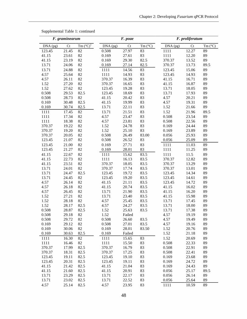
Chapter 2: Developing Fusarium qPCR Protocol
48
Supplemental Table 1: continued
F. graminearum
F. poae
F. proliferatum
DNA (pg) Ct Tm (°C)1
DNA (pg) Ct Tm (°C)
DNA (pg) Ct Tm (°C)
123.45 21.45 82
0.508 27.97 83
1111 12.27 89
41.15 23.61 82
0.169 27.61 83
1111 12.20 89
41.15 23.19 82
0.169 29.30 82.5
370.37 13.52 89
13.71 24.06 82
0.169 27.14 82.5
370.37 13.73 89.5
13.71 24.88 82
1111 14.56 83
123.45 15.06 89
4.57 25.64 82
1111 14.93 83
123.45 14.93 89
4.57 26.11 82
370.37 16.39 83
41.15 16.71 89
1.52 27.20 82
370.37 16.65 83
41.15 16.87 89
1.52 27.62 82
123.45 19.28 83
13.71 18.05 89
0.508 29.53 82.5
123.45 18.69 83
13.71 17.93 89
0.508 28.73 82
41.15 20.42 83
4.57 20.21 89
0.169 30.48 82.5
41.15 19.99 83
4.57 19.31 89
0.169 30.74 82.5
13.71 22.11 83
1.52 21.66 89
1111 17.45 82
13.71 21.51 83
1.52 21.96 89.5
1111 17.34 82
4.57 23.47 83
0.508 23.54 89
1111 18.38 82
4.57 23.81 83
0.508 22.56 89
370.37 19.22 82
1.52 24.78 83
0.169 24.44 89
370.37 19.20 82
1.52 25.10 83
0.169 23.89 89
370.37 20.05 82
0.508 26.49 83.00
0.056 25.93 89
123.45 21.07 82
0.508 26.52 83
0.056 25.09 89
123.45 21.00 82
0.169 27.71 83
1111 11.03 89
123.45 21.27 82
0.169 28.01 83
1111 11.25 89
41.15 22.67 82
1111 15.62 83.5
1111 11.5 89
41.15 22.73 82
1111 16.13 83.5
370.37 12.82 89
41.15 23.51 82
370.37 18.05 83.5
370.37 13.29 89
13.71 24.01 82
370.37 17.74 83.5
370.37 13.01 89
13.71 24.47 82.5
123.45 19.72 83.5
123.45 14.34 89
13.71 24.45 82
123.45 19.20 83.5
123.45 14.61 89
4.57 26.14 82
41.15 21.11 83.5
123.45 14.72 89
4.57 26.18 82
41.15 20.74 83.5
41.15 16.02 89
4.57 26.45 82
13.71 21.90 83.5
41.15 16.20 89
1.52 27.21 82
13.71 23.40 83.5
41.15 15.98 89
1.52 28.18 82
4.57 25.45 83.5
13.71 17.45 89
1.52 28.17 82.5
4.57 24.27 83.5
13.71 18.00 89
0.508 28.87 82.5
1.52 25.63 83.5
13.71 17.38 89
0.508 29.18 82
1.52 Failed
4.57 19.19 89
0.508 29.72 82
0.508 26.60 83.5
4.57 19.49 89
0.169 29.12 82
0.508 27.01 83.5
4.57 19.16 89
0.169 30.06 82
0.169 28.01 83.50
1.52 20.76 89
0.169 30.63 82.5
0.169 Failed
1.52 21.18 89
1111 16.30 82
1111 15.65 83
1.52 20.69 89
1111 16.46 82
1111 15.50 83
0.508 22.33 89
370.37 17.99 82.5
370.37 16.79 83
0.508 22.91 89
370.37 18.31 82.5
370.37 17.25 83
0.508 22.41 89
123.45 19.11 82.5
123.45 19.10 83
0.169 23.68 89
123.45 20.31 82.5
123.45 19.11 83
0.169 24.72 89
41.15 21.42 82.5
41.15 21.04 83
0.169 24.43 89
41.15 21.60 82.5
41.15 20.91 83
0.056 25.17 89.5
13.71 23.29 82.5
13.71 22.17 83
0.056 26.14 89
13.71 23.02 82.5
13.71 22.52 83
0.056 25.64 89
4.57 25.14 82.5
4.57 23.95 83
1111 10.59 89
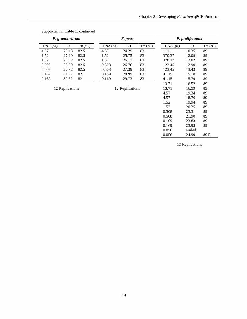
Chapter 2: Developing Fusarium qPCR Protocol
49
Supplemental Table 1: continued
F. graminearum
F. poae
F. proliferatum
DNA (pg) Ct Tm (°C)1
DNA (pg) Ct Tm (°C)
DNA (pg) Ct Tm (°C)
4.57 25.13 82.5
4.57 24.29 83
1111 10.35 89
1.52 27.10 82.5
1.52 25.75 83
370.37 12.09 89
1.52 26.72 82.5
1.52 26.17 83
370.37 12.02 89
0.508 28.99 82.5
0.508 26.76 83
123.45 12.90 89
0.508 27.92 82.5
0.508 27.39 83
123.45 13.43 89
0.169 31.27 82
0.169 28.99 83
41.15 15.10 89
0.169 30.52 82
0.169 29.73 83
41.15 15.79 89
13.71 16.52 89
12 Replications
12 Replications
13.71 16.59 89
4.57 19.34 89
4.57 18.76 89
1.52 19.94 89
1.52 20.25 89
0.508 23.31 89
0.508 21.90 89
0.169 23.83 89
0.169 23.95 89
0.056 Failed
0.056 24.99 89.5
12 Replications
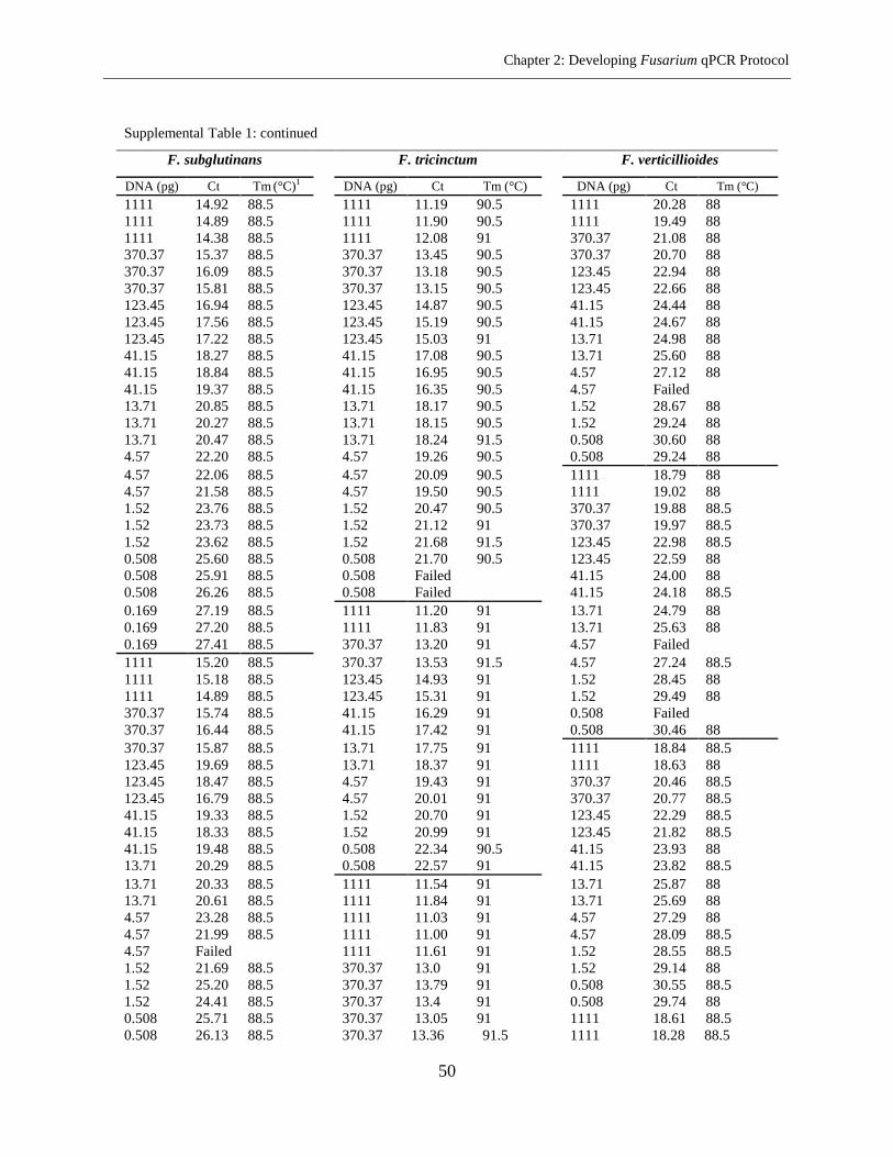
Chapter 2: Developing Fusarium qPCR Protocol
50
Supplemental Table 1: continued
F. subglutinans
F. tricinctum
F. verticillioides
DNA (pg) Ct Tm (°C)1
DNA (pg) Ct Tm (°C)
DNA (pg) Ct Tm (°C)
1111 14.92 88.5
1111 11.19 90.5
1111 20.28 88
1111 14.89 88.5
1111 11.90 90.5
1111 19.49 88
1111 14.38 88.5
1111 12.08 91
370.37 21.08 88
370.37 15.37 88.5
370.37 13.45 90.5
370.37 20.70 88
370.37 16.09 88.5
370.37 13.18 90.5
123.45 22.94 88
370.37 15.81 88.5
370.37 13.15 90.5
123.45 22.66 88
123.45 16.94 88.5
123.45 14.87 90.5
41.15 24.44 88
123.45 17.56 88.5
123.45 15.19 90.5
41.15 24.67 88
123.45 17.22 88.5
123.45 15.03 91
13.71 24.98 88
41.15 18.27 88.5
41.15 17.08 90.5
13.71 25.60 88
41.15 18.84 88.5
41.15 16.95 90.5
4.57 27.12 88
41.15 19.37 88.5
41.15 16.35 90.5
4.57 Failed
13.71 20.85 88.5
13.71 18.17 90.5
1.52 28.67 88
13.71 20.27 88.5
13.71 18.15 90.5
1.52 29.24 88
13.71 20.47 88.5
13.71 18.24 91.5
0.508 30.60 88
4.57 22.20 88.5
4.57 19.26 90.5
0.508 29.24 88
4.57 22.06 88.5
4.57 20.09 90.5
1111 18.79 88
4.57 21.58 88.5
4.57 19.50 90.5
1111 19.02 88
1.52 23.76 88.5
1.52 20.47 90.5
370.37 19.88 88.5
1.52 23.73 88.5
1.52 21.12 91
370.37 19.97 88.5
1.52 23.62 88.5
1.52 21.68 91.5
123.45 22.98 88.5
0.508 25.60 88.5
0.508 21.70 90.5
123.45 22.59 88
0.508 25.91 88.5
0.508 Failed
41.15 24.00 88
0.508 26.26 88.5
0.508 Failed
41.15 24.18 88.5
0.169 27.19 88.5
1111 11.20 91
13.71 24.79 88
0.169 27.20 88.5
1111 11.83 91
13.71 25.63 88
0.169 27.41 88.5
370.37 13.20 91
4.57 Failed
1111 15.20 88.5
370.37 13.53 91.5
4.57 27.24 88.5
1111 15.18 88.5
123.45 14.93 91
1.52 28.45 88
1111 14.89 88.5
123.45 15.31 91
1.52 29.49 88
370.37 15.74 88.5
41.15 16.29 91
0.508 Failed
370.37 16.44 88.5
41.15 17.42 91
0.508 30.46 88
370.37 15.87 88.5
13.71 17.75 91
1111 18.84 88.5
123.45 19.69 88.5
13.71 18.37 91
1111 18.63 88
123.45 18.47 88.5
4.57 19.43 91
370.37 20.46 88.5
123.45 16.79 88.5
4.57 20.01 91
370.37 20.77 88.5
41.15 19.33 88.5
1.52 20.70 91
123.45 22.29 88.5
41.15 18.33 88.5
1.52 20.99 91
123.45 21.82 88.5
41.15 19.48 88.5
0.508 22.34 90.5
41.15 23.93 88
13.71 20.29 88.5
0.508 22.57 91
41.15 23.82 88.5
13.71 20.33 88.5
1111 11.54 91
13.71 25.87 88
13.71 20.61 88.5
1111 11.84 91
13.71 25.69 88
4.57 23.28 88.5
1111 11.03 91
4.57 27.29 88
4.57 21.99 88.5
1111 11.00 91
4.57 28.09 88.5
4.57 Failed
1111 11.61 91
1.52 28.55 88.5
1.52 21.69 88.5
370.37 13.0 91
1.52 29.14 88
1.52 25.20 88.5
370.37 13.79 91
0.508 30.55 88.5
1.52 24.41 88.5
370.37 13.4 91
0.508 29.74 88
0.508 25.71 88.5
370.37 13.05 91
1111 18.61 88.5
0.508 26.13 88.5
370.37 13.36 91.5
1111 18.28 88.5
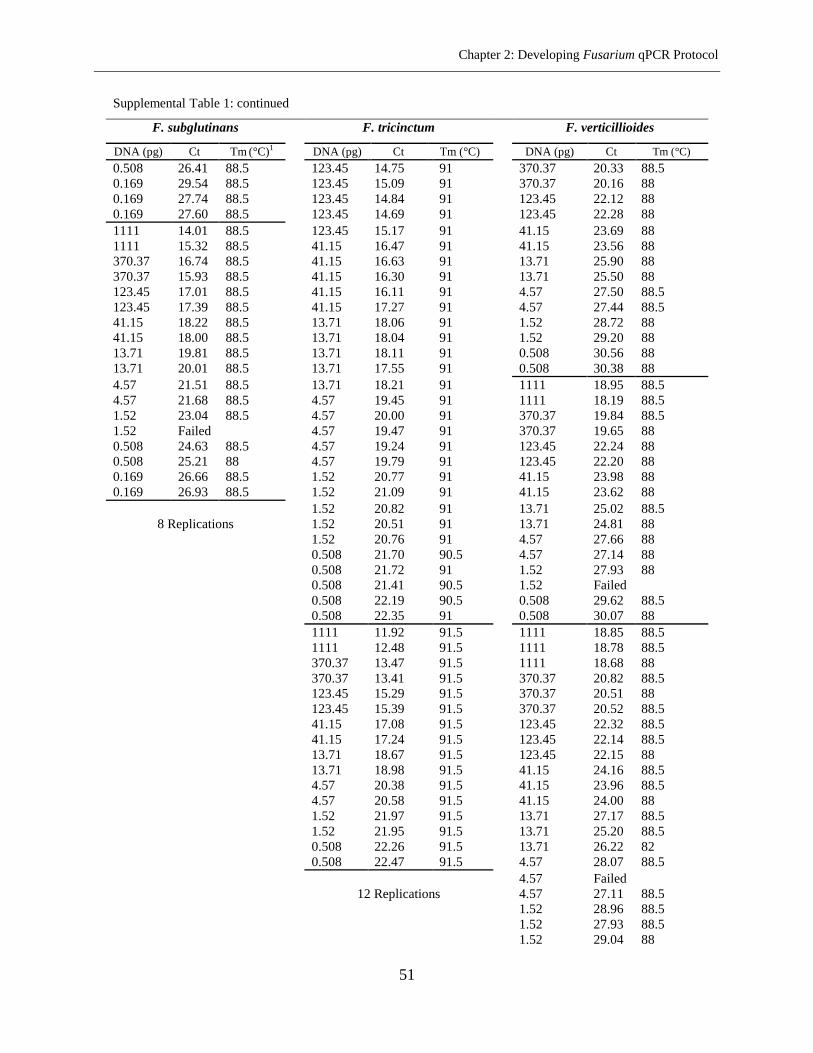
Chapter 2: Developing Fusarium qPCR Protocol
51
Supplemental Table 1: continued
F. subglutinans
F. tricinctum
F. verticillioides
DNA (pg) Ct Tm (°C)1
DNA (pg) Ct Tm (°C)
DNA (pg) Ct Tm (°C)
0.508 26.41 88.5
123.45 14.75 91
370.37 20.33 88.5
0.169 29.54 88.5
123.45 15.09 91
370.37 20.16 88
0.169 27.74 88.5
123.45 14.84 91
123.45 22.12 88
0.169 27.60 88.5
123.45 14.69 91
123.45 22.28 88
1111 14.01 88.5
123.45 15.17 91
41.15 23.69 88
1111 15.32 88.5
41.15 16.47 91
41.15 23.56 88
370.37 16.74 88.5
41.15 16.63 91
13.71 25.90 88
370.37 15.93 88.5
41.15 16.30 91
13.71 25.50 88
123.45 17.01 88.5
41.15 16.11 91
4.57 27.50 88.5
123.45 17.39 88.5
41.15 17.27 91
4.57 27.44 88.5
41.15 18.22 88.5
13.71 18.06 91
1.52 28.72 88
41.15 18.00 88.5
13.71 18.04 91
1.52 29.20 88
13.71 19.81 88.5
13.71 18.11 91
0.508 30.56 88
13.71 20.01 88.5
13.71 17.55 91
0.508 30.38 88
4.57 21.51 88.5
13.71 18.21 91
1111 18.95 88.5
4.57 21.68 88.5
4.57 19.45 91
1111 18.19 88.5
1.52 23.04 88.5
4.57 20.00 91
370.37 19.84 88.5
1.52 Failed
4.57 19.47 91
370.37 19.65 88
0.508 24.63 88.5
4.57 19.24 91
123.45 22.24 88
0.508 25.21 88
4.57 19.79 91
123.45 22.20 88
0.169 26.66 88.5
1.52 20.77 91
41.15 23.98 88
0.169 26.93 88.5
1.52 21.09 91
41.15 23.62 88
1.52 20.82 91
13.71 25.02 88.5
8 Replications
1.52 20.51 91
13.71 24.81 88
1.52 20.76 91
4.57 27.66 88
0.508 21.70 90.5
4.57 27.14 88
0.508 21.72 91
1.52 27.93 88
0.508 21.41 90.5
1.52 Failed
0.508 22.19 90.5
0.508 29.62 88.5
0.508 22.35 91
0.508 30.07 88
1111 11.92 91.5
1111 18.85 88.5
1111 12.48 91.5
1111 18.78 88.5
370.37 13.47 91.5
1111 18.68 88
370.37 13.41 91.5
370.37 20.82 88.5
123.45 15.29 91.5
370.37 20.51 88
123.45 15.39 91.5
370.37 20.52 88.5
41.15 17.08 91.5
123.45 22.32 88.5
41.15 17.24 91.5
123.45 22.14 88.5
13.71 18.67 91.5
123.45 22.15 88
13.71 18.98 91.5
41.15 24.16 88.5
4.57 20.38 91.5
41.15 23.96 88.5
4.57 20.58 91.5
41.15 24.00 88
1.52 21.97 91.5
13.71 27.17 88.5
1.52 21.95 91.5
13.71 25.20 88.5
0.508 22.26 91.5
13.71 26.22 82
0.508 22.47 91.5
4.57 28.07 88.5
4.57 Failed
12 Replications
4.57 27.11 88.5
1.52 28.96 88.5
1.52 27.93 88.5
1.52 29.04 88
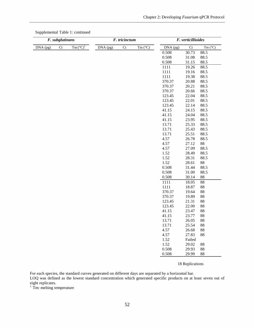
Chapter 2: Developing Fusarium qPCR Protocol
52
Supplemental Table 1: continued
F. subglutinans
F. tricinctum
F. verticillioides
DNA (pg) Ct Tm (°C)1
DNA (pg) Ct Tm (°C)
DNA (pg) Ct Tm (°C)
0.508 30.73 88.5
0.508 31.08 88.5
0.508 31.15 88.5
1111 19.26 88.5
1111 19.16 88.5
1111 19.38 88.5
370.37 20.88 88.5
370.37 20.21 88.5
370.37 20.66 88.5
123.45 22.04 88.5
123.45 22.01 88.5
123.45 22.14 88.5
41.15 24.15 88.5
41.15 24.04 88.5
41.15 23.95 88.5
13.71 25.33 88.5
13.71 25.43 88.5
13.71 25.51 88.5
4.57 26.78 88.5
4.57 27.12 88
4.57 27.09 88.5
1.52 28.49 88.5
1.52 28.31 88.5
1.52 28.61 88
0.508 31.44 88.5
0.508 31.00 88.5
0.508 30.14 88
1111 18.05 88
1111 18.87 88
370.37 19.64 88
370.37 19.89 88
123.45 21.31 88
123.45 22.00 88
41.15 23.47 88
41.15 23.77 88
13.71 26.05 88
13.71 25.54 88
4.57 26.68 88
4.57 27.83 88
1.52 Failed
1.52 29.02 88
0.508 29.93 88
0.508 29.99 88
18 Replications
For each species, the standard curves generated on different days are separated by a horizontal bar.
LOQ was defined as the lowest standard concentration which generated specific products on at least seven out of
eight replicates. 1.
Tm: melting temperature
Chapter 3: Weeds Colonization by Fusarium spp.
53
Chapter 3
Colonization of Weed Species with Fusarium spp. in Maize Fields
Raana Dastjerdi1, Katharina Döll
1, Horst-Henning Steinmann
2, Bärbel Gerowitt
3, Petr Karlovsky
1
1- Molecular Phytopathology and Mycotoxin Research, Georg-August-University Göttingen, Grisebachstr. 6, 37077
Göttingen, Germany
2- Centre of Biodiversity and Sustainable Land Use, Georg-August-University Göttingen, Grisebachstr. 6, 37077
Göttingen, Germany
3- Working Group Crop Health, Faculty of Agricultural and Environmental Sciences, University of Rostock,
Satowerstr. 48, 18051 Rostock, Germany
Corresponding author: P. Karlovsky; E-mail address: [email protected]
Abstract
The qPCR assay detected eight Fusarium species in 201 weed samples representing 36
weed species collected from maize fields. The highest frequency was observed for F. equiseti
(49%) and F. avenaceum (34.7%). It was followed by F. tricinctum, F. culmorum (each 18%),
F. proliferatum (11%) and F. graminearum (8%). The diversity of Fusarium spp. in comparison
between organic and conventional farming was similar. Isolations were made from 66 plants
belonging to 12 common weed species. Strains were identified based on morphology and
translation elongation factor 1-alpha (TEF-1α) gene sequence. The recovery rate was high for
F. equiseti (32.7%) and F. avenaceum (21%). We could not isolate F. poae and F. subglutinans
which were detected in low incidence rates (3% and 1%) in qPCR assessments. In contrast,
F. oxysporum (16%) and F. venenatum (8.5%) were obtained from 10 weed species except
Matricaria inodora and Galium aparine. None of the field samples as well as weed plants tested
in inoculation studies show obvious symptoms of Fusarium infection. Re-isolation of the strains
confirmed endophytic infection of weeds by Fusarium spp. The present study identified six new
alternative hosts for Fusarium species in maize fields. High incidence rates of beauvericin and
enniatins contamination were obtained in weed samples while trichothecenes, fumonisins and
zearalenone were not detected in any of the weeds studied.
Additional keywords: weed hosts, Fusarium spp., mycotoxins, maize
Chapter 3: Weeds Colonization by Fusarium spp.
54
Introduction
Genus Fusarium comprises phytopathogenic species causing important diseases of
cereals wherever corn (Zea mays L.), wheat (Triticum aestivum L.), barley (Hordeum vulgare L.)
or other small-grain cereals are grown. The most commonly reported Fusarium spp. associated
with maize plants include Fusarium avenaceum (Fr.) Sacc., F. culmorum (W.G. Smith) Sacc.,
F. graminearum Schwabe, F. proliferatum (Matsushima) Nirenberg, F. subglutinans
(Wollenweber & Reinking) P. E. Nelson, Toussoun & Marasas and F. verticillioides (Sacc.)
Nirenberg (Dornet al., 2009; Logrieco et al., 2002). Other Fusarium species also encounter
maize tissues and have a distinct effect on the level of infection (Munkvold, 2003). Since
Fusarium spp. have the potential to produce different mycotoxins, they had always been a
serious concern for human and animal health. The quality and quantity of crops are affected
when the Fusarium species colonize the ears and the kernels of wheat or corn produced for food
or feed. Main Fusarium mycotoxins usually reported from infected maize tissues include
trichothecenes, fumonisins, zearalenone, enniatins and moniliformin (Glenn, 2007; Placinta
et al., 1999).
Although several sources of inoculum are considered for the survival of Fusarium
pathogens, the primary source of inoculum in most fields is a remnant of plant residues. All
cereal-related Fusarium spp. can survive saprophytically on the crop residues that remain in the
field after harvesting (Parry et al., 1995). In maize fields, Fusarium species survive as mycelium
or other survival structures on maize crop debris (Sutton, 1982; Cotten & Munkvold, 1998; Naef
& De´fago, 2006). This crop debris is then the major reservoir for the infection of kernels of the
subsequent maize crop (reviewed in Munkvold, 2003). More recent studies in Europe and North
America have approved that in comparison with wheat residue, the maize residue has much more
effect as a source of inoculum for the infection of wheat to fusarium head blight (Dill-Macky &
Jones, 2000; Schaafsma et al., 2001). In Uruguayan cultivation systems, a lower contribution of
F. graminearum inoculum is allocated to corn (Pereyra & Dill-Macky, 2008).
In spite of many research on biology, pathology and toxicology of Fusarium species,
there is still need for improvement of the effective management of Fusarium diseases occurred in
maize crop. Current management protocols for maize Fusarium diseases mostly involve the
Chapter 3: Weeds Colonization by Fusarium spp.
55
avoidance of crop residues (Maiorano et al., 2008; Naef & De´fago, 2006) to reduce fungal
inoculum; but rarely consider weed hosts as a source for crop infection. Weed plants are mostly
considered as organisms which can provide favorable conditions such as humidity for developing
of Fusarium maize diseases. However, the role of weeds could be likely more. Weed and wild
plants can be hosts which provide an important source of inoculum (Parry et al., 1995). Although
the importance of weeds as a reservoir of fungal pathogens in fields was identified by Garrett in
1960, but still little literary work has been carried out on weeds as alternative hosts for fungal
pathogens and mostly has been concentrated on F. oxysporum. There are contradictory reports
for the interaction between weed plants density and fusarium head blight severity in different
experimental investigations (Teich & Nelson, 1984). It has been demonstrated that Fusarium
populations in the soil can colonize senescent tissues of weed plants as well as maize tissues and
produce different types of infectious propagules such as perithecia and ascospores or asexual
spores of microconidia and macroconidia (Parry et al., 1995; Munkvold, 2003). Recovery of
Fusarium spp. from 19 species of cereals and grasses and 24 species of common weed plants is
one of the first reports that introduce weeds as a harbor for Fusarium species (Gordon, 1959).
The reports from sugar beet, tomato, soybean and eggplant fields also show that F. oxysporum
pathogenic on these crops has survival capability on several common weed plants (McDonald &
Leach, 1976; Haware & Nene, 1982; Helbig & Carroll, 1984; Altinok, 2013).
Some Fusarium strains are weed pathogens and, therefore, have been used as biocontrol
agents. They are also used in the development of mycoherbicides and fungal phytotoxins for
control of weeds (Boyette & Walker, 1985; Abbas et al., 1991; Abbas & Boyette, 1992; Roy
et al., 1994). On the other hand, there are Fusarium strains that are not pathogenic to weeds. One
reason for the absence of pathogenicity would be the ectosymbiotic bacteria complexes that are
able to modulate the expression of pathogenicity genes in interaction with Fusarium strains
(Minerdi et al., 2008). The pathogenic Fusarium strains show protective behavior against other
antagonists while they harbor on non-host plants such as weeds. Subsequently, weed plants
occupied by such endophytic Fusarium strains act as symptomless hosts to increase inoculum
potential (Altinok, 2013). There is growing evidence that reveal weed plants are colonized by
pathogenic fungi as symptomless hosts (Roy, 1982; Cerkauskas et al., 1983; Helbig & Carrol,
1984; Roy et al., 1994; Jenkinson & Parry, 1994; Postic et al., 2012; Altinok, 2013). Exhibition
Chapter 3: Weeds Colonization by Fusarium spp.
56
no symptoms on weed plants by Fusarium species has always been a question. Some reports
assigned that only less aggressive strains can invade weeds and produce infection without
symptoms (Helbig & Carroll, 1984). Furthermore, lack of strong adaption performance of
Fusarium on alternative hosts could also explain the symptomless infection of weeds. The
reports show passage through alternative host causes reduction of the pathogenic fitness
(aggressiveness) but increment of the overall fungal reproductivity (saprophytic behavior). As a
result of transition through an alternative host, a conversion of pathogen behavior may be
occurred and colonization of primary host would be improved (Akinsanmi et al., 2007). All these
studies conclude that effective weed management can be a useful approach for reduction of
Fusarium diseases inoculum in maize fields (reviewed in Fandohan et al., 2003).
On the average, organically grown maize is to the same extent or even less contaminated
with mycotoxins than conventionally grown maize; but extremely high mycotoxin levels have
been occasionally reported for organic maize products over the years. Low frequency but regular
occurrence of unusually high mycotoxin values and fungal colonization has been observed in
well-controlled field trials too which is difficult to explain (Nutz & Karlovsky, unpublished).
There is a hypothesis that particular species of weed plants that host Fusarium spp. maybe
increase the infection pressure locally and account for high levels of Fusarium mycotoxins in
organic maize fields. International developments in mycotoxin regulation make high pressure for
finding novel strategies to manage the contamination of maize products by Fusarium
mycotoxins. Therefore, understanding of Fusarium spp. survival and the sources of Fusarium
inoculum potential as well as the factors influencing this potential is a prerequisite for plant
pathologists to prevent the build-up of inoculum sources and, subsequently, management of the
Fusarium diseases in maize fields.
This study was carried out to investigate the role of weed plants in the survival and
inoculum production of maize Fusarium pathogens and to assess their ability for producing of
the main mycotoxins. The question is if weed plants play a role as alternative hosts for Fusarium
species pathogenic to maize and if they can provide a significant source of inoculum that help to
increase maize mycotoxin contamination?
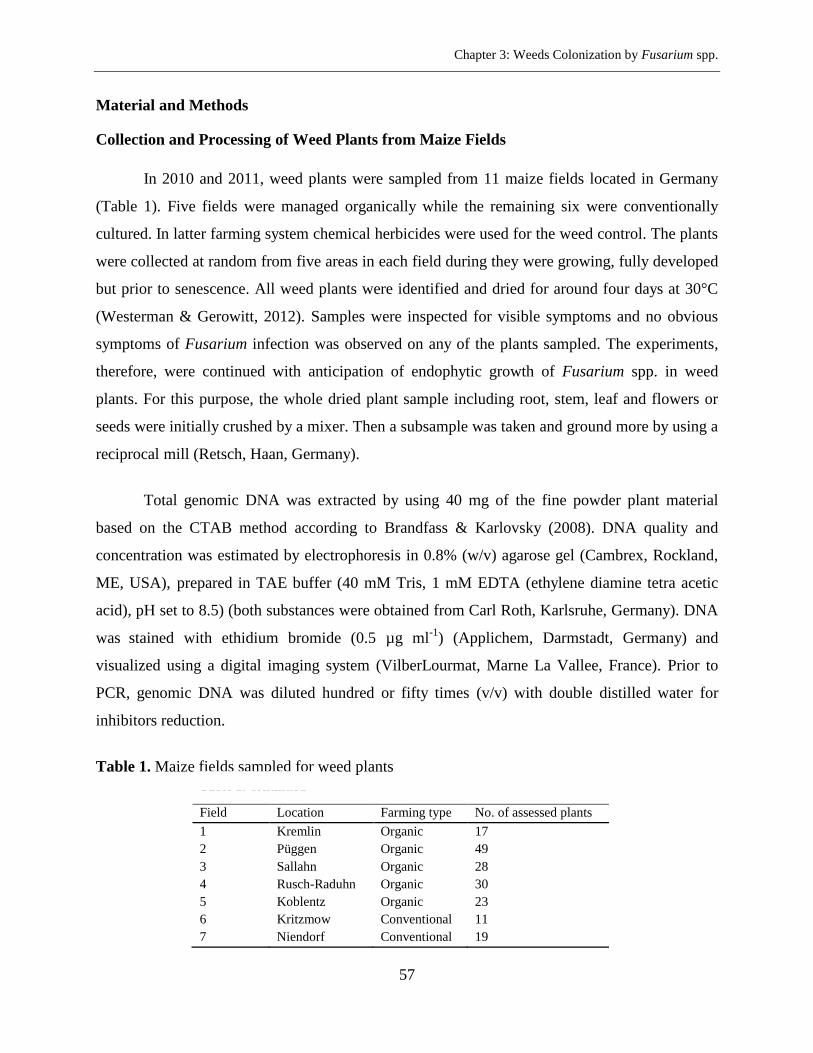
Chapter 3: Weeds Colonization by Fusarium spp.
57
Material and Methods
Collection and Processing of Weed Plants from Maize Fields
In 2010 and 2011, weed plants were sampled from 11 maize fields located in Germany
(Table 1). Five fields were managed organically while the remaining six were conventionally
cultured. In latter farming system chemical herbicides were used for the weed control. The plants
were collected at random from five areas in each field during they were growing, fully developed
but prior to senescence. All weed plants were identified and dried for around four days at 30°C
(Westerman & Gerowitt, 2012). Samples were inspected for visible symptoms and no obvious
symptoms of Fusarium infection was observed on any of the plants sampled. The experiments,
therefore, were continued with anticipation of endophytic growth of Fusarium spp. in weed
plants. For this purpose, the whole dried plant sample including root, stem, leaf and flowers or
seeds were initially crushed by a mixer. Then a subsample was taken and ground more by using a
reciprocal mill (Retsch, Haan, Germany).
Total genomic DNA was extracted by using 40 mg of the fine powder plant material
based on the CTAB method according to Brandfass & Karlovsky (2008). DNA quality and
concentration was estimated by electrophoresis in 0.8% (w/v) agarose gel (Cambrex, Rockland,
ME, USA), prepared in TAE buffer (40 mM Tris, 1 mM EDTA (ethylene diamine tetra acetic
acid), pH set to 8.5) (both substances were obtained from Carl Roth, Karlsruhe, Germany). DNA
was stained with ethidium bromide (0.5 µg ml-1
) (Applichem, Darmstadt, Germany) and
visualized using a digital imaging system (VilberLourmat, Marne La Vallee, France). Prior to
PCR, genomic DNA was diluted hundred or fifty times (v/v) with double distilled water for
inhibitors reduction.
Table 1. Maize fields sampled for weed plants
Table 1: continued
Field Location Farming type No. of assessed plants
1 Kremlin Organic 17
2 Püggen Organic 49
3 Sallahn Organic 28
4 Rusch-Raduhn Organic 30
5 Koblentz Organic 23
6 Kritzmow Conventional 11
7 Niendorf Conventional 19
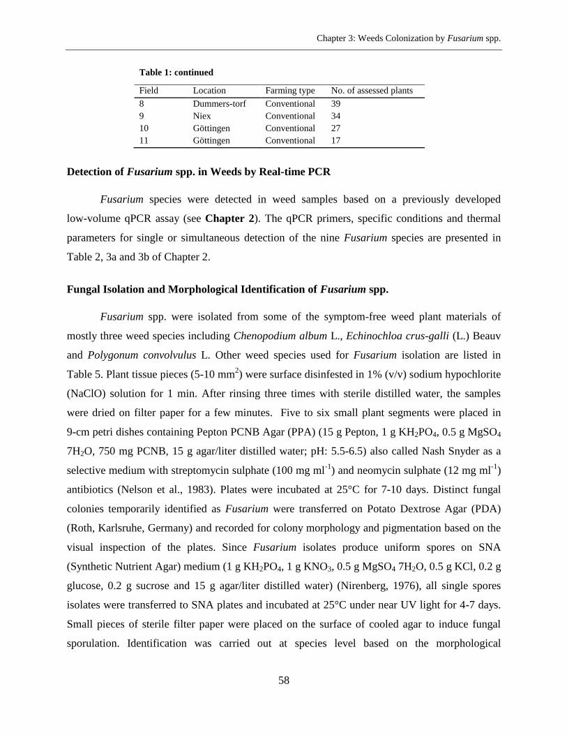
Chapter 3: Weeds Colonization by Fusarium spp.
58
Table 1: continued
Field Location Farming type No. of assessed plants
8 Dummers-torf Conventional 39
9 Niex Conventional 34
10 Göttingen Conventional 27
11 Göttingen Conventional 17
Detection of Fusarium spp. in Weeds by Real-time PCR
Fusarium species were detected in weed samples based on a previously developed
low-volume qPCR assay (see Chapter 2). The qPCR primers, specific conditions and thermal
parameters for single or simultaneous detection of the nine Fusarium species are presented in
Table 2, 3a and 3b of Chapter 2.
Fungal Isolation and Morphological Identification of Fusarium spp.
Fusarium spp. were isolated from some of the symptom-free weed plant materials of
mostly three weed species including Chenopodium album L., Echinochloa crus-galli (L.) Beauv
and Polygonum convolvulus L. Other weed species used for Fusarium isolation are listed in
Table 5. Plant tissue pieces (5-10 mm2) were surface disinfested in 1% (v/v) sodium hypochlorite
(NaClO) solution for 1 min. After rinsing three times with sterile distilled water, the samples
were dried on filter paper for a few minutes. Five to six small plant segments were placed in
9-cm petri dishes containing Pepton PCNB Agar (PPA) (15 g Pepton, 1 g KH2PO4, 0.5 g MgSO4
7H2O, 750 mg PCNB, 15 g agar/liter distilled water; pH: 5.5-6.5) also called Nash Snyder as a
selective medium with streptomycin sulphate (100 mg ml-1
) and neomycin sulphate (12 mg ml-1
)
antibiotics (Nelson et al., 1983). Plates were incubated at 25°C for 7-10 days. Distinct fungal
colonies temporarily identified as Fusarium were transferred on Potato Dextrose Agar (PDA)
(Roth, Karlsruhe, Germany) and recorded for colony morphology and pigmentation based on the
visual inspection of the plates. Since Fusarium isolates produce uniform spores on SNA
(Synthetic Nutrient Agar) medium (1 g KH2PO4, 1 g KNO3, 0.5 g MgSO4 7H2O, 0.5 g KCl, 0.2 g
glucose, 0.2 g sucrose and 15 g agar/liter distilled water) (Nirenberg, 1976), all single spores
isolates were transferred to SNA plates and incubated at 25°C under near UV light for 4-7 days.
Small pieces of sterile filter paper were placed on the surface of cooled agar to induce fungal
sporulation. Identification was carried out at species level based on the morphological
Chapter 3: Weeds Colonization by Fusarium spp.
59
descriptions of spores and conidiogenous cells according to Leslie & Summerell (2006).
Fusarium strains were stored as spore suspension in 15% (v/v) glycerol (Carl Roth, Karlsruhe,
Germany) at -70°C.
Fusarium DNA Sequencing and Taxonomic Analysis
Identification of recovered Fusarium isolates was performed according to the
combination of morphological and molecular characters. Typical Fusarium structures were
employed for morphological identification. Afterward, identification at species level was
confirmed by the results of DNA sequence of the “Translaton Elongation Factor 1-alpha”
(TEF-1α) gene, which is taxonomically most informative part in this fungal genus. For this
purpose, representatives of each Fusarium group (totally 61 isolates) have been employed.
Single spore cultures were cultivated in Potato Dextrose Broth (PDB) (Carl Roth, Karlsruhe,
Germany) for 5 to 7 days at 25°C. The mycelia were collected by filtration on sterile filter paper
and freeze-dried. Lyophilized mycelium was ground to a fine powder by using a reciprocal mill
in 2 ml Eppendorf tubes with 4-5 wolfram carbide spheres (diameter 3 mm, Retsch, Haan,
Germany). DNA was extracted as described by Brandfass & Karlovsky (2008).
Hot start PCR protocol was used to amplify the TEF-1α gene region using the fungal
specific primer set: ef1 (5'-ATGGGTAAGGA(A/G)GACAAGAC-3') and ef2
(5'-GA(G/A)GTACCAGT(G/C)ATCATGTT-3') described by O'Donnell et al. (1998). The
reaction mixture consisted of reaction buffer (16 mM (NH4)2SO4; 67 mM Tris-HCl; 0.01% (v/v)
Tween-20, pH: 8.8 at 25°C), 0.1 mM concentration of each of the four deoxynucleoside
triphosphates (Bioline, Lükenwalde, Germany), 2 mM of MgCl2, 1.75 U of hot-start DNA
polymerase (Immolase DNA Pol, Lükenwalde, Germany), 0.3 µM concentration of each primer.
Every reaction contained 1 µl template DNA or 1 µl sterile water as negative control. The PCR
reactions were performed in a total volume of 25 µl reactions with a peQ STAR Thermocycler
(96 Universal Gradient). The cycling conditions were as follows: 1 cycle of 10 min at 95°C, 30
cycles of 60 s at 94°C (denaturalization), 45 s at 58.5°C (annealing), 60 s at 72°C (extension) and
followed by a final extension cycle at 72°C for 5 min. Fungal amplified DNA fragments were
sequenced (LGC Genomics, Berlin, Germany) by Sanger method in one direction using ef1
primer. The chromatogram of TEF-1α sequence for each Fusarium species has been inspected
Chapter 3: Weeds Colonization by Fusarium spp.
60
visually and sequence reads edited when necessary. The sequences were, then, used as queries
for Fusarium-ID v. 1.0 database (Geiser et al., 2004). In order to construct a phylogenetic tree
sequences with the highest similarity to the query, together with some sequences retrieved from
the GenBank database (Supplemental Table 1) were aligned. The distance matrix was
constructed from pairwise similarities expressed as fractions of identical nucleotides with
Jukes-Cantor nucleotide distance measure model for multiple substitutions, and an UPGMA
(Un-weighted Pair Group Method with Averages) dendrogram was generated by using the
program CLC Main Workbench 6.9. The reliability of analysis was estimated by bootstrap
method with 1000 replications.
Inoculation Studies
Inoculation tests were conducted with six arbitrary selected Fusarium isolates belonging
to four species (RD22, RD98 for F. avenaceum; RD100, RD102 for F. culmorum; RD92 for
F. graminearum, and RD94 for F. proliferatum). The Fusarium strains had previously been
recovered from weed plants. The isolates were tested to young seedlings of 11 weed species
(Table 4) and also maize plants. The seeds of selected weed species were prepared from the
Herbiseed Company at UK and also from the Botanical Garden Department, Göttingen,
Germany. Mini maize cultivar (Gaspe Flint landrace, originally collected in Quebec, Canada,
provided by Prof. Dr. J. Schirawski, Department of Molecular Biology of Plant-Microbe
Interaction, University of Göttingen, Germany) was also used in this study. The seeds were sown
in plastic multi-pots containing sterile fine sand (< 2 mm granularity) and irrigated as required.
Seven to ten seeds of each weed species were planted per cavity and thinned to five plants when
seedlings appeared. For some of the weed species including Galium aparine L., Polygonum
convolvulus L., Polygonum persicaria L. and Amaranthus retroflexus L. vernalization at 4°C
under dark conditions for 3-4 weeks was necessary.
Two or three weeks after germination, the seedlings were inoculated with conidial spore
suspension of each species of Fusarium. Conidial inoculum was produced in liquid mung bean
media (Bai & Shaner, 1996) and has been stored in 15% (v/v) glycerol at -70°C till inoculation
time. Prior to plant inoculation, spore germination test was carried out on PDA plates to assess
the viability and rate of spore germination. The concentration of each conidial suspension was,
Chapter 3: Weeds Colonization by Fusarium spp.
61
then, determined using a Thoma Chamber (0.0025 mm²) and adjusted to 1 × 10⁶
spore ml-1
of
sterile tap water. Inoculum was contained 0.1% (v/v) Tween®
20 (Applichem, Darmstadt,
Germany) as a surfactant. Spore suspensions were kept on ice during inoculation and
homogenized well before using.
Root-dip inoculation method was employed for artificial inoculation in the greenhouse by
soaking the roots of individual plant in spore suspension for 30 min. Inoculations were carried
out in early morning. After inoculation the seedlings were transferred to new pots (9 × 9 ×
9.5cm) including mixed soil consisting of commercial plant substrate (Fruhstorfer Erde, Typ
T25, HAWITA Group, Vechta, Germany) and sand (1:1). All pots were arranged according to a
completely randomized design on the greenhouse benches. The plants were maintained at
25 ± 5°C with alternating 12-h light (mercury vapour lamps, 6600 lux at ear height) and dark
periods. During the experiment, relative humidity was 34-86%. Five replicate pots were set up
for each species of Fusarium and each pot included two plants. Control treatments were
inoculated with autoclaved tap water mixed with adhesion detergent of Tween®
20. Visual
symptoms were determined on a weekly basis and six weeks after inoculation the plants were
harvested. In order to verify the presence of fungus in artificial inoculated plants, subsequent
detection of Fusarium spp. in plant tissue was performed by qPCR assay. For a few inoculated
samples, fungal re-isolation was carried out from surface sterilized stem tissues.
Mycotoxin Analysis
The field weed samples which indicated a positive signal in qPCR experiments were
analyzed for a number of Fusarium related mycotoxins including deoxynivalenol (DON),
3ADON, 15ADON, nivalenol (NIV), zearalenone (ZEN), fumonisin B1 (FB1), beauvericin (BEA),
enniatins (B, B1, A, A1) by HPLC-MS/MS. Mycotoxin extraction was performed by adding 1 ml
acetonitrile-water (84:16) to 100 mg fine powder of weed plant tissues. The extract (600 µl) was
dried in a rotational-Vacuum-Concentrator RVC 2-25 (Martin Christ Gefriertrocknungsanlagen
GmbH, Osterode am Harz, Germany) at 40°C. The raw organic extract was resuspended in
600 µl of methanol-water (85:15) and defatted with the same volume of cyclohexane. After
centrigfugation the lower phase was taken for analysis.
Chapter 3: Weeds Colonization by Fusarium spp.
62
HPLC separation was carried out on a reverse-phase Polaris C18-ether column
(100 × 2 mm, 3 µm particle size; Agilent, Darmstadt, Germany) at 40°C and trichothecenes B
and zearalenone were detected by tandem mass spectrometry using triple quadrupol 1200 L
(Varian, Darmstadt, Germany) according to Adejumo et al. (2007a) and Adejumo et al. (2007b),
but two mass transitions were used for each mycotoxin. Beauvericin, fumonisin B1 and enniatins
were separated on a reverse phase Kinetex C18 column (50.0 × 2.1 mm, 2.6 µm particle
size; Phenomenex (Aschaffenburg, Germany) and detected using ion trap 500MS (Varian,
Darmstadt, Germany) as described by Nutz et al. (2011). For these mycotoxins three mass
transitions were used. Pure analytical standards in methanol/water (1:1) were used to construct
calibration curves.
Data Processing and Statistical Analysis
SigmaPlot 12.3 Notebook was used for statistical analysis. Non-parametric tests were
performed while data distribution was not normal. The qPCR data were assessed for finding the
positive samples based on the melting temperature (Tm), starting quantity (SQ) and threshold
cycle (Ct) value. The lowest standard DNA for each Fusarium spp. employed in the qPCR
assays was considered as the limit of quantification (LOQ). According to the qPCR data, the
quantity of fungal biomass (pg mg-1
) for the positive samples was calculated. DNA quantity for
the samples containing values lower than LOQ was substituted as LOQ/2 (Hornunga & Reeda,
1990). These samples indicated the species-specific melting temperature in the PCR cycles lower
than 30.
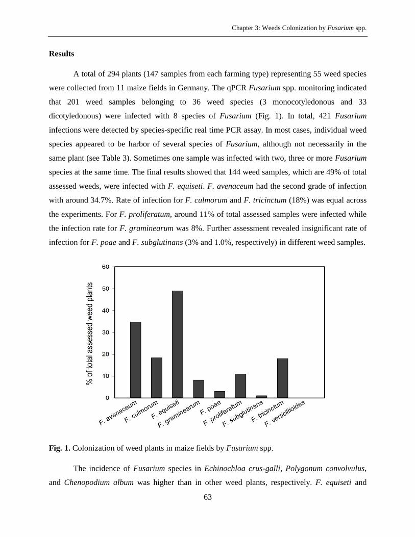
Chapter 3: Weeds Colonization by Fusarium spp.
63
Results
A total of 294 plants (147 samples from each farming type) representing 55 weed species
were collected from 11 maize fields in Germany. The qPCR Fusarium spp. monitoring indicated
that 201 weed samples belonging to 36 weed species (3 monocotyledonous and 33
dicotyledonous) were infected with 8 species of Fusarium (Fig. 1). In total, 421 Fusarium
infections were detected by species-specific real time PCR assay. In most cases, individual weed
species appeared to be harbor of several species of Fusarium, although not necessarily in the
same plant (see Table 3). Sometimes one sample was infected with two, three or more Fusarium
species at the same time. The final results showed that 144 weed samples, which are 49% of total
assessed weeds, were infected with F. equiseti. F. avenaceum had the second grade of infection
with around 34.7%. Rate of infection for F. culmorum and F. tricinctum (18%) was equal across
the experiments. For F. proliferatum, around 11% of total assessed samples were infected while
the infection rate for F. graminearum was 8%. Further assessment revealed insignificant rate of
infection for F. poae and F. subglutinans (3% and 1.0%, respectively) in different weed samples.
Fig. 1. Colonization of weed plants in maize fields by Fusarium spp.
The incidence of Fusarium species in Echinochloa crus-galli, Polygonum convolvulus,
and Chenopodium album was higher than in other weed plants, respectively. F. equiseti and
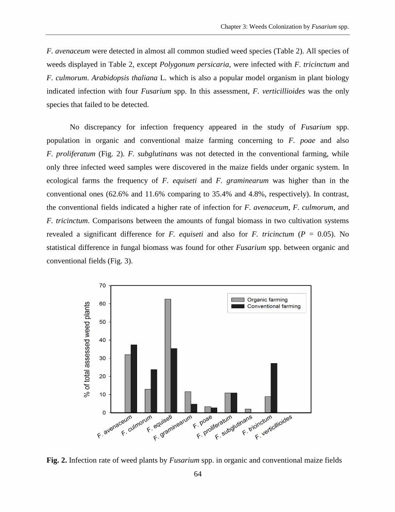
Chapter 3: Weeds Colonization by Fusarium spp.
64
F. avenaceum were detected in almost all common studied weed species (Table 2). All species of
weeds displayed in Table 2, except Polygonum persicaria, were infected with F. tricinctum and
F. culmorum. Arabidopsis thaliana L. which is also a popular model organism in plant biology
indicated infection with four Fusarium spp. In this assessment, F. verticillioides was the only
species that failed to be detected.
No discrepancy for infection frequency appeared in the study of Fusarium spp.
population in organic and conventional maize farming concerning to F. poae and also
F. proliferatum (Fig. 2). F. subglutinans was not detected in the conventional farming, while
only three infected weed samples were discovered in the maize fields under organic system. In
ecological farms the frequency of F. equiseti and F. graminearum was higher than in the
conventional ones (62.6% and 11.6% comparing to 35.4% and 4.8%, respectively). In contrast,
the conventional fields indicated a higher rate of infection for F. avenaceum, F. culmorum, and
F. tricinctum. Comparisons between the amounts of fungal biomass in two cultivation systems
revealed a significant difference for F. equiseti and also for F. tricinctum (P = 0.05). No
statistical difference in fungal biomass was found for other Fusarium spp. between organic and
conventional fields (Fig. 3).
Fig. 2. Infection rate of weed plants by Fusarium spp. in organic and conventional maize fields
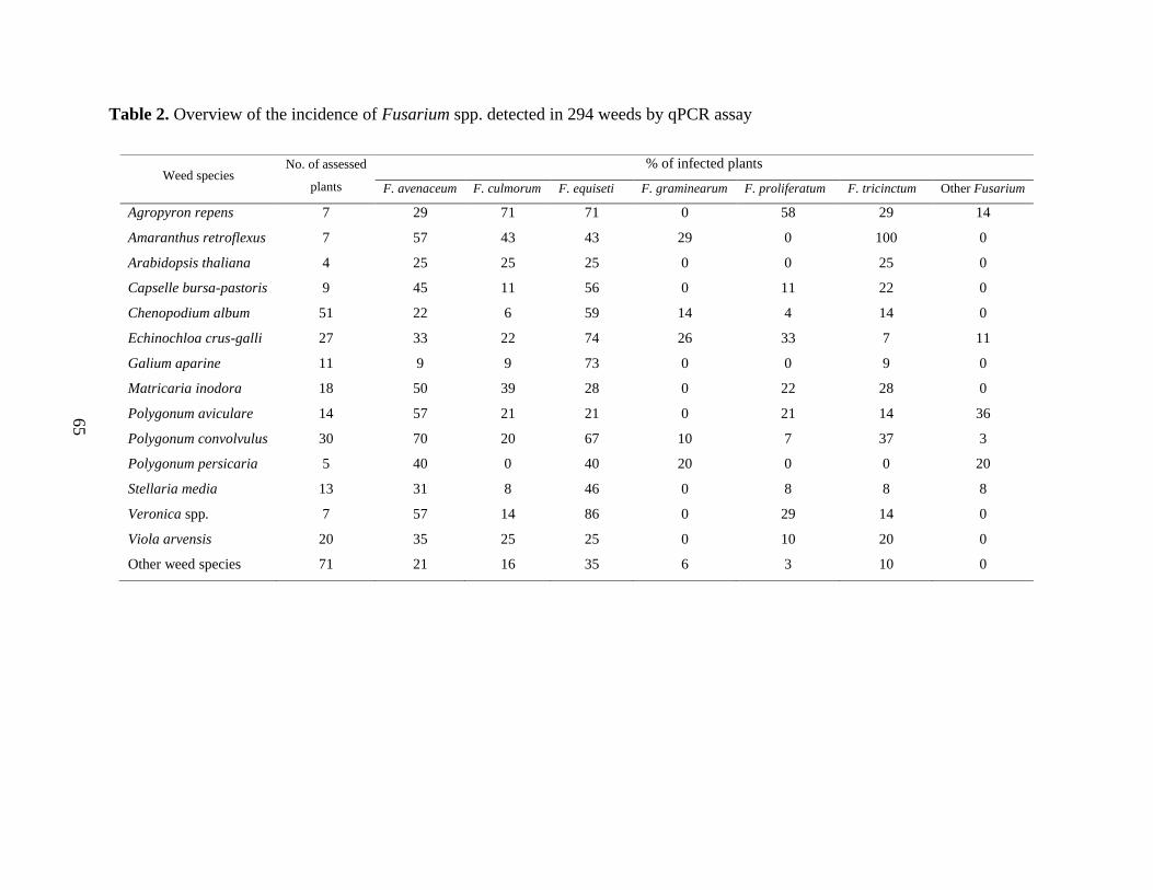
65
Table 2. Overview of the incidence of Fusarium spp. detected in 294 weeds by qPCR assay
Weed species No. of assessed
plants
% of infected plants
F. avenaceum F. culmorum F. equiseti F. graminearum F. proliferatum F. tricinctum Other Fusarium
Agropyron repens 7 29 71 71 0 58 29 14
Amaranthus retroflexus 7 57 43 43 29 0 100 0
Arabidopsis thaliana 4 25 25 25 0 0 25 0
Capselle bursa-pastoris 9 45 11 56 0 11 22 0
Chenopodium album 51 22 6 59 14 4 14 0
Echinochloa crus-galli 27 33 22 74 26 33 7 11
Galium aparine 11 9 9 73 0 0 9 0
Matricaria inodora 18 50 39 28 0 22 28 0
Polygonum aviculare 14 57 21 21 0 21 14 36
Polygonum convolvulus 30 70 20 67 10 7 37 3
Polygonum persicaria 5 40 0 40 20 0 0 20
Stellaria media 13 31 8 46 0 8 8 8
Veronica spp. 7 57 14 86 0 29 14 0
Viola arvensis 20 35 25 25 0 10 20 0
Other weed species 71 21 16 35 6 3 10 0
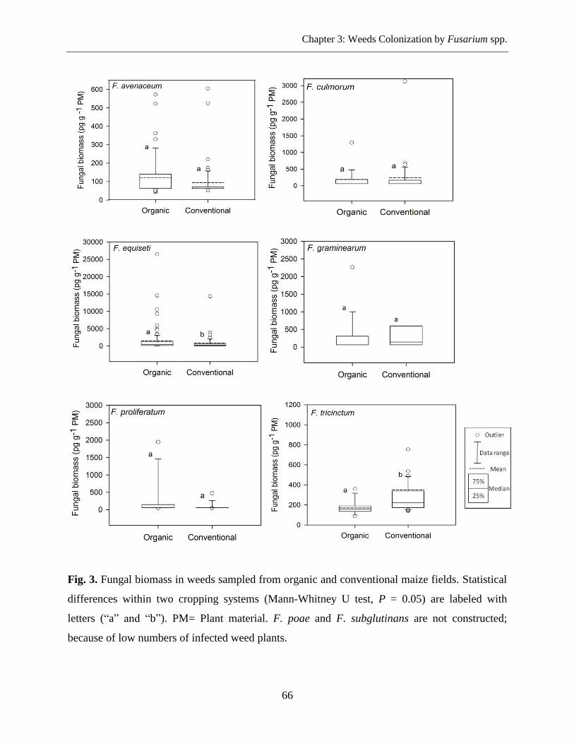
Chapter 3: Weeds Colonization by Fusarium spp.
66
Fig. 3. Fungal biomass in weeds sampled from organic and conventional maize fields. Statistical
differences within two cropping systems (Mann-Whitney U test, P = 0.05) are labeled with
letters (“a” and “b”). PM= Plant material. F. poae and F. subglutinans are not constructed;
because of low numbers of infected weed plants.
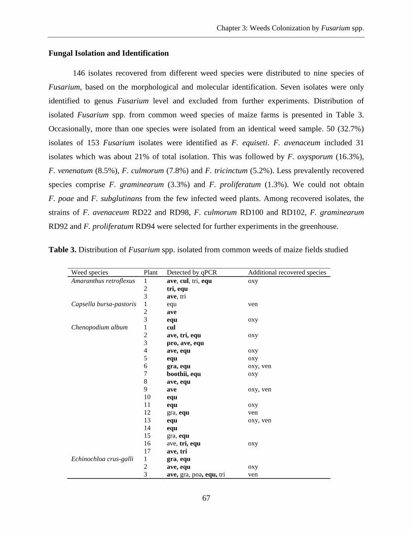
Chapter 3: Weeds Colonization by Fusarium spp.
67
Fungal Isolation and Identification
146 isolates recovered from different weed species were distributed to nine species of
Fusarium, based on the morphological and molecular identification. Seven isolates were only
identified to genus Fusarium level and excluded from further experiments. Distribution of
isolated Fusarium spp. from common weed species of maize farms is presented in Table 3.
Occasionally, more than one species were isolated from an identical weed sample. 50 (32.7%)
isolates of 153 Fusarium isolates were identified as F. equiseti. F. avenaceum included 31
isolates which was about 21% of total isolation. This was followed by F. oxysporum (16.3%),
F. venenatum (8.5%), F. culmorum (7.8%) and F. tricinctum (5.2%). Less prevalently recovered
species comprise F. graminearum (3.3%) and F. proliferatum (1.3%). We could not obtain
F. poae and F. subglutinans from the few infected weed plants. Among recovered isolates, the
strains of F. avenaceum RD22 and RD98, F. culmorum RD100 and RD102, F. graminearum
RD92 and F. proliferatum RD94 were selected for further experiments in the greenhouse.
Table 3. Distribution of Fusarium spp. isolated from common weeds of maize fields studied
Table 3: continued
Weed species Plant Detected by qPCR Additional recovered species
Amaranthus retroflexus 1 ave, cul, tri, equ oxy
2 tri, equ
3 ave, tri
Capsella bursa-pastoris 1 equ ven
2 ave
3 equ oxy
Chenopodium album 1 cul
2 ave, tri, equ oxy
3 pro, ave, equ
4 ave, equ oxy
5 equ oxy
6 gra, equ oxy, ven
7 boothii, equ oxy
8 ave, equ
9 ave oxy, ven
10 equ
11 equ oxy
12 gra, equ ven
13 equ oxy, ven
14 equ
15 gra, equ
16 ave, tri, equ oxy
17 ave, tri
Echinochloa crus-galli 1 gra, equ
2 ave, equ oxy
3 ave, gra, poa, equ, tri ven
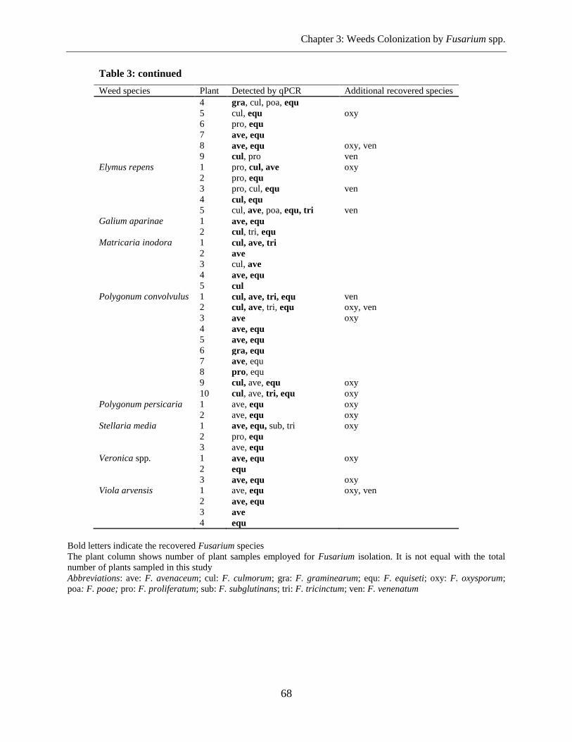
Chapter 3: Weeds Colonization by Fusarium spp.
68
Table 3: continued
Weed species Plant Detected by qPCR Additional recovered species
4 gra, cul, poa, equ
5 cul, equ oxy
6 pro, equ
7 ave, equ
8 ave, equ oxy, ven
9 cul, pro ven
Elymus repens 1 pro, cul, ave oxy
2 pro, equ
3 pro, cul, equ ven
4 cul, equ
5 cul, ave, poa, equ, tri ven
Galium aparinae 1 ave, equ
2 cul, tri, equ
Matricaria inodora 1 cul, ave, tri
2 ave
3 cul, ave
4 ave, equ
5 cul
Polygonum convolvulus 1 cul, ave, tri, equ ven
2 cul, ave, tri, equ oxy, ven
3 ave oxy
4 ave, equ
5 ave, equ
6 gra, equ
7 ave, equ
8 pro, equ
9 cul, ave, equ oxy
10 cul, ave, tri, equ oxy
Polygonum persicaria 1 ave, equ oxy
2 ave, equ oxy
Stellaria media 1 ave, equ, sub, tri oxy
2 pro, equ
3 ave, equ
Veronica spp. 1 ave, equ oxy
2 equ
3 ave, equ oxy
Viola arvensis 1 ave, equ oxy, ven
2 ave, equ
3 ave
4 equ
Bold letters indicate the recovered Fusarium species
The plant column shows number of plant samples employed for Fusarium isolation. It is not equal with the total
number of plants sampled in this study
Abbreviations: ave: F. avenaceum; cul: F. culmorum; gra: F. graminearum; equ: F. equiseti; oxy: F. oxysporum;
poa: F. poae; pro: F. proliferatum; sub: F. subglutinans; tri: F. tricinctum; ven: F. venenatum
Chapter 3: Weeds Colonization by Fusarium spp.
69
Fusarium DNA Sequencing and Phylogenetic Analysis
To confirm morphological identification, 62 representative isolates were sequenced based
on the TEF-1α gene which has been widely used for species identification. Thermal PCR
conditions suggested by Geiser et al. (2004), lead to a mixture of specific and non-specific
products. The situations were achieved by running a gradient PCR in the range of 53-63°C under
“Hot start PCR”. At the annealing temperature of 58.5°C, a single band in the size range of 610
to 653 bp was successfully amplified from all isolates. After DNA sequencing, each unknown
Fusarium strain was identified according to the deposited sequences in Fusarium-ID database
which had a high homology (≥ 99%) with them. The first three hits for the sequence of each
isolate have been assigned in Supplemental Table 2. The studied sequences were, then, aligned
with TEF-1α sequences of F. acuminatum, F. arthrosporioides, F. avenaceum, F. culmorum,
F. flocciferum, F. graminearum, F. torulosum, F. tricinctum, and F. venenatum retrieved from
sequence database (Supplemental Table 1) and exposed to phylogenetic analysis (Fig. 4, 5, 6).
The UPGMA dendrogram clearly clustered distinct clades regarding to Fusarium spp. studied.
Identification was well supported with bootstrap values. The studied strains, therefore, were
placed into groups matching with those determined by Fusarium-ID database. The strain RD99
which was identified morphologically as F. graminearum, made a distinct branch close to
F. graminearum. Fusarium-ID database identified this strain as F. boothii. Furthermore, the
isolates morphologically identified as F. equiseti were grouped in three different haplotypes of
Fusarium incarnatum-equiseti species complex (FIESC) based on the molecular identification.
Similarly, isolates identified morphologically as F. oxysporum placed in three different
sub-clades of Fusarium oxysporum species complex (FOSC).
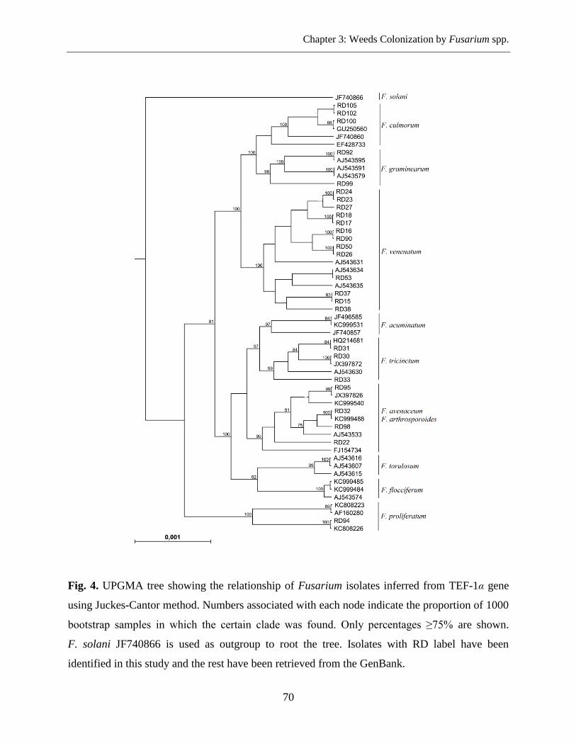
Chapter 3: Weeds Colonization by Fusarium spp.
70
Fig. 4. UPGMA tree showing the relationship of Fusarium isolates inferred from TEF-1α gene
using Juckes-Cantor method. Numbers associated with each node indicate the proportion of 1000
bootstrap samples in which the certain clade was found. Only percentages ≥75% are shown.
F. solani JF740866 is used as outgroup to root the tree. Isolates with RD label have been
identified in this study and the rest have been retrieved from the GenBank.
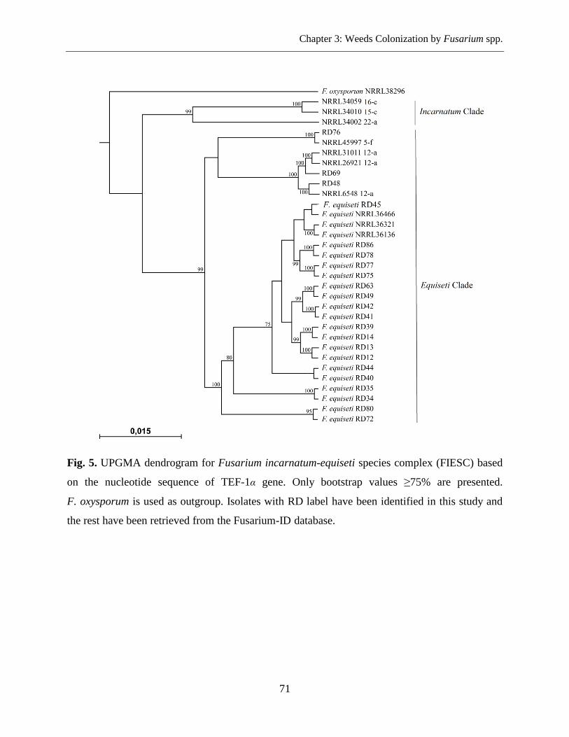
Chapter 3: Weeds Colonization by Fusarium spp.
71
Fig. 5. UPGMA dendrogram for Fusarium incarnatum-equiseti species complex (FIESC) based
on the nucleotide sequence of TEF-1α gene. Only bootstrap values ≥75% are presented.
F. oxysporum is used as outgroup. Isolates with RD label have been identified in this study and
the rest have been retrieved from the Fusarium-ID database.
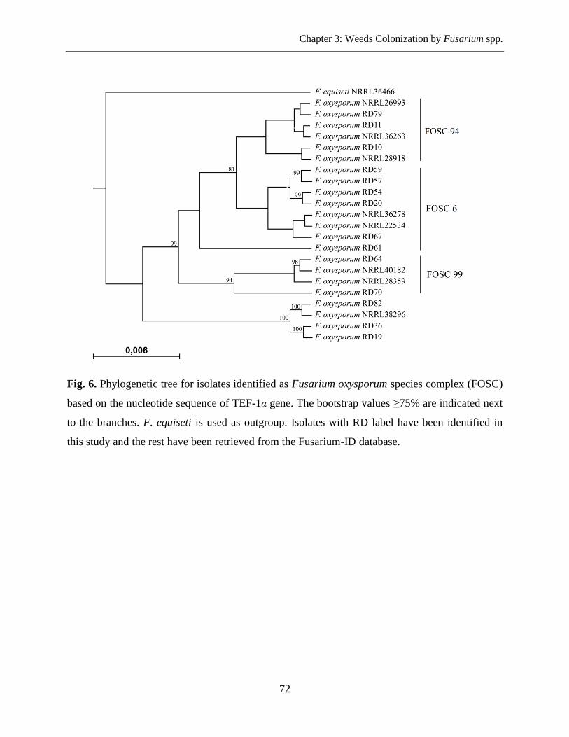
Chapter 3: Weeds Colonization by Fusarium spp.
72
Fig. 6. Phylogenetic tree for isolates identified as Fusarium oxysporum species complex (FOSC)
based on the nucleotide sequence of TEF-1α gene. The bootstrap values ≥75% are indicated next
to the branches. F. equiseti is used as outgroup. Isolates with RD label have been identified in
this study and the rest have been retrieved from the Fusarium-ID database.
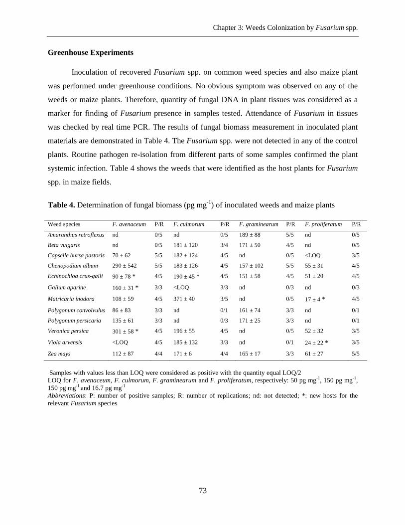
Chapter 3: Weeds Colonization by Fusarium spp.
73
Greenhouse Experiments
Inoculation of recovered Fusarium spp. on common weed species and also maize plant
was performed under greenhouse conditions. No obvious symptom was observed on any of the
weeds or maize plants. Therefore, quantity of fungal DNA in plant tissues was considered as a
marker for finding of Fusarium presence in samples tested. Attendance of Fusarium in tissues
was checked by real time PCR. The results of fungal biomass measurement in inoculated plant
materials are demonstrated in Table 4. The Fusarium spp. were not detected in any of the control
plants. Routine pathogen re-isolation from different parts of some samples confirmed the plant
systemic infection. Table 4 shows the weeds that were identified as the host plants for Fusarium
spp. in maize fields.
Table 4. Determination of fungal biomass (pg mg-1
) of inoculated weeds and maize plants
Samples with values less than LOQ were considered as positive with the quantity equal LOQ/2
LOQ for F. avenaceum, F. culmorum, F. graminearum and F. proliferatum, respectively: 50 pg mg-1
, 150 pg mg-1
,
150 pg mg-1
and 16.7 pg mg-1
Abbreviations: P: number of positive samples; R: number of replications; nd: not detected; *: new hosts for the
relevant Fusarium species
Weed species F. avenaceum P/R F. culmorum P/R F. graminearum P/R F. proliferatum P/R
Amaranthus retroflexus nd 0/5 nd 0/5 189 ± 88 5/5 nd 0/5
Beta vulgaris nd 0/5 181 ± 120 3/4 171 ± 50 4/5 nd 0/5
Capselle bursa pastoris 70 ± 62 5/5 182 ± 124 4/5 nd 0/5 <LOQ 3/5
Chenopodium album 290 ± 542 5/5 183 ± 126 4/5 157 ± 102 5/5 55 ± 31 4/5
Echinochloa crus-galli 90 ± 78 * 4/5 190 ± 45 * 4/5 151 ± 58 4/5 51 ± 20 4/5
Galium aparine 160 ± 31 * 3/3 <LOQ 3/3 nd 0/3 nd 0/3
Matricaria inodora 108 ± 59 4/5 371 ± 40 3/5 nd 0/5 17 ± 4 * 4/5
Polygonum convolvulus 86 ± 83 3/3 nd 0/1 161 ± 74 3/3 nd 0/1
Polygonum persicaria 135 ± 61 3/3 nd 0/3 171 ± 25 3/3 nd 0/1
Veronica persica 301 ± 58 * 4/5 196 ± 55 4/5 nd 0/5 52 ± 32 3/5
Viola arvensis <LOQ 4/5 185 ± 132 3/3 nd 0/1 24 ± 22 * 3/5
Zea mays 112 ± 87 4/4 171 ± 6 4/4 165 ± 17 3/3 61 ± 27 5/5
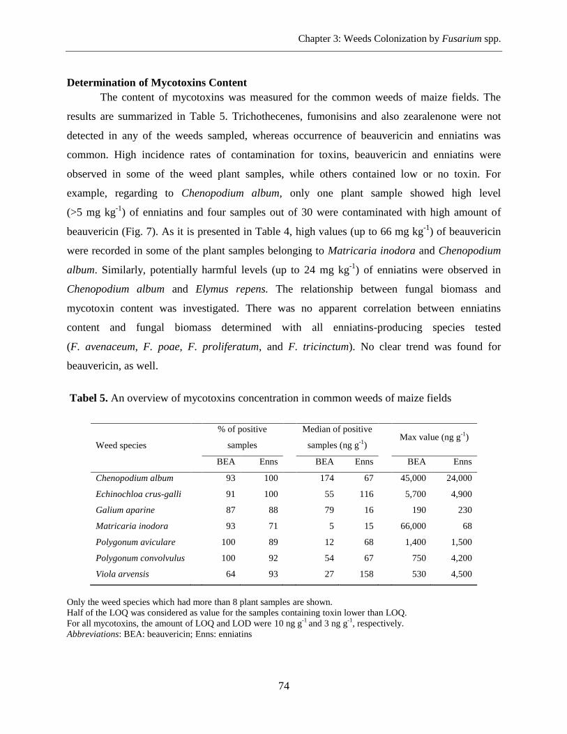
Chapter 3: Weeds Colonization by Fusarium spp.
74
Determination of Mycotoxins Content
The content of mycotoxins was measured for the common weeds of maize fields. The
results are summarized in Table 5. Trichothecenes, fumonisins and also zearalenone were not
detected in any of the weeds sampled, whereas occurrence of beauvericin and enniatins was
common. High incidence rates of contamination for toxins, beauvericin and enniatins were
observed in some of the weed plant samples, while others contained low or no toxin. For
example, regarding to Chenopodium album, only one plant sample showed high level
(>5 mg kg-1
) of enniatins and four samples out of 30 were contaminated with high amount of
beauvericin (Fig. 7). As it is presented in Table 4, high values (up to 66 mg kg-1
) of beauvericin
were recorded in some of the plant samples belonging to Matricaria inodora and Chenopodium
album. Similarly, potentially harmful levels (up to 24 mg kg-1
) of enniatins were observed in
Chenopodium album and Elymus repens. The relationship between fungal biomass and
mycotoxin content was investigated. There was no apparent correlation between enniatins
content and fungal biomass determined with all enniatins-producing species tested
(F. avenaceum, F. poae, F. proliferatum, and F. tricinctum). No clear trend was found for
beauvericin, as well.
Tabel 5. An overview of mycotoxins concentration in common weeds of maize fields
Only the weed species which had more than 8 plant samples are shown.
Half of the LOQ was considered as value for the samples containing toxin lower than LOQ.
For all mycotoxins, the amount of LOQ and LOD were 10 ng g-1
and 3 ng g-1
, respectively.
Abbreviations: BEA: beauvericin; Enns: enniatins
Weed species
% of positive
samples
Median of positive
samples (ng g-1
)
Max value (ng g
-1)
BEA Enns BEA Enns BEA Enns
Chenopodium album 93 100 174 67 45,000 24,000
Echinochloa crus-galli 91 100 55 116 5,700 4,900
Galium aparine 87 88 79 16 190 230
Matricaria inodora 93 71 5 15 66,000 68
Polygonum aviculare 100 89 12 68 1,400 1,500
Polygonum convolvulus 100 92 54 67 750 4,200
Viola arvensis 64 93 27 158 530 4,500
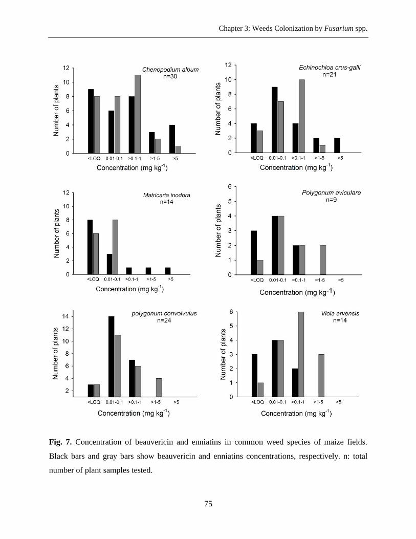
Chapter 3: Weeds Colonization by Fusarium spp.
75
Fig. 7. Concentration of beauvericin and enniatins in common weed species of maize fields.
Black bars and gray bars show beauvericin and enniatins concentrations, respectively. n: total
number of plant samples tested.
Chapter 3: Weeds Colonization by Fusarium spp.
76
Discussion
A high biodiversity of Fusarium species associated with maize diseases are
demonstrated. In this research, the qPCR assay successfully detected eight species of Fusarium
in weeds sampled from maize farms. Morphological and molecular characteristics also
confirmed the presence of eight different species, whereas F. equiseti and F. avenaceum were
predominant and most frequently found in common weeds of the maize fields. This finding
triggers the preliminary assumption that these two species are the most abundant Fusarium in the
northern parts of Germany which may help to incidence of maize Fusarium diseases. Different
reports confirm F. avenaceum as a common species associated with cereals in Europe while
F. equiseti has less frequently (Logrieco et al., 2002; Yli-Mattila, 2010; Kosiak et al., 2003).
Environmental and agronomic factors affect the occurrence of F. avenaceum on cereals
(Gäfenhan et al., 2013). F. tricinctum which is another important species in cereals of Northern
Europe (Yli-Mattila, 2010) was detected in 18% of total assessed weed samples. Regarding to
F. graminearum described as the most important Fusarium in Central and Northern Europe,
although the reports show replacement of this species with F. culmorum (Yli-Mattila, 2010), our
study revealed a higher incidence of F. culmorum (18%) in weeds than F. graminearum (8%).
We could not recover F. poae and F. subglutinans which were detected in very low frequency
(3% and 1%, respectively) by qPCR detection. F. oxysporum has been demonstrated from maize
and other cereal crops as a less common species (Logrieco et al., 2002; Kosiak et al., 2003). In
our sampling this species was isolated from most common weed plants. F. venenatum which has
been sporadically recovered from maize tissues (Logrieco et al., 2002) was isolated from six
different weed species. Therefore, the biodiversity of Fusarium of weeds sampled was as high as
reported in maize fields. In addition, prevalence of F. avenaceum and F. equiseti on almost all
common weeds studied indicates that these two species are more competitive Fusarium for weed
colonization compared to the main Fusarium of cereals such as F. graminearum. As we
expected, F. verticillioides was not detected in any of the weeds sampled. F. verticillioides is a
serious problem of maize production in southern parts of Germany, while our samples had been
collected from the north and eastern-north. Further work, thus, is needed to find the role of
weeds in survival of this species as well as in fumonisin production. The frequent detection of
Fusarium spp. on weed plants raised the importance of their role in the survival of Fusarium
fungus.
Chapter 3: Weeds Colonization by Fusarium spp.
77
The diversity of Fusarium species was similar in two farming types. Comparsion of
Fusarium population in two different cultivation systems indicated a higher incidence of
Fusarium spp., not necessarily significant, among organic fields except F. avenaceum,
F. culmorum and F. tricinctum. The comparison of fungal biomass for six Fusarium species (out
of eight) revealed no significant difference in aggressiveness of the fungi in two corn cropping
systems. However, there was a statistical difference in fungal biomass of F. equiseti and
F. tricinctum among two different farming types. F. equiseti had a higher level of biomass in
ecological fields while F. tricinctum was more aggressive in conventional farms. Lack of
fungicide application for the control management of Fusarium maize pathogens in both cropping
systems have been likely leaded to the similar Fusarium diversity in addition to equal fungal
biomass. According to Nutz & Karlovsky (unpublished) there is no significant difference
between conventional and organic farming in occurrence of fusarium ear rot and level of fungal
biomass. They demonstrated variety levels of infection in two cultivation methods.
Weed plants sampled from maize fields did not show any disease symptoms what was
obviously evidenced in inoculation experiments in the greenhouse. Furthermore, the Fusarium
strains were recovered from surface sterilized weed segments in both field and greenhouse
samples. This issue confirmed that the isolates have originated from inner parts of the
contaminated weeds. Symptomless colonization of weed plants has been observed by several
Fusarium spp. (Haware & Nene, 1982; Clark and Watson 1983; Jenkinson & Parry, 1994; Postic
et al., 2012; Altinok, 2013). Lack of visible symptoms on the weed plants and their re-isolation
would suggest that the infection of these hosts by Fusarium species could be endophytic.
In this study, weeds were clearly shown to harbor different species of Fusarium. To our
knowledge, some of the weed species demonstrated in this study (Table 4) are new alternative
hosts and have not been reported for the relevant Fusarium species. Our results increased the
possible role of weeds in the survival of fungus and production of primary inoculum in maize
fields. In the absence of susceptible host plants, Fusarium spp. can survive in weeds as an
alternative hosts and increase the inoculum potential for infecting the main crop during the
growing season. This is much more expected for the species such as F. poae or
F. subglutinans which are not able to survive saprotrophically on crop debris by producing
resting spores (Jenkinson & Parry, 1994). Although plant residues produce the main source of
Chapter 3: Weeds Colonization by Fusarium spp.
78
inoculum in maize fields (Cotten & Munkvold, 1998), different studies also show survival of
Fusarium species on non-host plants such as wild plants and weeds (Jenkinson & Parry, 1994;
Inch & Gilbert, 2003; Pereyra & Dill-Macky, 2008; Postic et al., 2012; Altinok, 2013). As a
result, for determination of the relative importance of weeds as a source of inoculum, the
pathogenicity of isolated Fusarium should be compared to that isolates obtained from maize.
High frequency of Fusarium spp. was detected in Echinochloa crus-galli, Polygonum
convolvulus, and Chenopodium album based on the qPCR assay. These plants were the main
weed species reported from maize fields sampled (Westerman & Gerowitt, 2012). The study also
pointed out very low incidence of Fusarium infection in some weed species. For example, the
only Fusarium species detected in Cirsium arvense L., Sinapis arvensis L., Alopecurus
myosuroides (L.) Huds. and Euphorbia helioscopia L. were F. proliferatum, F, equiseti,
F. culmorum and F. equiseti, respectively. Furthermore, in several weed species such as Sonchus
asper L., Anchusa arvensis L., Papaver rhoeas L., Solanum nigrum L., Geranium dissectum L.,
Lolium sp. and Erysimum cheirathoides L. no Fusarium infection was observed. Number of
relevant weed samples was not enough for resulting. Thus, the relative importance of the same
above mentioned weeds as Fusarium hosts or inoculum source in maize fields cannot be
determined from this work.
DNA sequences of genes such as TEF-1α have been widely used for supporting
morphological traits of Fusarium species (Yli-Mattila et al., 2002; Harrow et al., 2010; Geiser et
al., 2004; Hsuan et al., 2011). In the present study, molecular investigation based on the TEF-1α
gene leaded to more precise identification of Fusarium isolates. The isolates that were grouped
morphologically as F. equiseti were placed in FIESC according to the closest match of BLAST
search analysis using Fusarium-ID database. A high level of genetic diversity has been observed
in FIESC (O'Donnell et al., 2009a). This species complex with two clades (Incarnatum and
Equiseti) has been included 62 sub-clades in each one to eight isolates are introduced. Currently
reports have revealed 28 different genetic lineages in FIESC while Latin binomials were applied
with confidence to only three of the species and others were identified with the English numerals
(O'Donnell et al., 2009a). In our work, 22 isolates which have morphologically been identified as
F. equiseti showed a high identity to the sub-clades of FIESC 14-a, FIESC 12-a, and FIESC 5-f
(Supplemental Table 2). FIESC 14-a has been named F. equiseti while the next two
Chapter 3: Weeds Colonization by Fusarium spp.
79
sub-clades have still no Latin name (O'Donnell et al., 2009a). The percentage of similarity of the
isolates belonged to FIESC ranged from 99.06-99.84%. The possible reasons for not perfect
matching can be the occurrence of allelic variant, the existence of a new species, no sequence
representative in the database or poorly defining of the query sequence. In some cases, all known
isolates of a pathogen have identical TEF-1α sequence; but variation among TEF-1α alleles within
some species may vary (Geiser et al., 2004). Similarly, the isolates morphologically identified as
F. oxysporum placed in three different sub-clades of FOSC. Fusarium-ID database introduce 256
sub-caldes in this Fusarium complex. Based on the sequencing homology, strain RD61 was
identified as a member of FOSC6; but molecular phylogenetic analysis grouped it in a distinct
branch close to FOSC6. More investigation with other DNA sequences or mycotoxin profile is
necessary for finding the exact place of this strain in FOSC.
Furthermore, based on the morphological assessment strain RD99 was identified as
F. graminearum; but it was placed in a distinct branch close to F. graminearum. The TEF-1α
sequence of this strain indicated 99.68% identity with F. boothii NRRL29105. F. boothii
(= F. graminearum, lineage 3) was formerly described as one of the nine species lineages within
the F. graminearum clade. These two species are morphologically similar, but have slightly
different conidia. Recently, thirteen species lineage of F. graminearum clade have been
supported through the genealogical concordance and phylogenetic analysis of DNA sequences of
nuclear genes (O'Donnell et al., 2000, 2004, 2008; Starkey et al., 2007).
According to the morphological characters (orange color sporodochia and pyriform
microconidia) and the results of species-specific qPCR assay the strains RD22, RD32, RD95 and
RD98 were identified as F. avenaceum. In UPGMA dendrogram, constructed with TEF-1α
sequences from F. avenaceum and closely related species, the reference strains of
F. arthrosporioides did not form completely separated group (Fig. 4). They were nested within
F. avenaceum strains. The previous studies with the combined IGS and ß-tubulin sequences data
could not distinguish F. avenaceum and F. arthrosporioides strains to form separate groups
(Yli-Mattila et al., 2002). However, investigations based on the ATP Citrate Lyase (acl1) and
TEF-1α gene sequences were able to form a distinct lineage for F. arthrosporioides in the
F. avenaceum main clade (Gräfenhan et al., 2013).
Chapter 3: Weeds Colonization by Fusarium spp.
80
Common weeds were assessed for the main toxin contents by HPLC-MS/MS. Lack of
trichothecenes, zearalenone and fumonisin contamination in weed samples containing their
producers, made the assumption that these fungi are not active for toxin production in wild host
plants. We suppose that the production of toxins by some Fusarium strains may be affected by
inappropriate environmental factors. Uhlig et al. (2007) demonstrated that field situations in
Northern Europe are not favor for moniliformin production while enniatins are frequently
detected in cereal grains. On the other hand, poor performance of Fusarium pathogens such as
F. graminearum on weeds (Akinsanmi et al., 2007), may explain the lack of symptoms and
mycotoxin production on these alternative hosts. In this study, DON and NIV were not detected
in weed samples highly contaminated with F. equiseti. There are controversial reports regarding
DON and NIV production by F. equiseti strains (Kosiak et al., 2005; Marín et al., 2012).
Therefore, based on our knowledge of weeds, this question is still remained that whether
F. equiseti strains harbored in weeds have potential contribution for rising of contamination risk
of the DON and NIV in cereal grains.
Whereas, F. equiseti and F. avenaceum were found in 49% and 35% of all samples,
respectively, beauvericin and enniatins content were also determined in weed samples. The
results indicated that weeds were contaminated with beauvericin at the levels ranging from
65621 ng g-1
to 5 ng g-1
plant tissues. Beauvericin as a toxic contaminant has been reported from
maize ears infected with Fusarium pathogen (Logrieco et al., 1993; Bottalico et al., 1995; Ritieni
et al., 1997; Logrieco et al., 1995; Yli-Mattila, 2010). The values for enniatins contamination
were in the range of 24236 ng g-1
to 5 ng g-1
. Previous analytical surveys have indicated the
contamination of cereal grains by enniatins resulting of F. avenaceum. Single weed samples
showed extremely high level of toxins. There was no significant correlation between mycotoxin
level and Fusarium DNA. High level of fungal DNA in some beauvericin/enniatin-free samples
or beauvericin/enniatin-low samples revealed that some strains of fungal producers were not able
to yield relevant toxins. On the other hand, high contamination of both toxins with low levels of
fungal genomic DNA can be explained by the presence of other Fusarium species such as
F. acuminatum, F. sambucinum, F. venenatum which are able to produce related toxins.
In summary, the present results demonstrat the frequent occurrence of Fusarium species
in weed plants. Large number of recovered Fusarium spp. can support the role of weeds as
Chapter 3: Weeds Colonization by Fusarium spp.
81
symptomless hosts for Fusarim species. The possible importance of weeds in increasing of the
pressure of primary inoculum in maize fields raises the risk of toxin contamination in cereal
grains. Although the main toxins were not detected in weed samples, the significance of
Fusarium strains, harbored in weeds, for mycotoxin production in primary host plants should not
be disregarded. These findings indicate that weed management in maize fileds would aim to
reduce Fusarium inoculum and assist in the control of Fusarium maize diseases.
Acknowledgements
We acknowledge Dr. Barbara Edler who kindy helped us during the preparation of weed
plants for inoculation tests in the greenhouse.
Authors' Contributions
In this study, the weed samples were collected and identified by Prof. Dr. Bärbel
Gerowitt and Dr. Horst-Henning Steinmann. The mycotoxin experiments were carried out by
Dr. Katharina Döll.
Chapter 3: Weeds Colonization by Fusarium spp.
82
Literature Cited
Abbas, H. K., and Boyette, C. D. 1992. Phytotoxicity of fumonisin B1 on weeds and crop
species. Weed Technology 6:548-552.
Abbas, H. K., Boyette, C. D., Hogland, F. E., and Vesonder, R. F. 1991. Bioherbicidal
potential of Fusarium moniliforme and its phytotoxin. Weed Sci. 39:673-677.
Adejumo, T. O., Hettwer, U., and Karlovsky, P. 2007a. Occurrence of Fusarium species
and trichothecenes in Nigerian maize. Int. J. Food Microbiol. 116:350-357.
Adejumo, T. O., Hettwer, U., and Karlovsky, P. 2007b. Survey of maize from south
western Nigeria for zearalenone, fumonisin B1, alpha- and beta- zearalenols and enniatins
produced by Fusarium species. Food Add. Contam. 24:993-1000.
Akinsanmi, O. A., Chakraborty, S., Backhouse, D., and Simpfendorfer, S. 2007. Passage
through alternative hosts changes the fitness of Fusarium graminearum and Fusarium
pseudograminearum. Environ. Microbiol. 9:512-520.
Altinok, H. H. 2013. Fusarium species isolated from common weeds in eggplant fields
and symptomless hosts of Fusarium oxysporum f. sp. melongenae in Turkey. J.
Phytopathol. 161:335-340.
Bai, G. H., and Shaner, G. 1996. Variation in Fusarium graminearum and cultivar
resistance to wheat scab. Plant Dis. 80:975-979.
Baayen, R. P., O'Donnell, K., Bonants, P. J. M., Cigelnik, E., Kroon, L. P. N. M.,
Roebroeck, E. J. A., and Waalwijk, C. 2000. Gene genealogies and AFLP analyses in the
Fusarium oxysporum complex identify monophyletic and nonmonophyletic formae
speciales causing wilt and rot disease. Phytopathology 90:891-900.
Balmas, V., Migheli, Q., Scherm, B., and Garau, P. 2010. Multilocus phylogenetics show
high levels of endemic fusaria inhabiting Sardinian soils (Tyrrhenian Islands). Mycologia
102:803-812.
Bottalico, A., Logrieco, A., Ritieni, A., Moretti, A., Randazzo, G., and Corda, P. 1995.
Beauvericin and fumonisin B1 in preharvest Fusarium moniliforme maize ear rot in
Sardinia. Food Addit. Contam. 12:599-607.
Boyette, C. D., and Walker, H. L. 1985. Factors influencing biocontrol of Velvetleaf
(Abutilon theopbrasti) and Prickly Sida (Sida spinosa) with Fusarium lateritium. Weed
Sci. 33:209-211.
Chapter 3: Weeds Colonization by Fusarium spp.
83
Brandfass, C., and Karlovsky, P. 2008. Upscaled CTAB-based DNA extraction and
real-time PCR assays for Fusarium culmorum and F. graminearum DNA in plant
material with reduced sampling error. Int. J. Mol. Sci. 9:2306-2321.
Castanares, E., Stenglein, S. A., Dinolfo, M. I., and Moreno, M. V. 2011. Fusarium
tricinctum associated with head blight on wheat in Argentina. Plant Dis. 95:496.
Cerkauskas, R. F., Dhingra, O. D., Sinclair, J. B., and Asmus, G. 1983. Amaranthus
spinosus, Leonotis nepetaefolia, and Leonurus sibiricus: New hosts of Phomopsis spp. in
Brazil. Plant Dis. 67:821-824.
Clark, C. A., and Watson, B. 1983. Susceptibility of weed species of Convolvulaceae to
root-infecting pathogens of sweet potato. Plant Dis. 67:907-909.
Cotten, T. K., and Munkvold, G. P. 1998. Survival of Fusarium moniliforme,
F. proliferatum, and F. subglutinans in maize stalk residue. Phytopathology 88:550-555.
Dill-Macky, R., and Jones, R. K. 2000. The effect of previous crop residues and tillage on
fusarium head blight of wheat. Plant Dis. 84:71-76.
Dorn, B., Forrer, H. R., Schürch, S., and Vogelgsang, S. 2009. Fusarium species complex
on maize in Switzerland: occurrence, prevalence, impact and mycotoxins in commercial
hybrids under natural infection. Eur. J. Plant Pathol. 125:51-61.
Fandohan, P., Hell, K., Marasas, W. F. O., and Wingfield, M. J. 2003. Infection of maize
by Fusarium species and contamination with fumonisin in Africa. Afr. J. Biotechnol.
2:570-579.
Garrett, S. D. 1960. Inoculum potential. In Plant Pathology. An Advanced Treatise. Vol.
3 (ed. J. G. Horsfall & A. E. Dismond), p. 23-56. Academic press: New York.
Geiser, D. M., Jime´nez-Gasco, M. D. M., Kang, S., Makalowska, I., Veeraraghavan, N.,
Ward, T. J., Zhang, N., Kuldau, G. A., and O'Donnell, K. 2004. FUSARIUM-ID v. 1.0: A
DNA sequence database for identifying Fusarium. Eur. J. Plant Pathol. 110:473-479.
Glenn, A. E. 2007. Mycotoxigenic Fusarium species in animal feed. Anim. Feed Sci.
Tech. 137:213-240.
Gordon, W. L. 1959. The occurrence of Fusarium species in Canada. VI. Taxonomic and
geographic distribution of Fusarium species on plants, insects and fungi. Can. J. Botany
37:257-290.
Chapter 3: Weeds Colonization by Fusarium spp.
84
Gräfenhan, T., Patrick, S. K., Roscoe, M., Trelka, R., Gaba, D., Chan, J. M., McKendry,
T., Clear, R. M., and Tittlemier, S. A. 2013. Fusarium damage in cereal grains from
western Canada. Phylogenetic analysis of moniliformin-producing Fusarium species and
their natural occurrence in mycotoxin-contaminated. J. Agric. Food Chem. 61:5425-5437.
Harrow, S. A., Farrokhi-Nejad, R., Pitman, A. R., Scott, I. A. W., Bentley, A., Hide, C.,
and Cromey, M. G. 2010. Characterization of New Zealand Fusarium populations using a
polyphasic approach differentiates the Fusarium avenaceum/ F. acuminatum/
F. tricinctum species complex in cereal and grassland systems. Fungal Biol. 114:293-311.
Haware, M. P., and Nene, Y. L. 1982. Symptomless carriers of the chickpea wilt
Fusarium. Plant Dis. 66:250-251.
Helbig, J. B., and Carroll, A. B. 1984. Dicotyledonous weeds as a source of Fusarium
oxysporum pathogenic on soybean. Plant Dis. 68:694-696.
Hornunga, R. W., and Reeda, L. D. 1990. Estimation of average concentration in
presence of nondetectable values. Applied Occupational and Environmental Hygiene
5:46-51.
Hsuan, H. M., Salleh, B., and Zakaria, L. 2011. Molecular identification of Fusarium
species in Gibberella fujikuroi species complex from rice, sugarcane and maize from
Peninsular Malaysia. Int. J. Mol. Sci. 12:6722-6732.
Inch, S., and Gilbert, J. 2003. The incidence of Fusarium species recovered from
inflorescences of wild grasses in southern Manitoba. Can. J. Plant Pathol. 25:379–383.
Jenkinson, P., and Parry, D. W. 1994. Isolation of Fusarium species from common
broad-leaved weeds and their pathogenicity to winter wheat. Mycol. Res. 98:776-780.
Kosiak, E. B., Holst-Jensen, A., Rundberget, T., González-Jaén, M. T., and Torp, M. 2005.
Morphological, chemical and molecular differentiation of Fusarium equiseti isolated
from Norwegian cereals. Int. J. Food Microbiol. 99:195-206.
Kosiak, B., Torp, M., Skjerve, E., and Thrane, U. 2003. The prevalence and distribution
of Fusarium species in Norwegian cereals: a survey. Acta Agr. Scand. (Section B- Soil
and Plant Science) 53:168-176.
Kristensen, R., Torp, M., Kosiak, B., and Holst-Jensen, A. 2005. Phylogeny and
toxigenic potential is correlated in Fusarium species as revealed by partial translation
elongation factor 1 alpha gene sequences. Mycol. Res. 109:173-186.
Chapter 3: Weeds Colonization by Fusarium spp.
85
Leslie, J. F., and Summerell, B. A. 2006. The Fusarium Laboratory Manual. Blackwell
Publishing. 388 pp.
Logrieco, A., Mulè, G., Moretti, A., and Bottalico, A. 2002. Toxigenic Fusarium species
and mycotoxins associated with maize ear rot in Europe. Eur. J. Plant Pathol. 108:
597-609.
Logrieco, A., Moretti, A., Ritieni, A., Bottalico, A., and Corda, P. 1995. Occurrence and
toxigenicity of Fusarium proliferatum from preharvest maize ear rot, and associated
mycotoxins, in Italy. Plant Dis. 79:727-731.
Logrieco, A., Moretti, A., Ritieni, A., Chelkowski, J., Altomare, C., Bottalico, A., and
Randazzos, G. 1993. Natural occurrence of beauvericin in preharvest Fusarium
subglutinans infected corn ears in Poland. J. Agric. Food. Chem. 41:2149-2152.
Maiorano, A., Blandino, M., Reyneri. A., and Vanara, F. 2008. Effects of maize residues
on the Fusarium spp. infection and deoxynivalenol (DON) contamination of wheat grain.
Crop Protection 27:182-188.
Marín, P., Moretti, A., Ritieni, A., Jurado, M., Vázquez, C., and González-Jaén, M. T.
2012. Phylogenetic analyses and toxigenic profiles of Fusarium equiseti and Fusarium
acuminatum isolated from cereals from Southern Europe. Food Microbiol. 31:229-237.
Minerdi, D. Moretti, M., Gilardi, G., Barberio, C., Gullino, M. L., and Garibaldi, A.
2008. Bacterial ectosymbionts and virulence silencing in a Fusarium oxysporum strain.
Environ. Microbiol. 10: 1725-1741.
Munkvold, G. P. 2003. Epidemiology of Fusarium diseases and their mycotoxins in
maize ears. Eur. J. Plant Pathol. 109:705-713.
Naef, A., and De´fago, G. 2006. Population structure of plant-pathogenic Fusarium
species in overwintered stalk residues from Bt-transformed and non-transformed maize
crops. Eur. J. Plant Pathol. 116:129-143.
Nalim, F. A., Elmer, W. H., McGovern, R. J., and Geiser, D. M. 2009. Multilocus
Phylogenetic diversity of Fusarium avenaceum pathogenic on Lisianthus.
Phytopathology 99:462-468.
Nelson, P. E., Toussoun, T. A., and Marasas, W. F. O. 1983. Fusarium species: An
Illustrated Manual for Identification. Pennsylvania State University Press, University
Park. 193 pp.
Chapter 3: Weeds Colonization by Fusarium spp.
86
Niessen, L., Gräfenhan, T., and Vogel, R. F. 2012. ATP citrate lyase 1 (acl1) gene-based
loop-mediated amplification assay for the detection of the Fusarium tricinctum species
complex in pure cultures and in cereal samples. Int. J. Food Microbiol. 158:171-185.
Nirenberg, H. I. 1976. Untersuchungen über die morphologische und biologische
Differenzierung in der Fusarium Sektion Liseola. Mitt. Biol. Bund. Land-Forst.
(Berlin-1. Dahlem) 169:1-117.
Nutz, S., Döll, K., and Karlovsky, P. 2011. Determination of the LOQ in real-time PCR
by receiver operating characteristic curve analysis: application to qPCR assays for
Fusarium verticillioides and F. proliferatum. Anal. Bioanal. Chem. 401:717-726.
O'Donnell, K., Humber, R. A., Geiser, D. M., Kang, S., Park, B., Robert, V. A., Crous, P.
W., Johnston, P. R., Aoki, T., Rooney, A. P., and Rehner, S. A. 2012. Phylogenetic
diversity of insecticolous fusaria inferred from multilocus DNA sequence data and their
molecular identification via FUSARIUM-ID and Fusarium MLST. Mycologia 104:
427-445.
O'Donnell, K., Sutton, D. A., Rinaldi, M. G., Gueidan, C., Crous, P. W., and D. M.
Geiser. 2009a. Novel multilocus sequence typing scheme reveals high genetic diversity
of human pathogenic members of the Fusarium incarnatum-F. equiseti and
F. chlamydosporum species complexes within the united states. J. Clin. Microbiol.
47:3851-3861.
O'Donnell, K., Gueidan, C., Sink, S., Johnston, P. R., Crous, P. W., Glenn, A., Riley, R.,
Zitomer, N. C., Colyer, P., Waalwijk, C., van der Lee, T., Moretti, A., Kang, S., Kim, H.
S., Geiser, D. M., Juba, J. H., Baayen, R. P., Cromey, M. G., Bithell, S., Sutton, D. A.,
Skovgaard, K., Ploetz, R., Kistler, H. C., Elliott, M., Davis, M., and Sarver, B. A. J.
2009b. A two-locus DNA sequence database for typing plant and human pathogens
within the Fusarium oxysporum species complex. Fungal Genet. Biol. 46:936-948.
O'Donnell, K., Ward, T. J., Aberra, D., Kistler, H. C., Aoki, T., Orwig, N., Kimura, M.,
Bjørnstad, A., and Klemsdal, S. S. 2008. Multilocus genotyping and molecular
phylogenetics resolve a novel head blight pathogen within the Fusarium graminearum
species complex from Ethiopia. Fungal Genet. Biol. 45:1514-1522.
O'Donnell, K., Ward, T. J., Geiser, D. M., Kistler, H. C., and Aoki, T. 2004. Genealogical
concordance between the mating type locus and seven other nuclear genes supports
formal recognition of nine phylogenetically distinct species within the Fusarium
graminearum clade. Fungal Genet. Biol. 41:600-623.
Chapter 3: Weeds Colonization by Fusarium spp.
87
O'Donnell, K., Nirenberg, H. I., Aoki, T., and Cigelink, E. 2000. A multigene phylogeny
of the Gibberella fujikuroi species complex: Detection of additional phylogenetically
distinct species. Mycoscience 41:61-78.
O'Donnell, K., Kistler, H. C., Cigelnik, E., and Ploetz, R. C. 1998. Multiple evolutionary
origins of the fungus causing Panama disease of banana: Concordant evidence from
nuclear and mitochondrial gene genealogies. Proceedings of the National Academy of
Sciences of the United States of America 95:2044-2049.
Parry, D. W., Jenkinson, P., and McLeod, L. 1995. Fusarium ear blight (scab) in small
grain cereals- a review. Plant Pathol. 44:207-238.
Pereyra, S. A., and Dill-Macky, R. 2008. Colonization of the residues of diverse plant
species by Gibberella zeae and their contribution to fusarium head blight inoculum. Plant
Dis. 92:800-807.
Placinta, C. M., D'Mello, J. P. F., and Macdonald, A. M. C. 1999. A review of worldwide
contamination of cereal grains and animal feed with Fusarium mycotoxins. Anim. Feed
Sci. Technol. 78:21-37.
Postic, J., Cosic, J., Vrandecic, K., Jurkovic, D., Saleh, A. A., and Leslie, J. F. 2012.
Diversity of Fusarium species isolated from weeds and plant debris in Croatia. J.
Phytopathol. 160:76-81.
Ritieni, A., Moretti, A., Logrieco, A., Bottalico, A., Randazzo, G., Monti, S. M.,
Ferracane, R., and Fogliano, V. 1997. Occurrence of fusaproliferin, fumonisin B1, and
beauvericin in maize from Italy. J. Agric. Food. Chem. 45:4011-4016.
Roy, K. W. 1982. Cercospora kikuchii and other pigmented species: cultural and
reproductive characteristics and pathogenicity to soybean. Can. J. Plant Pathol. 4:
226-232.
Roy, K. W., Miller, W. A., and McLean, K. S. 1994. Survey of pathogenic genera on
fungi on foliage of weeds in Mississippi. Can. J. Plant Pathol. 16:25-29.
Schaafsma, A. W., Tamburic-Ilinic, L., Miller, J. D., and Hooker, D. C. 2001. Agronomic
considerations for reducing deoxynivalenol in wheat grain. Can. J. Plant Pathol. 23:
279-285.
Starkey, D. E., Ward, T. J., Aoki, T., Gale, L. R., Kistler, H. C., Geiser, D. M., Suga, H.,
To´th, B., Varga, J., and O’Donnell, K. 2007. Global molecular surveillance reveals
Chapter 3: Weeds Colonization by Fusarium spp.
88
novel fusarium head blight species and trichothecene toxin diversity. Fungal Genet. Biol.
44:1191-1204.
Sutton, J. C. 1982. Epidemiology of wheat head blight and maize ear rot caused by
Fusarium graminearum. Can. J. Plant Pathol. 4:195-209.
Teich, A. H., and Nelson, k. 1984. Survey of fusarium head blight and possible effects of
cultural practices in wheat fields in Lambton County in 1983. Can. Plant Dis. Surv.
64:11-13.
Uhlig, S., Jestoi, M., and Parikka, P. 2007. Fusarium avenaceum-The North European
situation. Int. J. Food Microbiol.119:17-24.
Ward, T. J., Clear, R. M., Rooney, A. P., O'Donnell, K., Gaba, D., Patrick, S., Starkey, D.
E., Gilbert, J., Geiser, D. M., and Nowicki, T. W. 2008. An adaptive evolutionary shift in
fusarium head blight pathogen populations is driving the rapid spread of more toxigenic
Fusarium graminearum in North America. Fungal Genet. Biol. 45:473-484.
Westerman, P. R., and Gerowitt, B. 2012. Probability of maize biomass contamination
with weed seeds. J. Plant Dis. Protect. 11:68-73.
Yli-Mattila, T. 2010. Ecology and evolution of toxigenic Fusarium species in cereals in
Northern Europe and Asia. J. Plant Pathol. 92:7-18.
Yli-Mattila, T., Paavanen-Huhtala, S., Bulat, S. A., Alekhina, I. A., and Nirenberg, H. I.
2002. Molecular, morphological and phylogenetic analysis of the Fusarium avenaceum/
F. arthrosporioides/ F. tricinctum species complex- a polyphasic approach. Mycol. Res.
106:655-669.

Chapter 3: Weeds Colonization by Fusarium spp.
89
Supplemental Table 1. Discription of Fusarium accessions for TEF-1α sequence used in this
study
Accession No. Strain Species Reference
JF496585 H2RA.033 F. acuminatum Marín et al., 2012
JF740857 NRRL52789 F. acuminatum O'Donnell et al., 2012
KC999531 S4B-10-15 F. acuminatum Gräfenhan et al., 2013
JX397826 BBA:67701 F. arthrosporioides Niessen et al., 2012
KC999488 BBA:64134 F. arthrosporioides Gräfenhan et al., 2013
AJ543533 VI01199 F. avenaceum Kristensen et al., 2005
FJ154734 R-3053 F. avenaceum Nalim et al., 2009
KC999540 S7A-10-15 F. avenaceum Gräfenhan et al., 2013
EF428733 NRRL34698 F. culmorum Ward et al., 2008
GU250560 NRRL46661 F. culmorum Balmas et al., 2010
JF740860 NRRL52792 F. culmorum O'Donnell et al., 2012
NRRL6548 FIESCa 12-a
b O'Donnell et al., 2009a
NRRL26921 FIESC 12-a O'Donnell et al., 2009a
NRRL31011 FIESC 12-a O'Donnell et al., 2009a
NRRL34002 FIESC 22-a O'Donnell et al., 2009a
NRRL34010 FIESC 15-c O'Donnell et al., 2009a
NRRL34059 FIESC 16-c O'Donnell et al., 2009a
NRRL36136 FIESC 14-a O'Donnell et al., 2009a
NRRL36321 FIESC 14-a O'Donnell et al., 2009a
NRRL36466 FIESC 14-a O'Donnell et al., 2009a
NRRL45997 FIESC 5-f O'Donnell et al., 2009a
AJ543574 VI02440 F. flocciferum Kristensen et al., 2005
KC999484 F-424 F. flocciferum Gräfenhan et al., 2013
KC999485 F5-89 F. flocciferum Gräfenhan et al., 2013
AJ543579 VI01020 F. graminearum Kristensen et al., 2005
AJ543591 VI01177 F. graminearum Kristensen et al., 2005
AJ543595 VI01182 F. graminearum Kristensen et al., 2005
FJ985267 NRRL22534 F. oxysporum f.sp. apii O'Donnell et al., 2009b
AF246846 NRRL26993 F. oxysporum f.sp. gladioli race lit Baayen et al., 2000
AF246856 NRRL28359 F. oxysporum Baayen et al., 2000
AF246874 NRRL28918 F. oxysporum f.sp. gladioli Baayen et al., 2000
NRRL36263 F. oxysporum f.sp. gladioli
NRRL36278 F. oxysporum
FJ985372 NRRL38296 F. oxysporum f.sp. betae O'Donnell et al., 2009b
NRRL40182 F. oxysporum
KC808223 NRRL62546 F. proliferatum O'Donnell et al., 2013, unpublished
KC808226 NRRL54994 F. proliferatum O'Donnell et al., 2013, unpublished
AF160280 NRRL22944 F. proliferatum O'Donnell et al., 2000
FJ740866c NRRL52798 F. solani O'Donnell et al., 2012
AJ543607 VI01184 F. torulosum Kristensen et al., 2005
AJ543615 VI01424 F. torulosum Kristensen et al., 2005
AJ543616 VI01194 F. torulosum Kristensen et al., 2005
HQ214681 TTAz1a F. tricinctum Castanares et al., 2011
JX397872 DAOM:235659 F. tricinctum Niessen et al., 2012
AJ543630 VI01423 F. tricinctum Kristensen et al., 2005
AJ543631 VI01412 F. venenatum Kristensen et al., 2005
AJ543634 VI01176 F. venenatum Kristensen et al., 2005
AJ543635 VI01179 F. venenatum Kristensen et al., 2005
a. Fusarium incarnatum-equiseti species complex b. English numeral identify species and small letters identify unique haplotypes within each species
c. Sequence of F. solani FJ740866 was used to root the phylogeny (Fig. 4)
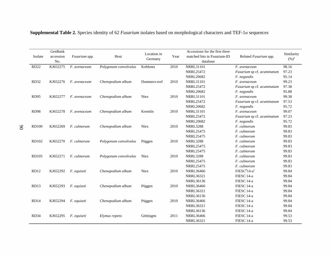
90
Supplemental Table 2. Species identity of 62 Fusarium isolates based on morphological characters and TEF-1α sequences
Supplemental Table 2: continued
Isolate
GenBank
accession
No.
Fusarium spp. Host Location in
Germany Year
Accessions for the first three
matched hits in Fusarium-ID
database
Related Fusarium spp. Similarity
(%)a
RD22 KJ652275 F. avenaceum Polygonum convolvulus Koblentz 2010 NRRL31101 F. avenaceum 98.16
NRRL25472 Fusarium sp cf. acuminatum 97.23
NRRL20682 F. negundis 95.14
RD32 KJ652276 F. avenaceum Chenopodium album Dummers-torf 2010 NRRL31101 F. avenaceum 99.23
NRRL25472 Fusarium sp cf. acuminatum 97.38
NRRL20682 F. negundis 95.88
RD95 KJ652277 F. avenaceum Chenopodium album Niex 2010 NRRL31101 F. avenaceum 99.38
NRRL25472 Fusarium sp cf. acuminatum 97.53
NRRL20682 F. negundis 95.72
RD98 KJ652278 F. avenaceum Chenopodium album Kremlin 2010 NRRL31101 F. avenaceum 99.07
NRRL25472 Fusarium sp cf. acuminatum 97.23
NRRL20682 F. negundis 95.72
RD100 KJ652269 F. culmorum Chenopodium album Niex 2010 NRRL3288 F. culmorum 99.83
NRRL25475 F. culmorum 99.83
NRRL25475 F. culmorum 99.83
RD102 KJ652270 F. culmorum Polygonum convolvulus Püggen 2010 NRRL3288 F. culmorum 99.83
NRRL25475 F. culmorum 99.83
NRRL25475 F. culmorum 99.83
RD105 KJ652271 F. culmorum Polygonum convolvulus Niex 2010 NRRL3288 F. culmorum 99.83
NRRL25475 F. culmorum 99.83
NRRL25475 F. culmorum 99.83
RD12 KJ652292 F. equiseti Chenopodium album Niex 2010 NRRL36466 FIESCb14-ac 99.84
NRRL36321 FIESC 14-a 99.84
NRRL36136 FIESC 14-a 99.84
RD13 KJ652293 F. equiseti Chenopodium album Püggen 2010 NRRL36466 FIESC 14-a 99.84
NRRL36321 FIESC 14-a 99.84
NRRL36136 FIESC 14-a 99.84
RD14 KJ652294 F. equiseti Chenopodium album Püggen 2010 NRRL36466 FIESC 14-a 99.84
NRRL36321 FIESC 14-a 99.84
NRRL36136 FIESC 14-a 99.84
RD34 KJ652295 F. equiseti Elymus repens Göttingen 2011 NRRL36466 FIESC 14-a 99.53
NRRL36321 FIESC 14-a 99.53
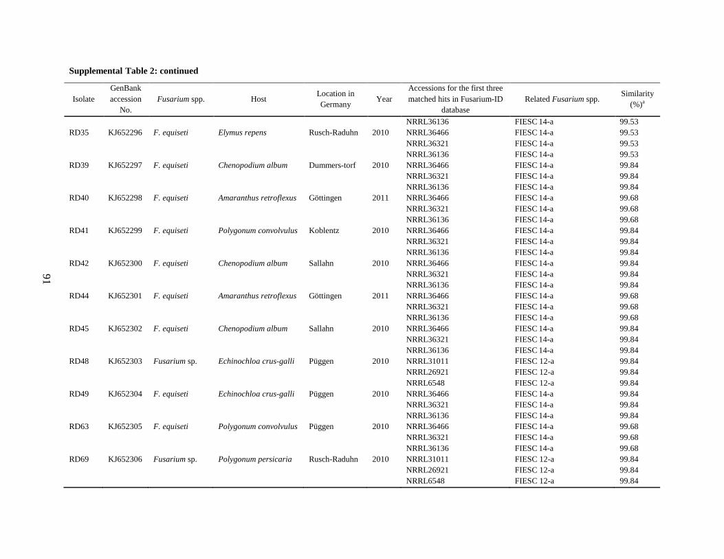
91
Supplemental Table 2: continued
Isolate
GenBank
accession
No.
Fusarium spp. Host Location in
Germany Year
Accessions for the first three
matched hits in Fusarium-ID
database
Related Fusarium spp. Similarity
(%)a
NRRL36136 FIESC 14-a 99.53
RD35 KJ652296 F. equiseti Elymus repens Rusch-Raduhn 2010 NRRL36466 FIESC 14-a 99.53
NRRL36321 FIESC 14-a 99.53
NRRL36136 FIESC 14-a 99.53
RD39 KJ652297 F. equiseti Chenopodium album Dummers-torf 2010 NRRL36466 FIESC 14-a 99.84
NRRL36321 FIESC 14-a 99.84
NRRL36136 FIESC 14-a 99.84
RD40 KJ652298 F. equiseti Amaranthus retroflexus Göttingen 2011 NRRL36466 FIESC 14-a 99.68
NRRL36321 FIESC 14-a 99.68
NRRL36136 FIESC 14-a 99.68
RD41 KJ652299 F. equiseti Polygonum convolvulus Koblentz 2010 NRRL36466 FIESC 14-a 99.84
NRRL36321 FIESC 14-a 99.84
NRRL36136 FIESC 14-a 99.84
RD42 KJ652300 F. equiseti Chenopodium album Sallahn 2010 NRRL36466 FIESC 14-a 99.84
NRRL36321 FIESC 14-a 99.84
NRRL36136 FIESC 14-a 99.84
RD44 KJ652301 F. equiseti Amaranthus retroflexus Göttingen 2011 NRRL36466 FIESC 14-a 99.68
NRRL36321 FIESC 14-a 99.68
NRRL36136 FIESC 14-a 99.68
RD45 KJ652302 F. equiseti Chenopodium album Sallahn 2010 NRRL36466 FIESC 14-a 99.84
NRRL36321 FIESC 14-a 99.84
NRRL36136 FIESC 14-a 99.84
RD48 KJ652303 Fusarium sp. Echinochloa crus-galli Püggen 2010 NRRL31011 FIESC 12-a 99.84
NRRL26921 FIESC 12-a 99.84
NRRL6548 FIESC 12-a 99.84
RD49 KJ652304 F. equiseti Echinochloa crus-galli Püggen 2010 NRRL36466 FIESC 14-a 99.84
NRRL36321 FIESC 14-a 99.84
NRRL36136 FIESC 14-a 99.84
RD63 KJ652305 F. equiseti Polygonum convolvulus Püggen 2010 NRRL36466 FIESC 14-a 99.68
NRRL36321 FIESC 14-a 99.68
NRRL36136 FIESC 14-a 99.68
RD69 KJ652306 Fusarium sp. Polygonum persicaria Rusch-Raduhn 2010 NRRL31011 FIESC 12-a 99.84
NRRL26921 FIESC 12-a 99.84
NRRL6548 FIESC 12-a 99.84
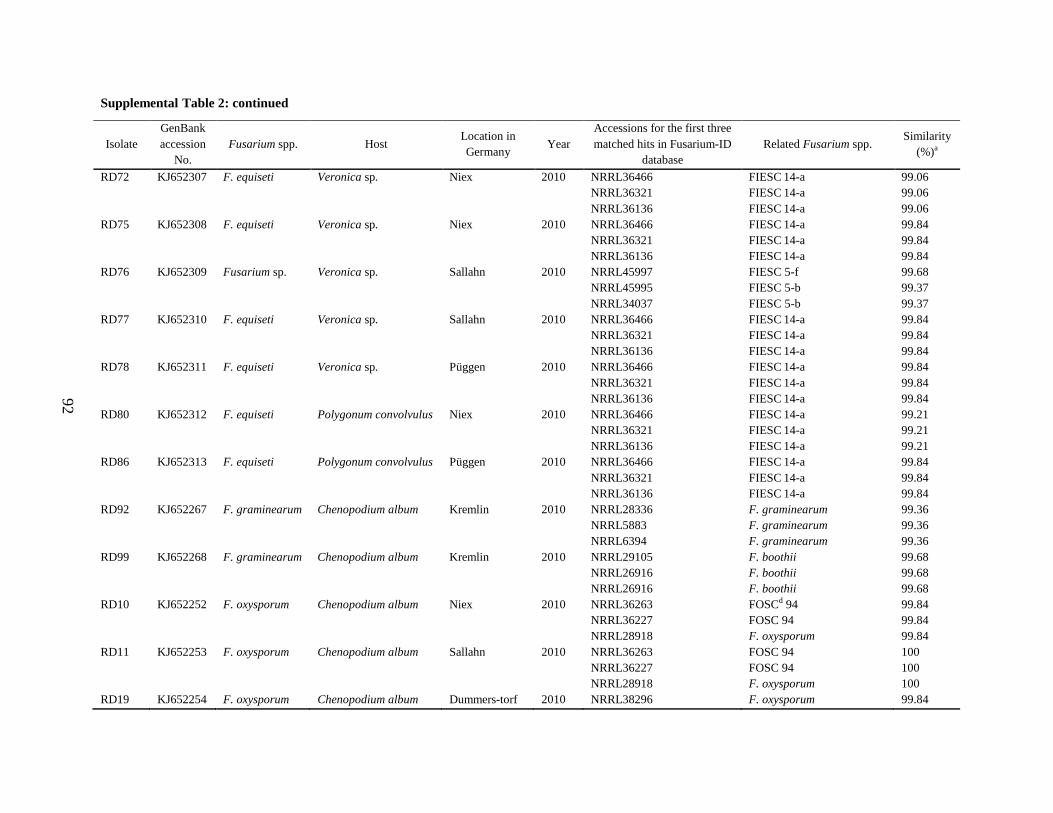
92
Supplemental Table 2: continued
Isolate
GenBank
accession
No.
Fusarium spp. Host Location in
Germany Year
Accessions for the first three
matched hits in Fusarium-ID
database
Related Fusarium spp. Similarity
(%)a
RD72 KJ652307 F. equiseti Veronica sp. Niex 2010 NRRL36466 FIESC 14-a 99.06
NRRL36321 FIESC 14-a 99.06
NRRL36136 FIESC 14-a 99.06
RD75 KJ652308 F. equiseti Veronica sp. Niex 2010 NRRL36466 FIESC 14-a 99.84
NRRL36321 FIESC 14-a 99.84
NRRL36136 FIESC 14-a 99.84
RD76 KJ652309 Fusarium sp. Veronica sp. Sallahn 2010 NRRL45997 FIESC 5-f 99.68
NRRL45995 FIESC 5-b 99.37
NRRL34037 FIESC 5-b 99.37
RD77 KJ652310 F. equiseti Veronica sp. Sallahn 2010 NRRL36466 FIESC 14-a 99.84
NRRL36321 FIESC 14-a 99.84
NRRL36136 FIESC 14-a 99.84
RD78 KJ652311 F. equiseti Veronica sp. Püggen 2010 NRRL36466 FIESC 14-a 99.84
NRRL36321 FIESC 14-a 99.84
NRRL36136 FIESC 14-a 99.84
RD80 KJ652312 F. equiseti Polygonum convolvulus Niex 2010 NRRL36466 FIESC 14-a 99.21
NRRL36321 FIESC 14-a 99.21
NRRL36136 FIESC 14-a 99.21
RD86 KJ652313 F. equiseti Polygonum convolvulus Püggen 2010 NRRL36466 FIESC 14-a 99.84
NRRL36321 FIESC 14-a 99.84
NRRL36136 FIESC 14-a 99.84
RD92 KJ652267 F. graminearum Chenopodium album Kremlin 2010 NRRL28336 F. graminearum 99.36
NRRL5883 F. graminearum 99.36
NRRL6394 F. graminearum 99.36
RD99 KJ652268 F. graminearum Chenopodium album Kremlin 2010 NRRL29105 F. boothii 99.68
NRRL26916 F. boothii 99.68
NRRL26916 F. boothii 99.68
RD10 KJ652252 F. oxysporum Chenopodium album Niex 2010 NRRL36263 FOSCd 94 99.84
NRRL36227 FOSC 94 99.84
NRRL28918 F. oxysporum 99.84
RD11 KJ652253 F. oxysporum Chenopodium album Sallahn 2010 NRRL36263 FOSC 94 100
NRRL36227 FOSC 94 100
NRRL28918 F. oxysporum 100
RD19 KJ652254 F. oxysporum Chenopodium album Dummers-torf 2010 NRRL38296 F. oxysporum 99.84
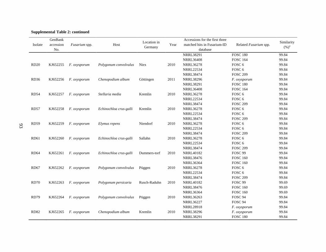
93
Supplemental Table 2: continued
Isolate
GenBank
accession
No.
Fusarium spp. Host Location in
Germany Year
Accessions for the first three
matched hits in Fusarium-ID
database
Related Fusarium spp. Similarity
(%)a
NRRL38291 FOSC 180 99.84
NRRL36408 FOSC 164 99.84
RD20 KJ652255 F. oxysporum Polygonum convolvulus Niex 2010 NRRL36278 FOSC 6 99.84
NRRL22534 FOSC 6 99.84
NRRL38474 FOSC 209 99.84
RD36 KJ652256 F. oxysporum Chenopodium album Göttingen 2011 NRRL38296 F. oxysporum 99.84
NRRL38291 FOSC 180 99.84
NRRL36408 FOSC 164 99.84
RD54 KJ652257 F. oxysporum Stellaria media Kremlin 2010 NRRL36278 FOSC 6 99.84
NRRL22534 FOSC 6 99.84
NRRL38474 FOSC 209 99.84
RD57 KJ652258 F. oxysporum Echinochloa crus-galli Kremlin 2010 NRRL36278 FOSC 6 99.84
NRRL22534 FOSC 6 99.84
NRRL38474 FOSC 209 99.84
RD59 KJ652259 F. oxysporum Elymus repens Niendorf 2010 NRRL36278 FOSC 6 99.84
NRRL22534 FOSC 6 99.84
NRRL38474 FOSC 209 99.84
RD61 KJ652260 F. oxysporum Echinochloa crus-galli Sallahn 2010 NRRL36278 FOSC 6 99.84
NRRL22534 FOSC 6 99.84
NRRL38474 FOSC 209 99.84
RD64 KJ652261 F. oxysporum Echinochloa crus-galli Dummers-torf 2010 NRRL40182 FOSC 99 99.84
NRRL38476 FOSC 160 99.84
NRRL36364 FOSC 160 99.84
RD67 KJ652262 F. oxysporum Polygonum convolvulus Püggen 2010 NRRL36278 FOSC 6 99.84
NRRL22534 FOSC 6 99.84
NRRL38474 FOSC 209 99.84
RD70 KJ652263 F. oxysporum Polygonum persicaria Rusch-Raduhn 2010 NRRL40182 FOSC 99 99.69
NRRL38476 FOSC 160 99.69
NRRL36364 FOSC 160 99.69
RD79 KJ652264 F. oxysporum Polygonum convolvulus Püggen 2010 NRRL36263 FOSC 94 99.84
NRRL36227 FOSC 94 99.84
NRRL28918 F. oxysporum 99.84
RD82 KJ652265 F. oxysporum Chenopodium album Kremlin 2010 NRRL38296 F. oxysporum 99.84
NRRL38291 FOSC 180 99.84
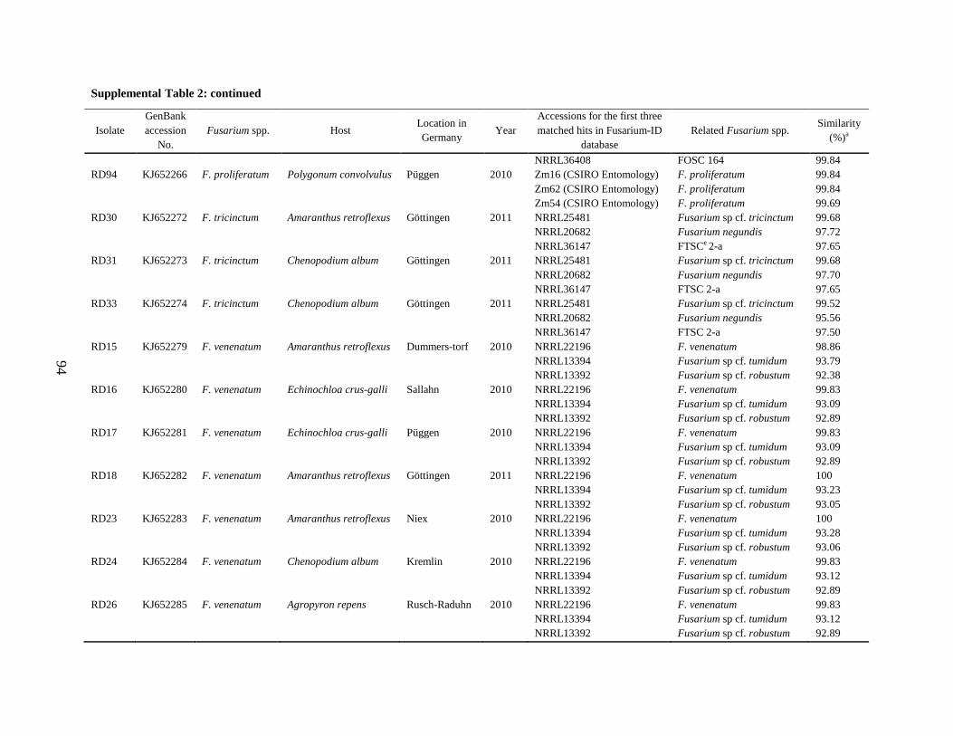
94
Supplemental Table 2: continued
Isolate
GenBank
accession
No.
Fusarium spp. Host Location in
Germany Year
Accessions for the first three
matched hits in Fusarium-ID
database
Related Fusarium spp. Similarity
(%)a
NRRL36408 FOSC 164 99.84
RD94 KJ652266 F. proliferatum Polygonum convolvulus Püggen 2010 Zm16 (CSIRO Entomology) F. proliferatum 99.84
Zm62 (CSIRO Entomology) F. proliferatum 99.84
Zm54 (CSIRO Entomology) F. proliferatum 99.69
RD30 KJ652272 F. tricinctum Amaranthus retroflexus Göttingen 2011 NRRL25481 Fusarium sp cf. tricinctum 99.68
NRRL20682 Fusarium negundis 97.72
NRRL36147 FTSCe 2-a 97.65
RD31 KJ652273 F. tricinctum Chenopodium album Göttingen 2011 NRRL25481 Fusarium sp cf. tricinctum 99.68
NRRL20682 Fusarium negundis 97.70
NRRL36147 FTSC 2-a 97.65
RD33 KJ652274 F. tricinctum Chenopodium album Göttingen 2011 NRRL25481 Fusarium sp cf. tricinctum 99.52
NRRL20682 Fusarium negundis 95.56
NRRL36147 FTSC 2-a 97.50
RD15 KJ652279 F. venenatum Amaranthus retroflexus Dummers-torf 2010 NRRL22196 F. venenatum 98.86
NRRL13394 Fusarium sp cf. tumidum 93.79
NRRL13392 Fusarium sp cf. robustum 92.38
RD16 KJ652280 F. venenatum Echinochloa crus-galli Sallahn 2010 NRRL22196 F. venenatum 99.83
NRRL13394 Fusarium sp cf. tumidum 93.09
NRRL13392 Fusarium sp cf. robustum 92.89
RD17 KJ652281 F. venenatum Echinochloa crus-galli Püggen 2010 NRRL22196 F. venenatum 99.83
NRRL13394 Fusarium sp cf. tumidum 93.09
NRRL13392 Fusarium sp cf. robustum 92.89
RD18 KJ652282 F. venenatum Amaranthus retroflexus Göttingen 2011 NRRL22196 F. venenatum 100
NRRL13394 Fusarium sp cf. tumidum 93.23
NRRL13392 Fusarium sp cf. robustum 93.05
RD23 KJ652283 F. venenatum Amaranthus retroflexus Niex 2010 NRRL22196 F. venenatum 100
NRRL13394 Fusarium sp cf. tumidum 93.28
NRRL13392 Fusarium sp cf. robustum 93.06
RD24 KJ652284 F. venenatum Chenopodium album Kremlin 2010 NRRL22196 F. venenatum 99.83
NRRL13394 Fusarium sp cf. tumidum 93.12
NRRL13392 Fusarium sp cf. robustum 92.89
RD26 KJ652285 F. venenatum Agropyron repens Rusch-Raduhn 2010 NRRL22196 F. venenatum 99.83
NRRL13394 Fusarium sp cf. tumidum 93.12
NRRL13392 Fusarium sp cf. robustum 92.89
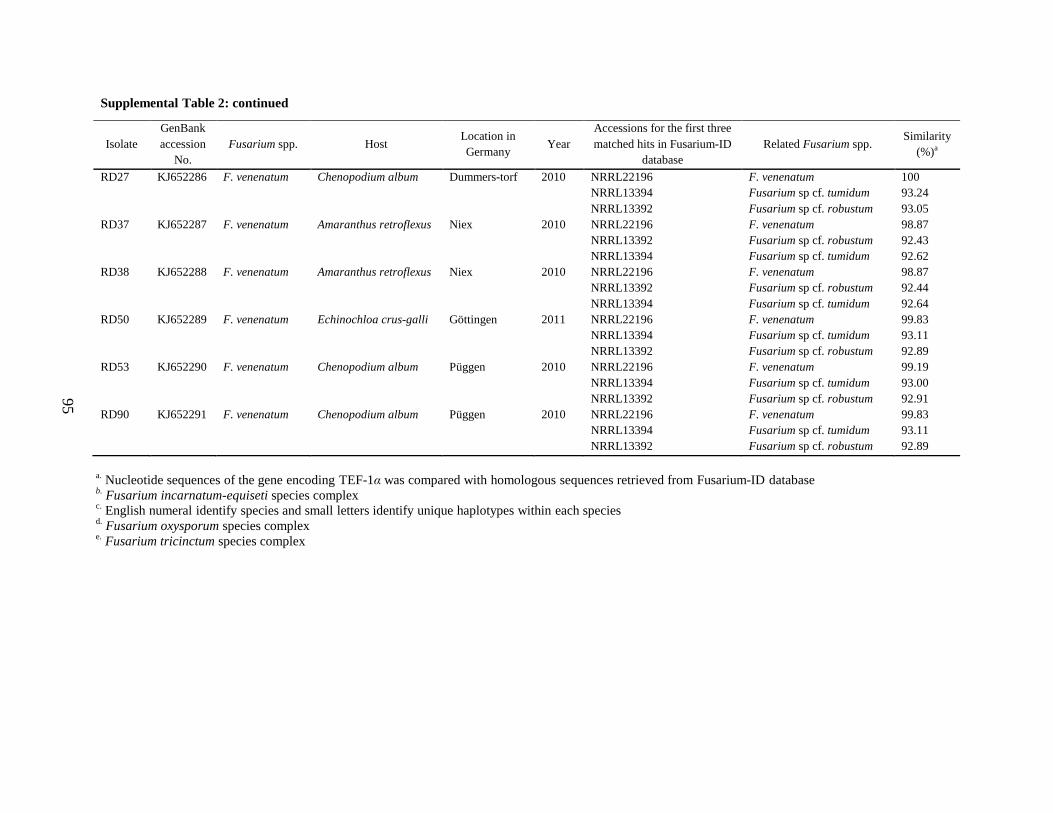
95
Supplemental Table 2: continued
Isolate
GenBank
accession
No.
Fusarium spp. Host Location in
Germany Year
Accessions for the first three
matched hits in Fusarium-ID
database
Related Fusarium spp. Similarity
(%)a
RD27 KJ652286 F. venenatum Chenopodium album Dummers-torf 2010 NRRL22196 F. venenatum 100
NRRL13394 Fusarium sp cf. tumidum 93.24
NRRL13392 Fusarium sp cf. robustum 93.05
RD37 KJ652287 F. venenatum Amaranthus retroflexus Niex 2010 NRRL22196 F. venenatum 98.87
NRRL13392 Fusarium sp cf. robustum 92.43
NRRL13394 Fusarium sp cf. tumidum 92.62
RD38 KJ652288 F. venenatum Amaranthus retroflexus Niex 2010 NRRL22196 F. venenatum 98.87
NRRL13392 Fusarium sp cf. robustum 92.44
NRRL13394 Fusarium sp cf. tumidum 92.64
RD50 KJ652289 F. venenatum Echinochloa crus-galli Göttingen 2011 NRRL22196 F. venenatum 99.83
NRRL13394 Fusarium sp cf. tumidum 93.11
NRRL13392 Fusarium sp cf. robustum 92.89
RD53 KJ652290 F. venenatum Chenopodium album Püggen 2010 NRRL22196 F. venenatum 99.19
NRRL13394 Fusarium sp cf. tumidum 93.00
NRRL13392 Fusarium sp cf. robustum 92.91
RD90 KJ652291 F. venenatum Chenopodium album Püggen 2010 NRRL22196 F. venenatum 99.83
NRRL13394 Fusarium sp cf. tumidum 93.11
NRRL13392 Fusarium sp cf. robustum 92.89
a. Nucleotide sequences of the gene encoding TEF-1α was compared with homologous sequences retrieved from Fusarium-ID database
b. Fusarium incarnatum-equiseti species complex
c. English numeral identify species and small letters identify unique haplotypes within each species
d. Fusarium oxysporum species complex
e. Fusarium tricinctum species complex
Chapter 4: Aggressiveness of Fusarium verticiliioides Strains
96
Chapter 4
Aggressiveness of Fusarium verticillioides Strains Differing in Ability to
Produce Fumonisin in Maize, Sorghum, Rice, and Beetroot Seedlings
Raana Dastjerdi and Petr Karlovsky
Molecular Phytopathology and Mycotoxin Research, Georg-August-University Göttingen, Grisebachstrasse 6,
37077 Göttingen, Germany
Corresponding author: P. Karlovsky; E-mail address: [email protected]
Abstract
Two fumonisin-nonproducing strains of Fusarium verticillioides (fum 1-3 and fum 1-4)
and their progenitors (FUM 1-1) were tested for aggressiveness toward different plant species
including maize, sorghum, rice and beetroot seedlings grown under greenhouse conditions. None
of the plants showed obvious disease symptoms following root inoculation. The quantity of
species-specific fungal biomass was measured by real-time PCR. No significant (P = 0.05)
differences in colonization of tissues were detected between each wild type and the relevant
mutant. F. verticillioides was not detected in any of the non-inoculated control plants. The
fungus could transfer from the roots to the first two internodes/leaves of maize, rice/beetroot,
regardless of fumonisin production. The symptomless systemic transmission rate, however, was
low for sorghum. This phenomenon could be explained by different host preferences of
biological species of Gibberella fujikuroi. The results indicate that in our system, fumonisin
production was not required as a virulence factor for F. verticillioides to colonize the maize, rice
and beetroot tissues.
Additional keywords: maize, Fusarium verticillioides, fumonisins
Chapter 4: Aggressiveness of Fusarium verticiliioides Strains
97
Introduction
Fumonisins are polyketide mycotoxins that contaminate commercial maize-based human
foodstuffs and animal feeds worldwide. Although number species of Fusarium, including
F. anthophilum, F. fujikuroi, F. nygamai, F. oxysporum and F. proliferatum produce fumonisins,
one species of particular concern is F. verticillioides (Sacc.) Nirenberg (syn. F. moniliforme
Scheld.), teleomorph Gibberella fujikuroi (syn. G. moniliformis Wineland) (Munkvold &
Desjardins, 1997). This fungus is a common ear rot pathogen of maize (Zea mays) and can also
infect maize stalks and roots without any visible symptoms of disease (Nelson et al., 1993;
Munkvold & Desjardins, 1997). Latent infections of maize stalk for extended periods of time
have been demonstrated (Munkvold et al., 1997). F. verticillioides reduces yield and quality of
contaminated kernels and also produces mycotoxins such as fumonisins. Since the discovery of
fumonisins (Bezuidenhout et al., 1988), these toxins have been studied due to the presence of
sphingolipid metabolism at the cellular level (Wang et al., 1991) and subsequently the
impairment of animal health (Nelson et al., 1993). Fumonisin-contaminated maize causes various
animal mycotoxicoses including leucoencephalomalacia in horses, pulmonary edema and hepatic
syndrome in pigs, poor performance in poultry, liver cancer in rats and alteration in hepatic and
immune function in cattle (reviewed in Logrieco et al., 2002). Studies have also indicated an
epidemiological correlation between human esophageal cancer and consumption of fumonisin-
contaminated maize (Marasas, 1995). In plants, toxicity of fumonisins is associated with
inhibition of Acyl CoA-dependent ceramide synthase (Williams et al., 2006). More than 10
fumonisins have been characterized, of which fumonisin B1 (FB1) is the most abundant in
naturally contaminated maize. Other B-series fumonisin homologues including FB2, FB3 and
FB4 make up 10-20% of the total fumonisins in infected maize (Nelson et al., 1993). The
fumonisin biosynthetic (FUM) gene cluster includes 16 genes (Brown et al., 2007). According
genetic studies, four closely linked loci, fum1, fum2, fum3 and fum4 have been identified as
being responsible in fumonisin biosynthesis (Desjardins et al., 1992, 1996). The lack of the
correct gene at the fum1 locus blocks fumonisin production, while strains defective at fum2
produce FB2, but not FB1 or FB3. Similarly, changes at fum3 could stop FB1 and FB2, but not
FB3 (Desjardins et al., 1992, 1996).
Chapter 4: Aggressiveness of Fusarium verticiliioides Strains
98
There is growing evidence of the occurrence of FB1 in maize all over the world,
including Europe (reviewed in Logrieco et al., 2002). In southern European countries such as
Italy, F. verticillioides is the most frequently isolated fungus from infected maize and high levels
of FB1 have been reported (reviewed in Logrieco et al., 2002). F. proliferatum as another source
of FB1 production has been found with F. verticillioides in southern Europe (Logrieco et al.,
1995; Bottalico et al., 1995). In contrast, the levels of FB1 in central and north-eastern European
areas such as Germany had been significantly lower (Usleber et al., 1994). Furthermore,
F. verticillioides is well-known pathogen on a variety of plants such as wheat, barley, soybean
(Castella et al., 1999), sorghum (da Silva et al., 2000; dos Reis et al., 2010), rice (Bhargava et al.,
1979; Cartwright et al., 1995; Kushiro et al., 2008; Maheshwar et al., 2009; Tansakul et al.,
2012) and banana (Glenn et al., 2008).
Toxicity of fumonisins to plants and animals has been clearly demonstrated (Lamprecht
et al., 1994), but there are controversial reports regarding the potential function of fumonisins in
virulence on maize. Some indirect evidence indicates that fumonisins may play a role in maize
seedling diseases caused by F. verticillioides. Fumonisins have similar structure to AAL toxins,
which are responsible for pathogenicity of Alternaria alternata on tomato cultivars (Gilchrist,
1998). AAL toxin-nonproducing strains were not able to provide leaf necrosis on susceptible
tomato cultivars (Akamatsu et al., 1997). Other studies have been shown decreasing shoot and
root length as well as leaf necrosis symptoms of maize and tomato seedlings while exposed to
low concentrations of purified fumonisins (Gilchrist et al., 1992; Lamprecht et al., 1994).
Although Desjardins et al. (1995) reported the importance of fumonisin production in virulence
on maize seedlings; they mentioned that fumonisin production is not necessary or sufficient to
cause maize blight disease. On the other hand, the greenhouse results of Jardine & Leslie (1999)
showed a fumonisin-nonproducing natural variant could infect the mature maize plants with the
same aggressiveness as the fumonisin producing strain, and caused stalk rot. Fumonisin
bioavailability to maize roots has been shown to cause a reduction in stalk weight and root mass
and increased leaf lesions (Williams et al., 2006). These findings have supported the importance
of fumonisins in plant pathogenesis. Recently, studies have shown that the expression of foliar
maize diseases is associated with fumonisin production, and this toxin can contribute to all
aspects of F. verticillioides maize seedling diseases (Williams et al., 2007; Glenn et al., 2008).
Chapter 4: Aggressiveness of Fusarium verticiliioides Strains
99
In relation to maize ear rot, Desjardins & Plattner (2000) employed three different
natural FB1-nonproducing strains and compared them with fumonisin-producing (FB1, FB2,
FB3) strains under field conditions. Both groups of the strains had the same capability to produce
maize ear rot. The studies conducted with natural variants were improved by generating isogenic
fumonisin non-producing mutants. Using these identical strains, which differ only in a fumonisin
production gene, helped to demonstrate the importance of fumonisins as a virulence factor for
F. verticillioides on maize (Desjardins et al., 2002). The results with two independent fumonisin
non-producing (fum1-3 and fum1-4) mutants in field tests indicated that fumonisins are not
necessary to cause maize ear infection by F. verticillioides (Desjardins et al., 2002).
On the basis of these observations, it has been proposed that the biological function of
fumonisins regarding their possible role in maize ear rot is still not completely clear (Munkvold,
2003). Fumonisins may have no real function in virulence, which is a complex physiological
process; however, it is possible to affect other host plants and enhance the pathogenicity (Proctor
et al., 2002). This is what has been demonstrated for trichothecenes. These toxins are the
virulence factor of Gibberella pulicaris (anamorph: F. sambucinum) and F. sporotrichioides on
parsnip roots; but there is no known function for trichothecenes in the virulence on potato tubers
(Desjardins et al., 1993). On the other hand, fumonisins could be responsible for other aspects of
the ecology of F. verticillioides (Proctor et al., 2002).
In this study, we examined the biological function of fumonisins in the growth of
F. verticillioides in maize and other possible hosts. We believed that maize silks/cobs may be a
very special environment that only became available for F. verticillioides a couple of thousands
years ago, which is too short to develop fumonisin biosynthesis. This means that fumonisin
synthesis would be originated on hosts other than maize and in tissues other than silks/cobs.
Therefore, we employed fumonisin-nonproducing strains as well as wild type strains to
determine whether fumonisin production is a potential and general virulence factor for
F. verticillioides on different plants such as sorghum, rice and beetroot as well as maize.
Understanding the role of fumonisin in pathogenicity can help pathologists in finding a strategy
to control cereal crop diseases and subsequently reduction of mycotoxin contamination.
Chapter 4: Aggressiveness of Fusarium verticiliioides Strains
100
Material and Methods
Fungal Strains and Inoculum Preparation
Two mutants of F. verticillioides and their progenitor strains were obtained from
Mycotoxin Research, National Center for Agricultural Utilization Research, United States
Department of Agriculture-Agricultural Research Service (USDA-ARS), Peoria, USA.
GFA2364 (Proctor et al., 1999) is a fumonisin non-producing mutant derived from virulent
wild-type strain M-3125 (Proctor et al., 2002), which is representative for FB1, FB2, and FB3
producing strains. Another mutant used in this study was GFA2556, a fumonisin non-producing
mutant (Desjardins et al., 2002) and its progenitor 109-R-7 (Desjardins et al., 1996) as a
FB2-producing strain. Both M-3125 and 109-R-7 were designated as wild type FUM 1-1 alleles.
This wild type functional allele was replaced with the mutant fum 1-3 allele to generate
GFA2364. Mutant GFA2556 with mutant allele fum 1-4 was generated by gene disruption in
109-R-7.
Conidia from all four strains were produced for inoculum by growing the fungus on
liquid mung bean media (Bai & Shaner, 1996) at 25°C for 5-7 days. The cultures were filtered
through sterile cheesecloth and centrifuged at 7300 g for 10 min. The spores were re-dissolved in
15% (v/v) glycerol (Carl Roth, Karlsruhe, Germany) and the number of spores was counted with
a Thoma Chamber (0.0025 mm²). The spore suspensions were stored at -70°C. Prior to
inoculation, spores were checked for viability on PDA plates. The concentration of each conidial
suspension was then determined and diluted to 1 × 10⁶
spore ml-1
with sterile tap water. The
inoculum contained 0.1% (v/v) Tween®20 (Applichem, Darmstadt, Germany) as a surfactant.
Spore suspensions were kept on ice during inoculation and homogenized well before using.
Greenhouse Tests
Mini maize cultivar ‘Gaspe Flint landrace’ (originally collected in Quebec, Canada,
provided by Prof. Dr. J. Schirawski, Albrecht-von-Haller Institute for Plant Sciences, University
of Göttingen, Germany), Rice genotype ‘Taichung Sen 10 (TCS 10)’ obtained from Africa Rice
Center, Cotonou, Benin, Sorghum bicolor ‘Tall Polish’ cultivar (originating from Leibniz
Institute of Plant Genetics and Crop Plant Research in Gatersleben, provided by Prof. Dr. J.
Schirawski, Albrecht-von-Haller Institute for Plant Sciences, University of Göttingen, Germany),
Chapter 4: Aggressiveness of Fusarium verticiliioides Strains
101
and Weiße Bete (Beetroot) cultivar ‘Weißer Kegel’ (Bio-Saatgut Gaby Krautkrämer, Armsheim)
were used in this study. The seeds of beetroot, maize and sorghum were surface disinfected in
1% (v/v) sodium hypochlorite (NaClO) solution for 1 min and absence of Fusaium spp.
propagules in or on the seeds was verified by plating a representative sample on potato dextrose
agar (PDA) (Roth, Karlsruhe, Germany) prior to the greenhouse experiments. The seeds were
then sown in plastic multi-pots containing sterile fine sand (< 2 mm granularity). Two to three
seeds of each cultivar were planted per cavity and thinned to one plant when seedlings appeared.
The multi-pots were maintained at 22°C and a relative humidity of 60% with alternating 12-h
light (mercury vapour lamps, 6600 lux at ear height) and dark periods. The rice seeds were
placed on wet paper towel and incubated at 25°C at darkness. Germinated rice seeds were
transferred to pots containing a mixture of sterile commercial plant substrate (Fruhstorfer Erde,
Typ T25, HAWITA Group, Vechta, Germany), sand and compost (1: 1/2: 1) and placed at 30°C
with 70-75% humidity. Two or three weeks after germination, all seedlings were inoculated with
conidial spore suspension.
Root-dip inoculation method was employed by soaking the roots of individual plants in
spore suspension for 30 min. After inoculation, the seedlings were transferred to new pots (9 × 9
× 9.5cm) with sterile mixed soil consisting of commercial plant substrate and sand (1:1). The
plants were maintained under the same conditions as described above for each cultivar. Plants
were irrigated with tap water as required. After one week of inoculation, the plants were
supplemented (twice during the growth) with a 15-10-15 (N- P- K) soluble fertilizer. Rice
seedlings were irrigated weekly with fertilizer containing N-P-K and Fe. Ten replicate pots were
set up for each treatment. For rice, number of replications was ten with two plants per pot and the
experiment was conducted two times. Control treatments were inoculated with autoclaved tap
water mixed with adhesion detergent Tween®20. The plants were inspected at weekly intervals
for symptoms. Seven to nine (for sorghum) weeks after inoculation, the plants were harvested
and the roots were washed carefully under tap water. The sorghum, maize and rice plants were
cut into three parts including roots, the first two or three internodes above the soil line and upper
plant part and then placed separately in plastic bags. In the case of beetroot plants, they were
divided into two parts: root and shoot. In order to quantify the fungal biomass in artificial
Chapter 4: Aggressiveness of Fusarium verticiliioides Strains
102
inoculated plants and determine the function of fumonisins, the harvested plant materials were
freeze dried and prepared for further experiments by real-time PCR (qPCR) assay.
DNA Extraction and Determination of Matrix Effects
Total genomic DNA was extracted by using 30-60 mg of fine powder plant material
based on the CTAB method according Brandfass & Karlovsky (2008). DNA quality and
concentration was estimated by electrophoresis in 0.8% (w/v) agarose gel (Cambrex, Rockland,
ME, USA), prepared in TAE buffer (40 mM Tris, 1mM EDTA (ethylene diamine tetra acetic
acid), pH set to 8.5) (both substances were obtained from Carl Roth, Karlsruhe, Germany). DNA
was stained with ethidium bromide (0.5 µg ml-1
) (Applichem, Darmstadt, Germany) and
visualized using a digital imaging system (VilberLourmat, Marne La Vallee, France).
For accurate quantification of fungal DNA in inoculated samples, DNA standards of
F. verticillioides and DNA from plant samples should be amplified under identical conditions.
Therefore, to perform the qPCR without matrix effects, the inhibitors were tested under a
common technique. DNA from an uncontaminated sample of each plant cultivar was mixed in
varying dilutions (1:25, 1:50, 1:100) with defined concentration of standard DNA, and
amplification was then carried out by qPCR. In this way, we also run the uncontaminated DNA
sample as well as DNA standard individually to be able to compare the results in different
treatments. According to the threshold value (Ct), the best dilution factor that could imitate
matrix effects was chosen.
Molecular Quantification of Fungal DNA
A thermocycler (CFX384TM
Real-Time System, C1000TM
Thermal Cycler, BioRad,
USA) with 384-well microplates (Kisker Biotech GmbH, Steinfurt, Germany) was used for the
qPCR analysis. F. verticillioides was assessed in inoculated samples based on a previously
developed low-volume qPCR protocol (Dastjerdi et al., 2013, Chapter 2, unpublished).
The Primer pairs VER 1 (CTTCCTGCGATGTTTCTCC) and VER 2
(AATTGGCCATTGGTATTATATATCTA) designed by Mul`e et al. (2004) were used to
amplify a 578 bp fragment, specific for F. verticillioides. The reaction mixture consisted of
reaction buffer (16 mM (NH4)2SO4; 67 mM Tris-HCl; 0.01% (v/v) Tween-20, pH 8.8 at 25°C,
Chapter 4: Aggressiveness of Fusarium verticiliioides Strains
103
Bioline, Lükenwalde, Germany), 0.1 mM of each of the four deoxynucleoside triphosphates
(dNTPs; Bioline, Lükenwalde, Germany), 2.5 mM MgCl2, 0.1U of Taq DNA polymerase
(BIOTaq, Bioline, Lükenwalde, Germany), 0.3 µM of each primer, 0.1x SYBR Green I
(Invitrogen, Karlsruhe, Germany). The cycling conditions were as follows: 1 cycle of 2 min at
95°C, 34 cycles of 30 s at 94°C (denaturalization), 30 s at 62.5°C (annealing), 40 s at 72°C
(extension) and followed by a final extension cycle at 72°C for 5 min. Fluorescent data were
obtained during the annealing phase to construct a melting curve at the end of assay. The qPCR
was completed by running a melting curve analysis. Quantity of fungal biomass was assessed as
a parameter of fungal development.
Data Processing and Statistical Analysis
The software SigmaPlot 12.3 Notebook was used for statistical analysis. Non-parametric
tests were performed when data distribution was not normal. The qPCR data were assessed to
find the positive samples based on the melting temperature (Tm), starting quantity (SQ) and Ct
value. The lowest DNA standard employed in qPCR assays was considered as the limit of
quantification (LOQ). According to the qPCR data, the quantity of fungal biomass (pg mg-1
) for
the positive samples was calculated. DNA quantity for the samples containing values lower than
LOQ was substituted as LOQ/2 (Hornunga & Reeda, 1990). These samples had the right melting
temperature and the logical Ct values.
Results
A common method was used to evaluate the influence of plant inhibitors on successful
qPCR performance. The results indicated a significant effect for all plant extracts tested. The
difference in Ct values between the standard reference DNA and the DNA from uncontaminated
plant material was considered as an indicator to find the matrix effects. The results are presented
in Table 1. High qPCR inhibition was seen in the root extracts; so that 100 dilution for maize and
sorghum and 50 for beetroot samples were necessary to imitate matrix effects. Diluting to 1:25
(v/v) (for aboveground tissues of sorghum and beetroot) or 1:50 (v/v) (for aboveground maize
parts) with double distilled water was preferred to reduce the Ct value to the same level as the
standard-DNA. In the case of rice plant material, DNA dilution, even 100-fold, was insufficient
for reduction of plant inhibitor effects. Adding 1 mg ml-1
bovine serum albumin (BSA) to the
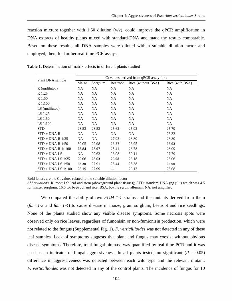
Chapter 4: Aggressiveness of Fusarium verticiliioides Strains
104
reaction mixture together with 1:50 dilution (v/v), could improve the qPCR amplification in
DNA extracts of healthy plants mixed with standard-DNA and made the results comparable.
Based on these results, all DNA samples were diluted with a suitable dilution factor and
employed, then, for further real-time PCR assays.
Table 1. Determination of matrix effects in different plants studied
Plant DNA sample Ct values derived from qPCR assay for :
Maize Sorghum Beetroot Rice (without BSA) Rice (with BSA)
R (undiluted) NA NA NA NA NA
R 1:25 NA NA NA NA NA
R 1:50 NA NA NA NA NA
R 1:100 NA NA NA NA NA
LS (undiluted) NA NA NA NA NA
LS 1:25 NA NA NA NA NA
LS 1:50 NA NA NA NA NA
LS 1:100 NA NA NA NA NA
STD 28.53 28.53 25.62 25.92 25.79
STD + DNA R NA NA NA NA 28.33
STD + DNA R 1:25 NA NA 27.93 28.80 26.80
STD + DNA R 1:50 30.05 29.98 25.27 28.95 26.03
STD + DNA R 1: 100 28.84 28.07 25.41 28.78 26.09
STD + DNA LS NA 29.63 28.08 30.11 27.79
STD + DNA LS 1:25 29.06 28.63 25.98 28.18 26.06
STD + DNA LS 1:50 28.30 27.91 25.44 28.38 25.90
STD + DNA LS 1:100 28.19 27.99 --- 28.12 26.08
Bold letters are the Ct values related to the suitable dilution factor
Abbreviations: R: root; LS: leaf and stem (aboveground plant tissues); STD: standard DNA (pg µl-1
) which was 4.5
for maize, sorghum; 16.6 for beetroot and rice; BSA: bovine serum albumin; NA: not amplified
We compared the ability of two FUM 1-1 strains and the mutants derived from them
(fum 1-3 and fum 1-4) to cause disease in maize, grain sorghum, beetroot and rice seedlings.
None of the plants studied show any visible disease symptoms. Some necrosis spots were
observed only on rice leaves, regardless of fumonisin or non-fumionisin production, which were
not related to the fungus (Supplemental Fig. 1). F. verticillioides was not detected in any of these
leaf samples. Lack of symptoms suggests that plant and fungus may coexist without obvious
disease symptoms. Therefore, total fungal biomass was quantified by real-time PCR and it was
used as an indicator of fungal aggressiveness. In all plants tested, no significant (P = 0.05)
difference in aggressiveness was detected between each wild type and the relevant mutant.
F. verticillioides was not detected in any of the control plants. The incidence of fungus for 10
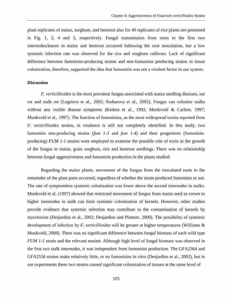
Chapter 4: Aggressiveness of Fusarium verticiliioides Strains
105
plant replicates of maize, sorghum, and beetroot also for 40 replicates of rice plants are presented
in Fig. 1, 2, 4 and 3, respectively. Fungal transmission from roots to the first two
internodes/leaves in maize and beetroot occurred following the root inoculation, but a low
systemic infection rate was observed for the rice and sorghum cultivars. Lack of significant
difference between fumonisin-producing strains and non-fumunisin producing strains in tissue
colonization, therefore, supported the idea that fumonisin was not a virulent factor in our system.
Discussion
F. verticillioides is the most prevalent fungus associated with maize seedling diseases, ear
rot and stalk rot (Logrieco et al., 2002; Srobarova et al., 2002). Fungus can colonize stalks
without any visible disease symptoms (Kedera et al., 1992; Munkvold & Carlton. 1997;
Munkvold et al., 1997). The function of fumonisins, as the most widespread toxins reported from
F. verticillioides strains, in virulence is still not completely identified. In this study, two
fumonisin non-producing strains (fum 1-3 and fum 1-4) and their progenitors (fumonisin-
producing) FUM 1-1 strains were employed to examine the possible role of toxin in the growth
of the fungus in maize, grain sorghum, rice and beetroot seedlings. There was no relationship
between fungal aggressiveness and fumonisin production in the plants studied.
Regarding the maize plants, movement of the fungus from the inoculated roots to the
remainder of the plant parts occurred, regardless of whether the strain produced fumonisin or not.
The rate of symptomless systemic colonization was lower above the second internodes in stalks.
Munkvold et al. (1997) showed that restricted movement of fungus from maize seed or crown to
higher internodes in stalk can limit systemic colonization of kernels. However, other studies
provide evidence that systemic infection may contribute to the contamination of kernels by
mycotoxins (Desjardins et al., 2002; Desjardins and Plattner, 2000). The possibility of systemic
development of infection by F. verticillioides will be greater at higher temperatures (Williams &
Munkvold, 2008). There was no significant difference between fungal biomass of each wild type
FUM 1-1 strain and the relevant mutant. Although high level of fungal biomass was observed in
the first two stalk internodes, it was independent from fumonisin production. The GFA2364 and
GFA2556 strains make relatively little, or no fumonisins in vitro (Desjardins et al., 2002), but in
our experiments these two strains caused significant colonization of tissues at the same level of
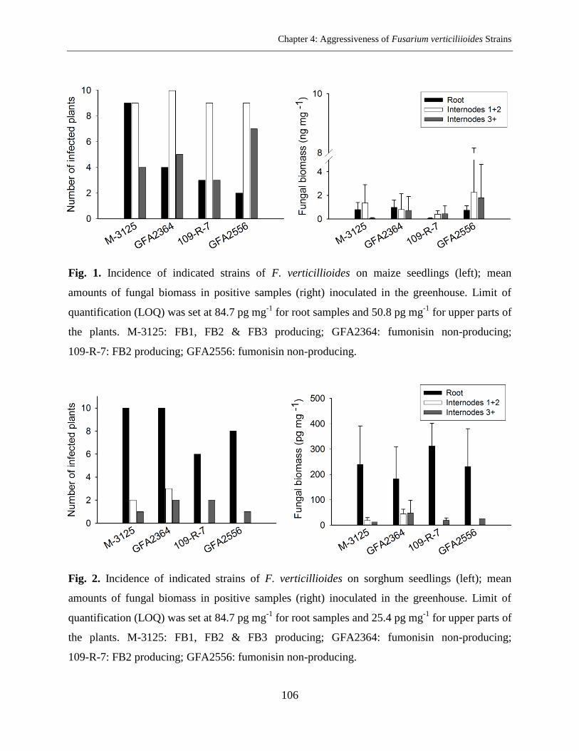
Chapter 4: Aggressiveness of Fusarium verticiliioides Strains
106
Fig. 1. Incidence of indicated strains of F. verticillioides on maize seedlings (left); mean
amounts of fungal biomass in positive samples (right) inoculated in the greenhouse. Limit of
quantification (LOQ) was set at 84.7 pg mg-1
for root samples and 50.8 pg mg-1
for upper parts of
the plants. M-3125: FB1, FB2 & FB3 producing; GFA2364: fumonisin non-producing;
109-R-7: FB2 producing; GFA2556: fumonisin non-producing.
Fig. 2. Incidence of indicated strains of F. verticillioides on sorghum seedlings (left); mean
amounts of fungal biomass in positive samples (right) inoculated in the greenhouse. Limit of
quantification (LOQ) was set at 84.7 pg mg-1
for root samples and 25.4 pg mg-1
for upper parts of
the plants. M-3125: FB1, FB2 & FB3 producing; GFA2364: fumonisin non-producing;
109-R-7: FB2 producing; GFA2556: fumonisin non-producing.
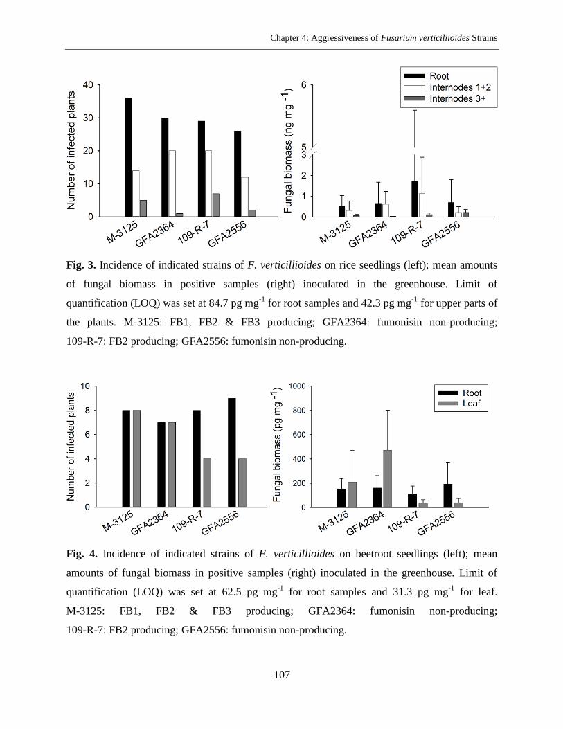
Chapter 4: Aggressiveness of Fusarium verticiliioides Strains
107
Fig. 3. Incidence of indicated strains of F. verticillioides on rice seedlings (left); mean amounts
of fungal biomass in positive samples (right) inoculated in the greenhouse. Limit of
quantification (LOQ) was set at 84.7 pg mg-1
for root samples and 42.3 pg mg-1
for upper parts of
the plants. M-3125: FB1, FB2 & FB3 producing; GFA2364: fumonisin non-producing;
109-R-7: FB2 producing; GFA2556: fumonisin non-producing.
Fig. 4. Incidence of indicated strains of F. verticillioides on beetroot seedlings (left); mean
amounts of fungal biomass in positive samples (right) inoculated in the greenhouse. Limit of
quantification (LOQ) was set at 62.5 pg mg-1
for root samples and 31.3 pg mg-1
for leaf.
M-3125: FB1, FB2 & FB3 producing; GFA2364: fumonisin non-producing;
109-R-7: FB2 producing; GFA2556: fumonisin non-producing.
Chapter 4: Aggressiveness of Fusarium verticiliioides Strains
108
fumonisin-producing strains. The results indicate that fumonisin production was not required for
F. verticillioides to colonize the maize tissues following root inoculation.
In the case of sorghum, after root inoculation the fungus remained in the below ground
tissues and fungal transmission did not occur. Only 20-30% of the plants inoculated with the
M-3125 strain and its mutant show the fungus in the first two internodes. This would be
explained by host preference of biological species of G. fujikuroi in which mating population A
(MPA) is predominant on maize while G. fujikuroi mating population F (MPF) can infect
sorghum plants (Leslie et al., 1992). These differences may cause differences in the pathogenic
potential of the strains (Leslie, 1991). The selection of suitable strains is, therefore, important in
breeding programs (Jardine & Leslie, 1999). It is also possible that the sorghum variety used in
this study reacted to the fungus with a partial resistance response, whereby the pathogen was
unable to spread through the plant. However, sorghum root colonization and lack of fungal
movement to the upper parts of the plant suggest that F. verticillioides is able to survive in the
roots of non-host plants when the main host is not available. This will clearly help to maintain
fumonisin synthesis. We believe infection of maize silks or cobs may be a very special
environment that only became available for the fungus a couple of thousands of years ago, which
is a short time period to develop fumonisin biosynthesis. For millions of years previously, the
fungus had infected roots or stems, living as endophyte. This means that fumonisin synthesis
would be maintained by the selection pressure exerted on hosts other than maize and in tissues
other than silks or cobs. The presence of fungus in upper internodes while it is absent in lower
nodes (as was the case for two plants inoculated with 109-R-7 strain and one plant inoculated
with GFA2556 strain) could be explained by spore splashing during the inoculation. Based on
the data obtained from this work, it is not possible to make a conclusion with confidence
regarding the function of fumonisin in pathogenicity on sorghum seedlings.
Although the fungus could have developed inside the young plants of rice and beetroot,
the aggressiveness of wild type strains and their mutants to the rice or beetroot seedlings was not
statistically different. Occurrence of fumonisins in rice plants has not been found as frequently as
in maize, but more recently contamination of rice with FB1 and FB2 toxins has been reported
(Kushiro et al., 2008; Maheshwar et al., 2009; Tansakul et al., 2012). The fungus has been
demonstrated to causes panicle rot of rice (Bhargava et al., 1979). G. fujikuroi complex including
Chapter 4: Aggressiveness of Fusarium verticiliioides Strains
109
MPA (anamorph, F. verticillioides), MPC (anamorph, F. fujikuroi) and MPD (anamorph,
F. proliferatum) have been demonstrated in infected rice seeds (Desjardins et al., 2000). They
show that some strains of G. fujikuroi MPD are capable of producing fumonisin as well as
beauvericin and moniliformin; but not gibberellic acid, which has been reported in bakanae. The
prevalence of MPD may be related to the role of this biological species in the complex symptoms
of bakanae disease of rice (Desjardins et al., 2000). F. verticillioides causes seedling
damping-off in sugar beet and has been reported as a pathogen which can produce symptoms
similar to fusarium yellows (Hanson & Hill, 2004; Drycott, 2006). In the present study, 15-20
days after inoculation some necrosis spots were observed on rice leaves. The spots were
irregular, small, light brown in the center and dark brown at the margins in the initial stage. The
necrotic lesions usually developed, joined together, made larger spots and extended along the
length of the leaf. The spots were seen regardless of fumonisin or non-fumonisin producing
strain tested. However, the qPCR assay could not detect the fungus in the leaf extracts. Control
plants did not show any necrotic spots. Rice experiments were conducted twice (in summer 2012
and winter 2013) and the results were identical. Further assessments are, therefore, necessary to
find the agent of these necrosis spots.
Potential plant inhibitors such as proteins, polyphenols, polysaccharides, secondary
metabolites (Horne et al., 2004) are released during the DNA extraction process. These
compounds in DNA extracts can affect the qPCR reactions. They prevent the amplification of
target DNA, increase the Ct values or lead to reduction of overall efficiency and reproducibility
of PCR and eventually may contribute to inaccurate results (Demeke & Jenkins, 2010; Cankar
et al., 2006). In this study, the effect of sample matrix properties on real-time PCR reactions was
assessed for maize, sorghum, rice and beetroot tissues by adding standard reference DNA to the
DNA of uncontaminated plant material. Initially experiments indicated a high potency of plant
inhibitors, so that the qPCR assays were completely inhibited and no amplification was detected
(Table 1). Dilution of DNA samples is a simple method to reduce the inhibitor concentration in
plant extracts especially in samples involving complex matrices such as root extracts. On the
other hand, for the samples with lower DNA concentration, a high DNA dilution factor may
decrease PCR sensitivity (Demeke & Jenkins, 2010). In this study, a dilution factor of 1:25 or
1:50 for the aboveground plant tissues and 1:100 for the root samples could decrease the PCR
Chapter 4: Aggressiveness of Fusarium verticiliioides Strains
110
inhibition and the Ct values then reached to the same level of pure fungal DNA. In the case of
rice plant tissues, although 100-fold dilution could generate PCR products, the Ct values were
still far from the Ct expected for target molecules added to the reaction. More dilution was not
possible due to decreasing qPCR sensitivity. BSA is a chemical which has been employed for
deactivation of PCR inhibitors in various sample types and recently for plant materials as well
(Wei et al., 2008; Plante et al., 2010). Adding this chemical directly into the PCR reactions was
sufficient to enhance the qPCR results.
In this work, maize seedlings colonization with fumonisin non-producing strains and
fumonisin-producing strains was similar. It is possible that such non-producing toxin strains can
be used as biocontrol agents of maize fusarium diseases if they provide a substantial level of
symptomless colonization in different maize plant tissues without causing any ear rot (Desjardins
& Plattner, 2000). In summary, the present results demonstrate that fumonisins may have a
function in pathogenicity of F. verticillioides, but in our system, the ability to synthesize these
toxins was not related to the fungal aggressiveness and fumonisins did not determine the
pathogenicity of F. verticillioides pathogen.
Acknowledgements
We thank Prof. Dr. J. Schirawski (Albrecht-von-Haller Institute for Plant Sciences,
University of Göttingen, Germany) for kindly providing the dwarf maize seeds and sorghum
cultivar. We further thank Prof. Dr. R. H. Proctor (Mycotoxin Research, National Center for
Agricultural Utilization Research, United States Department of Agriculture-Agricultural
Research Service (USDA-ARS), Peoria, USA.) who kindly supplied the F. verticillioides strains.
Chapter 4: Aggressiveness of Fusarium verticiliioides Strains
111
Literature Cited
Akamatsu, H., Itoh, Y., Kodama, M., Otani, H., and Kohmoto, K. 1997.
AAL-toxin-deficient mutants of Alternaria alternata tomatoa pathotype by restriction
enzyme-mediated integration. Phytopathology 87:967-972.
Bai, G. H., and Shaner, G. 1996. Variation in Fusarium graminearum and Cultivar
Resistance to Wheat Scab. Plant Dis. 80:975-979.
Bezuidenhout, S. C., Gelderblom, W. C. A., Gorst-Allman, C. P., Horak, R. M., Marasas,
W. F. O., Spiteller, G., and Vleggaar, R. 1988. Structure elucidation of the fumonisins,
mycotoxins from Fusarium moniliforme. J. Chem. Soc. Chem. Commun. 11:743-745.
Bhargava, S. N., Shukla, D. N., Singh, N. K., and Singh, N. 1979. Fusarium moniliforme
causing panicle rot of rice. Indian Phytopathology 31:367-369.
Bottalico, A., Logrieco, A., Ritieni, A., Moretti, A., Randazzo, G., and Corda, P. 1995.
Beauvericin and fumonisin B1 in preharvest Fusarium moniliforme maize ear rot in
Sardinia. Food Addit. Contam. 12:599-607.
Brown, D. W., Butchko, R. A. E., Busman, M., and Proctor, R. H. 2007. The Fusarium
verticillioides FUM gene cluster encodes a Zn(II)2Cys6 protein that affect FUM gene
expression and fumonisin production. Eukaryotic Cell 6:1210-1218.
Cankar, K., Stebih, D., Dreo, T., Zel, J., and Gruden, K. 2006. Critical points of DNA
quantification by real-time PCR effects of DNA extraction method and sample matrix on
quantification of genetically modified organisms. BMC. Biotech. 6:37-51.
Cartwright, R. D., Correll, J. C., and Crippen, D. L. 1995. Fusarium sheath rot of rice in
Arkansas. (Abstr.) Phytopathology 85:1199.
Castellá, G., Bragulat, M. R., and Cabañes, F. J. 1999. Surveillance of fumonisins in
maize-based feeds and cereals from Spain. J. Agric. Food Chem. 47:4707-4710.
da Silva, J. B., Pozzi, C. R., Mallozzi, M. A., Ortega, E. M., and Corrêa, B. 2000.
Mycoflora and occurrence of aflatoxin B1 and fumonisin B1 during storage of Brazilian
sorghum. J. Agric. Food Chem. 48:4352-4356.
Demeke, T., and Jenkins, G. R. 2010. Influence of DNA extraction methods, PCR
inhibitors and quantification methods on real-time PCR assay of biotechnology-derived
traits. Anal. Bioanal. Chem. 396:1977-1990.
Chapter 4: Aggressiveness of Fusarium verticiliioides Strains
112
Desjardins, A. E., Munkvold, G. P., Plattner, R. D., and Proctor, R. H. 2002. FUM1- A
gene required for fumonisin biosynthesis but not for maize ear rot and ear infection by
Gibberella moniliformis in field tests. Mol. Plant-Microbe Interact. 15:1157-1164.
Desjardins, A. E., Manandhar, H. K., Plattner, R. D., Manandhar, G. G., Poling, S. M.,
and Maragos, C. M. 2000. Fusarium species from Nepalese rice and production of
mycotoxins and gibberellic acid by selected species. Appl. Environ. Microb. 66:
1020-1025.
Desjardins, A. E., and Plattner, R. D. 2000. Fumonisin B1-nonproducing strains of
Fusarium verticillioides cause maize (Zea mays) ear infection and ear rot. J. Agric. Food
Chem. 48:5773-5780.
Desjardins, A. E., Plattner, R. D., and Proctor, R. H. 1996. Linkage among genes
responsible for fumonisin biosynthesis in Gibberella fujikuroi mating population A.
Appl. Environ. Microb. 62:2571-2576.
Desjardins, A. N., Plattner, R. D., Nelsen, T. C., and Leslie, J. F. 1995. Genetic analysis
of fumonisin production and virulence of Gibberella fujikuroi mating population A
(Fusarium moniliforme) on maize (Zea mays) seedlings. Appl. Environ. Microb. 61:
79-86.
Desjardins, A. E., Hohn, T. M., and McCormick, S. P. 1993. Trichothecene biosynthesis
in Fusarium species: chemistry, genetics, and significance. Microbiological Reviews
57:595-604.
Desjardins, A. E., Plattner, R. D., Shackelford, D. D., Leslie, J. F., and Nelson, P. E.
1992. Heritability of fumonisin B1 production in Gibberella fujikuroi mating population
A. Appl. Environ. Microb. 58:2799-2805.
dos Reis, T. A., Zorzete, P., Pozzi, C. R., da Silva, V. N., Ortega, E., and Corrˆea, B.
2010. Mycoflora and fumonisin contamination in Brazilian sorghum from sowing to
harvest. J. Sci. Food Agric. 90:1445-1451.
Gilchrist, D. G. 1998. Programmed cell death in plant disease: the purpose and promise
of cellular suicide. Annu. Rev. Phytopathol. 36:393-414.
Gilchrist, D. G., Ward, B., Moussato, V., and Mirocha, C. J. 1992. Genetic and
physiological response to fumonisin and AAL-toxin by intact tissue of a higher plant.
Mycopathologia 117:57-64.
Chapter 4: Aggressiveness of Fusarium verticiliioides Strains
113
Glenn, A. E., Zitomer, N. C., Zimeri, A. M., Williams, L. D., Riley, R. T., and Proctor, R.
H. 2008. Transformation-mediated complementation of a FUM gene cluster deletion in
Fusarium verticillioides restores both fumonisin production and pathogenicity. Mol.
Plant-Microbe Interact. 21:87-97.
Hanson, L. E., and Hill, A. L. 2004. Fusarium species causing fusarium yellows of
sugarbeet. J. Sugar Beet Res. 41:163-178.
Horne, E. C., Kumpatla, S. P., Patterson, K. A., Gupta, M., and Thompson, S. A. 2004.
Improved high-throughput sunflower and cotton genomic DNA extraction and PCR
fidelity. Plant Mol. Biol. Rep. 22:83a-83i.
Hornunga, R. W., and Reeda, L. D. 1990. Estimation of average concentration in
presence of nondetectable values. Applied Occupational and Environmental Hygiene
5:46-51.
Jardine, D. J., and Leslie, J. F. 1999. Aggressiveness to mature maize plants of Fusarium
strains differing in ability to produce fumonisin. Plant Dis. 83:690-693.
Kedera, C. J., Leslie, J. F., and Claflin, L. E. 1992. Systemic infection of corn by
Fusarium moniliforme. (Abstr.) Phytopathology 82:1138.
Kushiro, M., Nagata, R., Nakagawa, H., and Nagashima, H. 2008. Liquid
chromatographic deletion of fumonisins in rice seeds. Rep. Nat,l. Food Res. Inst. 72:
37-44.
Lamprecht, S. C., Marasas, W. F. O., Alberts, J. F., Cawood, M. E., Gelderblom, W. C.
A., Shephard, G. S., Thiel, P. G., and Calitz, F. J. 1994. Phytotoxicity of fumonisins and
TA-toxin to corn and tomato. Phytopathology 84:383-391.
Leslie, J. F., Plattner, R. D., Desjardins, A. E., and Klittich, C. J. R. 1992. Fumonisin B1
production by strains from different mating populations of Gibberella fujikuroi
(Fusarium section Liseola). Phytopathology 82: 341-345.
Leslie, J. F. 1991. Mating population in Gibberella fujikuroi (Fusarium section Liseola).
Phytopathology 81:1058-1060. :
Logrieco, A., Mul`e, G., Moretti, A., and Bottalico, A. 2002. Toxigenic Fusarium species
and mycotoxins associated with maize ear rot in Europe. Eur. J. Plant Pathol. 108:
597-609.
Chapter 4: Aggressiveness of Fusarium verticiliioides Strains
114
Logrieco, A., Moretti, A., Ritieni, A., Bottalico, A., and Corda, P. 1995. Occurrence and
toxigenicity of Fusarium proliferatum from preharvest maize ear rot, and associated
mycotoxins, in Italy. Plant Dis. 79:727-731.
Maheshwar, P. K., Moharram, S. A., and Janardhana, G. R. 2009. Detection of fumonisin
producing Fusarium verticillioides in paddy (Oryza sativa L.) using polymerase chain
reaction (PCR). Braz. J. Microbiol. 40:134-138.
Marasas, W. F. O. 1995. Fumonisins: their implications for human and animal health.
Nat. Toxins. 3:193-198.
Mul`e, G., Susca, G. S., and Moretti, A. 2004. A species-specific PCR assay based on the
calmodulin partial gene for identification of Fusarium verticillioides, F. proliferatum and
F. subglutinans. Eur. J. Plant Pathol. 110:495-502.
Munkvold, G. P. 2003. Epidemiology of Fusarium diseases and their mycotoxins in
maize ears. Eur. J. Plant Pathol. 109:705-713.
Munkvold, G. P., and Carlton, W. M. 1997. Influence of inoculation method on systemic
Fusarium moniliforme infection of maize plants grown from infected seeds. Plant Dis.
81:211-216.
Munkvold, G. P., and Desjardins, A. E. 1997. Fumonisins in maize: can we reduce their
occurrence. Plant Dis. 81:556-565.
Munkvold, G. P., McGee, D. C., and Carlton, W. M. 1997. Importance of different
pathways for maize kernel infection by Fusarium moniliforme. Phytopathology 87:
209-217.
Nelson, P. E., Desjardins, A. E., and Plattner, R. D. 1993. Fumonisins, mycotoxins
produced by Fusarium species: biology, chemistry, and significance. Annu. Rev.
Phytopathol. 31:233-252.
Plante, D., Be´langer, G., Leblanc, D., Ward, P., Houde, A., and Trottier, Y. L. 2010. The
use of bovine serum albumin to improve the RT-qPCR detection of foodborne viruses
rinsed from vegetable surfaces. Letters in Applied Microbiology 52:239-244.
Proctor, R. H., Desjardins, A. E., McCormick, S. P., Plattner, R. D., Alexander, N. J., and
Brown, D. W. 2002. Genetic analysis of the role of trichothecene and fumonisin
mycotoxins in the virulence of Fusarium. Eur. J. Plant Pathol. 108: 691-698.
Chapter 4: Aggressiveness of Fusarium verticiliioides Strains
115
Proctor, R. H., Desjardins, A. E., Plattner, R. D., and Hohn, T. 1999. A polyketide
synthase gene required for biosynthesis of fumonisin mycotoxins in Gibberella fujikuroi
mating population A. Fungal. Genet. Biol. 27:100-112.
Srobarova, A., Moretti, A., Ferracane, R., Ritieni, A., and Logrieco, A. 2002. Toxigenic
Fusarium species of Liseola section in pre-harvest maize ear rot, and associated
mycotoxins in Slovakia. Eur. J. Plant Pathol. 108:299-306.
Tansakul, N., Limsuwan, S., and Trongvanichnam, K. 2012. Fumonisin monitoring in
Thai red cargo rice by reversed-phase high-performance liquid chromatography with
electrospray ionization ion trap mass spectrometry. Int. Food Res. J. 19:1561-1566.
Usleber, E., Straka, M., and Terplan, G. 1994. Enzyme immunoassay for fumonisin B1
applied to corn-based food. J. Agric. Food Chem. 42:1392-1396.
Wang, E., Norred, W. P., Bacon, C. W., Riley, R. T., and Merrill, Jr, A. H. 1991.
Inhibition of sphingolipid biosynthesis by fumonisins. Implications for diseases
associated with Fusarium moniliforme. J. Biol. Chem. 266:14486-14490.
Williams, L. D., and Munkvold, G. P. 2008. Systemic infection by Fusarium
verticillioides in maize plants grown under three temperature regimes. Plant Dis.
92:1695-1700.
Williams, L. D., Glenn, A. E., Zimeri, A. M., Bacon, C. W., Smith, M. A., and Riley, R.
T. 2007. Fumonisin disruption of ceramide biosynthesis in maize roots and the effects on
plant development and Fusarium verticillioides-induced seedling disease. J. Agric. Food
Chem. 55:2937-2946.
Williams, L. D., Glenn, A. E., Bacon, C. W., Smith, M. A., and Riley, R. T. 2006.
Fumonisin production and bioavailability to maize seedlings grown from seeds
inoculated with Fusarium verticillioides and grown in natural soils. J. Agric. Food Chem.
54:5694-5700.
Wei, T., Lu, G., and Clover, G. 2008. Novel approaches to mitigate primer interaction
and eliminate inhibitors in multiplex PCR, demonstrated using an assay for detection of
three strawberry viruses. J. Virol. Methods. 151:132-139.
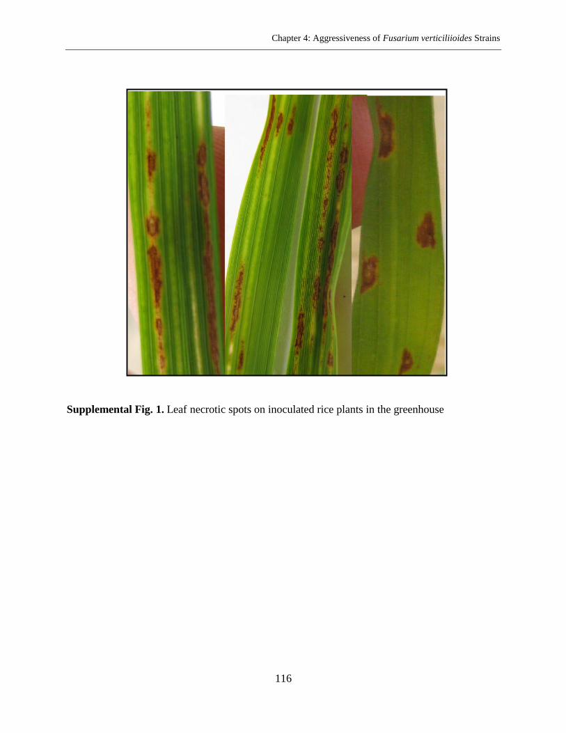
Chapter 4: Aggressiveness of Fusarium verticiliioides Strains
116
Supplemental Fig. 1. Leaf necrotic spots on inoculated rice plants in the greenhouse

Chapter 5: General Discussion
117
Chapter 5
General Discussion
Phytopathogenic fungi of the genus Fusarium are distributed worldwide and cause
economically serious diseases in strategic cereals (Jurado et al., 2005). Additionally, some
species are able to produce significant quantities of mycotoxins such as trichothecenes,
fumonisins, enniatins, zearalenone and beauvericin (Logrieco et al., 2002). Owing to the positive
correlation between fungal biomass and mycotoxin content (Waalwijk et al., 2004; Schnerr et al.,
2002; Yli-Mattila et al., 2008), one possible method of predicting mycotoxin contamination is
the quantification of fungal biomass in infected plant tissues. Among several quantification
methods, using the well-known real-time PCR (qPCR) technique is growing. In this study, we
developed a real-time PCR assay for quantification of the nine most important Fusarium
pathogens of maize and small-grain cereals in 384-well microplates and in a total volume of 4 µl.
The method is suitable for fast and cost-effective analysis of a large number of samples in a short
time. The qPCR assay for all nine species utilizes the same thermocycler profile and, therefore,
can be performed in the same microplate.
In spite of availability of thermocyclers with 384-well blocks, most of the published
qPCR assays are set for screening only one target DNA in each assay and the protocols still work
in a total volume of 15 to 25 µl. Similarly, the qPCR conditions were set up in our laboratory for
F. culmorum, F. graminearum, F. proliferatum, and F. verticillioides (Brandfass & Karlovsky,
2006; Nutz et al., 2011). The protocols, however, did not work well when the total volume was
decreased to 4 µl reactions. A second peak prior to specific melting point resulted in issues for
the qPCR melting curve, especially in low DNA concentrations. Modification of thermal
parameters (mostly annealing and elongation steps) and re-optimization of PCR reagents for each
Fusarium spp. enhanced the qPCR assay and the wide shape peaks disappeared.
The PCR efficiency, one of the most important qPCR parameters, is defined as the
amount of amplified template DNA during one cycle. Correct determination of this parameter is
necessary while the PCR data are used for the quantitative purposes (Rebrikov & Trofimov,
2006). In present optimized multi-species qPCR protocol, the PCR efficiency ranged from 81%
Chapter 5: General Discussion
118
(F. verticillioides) to 120% (F. equiseti). In this work, sensitivity which is the crucial factor for
interpretation of the qPCR results was defined as the lowest standard DNA that was able to be
amplified at the expected melting temperature in at least seven out of eight replicates. Based on
this definition, sensitivity trials showed a detection limit of 0.05 pg µl-1
of standard DNA for
F. proliferatum, 0.17 pg µl-1
for F. graminearum, F. poae, F. subglutinans, 0.50 pg µl-1
for
F. avenaceum, F. culmorum, F. tricinctum and 1.52 pg µl-1
for F. equiseti, F. verticillioides. The
developed multi-species qPCR assay did not show a delicate sensitivity for the last two Fusarium
species. Therefore, in order to quantify these species while low amounts of fungal biomass are
present in plant materials, it is suggested to follow the single species qPCR protocol developed in
the present research. For both species, thus, sensitivity will be improved to 0.50 pg µl-1
in a
background free of plant tissues and other contaminants. In summary, both single and
multi-species qPCR methods developed in this project are suitable for quick, cost-effective and
high-throughput quantification of the nine Fusarium spp. in plant material.
Inhibitors have been reported as a challenge for the successful detection and reliable
quantification of fungal biomass in plant tissues. Efficiency and sensitivity of qPCR assay will
be affected by the inhibitors present in DNA extracts. Developed high sensitive qPCR protocols,
therefore, maybe could not generate reliable data when a set of DNA extracts containing
considerable inhibitors are used (Gao et al., 2004; Demeke & Jenkins., 2010). Potential plant
inhibitors such as proteins, polyphenols, polysaccharides, secondary metabolites (Horne et al.,
2004), as well as components of DNA isolation buffers (Rossen et al., 1992) influence the qPCR
reaction and cause a reduction in efficiency (Gao et al., 2004). In some cases, samples with
strong matrix properties are able to completely block PCR amplification and result in a false
negative reaction. Significant PCR inhibition is demonstrated from soil and root DNA extracts
(Braid et al., 2003; Gao et al., 2004; Van de Graaf et al., 2003) in addition to DNA from
senescing plant material (Turner et al., 1998). In this investigation, the DNA extracts obtained
from different parts of maize, sorghum, rice and beetroot indicated different matrix effects.
Similarity between the PCR efficiency of unknown samples and standard reference material is a
prerequisite to determine the accurate quantity of fungal biomass (Cankar et al., 2006). For this
purpose, diluting DNA is a simple way to overcome the qPCR inhibition. A dilution factor of 25,
50 or 100 fold could decrease the threshold values (Ct) to the same level of the reference DNA

Chapter 5: General Discussion
119
standard and enhanced the qPCR efficiency. One hundred times dilution was not sufficient for
the DNA of the rice plants. Since a lower DNA concentration could decrease the PCR
sensitivity, 1 mg ml-1
bovine serum albumin (BSA) was directly added to the PCR reaction
mixture. This chemical has been successfully employed for deactivation of PCR inhibitors
(Wei et al., 2008; Plante et al., 2010).
Both developed qPCR protocols were successfully applied for detecting the nine
Fusarium spp. in weeds sampled from maize fields. These species are known as the most
common Fusarium associated with maize diseases. The qPCR analysis revealed that 201 weed
samples out of 294 were infected with at least one Fusarium species. Sometimes more than one
species of Fusarium were detected in individual sample. Weeds have been demonstrated to be
reservoirs of potential fungal pathogens. They serve as alternative hosts for several species of
Fusarium, usually when the economically important host plants are not present (Helbig &
Carroll, 1984; Jenkinson & Parry, 1994; Postic et al., 2012; Altinok, 2013). In this study, 36
weed species in the maize fields had been colonized with ten species of Fusarium. The rate of
infection was high for F. equiseti (49%) and F. avenaceum (34.7%). F. avenaceum is
well-known as a common species associated with cereals in Europe, while F. equiseti has less
frequently (Logrieco et al., 2002; Yli-Mattila, 2010; Kosiak et al., 2003). F. culmorum and
F. tricinctum indicated the same infection ratio (18%). It was followed by F. proliferatum (11%)
and F. graminearum (8%). F. poae and F. subglutinans were detected at very low frequencies
(3% and 1%, respectively). In isolation experiments, F. oxysporum and F. venenatum were also
recovered from weed samples in addition to other species.
The present study identified some of the weed species as new hosts for each Fusarium
spp. tested in the greenhouse (see Chapter 3, Table 4). The frequent screening of Fusarium on
common weeds (by qPCR or isolation on selective medium culture) increased their possible role
in the survival of fungus. The previous studies have indicated that plant residues are the main
inoculum source of Fusarium diseases (Cotten & Munkvold, 1998). Other works have also
demonstrated survival of Fusarium spp. on non-host plants such as weeds (Jenkinson & Parry,
1994; Inch & Gilbert, 2003; Postic et al., 2012; Altinok, 2013). Therefore, weeds as a harbor for
Fusarium have the potential to provide an inoculum reservoir for the infection of main crop
during the growing season. In this work, high incidence of Fusarium spp. occurred in
Chapter 5: General Discussion
120
Echinochloa crus-galli, Polygonum convolvulus and Chenopodium album which have been
reported as the main weed species from maize farms (Westerman & Gerowitt, 2012). Prevalence
of F. avenaceum and F. equiseti on almost all common weed species studied (see Chapter 3,
Table 2) may indicate that these Fusarium are more competitive species for colonization of
weeds compared to the main cereal fusaria such as F. graminearum.
In this work, weeds were symptomless carriers of Fusarium spp. No obvious disease
symptoms were observed on weeds assayed from the maize fields and also after inoculation in
the greenhouse conditions. This finding is in agreement with others (Haware & Nene, 1982;
Clark & Watson, 1983; Helbig & Carroll, 1984; Jenkinson & Parry, 1994; Postic et al., 2012;
Altinok, 2013). Akinsanmi et al. (2007) reported that the pathogenic fitness of Fusarium
pathogens was significantly reduced during passage through alternative hosts, while their fertility
on the primary host increased. Other reports show only less aggressive isolates could attack
weeds (Helbig & Carroll, 1984). The above reasons may explain the symptomless infection of
the weeds. On the other hand, Fusarium strains were recovered from surface sterilized roots and
stems of weeds in both field and artificially inoculated samples. Successful infection of weed
tissues in addition to the lack of symptoms on weeds suggested that the infection of weeds by
Fusarium spp. could be endophytic.
Common weeds were assessed for the main toxic secondary metabolites by
HPLC-MS/MS. All samples tested were negative (below the LOD of 3 ng g-1
) for contamination
of trichothecenes, zearalenone and fumonisins, even the samples containing the related
producers. As it is mentioned above, poor performance of Fusarium strains on alternative hosts
(Akinsanmi et al., 2007) may explain the lack of symptoms and mycotoxin production. Another
possibility is field conditions that did not favor mycotoxin production. In contrast, high levels of
beauvericin and enniatins were detected in the samples contaminated with F. equiseti and
F. avenaceum. Enniatins were measured most often at high levels (up to 24 mg kg-1
) in
Chenopodium album, Elymus repens and Polygonum aviculare. Concentration of beauvericin in
some weed species, such as Matricaria inodora, ranged from 0.0 to 65.6 mg kg-1
. However,
there was no correlation between mycotoxin content and fungal biomass. Contamination of
cereal grains by enniatins, resulting from F. avenaceum, and the natural occurrence of
beauvericin in maize ears has been reported (Logrieco et al., 1993; Bottalico et al., 1995; Ritieni
Chapter 5: General Discussion
121
et al., 1997; Logrieco et al., 1995; Yli-Mattila, 2010). In summary, although the main cereal
toxins were not found in the weed samples tested, the significance of toxin production in the
main host should not be disregarded.
There are limits on the use of morphological characters for identification of Fusarium
spp. These limitations increase when differentiation is within members belonging to a species
complex with highly similar morphology. In recent years, molecular techniques based mostly on
DNA sequencing of genes have supported morphological identification of Fusarium species
(Yli-Mattila et al., 2002; Harrow et al., 2010; Geiser et al., 2004; Hsuan et al., 2011). Nucleotide
sequencing of genes is performed particularly in informative parts of genome such as translation
elongation factor 1-alpha (TEF-1α). In this work, typical Fusarium structures were utilized to
identify the recovered isolates according to Leslie & Summerell (2006). Molecular information
obtained from the DNA sequence of TEF-1α and phylogenetic analysis were then employed to
confirm the morphological identification and probably distinguish closely related strains. The
isolates morphologically identified as F. equiseti, were placed in the Fusarium incarnatum-
equiseti species complex (FIESC) according to the closest match of BLAST search analysis
using the Fusarium–ID database (Geiser et al., 2004). The results show they belong to three
sub-clades of FIESC, only one of which has been called F. equiseti, and the next two sub-clades
still have no latin name (O'Donnell et al., 2009a). Fusarium oxysporum species complex (FOSC)
also comprises different sub-clades (O'Donnell et al., 2009b). Members of FOSC are ubiquitous
soil borne pathogens and have been demonstrated from maize and other cereals to be a less
common species (Logrieco et al., 2002; Kosiak et al., 2003). In this study, although closely
related species to F. avenaceum such as F. torulosum, F. flocciferum and F. accuminatum
formed different clades in the constructed UPGMA dendrogram, the reference strains of
F. arthrosporioides, however, fell in the F. avenaceum clade. These two species (F. avenaceum,
F. arthrosporioides) are morphologically very similar. They are distinguished by lack of orange
sporodochia in F. arthrosporioides after growing on SNA (Synthetic Nutrient Agar) medium in
darkness (Yli-Mattila et al., 2002). A combined DNA sequence data is necessary to be able to
distinguish these two species. The ATP Citrate Lyase (acl1) sequence together with the TEF-1α
gene could make a distinct lineage for F. arthrosporioides in the main clade of F. avenaceum
(Gräfenhan et al., 2013).
Chapter 5: General Discussion
122
F. verticillioides, as the most important maize pathogen, is able to cause a wide range of
symptoms, from asymptomatic infection to severe rotting and wilting. The fungus can colonize
maize stalks with no obvious disease symptoms (Kedera et al., 1992; Bacon & Hinton, 1996;
Munkvold et al., 1997; Logrieco et al., 2002; Srobarova et al., 2002). This species was not
detected in any of the weed samples. Furthermore, we could not recover it from different weed
samples. Therefore, the importance of weeds as a possible source of F. verticillioides inoculum
in maize fields remains still unknown. Additional studies need to be performed to evaluate if
weed plants can serve as an alternative host for F. verticillioides in maize fields. Further weed
sampling from maize fields of Italy and southern belt of Germany, where the fungus is a serious
problem, has been carried out and the experiments are running in our group. We hope the results
help us to find out if weed plants harbor F. verticillioides in maize fields and whether they can
increase the infection pressure locally. The answer may also be helpful for finding the biological
role of fumonisins in the growth of fungus in the host plants other than maize. We believe the
fumonisin synthesis has been maintained by natural selection on weeds (or hosts other than
maize) and in plant tissues other than silk/cobs (such as roots) for millions of years.
Fumonisins are polyketide mycotoxins produced by several species of Fusarium among
which F. verticillioides is well-known as a main producer (Munkvold & Desjardins, 1997). The
biological role of fumonisins in virulence of F. verticillioides has been an eristic issue. In this
study, two fumonisin non-producing strains (fum 1-3 and fum 1-4) and their progenitor
(fumonisin-producing) FUM 1-1 strains were employed to determine the potential role of toxins
in the growth of the fungus in maize, grain sorghum, rice and beetroot seedlings. The results of
this study confirmed the previous reports (Kedera et al., 1992; Bacon & Hinton, 1996; Munkvold
et al., 1997) that F. verticillioides can infect maize plants without causing symptoms. Systemic
movement of fungus within the root inoculated plants was detected by qPCR. Transmission of
the fungus from roots to the first two internodes in maize plants occurred regardless of toxin
production. Our experiments show that the infection through the roots has been an effective
pathway for developing fungus within the plant. However, the rate of symptomless systemic
colonization decreased after the second internode. This restricted movement of fungus may limit
the infection of kernels. Williams & Munkvold (2008) indicated that the systemic development
of F. verticillioides could be increased in high temperatures. Movement of the fungus appeared
Chapter 5: General Discussion
123
to be limited in sorghum plants. Only in 20-30% of inoculated plants with the M-3125 strain or
its mutant, GFA2364, the fungus was detectable in the first two internodes. Host preference of
the biological species of Gibberella fujikuroi has been demonstrated (Leslie et al., 1992) and it
could explain the lack of movement of fungus in sorghum plants. Another possibility would be
the partial resistance of the sorghum variety which was utilized in this study. The relative
distribution of the fungus in rice and beetroot seedlings was similar to maize plants. The
quantified fugal biomass was considered as a marker to show the aggressiveness of the fungus.
No infection was detected in control plants inoculated with sterile water. There was no
significant difference (P = 0.05) between fumonisin-producing strains and fumonisin
non-producing strains in colonization of plant tissues. The high level of fungal biomass was
detected in roots and also in the first two internodes of the maize stalks; but it was independent
of fumonisins production. Based on the results in our system, fumonisins have no role in fungal
growth of F. verticillioides in maize, rice and beetroot seedlings and the aggressiveness of
fungus was independent of toxin production.
Chapter 5: General Discussion
124
Literature Cited
Akinsanmi, O. A., Chakraborty, S., Backhouse, D., and Simpfendorfer, S. 2007. Passage
through alternative hosts changes the fitness of Fusarium graminearum and Fusarium
pseudograminearum. Environ. Microbiol. 9:512-520.
Altinok, H. H. 2013. Fusarium species isolated from common weeds in eggplant fields
and symptomless hosts of Fusarium oxysporum f. sp. melongenae in Turkey. J.
Phytopathol. 161:335-340.
Bacon, C. W., and Hinton, D. M. 1996. Symptomless endophytic colonization of maize
by Fusarium moniliforme. Can. J. Bot. 74:1195-1202.
Bottalico, A., Logrieco, A., Ritieni, A., Moretti, A., Randazzo, G., and Corda, P. 1995.
Beauvericin and fumonisin B1 in preharvest Fusarium moniliforme maize ear rot in
Sardinia. Food Addit. Contam. 12:599-607.
Braid, M. D., Daniels, M. L., and Kitts, C. L. 2003. Removal of PCR inhibitors from soil
DNA from chemical flocculation. J. Microbiol. Meth. 52:389-393.
Brandfass, C., and Karlovsky, P. 2006. Simultaneous detection of Fusarium culmorum
and F. graminearum in plant material by duplex PCR with melting curve analysis. BMC
Microbiol. 6 (4). Available from: http://www.biomedcentral.com/1471-2180/6/4 : URL.
DOI.
Cankar, K., Stebih, D., Dreo, T., Zel, J., and Gruden, K. 2006. Critical points of DNA
quantification by real-time PCR effects of DNA extraction method and sample matrix on
quantification of genetically modified organisms. BMC. Biotech. 6:37-51.
Clark, C. A., and Watson, B. 1983. Susceptibility of weed species of Convolvulaceae to
root-infecting pathogens of sweet potato. Plant Dis. 67:907-909.
Cotten, T. K., and Munkvold, G. p. 1998. Survival of Fusarium moniliforme,
F. proliferatum and F. subglutinans in maize stalk residue. Phytopathology 88:550-555.
Demeke, T., and Jenkins, G. R. 2010. Influence of DNA extraction methods, PCR
inhibitors and quantification methods on real-time PCR assay of biotechnology-derived
traits. Anal. Bioanal. Chem. 396:1977-1990.
Gao, X., Jackson, T. A., Lambert, K. N., Li, S., Hartman, G. L., and Niblack, T. L. 2004.
Detection and quantification of Fusarium solani f.sp. glycines in soybean roots with
real-time quantitative polymerase chain reaction. Plant Dis. 88:1372-1380.
Chapter 5: General Discussion
125
Geiser, D. M., Jime´nez-Gasco, M. D. M., Kang, S., Makalowska, I., Veeraraghavan, N.,
Ward, T. J., Zhang, N., Kuldau, G. A., and O'Donnell, K. 2004. FUSARIUM-ID v. 1.0: A
DNA sequence database for identifying Fusarium. Eur. J. Plant Pathol. 110:473-479.
Gräfenhan, T., Patrick, S. K., Roscoe, M., Trelka, R., Gaba, D., Chan, J. M., McKendry,
T., Clear, M., and Tittlemier, S. A. 2013. Fusarium damage in cereal grains from western
Canada. Phylogenetic analysis of moniliformin-producing Fusarium species and their
natural occurrence in mycotoxin- contaminated. J. Agric. Food Chem. 61:5425-5437.
Harrow, S. A., Farrokhi-Nejad, R., Pitman, A. R., Scott, I. A. W., Bentley, A., Hide, C.,
and Cromey, M. G. 2010. Characterization of New Zealand Fusarium populations using a
polyphasic approach differentiates the Fusarium avenaceum/ F. acuminatum/
F. tricinctum species complex in cereal and grassland systems. Fungal Biol. 114:293-311.
Haware, M. P., and Nene, Y. L. 1982. Symptomless carriers of the chickpea wilt
Fusarium. Plant Dis. 66:250-251.
Helbig, J. B., and Carroll, A. B. 1984. Dicotyledonous weeds as a source of Fusarium
oxysporum pathogenic on soybean. Plant Dis. 68:694-696.
Horne, E. C., Kumpatla, S. P., Ptterson, K. A., Gupta, M., and Thompson, S. A.
Improved high-throughput sunflower and cotton genomic DNA extraction and PCR
fidelity. Plant Mol. Biol, Rep. 22:83a-83i.
Hsuan, H. M., Salleh, B., and Zakaria, L. 2011. Molecular identification of Fusarium
species in Gibberella fujikuroi species complex from rice, sugarcane and maize from
Peninsular Malaysia. Int. J. Mol. Sci. 12:6722-6732.
Inch, S., and Gilbert, J. 2003. The incidence of Fusarium species recovered from
inflorescences of wild grasses in southern Manitoba. Can. J. Plant Pathol. 25:379-383.
Jenkinson, P., and Parry, D. W. 1994. Isolation of Fusarium species from common
broad-leaved weeds and their pathogenicity to winter wheat. Mycol. Res. 98:776-780.
Jurado, M., Vazquez, C., Patino, B., and González-Jaén, M. T. 2005. PCR detection
assays for the trichothecene-producing species Fusarium graminearum, Fusarium
culmorum, Fusarium poae, Fusarium equiseti and Fusarium sporotrichioides. Syst. Appl.
Microbiol. 28:562-568.
Kedera, C. J., Leslie, J. F., and Claflin, L. E. 1992. Systemic infection of corn by
Fusarium moniliforme. (Abstr.) Phytopathology 82:1138.
Chapter 5: General Discussion
126
Kosiak, B., Torp, M., Skjerve, E., and Thrane, U. 2003. The prevalence and distribution
of Fusarium species in Norwegian cereals: a survey. Acta Agr. Scand. (Section B- Soil
and Plant Science) 53:168-176.
Leslie, J. F., and Summerell, B. A. 2006. The Fusarium Laboratory Manual. Blackwell
Publishing. 388 pp.
Leslie, J. F., Plattner, R. D., Desjardins, A. E., and Klittich, C. J. R. 1992. Fumonisin B1
production by strains from different mating populations of Gibberella fujikuroi
(Fusarium section Liseola). Phytopathology (Abstr.) 82: 34145.
Logrieco, A., Mule, G., Moretti, A., and Bottalico, A. 2002. Toxigenic Fusarium species
and mycotoxins associated with maize ear rot in Europe. Eur. J. Plant Pathol. 108:
597-609.
Logrieco, A., Moretti, A., Ritieni, A., Bottalico, A., and Corda, P. 1995. Occurrence and
toxigenicity of Fusarium proliferatum from preharvest maize ear rot, and associated
mycotoxins, in Italy. Plant Dis. 79:727-731.
Logrieco, A., Moretti, A., Ritieni, A., Chelkowski, J., Altomare, C., Bottalico, A., and
Randazzos, G. 1993. Natural occurrence of beauvericin in preharvest Fusarium
subglutinans infected corn ears in Poland. J. Agric. Food. Chem. 41:2149-2152.
Munkvold, G. P., and Desjardins, A. E. 1997. Fumonisins in maize: can we reduce their
occurrence. Plant Dis. 81:556-565.
Munkvold, G. P., McGee, D. C., and Carlton, W. M. 1997. Importance of different
pathways for maize kernel infection by Fusarium moniliforme. Phytopathology 87:
209-217.
Nutz, S., Döll, K., and Karlovsky, P. 2011. Determination of the LOQ in real-time PCR
by receiver operating characteristic curve analysis: application to qPCR assays for
Fusarium verticillioides and F. proliferatum. Anal. Bioanal. Chem. 401:717-726.
O'Donnell, K., Sutton, D. A., Crous, P. W., Rinaldi, M. G., and Geiser, D. M. 2009a.
Novel multilocus sequence typing scheme reveals high genetic diversity of human
pathogenic members of the Fusarium incarnatum-F. equiseti and F. chlamydosporum
species complexes within the united states. J. Clin. Microbiol. 47:3851-3861.
O'Donnell, K., Gueidan, C., Sink, S., Johnston, P. R., Crous, P. W., Glenn, A., Riley, R.,
Zitomer, N. C., Colyer, P., Waalwijk, C., Lee, T., Moretti, A., Kang, S., Kim, H. S.,
Geiser, D. M., Juba, J. H., Baayen, R. P., Cromey, M. G., Bithell, S., Sutton, D. A.,
Chapter 5: General Discussion
127
Skovgaard, K., loetz, R., Corby Kistler, H., Elliott, M., Davis, M., and Sarver, B. A.
2009b. A two-locus DNA sequence database for typing plant and human pathogens
within the Fusarium oxysporum species complex. Fungal Genet. Biol. 46:936-948.
Plante, D., Belanger, G., Leblanc, D., Ward, P., Houde, A., and Trottier, Y. L. 2010. The
use of bovine serum albumin to improve the RT-qPCR detection of foodborne viruses
rinsed from vegetable surfaces. Letters in Applied Microbiology 52:239-244.
Postic, J., Cosic, J., Vrandecic, K., Jurkovic, D., Saleh, A. A., and Leslie, J. F. 2012.
Diversity of Fusarium species isolated from weeds and plant debris in Croatia. J.
Phytopathol. 160:76-81.
Rebrikov, D. V., and Yu. Trofimov, D. 2006. Real-time PCR: a review of approaches to
data analysis. Appl. Biochem. Microbiol. 42:520-528.
Ritieni, A., Moretti, A., Logrieco, A., Bottalico, A., Randazzo, G., Monti, S. M.,
Ferracane, R., and Fogliano, V. 1997. Occurrence of fusaproliferin, fumonisin B1, and
beauvericin in maize from Italy. J. Agric. Food. Chem. 45:4011-4016.
Rossen, L., Norskov, P., Holmstrom, K., and Rasmussen, F. 1992. Inhibition of PCR by
components of food samples, microbial diagnostic assays and DNA-extraction solutions.
Int. J. Food Microbiol. 17:37-45.
Schnerr, H., Vogel, R. F., and Niessen, L. 2002. Correlation between DNA of
trichothecene-producing Fusarium species and deoxynivalenol concentrations in
wheat-samples. Lett. Appl. Microbiol. 35:121-125.
Srobarova, A., Moretti, A., Ferracane, R., Ritieni, A., and Logrieco, A. 2002. Toxigenic
Fusarium species of Liseola section in preharvest maize ear rot, and associated
mycotoxins in Slovakia. Eur. J. Plant Pathol. 108:299-306.
Turner, A. S., Lees, A. K., Rezanoor, H. N., and Nicholson, P. 1998. Refinement of
PCR-detection of Fusarium avenaceum and evidence from DNA marker studies for
phonetic relatedness to Fusarium tricinctum. Plant Pathol. 47:278-288.
Van de Graaf, P., Lees, A. K., Cullen, D. W., and Duncan, J. M. 2003. Detection and
quantification of Spongospora subterranea in soil, water and plant tissue samples using
real-time PCR. Eur. J. Plant Pathol. 109:589-597.
Waalwijk, C., van der Heide, R., de Vries, I., van der Lee, T., Schoen, C., Costrel-de
Corainville, G., Häuser-Hahn, I., Kastelein, P., Köhl, J., Lonnet, P., Demarquet, T., and
Chapter 5: General Discussion
128
Kema, G. H. J. 2004. Quantitative detection of Fusarium species in wheat using TaqMan.
Eur. J. Plant Pathol. 110:481-494.
Wei, T., Lu, G., and Clover, G. 2008. Novel approaches to mitigate primer interaction
and eliminate inhibitors in multiplex PCR, demonstrated using an assay for detection of
three strawberry viruses. J. Virol. Methods. 151:132-139.
Westerman, P. R., and Gerowitt, B. 2012. Probability of maize biomass contamination
with weed seeds. J. Plant Dis. Protect. 11:68-73.
Williams, L. D., and Munkvold, G. P. 2008. Systemic infection by Fusarium
verticillioides in maize plants grown under three temperature regimes. Plant Dis.
92:1695-1700.
Yli-Mattila, T. 2010. Ecology and evolution of toxigenic Fusarium species in cereals in
northern Europe and Asia. J. Plant Pathol. 92:7-18.
Yli-Mattila, T., Paavanen-Huhtala, S., Jestoi, M., Parikka, P., Hietaniemi, V., Gagkaeva,
T., Sarlin, T., Haikara, A., Laaksonen, S., and Rizzo, A. 2008. Real-time PCR detection
and quantification of Fusarium poae, F. graminearum, F. sporotrichioides and
F. langsethiae in cereal grains in Finland and Russia. Archives of Phytopathology and
Plant Protection 41:243–260.
Yli-Mattila, T., Paavanen-Huhtala, S., Bulat, S. A., Alekhina, I. A., and Nirenberg, H. I.
2002. Molecular, morphological and phylogenetic analysis of the Fusarium avenaceum/
F. arthrosporioides/ F. tricinctum species complex- a polyphasic approach. Mycol. Res.
106:655-669.
Summary
129
Summary
Fusarium is a large and complex genus, comprises important pathogens most of which
are able to produce mycotoxins. Maize is a host of several Fusarium species that cause ear rot,
kernel rot, stalk rot and also seedling blight. Fusarium contamination of kernels and maize
products had always been a serious concern. Owing to the positive correlation between fungal
biomass and mycotoxin content, quantification of fungal DNA in plant materials would be
considered as an initial fast and cost-effective mean to evaluate the risk of grain contamination.
Several quantification methods are well-known of which real-time PCR (qPCR) has been used as
an effective tool for species-specific quantification of fungal biomass in plant tissues. The
method mostly relies on standard thermocyclers (96-well or 384-well blocks) with a separate run
for each template and usually set in total reaction volume of 15 to 25 µl. In this investigation, we
developed a multi-species qPCR assay for simultaneous quantification of genomic DNA of the
nine Fusarium species with 384-well microplates in a total volume of 4 µl. The sensitivity of the
method ranged from 0.05 - 1.52 pg DNA per reaction, and the repeatability ranged from 0.81%
to 1.71% RSD.
Developed low volume qPCR assay was successfully employed for the analysis of weed
plants to the infection of nine Fusarium spp. The main objectives were determination the role of
weeds in the survival of maize fusarium pathogens and assessment of their ability for producing
of the main mycotoxins. The Real-time PCR detected eight Fusarium species in 201 weed
samples representing 36 weed species, collected from maize fields. The highest frequency was
observed for F. equiseti (49%) and F. avenaceum (34.7%). Similar diversity of Fusarium spp.
was observed in both conventional and organic farming systems. Isolation of Fusarium strains
from 12 common weed species were carried out, the strains were identified based on the
morphological characters and then identification was confirmed by using the translation
elongation factor 1-alpha (TEF-1α) gene sequence. The recovery rate was high for F. equiseti
(32.7%) and F. avenaceum (21%). None of the field samples as well as weed plants tested in
inoculation studies show obvious symptoms of Fusarium infection. Re-isolation of the strains
from artificially inoculated plants confirmed the endophytic infection of weeds by Fusarium spp.
The present study reports five new alternative hosts for Fusarium species in maize fields. High
Summary
130
incidence rate of beauvericin and enniatins contamination was obtained in weed samples while
trichothecenes, fumonisins and zearalenone were not detected in any of the weeds studied.
Although toxicity of fumonisins to plants and field animals has been clearly
demonstrated, the function of this toxin, however, in virulence of F. verticillioides toward maize
plants is still unknown. In present study, virulence of two non-fumonisins producing strains
(fum 1-3 and fum 1-4) and their progenitors (FUM 1-1) was assessed on different plants
including maize, sorghum, rice and beetroot seedlings grown under greenhouse conditions. The
quantity of fungal biomass in plant tissues was considered as an indicator of fungal
aggressiveness and it was measured by the developed low volume qPCR protocol. There was no
significant (P = 0.05) differences between each wild type and the relevant mutant for
colonization of plant tissues. In inoculated maize, rice and beetroot seedlings, systemic fungal
infection was observed from roots to the aboveground parts; but rate of systemic transmission
was low in sorghum plants. Although our results are not enough to make a final conclusion of
fumonisin function in virulence of F. verticillioides on rice and sorghum seedlings, however, the
results show diseases incidence was independent of fumonisins production in maize seedlings. In
our system, therefore, fumonisins had no role as a pathogenicity factor.
Acknowledgements
131
Acknowledgements
I would like to deeply thank my great supervisor Prof. Dr. Petr Karlovsky for giving me the
opportunity to complete this work in his group. My sincere gratitude is for his scientific guidance and
also for giving me the chance to gain extraordinary experiences by working on side projects along
my study. Working in his group enriched my life experiences, as well.
I am very grateful to have Dr. Horst-Henning Steinmann and Prof. Dr. Andreas von
Tiedemann as my Ph. D. committee members.
Many friendly people of Karlovsky lab helped me scientifically or morally to be able to do
this work. I am indebted to Heike Rollwage, Ruth Pilot and Patricia Bartoschek for their advices and
technical support. They always were beside me and shared good moments with me. Special gratitude
to Dr. Katharina Döll for her friendship, constant support, numerous scientific advices and positive
thinking even during difficult moments. I am extremely grateful to Andrea Breitkopf, Jacqueline
Moser and Daniel Kretzschmar for technical assistance. It was a great honour to be with all of you
during my Ph. D. study; and Special thanks to Yi Kuang, Somayyeh Sedaghatjoo, Nahid Shafiei, my
friends, and all Ph. D. students who made my moments more enjoyable in Germany.
I would like to get this opportunity to say thank to all the administrative staff of molecular
phytopathology and mycotoxin research department for a good working atmosphere, especially to
Mrs. Evelin Kistner, Mr Thomas Oesterreich and Mr Siegfried Opolka. They kindly accompanied me
on this research project.
So many thanks to Seed and Plant Improvement Research Institute (SPII) authorities, in Iran,
especially Dr. R. Chokan and Dr. D. Hassani that authorized me for the highest level of my
education.
I want to express my deepest thank to Dr. Seyed Mostafa Pirseyedi, my colleague, for his
fruitfull scientific discussions and valuable suggestions.
I owe my gratitude to all my friends in Iran, especially Mrs. Sima Damyar for
encouragements and great emotional support during my Ph. D. study.

Acknowledgements
132
Ultimately, love and thanks to my parents who sacrificed everything to give me the most and
best education possible. They taught me to stand up against the problems and compromise with life.
Now, I am honoered to show them the beautiful world where that they have let me be a part of. I am
grateful to my lovely sister, Roya, who always supported me to follow my dreams; and many thanks
to my brothers, Hamid, Majid and Vahid. I am proud of you. During the last three years, I had very
nice and happy moments through “ooVoo” with my cute nephew, Ali, and my sweet niece, Baran,
who were new members of our family when I started my Ph. D. Hope, someday, when they read their
names in my dissertation be reassured of my love to themselves.
You, all, made my dreams come true. Thanks !, and Here, I am excited for the future…
Raana Dastjerdi
19 March, 2014, Göttingen, Germany
Curriculum Vitae
133
Curriculum Vitae
Personal information
Name: Raana Dastjerdi
Born: Ghazvin, Iran
E-mail [email protected]
Academic Education
From 03/2011 International Ph. D. Program for Agricultural Sciences,
Molecular Phytopathology and Mycotoxin Research Unit,
Division of Plant Pathology and Plant Protection, Georg-
August-University Göttingen, Germany
09/1999-02/2002 Master Program in Agricultural Sciences, Field: Plant
Pathology, Ferdowsi University of Mashhad, Iran. Master
thesis: “Investigation on genetic diversity in population of
Fusarium oxysporum from sugar beet in Khorasan using
vegetative compatibility groups (VCG) and RAPD molecular
marker”
09/1993-07/1997 Bachelor Program in Agricultural Engineering, Field: Plant
Protection, Ferdowsi University of Mashhad, Iran. Bachelor
thesis: “Biological control and applying of PGPR in plant
diseases management”
Work Experience
Since 05/2002 Faculty member and research leader of Horticulture Crops
Research Department, Seed and Plant Improvement Research
Institute (SPII), Tehran-Karaj, Iran
Publications
134
Publications
Publications During the Ph. D. Study
Dastjerdi, R., Döll, K., Gerowitt, B., Steinmann, H. H., and Karlovsky, P. 2013.
Fusarium species and mycotoxins in weed plants. 35th
Mycotoxin Workshop. Ghent,
Beljium.
Thirumalai, A. E., Dastjerdi, R., Döll, K., Venkatachalam, A., Karlovsky, P., and
Suryannarayanan, T. S. 2013. Mycotoxins of endophytic Fusarium mangiferae and
F. pallidoroseum from betel leaves (Piper betle L.). 35th
Mycotoxin Workshop. Ghent,
Beljium.
Other Publications
Journal Publications
Damyar, S., Hassani, D., Dastjerdi, R., Hajnajari, H., Zeinanloo, A. A., and Fallahi, E.
2007. Evaluation of Iranian native apple cultivars and genotypes. Journal of Food,
Agriculture and Environment 5(3&4):207-211.
Dastjerdi, R., and Damyar, S. 2010. Evaluation of relative resistance in eleven apple
rootstocks to crown rot caused by Phytophthora cactorum. Seed and Plant Improvement
Journal 26(3): 297-311.
Dastjerdi, R., Falahati-Rastegar, M., and Jafarpour, B. 2003. Identification of Fusarium
species associated with sugar beet root in Khorasan province and investigation of the
pathogenicity of Fusarium oxysporum. Iranian Journal of Sugar Beet 18 (2):143-154.
Dastjerdi, R., and Hassani, D. 2009. Response of 11 walnut cultivars and genotypes to
Gnomonia leptostyla. Seed and Plant Improvement Journal 25(3):433-449.
Dastjerdi, R., Hassani, D., and Javan-Nikkhah, M. 2009. Study on some characteristics,
assessment of Pathogenicity and diversity in Gnomonia leptostyla isolates casual agent of
walnut anthracnose in Iran. Iranian Journal of Plant Pathology 45(1):61-73.
Dastjerdi, R., Mozafari, J., Falahati-Rastegar, M., and Jafarpour, B. 2006. Population
diversity of Fusarium oxysporum associated with sugar beet in Khorasan based on
vegetative compatibility groups (VCGs). Agricultural Sciences and Technology Journal
(Ferdowsi University of Mashhad, Iran) 20(2):78-91.
Dastjerdi, R., Mozafari, J., Falahati-Rastegar, M., and Jafarpour, B. 2004. RAPD
Analysis of genetic diversity within and among vegetative compatibility groups (VCGs)
of Fusarium oxysporum f. sp. betae in sugar beet fields of Khorasan, Iran. Applied
Entomology and Phytopathology 72(1):1-19.
Publications
135
Hassani, D., Atefi, J., Haghjooyan, R., Dastjerdi, R., Keshavarzi, M., Mozaffari, M. R.,
Soleimani, A., Rahmanian, A. R., Nematzadeh, F., and Malmir. A. 2012. Jamal, a new
persian walnut cultivar for moderate-cold area of Iran. Seed and Plant Improvement
Journal 28(3):525-527.
Hassani, D., Atefi, J., Haghjooyan, R., Dastjerdi, R., Keshavarzi, M., Mozaffari, M. R.,
Soleimani, A., Rahmanian, A. R., Nematzadeh, F., and Malmir. A. 2012. Damavand, a
new persian walnut cultivar as a pollinizer for Iranian walnut cultivars and genotypes.
Seed and Plant Improvement Journal 28(3):529-531.
Presentations and Posters
Damyar, S., Dastjerdi, R., and Hassani, D. 2009. Determination of the best harvesting
time for Braeburn, Gala and Fuji apple cultivars in Iran. 6th
International Postharvest
Symposium. Antalya, Turkey. p: 227.
Damyar, S., Keshavarzi, M., Hassani, D., Dastjerdi, R., and samie, N. 2007. Non
chemichal control of superfacial scald in apple cultivar Red Delicious (Full Text).
International Scientific Conference. Minsk, Belarus. 128-131.
Dastjerdi, R., Damyar, S., and Farhadnejad, A. 2008. Evaluation of apple vegetative
rootstocks for resistance to Phytophthora cactorum. 18th
Iranian Plant Protection
Congress. Hamedan, Iran.
Dastjerdi, R., and Hassani, D. 2010. Study of walnut anthracnose and determination of
pathogenicity diversity in Gnomonia leptostyla isolates in greenhouse. 19th
Iranian Plant
Protection Congress. Tehran, Iran.
Dastjerdi, R., and Hassani, D. 2010. Survy on reaction of thirteen walnut cultivars and
genotypes to black spot in greenhouse. 19th
Iranian Plant Protection Congress. Tehran,
Iran.
Dastjerdi, R., Hassani, D., Javan-Nikkhah, M., Farhadnejad, A., and Salahi, S. 2008.
Evaluation of some walnut cultivars and genotypes for relative resistance to anthracnose.
18th
Iranian Plant Protection Congress. Hamedan, Iran.
Salahi, S., Javan-Nikkhah, M., Zad, J., Hassani, D., Dastjerdi, R., and Jamshidi, S. 2006.
Geographical distribution and determining of some traits of Gnomonia leptostyla isolates
on walnut trees in East Azarbaijan. 17th
Iranian Plant Protection Congress. Tehran, Iran.
Saman, S.M., Mozafari, J., Abasi moghadam, A., Vaezi, S., and Dastjerdi, R. 2010.
Evaluation for resistance to Fusarium oxysporum f. sp. lentil in the lentil collection of
National Plant Gene-Bank of Iran. 19th
Iranian Plant Protection Congress. Tehran, Iran.