Embed Size (px)
Citation preview

BIO 318 MODULE 4
79
NATIONAL OPEN UNIVERSITY OF NIGERIA
SCHOOL OF SCIENCE AND TECHNOLOGY
COURSE CODE: BIO 318
COURSE TITLE: IMMUNOLOGY AND IMMUNOCHEMISTRY III

BIO 318 IMMUNOLOGY AND IMMUNOCHEMISTRY III
80
BIO 318
IMMUNOLOGY AND IMMUNOCHEMISTRY III
Course Team Prof. J. Okpuzor (Course Developer/Writer) -
UNILAG
Dr. T. Taiwo (Course Editor) - UNILAG
Mr. Adams Abiodun E. (Course Coordinator) -
NOUN
Prof. A. Adebanjo (Programme Leader) - NOUN
NATIONAL OPEN UNIVERSITY OF NIGERIA
COURSE
GUIDE

BIO 318 MODULE 4
81
National Open University of Nigeria
Headquarters
14/16 Ahmadu Bello Way
Victoria Island, Lagos
Abuja Office
5 Dar es Salaam Street
Off Aminu Kano Crescent
Wuse II, Abuja
e-mail: [email protected]
URL: www.nou.edu.ng
Published by
National Open University of Nigeria
Printed 2013
ISBN: 978-058-988-0
All Rights Reserved
BIO 318 IMMUNOLOGY AND IMMUNOCHEMISTRY III
82
CONTENTS PAGE
Introduction............................................................................. iv
What you will Learn from this Course................................... iv
Course Aims............................................................................ iv
Course Objectives.................................................................... iv
Working through this Course.................................................. v
Course Materials...................................................................... v
Study Units.............................................................................. v
Textbooks and References....................................................... vi
Assessment............................................................................... vii
Self-Assessment Exercise........................................................ vii
Tutor-Marked Assignment....................................................... vii
Final Examination and Grading............................................... vii
Summary.................................................................................. vii

BIO 318 MODULE 4
83
INTRODUCTION
BIO 318: Immunology and Immunochemistry is a second semester, 3
credit units’ course in Biology. It is a 300 level, second semester
Biology course offered to students admitted in the School of Science
and Technology
The course guide tells you briefly what the course is all about, what
course materials you will be using and how you can work your way
through these materials. It gives you some guidance on your Tutor-
Marked Assignments.
There are Self-Assessment Exercise(s) within the body of a unit and/or
at the end of each unit. The exercise(s) are an overview of the unit to
help you assess yourself at the end of every unit.
WHAT YOU WILL LEARN FROM THIS COURSE
This course contains twenty units which cover general topics in
Immunology, Immunochemistry and related topics.
Principles of immune response and how it works, autoimmunity,
autopathology, immune response to tissue and organ transplant,
chemotherapy and recent techniques utilised in immunology will be
discussed.
COURSE AIMS
The aim of this course is to educate and introduce the students to
Immunology, Immunochemistry and related topics.
COURSE OBJECTIVES
In addition to the aim of this course, the course sets an overall objective
which must be achieved. In addition to the course objectives, each of the
units has its own specific objectives. You are advised to read properly
the specific objectives for each unit at the beginning of that unit. This
will help you to ensure that you achieve the set objectives. As you go
through each unit, you should from time to time go back to these
objectives to ascertain the level at which you have progressed.
At the end of this course, you should be able to:
discuss immune response in humans
explain antigen-antibody interactions
describe the causes of autoimmunity

BIO 318 IMMUNOLOGY AND IMMUNOCHEMISTRY III
84
define the concept of chemotherapy
discuss tissue and organ transplant tolerance in recipients
examine the modes of action of antimicrobials.
WORKING THROUGH THIS COURSE
In this course, you are advised to devote your time in reading through
the course materials. You would be required to do all that has been
stipulated in the course: study the course units, read the recommended
reference textbooks and do all the unit(s) self-assessment exercise(s) and
at some point, you are required to submit your assignment (TMAs) for
assessment purpose. You should therefore avail yourself of the
opportunity of being present during the tutorial sessions so that you
would be able to compare knowledge with your colleagues.
COURSE MATERIALS
You are to be provided with the two major course materials. These are:
Course Guide
Study Units
The course comes with a list of recommended textbooks. These
textbooks are to complement the course materials so that you can avail
yourself of reading further. Therefore, it is advisable you source for
some of these textbooks and read them to broaden your scope of
understanding.
STUDY UNITS
This course is divided into 4 modules with a total of 20 units, divided as
follows;
Module 1
Unit 1 Basic Concepts in Immunology
Unit 2 Structure of Antigenic Determinants
Unit 3 Cellular Response
Unit 4 Genetics of Response to Antigenic Stimulation
Unit 5 Structure and Classification of Immunoglobulins

BIO 318 MODULE 4
85
Module 2
Unit 1 Mechanism of Antibody Formation
Unit 2 Antigen/Antibody Interaction: Role of Lymphoid Tissues
and Thymus in Immune Response
Unit 3 Hypersensitivity
Unit 4 Immunopathology
Unit 5 Auto-Pathology and Auto-Immunology
Module 3
Unit 1 Tissue and Transplantation Immunology
Unit 2 Immunoprophylaxis
Unit 3 Modern Techniques in Immunology and
Immunochemistry
Unit 4 History of Chemotherapy
Unit 5 Principles of Chemotherapy
Module 4
Unit 1 Chemotherapeutic Agents
Unit 2 Modes of Action of Antimicrobials
Unit 3 Chemotherapy of Specific Diseases
Unit 4 Pharmaco-Dynamics and Pharmaco-Kinetics
Unit 5 Drug Bioassay and Sensitivity Tests
TEXT BOOKS AND REFERENCES
King, P. & Perry, M. (2001). “Hepatotoxicity of Chemotherapy”.
Oncologist 6:162–176.
Therasse, P. et al.(2000). “New Guidelines to Evaluate the Response to
Treatment of Solid Tumors”. J Natl Cancer Inst 92:205–216.
David, E. G. et al. (2005). Principles of Pharmacology. (1st ed.).
Lippincott: Williams & Wilkins.
Charles, A.J. et al. (1999). Immunobiology: The Immune System in
Health and Disease. Elsevier Science Ltd/ Garland Publishing.
Baron, S. (1996). Medical Microbiology.( 4th
ed.).
Murray, B. (1991). “New Aspects of Antimicrobial Resistance and the
resulting Therapeutic dilemmas”. J Infect Dis.163:1185.

BIO 318 IMMUNOLOGY AND IMMUNOCHEMISTRY III
86
Schaberg, D. (1994). “Resistant gram-positive organisms”. Ann
Emergency Med. 24(3):462
Julio,A.U. & Roberto, D. (2003). “Specific chemotherapy of Chagas
disease: controversies and advances”. Trends in Parasitology.
.19:495-501.
Martin, W. et al. (2005). "Bleomycin pulmonary toxicity has a negative
impact on the outcome of patients with Hodgkin's lymphoma". J
Clin Oncol 23 (30): 7614–20.
Canellos, G. et al. (2004). "How important is bleomycin in the
adriamycin + bleomycin + vinblastine + dacarbazine regimen?". J
Clin Oncol 22 (8): 1532–3.
ASSESSMENT
There are two components of the assessment for this course:
The Self-Assessment Exercise
The Tutor-Marked Assignment (TMAs)
The End of Course Examination
SELF-ASSESSMENT EXERCISE
The exercise within each unit is/are meant to probe your understanding
of the concepts in the unit. It is non-grading and as such does not add up
to your grade in the course.
TUTOR-MARKED ASSIGNMENT (TMA)
The TMA is the continuous assessment component of your course. It
accounts for 30 percent of the total score you will obtain in this course.
FINAL EXAMINATION AND GRADING
The course is to be concluded by the final examination. The final
examination constitutes 70 percent of the whole course. You will be
adequately informed of the time of the examination. The examination
will consist of questions which reflect all the basic concepts you would
have learnt through the duration of the course.

BIO 318 MODULE 4
87
SUMMARY
This is intended for you to have an underlying knowledge of the
principles of Immunology & Immunochemistry and other related topics.
By the time you complete this course, you should be able to answer
conveniently questions on the following:
immune response in humans
antigen-antibody interactions
the causes of autoimmunity
the concept of chemotherapy
tissue and organ transplant tolerance in recipients
the modes of action of antimicrobials.
We wish you all the best in your study of this course.
Best wishes.
BIO 318 IMMUNOLOGY AND IMMUNOCHEMISTRY III
88
CONTENTS PAGE
Module 1 ................................................................................... 1
Unit 1 Basic Concepts in Immunology............................... 1
Unit 2 Structure of Antigenic Determinants................... .... 7
Unit 3 Cellular Response..................................................... 11
Unit 4 Genetics of Response to Antigenic Stimulation..... 16
Unit 5 Structure and Classification of Immunoglobulins 24
Module 2 .................................................................................. 32
Unit 1 Mechanism of Antibody Formation........................ 32
Unit 2 Antigen/Antibody Interaction: Role of Lymphoid
Tissues and Thymus in Immune Response............. 37
Unit 3 Hypersensitivity........................................................ 42
Unit 4 Immunopathology..................................................... 47
Unit 5 Auto-Pathology and Auto-Immunology................. 52
Module 3 ................................................................................. 57
Unit 1 Tissue and Transplantation Immunology............... 57
Unit 2 Immunoprophylaxis.................................................. 64
Unit 3 Modern Techniques in Immunology and
Immunochemistry.................................................... 69
Unit 4 History of Chemotherapy......................................... 73
Unit 5 Principles of Chemotherapy..................................... 76
Module 4 .................................................................................. 79
Unit 1 Chemotherapeutic Agents......................................... 79
Unit 2 Modes of Action of Antimicrobials......................... 84
Unit 3 Chemotherapy of Specific Diseases......................... 98
Unit 4 Pharmaco-Dynamics and Pharmaco-Kinetics......... 102
Unit 5 Drug Bioassay and Sensitivity Tests....................... 106
MAIN
COURSE

BIO 318 MODULE 4
89
MODULE 1
Unit 1 Basic Concepts in Immunology
Unit 2 Structure of Antigenic Determinants
Unit 3 Cellular Response
Unit 4 Genetics of Response to Antigenic Stimulation
Unit 5 Structure and Classification of Immunoglobulins
UNIT 1 BASIC CONCEPTS IN IMMUNOLOGY
CONTENTS
1.0 Introduction
2.0 Objectives
3.0 Main Content
3.1 Organs of the Immune System
3.2 Components of the Immune System
3.3 The Lymphoid cells
3.4 The Myeloid cells
4.0 Conclusion
5.0 Summary
6.0 Tutor-Marked Assignment
7.0 References/Further Reading
1.0 INTRODUCTION
Immunology is a science that dates back to 1796 when Edward Jenner
discovered that cow pox or vaccinia induced protection against human
small pox is a fatal disease. This breakthrough led the World Health
Organisation (WHO) to announce that small pox had been eradicated in
1979 and is regarded as one of the greatest achievements in modern
medicine. The protection conferred against infectious diseases after an
initial encounter with a pathogen, or through immunisation or other non-
immunologic factors is termed immunity. The immune system first tries
to deny access to invading microbes by using physical barriers such as
skin and mucous membranes lining the respiratory, gastrointestinal and
reproductive tracts. However, once the pathogen enters the body, the
immune system goes straight into action by alerting the cells responsible
for defending the body so as to offer protection against such intruders.

BIO 318 IMMUNOLOGY AND IMMUNOCHEMISTRY III
90
2.0 OBJECTIVES
At the end of this unit, you should be able to:
list the organs that make up the immune system
have a general knowledge of the immune system and how it works
discuss the different types of cells of the immune system.
3.0 MAIN CONTENT
3.1 Organs of the Immune System
The organs of the immune system found throughout the body and are
referred to as lymphoid organs include the tonsils and adenoids, thymus,
lymph nodes, lymphatic vessels, Peyer’s patches, bone marrow, spleen,
and appendix. These organs take part in the growth, development and
sending of lymphocytes to wherever the body’s defence system is
compromised. There are two classes of lymphocytes - B and T
lymphocytes and this classification is based on where they mature. B
lymphocytes complete their maturation in the bone marrow while the T
lymphocytes migrate to the thymus where they multiply and mature into
cells that become immunocompetent. The T cells become educated in
order to be able to distinguish self cells from non-self cells.
Fig. 1.1: Diagram of the Components of the Immune System

BIO 318 MODULE 4
91
3.2 Components of the Immune System
The immune system provides defence against infectious micro
organisms, and also non infectious foreign substances may also elicit an
immune reaction. The cells of the immune system originate from the
bone marrow and the cells remain there till maturity. The principal
components of the immune system are lymphocytes, accessory cells and
effector cells. They migrate to the tissues and circulate in the blood in a
specialised system of vessels known as the lymphatic system. The red
blood cells, platelets and white blood cells are derived from precursor
cells called the hematopoietic stem cells found in the bone marrow.
Hematopoietic stem cells are said to be pluripotent because they give
rise to different types of blood cells. The myeloid precursor is also
important in immune response and they give rise to the granulocytes,
macrophages, dendritic cells and mast cells.
Fig.1. 2: Production and Development of the Lymphocytes

BIO 318 IMMUNOLOGY AND IMMUNOCHEMISTRY III
92
3.3 The Lymphoid Cell
Lymphocytes are the derived from the bone marrow like the blood cells
and go through complex maturation process before they are able to
acquire their characteristics. The lymphocytes are the only cells in the
body that are able to recognise and distinguish different antigenic
determinants. Small lymphocytes that have not encountered an antigen
before are called naïve lymphocytes and on activation by an antigen
they increase in size and are then called large lymphocytes or
lymphoblasts. Lymphocytes are heterogeneous and their subsets have
functions and protein products but they are the same in morphology. The
B cells or lymphocytes are responsible for antibody production and are
so called because they develop in the bone marrow of mammals whereas
in birds they are present and reach maturity in an organ called bursa of
Fabricus. The T cells or T lymphocytes though derived from the bone
marrow migrate to the thymus where they mature. Two subsets of T
cells exist, whic are the helper T cells and cytolytic or cytotoxic T
lymphocytes (CTLs). Another type of lymphocyte cells called natural
killer cells have receptors that are different from B and T lymphocytes,
they are important in innate immunity. Functional characteristics of
lymphocytes are identified using their membrane proteins such that most
helper T cells are recognised by expressing a surface protein called CD4
while most cytotoxic T cells express CD8 surface proteins. The
lymphoid marker CD, known as Cluster of Differentiation, is the
accepted nomenclature to identify the lineage or stage of differentiation
of the lymphocytes.
3.4 Myeloid Cells
These cells, as earlier stated consist of granulocytes, macrophages,
dendritic and mast cells of the immune system. The granulocytes or
polymorphs consisting of neutrophils, esinophils, basophils, monocytes
etc are so called because they contain granules having acidic and
alkaline phosphatases, defensins and peroxidases. These latter molecules
are required for the clearing of the pathogen. Macrophages are long
lived and function to engulf any pathogen by means of phagocytosis on
passing the membrane barrier. Once the pathogen is engulfed, a
phagosome is formed and the lysosomes secrete their enzyme leading to
the destruction of the pathogen. Macrophages secrete cytokines whose
function includes attracting other cells such as short lived neutrophils, as
well as increasing the permeability of the endothelium and the
production of neutrophils in the bone marrow. Dendritic cells take up
antigens and display them for recognition by lymphocytes. The
immature dendritic cells migrate from the blood to stay in tissues
displaying both phagocytic and macropinocytic activities by taking up
large quantities of fluid, but once they encounter a pathogen they rapidly

BIO 318 MODULE 4
93
mature and migrate to lymph nodes. Mast cells contain electron dense
granules in their cytoplasm and differentiate in the tissues. They stay
near blood vessels and upon activation, release molecules which affect
membrane permeability. They are also involved in eliciting allergic
reactions.
Fig.1.3: Classification of Immune Response
4.0 CONCLUSION
The immune system bears the responsibility of defending the body
against foreign pathogen. Once the pathogen enters the body, the
immune system goes into action by mobilising all the necessary cellular
machinery to defend the body from such intruders. The principal
components of the immune system are lymphocytes, accessory and
effector cells. There are two types of lymphocytes; B and T lymphocytes
classified on the basis of where they mature.
5.0 SUMMARY
In this unit, you have learnt that:
the immune system defends the body against foreign intruders such
as microbes
there are different cells that make up the immune system
the lymphocytes are the only cells able to recognise and distinguish
different antigenic determinants

BIO 318 IMMUNOLOGY AND IMMUNOCHEMISTRY III
94
macrophages live long in the body and engulf any pathogen that
enters the body.
6.0 TUTOR-MARKED ASSIGNMENT
i. Why are hematopoietic cells said to be pluripotent?
ii. Which type of cell gives rise to granulocytes, macrophages,
dendritic cells and mast cells?
iii. Distinguish between innate and acquired immunity.
7.0 REFERENCES/FURTHER READING
Abul Abbas et al. (N.D.). Cellular and Molecular Immunology. (4th
ed.).
Taylor, D.J. (1997). Biological Science. (3rd
ed.). Cambridge University.

BIO 318 MODULE 4
95
UNIT 2 STRUCTURE OF ANTIGENIC
DETERMINANTS
CONTENTS
1.0 Introduction
2.0 Objectives
3.0 Main Content
3.1 The Features of Antigenic Determinants
3.2 Structural Basis of Antigen Recognition
3.3 Types of Antigens
4.0 Conclusion
5.0 Summary
6.0 Tutor-Marked Assignment
7.0 References/Further Reading
1.0 INTRODUCTION
Antigens are substances capable of reacting with antibodies or T cell
receptors. Before we discuss antigenic determinants we must first of all
define terms that would enable us to understand the meaning of the term
“Antigenic determinant”. Immunogens are substances capable of
eliciting immune reactions. There are some small molecules which are
not capable of eliciting an immune response unless attached to a
macromolecule called a carrier before immunisation. Such small
molecules are called haptens and nitrophenol belongs to this group of
small molecules. It is also possible for these small molecules to bind to
an antibody without being immunogenic. Antigens bind only to a small
portion of antibody molecule and since macromolecules are larger than
the antigen binding region of the antibody, only a small portion of the
macromolecule is bound to the antibody. This small portion of the
macromolecule bound to antibody molecule is called Epitope or
antigenic determinant but the portion of the epitope that recognises an
antibody is called a paratope.
2.0 OBJECTIVES
At the end of this unit, you should be able to:
understand the meaning of certain terms
determine the features of antigenic determinants
discuss structural basis of antigen recognition
list types of antigens.

BIO 318 IMMUNOLOGY AND IMMUNOCHEMISTRY III
96
3.0 MAIN CONTENT
There is a wide range of biological molecules which are capable of
binding to an antigen. The molecules include lipids, proteins,
carbohydrates, sugars and nucleic acids that can bind to antibody unlike
T cell receptors which can only bind to peptides antigens. There are
multiple epitopes in macromolecules, and some of them may be
repeated and can bind to an antibody. Polyvalency or multivalency is
observed in cases where an antigen has multiple identical epitopes and
this type of antigenic determinants are not commonly present in globular
proteins except where aggregates of such proteins are found. However,
polyvalency is observed in polysaccharides and nucleic acids where
many identical epitopes are regularly spaced.
3.1 The Features of Antigenic Determinants
Epitopes may be formed depending on the protein folding as well as the
covalent structure of the molecule. The antigenic determinants of
phospholipids and complex carbohydrates are formed by covalent
structures whereas those of some proteins depend on the covalent
structure and others on the tertiary structure of the protein. In the case of
nucleic acids and proteins, antigenic determinants depend on the non
covalent folding of the molecule. Also, spatial arrangements may affect
antigen binding such that when epitopes are well separated from each
other two antibody molecules could bind to the same protein without
having any effect on each other. Formation of epitopes may be linear,
conformational or neo-antigenic. Linear epitopes are formed when there
are several adjacent amino acid residues, while conformational
determinants are formed when there is juxtapositioning of amino acid
residues in a folded state. The neo-antgenic determinant may be formed
when there is protein modification such as phosphorylation and
proteolysis leading to a change in the covalent structure of the protein.
New epitopes are subsequently produced which could be recognised by
antibodies.
3.2 Structural Basis of Antigen Recognition
The study of the protein lysozyme showed that the structure of antigenic
determinant recognised by antibody binding site has three principal
characteristics namely: must be large about 750 Ǻ2 in area, formed by
two segments of amino acids that are widely separated in primary
structure but adjacent in its three dimensional structure and finally
exposed on the surface of the antigen. The size of the antigenic
determinants therefore would depend on the number of CDRs it has
contact with. Two structural antigenic determinants have been proposed,
continuous and non continuous determinants. The residues in contact

BIO 318 MODULE 4
97
with antibody are all housed within a single segment of the amino acid
sequence of the antigen in the continuous determinants while in the non
continuous determinants, the amino acid residues are far apart but come
together as a result of protein folding in its native conformation.
3.3 Types of Antigens
B cells respond to three types of antigens. Antigens could be T-
independent or T- dependent. T-independent antigens can produce
antibodies on stimulation by B cells without the involvement of T cells
e.g. polysaccharides. When B cells are stimulated the responses received
from these antigens are different from other antigens. T- Independent
antigens are characterised by having polymeric structure, resist
degradation and have the ability to polyclonally activate other cells.
However, T-independent antigens may also be classified into Types 1
and 2 based on their ability to polyclonally activate B cells, the former
activate B cell clones while the latter do not. The T dependent antigens
require the help of helper T cells to stimulate antibody production by B
cells.
4.0 CONCLUSION
Different types of biological molecules are capable of binding to an
antigen to elicit an immune response. An epitope or antigenic
determinant is a small portion of a macromolecule that can bind to an
antibody molecule and multiple epitopes exist in macromolecules. The
structure of antigenic determinants varies depending on the
macromolecule. There are three structural characteristics of an epitope
that are recognised by an antibody.
5.0 SUMMARY
In this unit, you have learnt that:
there are multiple epitopes in macromolecules
these macromolecules could be proteins, complex carbohydrates,
lipids or nucleic acids
some structural features of an epitope are necessary in order to be
recognised by an antibody
B cells respond to three types of antigens.
6.0 TUTOR-MARKED ASSIGNMENT
i. Distinguish between epitope and paratope.
ii. What are the structural features that are important for antigen/
antibody binding?
iii. List the different ways in which epitopes may be formed.

BIO 318 IMMUNOLOGY AND IMMUNOCHEMISTRY III
98
7.0 REFERENCES/FURTHER READING
Gene, M. (N.D.). “The structural basis of antigen-antibody
Immunology” (chapter three). In: Antigens by Dr. Gene Mayer.
Mariuzza ,R. A & Ann. Rev. (1987). “Recognition.Biophys”.
Chem.16:139-59.

BIO 318 MODULE 4
99
UNIT 3 CELLULAR RESPONSE
CONTENTS
1.0 Introduction
2.0 Objectives
3.0 Main Content
3.1 Properties of Innate Immune Response
3.2 Properties of Adaptive Immune Response
3.3 Types of Adaptive Immune Response
3.4 Active and Passive Immunity
4.0 Conclusion
5.0 Summary
6.0 Tutor-Marked Assignment
7.0 References/Further Reading
1.0 INTRODUCTION
The body responds to the introduction of foreign compounds by eliciting
a cellular response provided by the immune system. There are two types
of immune response namely; adaptive immune response which provides
life-long protection against a particular pathogen and has memory and
specificity, and the innate immune response, an inherent immunity,
provides the first line of defence against pathogens and is non-specific.
It is to be noted that the same innate immune mechanisms, providing the
early lines of defence against infectious agents may be used by
subsequent adaptive immune response to eliminate microbes.
A link exists between the innate and adaptive immune response in two
ways. The first is that the innate immune response to pathogens is
responsible for the nature of adaptive immune responses. The second is
that many effector mechanisms of innate immunity are important to
eliminate microbes in adaptive immunity. The host defence mechanisms
employed by invertebrates against pathogens are mediated by innate
immunity but in the course of evolution in vertebrates, adaptive
immunity consisting of lymphocytes and antibodies became increasingly
specialised. There is usually a delay of 4-7 days before adaptive
immunity becomes operational; therefore the innate immune response
plays a crucial role during this period.

BIO 318 IMMUNOLOGY AND IMMUNOCHEMISTRY III
100
2.0 OBJECTIVES
At the end of this unit, you should be able to:
give the general properties of immune response
list the types of immune response
discuss adaptive immune response
mention components of innate and adaptive immune response.
3.0 MAIN CONTENT
3.1 Properties of Innate Immune Response
The innate immune response which is the first line of defence against an
intruding pathogen is very rapid and unable to memorise the intruder
should it occur a second time. This type of immunity is inherent and
exists before infection by the pathogen and will react in a similar
manner if that same infection reoccurs. Innate immunity comprises of
principal components such as the physical and chemical barriers,
namely; epithelia and antimicrobial substances produced by epithelia
surfaces, phagocytic cells mainly neutrophils, macrophages and natural
killer cells, blood proteins including members of the complement system
and mediators of inflammation and cytokines which are responsible for
the regulation and coordination of the cells of the innate immunity.
Innate immunity is affected by structures that the pathogens have in
common and is unable to distinguish the differences between intruders.
The ability of the microbe or pathogen to resist mechanisms of innate
immunity shows the measure of its pathogenicity.
Table 3.1: Components of the Innate and Adaptive Immune Systems
Innate system Adaptive system
Cellular components Monocytes/macrophages
Neutrophils
Eosinophils
Basophils
Mast cells
Natural killer cells
B cells/plasma
cells
T cells
Secreted components Complement
Cytokines
Lysozyme
Acute phase proteins
Interferons
Antibody
Cytokines

BIO 318 MODULE 4
101
3.2 Properties of Adaptive Immune Response
This type of immune response also called specific or acquired immune
response is specific because the pathogen has been encountered before
and the cells still have immunologic memory that make them respond
vigorously on a second encounter with the same microbe. It is able to
detect subtle differences even among closely related microbes and
macromolecules. The major components of the adaptive immune
response are lymphocytes and their products. This type of immune
response has six features which are:
1) Specificity: It is specific for distinct antigen or different structural
component of single complex proteins. The individual
lymphocytes express membrane receptors that recognise minute
differences in structure between antigens.
2) Diversity: The total number of lymphocytes repertoire in an
individual is extremely large and this makes it possible for antigen
binding sites of lymphocytes to be also diverse.
3) Memory: Once exposed to a particular antigen, the immune
system reacts rapidly to a reoccurrence of the same infection.
Immunologic memory results from the expansion of lymphocyte
clones after exposure to the antigen, and memory cells are more
efficient in destroying the antigen than naïve cells.
4) Specialisation: The immune system responds in a distinct manner
and in special ways to different microbe.
5) Self limitation: The immune system after responding to antigenic
stimulation return to basal state, a process called homeostasis that
is they wane with time.
6) Non-reactivity to self: The immune system in a normal individual
should be able to distinguish between foreign and self antigens,
that is, they are should be tolerant to self antigens.
3.3 Types of Adaptive Immune Response
There are two classes of adaptive immune response carried out by
lymphocytes. They are cell mediated and humoral (antibody) immune
response. The cell mediated immune response is carried out by T
lymphocytes and the B lymphocytes are involved in humoral immune
response. Cell mediated or cellular immunity defends against
intracellular microbes e.g. virus and bacteria which can survive and
proliferate inside phagocytes and other host cells. When the microbes
are intracellular, they become inaccessible to circulating antibodies and
it is only cell mediated immunity that can eliminate microbes inside
phagocytes as well as able to lyse the infected cell. The humoral
immunity is mediated by molecules in the blood known as antibodies
produced by B lymphocytes. It is the principal line of defense against
extracellular microbes and their toxins. Antibodies evoke different

BIO 318 IMMUNOLOGY AND IMMUNOCHEMISTRY III
102
effector mechanisms to attack a pathogen because they are specialized.
Some antibodies lead to phagoctosis of the pathogen while there are
others that mediate the release of inflammatory substances from mast
cells.
3.4 Active and Passive Immunity
There is also what is called active immunity where the individual is
actively involved in responding to the antigen. Here, immunity is
induced either from the host’s response to the microbe, antibodies
transferred to the host or lymphocytes specific to the host. It is also
possible to have immunity conferred to an individual without
encountering the pathogen. This is possible in cases where serum or
lymphocytes from specially immunised individual is transferred to an
unimmunised individual, without having to be challenged by the
pathogen. This is known as passive immunity and it is the form of
immunity conferred to an unborn baby by the mother. This helps the
unborn baby to resist infections before the baby is able to produce
antibodies.
4.0 CONCLUSION
There are two types of immune response namely: adaptive and innate
immune response. The innate immune response provides the first line of
defence against pathogens and does not have immunologic memory. The
adaptive immune response is acquired and the body responses more
vigorously when the same pathogen is encountered a second time. Its
features include specificity, diversity, memory, specialisation and do not
react to self antigens. Adaptive immunity has two classes which are cell
mediated immunity and this type of immunity protects against
intracellular microbes while humoral immunity is mediated by
molecules in the blood called antibodies produced by B-lymphocytes.
5.0 SUMMARY
In this unit you have learnt that:
there are two types of cellular response to pathogens
innate immune response does not have immunologic memory,
while adaptive immune response remembers the same pathogen
when encountered a second time
adaptive immune response comprises of cell mediated and humoral
immunity
the type of immunity conferred on the mother to her baby is called
passive immunity.
6.0 TUTOR-MARKED ASSIGNMENT
BIO 318 MODULE 4
103
i. Why does the immune system react vigorously when it encounters
the same pathogen again?
ii. Why is innate immune response unable to distinguish between
pathogens?
7.0 REFERENCES/FURTHER READING
Peter, J.D. et al. (N.D.).Immunology: Roitt's Essential Immunology
Includes FREE Desktop Edition (Essentials).
David, M. et al. (N.D.). Immunology: With STUDENT CONSULT Online
Access (Immunology Roitt).
Charles, A. J. et al. (2001). “Immunobiology’’ In: The Immune System
in Health and Disease. (5th ed.).
UNIT 4 GENETICS OF RESPONSE TO ANTIGENIC

BIO 318 IMMUNOLOGY AND IMMUNOCHEMISTRY III
104
STIMULATION
CONTENTS
1.0 Introduction
2.0 Objectives
3.0 Main Content
3.1 Recall: Adaptive Immune Response (AIR)
3.2 Clonal Selection Theory
3.3 Generation of Diversity in Adaptive Immune Response:
Genetics of Response
3.3.1 Diversity in the B lymphocyte
3.3.2 Diversity in the Tlymphocyte: MHC Molecules
3.4 Response to Antigenic Stimulation: A Practical Scenario
4.0 Conclusion
5.0 Summary
6.0 Tutor-Marked Assignment
7.0 References/Further Reading
1.0 INTRODUCTION
In response to antigenic invasion (antigenic stimulation), both the Innate
and Adaptive Immune response are elicited. The adaptive immune
response (comprising of the B & T lymphocytes) is the most diverse and
specific, while innate immune response, serving as the first line of
defence is unspecific usually reacting to all antigens the same way.
This property of the adaptive immune response, as shown by studies is
due to the high level of genetic recombination that occurs during the
course of the production of the antigen recognition sites of both the B&
T lymphocytes as they mature in the bone marrow and thymus
respectively.
2.0 OBJECTIVES
At the end of this unit, you should be able to:
state the postulates of clonal selection theory
give reasons for the specific and diverse nature of the adaptive
immune response
describe the mechanism of immune response to antigenic
stimulation.
3.0 MAIN CONTENT

BIO 318 MODULE 4
105
3.1 Recall: The Adaptive Immune Response
As earlier mentioned in the previous unit, the adaptive immune response
is divided into humoral and cell-mediated immune response (table 1).
The humoral immunity consists mainly of the B lymphocytes involved
in the secretion of highly specific antibodies that bind and destroy the
recognized antigen with great affinity.Antibodies eliminate soluble
extracellular pathogens and their toxins found in the blood.The cell
mediated adaptive immune response however mainly consist of the T
lymphocytes which are needed to eliminate intracellular pathogens and
the activation of other components of immune response in the
elimination of antigens.There are two major types of T lymphocytes; the
Helper T lymphocytes and the cytolytic/cytotoxic T lymphocytes (CTL).
The actions of the CTL are the most direct. They recognize, cells
infected with viruses, bind to them and initiate apoptosis in the infected
cells. The Helper T lymphocytes on the other hand, play a major role in
the activation of other components of the immune system, particularly
the B lymphocytes (Figure 1).
The mechanisms of action of the adaptive immune response are mainly
regulated by the activities of the T lymphocytes, particularly the Helper
T cells.
Table 4.1: A Brief Comparison of the Humoral and Cell-mediated
Adaptive Immune Response Properties Humoral Immunity Cell- Mediated Immunity
Lymphocytes B Lymphocytes T Lymphocytes
Antigens Recognised
Protein, nucleotide, phospholipids,
carbohydrates
Protein antigen mainly
Recognition
Sensitivity
Detects only
conformational Changes
Detects even a single amino acid residue change
Mode of Action Secrete soluble
antibodies that bind and
destroy antigen
Helper T cell with the help of Antigen Presenting
Cell (APC), detect antigen invasion and thus
stimulate the proliferation and differentiation of
other components of the immune response. While the cytolytic T Cell, in response to Helper T cell
stimulation, bind and destroy virally infected cells.
Detection
Ability
1011
different specific
antibodies produced
Almost a limitless antigenic determinants can be
detected
Antigen Binding sites
Found on the variable region of secreted
Immunoglobulin
molecules (antibodies)
Found on the surface of Cell receptors
BIO 318 IMMUNOLOGY AND IMMUNOCHEMISTRY III
106
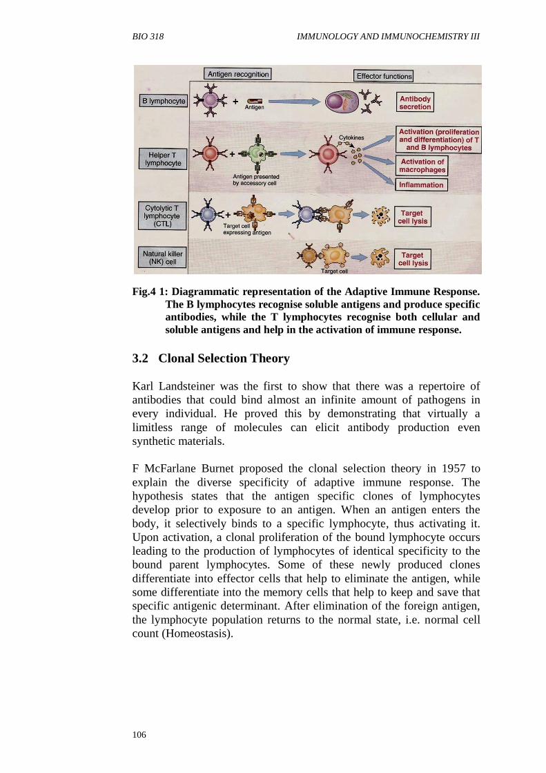
Fig.4 1: Diagrammatic representation of the Adaptive Immune Response.
The B lymphocytes recognise soluble antigens and produce specific
antibodies, while the T lymphocytes recognise both cellular and
soluble antigens and help in the activation of immune response.
3.2 Clonal Selection Theory
Karl Landsteiner was the first to show that there was a repertoire of
antibodies that could bind almost an infinite amount of pathogens in
every individual. He proved this by demonstrating that virtually a
limitless range of molecules can elicit antibody production even
synthetic materials.
F McFarlane Burnet proposed the clonal selection theory in 1957 to
explain the diverse specificity of adaptive immune response. The
hypothesis states that the antigen specific clones of lymphocytes
develop prior to exposure to an antigen. When an antigen enters the
body, it selectively binds to a specific lymphocyte, thus activating it.
Upon activation, a clonal proliferation of the bound lymphocyte occurs
leading to the production of lymphocytes of identical specificity to the
bound parent lymphocytes. Some of these newly produced clones
differentiate into effector cells that help to eliminate the antigen, while
some differentiate into the memory cells that help to keep and save that
specific antigenic determinant. After elimination of the foreign antigen,
the lymphocyte population returns to the normal state, i.e. normal cell
count (Homeostasis).

BIO 318 MODULE 4
107
Postulates of the clonal selection hypothesis:
each lymphocytes bear a single type of receptor with a unique
specificity
interaction between a foreign peptide and the lymphocyte receptor
cable of binding that molecule with high affinity usually leads to
lymphocyte action
the differentiated effector cells derived from an activated
lymphocyte bear receptors of identical specificity to those of the
parental cells which the lymphocyte was derived from
lymphocytes bearing receptor specificity for ubiquitous self
molecules are deleted early in the state of the development. They
are usually absent in the repertoire of mature lymphocytes.
3.3 Generation of Diversity in Adaptive Immune Response:
Genetics of Response
3.3.1 Generation of Diversity in Antibodies
The total collection of antibody specificity available in an individual is
known as the antibody repertoire. During maturation of the B
lymphocytes in the bone marrow, the genes responsible for the
formation of the Immunoglobulin molecules (antibody) are expressed.
Thus the specificity of each antibody in the repertoire of every
individual is determined even before B lymphocyte maturation. The
amino acid sequence of the variable region (V regions) of
Immunoglobulin molecules is responsible for the specificity of each
antibody.
Studies have revealed that although the genes coding for each variable
region occurs in multiples, majority of them are deleted during B
lymphocyte maturation. Thus, the diverse specificity of the antibody
repertoire is achieved through the Somatic Recombination (random
rearrangement of different genes) of the remaining variable genes. After
this, point mutations (single nucleotide changes in the genes) are
induced within the rearranged genes, further increasing the level of
variability. Finally, the alternative trimming and splicing of the primary
mRNA transcript before its translation into Immunoglobulin molecules
leads to the specificity and diversity observed in antibodies. All these
mechanisms, permits the response even to a single antigen diverse
(Figure 4.2).

BIO 318 IMMUNOLOGY AND IMMUNOCHEMISTRY III
108
Fig.4.2: Illustrates the various processes by which variability is
observed in the V regions of Antibodies
3.3.2 Diversity in the Tlymphocyte: MHC Molecules
T lymphocytes respond to peptide fragments of protein antigens that are
displayed by Antigen presenting cells (APCs). The task of displaying
the antigens of cell-associated microbes for recognition by T
lymphocytes is performed by specialised proteins that are encoded by
genes in the Major Histocompatibiliy Complex (MHC).
There are two different types of MHC gene products, called the class I
MHC molecules and the class II MHC molecules, which sample
different pools of protein antigens (extracellular antigens that have
undergone endocytosis and cytosolic intracellular antigens). The human
MHC, also referred to as the Human Leucocyte Antigen (HLA) locus is
located on the short arm of chromosome 6 spanning about 4×10⁶ base
pairs. It encodes for about 200 genes that are highly polygenic (i.e.
encodes different ranges of peptides binding specificity) and
polymorphic (i.e. there are multiple alleles of each gene). The HLA
locus and gene products are homologous to the mouse H2 locus and
gene product (figure 3).
MHC molecules are transmembrane proteins (antigens) that have
structure similar to the antibodies (immunoglobulin). They are
ubiquitously expressed on all nucleated cell found in the body.The class
I MHC molecules presents antigens to the CD4+ helper T cells that help
to activate other components of the immune response while the class II
MHC molecules present antigens to the CD8+cytolytic T lymphocytes
that help to lyse virus infected cells (Figure 4).

BIO 318 MODULE 4
109
T lymphocytes play a dual role of specificity in that they have the ability
to recognize and differentiate self antigen from foreign antigens during
immune response. Lymphocytes that actively react to self antigens are
destroyed during maturation in the thymus.
Fig.4. 3: Schematic maps of the human (HLA) and mouse (H2)
MHC loci
Fig. 4. 4: Antigen presentation by MHC molecules
3.4 Response to Antigenic Stimulation: A Practical Scenario

BIO 318 IMMUNOLOGY AND IMMUNOCHEMISTRY III
110
Let us consider a case study of a bacterial infection, after the first day of
infection (i.e. the pathogenic escape of the innate immune response). As
we know all microbes (as well as most other cell types) have surface
proteins expressed on their membrane.
The B lymphocyte that recognises the bacterial epitope from the
repertoire will selectively bind it (i.e. the antigenic determinant).
Simultaneously, antigen presenting cells presents the extracellular
antigens of the helper T lymphocytes or in some cases the B lymphocyte
itself presents the antigen protein to the helper T cell (Figure 4.5). The
helper T lymphocytes in turn recognises it has a foreign antigen, releases
cytokines that activates the rapid proliferation of that particular
Blymphocytes bound to the bacterial epitope to produce antibodies that
collectively eliminate the bacterial and its toxins.
Fig.4.5: A- Extracellular antigens are presented by macrophages or
B lymphocytes to CD4+ helper T lymphocytes which activates
the macrophages and B lymphocytes to eliminate the
extracellular antigen. B- Cytosolic antigens are presented
nucleated cells to the CD8+ cytolytic T lymphocytes for
elimination. MCH- Major Histocompatibility Complex

BIO 318 MODULE 4
111
4.0 CONCLUSION
In terms of specificity and diversity, the adaptive immune response is
more specific and diverse than the Innate Immunity. This specific and
diverse nature of the adaptive immunity is accomplished through the
genetic recombination of genes involved in the formation of antibody
and receptor specificity during the development of B and T lymphocytes
in the bone marrow and thymus respectively.
5.0 SUMMARY
In this unit, you have learnt that:
the adaptive immunity are the most diverse and specific form of
immune response
through immense genetic recombination, the specificity and
diversity of the adaptive immune response is obtained
genetic diversity of the T lymphocyte receptor is controlled by the
genes in the Major Histocompatibility (MHC) complex loci.
6.0 TUTOR-MARKED ASSIGNMENT
i. Briefly discuss the adaptive immune response.
ii. Discuss the Clonal Selection Theory.
iii. How is genetic diversity attained in the humoral immune response?
7.0 REFERENCES/FURTHER READING
Abbas, A.K. et al. (2000). Cellular and Molecular Immunology.(4th
ed.).
New York: W.B. Sanders Company.
Janeway, C.A. et al. (1999). Immunology: The Immune System in Health
and Disease. (4th
ed.). London: Current Biology Publications.

BIO 318 IMMUNOLOGY AND IMMUNOCHEMISTRY III
112
UNIT 5 STRUCTURE AND CLASSIFICATION OF
IMMUNOGLOBULINS
CONTENTS
1.0 Introduction
2.0 Objectives
3.0 Main Content
3.1 Molecular Structure of Antibodies
3.2 Classification of Antibodies
3.3 Structure - Function Relationship of Antibodies
4.0 Conclusion
5.0 Summary
6.0 Tutor-Marked Assignment
7.0 References/Further Reading
1.0 INTRODUCTION
The term immunoglobulin was coined by Elvin Kabat and colleagues
after they discovered that most antibodies after electrophoresis are
detected in the third fastest migrating group named gamma globulins for
the third letter of the Greek alphabet. Immunoglobulins (Ig) otherwise
known as antibodies are soluble glycoproteins produced in a membrane
bound form on B lymphocytes to recognise and bind to antigens. They
are present in serum, tissue fluids, or on cell membranes. The B cells are
the only cells that synthesise antibodies. Antibodies act as adaptors for
immune system effector molecules by linking antigens to receptor
molecules. So basically antibodies perform two main functions which
are to recognise and bind to foreign material and to trigger the
elimination of the foreign material through complex system involving
different proteins. The study of antibodies and how they react with
antigens is known as serology. When blood or plasma is made to clot,
antibodies remain in the fluid called serum and a serum containing
antibodies against a particular antigen is known as antiserum.
2.0 OBJECTIVES
At the end of this unit, you should be able to:
understand the molecular structure of antibodies
list the different classes of antibodies
describe how immunoglobulins recognise and bind to antigens
explain the structure-function relationships found in antibody
molecules.

BIO 318 MODULE 4
113
3.0 MAIN CONTENT
3.1 Molecular Structure of Antibodies
The antibody is a Y-shaped molecule consisting of three equal
fragments connected to each other by a flexible hinge. Two of these
fragments called Fab (fragment antigen binding) are identical to one
another are involved in antigen binding and the other fragment known as
Fc (fraction crystallisable) binds to effector molecules. These fragments
were identified after proteolytic digestion with papain split the antibody
molecule into two fragments in the hinge region before the hydrogen-
hydrogen inter-chain disulfide bond. The antibody molecule is flexible
thus allowing binding to different arrays of multivalent antigen.
The basic structure of immunoglobulins revealed a four chain unit
polypeptide comprising of two identical light and heavy chains.
Fig.5.1: The structure of an antibody molecule

BIO 318 IMMUNOLOGY AND IMMUNOCHEMISTRY III
114
Fig.5. 2: Detailed structure of an antibody
The light chains ( L ) have a molecular weight of about 24 KD and the
heavy chains ( H ) molecular weight of between 55 -70 KD. The heavy
chains are connected to one another via disulphide bonds, while each
heavy chain links a light chain by disulphide and non covalent bonds.
Fig.5. 3: Linear representation of an antibody molecule
The heavy and light chains consist of repeating, homologous units of
110 amino acid residues in length that fold independently to form an
immunoglobulin domain. Both the heavy and light chains have regions
called variable and the constant regions represented as CH and CL and
VH and VL where the subscript denotes heavy or light chain. The amino
BIO 318 MODULE 4
115
acid sequences of the variable region of both heavy and light chains
consists of 110 amino acid residues whereas in the case of the heavy
chain constant region, the amino acids vary , between 110 to 440
residues. The variable region consists of N-terminal of the amino acid
sequence and the carboxy terminal, the constant region. The variable
region defines the region where the amino acid sequence varies and this
distinguishes antibodies of one clone from another clone. It has been
observed that most of the variability in amno acid sequences occur in the
three regions known as hypervariable or complementarity determining
region ( CDRs). Hypervariable or complementarity determining region
are found in both the light and heavy chain regions and function in
binding and recognition of an antigen
3.2 Classification of Antibodies
Immunoglobulins may be classified into five distinct classes based on
differences in size, amino acid sequence, carbohydrate content and
charge. The different classes are IgA, IgE, IgD, IgG and IgM. Some of
these classes may be divided into subclasses egIgG (ϒ) has four
subclasses namely IgG1, IgG2, IgG3 and IgG4 and IgA(α) has two
subclasses –IgA1 and IgA2. A unique hinge region exists for each IgG
subclass. The classes and the subdivisions represent what is referred to
as isotypes and these nine isotypes are found in normal human beings.
The other immunoglobulin types IgE, IgM and IgD do not have
subclasses. The structure of the heavy chain of the Ig molecule is
important in defining the isotype. Other terms used in classifying
immunoglobulins are allotypes and idiotypes. The amino acid sequence
and the three dimensional structure of the constant region of an Ig
molecule determines the allotype , however, allotypes shows the genetic
differences within the same species, consequently all members of the
species will not possess any particular allotype. Idiotype defines the
variation in amino acid sequence and three dimensional structure of the
Ig molecule and the antigen binding specificity of any particular
antibody.

BIO 318 IMMUNOLOGY AND IMMUNOCHEMISTRY III
116
a)
b)
c)
Fig.5. 4: a-Ig A, b)-Ig D , c)- Ig E
There are two classes or isotypes of light chains called Lambda (λ) and
Kappa (k) after the Greek alphabet. Only one particular type of the light
chain is present in an antibody molecule but never one of each. These
two isotypes of light chains can be distinguished based on the carboxy
terminal of the constant region.

BIO 318 MODULE 4
117
3.3 Structure - Function Relationship of Antibodies
Different roles are played by different classes of immunoglobulins.
Basically immunoglobulins recognise and bind to antigens and trigger
off effector functions thus ensuring that the effector functions are
specifically targeted towards elimination of that particular antigen. All
immunoglobulin isotypes except IgD play a dual role which is to bind to
antigen and exhibit one or more effector functions. There are certain
features required in order for an antibody to be able to perform these
roles. These features include specificity, diversity, affinity and avidity.
Antibodies are able to distinguish subtle differences between antigens
and these differences applies to recognition of all classes of molecules
eg two linear protein determinants could be distinguished from each
other based on the substitution of a single amino acid sequence that may
or may not affect the secondary structure. The recognition of this fine
specificity is important because biochemical constituents of all living
organisms are basically related so that antibodies produced against
microbial molecules do not react with structurally similar molecules. In
some cases though, antibodies generated against a particular antigen
reacts with a structurally similar antigen and this is what is regarded as
cross-reaction. Diversity is generated from the large numbers of
antibodies necessary to bind antigen as a result of genetic mutations that
may bring about random variations in structure in the hypervariable
regions of both the heavy and light chains. It is necessary that antibodies
bind tightly to antigens to be able to eliminate a pathogen, therefore high
affinity binding antibodies are generated by changes that occur during
somatic mutation in antigen stimulated B lymphocytes. These B
lymphocytes give rise to new V domain structures that bind antigens
with greater affinity than the original V domains. The Fc fragment
mediates the effector functions of an antibody and different domains of
this fragment are responsible for carrying out different functions. During
humoral immune response the isotype of the antibody influences the
response of that antibody to an antigen, and this is because stimulation
of B clone cells would produce different isotypes with identical V
domains hence identical antigen binding sites. This is called isotype
switching where naïve B cells would produce IgM and IgD with
identical binding sites on activation. In isotype switching, there is a
change in the type of the constant region heavy chain (CH) or antibody
isotype produced by the B cell whereas the V regions and antigen
specificity remains the same. Different isotypes and subtypes may be
produced from the original B cells as a result of isotype switching.

BIO 318 IMMUNOLOGY AND IMMUNOCHEMISTRY III
118
Fig.5.5: Changes in the antibody structure
4.0 CONCLUSION
The antibody molecule is Y – shaped consisting of two fragments called
Fab (fragment antigen binding) and another fragment called Fc
(fragment crystallisable). There are two identical heavy and light chains
and both chains have regions called variable and the constant regions
represented as CH and CL and VH and VL where the subscript denotes
heavy or light chain. There are three regions called hypervariable or
complementarity determining region ( CDRs) found in both the light and
heavy chain regions and function in binding and recognition of an
antigen.
There are five classes of immunoglobulins known as IgA, IgE, IgD, IgG
and IgM and these classes vary based on differences in size, amino acid
sequence, carbohydrate content and charge. Structural and functional
relationship exists among immunoglobulins which enables them to
perform their roles. Features such as specificity, diversity, affinity and
avidity are very important and again antibodies are able to distinguish
subtle differences between antigens and these differences assist in the
recognition of all classes of molecules.
5.0 SUMMARY
In this unit, you have learnt that:
the antibody is a Y- shaped molecule
there are two heavy and two light chains
there are five classes of immunoglobulins

BIO 318 MODULE 4
119
Fc molecules mediate the effector functions of an antibody
molecule
isotype switching occurs in which naive B cells would produce
IgM and IgE with identical binding sites on activation.
6.0 TUTOR-MARKED ASSIGNMENT
i. How do you distinguish antibodies of one clone from that of
another?
ii. Distinguish between isotype, allotype and idiotype?
iii. Define isotype switching?
7.0 REFERENCES/FURTHER READING
Abbas, A.K. et al. (2000). Cellular and Molecular Immunology. (4th
ed.). New York: W.B. Sanders Company.
Janeway, C.A. et al. (1999). Immunobiology: The Immune System in
Health & Disease. (4th
ed.). London: Current Biology Publications.
http://www.biology.arizona.edu/immunology/tutorials/antibody/structur
e.html
Yang, L. (2003). “Study of Binding between Protein A and
Immunoglobulin G Using a Surface Tension Probe”. . Biophys
J.84(1): 509–522.

BIO 318 IMMUNOLOGY AND IMMUNOCHEMISTRY III
120
MODULE 2
Unit 1 Mechanism of Antibody Formation
Unit 2 Antigen/Antibody Interaction: Role of Lymphoid Tissues
and Thymus in Immune Response
Unit 3 Hypersensitivity
Unit 4 Immunopathology
Unit 5 Auto-Pathology and Auto-Immunology
UNIT 1 MECHANISM OF ANTIBODY FORMATION
CONTENTS
1.0 Introduction
2.0 Objectives
3.0 Main Content
3.1 Theories of Antibody Formation
3.2 Production of Antibodies
3.3 Qualitative Changes and Cellular Events during Primary and
Secondary Response
4.0 Conclusion
5.0 Summary
6.0 Tutor-Marked Assignment
7.0 Reference/Further Reading
1.0 INTRODUCTION
Antibodies are present in biologic fluids throughout the body and also
on the surface of a few cell types. The B cells are the only cells that
synthesises antibody molecule. They are found in cytoplasmic
membrane compartments such as endoplasmic reticulum and golgi
complex and on the surface of B cells and are expressed as integral
membrane proteins. There are also secreted forms of antibody found on
the plasma, mucosal secretions and in the interstitial fluid of tissues in a
small amount.
2.0 OBJECTIVES
At the end of this unit, you should be able to:
discuss the natural distribution of antibodies
describe the theories of antibody formation
discuss the process of antibodies production
differentiate between primary and secondary antibody response.

BIO 318 MODULE 4
121
3.0 MAIN CONTENT
3.1 Theories of Antibody Formation
Two theories have been proposed to show how antibodies are produced.
They are the selective and instructive theories. The first theory known as
the instructive theory by Linus Pauling proposed that all antibodies had
only one polypeptide structure which on induction by combining with an
antigen folded in diverse ways. This proposal was rejected because it
was observed that all antibodies still retain their specificity even after
denaturation and renaturation in the presence of an antigen and
moreover antigen specific antibodies were detected on lymphocyte
surfaces before exposure to the antigen. The selective theory which
replaced the instructive theory was put forward through the research
efforts of these scientists: Jerne, Niels; Talmadge, David and Burnet
MacFarlane (1955-1957). There were many versions of the theory that
emerged but the clonal selection theory of antibody production was the
most accepted. According to this theory antigen specific clones of
lymphocytes develop prior to and it is independent of exposure to an
antigen. The model of the clonal selection theory occurs in two phases,
the first phase involves the generation of diverse immunoglobulins
without being challenged by an antigen and subsequent incorporation
into the cell surface. This stage is now followed when on exposure to an
antigen, there is selection by B lymphocytes of antigens and subsequent
proliferation and differentiation to become either memory or antibody
plasma producing cells. The second stage occurs in the circulation and
peripheral lymphoid tissues.
3.2 Production of Antibodies
When the body is exposed to an antigen on immunisation, a stimulation
of antibody production follows but continued exposure to that same
antigen would ensure the production of high affinity antibodies. B cells
are activated to produce IgM antibody on initial exposure but 3-5 days
later IgMisotype appears in the serum and reaches its peak at between
10- 14 days. Antibody levels begin to decline thereafter till it reaches the
pre-immunisation or baseline levels. The figure below shows that this
response goes through four phases namely the lag, log, and plateau and
decline phases. In the lag phase also referred to as latent or inductive
phase, on introduction of the antigen, it is seen as a foreign substance
and the cells begin to proliferate and differentiate in response to the
antigen usually 3-5 days. This is subsequently followed by the log or
exponential phase in which the B cells different into plasma cells that
produce antibody of the IgM type. In the third phase which is the plateau
or steady state phase there is no net increase in antibody production
because antibody synthesis is balanced by its decay and this happens

BIO 318 IMMUNOLOGY AND IMMUNOCHEMISTRY III
122
between 10-14 days. The final stage decline or decay, the rate at which
antibodies are degraded outweighs the synthesis and this brings a
decline in the level of antibody in the body till it reaches the baseline or
pre-immunisation state. This is what happens during primary response to
an antigen. However, a subsequent or secondary exposure to the same
antigen results in a quicker response because of immunologic memory.
The lag phase is shorter while the log phase responds quickly to the
presence of an antigen. There seems to be no steady state and the
response proceeds to the decline stage which is not as rapid and the
antibody persists for months and even for years or a lifetime.
Fig. 1.1: Kinetics of Primary immune response to an antigen.
Fig.1. 2: Kinetics of Secondary immune response to an antigen

BIO 318 MODULE 4
123
3.3 Qualitative Changes and Cellular Events during Primary
and Secondary Response
The qualitative changes that occur during primary and secondary
response involve the isotype of the antibody. The IgMisotypes are
predominant antibodies produced but in secondary response, the
isotypes present are the IgA, IgG and IgEisotypes and the IgGisotypes
are the predominant antibodies. There is affinity maturation of the IgG
antibody which implies that initial response to low doses of an antigen is
low but it progresses with time. This explanation is explained by the
clonal selection theory. Increased affinity leads to avidity and higher
affinity is much likely to result in cross reactivity. Avidity refers to the
strength with which an antibody binds to an antigen. Antibodies are
multivalent in their reactions with multivalent antigens, thus creating a
stronger bond. However, remember that a univalent antibody fragment
could bind to a single antigenic determinant thus the avidity of an
antibody for its antigen depends on the affinities of individual antigen
binding sites o for the epitopes on the antigen. Cross reactivity may
occur when an antibody/antigen reactions show a high level of
specificity and in that case it is possible for an antibody to bind to an
antigen that is structurally related though different.
The cellular events though earlier stated are seen in the phases of both
primary and secondary immune response, where in the lag phase the B
cells differentiate in plasma cells, followed by exponential increase in
antibody concentration, then the plateau and decline phases. In the
secondary response, all the differentiated B and T cells after
encountering an antigen die off and some become memory cells. When
challenged again by an antigen it is not only the naïve T and B cells that
are activated but also the memory cells and that is the reason for a short
lag period during secondary response.
4.0 CONCLUSION
The theories of antibody production were proposed and the clonal
selection theory is the most acceptable. According to the theory, diverse
immunoglobulins could be generated without being challenged by an
antigen. Once challenged by an antigen, the B lymphocytes selects
antigens, followed by proliferation and differentiation of cells that lead
to such cells becoming either memory or antibody producing cells.
Antibodies are produced when the body is exposed to an antigen on
immunisation, and high affinity antibodies are produced on continued
exposure to the same antigen with a quicker response.

BIO 318 IMMUNOLOGY AND IMMUNOCHEMISTRY III
124
5.0 SUMMARY
In this unit, you have learnt that:
antibodies are naturally distributed in biological fluids
there are two theories of antibody production
antibodies are produced when the body is challenged by a
pathogen
there are primary and secondary phases in immune response
when the body is challenged again by the same antigen, the
response period is shorter than the first time.
6.0 TUTOR-MARKED ASSIGNMENT
i. Why is the clonal selection theory the most acceptable of the
theories of antibody production.
ii. Identify at least two cellular changes that occur during primary and
secondary immune response.
iii. Compare and contrast primary and secondary antibody response.
7.0 REFERENCE/FURTHER READING
Janeway, C.A. et al. (1999). Immunology: Then Immune System &
Disease. (4th
ed.). London: Current Biology Publications.

BIO 318 MODULE 4
125
UNIT 2 ANTIGEN/ANTIBODY INTERACTIONS: ROLE
OF LYMPHOID TISSUES AND THYMUS IN
IMMUNE-RESPONSE
CONTENTS
1.0 Introduction
2.0 Objectives
3.0 Main Content
3.1 What are Lymphocytes?
3.2 What are B-Lymphocytes?
3.3 What are T-Lymphocytes?
3.4 Development of T-Lymphocytes within the Thymus
4.0 Conclusion
5.0 Summary
6.0 Tutor-Marked Assignment
7.0 References/Further Reading
1.0 INTRODUCTION
Immune responses are not carried out in any single organ, but in a wide
variety of structures collectively known as lymphoid tissue. Lymphoid
tissue can be generally categorised as central (or primary), and
peripheral (or secondary). Central lymphoid tissues are those which act
as a source of immunocompetent cells, these cells then migrate to the
peripheral lymphoid tissues which are the sites of immune responses.
Primary lymphoid tissues are the tissues in which lymphocytes are
generated and differentiate into mature naïve lymphocytes; these are the
bone marrow for B cells, and the bone marrow and the thymus for T
cells. Secondary lymphoid tissues are the tissues in which immune
responses are initiated, and the lymphatic vessels that connect them to
the tissues and the bloodstream and thus to sites of infection.
The main function of the immune system is to defend the body against a
wide variety of pathogenic infectious agents with vastly differing
natures, i.e. viruses, bacteria, fungi, protozoa and parasitic worms. The
complexity of this task requires a sophisticated repertoire of
mechanisms for the recognition of, and defense of the body against,
these pathogens. This is achieved by an array of cells (and molecules
which they secrete) which are dispersed throughout the body and
collectively constitute the immune system. Most of the major cell types
of the immune system are derived from progenitors (stem cells) in the
bone marrow.
Many of the mature cells circulate in the bloodstream and are dispersed
throughout tissues of the body, while some also congregate in

BIO 318 IMMUNOLOGY AND IMMUNOCHEMISTRY III
126
specialised lymphoid tissues. Furthermore, in order to generate effective
immunity, the various cell types cooperate with each other by means of
direct interactions between cell surface molecules and via the molecules
that they secrete.
2.0 OBJECTIVES
At the end of this unit, you should be able to:
describe what lymphoid tissue and lymphocytes are
discuss both B lymphocytes and T lymphocytes
relate the function of B-lymphocytes with T-lymphocytes
describe the development of T-lymphocytes in the thymus.
3.0 MAIN CONTENT
3.1 What are Lymphocytes?
They arise from lymphoid progenitors in the bone marrow mammalian
B cells fully develop here, whereas T cell precursors migrate to the
thymus for selection and maturation. The bone marrow and thymus are
thus known as primary lymphoid organs. The three major types of
lymphocytes are called B cells, T cells and NK (natural killer) cells.
Mature B and T cells circulate in the bloodstream and lymphatic system,
spending some time in the secondary lymphoid tissues, i.e. the spleen,
lymph nodes and mucosa-associated lymphoid tissues (MALT). Two
morphological types of resting lymphocytes can be distinguished: B
cells and the majority of T cells are small lymphocytes with a thin rim of
cytoplasm surrounding the nucleus, whereas natural killer cells and
some T cells are larger, have more cytoplasm and distinct cytoplasmic
granules, and are known as large granular lymphocytes. B and T
lymphocytes are entirely responsible for adaptive or acquired immunity,
i.e. the ability to recognise each pathogen in a specific way and to mount
a faster and bigger response on repeated exposure to a particular
pathogen (immunological memory). This is because B and T cells
express surface receptors which specifically bind to materials that are
foreign to the body (known as antigens). The receptors of a single
lymphocyte are identical to each other and recognise a single antigen.
However, millions of different antigen receptors are collectively
expressed by the whole population of lymphocytes in the human body,
thus conferring the ability to recognise many great foreign antigens.
3.2 What are B-Lymphocytes?
Cells of the immune system which are specialised to make antibodies
are termed B-lymphocytes, they are produced in the bone marrow of
BIO 318 MODULE 4
127
adult mammals. Without B-cells and the antibodies they produce, one
can only survive if given frequent gamma-globulin injections. The B
cells constitute 5–15% of human blood lymphocytes. The main function
of a B cell is to secrete soluble recognition molecules called antibodies
which specifically bind to an antigen recognised by that B cell. These
antibodies (also known as immunoglobulins) are, in fact, the secreted
form of a B cell’s surface antigen receptors and bind to exactly the same
antigen. A B cell will only produce antibodies when it has been
activated by binding antigen; this activation process also usually
requires help from T cells. The activated B cell undergoes multiple
divisions and some of the resulting cells differentiate into antibody-
secreting cells. These are known as plasma cells, and they possess
copious rough endoplasmic reticulum involved in antibody synthesis.
The antibodies serves to neutralise toxins, prevents organisms adhering
to mucosal surfaces, activates complement, opsonises bacteria for
phagocytosis, and sensitisestumour and infected cells for antibody
dependent cytotoxic attack by killer cells. Thus antibody acts to enhance
elements of the innate system. Although ultimately antibody is the
secreted product of activated B cells with the functions listed, early in
B-cell development, it is a membrane bound molecule that acts as the B-
cell receptor. In this role it internalises antigen and processes it to act as
an antigen-presenting cell for T-cell responses.
Different classes of antibody predominate at different compartments of
the body (IgM being intravascular, IgG the main antibody of the blood
and tissues, IgA in secretions). Mucosa associated lymphoid tissue
consists of lymphoid tissue at several mucosal sites (bronchus, gut,
urogenital tract). However, these are all linked functionally as
subpopulations of B cells home to these tissues specifically. A response
generated at one site will induce immune responses to the same antigen
at other sites.
The main structural difference between antibodies of different classes is
in the non-antigen binding portion of the molecules, called the Fc
region, which is constant in structure between antibodies of the same
class produced by different B cells. An important aspect of antigen
recognition by B cells is that these lymphocytes, and the antibodies they
produce, bind to antigens in their natural or naive form, i.e. as they
occur as constituents of pathogens.
3.3 What are T-Lymphocytes?
About 70% of human blood lymphocytes are T cells. The main
functions of T lymphocytes are to exert effects on other cells, either
regulating the activity of cells of the immune system or killing cells that

BIO 318 IMMUNOLOGY AND IMMUNOCHEMISTRY III
128
are infected or malignant. Like B lymphocytes, T cells have surface
antigen receptors, but there is no secreted form of this equivalent to
antibodies. Furthermore, T cells cannot recognise antigens in their
native forms, but only when they are presented on the surface of
antigen-presenting cells (APCs). The antigen receptors of most T cells
(alpha-beta T cells) are composed of two polypeptides called alpha and
beta chains, and they interact with peptides derived from the degradation
(processing) of foreign antigenic proteins. These peptides are bound to
molecules of the major histocompatibility complex (MHC) on the
surface of APCs. The interaction between the T-cell antigen receptors
and the peptide–MHC complexes binds a T cell to the surface of an
APC, thus targeting the T cell to exert effects on the APC. There are two
types of MHC molecules, called class I and class II, which present
antigen peptides to alpha-beta T cells expressing the surface proteins
CD8 or CD4, respectively. This is because CD8 binds to MHC class I
and CD4 binds to MHC class II.
3.4 Development of T-Lymphocytes in Thymus
As soon as receptor rearrangement has occurred, T and B cells are able
to respond to their antigen and induce an immune response. However,
cell activation is tightly regulated to ensure that only damaging antigens
elicit a reaction. Regulation particularly involves the initiation of T
lymphocyte activation. This requires that antigen is presented to the T
cell within the peptide binding groove of a self MHC molecule. This is
because the T-cell receptor does not just recognise the antigenic epitope,
but also the complex of the peptide in association with the self-MHC
molecule. The delicate process of positive selection of T cells that can
react with self-MHC and peptide adequately to induce immune
responses occurs in the thymus. These cells could sometime not be
excessively MHC-reactive to the extent which would cause self-tissue
destruction.
The cells that emerge from the thymus and bone marrow having
undergone gene rearrangement are naive i.e. they have not yet
encountered their specific antigen within an immune response. These
cells populate the secondary lymphoid tissues of the lymph nodes,
spleen, tonsils, and mucosa associated lymphoid tissue. Because there
are only a few naive T and B cells capable of reacting specifically with a
foreign particle, in order for them to encounter their specific antigen,
there has to be a system to bring them together. The lymphoid tissues
provide the microenvironment for this process. In addition to T and B
lymphocytes, they contain efficient antigen-presenting cells and are able
to produce the cytokines necessary to maintain T and B lymphocytes.
Lymphoid tissues express adhesion molecules in an ordered array,
allowing cells to move through the tissue and increase the chance of

BIO 318 MODULE 4
129
lymphocytes being brought into contact with antigen. The lymphoid
organs communicate with the tissues using lymphatic and blood vessels.
4.0 CONCLUSION
B cells are generated throughout life in the specialised environment of
the bone marrow and may require a second environment; the lymphoid
tissue helps to maintain the existence as mature recirculation B cells.
The thymus provides a specialised and architecturally organised
microenvironment for the development of mature T cells.
5.0 SUMMARY
In this unit, you have learnt that:
the bone marrow and thymus are the primary lymphoid organs
involved in the production and differentiation of lymphocytes
there are two major types of lymphocytes; the B-lymphocytes and
the T-lymphocytes
the development of T-lymphocytes occurs in the thymus.
6.0 TUTOR-MARKED ASSIGNMENT
1. Briefly describe different types of lymphocytes.
2. What are the importance of B cells and T cells?
3. Describe the development of T-Lymphocytes in the Thymus
7.0 REFERENCES/FURTHER READING
Charles A.J (Jr). et al.. (1999). ImmunoBiology: The Immune System in
Health and Diseases.( 4th ed.). Elsevier Science Ltd/Garland
Publishing
Lydyard, P. & Grossi, C. (1998). “Cells involved in the immune
response”. In: Roitt I, Brostoff J and Male D (Eds.) Immunology
(5th ed.). London: Mosby. pp. 13–30.

BIO 318 IMMUNOLOGY AND IMMUNOCHEMISTRY III
130
UNIT 3 HYPERSENSIVITY
CONTENTS
1.0 Introduction
2.0 Objectives
3.0 Main Content
3.1 Types of Hypersensitivity Reactions
3.2 Diseases Associated with Hypersensitivity Reactions
4.0 Conclusion
5.0 Summary
6.0 Tutor-Marked Assignment
7.0 References/Further Reading
1.0 INTRODUCTION
Hypersensitivity may be defined as a process in which the adaptive
immune response becomes over sensitive to a variety of infectious and
innocuous antigens thereby inflicting injury to the host tissue. The
disorders that result from such reactions are termed hypersensitivity
diseases.
2.0 OBJECTIVES
At the end of this unit, you should be able to:
understand the term hypersensitivity
classify hypersensitivity reactions
discuss some diseases associated with hypersensitivity reactions.
3.0 MAIN CONTENT
The most important function of adaptive immune response is in host
defense against microbes. However, it could also be responsible for
tissue injury and disease. This reaction could produce discomfort and
sometimes may result in fatality. A common cause of hypersensitivity
reaction is inability to tolerate self antigens, a property of the immune
system whose responsibility it is to ensure that no reaction occurs
between individuals and their own antigens. Inability to self tolerance
and subsequent immune response against self or autologous antigens
results in autoimmune diseases.

BIO 318 MODULE 4
131
3.1 Types of Hypersensitivity Reactions
There are five types referred to as Type I, II, III, IV and V. Once contact
is established with antigen a variation in timing exists for each of the
hypersensitivity reactions (Table 3.1).
Table 3.1: Hypersensitivity Reactions Hypersensitivity
type
Appearance
time
Mediators Mechanism
i. Immediate 2-30 mins IgE Mast cell response
(enhance acute
inflammation)
ii. Cytotoxic 5-8 hours IgM and IgG Antibody and
complement
iii.Immune
complex
2-8 hours IgM and IgG
immune
complexes
Antibody/antigen
Complexes
iii. Delayed type 24-72 hours CD4 and CD8 T
cells, APCs
T cell mediated
Macrophages activated
Include granulomatous
reactions
iv. Stimulatory Autoantibodies Autoantibodies against
hormone
The Type 1 hypersensitivity reaction which is allergy affects about 17%
of the population and it exists in two phases and mediated by IgE. It is
an immediate hypersensitivity reaction because antibodies are involved
and starts within minutes of contact with the antigen. The mechanism of
action involves mast cells and basophils, both of which have receptors
for IgE. After interaction of antibody/antigen some physiological
substances such as histamine, serotonin and other mediators of
inflammation are released thus inducing spasms of the smooth muscle.
The hypersensitivity Type II reaction also called cytotoxic reaction
involves IgE and IgM antibodies. These antibodies interact with the
antigen in the blood as well as analogous antigens on the surface of
human body cells. This is done by opsonization and phagocytosis of
cells. These mechanisms set off the complement system that lyses the
cell.
Hypersensitivity type III mobilises not only IgE and IgM but also
immune complexes. It can rise from soluble antigens. The immune
complexes could vary in size, amount, affinity and the isotype of the
responding antibody. Consequently, large immune complexes fix the
complements and are disposed by mononuclear phagocytes from
circulation, whereas the smaller complexes deposit in blood vessel walls

BIO 318 IMMUNOLOGY AND IMMUNOCHEMISTRY III
132
and can ligate Fc receptors on mast cells and other leucocytes which can
lead to leucocyte activation and tissue injury.
Type IV hypersensitivity called delayed type hypersensitivity (DTH)
reaction occurs within 24-72 hours and it is mediated by CD4, CD8 T
cells as well as antigen peptide cells (APCs) eg Langerhans’ cells. The
activated T cells migrate to the site of antigenic entry where pro-
inflammatory mediators such as Tumor necrosis factor (TNF) are
released. The pro-inflammatory mediators facilitate blood flow and
extravasation of plasma contents to the area. CD4 and CD8 cells are
released resulting in the interferon gamma (IFN-ᵞ) thus enhancing
macrophage activity in the area.
The stimulatory or type V hypersensitivity are mediated by auto-
antibodies. It is classified as a distinct type of hypersensitivity where the
auto-antibodies bind to hormone receptors that imitate the hormone
itself and this leads to stimulation of target cells. Autoimmune response
defines the ability of an individual immune system to react with self
antigens. There are some individuals who have auto-antibodies and will
show no symptoms, but autoimmune disease results if the regulatory
mechanisms breakdown. It is to be noted that the causes of autoimmune
diseases are multi-factorial.
3.2 Diseases Associated With Hypersensitivity Reactions
Hypersensitivity diseases are clinically heterogeneous group of
disorders whose manifestations are defined by the type of immune
response that leads to cell and tissue injury. The mechanisms of immune
response that leads to different types of hypersensitivity reactions have
been stated above. There are some individuals prone to produce IgE in
response to environmental allergens and show strong immediate
hypersensitivity are said to be atopic and they suffer from allergy. This
form of allergy may present in different forms such as hay fever,
asthma, urticaria (hives) or chronic skin irritation. In extreme cases
known as anaphylaxis, the mast cell and basophil derived mediators
restrict the airways to the point of asphyxiation and produce
cardiovascular collapse leading to death. Myasthenia gravis is an
example of type II hypersensitivity reaction in which the antibody
inhibits acetylcholine binding as well as down modulating receptors.
Type III hypersensitivity reaction is presented in immune complexes
mediated reactions such as Arthus reaction. The delayed type
hypersensitivity results from tissue injury as a result of the production of
reactive oxygen species, hydrolytic enzymes, nitric oxide and some pro-
inflammatory mediators such cytokines. Insulin-dependent diabetes
which is an organ specific auto immune disease is caused by DTH
reactions induced by auto T cells. Diseases caused from

BIO 318 MODULE 4
133
hypersensitivity reactions are shown in the Tables below.
Table 3. 2: Diseases caused by Cell- or Tissue Specific Antibodies
Table 3. 3: Examples of Human Immune Complex-Mediated
Diseases
4.0 CONCLUSION
Hypersensitivity reactions are caused by disorders of immune response.
The reactions are classified into five types based on the type of immune
response and the effector mechanisms that are responsible for cell and
tissue injury. The immune response could be autoimmune responses
against self antigens or uncontrolled or over reaction to foreign antigens.
The immediate hypersensitivity occurs within few minutes of being

BIO 318 IMMUNOLOGY AND IMMUNOCHEMISTRY III
134
challenged with an antigen and in extreme cases death may occur due to
asphyxiation and circulatory collapse.
5.0 SUMMARY
In this unit, you have learnt that:
hypersensitivity reaction is a disorder of the immune response
there are five types of hypersensitivity reactions
different types of hypersensitivity reactions are mediated by
different effector mechanisms.
some diseases are associated with hypersensitivity reactions.
6.0 TUTOR-MARKED ASSIGNMENT
i. Explain the differences between Type II and Type III
hypersensitivity reactions.
ii. Why are some individuals hypersensitive to certain drugs or
chemicals while some others are not?
iii. How does the mechanism of opsonization mediate in Type II
hypersensitivity reaction?
7.0 REFERENCES/FURTHER READING
Abbas, A.K. et al. (2000). Cellular and Molecular Immunology. (4th
ed.). New York: W.B. Sanders Company.
Janeway, C.A. et al. (1999). Immunobiology: The Immune System in
Health and Disease. (4th
ed.). London: Current Biology
Publications.
Gregory, H. (N.D.). Basic Immunology.
BIO 318 MODULE 4
135
UNIT 4 IMMUNOPATHOLOGY
CONTENTS
1.0 Introduction
2.0 Objectives
3.0 Main Content
3.1 Pathology of Immune Response
3.2 Immunopathology of Infectious Diseases
3.3 Immunopathology of Parasites
3.4 Diseases of the Blood
4.0 Conclusion
5.0 Summary
6.0 Tutor-Marked Assignment
7.0 References/Further Reading
1.0 INTRODUCTION
Immunopathology is the branch of immunology that deals with the
pathologies of the immune system. It can also be described as the branch
of medicine that deals with immune responses associated with diseases.
The immune system helps fight off infections but could also turn around
to be harmful due to over activity and that is immunopathology. Early
immunology studies looked at the ambivalence of the manifestations of
allergy or hypersensitivity. This is because the immune system which
was supposed to defend the body goes astray to create a variety of
pathological conditions. However, with deeper understanding of the
pathogenesis of some diseases such as tuberculosis and leprosy as well
as the development of experimental models of some diseases like
Masugi nephritis, and lymphocytic choriomeningities
immunopathology was then viewed in a wider context of immunologic
event. A few years after the development of the basic concepts of
immunopathology, it was then divided into four classes viz anaphylaxis,
delayed hypersensitivity, immune-complex disease and complement
mediated lysis.
2.0 OBJECTIVES
At the end of this unit, you should be able to:
understand the pathologies of the immune system
relate immune responses to certain diseases
assess the pathology of Immune responses.

BIO 318 IMMUNOLOGY AND IMMUNOCHEMISTRY III
136
3.0 MAIN CONTENT
3.1 Pathology of Immune Response
The reaction caused as a result of the response of the immune system to
infections, could be beneficial, without effect or detrimental to the
patient. When a reaction is beneficial, there are no signs or symptoms on
the affected person, and a lifelong immunity is conferred on the
individual. A role carried out effectively by the Adaptive immune
response. If the effect on the patient is neutral, the individual experience
symptoms, as a result of the response of the immune system to
infections, mainly cytokines released from phagocytes and toxins
released from dying bacteria. Cytokines affect the local blood vessels,
causing inflammation; redness and swelling of the skin. Cytokines also
play an imperative role in the adhesive properties of the endothelium,
causing circulating leukocytes to stick to the endothelial cells of the
blood vessel wall and migrate between them to the site of infection, to
which they are attracted by other cytokines. This action accounts for the
pain experienced during infection. A reaction is described harmful to an
individual if it causes a variation of symptoms that are quite different
from the normal symptoms associated with the infection. Symptoms of
this kind of immune response are unique to the patient and are
considered life threatening e.g. diarrhea, fatigue, muscle weakness,
constipation, cough, depression, fever, flu e.t.c. The length or duration
of immunopathological symptoms depends on the nature of infection
and the defense system of the affected person. However, this does not
mean that a rise in the intensity of symptoms is an indication of
advancement in infection or disease, but in most cases suggests that the
immune response system is effective in combating infectious pathogens
e.g. diarrhea and vomiting associated with food poisoning are quite
necessary in eliminating harmful toxins.
The adaptive immune response involves production of antibodies
against a particular pathogen. These antibodies continually provide an
adaptation to infection from that pathogen, as a result may confer a life-
long protective immunity to re-infection with the same pathogen.
Antibodies are produced only in response to specific infections, hence
antibodies present in an individual directly represents the number of
pathogens the individual has been exposed to. Adaptive immune
responses depend on the activities of lymphocytes. Lymphocytes help to
identify and defend against pathogenic molecules not recognised by the
phagocytes and macrophages e.g. Viruses and bacteria with protective
capsules. All lymphocytes require two signals in order to be activated:
one that results from antigen binding and a second signal from a T-cell
if the lymphocyte is a B-cell, or from macrophages, dendritic cell or B-
cell if it is a T-lymphocyte. Mature dendritic cells are found extensively

BIO 318 MODULE 4
137
in the T-cell. The surface receptors of B cells and T cells are adapted to
recognise antigen in two different ways. B cells of the lymphocytes
recognize antigen that is present outside the cells of the body, where
most bacteria are found; T cells by contrast can detect antigen generated
inside host cells such as Viruses. Antibodies produced by the
lymphocytes are found in the plasma or extracellular fluid, and they
protect from pathogens by either binding to them, thereby blocking their
access to cells that they may infect or destroy, a process known as
Neutralisation, or through Opsonization. Opsonization involves the
coating of pathogens by antibodies, enabling ingestion and digestion by
phagocytes. Another form of protection by antibodies is through
activation of a system of plasma proteins known as Complement. The
function of the complement is similar to Opsonization. T-cells are very
crucial in detecting pathogens that replicate inside cells. As earlier stated
innate immune response, involves ingestion and digestion of pathogens
by phagocytic cells called macrophages. These phagocytic cells are
always available to combat a wide range of microorganisms, without
requiring prior exposure, and they act in the same way in all normal
individuals. The innate responses largely involve granulocytes and
macrophages. The defence system of the innate immunity is effective in
combating many pathogens. However, they cannot recognise novel
pathogens that lack surface molecules, common to many pathogens.
The adaptive and innate immune responses work together to provide an
effective defensive system, ensuring successful destruction of all forms
of pathogens that in most instances cause no pathology.
3.2 Immunopathology of Infectious Diseases:
Immunopathology captures both hypersensitivity and autoimmunity as a
result of overactive immune system and immunodifficiency, which is
the inability of an individual to fight infection. If a virus infects a host,
an immune response is generated which could determine the survival or
the death of the host in the case of acute infection or persistent/chronic
infection. This type of infection could result in tissue damage or
pathology and it is different from direct virus attack on cells resulting in
tissue damage or apoptosis. Thus there must be a balance between viral
clearance and immunopathology (Fig 1).Several factors determine
immunopathology which include age of the host, genetic makeup of the
genes, route and /or site of infection, as well as whether the immune
system is compromised and the strain or type and the size of the
inoculums/dose.

BIO 318 IMMUNOLOGY AND IMMUNOCHEMISTRY III
138
▲
Fig.4.1: The antiviralimmune response is a balance between viral
clearance and immunopathology. One direction leads to survival of
the host, whereas the other can lead to disease.
3.3 Immunopathology of Parasites
Tissue damaging reactions occur when there is persistent infection of
parasites e.g. Plasmodium malariae, Schistosomajaponicum. This is
because the persistence of the parasite antigen in the blood leads to
immunopathological disorders such as immune complex nephrotic
syndrome, liver granuloma and autoimmune lesions of the heart. It is
believed that cross reactions between parasite and self could result in
autoimmunity and is the basis for the cardiomyopathy in Chagas
disease. Generalised immunosupression increases susceptibility to
bacterial and viral infections.
3.4 Diseases of the Blood
There are some immunological diseases known as cytopenia are blood
diseases eg autoimmune neutropenia, immune thrombocytopenic
purpurae.t.c. These are believed to be autoimmune diseases caused by
the destruction of blood components by autoantibodies.
4.0 CONCLUSION
Understanding Immunopathology is very critical in the treatment of
diseases and infections. Since the symptoms associated with infections
are as a result of the combat effect of the Adaptive and Innate immune
responses to the causative pathogens, assessment of
immunopathological symptoms, through visual analysis of patients or
laboratory test helps to determine the pace of therapy. Accurate analysis
Antiviral immune response
Viral clearance Immunopathology
Health/survival Disease
BIO 318 MODULE 4
139
of Immunopathology is sometimes complicated by chance events and
environmental factors. In most cases, the goal of a treatment is to
generate a tolerable immunopathology. In other words, reducing the
symptoms associated with an infection to a less harmful level in the
patient.
5.0 SUMMARY
In this unit, you have learnt that:
the immune response to infections is carried out effectively by the
combination of the Adaptive and Innate immune response system
tissue damage to host due to immunological event could be as a
result of hypersensitivity reaction to innococus materials, vigorous
rejection to foreign grafts and the loss of self tolerance
consequently leads to autoimmune disease
sometimes, the immune response to these pathogens could be
beneficial, or lead to symptoms in infected areas; inflammation or
pain, or cause other symptoms not usually associated with the
infection
tissue damaging reactions occur when there is persistent infection
of parasites.
6.0 TUTOR-MARKED ASSIGNMENT
i. What is the difference between immunopathology and auto-
pathology?
ii. Categorise immunopathology into two broad groups based on
reaction of the cells or tissues to antibody reaction with foreign
material.
iii. Under what condition could the immune mechanism be
disadvantageous to the host?
7.0 REFERENCES/FURTHER READING
Smith, G., Hall, L. & Walker. M. (2002). Handbook of Toxicologic
Pathology. (2nd
ed.). Volume 1. Academic Press. p156
Dey, N.G. (1970). A Text Book of Pathology
Oldstone, B.A. & Fujinami, R.S. (2008). Encyclopedia of Virology (3rd
ed.). pp. 78-83
Weingand,,K.et al. (1996). Fundamental and Applied Toxicology (29),
198-201
BIO 318 IMMUNOLOGY AND IMMUNOCHEMISTRY III
140
UNIT 5 AUTO-IMMUNOLOGY AND AUTO-
PATHOLOGY
CONTENTS
1.0 Introduction
2.0 Objectives
3.0 Main Content
3.1 Possible Causes of Autoimmunity
3.1.1 Genetic factors
3.1.2 Sex
3.1.3 Environmental Factors
3.2 Pathology of Autoimmunity
4.0 Conclusion
5.0 Summary
6.0 Tutor-Marked Assignment
7.0 References/Further Reading
1.0 INTRODUCTION
While Auto-immunology is the branch of immunology that deals with
Autoimmunity, Auto-pathology is the pathology that describes the
autoimmune system. The immune system of an individual is well
equipped to protect it against invasion by harmful microorganisms, but
sometimes the immune system fails to recognise its own constituent
parts, thereby attacking its own cells and tissues. This process of
allowing immune responses against itself is termed Autoimmunity.
Autoimmunity also referred to as ‘Self Immunity’ is known to be
present in everyone, and diseases resulting from it are termed
autoimmune diseases.
2.0 OBJECTIVES
At the end of this unit, you should be able to:
explain the possible causes of autoimmunity
describe the pathology of autoimmunity.
3.0 MAIN CONTENT
3.1 Possible Causes of Autoimmunity
The adaptive immune responses are responsible for autoimmunity, and
its actions are similar to the way it acts against foreign antigens.
However, in the case of self antigens, it is almost impossible for the
immune response mechanism to eliminate the antigen completely,
thereby prolonging the response, which could be detrimental to the

BIO 318 MODULE 4
141
affected tissues in most cases. There is no specific explanation to why
the adaptive immune response attacks its own self; rather susceptibility
to autoimmune diseases seems to be caused by a combination of factors;
genetics, sex and the environment.
3.1.1 Genetic factors
Certain individuals that have a family or personal history of autoimmune
diseases are likely to be susceptible to autoimmunity. This susceptibility
is discovered to be associated with a combination or group of genes.
Based on twin and family studies carried out for several autoimmune
diseases e.g. insulin-dependent diabetes mellitus, genes related to
immunoglobulins, T-cell receptors, and the major histocompatibility
complexes (MHC) have been suspected to be mainly responsible for
autoimmunity. However, the most consistent association for
susceptibility to autoimmunity has been linked to the MHC genotype.
This correlation of MHC genotype with autoimmune diseases is not
surprising, because autoimmune responses is linked to the adaptive
immune response mechanism which involves T cells, and the ability of
T cells to respond to a particular antigen depends on MHC genotype.
Various studies also carried out on affected patients suggested that
siblings affected with the same autoimmune disease are far more likely
than expected to share the same MHC type. However the MHC
genotype alone does not determine susceptibility, in fact monozygotic
twins are far more susceptible to autoimmunity than MHC- identical
siblings, which is a confirmation to the fact that genetic factors besides
MHC affect susceptibility. Genes linked to immunoglobulins and T-cell
receptors, involved in antigen recognition, are inherently variable and
susceptible to recombination. This attribute enables the immune system
to respond to a wide variety of harmful pathogens, and as a result may
give rise to lymphocytes capable of self- immunity.
3.1.2 Sex
It has been observed that the frequency of autoimmune diseases is
higher in females than males, although studies also show that the
autoimmune diseases present in males tend to be severe. However, the
reasons for this anomaly are unclear, but there are few explanations to
this wide range in frequency between the two sexes. Compared to males,
females are shown to exhibit larger inflammatory responses, when their
immune system is triggered, hence increasing the risk of autoimmunity.
Hormonal levels also tend to play a role in autoimmunity, based on
indications that autoimmune diseases fluctuate with hormonal changes
during menstrual cycle or pregnancy. Some studies carried on
autoimmune thyroiditis, suggests that the high frequency of the females
to autoimmunity is as a result of the imbalanced X chromosome
inactivation.
BIO 318 IMMUNOLOGY AND IMMUNOCHEMISTRY III
142
3.1.3 Environmental Factors
Various studies have shown that the expression of a disease could be
influenced by environmental factors. Since the presence of an
autoantibody requires an autoantigen to cause autoimmunity, certain
factors have been observed to inhibit or affect the binding of the
autoantibody to the autoantigen, thereby modulating the expression of
these diseases among different individuals. For example, in the
Goodpasture’s disease, it was observed that cigarette smokers
exclusively exhibited pulmonary haemorrhage while non cigarette
smokers showed no sign of pulmonary haemorrhage. This is because
cigarette smoke stimulates an inflammatory response in the lungs, which
damages alveolar capillaries, as a result exposing autoantigen to
autoantibody. Some studies also show the existence of an inverse
relationship between infectious diseases and autoimmune diseases. It
was observed that autoimmunity seems to prevail in areas where there
are no infectious diseases, while autoimmune diseases are very rare in
areas where infectious diseases are endemic. Various hypotheses have
tried to explain this correlation, but the details of the mechanism are not
fully known. Certain chemicals and drugs have also been implicated in
the expression of autoimmunity e.g. in the drug- induced lupus
erythematosus, withdrawal of the drug removes the symptoms
automatically. Exposure to heavy metals such as mercury and lead, have
been shown to induce autoantibodies, hence causing autoimmunity.
3.2 Pathology of Autoimmunity
The pathology and clinical expression of an autoimmune disease is
determined by the antigens or group of antigens, together with the
mechanism by which the antigen-bearing tissue is damaged. Antigens
responsible for autoimmunity are known to be few in number (DNA,
receptor), and several have been observed to play signaling roles in the
immune response, while the mechanisms operative in the pathology of
an autoimmune disease are numerous, and are mostly mediated by the
actions of autoantibodies and T cells. Studies carried out on autoimmune
haemolyticanaemia, showed that responses of immunoglobulin G (IgG)
or immunoglobulin M (IgM) autoantibodies to autoantigens on cell
surfaces of red blood cells triggered the destruction of red blood cells. It
has also been observed that IgG or IgM may bind to cells in tissues for a
prolong period of time, causing inflammatory damage or may bind to
receptors on cell surfaces, stimulating receptor or blocking the binding
of cell surface receptors to its natural ligand. Tissue damage can also be
due to the binding of autoantibodies to autoantigens in the extracellular
matrix or the binding of autoantibodies to soluble autoantigens. T cell
responses have been directly implicated in causing tissue damage or
indirectly by activation of macrophages. It was observed in the

BIO 318 MODULE 4
143
autoimmune disease insulin-dependent diabetes mellitus, that
specifically reactive T cells selectively destroyed the insulin-producing
β cells of the pancreatic islets. Further studies on the immune response
also showed that the requirement for the activation of the B cell by the T
cell, in order for the B cell to produce antibodies maybe sometimes
bypassed, hence causing the continual production of antibodies that may
damage tissues. Molecular mimicry, an important mechanism
discovered to be important in autoimmunity, involves the production of
antibodies to an antigen that mimics a self antigen, thereby causing the
binding of the antibodies to the self antigens. There are still more
research going on the mechanisms associated with the pathology of
autoimmunity, especially the roles of T cells.
4.0 CONCLUSION
In some studies, conclusion that various diseases had an autoimmune
basis was as a result of the fact that autoantibodies were present in the
serum of affected patients. However, further studies revealed that
similar autoantibodies were present in the serum of patients, if the tissue
damage was caused by trauma or infection. Thus, suggesting that
autoantibodies may be as a result of tissue damage, rather than be the
cause of it. Research is still going on to further understand
autoimmunity and the mechanism of pathology involved, since a disease
cannot be classified as autoimmune unless the autoantibody isolated is
proven to be pathogenic.
5.0 SUMMARY
In this unit, you have learnt that:
a disease can only be classified as autoimmune, if it is shown
clearly that the response of the adaptive immune system to the
autoantigen causes the pathology observed
various factors such as genetic, sex and environmental are known
to modulate autoimmunity
the pathology of autoimmunity is determined by the antigens and
mechanism of the tissue damage, and autoimmune diseases are
observed to be mediated mainly by auto-antibodies and T cells
the MHC genotype of individuals have been linked to the
susceptibility of individuals to autoimmunity
6.0 TUTOR-MARKED ASSIGNMENT
i. List the factors that influence autoimmunity
ii. Discuss the pathology of autoimmunity

BIO 318 IMMUNOLOGY AND IMMUNOCHEMISTRY III
144
7.0 REFERENCES/FURTHER READING
Smith, G., Hall, L. & Walker. M. (2002). Handbook of Toxicologic
Pathology. (2nd
ed.). Volume 1. Academic Press. p156
Dey, N.G. (1970). A Text Book of Pathology
Oldstone, B.A. & Fujinami, R.S. (2008). Encyclopedia of Virology (3rd
ed.). pp. 78-83
Weingand,,K.et al. (1996). Fundamental and Applied Toxicology (29),
198-201

BIO 318 MODULE 4
145
MODULE 3
Unit 1 Tissue and Transplantation Immunology
Unit 2 Immunoprophylaxis
Unit 3 Modern Techniques in Immunology and
Immunochemistry
Unit 4 History of Chemotherapy
Unit 5 Principles of Chemotherapy
UNIT 1 TISSUE AND TRANSPLANTATION
IMMUNOLOGY
CONTENTS
1.0 Introduction
2.0 Objectives
3.0 Main Content
3.1 Immune Responses to Tissue Grafts
3.2 Transplant Tolerance
3.3 Immunosuppressive Therapy
3.3.1 Calcineurin Blockers
3.3.2 Antiproliferative Agents
3.3.3 Corticosteroids
3.3.4 m-TOR inhibitors
3.3.5 Antibodies
4.0 Conclusion
5.0 Summary
6.0 Tutor-Marked Assignment
7.0 References/Further Reading
1.0 INTRODUCTION
Transplantation can be defined as the process or act of transferring cells,
tissues or organs from one site to another. Tissue transplantation as a
form of medical therapy has grown to be very important and preferred
form of treatment in a range of life threatening clinical conditions such
as liver, kidney or heart failures. However, the major obstacle to the use
of tissue transplantation as a routine medical treatment is the adaptive
immune response. Blood transfusion, which is the earliest and
commonest form of tissue transplantation involves only four major ABO
blood types and two Rhesus blood types, hence matching between
individuals to avoid destruction by antibodies is relatively easy. When
tissues containing nucleated cells are transferred from one individual to
another, the responses of T cells to the Major Histocompatibility
Complex molecules, triggers an elaborate response against the grafted

BIO 318 IMMUNOLOGY AND IMMUNOCHEMISTRY III
146
organ, causing destruction. For a successful tissue transplant to take
place there must be a perfect match of the MHC molecules of the donor
with that of the recipient, which is only possible amongst related
individuals. In spite of that, genetic differences at other loci can still
trigger tissue rejection.
2.0 OBJECTIVES
At the end of this unit, you should be able to:
discuss the immune responses to tissue grafts
explain transplant tolerance
describe immunosuppressive therapy.
3.0 MAIN CONTENT
3.1 Immune Responses to Tissue Grafts
The extent to which the immune system responds to a tissue graft will
depend on the level of genetic differences between the graft and the
host. A number of terms have been used to classify a graft, according to
its origin and possible outcome in the host.
Autograft: Transplantation of tissues or organs from the same
individual e.g. skin. Transplantation is always 100% successful,
since they do not elicit rejection.
Syngeneic graft: Transplantation of tissues or organs between
genetically identical animals or individuals. Similar to Autograft,
Syngeneic graft undergoes no rejection.
Xenograft: Transplantation of tissues or organs from one species
to another. This elicits the maximum immune response.
Allograft: Transplantation of tissues or organs between genetically
unrelated individuals. This is the most common form of
transplantation, and the graft is initially accepted but it is then
rejected after some days. The degree of rejection is dependent on
the level of disparity in the MHC molecules between the donor and
the recipient. MHC molecules present endogenous and exogenous
peptides to T lymphocytes, which decide whether the peptide-
MHC complex is a potentially threatening antigen, thereby
triggering an immune response. In humans, the MHC is known as
the human leukocyte antigen (HLA), located on the short arm of
chromosome 6, near the complement genes. HLA matching has
been shown to improve the success rate of tissue or organ

BIO 318 MODULE 4
147
transplant, but in itself does not prevent the risk of rejection. This
is due to the imprecise nature of HLA matching, as unrelated
individuals who possess identical HLA type with antibodies
against MHC proteins rarely have similar MHC genotype.
However, this is not the case with HLA identical siblings because
the siblings inherit their MHC gene as a haplotype; that is one
sibling in four is expected to be HLA identical. In spite of this,
graft rejection is still possible although less rapid. The minor H
antigens also known as minor histocompatibility antigens are
responsible for the rejection of HLA identical grafts between donor
and recipient. Two mechanisms are involved in the recognition of
alloantigens in grafted organs, the direct recognition; whereby T
cells bind directly to donor MHC-peptide complex, causing graft
rejection, and the Indirect Recognition; is similar to the process
whereby T cells become activated in response to a pathogen. In
contrast to direct recognition pathway, T cells bind to allogeneic
peptides presented by the recipient antigen presenting cells and not
MHC molecules expressed on the donor graft itself. The
contribution of direct and indirect recognition pathway to the graft
rejection response is still unknown. Rejection of transplant is
divided into stages, Hyperacute rejection; this is the most
aggressive form of graft rejection, as it occurs within minutes or
hours. This stage of rejection is mediated by antibodies that are
thought to be induced by prior blood transfusion, prior transplants,
xenografts or even multiple pregnancies. Damage to the transplant
is as a result of these antibodies binding to the target antigens in
the graft, activating the complement and blood clotting cascades
and blocking the vessels of the graft, which leads to the immediate
death of the transplant. Kidney are observed to be more susceptible
to hyperacute rejection, because preservation time is longer when
compared to the heart and liver, due to pretransplant cross match
test often required in kidney transplant. Acute rejection; direct
recognition is thought to be responsible for acute rejection. This is
as a result of the immune system recognising new foreign antigens,
and is mediated either by antibodies or T lymphocytes. Acute
rejection usually occurs within the first few weeks after
transplantation and may be triggered at a much later date mostly by
infection. Chronic rejection; this is the major form of graft
rejection, and it takes place months to years after transplant. This is
because tissue typing methods and immunosuppressive drugs have
assisted in the survival of allografts within the first year of
transplant. However, chronic rejection is yet to benefit from these
recent technological advancements. Chronic rejection is mostly
likely caused by antigraft immune response, as supported by the
fact that previous episodes of acute rejection determine the degree
of chronic rejection. It is also associated with the level of HLA

BIO 318 IMMUNOLOGY AND IMMUNOCHEMISTRY III
148
mismatch between donor and recipient, and is characterised by
gradual decline in transplant functions. Antibodies are observed to
play a major role in chronic rejection, although studies have also
implicated T lymphocytes.
The response of the immune system to transplants also varies with the
type or source of grafts. Some parts of the body are deemed to be
immunologically privileged sites, since they rarely evoke any immune
response e.g. eyes, the brain and the testis. Similarly, the developing
foetus, which expresses both maternal MHC antigen and the foreign
paternal MHC antigen, is protected from the mother’s immune system.
Transplants at this site relatively require no tissue matching and they are
useful sites for the study of experimental transplantations and
immunological tolerance mechanisms.
3.2 Transplant Tolerance
Studies have shown that even though tissue or organ rejection cannot be
completely prevented, immunological tolerance can be achieved. In a
group of liver transplant patients, it was observed that stopping the use
of immunosuppressive therapy did not result to rejection of the
transplant. Several mechanisms have been postulated to explain
transplant tolerance; clonal deletion, suppression and anergy. Clonal
deletion involves the removal of all T cells that can aggressively
respond to self MHC molecules. In suppression, T regulatory cells
which are known to maintain the state of immunological
unresponsiveness in individuals have been observed to prevent allograft
rejection when infused into transplanted mice. Allograft tolerance can
also be achieved by altering the balance of local and circulating
cytokines in favour of those that maintain tolerance, through gene
therapy and antibody treatment. Anergy also known as
unresponsiveness, results in transplant tolerance when T cells are not
completely activated, hence causing the generation of T regulatory cells.
Despite the reduction in drug treatment for maintenance therapy after
transplant, due to the use of modern immunosuppressive induction
techniques, immunological or transplant tolerance is still a field that
requires intense research as most patients are yet to benefit from these
techniques.
3.3 Immunosuppressive Therapy
There are various immunosuppressive agents utilised in the reduction of
transplant rejection, and they are grouped according to their mode of
actions.

BIO 318 MODULE 4
149
3.3.1 CalcineurinBlockers (Ciclosporin and Tacrolimus)
Calcineurin is a protein phosphatase that helps to activate transcription
factors necessary for the initiation of transcription of a number of genes.
Ciclosporin and tacrolimus are calcineurin inhibitors that bind to
immunophilins within the lymphocyte, as a result suppressing activation
of T lymphocytes through the inhibition of cytokines. Although
tacrolimus have similar side effects with ciclosporin, such as
nephrotoxicity, hypertension, and diabetes, its use is associated with a
limited number of acute rejection episodes compared to ciclosporin.
Ciclosporin is secreted by the fungus Tolyplocadiuminflatum, while
tacrolimus is an antibiotic derived from the soil fungus
Streptomycestsukubaensis.
3.3.2 AntiproliferativeAgents (Azathioprine and
MycophenolateMofetil)
Azathioprine and mycophenolatemofetil are the most commonly used in
this category, and they act by inhibiting DNA replication through the
limitation of the availability of purines, thereby suppressing the
proliferation of T and B lymphocytes. Both have similar side effects,
which is nausea and diarrhea. They are usually used in conjunction with
calcineurin blockers, since their normal dose ranges results in
suppression of the bone marrow.
3.3.3 Corticosteroids
Corticosteroids are thought to be the cornerstone of immunosuppression,
because they have a wide range of immunosuppressive activities.
Corticosteroids are mostly used for autoimmune diseases and
inflammatory conditions. The numerous side effects associated with
corticosteroids have caused many to deviate from its use. However, they
are still very imperative in the treatment of acute rejection episodes.
Mostly commonly used corticosteroids in transplantation are oral
prednisolone and intravenous methylprednisolone.
3.3.4 m-TOR Inhibitors (Sirolimus and Everolimus)
Sirolimus and its derivatives are similar to tacrolimus in structure, and
also bind to immunophilins but they do not inhibit calcineurin but m-
TOR. m-TOR, which is the mammalian target for rapamycin, regulates
gene translation and protein synthesis. The binding of sirolimus to m-
TOR inhibits lymphocyte activation. Due to its numerous side effects,
especially delayed wound healing and delayed graft function, the use of
sirolimus is often delayed until several weeks after transplantation.
Sirolimus is derived from the soil bacterium
Streptomyceshygroscopicus.

BIO 318 IMMUNOLOGY AND IMMUNOCHEMISTRY III
150
3.3.5 Antibodies
Approval of antibodies in the therapeutic use in transplantation is simply
based on the rationale that removal of the target cell will diminish an
immune response against an allograft. Antibodies react with lymphocyte
surface antigens depleting lymphocytes and interfering with immune
responses. Common adverse effects associated with the use of
antibodies are fevers, chills and headaches.
Apart from the groups of immunosuppressive agents highlighted above,
several agents are still being developed to help counter acute rejection
episodes and maintain transplant tolerance.
4.0 CONCLUSION
Over the years, tremendous progress has been made in tissue or organ
transplantation, especially in the areas of abating transplant rejections,
treatment of acute rejection episodes, and tissue transplant tolerance.
However, much work still needs to be done in areas of understanding
the contribution of allograft recognition pathways to the immune
response, and also elimination or further reduction of the adverse effects
caused by the use of the various immunosuppressive agents.
5.0 SUMMARY
In this unit, you have learnt that:
based on the genetic differences between the graft and the host,
tissue transplants are divided into sygeneic grafts, autografts,
xenografts and allografts
allografts are the major forms of transplants, and rejection is often
as a result of differences in MHC molecules between the donor and
the recipient
some sites on the body are known to exhibit little or no immune
responses and are termed immunological privileged sites and these
sites are useful in the study of transplantation mechanisms and
transplant tolerance
tolerance of grafts by the host is attributed to three mechanisms;
suppression, clonal deletion and anergy
various immunosuppressive agents are utilised in transplantation
which are very useful in the treatment of acute rejection episodes
in tissue or organ transplant, they also help to reduce graft
rejection.

BIO 318 MODULE 4
151
6.0 TUTOR-MARKED ASSIGNMENT
i. Discuss the immune responses to tissue grafts.
ii. Discuss the process of transplant tolerance.
iii. List and discuss the various immunosuppressive agents utilised in
transplant rejection.
7.0 REFERENCES/FURTHER READING
Smith, G., Hall, L. & Walker. M. (2002). Handbook of Toxicologic
Pathology. (2nd
ed.). Volume 1. Academic Press. p156
Dey, N.G. (1970). A Text Book of Pathology
Oldstone, B.A. & Fujinami, R.S. (2008). Encyclopedia of Virology (3rd
ed.). pp. 78-83
Weingand,,K.et al. (1996). Fundamental and Applied Toxicology (29),
198-201

BIO 318 IMMUNOLOGY AND IMMUNOCHEMISTRY III
152
UNIT 2 IMMUNO-PROPHYLAXIS
CONTENTS
1.0 Introduction
2.0 Objectives
3.0 Main Content
3.1 Types of Immunisation
3.1.1 Passive Immunisation
3.1.2 Active Immunisation
3.2 Vaccine
3.2.1 Dead/Killed
3.2.2 Live/Attenuated
3.2.3 Toxoid
3.2.4 Subunit
3.2.5 Conjugate
3.2.6 Experimental
3.2.7 Valence
4.0 Conclusion
5.0 Summary
6.0 Tutor-Marked Assignment
7.0 References/Further Reading
1.0 INTRODUCTION
Immunoprophylaxis is a branch of immunology that deals with the
prevention of infectious diseases, through the administration of
immunological preparations, such as vaccines, gamma globulins and
hyperimmunesera, so as to create immunity. Immunoprophylaxis has
helped in the eradication of many diseases such as smallpox, tetanus and
poliomyelitis in some parts of the world, and its study is considered to
be important in the prevention and further eradication of infectious and
parasitic diseases.
2.0 OBJECTIVES
At the end of this unit, you should be able to;
discuss the two major types of immunisation
elaborate on the roles of vaccines in Immunoprophylaxis.

BIO 318 MODULE 4
153
3.0 MAIN CONTENT
3.1 Types of Immunisation
Immunisation is simply the process of rendering an individual immune
against infectious diseases. Immunisation can be passive or active,
depending on the source of immune response.
3.1.1 Passive Immunisation
This involves the administration of sensitised lymphoid cells or serum
from immune individuals to non-immune individuals, to create
immunity. In passive immunity, recipients do not produce antibodies to
the infectious disease; rather the immune response is acquired. Passive
immunity can also be natural e.g. the transfer of antibodies of a mother
to the foetus, and recently the cure of HIV through bone marrow
transplant from immune donors. In most cases, especially in the artificial
form of passive immunity, the conferred immunity is short-lived,
usually 4-6 weeks. Examples of artificial passive immunisations are the
injection of serum for the treatment of tetanus or diphtheria, and the
administration of gamma globulin to hypogammaglobulin children.
3.1.2 Active Immunisation
This is the treatment that provides immunity to an individual, through
the administration of a specific antigen, hence stimulating the recipient’s
immune system to produce antibodies against the organism. Immunity
from this treatment develops slowly, but it’s long lasting, sometimes
over the course of the entire life of the recipient. Active immunisation
can also be natural, through previous exposure to live pathogen. Active
immunisation is also referred to as inoculation or vaccination, since the
artificial form of active immunisation is through the administration of
vaccines.
3.2 Vaccine
Vaccine is an immunological preparation that provides immunity to a
specific disease. Vaccines are made to resemble a pathogen or disease
causing microorganism, hence stimulating the immune system to initiate
a response against it. As a result, the immune system remembers ‘it’ and
provides an attack towards ‘it’ or any similar pathogen the immune
system encounters in future. In as much as the immune system generates
a response against the vaccine, the response does not result in symptoms
associated with the disease. Vaccines are mostly made from weakened
or killed form of microbe or its toxins, and they can be therapeutic or
prophylactic. Vaccines help to prevent diseases such as cholera,
BIO 318 IMMUNOLOGY AND IMMUNOCHEMISTRY III
154
diphtheria, rabies, poliomyelitis, tetanus and measles. There are different
types of vaccines, based on the strategy to try to reduce risks while
retaining the ability to generate a beneficial immune response.
3.2.1 Dead/Killed
These are vaccines that are made from the destruction of previously
virulent microbes. The microorganisms are destroyed either through
heat (60°C), radiation, chemicals (formalin or phenol) or antibiotics.
Killed vaccines most times do not provide long lasting immunity, they
do not stimulate cytotoxic T cell response, they are safe and can be
given to pregnant women, and they are stable to heat. Examples are
influenza vaccine, polio vaccine, cholera vaccine and rabies vaccine.
3.2.2 Live/Attenuated
These are vaccines made from living microorganisms that have lost their
virulent properties, but are still able to generate an immune response
when administered to a recipient. Many of the microorganisms used are
viruses, while some are bacteria and they are known to provoke a
durable immune response, hence making them more preferable for
healthy adults to other forms of vaccines. Live vaccines are not stable to
heat and are not safe for pregnant women. Examples are vaccines
developed against the viral diseases; measles, rubella and yellow fever,
and the bacterial disease; typhoid fever.
3.2.3 Toxoid
These vaccines are made from the detoxification of bacteria toxins,
rather than the bacteria themselves. The toxins from bacteria are treated
with formalin to destroy toxicity and retain antigenic properties.
Vaccines made from toxins are known to be efficient, but not all Toxoid
vaccines are made from microorganisms. Examples are vaccines for
tetanus and diphtheria.
3.2.4 Subunit
These are vaccines made from part or fragment of a microorganism.
Examples are the vaccine against Hepatitis B virus, which is composed
only of cell surface proteins of the virus, the vaccine against Human
papillomavirus, composed mainly of the major capsid proteins of the
virus.

BIO 318 MODULE 4
155
3.2.5 Conjugate
These are vaccines created by linking the polysaccharide outer coat of
bacteria with a protein or toxin, so as to enable recognition of the
bacteria by the immune response team. Since the outer polysaccharide
coat on its own generates little or no immune response. An example is
Haemophilusinfluenzae type B virus vaccine.
3.2.6 Experimental
These are vaccines created from innovations, such as DNA
recombination technology. They are also known as synthetic vaccines,
and they are very easy to produce and store. Experimental or synthetic
vaccines are composed mainly of carbohydrates, antigens or synthetic
proteins.
3.2.7 Valence
Sometimes vaccines are designed to provide immunity to more than one
microorganism or more than one strain of a microorganism, and such are
termed polyvalent vaccines. While those that provide immunity to just
one microorganism is termed monovalent vaccine.
In spite of the numerous types of vaccines, and the success achieved so
far in the eradication of infectious diseases such as small pox through
the administration of vaccines, vaccination does not guarantee
protection from diseases. The immune response triggered by the
administration of vaccines, can be affected by various factors such as the
use of steroid by the recipient, age of the recipient or inability of the
recipient immune system to trigger production of antibodies to the
specific antigen. It has been observed that the more vaccines
administered to an individual, the more the likelihood of that individual
being affected by the adverse effects of the vaccine, which is rarely
more harmful than the intended disease the vaccine, was meant to
protect against. Nevertheless, the administration of vaccines is known to
reduce the risk of individuals to specific diseases.
4.0 CONCLUSION
Conferment of immunity through passive or active immunisation helps
to prevent and reduce the spread of infectious diseases. However, the
immune response to active immunisation depends partly on the recipient
immune system, which may not produce antibodies to the given
immunological preparation. As a result, immunisation may produce
different observable result in genetically different individuals; providing
immunity against a disease in some, while exposing others to the same

BIO 318 IMMUNOLOGY AND IMMUNOCHEMISTRY III
156
disease. There are various economic, social and ethnic challenges
associated with vaccination, therefore the adverse effects of vaccine
administration is still a subject of much debate.
5.0 SUMMARY
In this unit, you have learnt that:
immunoprophylaxis is the prevention of infectious diseases
through immunisation by the administration of vaccines and
hyperimmune sera
immunisation could either be active (administration of vaccines) or
passive (administration of hyperimmune sera
in passive immunisation, immune response is acquired and short-
lived while active immunisation stimulates the recipient immune
response to produce antibodies against the administered antigen
and similar pathogens in future, hence conferring a long lasting
immunity to the recipient
Vaccines are important in the prevention of diseases.
6.0 TUTOR-MARKED ASSIGNMENT
i. Define immunoprophylaxis.
ii. List and discuss the two major types of immunisation.
iii. List the different types of vaccines.
7.0 REFERENCES/FURTHER READING
Smith, G., Hall, L. & Walker. M. (2002). Handbook of Toxicologic
Pathology. (2nd
ed.). Volume 1. Academic Press. p156
Dey, N.G. (1970). A Text Book of Pathology
Oldstone, B.A. & Fujinami, R.S. (2008). Encyclopedia of Virology (3rd
ed.). pp. 78-83
Weingand,,K.et al. (1996). Fundamental and Applied Toxicology (29),
198-201

BIO 318 MODULE 4
157
UNIT 3 MODERN TECHNIQUES IN IMMUNOLOGY
AND IMMUNOCHEMISTRY
CONTENTS
1.0 Introduction
2.0 Objective
3.0 Main Content 3.1 Recent Techniques in Immunology
3.2 Recent Techniques in Immunochemistry
4.0 Conclusion
5.0 Summary
6.0 Tutor-Marked Assignment
7.0 References/Further Reading
1.0 INTRODUCTION
The development of new techniques in the field of immunology and
immunochemistry has led to the detailed and critical understanding of
the immune system and their response to pathogens. Many years back,
scientists’ comprehension of the immune response system was confined
to visual examination of living cells of blood samples through the
microscope. Now it is possible to observe whole organisms, cells,
tissues and organs and view the response of the immune system to
different pathogenic diseases. Through present day immunological
techniques, the mechanisms of the immune response system have been
made easier to understand and various approaches are being made
through this knowledge to prevent and control the spread of many
diseases, as well as their treatment.
2.0 OBJECTIVE
At the end of this unit, you should be able to
discuss the recent techniques in immunology and
immunochemistry.
3.0 MAIN CONTENT
3.1 Recent Techniques in Immunology
The reliance of scientists on new and improved tools and techniques to
aid their proper understanding of the immune system cannot be
overstated, and in fact it is attributed to be the main cause for the
advancements in technology. Cell based assays have been improved

BIO 318 IMMUNOLOGY AND IMMUNOCHEMISTRY III
158
tremendously to be able to measure multiple cytokines level
simultaneously, and also cytotoxic T cells against infected targets in
humans. This improvement enables researchers to know if the T cells
recognize the epitopes to activate them. The development of tetramer
technology, which consists of four HLA molecules of the same kind
bound to a peptide, has helped to provide reagents necessary to identify
specific T cells. Hence, no need for the use of antigen stimulation to
detect antigen specific T cells. Monoclonal and polyclonal antibodies
are also being improved to enhance the detection of antibodies,
especially in combination with western blotting. Culture systems needed
to generate large quantities of specific antibodies are also being
improved. This advancement has made it easier for researchers to get
specific polyclonal antibodies, since they are cheap and come in large
amounts; hence there is no need to raise antibodies. However, creation
of monoclonal antibodies is a more complex process, but once
completed, there is an unlimited generation of specific antibodies
needed for research. Separation techniques needed to separate a
required cell from a mixture have also benefited from these
advancements. Antibodies can be attached to a chromatography column
and then used to bind a cell carrying the antigen recognised by the
specific antibody. Another separation technique, known as magnetic
attraction, involves bonding antibodies specific for a particular cell
target to magnetic particles. Flow cytometers which is a form of cell
separation techniques, involves the tagging of antibodies with
fluorescent labels, which are then identified and sorted out with the aid
of a flow cytometer. This technique helps to eliminate several separate
runs needed to measure more than one parameter, since it’s a parametric
method. ELISA, which is a common technique for immunological
research, and used in detection of antibody-antigen complex, is being
modified such that researchers can now obtain antibodies conjugated to
a variety of labels, unlike the past when a researcher would have to
undertake a complicated task of having to conjugate their own
antibodies and label.
3.2 Recent Techniques in Immunochemistry
Immunochemistry is a branch of chemistry that studies the reactions and
components of the immune system, hence any advancements in
immunology definitely impacts immunochemistry. Various techniques
in immunochemistry are being refined, developed and used in scientific
study. Similar to immunology, immunoassays are being modified to
help quantify specific substances through the formation of antigen-
antibody complex. The antibody is labeled with enzyme, radioisotope or
fluorescent dye to enable detection and separation. Another technique is
western blotting, also known as immunoblotting, which helps to
analyse soluble antigens in a mixture, through the resolution of gel

BIO 318 MODULE 4
159
electrophoresis. Immunochemistry is also studied from the aspect of
using antibodies to label epitopes of interest in cells and tissues. The use
of enzyme labels instead of dyes opened the door to technological
advancements in immunochemistry. The development of EPOS
(Enhanced polymer one step) system, enables the entire
immunochemical staining procedure, from primary antibody to enzyme
to be accomplished within a single step, compared to the widely use of
streptavidin-biotin method, which is complex and associated with
frequent background staining. Rolling circle amplification, a signal
amplification system, that generates a local signal via extension and
amplification of an oligonucleotide tail, was developed for the detection
of nucleic acid. However, its application in immunochemistry is very
useful in the detection of a variety of cell surface and intracellular
molecules.
The various techniques highlighted are not without their weaknesses,
and as a result much study is still being carried out to tackle these
differences and also to delve into more complicated areas, that are yet to
achieve any significant improvements. The use of vaccines to counter
the effect of HIV is of much interest to the scientific society, especially
since the human genome project has been successfully completed. Also
of importance in immunology is the identification of the epitopes that
are recognised by the T cells, the portions of specified proteins bound by
HLA molecules presented to the T cells and ultimately recognised by
the lymphocytes. In fact, the identification, optimisation and analysis of
epitopes are areas of intense study in immunology.
4.0 CONCLUSION
Advancements in immunology and immunochemistry techniques serves
as the backbone of success recorded by researchers in the areas of
disease prevention, understanding the mechanisms of pathogen
infections and also in the study of immune system response to
microorganisms. However, as new technologies are being unveiled, it is
expected that areas yet to be covered or understood by scientists will
become clearer.
5.0 SUMMARY
In this unit, you have learnt that:
aspects of immunology and immunochemistry such as detection,
isolation and analysis of antibodies, improvements in immunoassays and
cultures have benefited immensely from modern techniques.

BIO 318 IMMUNOLOGY AND IMMUNOCHEMISTRY III
160
6.0 TUTOR-MARKED ASSIGNMENT
Briefly discuss the recent techniques in immunology and
immunochemistry.
7.0 REFERENCES/FURTHER READING
Smith, G., Hall, L. & Walker. M. (2002). Handbook of Toxicologic
Pathology. (2nd
ed.). Volume 1. Academic Press. p156
Dey, N.G. (1970). A Text Book of Pathology
Oldstone, B.A. & Fujinami, R.S. (2008). Encyclopedia of Virology (3rd
ed.). pp. 78-83
Weingand,,K.et al. (1996). Fundamental and Applied Toxicology (29),
198-201

BIO 318 MODULE 4
161
UNIT 4 HISTORY OF CHEMOTHERAPY
CONTENTS
1.0 Introduction
2.0 Objective
3.0 Main Content
3.1 History of Chemotherapy
4.0 Conclusion
5.0 Summary
6.0 Tutor-Marked Assignment
7.0 References/Further Reading
1.0 INTRODUCTION
Chemotherapy is simply the treatment of diseases with chemicals, but it
is commonly referred to as cancer treatment. The use of antibiotics in
treatment of bacteria is referred to as antibacterial chemotherapy, thus
the first chemotherapeutic agent used is arsephenamine used to treat
syphilis. It was discovered in 1909. Other forms of chemotherapy are
the treatment of autoimmune diseases and transplant suppressions.
2.0 OBJECTIVE
At the end of this unit, you should be able to:
highlight the history of chemotherapy.
3.0 MAIN CONTENT
3.1 History of Chemotherapy
Paul Ehrlich considered to be the father of immunology and
chemotherapy is reported to have laid down the principles on which
chemotherapy is based. The first use of drugs to treat cancer was in the
early 20th
century, when mustard gas was used as a chemical warfare
agent in World War 1. It was observed that individuals exposed to
mustard gas had low white blood cell count, and similar compound
nitrogen mustard was studied during World War II. Based on the
hematopoeisis properties of mustard gas, in December 1942 several
patients with lymphomas were given intravenous injection of mustard
gas, and there was a temporary remarkable improvement in the illness. It
was also observed during a military operation in World War II,
survivors of the hundreds of people exposed to mustard gas after an air
raid by Germany on the Italian harbor of Bari, had low white blood cell
counts. This incidence led scientists to start thinking of other substances

BIO 318 IMMUNOLOGY AND IMMUNOCHEMISTRY III
162
that would be of help in the treatment of cancer, and the first drug
produced from this line of research is known as mustine. Also after
World War II, folic acid was also discovered to be effective against the
treatment of leukaemia in children, by blocking certain chemical agents
such as aminopterin and amethopterin. In 1965, POMP regimen, which
is a combination of different drugs to fight against cancer, was
discovered. Through this combination, long term remission of leukaemia
was made possible in children. This discovery is suggested to be
responsible for the improvements made so far in the field of
chemotherapy and nearly all successful chemotherapies are known to
use this combination approach. Recently is the discovery of adjuvant
therapy, whereby chemotherapy is applied to tumor at a very early stage
or the application of chemotherapy to remaining cancer cells after the
removal of tumor by surgery. It was discovered that high doses of
chemotherapy after surgery prevented the cancer cells from returning.
Another chemical agent 5- fluorouracil, a DNA inhibitor was discovered
to improve the rate of colon cancer when combined with surgery. A lot
of chemicals were tested during the 1970s, to fight against cancer and
even till present moment, drugs used to treat malaria, plant extracts have
all been used in the treatment of cancer, and improvements have been
recorded in certain places and areas. Nevertheless the use of
chemotherapy in the fight against cancer is losing grounds based on the
couple of side effects attached to it. Most of the chemical agents used
are poisons and are known to suppress bone marrow function, hence
exposing the patients to risk of infections.
4.0 CONCLUSION
Chemotherapy is many decades old, and a lot of improvements and
achievements have been recorded within this time frame, especially in
areas of drug improvements to help reduce side effects of chemotherapy.
However, there is still a lot to be done in this field as the cure for cancer
is yet to be discovered, even though it has been known to be suppressed
by the use of a couple of chemical agents. The history of chemotherapy
shows that researchers are drawing closer to the cure of cancer.
5.0 SUMMARY
In this unit, you have learnt that:
the first use of chemotherapy was by coincidence, through the
discovery of mustard gas as a hematopoietic agent
achievements have been recorded in areas of cancer growth
suppression through the use of drugs like folic acid, but a lot is still
required to be done to reduce the adverse effects associated with
chemotherapy as well to eradicate cancer.

BIO 318 MODULE 4
163
6.0 TUTOR-MARKED ASSIGNMENT
Briefly discuss the history of chemotherapy.
7.0 REFERENCES/FURTHER READING
Smith, G., Hall, L. & Walker. M. (2002). Handbook of Toxicologic
Pathology. (2nd
ed.). Volume 1. Academic Press. p156
Dey, N.G. (1970). A Text Book of Pathology
Oldstone, B.A. & Fujinami, R.S. (2008). Encyclopedia of Virology (3rd
ed.). pp. 78-83
Weingand,,K.et al. (1996). Fundamental and Applied Toxicology (29),
198-201

BIO 318 IMMUNOLOGY AND IMMUNOCHEMISTRY III
164
UNIT 5 PRINCIPLES OF CHEMOTHERAPY
CONTENTS
1.0 Introduction
2.0 Objectives
3.0 Main Content
3.1 Process of Cell Division
3.2 Principles of Chemotherapy
4.0 Conclusion
5.0 Summary
6.0 Tutor-Marked Assignment
7.0 References/Further Reading
1.0 INTRODUCTION
Cancer is uncontrolled growth of cells coupled with invasion, and is
believed to be caused by genetic and environmental factors. The
successful use of chemotherapy in treatment of cancer is based on the
mode of action of the chemical agents used, and the genetic constituents
of the patients.
2.0 OBJECTIVES
At the end of this unit, you should be able to:
explain the cell division process in relation to cancer growth
describe the principles of chemotherapy.
3.0 MAIN CONTENT
3.1 Process of Cell Division
Numerous studies have shown that kinetics of cell growth determines
tumor growth characteristics, and also partly explains the biological
behavior and heterogeneity of tumors. In cell cycle, mitosis results in
three subpopulations of daughter cells; non-dividing and terminally
differentiated cells, continually proliferating cells and stem cells, which
are resting cells and may be recruited in the cell cycle. These cells are
also observed in tumors. There are four distinct phases in the cell cycle
division process, and they are the G-phase, where preliminary synthetic
process occurs that prepare the cells for the DNA synthetic phase,
known as the S-phase. In the S-phase, specific protein signals regulate
the cell cycle and allow replication of the genome where the DNA
contents become tetraploid. G2-phase is another resting stage for cells,
before heading on to mitotic phase, which is the M-phase. M-phase is

BIO 318 MODULE 4
165
characterised with chromosome condensation and separation, as well as
cell division into two daughter cells. Studies reveal that the variations in
the growth of tumor or cancer reflect the proportion of actively dividing
cells, the length of the cell cycle and the rate of cell loss. Tumors have
been observed to have a sigmoid shaped growth curve, which suggests
that tumors rate of growth increases at a faster rate when the size is
small, and growth reduces as the size increases. This slow pace in the
rate of growth of cancer cells at large sizes is as a result of the complex
process that results from cell loss and tumor blood and oxygen supply.
To increase the chances of cancer cure using chemotherapy, researchers
have shown that chemical agents must be able to achieve a fractional
cell kill in logarithmic fashion. It is from this concept that chemotherapy
models are designed and fashioned.
3.2 Principles of Chemotherapy
Cells targeted by chemical agents in chemotherapy are cells that have
the indefinite capacity to reproduce themselves, which are stem cells.
Most chemical agents act by impairing mitosis or altering the delicate
mechanisms of cell division, effectively targeting fast dividing cells,
while ignoring slowly developing tumor cells. Some agents cause the
tumor cells to undergo programmed cell death, known as apoptosis.
Since most chemical agents target cell division process, tumors with
high growth fraction such as aggressive lymphomas, acute myeloid
leukaemia and Hodgkin’s disease are more likely to be affected by
chemotherapy as a larger part of the cells are undergoing cell division at
any point in time. Indolent lymphomas tend to respond to chemotherapy
at a very slow and modest pace. Tumors still at a young stage at affected
more effectively by chemotherapy, because the mechanisms regulating
cell growth are still very much preserved, since they are more
differentiated. As tumor or cancer cells ages, they become less
differentiated and growth becomes less regulated. Hence tumor cells
response to chemical agents or chemotherapy is low, and at this stage
surgery or radiation becomes more imperative than chemotherapy. Near
the centre of some solid tumors, there is absolute cessation of cell
division, and in most cases chemical agents do not have access to core
of the tumor, so they are highly insensitive to chemotherapy. The
targeting of rapidly dividing cells in the cell division cycle by chemical
agents, also leads to destruction of cells responsible for hair growth and
replacement of intestinal epithelium. These side effects have pressured
researchers to find ways to identify malignant cells, so that they can be
uniquely targeted without causing further damage to the patients.
Imatinib, a monoclonal antibody drug was suggested to have been
developed to target the Philadelphia chromosome, mostly seen in
patients with specific forms of tumors. Efforts are on to identify much
more features in order to develop efficient treatment modalities. Small
BIO 318 IMMUNOLOGY AND IMMUNOCHEMISTRY III
166
pumps have been identified on the surface of cancer cells that actively
move chemotherapy from inside the cell to the outside. Also,
medications to alter the effect of p-glycoprotein are being tested to
enhance the efficacy of chemotherapy.
4.0 CONCLUSION
The principles on which chemical agents work to inhibit the growth of
cancer cells are similar, and as a result, cancer cells have developed high
resistance to chemotherapy treatments. Chemical agents are now being
used in conjunction with other methods, such as radiation to effectively
curb growth of tumor cells.
5.0 SUMMARY
In this unit, you have learnt that:
understanding the process of cell division is very critical in
chemotherapy treatments
variations in the growth of cancer cells have been attributed to
three different factors; proportion of actively dividing cells, length
of the cell cycle and the rate of cell loss
chemotherapeutic models are designed on the basis that, chemical
agents must be able to kill tumor cells in logarithmic fashion
chemical agents act by altering or impairing the cell division
process, and as a result tumor cells undergoing rapid cell division
are easily affected by chemotherapy.
6.0 TUTOR-MARKED ASSIGNMENT
i. Explain the process of cell division.
ii. Describe the principles involved in chemotherapy treatment.
7.0 REFERENCES/FURTHER READING
King, P. & Perry, M. (2001). “Hepatotoxicity of Chemotherapy”.
Oncologist 6:162–176.
Therasse, P. et al. (2000). “New guidelines to evaluate the response to
treatment of solid tumors”. J Natl Cancer Inst 92:205–216.
David, E.G. et al.(2005). Principles of Pharmacology. (1st ed.).
Lippincott: Williams & Wilkins.

BIO 318 MODULE 4
167
MODULE 4
Unit 1 Chemotherapeutic Agents
Unit 2 Modes of Action of Antimicrobials
Unit 3 Chemotherapy of Specific Diseases
Unit 4 Pharmaco-Dynamics and Pharmaco-Kinetics
Unit 5 Drug Bioassay and Sensitivity Tests
UNIT 1 CHEMOTHERAPEUTIC AGENTS:
ANTIBACTERIAL, ANTIFUNGAL,
ANTIVIRAL, ANTIPROTOZOAN, AND ANTI-
HELMINTH
CONTENTS
1.0 Introduction
2.0 Objectives
3.0 Main Content
3.1 Antibacterial Chemotherapeutic Agents
3.1.1 Inhibition of Bacteria Cell Wall Synthesis
3.1.2 Disruption of Cell Membrane Functions
3.1.3 Protein Synthesis Inhibition
3.1.4 Nucleic Acid Synthesis Inhibition
3.1.5 Antimetabolites
3.2 Antifungal Chemotherapeutic Agents
3.3 Antiviral Chemotherapeutic Agents
4.0 Conclusion
5.0 Summary
6.0 Tutor-Marked Assignment
7.0 References/Further Reading
1.0 INTRODUCTION
Antimicrobial chemotherapeutic agents are the oldest known form of
chemotherapy, commonly referred to as antibiotics. Antibiotics refer to
substances of biological origin, while chemotherapeutic agents are
substances derived from chemical components. However, most new
antibiotics are chemically modified or chemically synthesised biological
products, so the unique differences have been lost over time. The
common name for antibiotics and chemotherapeutic agents is
antimicrobial agents. Typically, chemotherapeutic agents work in two
different ways; they either kill the microbes or interfere with its growth.
An effective chemotherapeutic agent is one that poisons the pathogens,
with little side effects to the host. Antimicrobial chemotherapeutic
agents come in different forms, and are differentiated by their source,
mode of action, toxicity and spectrum of activity.
BIO 318 IMMUNOLOGY AND IMMUNOCHEMISTRY III
168
2.0 OBJECTIVES
At the end of this unit, you should be able to:
discuss the various antimicrobial chemotherapeutic agents
list the different types of antimicrobial chemotherapeutic agents.
3.0 MAIN CONTENT
3.1 Antibacterial Chemotherapeutic Agents
The known various antibacterial chemotherapeutic agents are derived
directly or indirectly mostly from microbes and plants. Antibacterial
chemotherapeutic agents can be bactericidal or bacteriostatic. A
bactericidal effect occurs when a chemotherapeutic agent binds
irreversibly to bacteria, while a bacteriostatic effect occurs when the
chemotherapeutic agent poorly binds to the bacteria, hence binding is
reversible. Bactericidal agents destroy the bacteria to which they bind,
while bacteriostatic agents inhibit the growth of the bacteria to which
they bind. In other words, bactericidal agents are more effective, while
bacteriostatic agents permit the host defense mechanism to eliminate the
bacteria. Regardless of the method of binding, antibacterial
chemotherapeutic agents act against the bacteria in different ways.
3.1.1 Inhibition of Bacteria Cell Wall Synthesis
Inhibition alters bacteria growth because most bacteria require
peptidoglycan cell wall to avoid osmotic lysis. Bacitracin, an antibiotic
inhibits the second stage of cell wall synthesis in eubacteria. β lactam
antibiotics such as penicillin, cephalosporins, thienamycins and
aztreonam, inhibit the attachment of peptidoglycan to the bacteria cell
wall. Vancomycin interrupts cell wall synthesis, and is useful in
treatment of bacteria that have grown resistance to β lactam antibiotics.
3.1.2 Disruption of Cell Membrane Functions
Polymyxin B and polymixin E are known chemotherapeutic agents that
disorganise the cell membranes of gram negative bacteria.
3.1.3 Protein Synthesis Inhibition
The ability of these chemotherapeutic agents to inhibit protein synthesis
is based on the structural difference between bacteria ribosome and
eukaryotic ribosome. Aminoglycosides such as gentamicin and
streptomycin bind irreversibly to the 30S ribosome. While,
tetracycline bind irreversibly to the 30S ribosome.

BIO 318 MODULE 4
169
3.1.4 Nucleic Acid Synthesis Inhibition
Antibacterial chemotherapeutic agents that inhibit nucleic acid synthesis
act at different levels. Rifampin inhibits bacteria RNA synthesis.
3.1.5 Antimetabolites
Bacteria cannot use preformed folic acid for growth; hence they have to
synthesise their own. Chemotherapeutic agents such as sulfonamides
inhibit the synthesis of folic acid.
3.2 Antifungal Chemotherapeutic Agents
Over time, many antifungal chemotherapeutic agents have been
developed, but only a few are active. Some cannot be used in humans
because they do not diffuse into tissues, are inactivated by induced
enzymes, or are too toxic. Factors that affect the permeability of fungi to
chemotherapeutic agents are; the structure of the fungal cell wall and the
cytoplasmic membranes. As a result, most antifungal chemotherapeutic
agents are developed to break the cell wall of fungi, by inhibiting the
synthesis of chitin which is component of fungi cell wall, or inhibiting
the synthesis of ergosterol which is a major component of the fungi cell
membranes. The three major categories of antifungal chemotherapeutic
agents are; azoles, polyenes and allylamine/ thiocarbamates.
Azoles: This group of chemotherapeutic agents inhibits the
synthesis of ergosterol and is active against dermatophytes group
of fungi. Examples are clotrimazole, ketoconazole, miconazole
and tioconazole.
Polyenes: This group binds to fungal membrane sterols, causing it
to leak, and as a result tiny molecules are lost. Examples are
nystatin, and amphotericin B.
Allylamines: This group inhibits fungal sterol synthesis, by
obstructing a major part of ergosterol production pathway.
Examples are Terbinafine, butenafine, naftifine and amorolfine.
Another important chemotherapeutic agent is flucytosine, which is very
important in the treatment of yeast, by interfering with nucleic acid
synthesis.
3.3 Antiviral Chemotherapeutic Agents
Most antiviral chemotherapeutic agents have been proven to have little
or no use, as this virus they intend to eradicate uses the host cell
metabolic reactions, thus these agents in the long run attack the cells
they are developed to protect. However few have been proven to be of
clinical useful;

BIO 318 IMMUNOLOGY AND IMMUNOCHEMISTRY III
170
Receptor Binding: AMD-3100 and maraviroc disrupts HIV- co
receptor interactions by binding to the receptors.
Fusion of viral and host cell membrane: Enfuvirtide is a
chemotherapeutic agent also necessary in treatment of HIV
infection, by blocking the conformational change of some of the
peptides required in interaction.
Uncoating: Some antiviral chemotherapeutic agents act by
inhibiting uncoating of the virus. Examples are arildone and
pleconaril.Other very important antiviral chemotherapeutic agents
are Amantadine and its derivative rimantadine. They act by
blocking the protein that functions as ion channels in the disease
influenza. Acyclovir inhibits the herpes DNA polymerase in
herpes simplex virus infections.
Antiprotozoan chemotherapeutic agents: There has been little
progress made in the search for new chemotherapeutic drugs to
combat protozoan. However due to the number of deaths caused by
AIDS, it has become urgent to develop drugs with novel
characteristics, since most AIDS patients die as a result of other
opportunistic diseases which are majorly protozoan in origin.
Some examples of antiprotozoan chemotherapeutic agents are
chloroquine against malaria, metronidazole and iodoquinol
against amebiasis.
ntihelminth chemotherapeutic agents: These are agents used to
treat infections with parasitic worms. Despite the high prevalence
of parasitic worms, the rate of drug discovery or therapy is low,
because most countries affected by these infections have little or
no money to fund research in these areas. As a result, the number
of antihelminth chemotherapeutic agents available for human
treatment is very small and most originates from veterinary
medicine. It was also observed that the success associated with the
development of the drug ivermectin over the last 20 years, further
decreased the motivation in the search for antihelminth
chemotherapeutic agents. Other antihelmint chemotherapeutic
agents are praziquantel,mebendazole and albendazole.
Over time, microbes have developed resistant mechanisms against the
chemotherapeutic agents, and as a result drug development is an
ongoing research. Also research on drug discovery is going on to reduce
the adverse effects of these agents on the host.

BIO 318 MODULE 4
171
4.0 CONCLUSION
The various antimicrobial chemotherapeutic agents have played a major
role over the years in controlling the spread of diseases. However some
microbes such as viruses still require a great deal of study, in order to
develop drugs with potent characteristics.
5.0 SUMMARY
In this unit, you have learnt that:
antimicrobial chemotherapeutic agents are used to fight and cure
most of the various diseases
these agents are known to have detrimental effects on the host after
use
antimicrobial chemotherapeutic agents could either be
antibacterial, antifungal, antiviral and so on.
6.0 TUTOR-MARKED ASSIGNMENT
List three antibacterial, antiviral, antiprotozoan, antifungal and
antihelminth chemotherapeutic agents, and explain their mode of action.
7.0 REFERENCES/FURTHER READING
Baron, S. (1996). Medical Microbiology.( 4th
ed.).
Murray, B. (1991). “New Aspects of antimicrobial resistance and the
resulting therapeutic dilemmas”. J Infect Dis.163:1185.
Schaberg, D. (1994). “Resistant gram-positive organisms”. Ann
Emergency Med. 24(3):462
BIO 318 IMMUNOLOGY AND IMMUNOCHEMISTRY III
172
UNIT 2 MODES OF ACTION OF ANTIMICROBIALS
CONTENTS
1.0 Introduction
2.0 Objectives
3.0 Main Content
3.1 Antibacterial Agents
3.2 Antifungal Agents
3.3 Antiviral Agents
3.4 Antiparasitic Agents
3.5 Antiprotozoal Agents
4.0 Conclusion
5.0 Summary
6.0 Tutor-Marked Assignment
7.0 References/Further Reading
1.0 INTRODUCTION
Different antimicrobial agents act by interfering with synthesis of
biological molecules essential for life (nucleic acids), synthesis of
microbial cell wall, folate synthesis, integrity of the plasma membrane
and ribosomal function.
2.0 OBJECTIVES
At the end of this unit, you should be able to:
discuss what antimicrobial agents are
list the different types of antimicrobial agents
discuss their modes of action.
3.0 MAIN CONTENT
3.1 Antibacterial Agents
Inhibitors of cell wall synthesis: Cell wall synthesis inhibitors are
bactericidal and cell wall synthesis can be inhibited by ß-lactams
e.g. Penicillins and Cephalosporins, which inhibit peptidoglycan
polymerization and transpeptidation reactionwith concomitant
release of C-terminal D-alanine. They act as artificial substrate for
D-alanyl-D-alanyltranspeptidases. Cycloserine and Vancomycin
binds to precursors of the peptidoglycan layer in bacterial cell
walls resulting in the inhibition of the synthesis of peptidoglycan.
Cycloserine, resembles alanine and inhibits addition of alanine into
peptide chain while Vancomycin combines with cell wall

BIO 318 MODULE 4
173
substrates, inhibits transglycosidase enzyme and prevents
peptidoglycan chain elongation. Ethambutol, Isoniazid and
pyrazinamide inhibit mycobacterial cell wall. Ethambutol inhibits
arabinosyl-transferase enzyme involved in addition of arabinose in
arabino-galactan chains while Isoniazid and pyrazinamide inhibit
enoylreductase, involved in fatty acid (mycolic acid) synthesis for
mycobacterial cell wall by inhibiting fatty-acid synthase enzyme.
Inhibitors of protein synthesis: Different steps in protein
synthesis are susceptible to inhibition by different groups of
antibiotics .Inhibitors may inhibit protein synthesis in prokaryotes,
eukaryotes or both e.g. the aminoglycosides permanently bind to
several sites at 30S and 50S subunits and freeze the 30S initiation
complex (30S-mRNA-tRNA), so initiation can no longer occur.
Antibiotics that act at the level of protein synthesis initiation
Fig. 2.1: Antibiotics that act at the level of protein synthesis
initiation
They slow down protein synthesis that was already initiated and impair
translational accuracy leading to the misreading of the mRNA sequence
and/or premature termination of protein synthesis e.g. Streptomycin
interferes with the formation of the 30S initiation complex. The irregular
proteins produced as a result of misreading may be inserted into the cell
membrane, changing permeability and enhancing the entry of
aminoglycoside into bacterial cells. It induces binding of wrong t-RNA-
AA complexes resulting in false proteins and also interferes with
polysome formation.

BIO 318 IMMUNOLOGY AND IMMUNOCHEMISTRY III
174
Tetracyclines inhibit bacterial protein synthesis by preventing the
association of aminoacyl-tRNA to the acceptor site on the mRNA-
ribosome complex thereby blocking the A (aminoacyl) site of the 30S
ribosome while chloramphenicol attaches to 50 S subunit of the 70 S
ribosome and inhibits peptide synthetase or peptidyltransferase
activities. It also prevents the binding of aminoacyl-tRNA to the active
site of peptidyltransferase. Macrolides bind to 50 S subunit of the
ribosome and interferes with translocation process by blocking peptide
bond formation and peptidyltRNA translocation from A to P site leading
to premature termination of the peptide chain. Tetracycline, macrolides
and chloramphenicol are bacterostatic while aminoglycosides are
bacteriocidal.Lincosamides prevent protein synthesis by binding to the
50 S subunit of bacterial ribosomes.
Fig.2. 2: Antibiotics that act at the level of protein synthesis
elongation phase
Someantimicrobials interfere with elongation factors e.g. Fusidic acid
binds to elongation factor G (EF-G) and inhibits release of EF-G from
the EF-G/GDP complex.
Inhibitors of nucleic acid synthesis and cell division: Rifampicin
blocks RNA synthesis and functions by binding to DNA-
dependent RNA polymerase thereby inhibiting the initiation of
RNA synthesis while the quinolonesbind to the A subunit of DNA
gyrase and inhibits bacterial DNA gyrase (topoisomerase II and
IV). It crosses the outer membrane of gram negative bacteria via
porins and prevents super coiling of DNA. This inhibits DNA

BIO 318 MODULE 4
175
synthesis and consequently disrupting the spatial arrangement of
DNA.
Inhibitors of other metabolic processes: The sulphonamides are
analogues of para-aminobenzoic acid and competitively inhibit the
formation of dihydropteric acid by dihydropteroate synthase during
which para-aminobenzoic acid is incorporated into the synthesis of
folic acid while Trimethoprim binds to dihydrofolatereductase and
inhibits the formation of tetrahydrofolic acid therebyinhibiting the
synthesis of folic acid. Sulfonamides and trimethoprim block the
synthesis of the folate needed for DNA replication.
Isoniazid inhibits synthesis of mycolic acids. These are called
antimetabolite antimicrobials. Polymyxinsbind with anionic
lipopolysaccharide molecules by displacing calcium and
magnesium from outer cell membrane of gram negative bacteria.
This results in leakage of cell contents and cell lysis.
Fig.2. 3: Folic acid metabolism and points of inhibition by
antimetabolites
3.2 Antifungal Agents
Different classes of antifungal agents target the plasma membrane, sterol
biosynthesis, DNA biosynthesis and β glucan biosynthesis. Antifungals
can be classified into three classes based on their mode of action.

BIO 318 IMMUNOLOGY AND IMMUNOCHEMISTRY III
176
Inhibitors of fungal cell membranes: The antifungal activities of
Azoles, polyenes, and allylamine/thiocarbamates, can be attributed
to their ability to inhibit the synthesis of ergosterol or their
directinteraction with ergosterol. Antifungal agents can inhibit
synthesis of ergosterol or form ergosterol binding compounds.
Azole antifungals such as miconazole, ketoconazole, and
itraconazole target the heme protein. They block the synthesis of
ergosterol by co-catalyzingcytochrome P-450-dependent 14 α -
demethylation of lanosterol causing inhibition of C14 α -
demethylase. This leads to depletion of ergosteroland accumulation
of sterol precursors in the membranes of fungi exposed to
imidazoles, including 14-methylatedsterols,resulting in the
formation of a plasma membrane with altered structure (holes in
the cell membrane)and function. Polyene antifungals
physicochemically interact with plasma membrane sterols e.g.
Amphotericin B binds to ergosterol in the cell membrane and
increases the permeability of the membrane resulting in the
production of aqueous pores. These pores consist of a ring-like
structuremade up of eight amphotericin B molecules coupled
hydrophobically to themembrane sterols leading to leakage of
essential elements and loss of macromolecules then followed by
cell lysis. Terbinafine selectively obstructs the biosynthesis of
ergosterol thus synthesis of fatty acids and phospholipids are
affected, most probably because of the abnormal intracellular
buildup of squalene and decrease in erogsterol levels in cellular
membranes and consequentlydisrupting the cellularstructure.
Inhibitors of DNA synthesis: Flucytosine a cytosine analogue
inhibits synthesis of the macromolecule thymidylate. It can act
directly on fungal organisms by aggressively inhibiting uptake of
nucleic acids and indirectly by intracellular metabolism to 5-
fluorouracil. The antifungal activity of 5- Fluorouracil is exerted
through its conversion into several active metabolites e.g. 5-
fluorodeoxyuridine monophosphate (DNA synthesis inhibitor) and
fluorouridine triphosphate (RNA synthesis inhibitor),
whichinhibits protein synthesis by being falsely incorporated into
fungal RNA or inhibits thymidylate synthase and interfers with the
biosynthesis of fungal DNA. This leads to unbalanced cell growth
and lysis. The sordarins selectively inhibit fungal protein synthesis
which consequently impairs the function of fungal translation
elongation factor 2 (EF2).
Inhibitors of fungal cell wall: The echinocandins are cyclic
hexapeptides linked to a long chain fatty acid e.g. Caspofungin.
They are noncompetitive inhibitors of the biosynthesis of 1, 3- β-
glucansynthetase and prevents more than 90 % of glucose
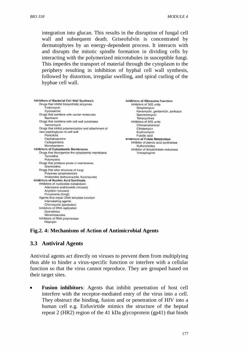
BIO 318 MODULE 4
177
integration into glucan. This results in the disruption of fungal cell
wall and subsequent death. Griseofulvin is concentrated by
dermatophytes by an energy-dependent process. It interacts with
and disrupts the mitotic spindle formation in dividing cells by
interacting with the polymerized microtubules in susceptible fungi.
This impedes the transport of material through the cytoplasm to the
periphery resulting in inhibition of hyphal cell wall synthesis,
followed by distortion, irregular swelling, and spiral curling of the
hyphae cell wall.
Fig.2. 4: Mechanisms of Action of Antimicrobial Agents
3.3 Antiviral Agents
Antiviral agents act directly on viruses to prevent them from multiplying
thus able to hinder a virus-specific function or interfere with a cellular
function so that the virus cannot reproduce. They are grouped based on
their target sites.
Fusion inhibitors: Agents that inhibit penetration of host cell
interfere with the receptor-mediated entry of the virus into a cell.
They obstruct the binding, fusion and or penetration of HIV into a
human cell e.g. Enfuvirtide mimics the structure of the heptad
repeat 2 (HR2) region of the 41 kDa glycoprotein (gp41) that binds

BIO 318 IMMUNOLOGY AND IMMUNOCHEMISTRY III
178
to the heptad repeat 1 (HR1) region and facilitates the fusion of the
viral envelope with the cell membrane. Binding of Enfuvirtide to
the HR1 region prevents the HR2 region from access to HR1
resulting in inhibition of the fusion process. Amantadin is a
tricyclic amine that inhibits penetration anduncoating, targeting
matrix protein and haemagglutinin. It blocks cellular membrane
ion channels and is effective against influenza A
viruses.Pleconaril a small cyclic drug binds to a canyon pore of the
picorna virus thereby blocking attachment and uncoating of the
viral particle. It fits into a hydrophobic compartment in the
nucleocapsid and disrupts the replication of the virus stopping the
shedding of nucleocapsid proteins from the RNA.
Inhibitors of Nucleic acid synthesis: Nucleoside and non-
nucleoside reverse-transcriptase inhibitors are two classes of
antiretroviral drugs that suppress HIV replication by affecting the
action of reverse transcriptase.Nucleoside-Analog Reverse
Transcriptase Inhibitors (NRTI) inhibit viral RNA-dependent DNA
polymerase (reverse transcription) and are incorporated into viral
DNA nucleoside chain to prevent ongoing viral DNA synthesis
causing chain-termination e.g. Zidovudine. Zidovudine, an
analogue of thymidine is phosphorylated by cellular enzymes
to the triphosphate form. Thereafter it competes with equivalent
cellular triphosphates which are essential substrates for the
formation of proviral DNA by viral RNA dependent DNA
polymerase. It terminates the chain when it gets inserted into the
growing viral DNA strand. Non-Nucleoside Reverse Transcriptase
Inhibitors (NNRTIs) are not incorporated into viral DNA but
inhibit HIV replication directly by binding non-competitively to
reverse transcriptase retarding its function. They attach to the
reverse transcriptase and affect the activity of the enzyme by
restricting its mobility and making it incapable to function.
Delavirdine a non-nucleoside reverse transcriptase inhibitor
(NNRTI) of HIV-1 binds directly to reverse transcriptase and
obstructs RNA-dependent and DNA-dependent DNA polymerase
activities. It does not compete with primer or deoxynucleoside
triphosphates.
Protein processing inhibitors: Protease Inhibitors
are substrate analogues for the HIV aspartyl protease enzyme,
involved in the processing of viral proteins. They bind to the HIV
protease active site and block it from further activity thereby
preventing the enzyme from cutting the viral protein molecules to
their correct sizes. This hinders the viral maturation process
leading to lack of functional virion formation. This protease does
not occur in humans because it is a virus specific protease. The

BIO 318 MODULE 4
179
viral particles cannot make copies capable of infecting other cells.
Protease inhibitors inhibit viral production in cell that have been
infected for a long time and newly infected cells compared to
NRTIs and NNRTIs that are active against only newly infected
cells e.g. Saquinavir. All Protease inhibitors inhibit the cytochrome
P450 enzymes.
Thymidine kinase substrates: Acyclovir (acycloguanosine) a
chain terminator inhibits viral DNA polymerase. It enters the cell
through the plasma membrane in form of a nucleoside and is then
distinctively phosphorylated inside the cell by herpes virus
thymidine kinase to an active form. It subsequently obstructs DNA
synthesis by hindering polymerisation.
DNA integration:Integrase inhibitors target the HIV enzyme
responsible for the integration of viral genetic material into human
DNA (integrase). Raltegravira 1-N-alkyl-5-hydroxypyrimidinone
inhibits the insertion of HIV-1 viral genomic DNA into the host
chromosome. It extends first phase viral decay and alters viral
decay kinetics of HIV by significantly reducing the second phase
and challenging current hypotheses of viral replication.Elvitegravir
is a specific inhibitor of the strand-transfer step of HIV integration
and is active against HIV-1 and HIV-2. It is also active against
organisms resistant to nucleoside reverse-transcriptase inhibitors
(NRTIs), non-nucleoside reverse-transcriptase inhibitors (NNRTIs)
and protease inhibitors (PIs).
RNA synthesis inhibitors: Ribavirin interferes with viral DNA
synthesis and can inhibit both DNA and RNA viruses. It introduces
multiple mutations into viral RNA making it unable to infect new
cells. Neplanocin A is an effective inhibitor of S-
adenosylhomocysteine hydrolase, and may also inhibit capping of
mRNA. They also hinder HIV replication by inhibiting the trans-
activation process.
Protein modification inhibitors: Castanospermine apotent
inhibitor of lysosomal alpha- and beta-glucosidasesmay interrupt
folding of some viral proteins by preventing the elimination of the
terminal glucose residue on N-linked glycans.Neuramidase
inhibitors such as Zanamivir and Oseltamivir inhibit
neuraminidase that is essential for viral replication and are
effective against influenza A and B virus.
Protein synthesis inhibitors: e.g.Fomivirsen a
phosphorothioateoligonucleotide inhibits the replication of human
cytomegalovirus (HCMV) by binding to the corresponding
BIO 318 IMMUNOLOGY AND IMMUNOCHEMISTRY III
180
sequence of the mRNA transcribed from the major immediate-
early transcriptional unit of CMV. Fomivirsen binds to the target
mRNA resulting in inhibition of IE2 protein synthesis, which then
inhibits virus replication.
Immunomodulators: Interferons induce in the host cells
ribosomes enzymes which inhibit viral mRNA e.g. hepatitis B
virus. Pavilizumab is a mononclonal antibody against infusion
proteins of respiratory syncytial virus.
3.4 Antiparasitic Agents
Antiparasitic agents are a class of drugs which are used for the treatment
of parasitic diseases e.g. Helminthiasis, amebiasis, malaria. Parasitic
infections belong to a class of infectious diseases that
have protozoa or helminths as the infectious agents.
Antihelmintic Drugs: Antihelmintics are divided into classes
based on the similarity of chemical structure and mode of action
e.ginhibitors of tubulin polymerisation, potentiation of inhibitory
transmitters, inhibitors of glucose uptake, uncouplers of oxidative
phosphorylation, muscle hyperpolarization, cholinesterase
inhibitors, cholinergic agonists and inhibitors of enzymes in the
glycolytic pathway.
Inhibitors of tubulin polymerization: Mebendazole binds to
beta-tubulin, interfers with beta-tubulin dependent glucose uptake
preventing microtubule formation and effects glycogen depletion.
It blocks the uptake of glucose leading to depletion of the
helminth’s own glycogen with a resultant decrease in adenosine
triphosphate formation. It causes degenerative alterations in the
outer body covering among members of
the phylum Platyhelminthes and intestinal cells of these worms by
binding to the colchicine-sensitive site of tubulin, thereby
inhibiting its polymerisation or assembly into microtubules. This
leads to immobilisation of the parasite and ultimately death.
However, mebendazole does not affect serum glucose
concentrations in humans and used in treatment of Ascariasis and
Enterobiasis.
Albendazole binds to beta-tubulin and prevents microtubule
assembly thus inhibiting beta-tubulin dependent glucose uptake. It
also inhibits fumaratereductase, decreases levels of NADH causing
degradation of endoplasmic reticulum and mitochondria with
decreased production of ATP.

BIO 318 MODULE 4
181
Potentiation of inhibitory transmitters: Macrocyclic lactones are
a group of hydrophobic compounds with broad spectrum
antinematodal and antiarthropodal properties derived from soil
microorganisms belonging to the genus Streptomyces. Inhibitors
likeivermectineprinomectin, selamectin. ivermectin activate the
opening of gated chloride channels. They are potent against many
immature nematodes and arthropods by binding to glutamate-gated
chloride channel receptors in nematode and arthropod nerve cells.
This results in the opening of the gated channel leading to an influx
of chloride ions with subsequent paralysis of the pharyngeal
pumping mechanism, body wall and uterine muscles of helminths.
Paralysis of uterine muscles results in disruption of reproduction.
Inhibition of glucose uptake: Blocking the uptake of glucose, the
primary source of worms’ energy would lead to starvation and
ultimate death of the parasite. This may be achieved by modulating
the cytosolic or mitochondrial enzymes. Praziqunatelan
isoquinolone analog increases cell membrane permeability in
susceptible worms and flukes leading to loss of intracellular
calcium, paralysis and contractions of the parasite’s muscles and
subsequently expulsion of the parasite. It is active against
Tapeworms. Inhibition of glucose uptake by Praziquantel in
Hymenolepisdiminuta may be mediated through modulation of
mitochondrial enzymes. Mebendazole is vermicidal in action and
causes degeneration of parasite's cytoplasmic microtubules and
thereby carefully and permanently blocks glucose uptake in
susceptible adult intestine-dwelling helminths and their tissue-
dwelling larvae leading to depletion of the parasite's store of
glycogen.
Uncouplers of oxidative phosphorylation in mitochondria e.g.
fasciolicides mainly salicylanilides and substituted phenols inhibit
the coupling between the electron transport and phosphorylation
reactions and thus inhibit Adenosine Triphosphate (ATP) synthesis
without affecting the respiratory chain and ATP synthase (H+-
ATPase). They exhibit protonophoric action in the H+-
impermeable mitochondrial membrane, permitting hydrogen ions
to leak through the inner mitochondrial membrane. They are active
against the hematophagous nematodes, eg, Haemonchus and
Bunostomum. Niclosamide inhibits oxidative phosphorylation in
the mitochondria of cestodes. When Clorsulon is ingested by
Fasciolahepatica they are killed since glycolysis is inhibited and
cellular energy production is disturbed.
Cholinesterase Inhibitors: Organophosphates bind to the enzyme
usually in charge for breaking down acetylcholine after it has

BIO 318 IMMUNOLOGY AND IMMUNOCHEMISTRY III
182
passed its message through the synapse. It makes the
cholinesterase unavailable for breaking down the acetylcholine so
the neurotransmitter continues to cause the neuron to transmit
electrical charges leading to overstimulation of the nervous system
causing death.Trichlorfon irreversibly binds to the active site of
acetylcholine esterase, thereby inactivating it. This leads to loss of
control by the parasite over its nervous system causing paralysis.
Cholinergic agonists or parasympathomimetics: are nicotinic
receptor agonists that combine with acetylcholine receptors and
imitate the effects of parasympathetic stimulation by causing
spastic muscle paralysis due to persistent activation of the
excitatory nicotinic acetylcholine receptors on the body wall
muscle of nematodese.g. imidazothiazoles and pyrimidines.The
antihelmintic activity of imidazothiazoles is attributed mainly to
their cholinomimetic activity, whereby they stimulate ganglion-
like structures in somatic muscle cells of nematodes. This results in
continued muscular contractions leading to neuromuscular
depolarising blockade causing paralysis. levamisole’sagnositic
activity as an antiparasitic agent is of the ganglionic nicotinic type.
It acts against the L-subtype nicotinic acetylcholine receptors in
nematode muscles thereby reducing the capacity of the males to
control their reproductive muscles and limit their ability to
copulate.Levamisole is highly effective against both adult and
immature stages of Ascarissuum.
Muscle hyperpolarisation: Drugs like Piperazine targets nervous
synaptic transmission. Piperazine, a pharmacological analogue of a
natural inhibitory transmitter blocks neuromuscular transmission in
the parasite by hyperpolarising the nerve membrane leading to
flaccid reversible paralysis of body wall muscle. The extent of
muscle hyperpolarisation by piperazine depends on the
concentration of extracellular chloride decreasing once a fraction
of the chloride ions is replaced by the sulphate anions. The parasite
is removed by peristalsis once it is paralysed and depleted of
energy. It also blocks succinate production by the parasite and is
active against ascarids.
3.5 Antiprotozoal Agents
Anti-protozoal drugs are used to treat infections or diseases caused by
Protozoa. The commonest diseases caused by protozoa are Malaria,
Trichomoniasis, Amoebiasis, Trypanosomiasis or sleeping sickness,
Toxoplasmosis e.t.c. These drugs destroy protozoa or prevent their
growth and ability to reproduce. The anti protozoal agent can target
either the enzymes found in both host and parasite but are indispensable

BIO 318 MODULE 4
183
only for the parasite, enzymes found exclusively in the parasites,
common biochemical functions found in both the parasite and the host
but with dissimilar pharmacological properties or exhibit a selective
toxicity for the parasite as compared to the host.
Enzymes found only in parasites:
Enzymes for dihydropteroate synthesis: 6-hydroxymethyl-7,8-
dihydropteroate synthase (DHPS) is an essential protein in lower
eukaryotes, prokaryotes and plants used in the synthesis of folate.
Folates are essential cofactors in the production of methionine,
thymidylate, glycine, purines etc for the organism. Therefore in
order to survive, intracellular sporozoan parasites e.g. Eimeriaspp,
Plasmodium and Toxoplasma must synthesize their own folate.
Plasmodia generate majority of their folates from the beginning,
and hence inhibition of dihydropteroate synthase by these drugs
leads to depletion of deoxythymidine triphosphate and decreased
DNA synthesis. In the treatment of human malaria caused by P.
falciparum, sulfadoxine has been used in combination with
pyrimethamine. Pyrimethamine is an antifolate drug that inhibits
dihydrofolatereductase. The sulfonamide group of compounds
such as sulfadoxine and dapsone, inhibit dihydropteroate synthase
which catalyzes the condensation of paraaminobenzoic acid
(PABA) with 6-hydroxymethyldihydropterin pyrophosphate to
yield 7,8-dihydropteroate. These drugs inhibit cell growth and
cause cell death through folate cofactor depletion. Sulfadoxine and
pyrimethamine are folic acid antagonists that sequentially inhibit
the activity of two enzymes involved in the biosynthesis of folinic
acid within the parasites dihydropteroate synthase and
dihydrofolatereductase respectively. The asexual erythrocytic
stages of Plasmodium falciparum are sensitive to sulfadoxine and
pyrimethamine.
Pyruvate:ferredoxinoxidoreductase are present in anaerobic
protozoa e.g. Entamoeba spp, Giardia and Trichomonas. This
enzyme can reduce the nitro group of metronidazole to form
cytotoxic reduced products that bind to DNA and proteins.
Nitazoxanide and Tenonitrozole are antiprotozoal agent that
interfere with the action of Pyruvate:ferredoxinoxidoreductase.
Nitazoxanide interferes with the pyruvate:ferredoxin 2-
oxidoreductase enzyme-dependent electron transfer reaction that
is vital to anaerobic energy metabolism in susceptible
organisms.Sporozoites and oocysts of Cryptosporidium parvum
and trophozoites of Giardia lamblia are sensitive to nitazoxanide
BIO 318 IMMUNOLOGY AND IMMUNOCHEMISTRY III
184
Nucleoside phosphotransferase: All of the protozoan parasites
studied thus far are deficient in de novo synthesis of purine
nucleotides. The purine nucleoside phosphotransferase found in
Leishmania can phosphorylate purine nucleoside analogs such as
allopurinol riboside, formycin B, 9-deazainosine, and
thiopurinolriboside converting them to the corresponding
nucleotides. These nucleotides are either further converted to
triphosphates or eventually incorporated into nucleic acids or
become inhibitors of other essential enzymes in purine
metabolism. T. vaginalis, T. foetus, and Giardia lamblia appear to
be deficient in de novo synthesis of both purines and pyrimidines.
Pyrimidine and purine salvage pathway becomes indispensable for
these parasites.
4.0 CONCLUSION
Bacterial cells have to replicate often in order to reach large numbers
required during an infection, therefore a large amount of biomolecules
are required to enable them grow and divide. There are some specific
processes essential for growth and or division of bacteria and these
processes are inhibited by antibacterial agents. These antimicrobial
agents are classified into groups based on their mode of action.
Antimicrobial agents may kill the target bacteria or fungus and are said
to be bactericidal or inhibit its growth (bacteriostatic).
5.0 SUMMARY
In this unit, you have learnt that:
antimicrobial agents inhibit specific cellular processes
some antimicrobial agents are bactericidal and others bacterioststic
antifungal agents target the plasma membrane, sterol, DNA and β
glucan biosynthesis
antiviral agents act directly on viruses to prevent them from
multiplying and inhibit a virus-specific function or interfere with a
cellular function to prevent them from replication
antiprotozoal agent can target either the enzymes found in both
host and parasite but are indispensable only for the parasite.
6.0 TUTOR-MARKED ASSIGNMENT
i. Why is folate very important in microbial metabolism?
ii. Could antimicrobial agents be both bactericidal and bacteriostatic?
iii. Why does the activity of salicylanilides not affect the respiratory
chain and ATP synthase?

BIO 318 MODULE 4
185
7.0 REFERENCES/FURTHER READING
Satyavan, S. & Nitya, A. (N.D). Approaches to Design and Synthesis of
Antiparasitic Drugs
Jozef, V. & Robert, S. R. (N.D). Macrocyclic Lactones in Antiparasitic
Therapy
Patrick R. Murray et al. (2009). Medical Microbiology. (6th
ed.).,
Baron, S,.(Ed.). (1996). .Galveston (TX): University of Texas Medical
Branch at Galveston.

BIO 318 IMMUNOLOGY AND IMMUNOCHEMISTRY III
186
UNIT 3 CHEMOTHERAPY OF SPECIFIC DISEASES
CONTENTS
1.0 Introduction
2.0 Objective
3.0 Main Content
3.1 Chemotherapy of Chagas Disease
3.2 Chemotherapy of Hodgkin’s Disease
4.0 Conclusion
5.0 Summary
6.0 Tutor-Marked Assignment
7.0 References/Further Reading
1.0 INTRODUCTION
A majority of infectious diseases can be treated with antibiotic or
chemotherapeutic agents. It is important to establish the diagnosis of the
kind of infection, prepare cultures to isolate the microorganism and
carry out sensitivity tests before commencement of chemotherapy.
2.0 OBJECTIVE
At the end of this unit, you should be able to:
describe the chemotherapy of Chagas and Hodgkin’s diseases.
3.0 MAIN CONTENT
3.1 Chemotherapy of Chagas Disease
Chagas disease is caused by the protozoan Trypanosomacruzi, and
it is prevalent in Latin America. The mode of transmission is
through hematophagousreduviid vectors such as
Triatomainfestans, which establish a habitat in human dwellings.
Over a long period, the pathogenesis of Chagas disease was
unclear and it was thought to be an autoimmune condition.
However, current knowledge indicates that parasitic invasion
coupled with an unbalanced immune response plays an important
role in the characteristic pathology observed in both acute and
chronic human chagas disease. Chemotherapeutic drugs most
frequently used in the treatment of chagas disease are
nitroheterocyclic compounds such as nitrofuran, nifurtimox
andbenznidazole a nitroimidazole derivative, radanil.
Trypanosomacruzi lacks the mechanisms to detoxify oxygen
metabolites especially hydrogen peroxides, and are thus more

BIO 318 MODULE 4
187
sensitive to oxidative stress than vertebrates. Nifurtimox thus acts
by reducing nitro groups to unstable nitroanion radicals, which in
turn react to produce toxic reduced oxygen metabolites.
Benznidazole acts through a different mechanism, by a covalent
modification of macromolecules by nitroreduction intermediates.
Nifurtimox and benznidazole have been observed to be very active
in the acute stage of chagas disease, while they are inactive at the
chronic stage. Due to their oxidative and reductive properties, they
are also associated with a lot of side effects such as anorexia,
vomiting, and allergies, and in some cases this usually leads to
discontinuation of the drugs. The major limitations of
nitroheterocyclic compounds is the difference in the level of
efficacy at the acute and chronic stages of the disease, even though
the reason for this disparity is yet unknown. Studies have also
shown that specific sterol inhibitors such as itraconazole, used in
the treatment of fungal diseases, inhibit sterols required by
Trypanosomacruzi for cell viability and proliferation at all stages
of cell division. Thus, important in the reduction of parasitic
load.Posaconazole and ravuconazole, triazole derivatives were
the first chemotherapeutic agents found to induce parasitological
cure in both acute and chronic stage of chagas disease. They were
also found to be able to eradicate nitrofuran and nitroimidazole
resistant strains of T. cruzi. A chemotherapeutic agent N-methyl-
piperazine-urea-F-HF-vinyl-sulphone-phenyl inhibits cruzain,
responsible for the major proteolytic activity of all stages of the
parasites life cycle, and as a result prolongs survival and induces
parasitological cure in acute and chronic stages of human chagas
disease with minimal toxicity. Inhibition of cruzain seems to be an
effective anti-T. cruzi target and so many current researches are
being modeled towards this mechanism.
3.2 Chemotherapy of Hodgkin’s Disease
Hodgkin’s disease is a lymphoma, a cancer of the lymphatic system.
Hodgkin’s disease is distinguished from non Hodgkin’s lymphoma by
the presence of large abnormal cells, known as Reed-Sternberg cells.
Hodgkin’s disease is classified into two types, classical Hodgkin’s
lymphoma which is more common and the less common nodular
lymphocyte-predominant Hodgkin’s disease. Hodgkin’s disease is
considered one of the most curable forms of cancer, if diagnosed and
treated early. The main types of treatments for Hodgkin’s disease are
radiation and chemotherapy. The standard chemotherapy regimens for
Hodgkin’s disease are ABVD and standard V. ABVD is a four drug
combination; doxorubicin, bleomycin, vinblastine and dacarbazine,
while standard V is a seven drug combination; etoposide, prednisone,
vincristine, mechlorethamine, doxorubicin, bleomycin, and

BIO 318 IMMUNOLOGY AND IMMUNOCHEMISTRY III
188
vinblastine. BEACOPP (doxorubicin, bleomycin,
vinblastine,cyclophosphamide,etoposide, prednisone, and
precarbazine) is a chemotherapy regimen reserved for high risk
patients, and has been proven to be very effective especially at the
advanced stage of the disease. Although it has been shown, that
treatment with this combination increases the risk of leukaemia in
patients. There are numerous side effects associated with the use of
chemotherapeutic regimens, and these effects increases with higher
doses and over the course of treatment of the disease. Common mild and
temporary side effects are vomiting, nausea, hair loss, weight loss,
diarrhea and depression. Serious side effects that may occur are;
neutropenia, which is a severe drop in white blood cells, anaemia, a drop
in red blood cells, liver and kidney damage, allergic reaction and
abnormal blood clotting. However, drugs have been developed to reduce
the intensity of the adverse effects of these combinations, but the
duration of the combat actions of these drugs are short. Long term
implications associated with the use of these combinations are infertility,
fatigue, menopause, heart failure, and bone thinning. Bleomycin has
been associated with lung toxicity, while vinblastine is observed to be
toxic when used in combination with radiation. The combination of
chemotherapy and radiation are used in the treatment of the advanced
stages of Hodgkin’s disease.
4.0 CONCLUSION
Chemotherapy has proved effective in the treatment of infectious
diseases, and is used as a major form of cancer treatment or in
combinations with other forms of therapy in cancer treatment. However,
the adverse effects associated with the use of chemotherapy is a cause of
concern, as it has been reported that sometimes most patients die as a
result of the effects of the drugs used.
5.0 SUMMARY
In this unit, you have learnt that;
the effective chemotherapeutic agents developed towards the
treatment of Chagas disease are modeled towards the inhibition of
cruzain
the effective chemotherapeutic agents used in the treatment of
Hodgkin’s disease are used in combination with radiation
the side effects of chemotherapy have become a subject of recent
research.

BIO 318 MODULE 4
189
6.0 TUTOR-MARKED ASSIGNMENT
Differentiate between the chemotherapy of Chagas disease and that of
Hodgkin’s disease.
7.0 REFERENCES/FURTHER READING
Julio, A.U. & Roberto, D. (2003). “Specific chemotherapy of chagas
disease: controversies and advances”. Trends in Parasitology.
19:495-501.
Martin, W. et al. (2005). "Bleomycin pulmonary toxicity has a negative
impact on the outcome of patients with Hodgkin's lymphoma". J
ClinOncol 23 (30): 7614–20.
Canellos, G. et al.(2004). "How important is bleomycin in the
adriamycin + bleomycin + vinblastine + dacarbazine regimen?". J
ClinOncol 22 (8): 1532–3.

BIO 318 IMMUNOLOGY AND IMMUNOCHEMISTRY III
190
UNIT 4 PHARMACO-DYNAMICS AND PHARMCO-
KINETICS
CONTENTS
1.0 Introduction
2.0 Objectives
3.0 Main Content
3.1 Pharmacodynamics
3.2 Pharmacokinetics
3.2.1 Absorption
3.2.2 Distribution
3.2.3 Metabolism
3.2.4 Excretion
4.0 Conclusion
5.0 Summary
6.0 Tutor-Marked Assignment
7.0 References/Further Reading
1.0 INTRODUCTION
Pharmacodynamics is study of the biochemical and physiological effects
of drugs on the body or on microorganisms or parasites within or on the
body and the mechanisms of drug action and the relationship between
drug concentration and effect. While pharamacokinetics involves the
determination of the fate of substances administered externally to a
living organism. Pharmacodynamics can be simply referred to as what
the drug does to the body, while pharmacokinetics is what the body does
to the drug.
2.0 OBJECTIVES
At the end of this unit, you should be able to:
describe pharmacodynamics and pharmacokinetics
list the processes of pharmacokinetics involved in drug action.
3.0 MAIN CONTENT
3.1 Pharmacodynamics
The mode of most drug actions is in form of mimicry of major vital
processes of parasites and microorganisms, or rather through inhibition
of these vital processes. Mode of drug actions, under these common
methods are; stimulating or depressing actions by directly binding to
receptors to trigger response, blocking action by directly binding to

BIO 318 MODULE 4
191
receptor without activating it, stabilizing action by acting neither as a
stimulant or depressant, replacing or accumulating substances to form a
reserve, scavenging free radicals obtained from oxidation, and direct
harmful chemical reaction that may result in the damage or destruction
of the cells through induced toxic or lethal damage. Only few drugs are
specific in the choice of receptor binding, but most have relative
specificity. The ability of a drug to affect a given receptor is determined
by its affinity and intrinsic efficacies, which are further determined by
its chemical composition. However, there are some drugs that induce
effects without binding to receptors or altering cellular functions, e.g.
antacids act by just decreasing gastric acidity through chemical reactions
with acids to produce salts. The desired effect of a drug on the body is
observed mainly due to one of the following reasons; interaction with
structural or carrier proteins, interaction with ion channels, or disruption
of the cell membranes. For example, aspirin acts by irreversibly
inhibiting the enzyme prostaglandin synthetase, as a result preventing
inflammatory response, and colchicine interferes with the structural
protein tubulin. Pharmacodynamics is not restricted to benefits alone,
there are also adverse effects associated with the use of drugs, e.g.
induced physiological damage or abnormal chronic conditions, or
increasing the probability of cell mutation. It has been observed that a
drug’s pharmacodynamics can be affected by physiological changes due
to disorders, aging or the effects of other drugs. Disorders such as
genetic mutations, malnutrition, Parkinson’s disease or insulin-resistant
diabetes mellitus are affected. These disorders could act by changing
receptor binding such as aging, alter the level of binding proteins or
decrease receptor sensitivity. Other drugs affect drug
pharmacodynamics by competing with receptor binding sites or
alteration of post-receptor responses.
3.2 Pharmacokinetics
This is often studied in association with pharmacodynamics.
Pharmacokinetics includes the study of the mechanisms of absorption
and distribution of an administered drug, the rate at which a drug action
begins and the duration of the effect, the chemical changes of the
substance in the body and the effects and routes of excretion of the
metabolites of the drug. Drug pharmacokinetics determines the onset,
duration and intensity of a drug’s effects. The processes of
pharmacokinetics can be explained and summarised by absorption,
distribution, metabolism and excretion.
3.2.1 Absorption
Drug absorption is determined by the drug’s physiochemical properties,
formulation and route of administration. Most drugs are weak organic

BIO 318 IMMUNOLOGY AND IMMUNOCHEMISTRY III
192
acid or bases, existing in ionized or non-ionised forms. The non-ionised
forms are lipid soluble, and readily diffuse across membranes, while the
ionized forms are water soluble. Drugs are mostly administered orally,
and must be in a solution in order to be absorbed. Drugs administered
orally, must cross several semi-permeable membranes before they reach
the systemic circulation. Hence mode of drug transport across
membranes is through passive diffusion, pinocytosis or active transport.
Other factors affecting absorption of drugs are differences in luminal
ρH, presence of bile and mucus, and nature of epithelial membranes.
However, drugs administered through IV injection enter the systemic
circulation immediately. Perfusion, which is blood flow per gram of
tissue, also affects absorption of drugs. In other words, site of injection
affects absorption rate. The rate and extent to which a drug enters
systemic circulation to gain access to the site of action, known as
bioavailability is dependent on properties of the dosage form, age, sex,
physical activity, stress, disorders e.t.c.
3.2.2 Distribution
Drugs are distributed to tissues after they gain entry into the systemic
circulation. Distribution is affected by differences in the regional ρH,
cell membrane permeability, tissue binding and blood perfusion, and
these results to an uneven distribution of drugs to tissues. The rate of
blood flow to the tissue, tissue mass, and partition characteristics
between blood and tissue determine the rate of entry of drug into the
tissue.
3.2.3 Metabolism
This is the irreversible transformation of a compound into daughter
metabolites, and the principal site for this is in the liver. Drugs can be
metabolised by oxidation, reduction, isomerisation, conjugation,
hydrolysis, condensation or hydration, mainly for easier excretion by the
body. Rate of drug metabolism is dependent on the individual genetic
components, disorders or drug effects affecting metabolic rates, and the
age of the patients.
3.2.4 Excretion
This is simply the removal or elimination of broken down metabolites
from the body, although rarely, these substances may accumulate in the
body tissues. Most drugs and their metabolites are excreted from the
body through renal and biliary excretion. Renal excretion provides the
most common form of mechanism of drug excretion.

BIO 318 MODULE 4
193
Patients- related factors such as genetic properties, sex and age can be
used to predict the pharmacological response of populations. For
example, the half-life of some drugs is remarkably longer in the elderly.
4.0 CONCLUSION
As a result of individual differences, administration of drugs should be
based on individual needs, adjusting dosage levels continually till the
target therapeutic condition is met. Although this approach can lead to
adverse effects, but knowledge of pharmacokinetics helps to ensure
dosages are adjusted appropriately and accurately.
5.0 SUMMARY
In this unit, you have learnt that:
Pharmacodynamics and pharmacokinetics are interrelated studies
Drugs act by either acting similarly to microorganisms or through
inhibition of their properties
Drugs that selectively bind to receptors, stimulate, depress or block
the actions of these receptors.
6.0 TUTOR-MARKED ASSIGNMENT
i. What is pharmacodynamics?
ii. Explain the processes of pharmacokinetics.
7.0 REFERENCES/ FURTHER READING
Lees, P, Cunningham, F.M. & Elliott, J (2004). "Principles of
pharmacodynamics and their applications in veterinary
pharmacology". J. Vet. Pharmacol.Ther. 27 (6): 397–414.
David, E.G. et al. (2005). Principles of Pharmacology. (1st ed.).
Lippincott: Williams & Wilkins.

BIO 318 IMMUNOLOGY AND IMMUNOCHEMISTRY III
194
UNIT 5 DRUG BIOASSAYS AND SENSITIVITY TESTS
CONTENTS
1.0 Introduction
2.0 Objective
3.0 Main Content
3.1 Drug Bioassays
3.2 Drug Sensitivity Tests
4.0 Conclusion
5.0 Summary
6.0 Tutor-Marked Assignment
7.0 References/Further Reading
1.0 INTRODUCTION
Biological assays or bioassays are scientific experiments used to test the
effects of a compound on a living organism, and are essential in drug
developments and monitoring of environmental pollutants. Sensitivity
tests are laboratory methods used to determine the susceptibility of
microorganisms to drug therapy.
2.0 OBJECTIVE
At the end of this unit, you should be able to:
describe drug bioassays and explain various drug sensitivity tests.
3.0 MAIN CONTENT
3.1 Drug Bioassays
Several drugs are isolated from natural products, and as a result
pharmacological screening or evaluation is necessary to guide the
isolation process towards the production of a pure active bio-component.
Bioassays determine the concentration of purity or biological activity of
substances such as hormones and plant extracts, while measuring the
effects on an organism or tissue cells compared to a standard
preparation. The purposes of drug bioassays are; measurement of the
pharmacological activities of new or chemically undefined compounds,
investigating the functions of endogenous mediators, to understand the
side effects of the drug to be used, and concentration measurement of
known substances. Drug bioassays are usually quantitative, in that they
measure the biological response evoked by the use of such drugs, and
they are usually analysed using biostatistics. A drug bioassay may be
primary or secondary assay depending on specific criteria such as cost

BIO 318 MODULE 4
195
and duration of results. A primary bioassay may not be quantitative, and
is applied to a large number of samples; it is cheap and produces results
quickly. While secondary bioassays are applied to more detailed
samples, are slow and costly. A primary bioassay should meet certain
criteria such as:
its results should be able to predict therapeutic potential of the
substance being tested either directly or indirectly by comparing
with other known effective drugs that have been screened through
the same process
a potentially useful pharmacological activity should not go
undetected even though the activity may be unexpected or unique
the probable nature of the activity should be highlighted such that
subsequent research can be carried out intelligently
the bioassay should be tolerant of many impurities present in the
crude extract, but also be able to detect the presence any interesting
substances in low concentration
the bioassay procedure should be unbiased and it should allow for
coding of all samples
results obtained should be reproducible
the screen should allow the use of both crude materials and pure
isolates so that the procedures can be used to direct the extraction,
isolation and purification work of the natural product chemist
the procedure should not require expensive equipments or
sophisticated laboratory environment so that the primary level of
screening experiment can be conducted synchronously with the
fractionation process
the procedure should be simple enough to be taught to laboratory
technicians so that there would not be a need to require highly
trained and qualified researchers to run the routine operation of
bioassay program
test animals, if required for the bioassays should be easily
obtainable, easily bred, easily handled and resistant to the infection
most importantly the bioassay should be economical to conduct
over a long period of time.
There are four approaches that serve as a bioassay guide in drug
discovery research, and the choice of screening approach depends on the
target disease and available information about the target organism to be
studied. The approaches are:
a. the use of a single bioassay technique to search for a specific type
of pharmacological activity e.g. anti-inflammatory activity
b. the use of specific bioassay technique, with each procedure
designed to capture specific useful properties
c. the use of single bioassay technique to discover multiple activities

BIO 318 IMMUNOLOGY AND IMMUNOCHEMISTRY III
196
d. the use of a variety of combination of bioassays to detect specific
and multiple activities.
There are basically two types of bioassays; Quantal and Graded. A
quantal bioassay requires all or none response, which has no room for
intermediate results. In the case of toxicity studies, the animal receiving
the drug either dies or not. Graded bioassay is based on the observation
that, proportionate increase in dose levels causes a similar increase in
response. Techniques involved in the determination of bioassays are:
a. Matching bioassays: The simplest type of bioassay and it involves
taking the response of the first substance and then matching it with
the standard response. This method is applied to small sample sizes
and the major limitation is that it does not take into consideration
sensitivity of the drug; hence it is not precise or reliable.
b. Interpolation method: This is conducted by determining the
amount of preparation of unknown potency required to produce a
definite effect on suitable tissues or test animals under a standard
condition, and the effect is compared to the standard.
Other techniques are bracketing method and multiple point assays.
Statistical methods are more reliable method of calculating
concentration of drugs in bioassay techniques.
3.2 Drug Sensitivity Tests
When a microorganism has been recovered from a clinical specimen, it
is cultured and tested against a variety of drugs. If the growth of the
microbe is inhibited by the action of the drug, it is reported as being
sensitive to the drug, but if the actions of the drug have little or no effect
on the growth of the microbe, the microbe is reported to be resistant to
the drug. This process is known as drug sensitivity test, and culture used
in this test is dependent on the type and kind of microorganism. Drug
sensitivity tests help to provide information about the potency of certain
drugs, if they are still effective and to what extent if they are. Without
drug sensitivity tests, drugs are prescribed on a trial and error basis,
which could take many days for an observed improvement and
sometimes may lead to several complications during the course of
illness such as kidney impairment, or even lead to death of the patients.
Culturing microorganisms before testing for drug sensitivity is done to
enable reproduction and growth of the microbe for easy identification of
results. It usually takes up to 18 hours to obtain results for bacteria, but
it could take several weeks to observe colonies in the bacterium that
causes tuberculosis. Culture mediums are usually sterile plastic dishes
containing nutrient gel on which they feed, and sometimes substances
are added to the medium to suppress the growth of other bacteria not

BIO 318 MODULE 4
197
needed. However, viruses require living cells to grow. After culturing, a
small portion of the colony is removed and stained for proper
identification under the microscope. Once properly identified, drug
sensitivity test is carried out by inserting paper discs containing the
drugs of specification, into the bacteria culture medium. The medium is
checked after 24 hours to observe the drug which the bacterium is most
sensitive to. An effective drug sensitivity test does not depend solely on
the kind of drugs used, but also on the use of proper culture medium,
careful extraction of bacteria from colony, and adequate storage
facilities for the culture medium to avoid contamination from outer
source.
4.0 CONCLUSION
Drug bioassays are necessary in drug discovery research, while drug
sensitivity tests ensure the success of treatment of infectious diseases.
5.0 SUMMARY
In this unit, you have learnt that:
pharmacological screening of compounds or drugs is necessary in
order to determine its beneficial and detrimental effects, as well as
its potential for future drug therapy
screening of drugs could be primary or secondary, depending on
costs and specificity required
drug bioassay may be quant or graded, and techniques used in
bioassays are matching bioassay, interpolation method, bracketing
method and multiple point bioassay
drug sensitivity testing is an important process in the successful
treatment of diseases, and method of propagation of microbe
growth is a relevant factor in this process.
6.0 TUTOR-MARKED ASSIGNMENT
i. Mention 5 criteria that should be met by a primary bioassay, and
differentiate between a primary bioassay and a secondary bioassay.
ii. Explain the process of drug sensitivity test.
7.0 REFERENCES/FURTHER READING
Atta-ur-raman et al. (2005). Bioassay Techniques for Drug
Development. e-library.
Kubo, I. (1991). “Method in Plant Biochemistry” In: Dey, P.M. and
Harborne ,J. B.. (Eds). London: Hostettman ,K. Academic Press.