Embed Size (px)
Citation preview

I Ill11 ll111111 Ill Ill11 Ill11 US006673597B2 IIIII IIIII 11111 Ill11 IIIII 11111 11111111111111 Ill1 (12) United States Patent (io) Patent No.: US 6,673,597 B2
Wolf et al. (45) Date of Patent: Jan. 6,2004
GROWTH STIMULATION OF BIOLOGICAL CELLS AND TISSUE BY ELECTROMAGNETIC FIELDS AND USES THEREOF
Inventors: David A. Wolf, Houston, TX (US); Thomas J. Goodwin, Friendswood, TX (US)
Assignee: The United States of America as represented by the Administrator of the National Aeronautics and Space Administration, Washington, DC (US)
Subject to any disclaimer, the term of this patent is extended or adjusted under 35 U.S.C. 154(b) by 0 days.
Notice:
Appl. No.: 09/798,854
Filed: Feb. 28, 2001
Prior Publication Data
US 200210009797 A1 Jan. 24, 2002
Related U.S. Application Data
Division of application No. 091587,028, filed on Jun. 2, 2000, now Pat. No. 6,485,963.
Int. Cl? ................................................. C12M 1/10 U.S. C1. .................................. 435/298.2; 4351299.1 Field of Search ........................... 4351173.1, 173.8,
4351298.2, 299.1
References Cited
U.S. PATENT DOCUMENTS
3,133,003 A * 511964 Schaefer et al. 3,871,961 A * 311975 Gianessi ......... 4,377,639 A * 311983 Lee ......................... 4351299.1 4,487,834 A * 1211984 Brighton ..................... 4351173 4,703,010 A * 1011987 Yunker et al. ........... 4351173.8 4,762,795 A * 811988 Masson ................... 4351173.8 4,939,151 A * 711990 Bacehowski et al. ....... 4351402 5,134,070 A * 711992 Casnig .................... 4351173.6 5,270,205 A * 1211993 Rogalsky 5,316,945 A * 511994 Minuth ... 5,344,454 A * 911994 Clarke et al. ............ 623123.72 5,919,679 A * 711999 Blackman et al. 4351173.1 6,022,733 A * 212000 Tam et al. ............... 4351287.1 6,066,495 A * 512000 Fofonoff et al. ......... 4351289.1
* cited by examiner
Primary Examinerqavid A. Redding (74) Attorney, Agent, or Firm-James M. Cate
(57) ABSTRACT
The present invention provides systems for growing two or three dimensional mammalian cells within a culture medium facilitated by an electromagnetic field, and preferably, a time varying electromagnetic field. The cells and culture medium are contained within a fixed or rotating culture vessel, and the electromagnetic field is emitted from at least one elec- trode. In one embodiment, the electrode is spaced from the vessel. The invention further provides methods to promote neural tissue regeneration by means of culturing the neural cells in the claimed system. In one embodiment, neuronal cells are grown within longitudinally extending tissue strands extending axially along and within electrodes com- prising electrically conductive channels or guides through which a time varying electrical current is conducted, the conductive channels being positioned within a culture medium.
2,996,429 A * 811961 Toulmin, Jr. ................ 4351391 25 Claims, 11 Drawing Sheets
1
9
https://ntrs.nasa.gov/search.jsp?R=20080007454 2020-06-29T22:46:48+00:00Z

U S . Patent Jan. 6,2004 Sheet 1 of 11 US 6,673,597 B2

U S . Patent Jan. 6,2004 Sheet 2 of 11 US 6,673,597 B2
U S . Patent Jan. 6,2004 Sheet 3 of 11 US 6,673,597 B2

U S . Patent Jan. 6,2004 Sheet 4 of 11 US 6,673,597 B2
FIGURE 4

U S . Patent Jan. 6,2004 Sheet 5 of 11 US 6,673,597 B2
FIGURE 5
U S . Patent Jan. 6,2004 Sheet 6 of 11 US 6,673,597 B2

U S . Patent Jan. 6,2004 Sheet 7 of 11 US 6,673,597 B2

U S . Patent Jan. 6,2004 Sheet 8 of 11 US 6,673,597 B2

U S . Patent Jan. 6,2004 Sheet 9 of 11 US 6,673,597 B2
U S . Patent Jan. 6,2004 Sheet 10 of 11
3
1
2 1 I 14A
US 6,673,597 B2
13A 4 /
FIGURE 10

U S . Patent Jan. 6,2004 Sheet 11 of 11 US 6,673,597 B2
FIG. I I B I
i
12-
6 2
6 \+
FIG. I I A
0
12
+ 2
11-

US 6,673,597 B2 3 4
In the preferred embodiment, the presence of the time may be introduced as necessary to achieve adequate mass varying electromagnetic field potentiates the growth of transfer for each embodiment. nerve and other tissue. The time varying field may be In one embodiment, illustrated in FIGS. 10 and 11, induced by either: 1) a time varying current within a slip-ring contacts or their equivalents are electrically con- conductor, or 2) a time varying voltage between fixed s nected to the ends of the channels, and an external power conductors. In one embodiment, for example, the culture is source is provided for applying the time varying electrical placed nearby a conductor through which a time varying current defining the waveform through the channels. In current is passed, or between parallel plates upon which a another embodiment, the channel consists of a pair of time varying voltage is applied. In both cases, a time varying parallel, mutually spaced conductors across which a time electromagnetic field results within the area of interest, i.e., varying voltage is applied. This also achieves the time in the region of the cell culture. varying electromagnetic field but restricts it to the region
The system and process are utilized in combination with between the parallel electrodes, which is advantageous for known tissue culture processes to produce enhanced cell directing localized growth according to a desired physical growth, directed cell growth, and tissue formation and pattern. The present invention also relates to a system and organization. 15 method for culturing primarily two dimensional mammalian
A will be understood from the description to follow, the cells facilitated by a time varying electromagnetic field. The system is operable to up or down the activity of electrodes may either be in direct galvanic contact or gal- specific genes, general, growth promoting genes are up vanically isolated from the target cells. The present inven- regulated and growth inhibitory genes are down regulated. tion Provides a strategy to re-engineer nerve tissue and The effect is shown to persist for some period after termi- 2o myoneural junctions and can be used medically for axonal nation of the applied time varying field. This persistent, regeneration. growth promoting effect subsides after a period of some In one embodiment of the present invention, there is days, and the cells return to a growth state characterized by provided a system for growing three dimensional mamma- controls, having never been exposed to the fields. This is lian cells, comprising a rotating wall vessel containing a
tions for clinical medical care, i.e. nerve regeneration, are varying electromagnetic field is applied to enhance tissue therefore safer than if the “pseudo transformed” state per- growth which may occur on a shaped substrate. The elec- sisted. The set of gene transformations, associated with the tromagnetic field may be generated by means such as by time varying electromagnetic field, also promote the ability directing the current waveform directly through a conduc- of the growing tissue to adhere and thrive on substrates by 30 tive substrate (or substrate layer) or by projecting the field the induction of genes leading to the secretion of extracel- from an external antenna, or electrode adjacent to and lular materials favorable to the tissue microenvironment. spaced from the medium, the spacing being sufficiently
of producing the tirne varying electro- small relative to the strength of the electromagnetic field to magnetic field in the vicinity of the living tissue culture are induce effectual levels of electromagnetic field within the encompassed. In one embodiment, an array of conductive 35 medium, in aCCOrdance with the Particular application. A current carrying elements (or voltaic electrodes) are time varying electromagnetic field may be emitted from a arranged so as to intensify or focus the time varying elec- nearby plate or other suitable “antennae,” or a time varying tromagnetic (EM) field onto the culture. Each embodiment voltage may be applied across suitable electrodes (such as is characterized by a method for application of the time Plates) to Produce the time varying electromagnetic field. varying field to the target tissue, such as neuronal, for 40 The field generation system may either be rotating with the stimulation of growth, or repair or induction of changes in vessel Or fixed, and spaced from, the rotating vessel. The gene activity patterns, The term “field generator” is used rotating wall vessel can be a rotating wall perfused vessel or herein to represent these various embodiments for generat- a rotating wall batch-fed vessel. ing the time varying electromagnetic fields. In its simplest The time varying electromagnetic field is advantageously form, it is a conductive electrode, placed near the target 45 produced by a varying electrical potential in the form of a cells, through which current is directed from a controlled square wave having a frequency of approximateb ten cycles waveform current source. per second. In one embodiment, a current of about ten
A suggested above, in one embodiment, the field gen- milliamps, conducted between opposite corners of a metallic erator is in the form of a conductive channel mounted on or conductor, Produces a stimulatory time varying electromag- embedded in a disc of biocompatab~e material, (FIG, 11) so netic field extending several centimeters from the plate one or within a surface. In practice, the range of frequency and oscillating rotational bioreactor so as to obtain the beneficial culture electromagnetic field strength is a Parameter which may be conditions associated therewith, The of the selected to for achieving the desired stimulation of particular stimulatory electromagnetic field with the rotational tissues, cells, Or genes, and for Providing the appropriate environment, known to permit morphological expression ss m ~ ~ n t of uP/down reda t ion of these genes. beyond conventional culture, is particularly effective. This is In one embodiment of the present invention, the cell because the induced pattern of growth enhancing genes is growth substrates or carriers are spherical disks containing permitted to be ultimately expressed, as cell growth and multiple parallel channels (FIG. 10) which are coated with tissue formation, without mechanical inhibition from the a bioattractive material. The bioattractive material has a culture apparatus. Also the inherent growth advantages well 60 longitudinal axis across which the time varying electrical known in the rotational systems is synergistic with the potential is applied and through which a time varying
beneficial in certain applications, in that medical applica- 25 cell-rich medium and a formed cell growth substrate. A time
several
of these discs may be then
growth stimulation derived from the time varying electro- magnetic field. The conditions may be further optimized by utilizing actual microgravity, in space. In this application, mechanical rotation of the cell culture vessel is not required 65 conium and platinum. but may be utilized to achieve mixing and sufficient mass transfer to sustain a healthy culture. Other forms of mixing
current is conducted. The mammalian cells adhere to the bioattractive material and are free to orient, as they grow. Representative bioattractive materials include titanium, zir-
The class of mammalian cells preferably is selected from the group consisting of neuronal cells, normal human neu-

US 6,673,597 B2 5
ronal progenitor cells (NHNP), and a cell responding to the time varying electromagnetic field. It will be understood by those of ordinary skill in the art that the teachings of the present invention apply to other cell types.
In another embodiment of the present invention, there is provided a method of culturing mammalian cells in the claimed system, comprising the steps of inoculating the cells into the vessel containing a culture medium, rotating the vessel to enhance the proliferation of the cells and, in one embodiment, to initiate the attachment of the cells to micro- carrier spheres or beads suspended within the culture medium, applying a time varying electromagnetic field to the culture medium, cells, and cell carriers, and measuring the growth of the cells. Preferably, the vessel is rotated at a speed from about 2 RPM to 30 RPM, and the time varying electromagnetic field is generated by a time varying current passed through a conductor with RMS value of about 1 to 1,000 ma. In one embodiment, a range of about 1 mA to 6 mA is used.
In still another embodiment of the present invention, there is provided a system for growing two-dimensional neural cells, comprising a petri dish containing a cell culture medium and an electrode placed in the center of the petri dish. In this embodiment, the electrode serves as the field generator. Preferably, the neural cells are applied directly on the electrode. As a result, the neutral cells exhibit acceler- ated growth.
In yet another embodiment of the present invention, there is provided a system for growing two-dimensional neural cells further comprising a slide placed on the electrode. Preferably, the neural cells are applied, e.g., bubbled, on the slide instead of directly contacting the electrode, and preferably, the current producing the waveform is applied at a strength range of from about 1 mA to about 100 mA, and, in one embodiment, suitably from about 1 mA to 6 mA.
In still another embodiment of the present invention, there is provided a method of treating an individual having diseased neuronal cells, comprising the steps of growing neuronal cells in the claimed two- or three- dimensional systems and transplanting the neuronal cells into the indi- vidual. Such diseases include Parkinson’s disease, diseases of neuromuscular junction and Alzheimer’s Disease. Neural trauma can also be treated in same methodology.
In yet another embodiment of the present invention, the time varying electromagnetic field (or electrical potential) induces cellular response including cellular control of growth and differentiation at gene level. Preferably, the cellular control of growth and differentiation is to suppress or enhance growth regulatory functions at gene level. Still preferably, the gene is associated with increased tissue and cell proliferation.
Other and further aspects, features, and advantages of the present invention will be apparent from the following description of the presently preferred embodiments of the invention given for the purpose of disclosure.
BRIEF DESCRIPTION OF THE DRAWINGS So that the matter in which the above-recited features,
advantages and objects of the invention, as well as others which will become clear, are attained and can be understood in detail, more particular descriptions of the invention briefly summarized above may be had by reference to certain embodiments thereof which are illustrated in the appended drawings. These drawings form a part of the specification. It is to be noted, however, that the appended drawings illustrate preferred embodiments of the invention and therefore are not to be considered limiting in their scope.
6 FIG. 1 shows a petri dish with cells in a concentrated
bubble placed on the metal electrode in the center of the dish.
FIG. 2 shows normal human neuronal progenitor (NHNP) 5 cells grown in conventional tissue culture procedures.
FIG. 3 shows the perimeter of non-waveform influenced normal human neuronal progenitor cells 24 hours after the experiment.
FIG. 4 shows neural tube formation within normal human neuronal progenitor cells under the influence of waveform.
FIG. 5 shows neural tube generation within normal human neuronal progenitor cells under the influence of waveform.
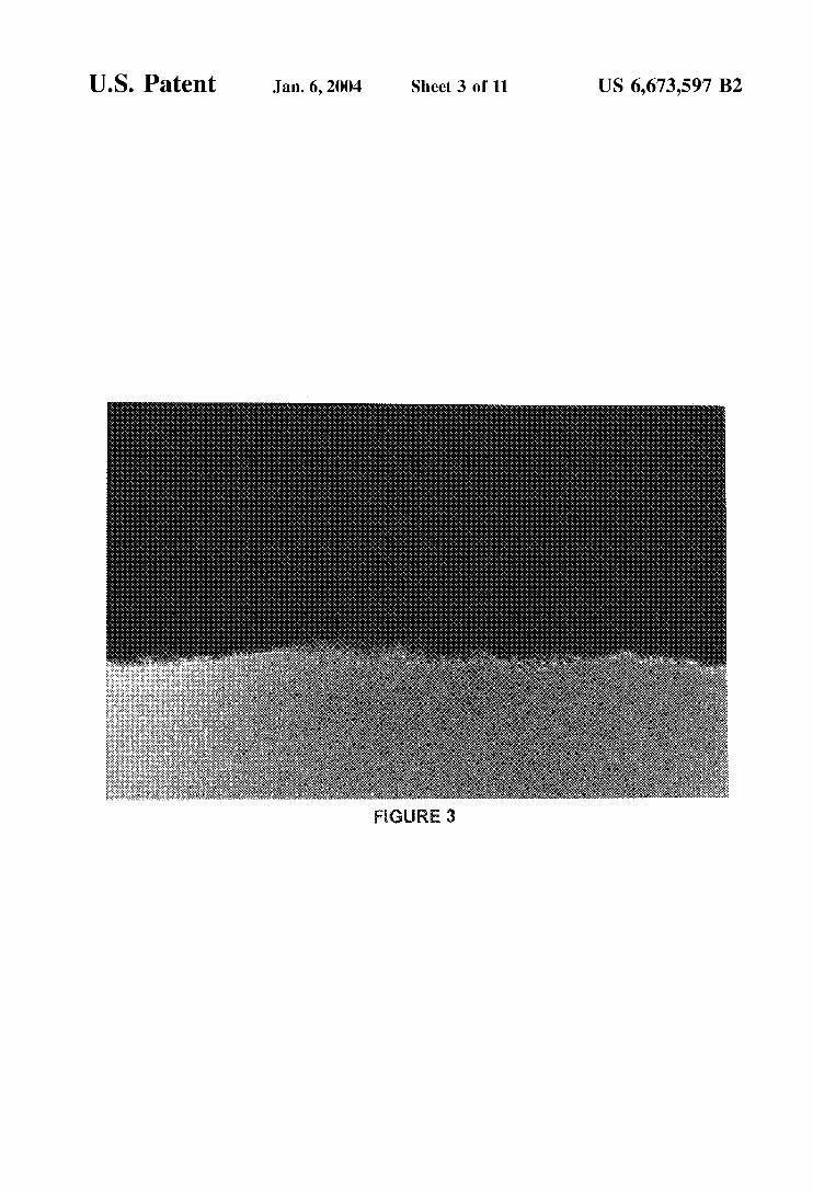
FIG. 6 shows the composition of waveform-influenced neural tissue 24 hours after the exposure.
FIG. 7 shows the waveform-influenced normal human neuronal progenitor cells 24 hours after the exposure.
FIG. 8 shows a close-up of waveform-influenced normal
FIG. 9 shows waveform-influenced normal human neu- ronal progenitor cells 24 hours after the exposure.
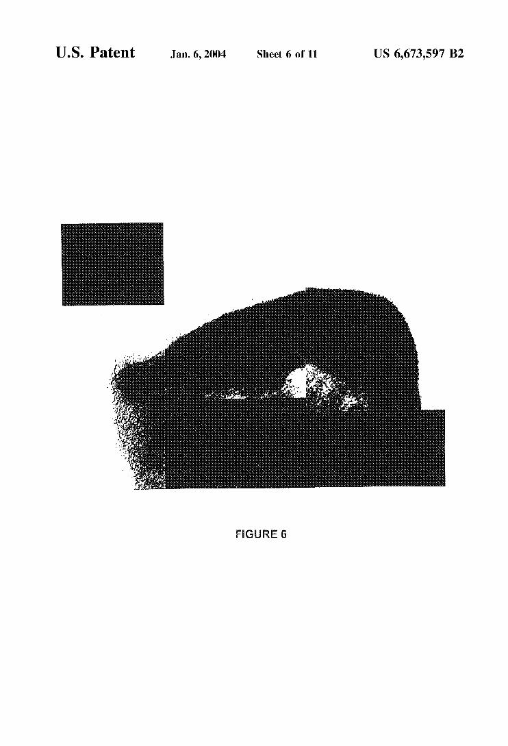
FIG. 10 shows prototype of the system 1, consisting of silicon plates 2, fluid coupling 3, slip rings 4, a rotating wall pressure vessel (RWPV) 5, electrical conductor 6 (i.e., an electrical conductive bioattractive inlay strip), a perfusion inlet 7, perfusion outlet 8, a stand for the rotating wall pressure vessel 9 and a source of time varying electrical
FIG. 11A is a plan view of one of the disc-shaped silicon plates 2, showing a central opening 11 about which rotation occurs, the electrical conductors 6, and electrical contacts 12 connected to the strips at opposite ends thereof.
FIG. 11B is a side view of the silicon plate 2, demon- strating the electrical contacts 12 and the electrical conduc- tive strips, in the form of bioattractive inlays 6.
DETAILED DESCRIPTION OF THE INVENTION
1 u
is
2o human neuronal progenitor cells.
2s
3o current 10.
35
40 As used herein, the term “bioattractive material” shall
refer to materials to which a cellular material will attach. As used herein, the term “longitudinally orient” shall refer
to orientation in an elongated cordlike fashion. As used herein, the term “parallel channels” shall refer to
electric channels which are designed to provide constant output to all the electrodes simultaneously.
As used herein, the term “cell carriers” shall refer to microcarrier beads, scaffolds and matrices which support the
As used herein, the term “rotating wall batch-fed vessel” shall refer to slow turning lateral vessel (STLV) and high aspect rotating vessel (HARV).
As used herein, the term “Corona Effect” shall refer to the accelerated growth pattern of neuronal cells electrically potentiated by waveform.
In one preferred embodiment, the present invention is directed to the growth of three dimensional mammalian
60 neuronal tissue using an electrically conductive strip in the form of a channel or mold coated with a bioattractive or biocompatable material to which an electrical potential is applied to longitudinally orient the neural cells or axons as they adhere to the bioattractive material which is suspended
65 in an axon rich medium. Specifically, in the present embodiment, the apparatus
includes a bioreactor chamber vessel employing electrically
4s
so growth and/or attachment of cellular materials.
ss

US 6,673,597 B2 7 8
insulative, biocompatable spherical disks of a material such diseased neuronal cells, comprising the steps of growing as silicon. These disks rotate inside the pressure vessel. Each neuronal cells in the two or three dimensional systems disk has multiple parallel channels cut into its surface. The disclosed herein and transplanting the grown neuronal cells channels have a semicircular cross-section and contain an into the individual. Such diseases include Parkinson’s electrically conductive inlay in the form of a channel-shaped s disease, diseases of neuromuscular junction and Alzheimer’s conductive strip of a bioattractive material such as Disease. Neural trauma can also be treated with this same zirconium, titanium and platinum. Each channel strip 6 has methodology. an electrical contact on each longitudinal end that is used to yet another embodiment of the present invention, the create and control an electrical potential along the length of waveform (or electrical potential) induces a cellular the strip. The vessel is filled with a medium and the disks are i o response including cellular control of growth and differen- rotated within a medium containing axons. The cells adhere tiation at gene level, preferably, the cellular control of to the electrically conductive bioattractive inlay material. growth and differentiation is to suppress or enhance growth The desired longitudinal cell orientation and therefore the regulatory functions at gene level, Still preferably, the gene structure of the resulting tissue is affected and/or controlled is associated with embryogenesis,
The following examples are given for the purpose of by the electrical stimulus. The present invention is also directed to the growth of two illustrating various embodiments of the invention and are
dimensional mammalian neuronal tissue using electrodes. not meant to limit the present invention in any fashion, The electrodes are either in direct contact or not in contact with the target cells.
provided a system for growing three dimensional mamma- lian cells, comprising a rotating wall vessel containing a cell-rich medium, cell carriers placed within the vessel and an electrical potential applied to the cell carrier. Preferably, the rotating wall vessel can be a rotating wall perfused vessel 25 culture following standard cell culturing procedures, or a rotating wall batch-fed vessel.
In one embodiment of the present invention, the cell carriers are spherical disks containing multiple parallel channels, which are coated with a bioattractive material. More preferably, the bioattractive material has a longitudinal axis across which the electrical potential is applied. The mammalian cells adhere to the bioattractive material and are therefore oriented longitudinally upon the electrical stimu- lus. Representative bioattractive materials include titanium, 35 coated or not coated), ~ l ~ ~ ~ ~ ~ d ~ ~ were made of zirconium and platinum.
In the methods of the present invention, the mammalian cell is selected from the group consisting of a neuronal cell, a normal human neuronal progenitor cell (NHNP) and a cell responding to waveform. A person having ordinary skill in 4o this art will be able to apply the teachings of the present invention to other cell types.
In another embodiment of the present invention, there is Provided a method of culturing mmmdian cells in the Initially, a metal electrode was placed inside a petri dish claimed system, comprising the steps of inoculating the cells 45 and centered. Normal human neuronal progenitor cells were into the vessel, rotating the vessel to initiate the attachment seeded at 2x10⁵ cells in 0.7 ml of media and carefully of the cell to the cell carriers, applying an electrical Potential dropped on the electrode in a concentrated bubble (FIG. 1). to the cell carriers and measuring the growth of the cells. Cells were incubated for 2 days. Second day after seeding is Preferably, the vessel is rotated at a speed from about 10 considered day 0 of the experiment. At day 0, each dish was RPM to 30 RPM, and the electrical Potential is applied at a 50 given 15 ml of media and waveform was applied to seven strength range of from about 1 mA to about 6 mA. electrodes. Cells were observed under a dissecting micro-
In still another embodiment of the present invention, there scope and fed with 15 ml of media at day 3, and 13 ml every is provided a system for growing two-dimensional neural three days at day 6, 9 and 12. At day 14, the cells were fed cells, comprising a petri dish containing a cell culture again with 13 ml of media. At day 17, the cells were medium and an electrode placed in the center of the petri 55 incubated for 10 minutes in a Collagenase/DNase cocktail, dish. The electrode is charged with a waveform. Preferably, then trypsin was directly applied to the cocktail and the cells the neural cells are bubbled directly on the electrode. As a were further incubated for 3 more minutes. Before the media result, the neutral cells exhibit accelerated growth. was added to deactivate trypsin, the cocktail mix was
In yet another embodiment of the present invention, there pipetted up and down several times. The cells were washed is provided a system for growing two-dimensional neural 60 twice with IxPBS, reapplied with the media and placed on cells further comprising a slide placed on the electrode. ice. The cells were counted, assessed for viability. Preferably, the neural cells are bubbled on the slide instead To examine the accelerated growth of cells 48 and 72 of directly contacting the electrode. Preferably, the wave- hours after waveform was discontinued, cells were treated form is applied at a strength range of from about 1 mA to the same as above, except that after day 14 treatment, instead about 6 mA. 65 of harvesting, two dishes from the non-waveform group
In still yet another embodiment of the present invention, (control) and two dishes from the waveform group were there is provided a method of treating an individual having randomly chosen and re-seeded at 9x10’ cells in two new
EXAMPLE 1
Cells In one embodiment of the present invention, there is 2o
~~~~~l human neuronal progenitor cells (NHNP) were pooled from three donors, controls, normal human neu- ronal progenitor cells were grown in conventional tissue
EXAMPLE 2
Materials 30
GTSF-2 medium with 10% FBS, ~ i ~ ~ ~ f l ~ ~ ~ ~ i ~ and F ~ ~ - gizone was used to culture the cells, l x p ~ s , collagenase, D N ~ ~ ~ and ~~~~~i~ were purchased from clonetics, The cells were grown on 12-100 mm Petri dishes (tissue culture
and stainless steel. A waveform generator was used to generate the waveform in a strength of 1-6 mA(AC) square wave, 10 Hz variable duty cycle,
EXAMPLE 3
Electrically Potentiating Cell Growth When Electrode is in Direct Contact with the Target Cells

US 6,673,597 B2 9
petri dishes each, with a total of four dishes. Cells from one set (#11 waveform and control #6) were counted and pho- tographed 48 hours after seeding, and cells from the second set (#12 waveform and control #7) were counted and pho- tographed 72 hours after seeding.
To examine accelerated growth pattern “Corona Effect” after the electrical potentiation, the same treatment was applied to the cells without harvesting. A dish each from waveform group and non-waveform group were chosen randomly. Cells still attached in sheet from were lifted off of the electrodes carefully and placed in new petri dishes with medium, and then photographed 24 hours later.
EXAMPLE 4
Electrically Potentiating Cell Growth When Electrode is not in Direct Contact with the Target
Cells
Initially, a metal electrode was placed inside a petri dish and centered. A slide was carefully placed on the electrode under sterile conditions. Normal human neuronal progenitor cells were seeded at 2x10’ cells in 0.7 ml of media and bubbled on the slide. Cells were incubated for 2 days. 25 ml of media were applied and two 1000 pl pipetman blue tips were placed in the dish to anchor the slide to bottom of the dish. The second day after seeding was considered day 0 of the experiment. At day 0, each dish was given 25 ml of media and waveform was applied to six of the twelve electrodes.
Cells were observed under a dissecting microscope and fed with 25 ml of media every three days at day 3, 6, 9 and 12. At day 14, the cells were fed again with 25 ml of media. At day 18, the cells were incubated for 10 minutes in a CollagenaseiDNase cocktail, then typsin was directly applied to the cocktail and the cells were further incubated for 3 more minutes. Before the media was added to deac- tivate trypsin, the cocktail mix was pipetted up and down several times. The cells were washed twice with IxPBS, reapplied with the media and placed on ice. The cells were counted, assessed for viability and then replated at 100,000 per plate. The remaining waveform and non waveform slides were fixed and refrigerated for staining at a later date.
EXAMPLE 5
Electrically Potentiated Growth of Cells
Normal human neuronal progenitor-pool cells exposed to a time varying electromagnetic field (waveform), either in direct contact or not in direct contact with the electrode, displayed an accelerated growth rate and different morphol- ogy as compared to non waveform cells (control), i.e., cells not subject to the time varying electromagnetic field (see Table 1 and Table 2, FIGS. 2-9). After the application of the time varying electromagnetic field or waveform, the cells preferentially aligned, while cells without waveform expo- sure showed random pattern. Cells in direct contact with the electrode remained stimulated up to at least 72 hours after waveform was removed (Table 3); while those not in direct contact with the electrode once removed from waveform continued to experience accelerated and long term stimula- tion growth pattern even after 168 hours (Table 4). Viability was also higher in the cells exposed to the waveform (Table 4). Cells were suspended easily with the CollagenaseiDNase then trypsin sequence.
10
TABLE 1
*Cell Count and Viability at Harvest (day 17)
5 NHNP-POOL CELL COUNT VIABILITY HARVEST
Waveform 1 860,000 98% 17 days Waveform 2 1,000,000 98% 17 days Waveform 3 1,000,000 98% 17 days
10 Waveform 4 1,300,000 98% 17 days Waveform 7 1,000,000 98% 17 days Waveform 8 940,000 98% 17 days Waveform 9 700,000 98% 17 days Waveform 10 1,000,000 98% 17 days
1s Control 1 500,000 98% 17 days Control 2 400,000 98% 17 days Control 3 300000 98% 17 days Control 4 500,000 98% 17 days Control 5 400,000 98% 17 days
*Cells were in direct contact with the electrode. 20
TABLE 2
2s *Cell Count and Viabilitv at Harvest (dav 18)
NHNP-POOL CELL COUNT VIABILITY HARVEST
Waveform 1 1,000,000 100% 18 days Waveform 5 1,100,000 100% 18 days
3o Control 1 800,000 100% 18 days Control 5 800,000 100% 18 days
*Cells were not in direct contact with the electrode.
3 s TABLE 3
*Cell Count at Various Times after Removal of Waveform
CELLS COUNT HOURS OFF ELECTRODE
Waveform 11 40 Control 6
Waveform 11 Control 6 Waveform 12 Control 7 Waveform 12
45 Control 7
520,000 112,000 274,000 48,000
576,000 96,000
228,000 120,000
Counted and re-seeded at 96,000iplate Counted and re-seeded at 96,000iplate 48 hours off electrode 48 hours off electrode Counted and re-seeded at 96,000iplate Counted and re-seeded at 96,000iplate 72 hours off electrode 72 hours off electrode
*Cells were in direct contact with the electrode.
TABLE 4 so
*Cell Count and Viabilitv at Various Times after Harvest
TIME AFTER HARVEST
NHNP-POOL CELL COUNT VIABILITY (Hours)
Waveform 1 56,000 85% 24 Waveform 5 40,000 85% 24 Control 1 36,000 65% 24 Control 5 28,000 65% 24 Waveform 1 188,000 98% 48
6o Waveform 5 212,000 98% 48 Control 1 74,000 87% 48 Control 5 162,000 90% 48
55
Waveform 1 3,400,000 100% 120 Waveform 5 3,400,000 100% 120 Control 1 900,000 99% 120 Control 5 900,000 99% 120
65 Waveform 1 4,000,000 100% 168 Waveform 5 3,800,000 100% 168

US 6,673,597 B2 11
TABLE 4-continued
12
TABLE 5-continued
*Cell Count and Viabilitv at Various Times after Harvest Down Regulated Genes in Descending Order (Highest to lowest)
Homo sapiens mRNA expressed in osteoblast, complete cds TIME AFTER 22. HARVEST {Incyte PD:2537863}
24. NHNP-POOL CELL COUNT VIABILITY (Hours) 23. EST {Incyte PD:3204745}
Control 1 980,000 97% 168 {Incyte PD:2732630} Control 5 900,000 95% 168 25. Homo sapiens serum-inducible kinase mRNA, complete cds
10 {Incyte PD:1255087} *Cells were not in direct contact with the electrode.
Homo sapiens mRNA for serineithreonine protein kinase SAK
26. 27. EST {Incyte PD:660376} 28. GRANCALCIN {Incyte PD:1671852} 29. N-CHIMAERIN {Incyte PD:1852659} 30.
31. Adenylosuccinate lyase {Incyte PD:1653326} 32. EST {Incyte PD:1798393} 33.
Carbonic anhydrase I1 {Incyte PD:2474163}
Homo sapiens Pig10 (PIG10) mRNA, complete cds {Incyte EXAMPLE 6 15 PD:1731061}
Waveform Gene Array Display (GAD) Results Homo sapiens HP protein (HP) mRNA, complete cds {Incyte PD:3084122) Normal Human Neural Progenitor cells or human adult
astrocytes were exposed to waveform and non-waveform growth conditions for 17 days. Upon completion of the exposure period cells were harvested via trypsinization and poly-RNA was prepared from the respective groups of cells. RNA samples were quick frozen and shipped to Synteni Corporation for GAD analysis. Below are the results of a survey of the response of over 10,000 human genes. The results were divided into two categories (Table 5 and Table 6). Those genes down regulated or suppressed by the wave- form and those up regulated or enhanced in activity by the waveform.
An analysis of the data indicates a significant down regulation of maturation and regulatory genes. These matu- ration and regulatory genes are normally associated with the differentiated or non-growth profile of normal cells. However, there is a significant up regulation of some 150 genes which are mainly associated with growth and cellular proliferation. Neither two nor three dimensional growth of neural cells has been achieved prior to this event with the positive outcome of enhanced growth and apparent gene regulatory control.
TABLE 5
Down Regulated Genes in Descending Order (Highest to lowest)
34.
2o 3s. 36.
37. 38.
25 39.
40.
41. 42.
43. 44. 4s. 46. 47.
48. 49.
so.
40 2;: 53. 54.
30
3 s
1. 2. 3. 4.
5. 6. 7. 8. 9.
10. 11.
12. 13. 14.
15. 16.
17. 18.
19. 20.
21.
Homo sapiens (clone Zap2) mRNA fragment {Incyte PD:1661837] CDC28 protein kinase 2 {Incyte PD:1384823} Synteni: YCFR 22 {YC 22.2OOO.W} ESTs, Moderately similar to cell growth regulating nucleolar protein LYAR [M.mwcuZus]{Incyte PD:2233551} KERHIN, TYPE I1 CYTOSKELETAL 7 {Incyte PD:1649959} MITOTIC KINESIN-LIKE PROTEIN-1 {Incyte PD:2640427} EST {Incyte PD:674714} Synteni: YCFR 22 {YC 22.2OOO.X} Synteni: YCFR 26 {YC 26.0062.N) Synteni: YCFR 22 {YC 22.2OOO.Z} Transcription factor 6-like 1 (mitochondrial transcription factor 1-like) {Incyte PD:3371995} Interferon-inducible 56-KDa protein {Incyte PD:1215596} EST {Incyte PD:1794375} Homo sapiens mitotic feedback control protein Madp2 homolog mRNA, complete cds {Incyte PD:2414624} EST {Incyte PD:151026} Homo sapiens Pig3 (PIG3) mRNA, complete cds {Incyte PD: 2395269) General transcription factor IIIA {Incyte PD:1527070} Cellular retinoic acid-binding protein [human, skin, mRNA, 735 nt] {Incyte PD:585432} EST {Incyte PD:1755159} Homo sapiens mRNA for KIAAO285 gene, complete cds {Incyte PD:1738053} ESTs, Weakly similar to F25HS.h [C.eZegans] {Incyte PD: 1923567)
45 56. 57.
58. 59.
SO 60.
61.
62. 63.
64.
65.
66. 60 67.
68.
55
69.
65 70.
ESTs, Modeiately similar to TlOC6.i [C.eZegans] {Incyte PD:1923186} Chromosome condensation 1 {Incyte PD:3180854} Calmodulin 1 (phosphorylase kinase, delta) {Incyte PD: 2803306) Centromere protein A (17kD) {Incyte PD:2444942} V-jun avian sarcoma virus 17 oncogene homolog {Incyte PD: 1920177) Human glutathione-S-transferase homolog mRNA, complete cds {Incyte PD:1862232} Homo sapiens gene for protein involved in sexual development, complete cds {Incyte PD:3033934} EST {Incyte PD:2630992} Human low-Mr GTP-binding protein (RAB32) mRNA, partial cds {Incyte PD:1662688} Annexin 111 (lipocortin 111) {Incyte PD:1920650} Hydroxymethylbilane synthase {Incyte PD:1509204} Synteni: HK 4 {HK 4.2000.Y) Ribosomal protein L7a {Incyte PD:2579602} Human mRNA for myosin regulatory light chain {Incyte PD: 78783) Ferredoxin reductase {Incyte PD:1819763} Human copper transport protein HAHl (HAH1) mRNA, complete cds {Incyte PD:2313349} Human G protein gamma-11 subunit mRNA, complete cds {Incyte PD:1988432} Synteni: HK 4 {HK 4.2000.W} Human XIST, coding sequence a mRNA (locus DXS399E) {Incyte PD:1514318) Ribosomal protein, large, PO {Incyte PD:3511355} Homo sapiens clone 23714 mRNA sequence {Incyte PD:1728368} Human mRNA for ApolLHuman (MERS(Aop1-Mouse)-like protein), complete cds {Incyte PD:2527879} Synteni: HK 4 {HK 4.2000.Z} Proteasome (prosome, macropain) subunit, beta type, 5 {Incyte PD:2503119} Human PINCH protein mRNA, complete cds {Incyte PD:126888} Homo sapiens peroxisome assembly protein PEXlO mRNA, complete cds {Incyte PD:998279} Homo sapiens short chain L-3-hydroxyacyl-CoA dehydrogenase (SCHAD) mRNA, complete cds {Incyte PD:1638850} Neuroblastoma RAS viral (v-ras) oncogene homolog {Incyte PD:2816984} H.sapiens mRNA for b4 integrin interactor {Incyte PD:1932850} Human forkhead protein FREAC-1 mRNA, complete cds {Incyte PD:1449920} Human mRNA for protein D123, complete cds {Incyte PD: 1920522) H.sapiens mRNA for A-kinase anchoring protein AKAP95 {Incyte PD:1628787} Carbonyl reductase {Incyte PD:1633249} EST {Incyte PD:2060973} ESTs, Highly similar to GUANINE NUCLEOTIDE-BINDING PROTEIN G(I)/G(S)/G(O) GAMMA-7 SUBUNIT [Rattus nowegicw] {Incyte PD:1640161} Homo sapiens Na+/Ca+ exchanger mRNA sequence {Incyte PD:2880435} STRESS-ACTIVHED PROTEIN KINASE JNKl {Incyte PD:3331719}

US 6,673,597 B2 13
TABLE 5-continued
14
TABLE 5-continued
Down Regulated Genes in Descending Order (Highest to lowest) Down Regulated Genes in Descending Order (Highest to lowest)
71.
72. 73. 74.
7s.
76. 77. 78. 79. 80.
81.
82. 83.
84.
85. 86. 87. 88. 89. 90. 91.
92. 93.
94. 9s.
96. 97. 98. 99.
100.
101.
102.
103. 104.
105. 106. 107.
108. 109.
110.
111.
112.
113. 114. 115.
116. 117. 118.
119.
Homo sapiens leupaxin mRNA, complete cds {Incyte PD: 1595756) CLEAVAGE SIGNAL-1 PROTEIN {Incyte PD:2054053} EST {Incyte PD:1798965} Human DNA from overlapping chromosome 19 cosmids R31396, F25451, and R31076 containing COX6B and UPKA, genomic sequence {Incyte PD:1320685} INTERFERON-INDUCED 17 KD PROTEIN {Incyte PD: 2862971) Human homolog of yeast IPP isomerase {Incyte PD:1526240} Translation elongation factor 1 gamma {Incyte PD:3138196} Tropomyosin alpha chain (skeletal muscle) {Incyte PD:1572555} Aplysia ras-related homolog 9 {Incyte PD:2733928} ATP SYNTHASE ALPHA CHAIN, MITOCHONDRIAL PRECURSOR {Incyte PD:3206210} Homo sapiens androgen receptor associated protein 24 (ARA24) mRNA, complete cds {Incyte PD:552654} Glucagon {Incyte PD:1333075} Human enhancer of rudimentary homolog mRNA, complete cds {Incyte PD:1704472} TRANSCRIPTIONAL ENHANCER FACTOR TEF-1 {Incyte PD:2957175} Ubiquitin-like protein {Incyte PD:1754454} Human RGP4 mRNA, complete cds {Incyte PD:617517} Cellular retinol-binding protein {Incyte PD:1612969} Ornithine decarboxylase 1 {Incyte PD:1930235} EST {Incyte PD:3605632} EST {Incyte PD:2057260} ESTs, Weakly similar to CAMP-DEPENDENT PROTEIN KINASE TYPE 2 [Saccharomyces cerevisiae] {Incyte PD:2055611} Human p37NB mRNA, complete cds {Incyte PD:1407110} Human mRNA for suppressor for yeast mutant, complete cds {Incyte PD:2888814} EST {Incyte PD:3142705} ESTs, Weakly similar to K01H12.1 [C.eZegans] {Incyte PD:56197} Cell division cycle 2, G1 to S and G2 to M {Incyte PD:1525795] EST {Incyte PD:1794175} EST {Incyte PD:1489557} ESTs, Weakly similar to PROTEIN PHOSPHATASE PPZA, 72 KD REGULATORY SUBUNIT [H. sapiens] {Incyte PD:2379045} CAMP-DEPENDENT PROTEIN KINASE TYPE 11-ALPHA REGULATORY CHAIN {Incyte PD:1649731} ESTs, Weakly similar to transcription factor [H.sapiens] {Incyte PD:1637517} ATP synthase, H+ transporting, mitochondrial F1 complex, 0 subunit (oligomycin sensitivity conferring protein) {Incyte PD:2193246} RAS-LIKE PROTEIN TC21 {Incyte PD:2505425} Small nuclear ribonucleoprotein polypeptides B and B1 {Incyte PD:2071473} EST {Incyte PD:1922084} Proliferating cell nuclear antigen {Incyte PD:2781405} ESTs, Highly similar to HIGH MOBILITY GROUP-LIKE NUCLEAR PROTEIN 2 [Saccharomyces cerevisiae] {Incyte PD:2669174} EST {Incyte PD:1844150} Human mRNA for proteasome subunit HsC10-11, complete cds {Incyte PD:1737833} Homo sapiens mRNA for STlC2, complete cds {Incyte PD:3993007} Human dual specificity phosphatase tyrosineiserine mRNA, complete cds {Incyte PD:1514573} Human stimulator of TAR RNA binding (SRB) mRNA, complete cds {Incyte PD:2057162} EST {Incyte PD:2507206} H.sapiens mRNA for Ndr protein kinase {Incyte PD:3318571} ESTs, Weakly similar to Grb2-related adaptor protein [H.sapiens] {Incyte PD:1857259} ESTs, Highly similar to Tbcl [M.muscuZus] {Incyte PD:1889147} GTPase-activating protein ras p21 (RASA) {Incyte PD:147344} Human mRNA for KIAA0123 gene, partial cds {Incyte PD:1752436} Synteni: YCFR 22 {YC 22.2OOO.Y}
5 120.
121. 122.
123. 10 124.
125.
126.
1s 127. 128. 129.
130.
2o 131. 132. 133. 134. 135. 136.
25 137.
138.
139.
30 140. 141. 142.
143. 144.
145. 146.
147.
4o 148. 149. 150.
151. 152.
45 153.
154.
155.
SO 156. 157.
158. 159. 160.
5s 161. 162.
163. 164. 165.
6o 166.
167.
168.
65 169.
3 s
Human non-histone chromosomal protein (NHC) mRNA, complete cds {Incyte PD:1748670} Thioredoxin {Incyte PD:2606240} FATTY ACID-BINDING PROTEIN, EPIDERMAL {Incyte PD:2537805} Proteasome component C2 {Incyte PD:2195309} Homo sapiens heat shock protein hsp40 homolog mRNA, complete cds {Incyte PD:2844989} Human amyloid precursor protein-binding protein 1 mRNA, complete cds {Incyte PD:1663083} Homo sapiens DNA binding protein homolog (DRIL 1) mRNA, complete cds {Incyte PD:2538333} Human Has2 mRNA, complete cds {Incyte PD:3602403} EST {Incyte PD:1749678} Homo sapiens golgi SNARE (GS27) mRNA, complete cds {Incyte PD:3279439} ESTs, Weakly similar to UBIQUlTIN-ACTIVHING ENZYME E l HOMOLOG [H.sapiens] {Incyte PD:1710472} Synteni: YCFR 22 {YC 22.2000.N) Voltage-dependent anion channel 2 {Incyte PD:2189062} Human rap2 mRNA for ras-related protein {Incyte PD:3334979] Acid phosphatase 1, soluble {Incyte PD:620871} Human clone 23840 mRNA, partial cds {Incyte PD:1830083} Human mRNA for K I A A O O O 8 gene, complete cds {Incyte PD:1970111} H.sapiens mRNA for protein-tyrosine-phosphatase (tissue type: foreskin) {Incyte PD:444957} Human B-cell receptor associated protein (hBAP) mRNA, partial cds {Incyte PD:2545562} ESTs, Highly similar to ring finger protein [H.sapiens] {Incyte PD:2860918) H.sapiens mRNA for CLPP {Incyte PD:2675481} APOPTOSIS REGULHOR BCL-X {Incyte PD:1855683} PROTEASOME COMPONENT C13 PRECURSOR {Incyte PD:2668334} Sorting nexin 1 {Incyte PD:1508407} Human voltage dependent anion channel form 3 mRNA, complete cds {Incyte PD:2051154} H.sapiens mRNA for translin {Incyte PD:986855} Human DEAD-box protein p72 (P72) mRNA, complete cds {Incyte PD:1750553} Ras homolog gene family, member G (rho G) {Incyte PD:1342744} EST {Incyte PD:1377794} Human FEZ2 mRNA, partial cds {Incyte PD:2623268} Human homolog of Drosophila discs large protein, isoform 2 (hdlg-2) mRNA, complete cds {Incyte PD:2203554} ALCOHOL DEHYDROGENASE {Incyte PD:1634342} 3-hydroxymethyl-3-methylglutaryl-Coenzyme A lyase (hydroxymethylglutaricaciduria) {Incyte PD:1695917} ENOYL-COA HYDRHASE, MlTOCHONDRIAL PRECURSOR {Incyte PD:2235870} Proteasome (prosome, macropain) subunit, beta type, 6 {Incyte PD:2989852} INTERFERON GAMMA UP-REGULATED 1-5111 PROTEIN PRECURSOR {Incyte PD:2211625} Epimorphin {Incyte PD:3438987} H.sapiens RY-1 mRNA for putative nucleic acid binding protein {Incyte PD:1805712} EST {Incyte PD:1905120} KD HOUSEKEEPING PROTEIN {Incyte PD:1819287} Cytochrome c oxidase subunit V I h {Incyte PD:2060789} EST {Incyte PD:661516} Homo sapiens nuclear VCP-like protein NVLp.2 (NVL.2) mRNA, complete cds {Incyte PD:1445507} EST {Incyte PD:1251588} EST {Incyte PD:1665871} Homo sapiens inositol polyphosphate 4-phosphatase type 11-alpha mRNA, complete cds {Incyte PD:3032739} Homo sapiens arsenite translocating H P a s e (ASNA1) mRNA, complete cds {Incyte PD:1666094} Human SnRNP core protein Sm D3 mRNA, complete cds {Incyte PD:1624865} Homo sapiens clone 23777 putative transmembrane GTPase mRNA, partial cds {Incyte PD:2554541} Homo sapiens regulator of G protein signaling RGS12 (RGS) mRNA, complete cds {Incyte PD:3618382}

US 6,673,597 B2 15
TABLE 5-continued
16
TABLE 6-continued
Down Regulated Genes in Descending Order (Highest to lowest)
170. Human Ki nuclear autoantigen mRNA, complete cds {Incyte 5 PD:1308112} Homo sapiens peroxisomal phytanoyl-CoA alpha-hydroxylase (PAHX) mRNA, complete cds {Incyte PD:4073867}
PD:1222317}
PRECURSOR {Incyte PD:1552335} Human clone C4E 1.63 (CAC)n/(GTG)n repeat-containing mRNA {Incyte PD:1928789} Human glioma pathogenesis-related protein (GliPR) mRNA, complete cds {Incyte PD:477045} Homeo box A9 {Incyte PD:459651}
171.
172. PLACENTAL CALCIUM-BINDING PROTEIN {Incyte
173. PRE-MRNA SPLICING FACTOR SF2, P32 SUBUNlT 10
174.
175.
176. 15
1. 2. 3.
4. 5. 6. 7. 8.
9. 10.
11. 12.
13. 14. 15. 16.
17. 18.
19.
20.
21. 22. 23.
24. 25.
26.
27.
28.
29. 30. 31.
32.
33.
34.
3s. 36.
37.
TABLE 6 20
Waveform Up Regulated Genes in Ascending Order (Lowest to Highest)
NEUROMEDIN B PRECURSOR (Incyte PD:2754315} Synteni: YCFR 21 {YC 21.0031.N) ATRIAL NATRIURETIC PEPTIDE CLEARANCE RECEPTOR PRECURSOR {Incyte PD:1353606} Synteni: YCFR 85 {YC 85.2OOO.Y} Homo sapiens CHD3 mRNA, complete cds {Incyte PD:1965248} EST {Incyte PD:565872} Synteni: YCFR 46 Cy3 {YC 46.2000.Z) ESTs, Weakly similar to metaxin [H.sapiens] {Incyte PD: 1754461) Plasminogen {Incyte PD2515873) Human mRNA for CC chemokine LARC precursor, complete cds {Incyte PD:2220923} Synteni: YCFR 21 {YC 21.0062.N) Homo sapiens Amplified in Breast Cancer (AIB1) mRNA, complete cds {Incyte PD:2634478} Homo sapiens clone 24640 mRNA sequence {Incyte PD:1560143} Synteni: YCFR 21 {YC 21.2000.N} EST {Incyte PD:143912} Human transcription factor SIM2 long form mRNA, complete cds {Incyte PD:996104} EST {Incyte PD:2841478} PUTATIVE DNA BINDING PROTEIN AZO {Incyte PD:1878791} Protein tyrosine phosphatase, receptor type, mu polypeptide {Incyte PD:987736} Human clone A9A2BRBS (CAC)n/(GTG)n repeat-containing mRNA {Incyte PD:1987975} Endothelin converting enzyme 1 {Incyte PD:1963819} BB1 {Incyte PD:1966148} Pleiotrophin (heparin binding growth factor 8, neurite growth-promoting factor 1) {Incyte PD:2989411} Argininosuccinate synthetase {Incyte PD: 1981145) Human breast epithelial antigen BA46 mRNA, complete cds {Incyte PD:1319020} Human clone 46690 brain expressed mRNA from chromosome X {Incyte PD:1669780} Human plectin (PLEC1) mRNA, complete cds {Incyte PD:1907232} Homo sapiens mRNA for calmegin, complete cds {Incyte PD:2498216} EST {Incyte PD:769182} Amyloid beta (A4) precursor-like protein 2 {Incyte PD:3876715} Polymerase (RNA) I1 (DNA directed) polypeptide A (220kD) {Incyte PD:1382059} GLUCOSE TRANSPORTER TYPE 3, BRAIN {Incyte PD:2745082} Homo sapiens sarco-/endoplasmic reticulum Ca-ATPase 3 (ATP2A3) mRNA, alternatively spliced, partial cds {Incyte PD:688411} Human c-jun proto oncogene (JUN), complete cds, clone h U - 1 {Incyte PD:1969563} Microtubule-associated protein 1A {Incyte PD:702684} Clusterin (complement lysis inhibitor; testosterone-repressed prostate message 2; apolipoprotein J) {Incyte PD:2966620} NADH-CYTOCHROME BS REDUCTASE {Incyte PD:1901142}
Waveform Up Regulated Genes in Ascending Order (Lowest to Highest)
38. Protein-tyrosine kinase 7 {Incyte PD:996229} 39. Alpha-1 type XVI collagen {Incyte PD:1963529} 40. EST {Incyte PD:2839121} 41. Homo sapiens mRNA for DEC1, complete cds
{Incyte PD:1732479} 42. Human endogenous retroviral protease mRNA, complete
cds {Incyte PD:1347636} 43. ATPase, Na+/K+ transporting, alpha 1 polypeptide
{Incyte PD:1730609} 44. Laminin, alpha 4 {Incyte PD:1851696} 45. Hexabrachion (tenascin C, cytotactin) {Incyte PD:1453450} 46. Human mRNA for KIAA0325 gene, partial cds
{Incyte PD:1995315} 47. Integrin beta-5 subunit {Incyte PD:418731} 48. Microfibrillar-associated protein 4 {Incyte PD:1659231} 49. Fibulin 1 {Incyte PD:1320658} SO. Protein serineithreonine kinase stk2 {Incyte PD:1518981} 51. ESTs, Weakly similar to HYPOTHETICAL 16.1 KD PROTEIN
IN SEC 17-QCR1 INTERGENIC REGION [Saccharomyces cerevisiae] {Incyte PD: 1923722) Homo sapiens carbonic anhydrase precursor (CA 12) mRNA, complete cds (Incyte PD:3766382}
52.
53. 54. 55.
25 56. 57.
58. 59.
30 60.
61.
62. 63.
64.
65. 66. 67.
40 68.
69.
70. 71.
45 72. 73.
74.
7s. SO 76.
77. 78.
79.
80. 81.
82.
60 83’
84.
85. 86.
65 87. 88.
3 s
55
H.sapiens mRNA for SIX1 protein {Incyte PD:3208486} Plasminogen activator inhibitor, type I {Incyte PD:1445767} Human mRNA for SHPS-1, complete cds {Incyte PD:2180684} Collagen, type V, alpha 1 {Incyte PD:1672442} Homo sapiens monocarboxylate transporter (MCT3) mRNA, complete cds {Incyte PD:1343253} Human follistatin gene {Incyte PD:1577614} Human putative RNA binding protein (RBP56) mRNA, complete cds {Incyte PD:1907369} Homo sapiens mRNA for PRP8 protein, complete cds {Incyte PD:3616229} Homo sapiens CAGH3 mRNA, complete cds {Incyte PD: 1432042) EST {Incyte PD:2953888} Intercellular adhesion molecule 1 (CD54), human rhinovirus receptor {Incyte PD:1556061} Human p120E4F transcription factor mRNA, complete cds {Incyte PD:1940164} Collagen, type VI, alpha 1 {Incyte PD:2672056} Human mRNA for pM5 protein {Incyte PD:1578951} ALZHEIMER’S DISEASE AMYLOID A4 PROTEIN PRECURSOR {Incyte PD:126370} Human mRNA for KIAA0062 gene, partial cds {Incyte PD:3138128} Human clone HSHl HMG CoA synthase mRNA, partial cds {Incyte PD:1807407} Filamin 1 (actin-binding protein-280) {Incyte PD:1708528} Synteni: YCFR 85 {YC 85.2OOO.X} Synteni: YCFR 46 Cy3 {YC 46.2000.W) Homologue of mouse tumor rejection antigen gp96 {Incyte PD:2679349} Tissue inhibitor of metalloproteinase 3 (Sorsby fundus dystrophy, pseudoinflammatory) {Incyte PD:418041} Human XMP mRNA, complete cds {Incyte PD:1887661} Cytochrome P450, subfamily XIA (cholesterol side chain cleavage) {Incyte PD:2368282} Granulin {Incyte PD:812141} Human extracellular matrix protein 1 (ECM1) mRNA, complete cds {Incyte PD:1965806} 78 KD GLUCOSE REGULHED PROTEIN PRECURSOR {Incyte PD:2884613} Synteni: YCFR 21 {YC 21.2OOO.X} Homo sapiens mRNA for serin protease with IGF-binding motif, complete cds {Incyte PD:1958902} Inhibitor of DNA binding 1, dominant negative helix-loop- helix protein {Incyte PD:1687060} Solute carrier family 6 (neurotransmitter transporter, taurine), member 6 {Incyte PD:1516886} Hormone receptor (growth factor-inducible nuclear protein N10) {Incyte PD:1958560} Fibulin 2 {Incyte PD:1901095} Kinase insert domain receptor (a type 111 receptor tyrosine kinase) {Incyte PD:2220338} Synteni: YCFR 45 {YC 45.2000.X} Syndecan 4 (amphiglycan, ryudocan) {Incyte PD:3214670}

US 6,673,597 B2 17
TABLE 6-continued
18
TABLE 6-continued
Waveform Up Regulated Genes in Ascending Order (Lowest to Highest)
89. 90.
91.
92.
93. 94.
9s.
96.
97. 98.
99.
100.
101. 102. 103.
104. 105. 106. 107.
108.
109.
110. 111.
112.
113. 114.
115.
116. 117.
118.
119. 120. 121. 122. 123. 124.
125. 126. 127. 128. 129. 130. 131.
132. 133. 134. 135.
136.
137. 138. 139. 140.
Synteni: YCFR 21 {YC 21.0500.N) Human pre-B cell enhancing factor (PBEF) mRNA, complete cds {Incyte PD:1641590} Cytochrome P450, subfamily IIC (mephenytoin 4-hydroxylase) {Incyte PD:168865} Latent transforming growth factor beta binding protein 1 {Incyte PD:1313183} Lysyl hydroxylase {Incyte PD:1759127} Human mRNA for KIAA0230 gene, partial cds {Incyte PD: 1449824) Human mRNA for dihydropyrimidinase related protein-2, complete cds {Incyte PD:2784546} H.sapiens garp gene mRNA, complete CDS {Incyte PD:3572014} EST {Incyte PD:724880} ESTs, Weakly similar to TRANSMEMBRANE PROTEIN SEX PRECURSOR [H.sapiens] {Incyte PD:1511346} Human contactin associated protein (Caspr) mRNA, complete cds {Incyte PD:2309047} Human cysteine-rich fibroblast growth factor receptor (CFR-1) mRNA, complete cds {Incyte PD:2204871} EST {Incyte PD:2580841} Collagen, type V, alpha {Incyte PD:1887959} H.sapiens RNA for type VI collagen alpha3 chain {Incyte PD:1314882} Protein kinase C substrate 80K-H {Incyte PD:1723971} Fibrillin 1 (Marfan syndrome) {Incyte PD:1448051} Collagen, type XI, alpha 1 {Incyte PD:3598222} H.sapiens mRNA for extracellular matrix protein collagen type XIV, C-terminus {Incyte PD:2208990} Collagen, type 11, alpha 1 (primary osteoarthritis, spondyloepiphyseal dysplasia, congenital) {Incyte PD:2518178} ESTs, Weakly similar to unknown [S.cerevisiae] {Incyte PD:2171401} EST {Incyte PD:1923572} Human insulin-like growth factor binding protein 5 (IGFBPS) mRNA {Incyte PD:1686585} Human mRNA for KIAA0242 gene, partial cds {Incyte PD:1940994} Complement component 1, s subcomponent {Incyte PD:1904751} Human chromosome 17 unknown product mRNA, complete cds {Incyte PD:2849603} Homo sapiens lysosomal pepstatin insensitive protease (CLN2) mRNA, complete cds {Incyte PD:3500996} Collagen, type IV, alpha 2 {Incyte PD:1906574} ESTs, Moderately similar to ZINC FINGER PROTEIN HE12 [Homo sapiens] {Incyte PD:3729155} Homo sapiens stanniocalcin precursor (STC) mRNA, complete cds {Incyte PD:2222921} P55-C-FOS PROTO-ONCOGENE PROTEIN {Incyte PD:341491] EST {Incyte PD:2424631} EST {Incyte PD:1940710} Thrombospondin 1 {Incyte PD:2055534} Complement component Cl r {Incyte PD:1664320} REGULATOR OF G-PROTEIN SIGNALLING 2 {Incyte PD: 1218114) INTEGRAL MEMBRANE PROTEIN E16 {Incyte PD:1911012} Collagen, type I, alpha 1 {Incyte PD:782235} H.sapiens mRNA for adipophilin {Incyte PD:1985104} EST {Incyte PD:1979450} EST {Incyte PD:690994} Cathepsin D (lysosomal aspartyl protease) {Incyte PD:3940755} Matrix metalloproteinase 2 (gelatinase A, 72kD gelatinase, 72kD type IV collagenase) {Incyte PD:1558081} Cyclin D2 {Incyte PD:1618422} EST {Incyte PD:2636514} COMPLEMENT C3 PRECURSOR {Incyte PD:1513989} Homo sapiens secreted frizzled related protein mRNA, complete cds {Incyte PD:428236} INSULIN-LIKE GROWTH FACTOR BINDING PROTEIN 3 PRECURSOR {Incyte PD:1447903} Fibronectin 1 {Incyte PD:3553729} Early growth response protein 1 {Incyte PD:1705208} Human autoantigen DFS70 mRNA, partial cds {Incyte PD:42920} Prostaglandin-endoperoxide synthase 2 (prostaglandin GIH synthase and cyclooxygenase) {Incyte PD:3139163}
5
10
15
20
25
30
3 s
40
4s
so
55
60
65
Waveform Up Regulated Genes in Ascending Order (Lowest to Highest)
141. 142. 143. 144. 145.
146. 147. 148. CARTILAGE GLYCOPROTEIN-39 PRECURSOR {Incyte
149. 150. 151. 152.
Synteni: YCFR 43 {YC 43.2000.W) Synteni: YCFR 43 {YC 43.2000.Z) Synteni: YCFR 23 {YC 23.0062.N) Synteni: YCFR 43 {YC 43.2000.Y) Homo sapiens phosphomevalonate kinase mRNA, complete cds {Incyte PD:1497123} Synteni: YCFR 43 {YC 43.2000.X) Synteni: YCFR 23 {YC 23.0031.N)
PD:157510} Synteni: YCFR 23 {YC 23.0125.N) Synteni: YCFR 23 {YC 23.0250.N) Synteni: YCFR 23 {YC 23.4000.N) Human germline oligomeric matrix protein (COMP) mRNA, complete cds {Incyte PD:2636634}
EXAMPLE 7
Prototype of the System
FIGS. 10 and 11 are partially diagrammatic representa- tions of one embodiment of the system 1. Spherical disks 2 of biocompatable material are arranged along the horizontal spin filter or horizontal oxygenator of a standard rotating wall perfused vessel or rotating wail batch-fed vessel 5 .
The electrically conductive, bioattractive strips 6 are each suitably embedded in or affixed to each of the disks such that each disk has a positive and negative pole 12, associated with positive and negative terminals 12 connected to respec- tive endportions of the strips, as shown in FIG. 11A. As will be more fully described, in one preferred embodiment in which an alternating current is applied to the strips, the polarity of the strips changes cyclically in correspondence with the change in polarity of the applied current. Each biocompatable disk is preferably two-sided, allowing growth of tissue on both the left and right portions of the spherical disk. Sterilization of the reactor core is effected by one of multiple sterilization procedure, either ethylene oxide sterilization, autoclave sterilization if the polymer permits, or in the case of the batch-fed vessel, sterilization with hydrogen peroxide. After sterilization and sufficient detoxi- fication procedures, cellular material is seeded into the reactor at a level to be determined according to the cell line of interest.
A source of time varying current 10, suitably a laboratory current source with adjustable wave-form output connected to a remote power source, not shown, is operable to provide a time varying current, suitably of a value of about 1 mA to about 1,000 mA, in the present embodiment. The time varying current is suitably an alternating current, as indi- cated above, although in other embodiments it is a pulsating DC current. The current is conducted from the source 10 along first and second conductors 13A and 13B to slip rings 4. The slip rings are non-rotatable relative to the vessel 5, and therefore rotate with the vessel during operation. Cur- rent received through conductors 13A and 13B is conducted through the associated slip rings to first and second sets of conductors, represented by first and second conductors 14A and 14B, which are preferably insulated with an insulative material, not shown, compatible with the fluids and products within the bioreactor chamber. Each conductor is mechani- cally connected to a respective peripheral portion of each of the respective discs, and electrically connected with an end portion of one of the conductive strips 6 (FIGS. 11A and 11B). As viewed in FIG. 10, conductor 14A and the asso-

US 6,673,597 B2 19
ciated slip ring are indicated to be of positive (+) polarity, and conductor 14B and its associated slip ring are indicated to be of relatively negative (-) polarity. As suggested above, upon the current changing in polarity, conductor 14A will momentarily have a negative potential relative to conductor 14B, thereby permitting a time varying current, which in this embodiment is an alternating current, to flow through con- ductor 6 which. In other embodiments, the time-varying current may be in the form of a pulsating DC current, suitably a square wave or other waveform, rather than an alternating current, in which case the conductors 14A and 14B and their associated slip rings remain of the same polarity but of differing potentials.
After inoculation, the rotating wall vessel 5 is rotated at an appropriate speed and single cellular material begins to attach onto the surface of the biocompatable material 6. After initial growth of one 24- or 48-hour period, electrical stimulation, i.e., potentiation, begins via continuous low- level or pulsatile electrical flow through each disk in series.
Discussion
Use of the methods of the present invention to control the proliferative rate of normal human adult astrocytes and normal human neural progenitor cells (NHNP) has been demonstrated. The procedure is applicable to, but not limited to, the control of normal human neural cells in both two- dimensional and three-dimensional culture. As presented in the molecular genetic data shown in Table 5 and Table 6, many of the genetic responses in both up regulated and down regulated genes are maturation and growth regulatory in nature. An inspection reveals these genes are also primarily involved in the embryogenic process. Therefore it is rea- sonable to conclude that control over the embryogenic development process can be achieved via the presently demonstrated methodology.
As shown in Table 6, specific genes such as human germline oligomeric matrix protein, prostaglandin endoper- oxide synthase 2, early growth response protein 1, and insulin like growth factor binding protein 3 precursor are highly up regulated, while Keratin Type I1 cytoskelatal 7, mytotic kinesin like protein 1, transcription factor 6 like 1, mytotic feedback control protein, and cellular retinoic acid binding protein are down regulated (Table 5). Each of these two sets or classes of genes are only examples from the sum of approximately 320 genes changes expressed as a conse- quence of exposure to electrical potentiation.
As is clearly demonstrated in the human body, the bioelectric, biochemical process of electrical nerve stimula- tion is a documented reality. The present invention demon- strates that the same phenomena can be potentiated in a synthetic atmosphere, i.e., in rotating wall cell culture ves- sels. As may be understood from the forgoing discussion, this electrical potentiation can be used for a number of purposes.
1. Schwarz et al., U.S. Pat. No. 4,988,623, (1991). 2. Schwarz et al., U.S. Pat. No. 5,026,650, (1991). 3. Goodwin, et al., In Vltvo Cell Dev. Biol., 2 8 A 47-60
4. Goodwin, et al., Pvoc. SOC. Exp. Biol. Med., 202: 181-192
5. Goodwin, et al., J . Cell Biochem., 51: 301-311 (1993). 6. Goodwin, et al., In Vltvo Cell Dev. Biol. Anim., 33:
7. Fukuda et al., U.S. Pat. No. 5,328,843 8. Aebischer, U.S. Pat. No. 5,030,225
The following references were cited herein.
(1992).
(1993).
366-374 (1997).
20 Any patents or publications mentioned in this specifica-
tion are indicative of the levels of those skilled in the art to which the invention pertains. These patents and publications are herein incorporated by reference to the same extent as if
5 each individual publication was specifically and individually indicated to be incorporated by reference.
One skilled in the art will readily appreciate that the present invention is well adapted to carry out the objects and obtain the ends and advantages mentioned, as well as those inherent therein. The present examples along with the methods, procedures, treatments, molecules, and specific compounds described herein are presently representative of preferred embodiments, are exemplary, and are not intended as limitations on the scope of the invention. Changes therein and other uses will occur to those skilled in the art which are encompassed within the spirit of the invention as defined by the scope of the claims.
What is claimed is: 1. A system for growing biological cells, comprising: a rotating cell culture vessel; a cell culture medium contained within said rotating cell
culture vessel; an electrode; and an electrical circuit connected to said electrode for pass-
ing a time varying electrical current through said elec- trode for generating a time varying electromagnetic field within said cell culture medium.
2. The system of claim 1, wherein said time varying
3. The system of claim 1, wherein said time varying
4. The system of claim 1, wherein said electrode is adjacent to and spaced from said rotational cell culture vessel.
5 . The system of claim 1, wherein said electrode is
6. The system of claim 1, wherein said electrical current is from about 1 mA to about 1,000 mA. 7. The system of claim 6, wherein said time varying
electrical current is about 1 milliamperes to about 6 milli-
8. The system of claim 1, wherein said time varying electromagnetic field induces a cellular response at gene level.
9. The system of claim 8, wherein said cellular response 45 is cellular control of growth and differentiation at gene level.
10. The system of claim 9, wherein said cellular control of growth and differentiation is to suppress or enhance growth regulatory functions at gene level.
11. The system of claim 10, wherein said gene is associ-
12. The system of claim 1, wherein said electrode is made of a material selected from the group consisting of titanium, zirconium, platinum and stainless steel.
13. The system of claim 1, wherein said electrode com- ss prises an antenna for emitting said time varying electromag-
netic field within said cell culture medium. 14. The system of claim 13, wherein said antenna is
positioned adjacent said rotating cell culture vessel, said antenna comprising means for inducing an electromagnetic
60 field within said medium within said rotating cell culture vessel.
15. The system for growing biological cells of claim 1, wherein the frequency of said time varying electrical current is approximately 10 Herz.
16. A system for growing biological cells in a two- dimensional array, wherein said cells exhibit accelerated growth, comprising:
10
20
25
electrical current is in the form of an alternating current.
30 electrical current is in the form of a square wave.
35 positioned within said rotational cell culture vessel.
40 amperes.
SO ated with embryogenesis.
65

US 6,673,597 B2 21 22
a cell culture medium; a container; an electrode positioned within said container; said biological cells, wherein said biological cells con-
tained in said cell culture medium are bubbled onto said electrode; and
an electrical circuit connected to said electrode for pass- ing a time varying electrical current through said elec- trode for generating a time varying electromagnetic field within said cell culture medium.
17. The system of claim 16, wherein said electrode is
18. The system of claim 16, wherein said electrode is
19. The system of claim 16, wherein said biological cells
20. The system of claim 16, further comprising: a slide placed on said electrode wherein said biological
cells contained in said cell culture medium are bubbled thereon.
21, The system of claim 16, wherein said time varying electrical current is in the form of an alternating current,
22. The system of claim 16, wherein said time varying electrical current is in the form of a square wave.
23. The system of claim 16, wherein said electrical current is from about 1 millamperes to about 1,000 1 millamperes.
24. The system of claim 16, wherein said electrical current is from about 1 millamperes to about 6 millamperes.
25. The system of claim 16, wherein said electrical is made of a material selected from the group consisting of
positioned adjacent said container.
positioned within said container. 1~ titanium, zirconium, platinum and stainless steel.
comprise neural cells. * * * * *