Embed Size (px)
Citation preview
RESEARCH Open Access
Nanoparticle treatment of maize analyzedthrough the metatranscriptome:compromised nitrogen cycling, possiblephytopathogen selection, and planthormesisWouter M. A. Sillen1* , Sofie Thijs1, Gennaro Roberto Abbamondi1,2, Roberto De La Torre Roche3, Nele Weyens1,Jason C. White3 and Jaco Vangronsveld1,4
Abstract
Background: The beneficial use of nanoparticle silver or nanosilver may be confounded when its potentantimicrobial properties impact non-target members of natural microbiomes such as those present in soil or theplant rhizosphere. Agricultural soils are a likely sink for nanosilver due to its presence in agrochemicals and land-applied biosolids, but a complete assessment of nanosilver’s effects on this environment is lacking because theimpact on the natural soil microbiome is not known. In a study assessing the use of nanosilver for phytopathogencontrol with maize, we analyzed the metatranscriptome of the maize rhizosphere and observed multipleunintended effects of exposure to 100 mg kg−1 nanosilver in soil during a growth period of 117 days.
Results: We found several unintended effects of nanosilver which could interfere with agricultural systems in thelong term. Firstly, the archaea community was negatively impacted with a more than 30% decrease in relativeabundance, and as such, their involvement in nitrogen cycling and specifically, nitrification, was compromised.Secondly, certain potentially phytopathogenic fungal groups showed significantly increased abundances, possiblydue to the negative effects of nanosilver on bacteria exerting natural biocontrol against these fungi as indicated bynegative interactions in a network analysis. Up to 5-fold increases in relative abundance have been observed forcertain possibly phytopathogenic fungal genera. Lastly, nanosilver exposure also caused a direct physiologicalimpact on maize as illustrated by increased transcript abundance of aquaporin and phytohormone genes, overallresulting in a stress level with the potential to yield hormetically stimulated plant root growth.
Conclusions: This study indicates the occurrence of significant unintended effects of nanosilver use on corn, whichcould turn out to be negative to crop productivity and ecosystem health in the long term. We therefore highlightthe need to include the microbiome when assessing the risk associated with nano-enabled agriculture.
Keywords: Plant microbiome, Maize, Silver nanoparticles, Rhizosphere, Metatranscriptome, Phytopathogens
© The Author(s). 2020 Open Access This article is licensed under a Creative Commons Attribution 4.0 International License,which permits use, sharing, adaptation, distribution and reproduction in any medium or format, as long as you giveappropriate credit to the original author(s) and the source, provide a link to the Creative Commons licence, and indicate ifchanges were made. The images or other third party material in this article are included in the article's Creative Commonslicence, unless indicated otherwise in a credit line to the material. If material is not included in the article's Creative Commonslicence and your intended use is not permitted by statutory regulation or exceeds the permitted use, you will need to obtainpermission directly from the copyright holder. To view a copy of this licence, visit http://creativecommons.org/licenses/by/4.0/.The Creative Commons Public Domain Dedication waiver (http://creativecommons.org/publicdomain/zero/1.0/) applies to thedata made available in this article, unless otherwise stated in a credit line to the data.
* Correspondence: [email protected] for Environmental Sciences, Hasselt University, Agoralaan Building D,3590 Diepenbeek, BelgiumFull list of author information is available at the end of the article
Sillen et al. Microbiome (2020) 8:127 https://doi.org/10.1186/s40168-020-00904-y

BackgroundThe development and application of nanotechnology hasimpacted numerous disciplines across a broad array ofsectors, including communications, medicine, energy,and agriculture. In order to ensure the sustainable andoptimal application of these products and materials, it isimportant to thoroughly understand both the risks andbenefits associated with their use. However, the diverseand highly complex interplay of biotic and abiotic factorsin environments such as soil serve as a major confound-ing factor to efforts designed to understand the potentialnegative or positive impacts associated with nanomater-ials and nano-enabled products. Studies in artificialin vitro conditions or with a single species cannot beeasily extrapolated to natural multispecies conditions.The rhizosphere or soil root-zone is a prime example ofsuch a complex environment. Both prokaryotic andeukaryotic microorganisms reside in root-affected soil,and these groups of microbes can interact directly or in-directly with each other or with the plant in ways thatmay be beneficial, antagonistic, or neutral [1–3]. How-ever, given the importance of the rhizosphere to bothagriculture and important ecosystem services such asnutrient cycling (e.g., nitrogen fixation), a thorough un-derstanding of the range of potential impacts of nano-material exposure in this complex environment is ofcritical importance [4].Silver nanoparticles are among the most widely used
nanomaterials, largely due to their known antimicrobialproperties which make them highly valuable in textiles,food packaging, cosmetics, medicinal products, andmany other applications [5, 6]. This is illustrated by the379 consumer products, such as textiles, food packaging,cosmetics, and medicinal applications listed in TheNanodatabase as containing nanosilver as of 2020 [7].Consequently, the number of studies addressing the en-vironmental impact of nanosilver has increased signifi-cantly during the last 5 years. As alluded to above,investigations in soil under realistic exposure scenarios,including multispecies environments, are rare. A numberof prominent researchers in this field have noted thatthe next important step in nanomaterial ecotoxicologyafter hazard-based screening studies is to increase theenvironmental realism of exposure by approximatingconditions closer to the field situation [8]. This obviouslyapplies to nanosilver, as its molecular toxicity and its ef-fects on various species need to be evaluated under real-istic exposure scenarios that can also account for otherunforeseen abiotic and biotic interactions that mayoccur. The rhizosphere of important crop species suchas maize are prone to nanosilver exposure given thatagricultural soils are an important sink for engineerednanomaterials through the application of nano-enabledagrochemicals and nanomaterial-containing biosolids
[9–11]. In this root-soil interface, nanosilver could im-pact both this economically important crop species andthe microbial network residing in its vicinity. Given theknown antimicrobial properties of nanosilver, under-standing the impact of this material on phytopathogenactivity, the rhizosphere microbiome, and on the cyclingand availability of nutrients [3] is of particular concern.In this study, we grew maize to maturity in nanosilver-
containing soil and analyzed a range of endpoints, in-cluding plant biomass, tissue element content, and therhizosphere metatranscriptome. The primary benefit of ametatranscriptomic analysis is that it offers an opportun-ity to investigate all active organisms in the corn rhizo-sphere as a function of nanosilver exposure. Specifically,the microbial community structure and the expressedgenes of both prokaryotic and eukaryotic microorgan-isms can be studied, as well as the expressed genes frommaize roots themselves. This allows to provide new in-formation on nanosilver’s direct and indirect effects onbiota at the individual and community level, as well ason important processes in the complex soil ecosystem.Hence, this creates a study framework where multipleinteractors of a natural system are studied, getting a stepcloser to the holistic view of nanosilver toxicology in anatural setting which is alluded to above. Because of theimportance and ubiquity of maize growth, the data pro-vided on this model crop has relevance to both nanotox-icology and to nano-enabled agriculture. Also, similarmechanisms of response can be expected in the rhizo-sphere of other crops, extending the results of this studybeyond this single species.
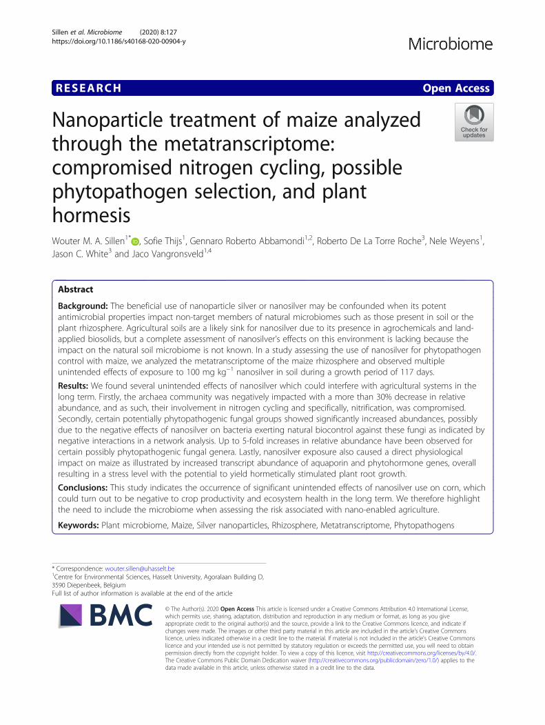
Results and discussionMixed responses by bacteria to nanosilver are likelydetermined by silver defense systemsNanosilver’s antimicrobial properties are expected toaffect the microbiome, and the bacterial communitycomposition in the maize rhizosphere does clearly reflectthis. Indeed, alpha diversity analysis indicates a signifi-cant decrease in richness (Fig. 1b). However, not all bac-teria displayed a similar response of decreasing inabundance. This can already be deduced from thePCoA-plot (Fig. 1e) as the communities of the controland nanosilver-exposed treatments appear to be sepa-rated, although the statistical significance of this couldnot be verified (ANOSIM p = 0.1). Nonetheless, taxo-nomic analysis of the bacterial communities clearly indi-cates already at the phylum level that different bacterialgroups responded differently to nanosilver exposure(Fig. 2). While Chloroflexi and Planctomycetes abun-dance decreased significantly (p < 0.05) in the activerhizosphere microbiome, Bacteroidetes, Alphaproteobac-teria, Gammaproteobacteria, and Acidobacteria were allincreased in response to nanosilver exposure. Other
Sillen et al. Microbiome (2020) 8:127 Page 2 of 17

groups, such as Actinobacteria and Deltaproteobacteria,showed a trend of decrease, although the effect was notstatistically significant. Figure 3 indicates that also withinthese non-significantly affected phyla, there are bacterialorders, e.g., Acidimicrobiales, Solirubrobacterales, andPseudonocardiales, that show significant decreases dueto nanosilver exposure. This lower taxonomic level alsoindicated that important bacteria orders such as Fimbrii-monadales, Chthonomonadales, Verrucomicrobiales,Myxococcales, Synthrophobacterales, and Rhodobacter-ales showed significant decreases.The impact of nanosilver on the bacterial community
is also illustrated by the functional characterization ofthe metatranscriptome. Table 1 shows the transcriptswith a significantly (p < 0.05) different abundance inrhizosphere soil. Gene transcripts of P-type ATPases,Cu+-exporting ATPases, and Cu(I)/Ag(I) efflux system
proteins showed increases in abundance. Some of theseare related to bacterial groups that increased in abun-dance due to nanosilver, such as Proteobacteria, Bacter-oidetes, and Acidobacteria, as well as to bacterial groupsthat decreased due to nanosilver, such as Actinobacteriaand Planctomycetes. Hence, regardless of their final de-crease or increase in abundance in response to nanosil-ver exposure, diverse bacterial groups initiate defenseresponses. However, the groups that thrive under nano-silver exposure display increased abundances of add-itional important genes such as cation efflux systemCzcA, copper-resistance protein CopA, and Copper-resistance protein K. These additionally expressed genescould explain the advantage that these bacterial groupshave over others which do not express these genes anddecrease in overall abundance. Such copper and othermetal resistance genes seem to be similar and
Fig. 1 Alpha and beta diversity of maize rhizosphere archaea, bacteria and fungi. Richness and Shannon diversity index for (a) archaea, (b)bacteria, and (c) fungi; and PCoA plot based on Bray-Curtis dissimilarities for (d) archaea, (e) bacteria, and (f) fungi for maize rhizosphere soilcommunities with (grey) or without (white) exposure to 100 mg kg−1 silver nanoparticles (20 nm) in soil, based on the metatranscriptome.Statistically significant differences for the alpha diversity measures are indicated on top of the graph: *p < 0.05, **p < 0.01, ***p < 0.001
Sillen et al. Microbiome (2020) 8:127 Page 3 of 17
evolutionarily linked to silver resistance systems such asthe sil silver-resistance system [12]. The sil system oftenis associated with the incompatibility group H1 (IncH1)plasmid pMG101, which is transferred horizontally andconfers resistance to Gram-negative but not to Gram-positive bacteria [13, 14]. This suggests that horizontalgene transfer of such plasmid-borne resistance systemscould play a vital role in determining the communityshifts taking place in soil under nanosilver exposure.These mechanisms may explain the observed success ofGram-negative groups such as Alphaproteobacteria,Gammaproteobacteria, and Bacteroidetes in nanosilver-exposed soil. Groups that cannot rely on the defensiveaction of this silver resistance system will be at a disad-vantage, and if these organisms are plant-beneficial mi-crobes, then plants could also be negatively affected.
Increase in fungal abundance, including potentiallyphytopathogenic groupsNext to bacteria, fungi are also susceptible to nanosilvertoxicity. However, the trend of increase in richness alreadyindicates that at least part of the fungal community bene-fited from nanosilver addition to the soil (Fig. 1c). Fungalcommunity shifts are hinted at by the PCoA plot (Fig. 1f),although statistical significance could not be inferred(ANOSIM p = 0.1). Community composition at the phylumlevel showed a significant increase in abundance of Ascomy-cota, which appear to benefit from the presence of
nanosilver in soil (Fig. 2). Figure 4 reveals more detail asseveral fungal orders were significantly altered in abun-dance, but still most fungal groups displayed an increase inabundance. Orders of Basidiomycota reacted variably tonanosilver, with positive and negative responses equally di-vided. Ascomycota showed a different pattern, with nearlyuniform increases associated with nanosilver exposure. Im-portantly, the orders Diaporthales, Eurotiales, and Botryo-sphaeriales experienced the greatest positive response tonanosilver exposure. These three orders all contain generawith well-known phytopathogens; significantly greaterabundance of these groups within the nanosilver-exposedrhizosphere microbiome is of concern. Certain species fromthe genus Diplodia, belonging to the Botryosphaeriales, arecausal agents of stalk and ear rot in maize, and this genusshowed a 5.5-fold increase in relative abundance undernanosilver exposure. In the Eurotiales, species from thegenera Aspergillus and Penicillium can cause maize ear rot,and these genera displayed 87% increase and a 6.25-fold in-crease under nanosilver exposure, respectively. The genusValsa, showing a 3.15-fold increase under nanosilver expos-ure, belongs to the Diaporthales, and certain species fromthis genus are well-described phytopathogens, although notknown for maize infection. Overall, such an increase inabundance of possible phytopathogens is not expected tobe directly caused by nanosilver but rather is likely to be in-duced by an indirect mechanism involving interactions be-tween microorganisms.
Fig. 2 Maize rhizosphere microbiome community composition at phylum level.Microbiome community composition at the phylum level in maizerhizosphere soil with or without 100 mg kg−1 silver nanoparticles (20 nm), based on the metatranscriptome. Relative abundance is shown andonly phyla making up more than 1% of the total community are taken into account. Phyla with statistically significant differences betweencontrol and nanosilver-exposed conditions are indicated next to the legend: *p < 0.05, **p < 0.01, ***p < 0.001
Sillen et al. Microbiome (2020) 8:127 Page 4 of 17

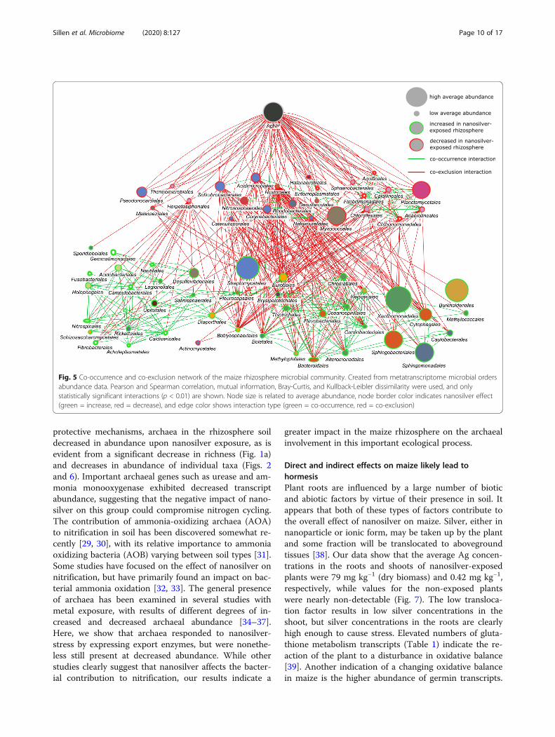
Disruption of natural biocontrol as a cause ofphytopathogen increaseNetwork analysis based on co-occurrence and co-exclusion interactions enabled differentiation betweengroups which play an important role in the rhizospheremicrobiome by influencing other groups from those thatact more individually. As shown in Fig. 5, nanosilver wasalmost exclusively involved in negative interactions, theexception being a positive interaction with Boletales.Therefore, nanosilver exposure was not associated withthe direct promotion of abundance of nearly any species.Hence, the cause of the increased abundance of thegroups noted above after nanosilver exposure is clearly
more complex. Several of the orders with significantly de-creased abundance as shown by the taxonomic analysis doappear as direct negative interactors with nanosilver in thenetwork analysis. Phyla belonging to this group includeActinobacteria, Aquificae, Armatimonadetes, Basidiomy-cota, Chloroflexi, Cyanobacteria, Firmicutes, Planctomy-cetes, Proteobacteria, Tenericutes, and Thaumarchaeota. Itis noteworthy that Actinobacteria are decreased as thisgroup are well-known producers of antibiotics [15] andhence, are involved in negative interactions with numer-ous species. Of the orders with negative interaction withnanosilver, Actinobacteria orders such as Solirubrobacter-ales, Acidimicrobiales, Corynebacteriales, Nakamurellales,
Fig. 3 Maize rhizosphere bacteria community composition at order level. Community composition of bacteria at the order level in maizerhizosphere soil with or without 100 mgkg−1 silver nanoparticles (20 nm), based on the metatranscriptome. Bar graphs indicate abundance, withthe effect of nanosilver exposure shown in green (increased compared to control) or red (decreased compared to control). Scale reference isindicated as dotted lines. Statistical significance (p < 0.05) of the nanosilver effect is indicated by a star
Sillen et al. Microbiome (2020) 8:127 Page 5 of 17

Table 1 Functional composition of the metatranscriptome. Metatranscriptome genes annotated to InterPro2GO and KEGGdatabases with significantly different abundances between control conditions and nanosilver-exposed (100 mg kg−1 soil) conditionsin maize rhizosphere soil. Taxonomic origin of the genes is given at a general level, as well as the lowest possible level
Databasereference
Database entry Taxonomic origin Lowest leveltaxonomicassignment
Log2 foldchange
Base level:mean numberof reads
Archaea Bacteria Maize Fungi Other
Higherincidencein AgNP-conditions
IPR021604 Copper resistanceprotein K
x Burkholderiales − 3.59 34.7
IPR027256 P-type ATPase,subfamily IB
x x Actinobacteria,Proteobacteria,Bacteroidetes,Thaumarchaeota,Euryarchaeota
− 1.30 122.4
IPR004763 Cation efflux systemCzcA/CusA/SilA/NccA/HelA/CnrA
x Bacteroidetes,Proteobacteria,Acidobacteria
− 2.53 29.3
IPR006376 Copper-resistanceprotein CopA
x Sphingomonadales,Xanthomonadales
− 2.26 5.3
K07787 Cu(I)/Ag(I) effluxsystem membraneprotein CusA/SilA
x Bacteroidetes,Proteobacteria,Acidobacteria
− 2.53 29.0
K07798 Membrane fusionprotein, Cu(I)/Ag(I)efflux system
x Proteobacteria,Acidobacteria,Nitrospira,Planctomycetes
− 1.81 9.3
K17686 Cu+-exportingATPase
x x Proteobacteria,Actinobacteria,Thaumarchaeota
− 1.25 83.5
IPR001806 Small GTPasesuperfamily
x Xanthomonadales − 0.64 171.1
IPR005294 ATPase, F1 complex,alpha subunit
x Bacteria general − 1.23 76.2
GO:0005634
Nucleus x x x Eukaryote general − 0.73 52.0
ko04121 Ubiquitin system x x x Eukaryote general − 0.59 218.9
ko04144 Endocytosis x x x Eukaryote general − 0.62 126.7
ko01009 Protein phosphataseand associatedproteins
x x x Eukaryote general − 0.42 229.1
IPR005093 RNA-directed RNApolymerase beta-chain
x Leviviridae(Bacteriophage)
− 3.12 113.8
K09872 aquaporin PIP x NA − 1.13 1.8
ko04075 Plant hormone signaltransduction
x NA − 2.37 12.1
ko00940 Phenylpropanoidbiosynthesis
x NA − 1.26 49.7
IPR005922 Phenylalanineammonia-lyase
x NA − 2.98 3.1
K01188 Beta-glucosidase x NA − 2.15 6.2
IPR015655 Protein phosphatase2C family
x NA − 1.27 17.4
IPR001246 Lipoxygenase, plant x NA − 2.57 2.8
K04733 Interleukin-1 receptor-associated kinase 4
x NA − 1.77 6.2
IPR000864 Proteinase inhibitor x NA − 1.89 1.7
Sillen et al. Microbiome (2020) 8:127 Page 6 of 17

Table 1 Functional composition of the metatranscriptome. Metatranscriptome genes annotated to InterPro2GO and KEGGdatabases with significantly different abundances between control conditions and nanosilver-exposed (100 mg kg−1 soil) conditionsin maize rhizosphere soil. Taxonomic origin of the genes is given at a general level, as well as the lowest possible level (Continued)
Databasereference
Database entry Taxonomic origin Lowest leveltaxonomicassignment
Log2 foldchange
Base level:mean numberof reads
Archaea Bacteria Maize Fungi Other
I13, potato inhibitor I
IPR001929 Germin x NA − 2.21 3.2
ko00480 Glutathione metabolism x x Proteobacteria,Actinobacteria
− 0.48 119.1
IPR003855 Potassium transporter x NA − 1.89 4.7
IPR009716 Ferroporti-1 x NA − 1.77 1.3
K08176 MFS transporter,PHS family, inorganicphosphate transporter
x NA − 1.50 2.5
K00695 Sucrose synthase x NA − 1.90 5.2
K10592 E3 ubiquitin-proteinligase HUWE1
x NA − 1.58 4.0
IPR005150 Cellulose synthase x NA − 1.93 3.6
IPR016461 O-methyltransferaseCOMT-type
x NA − 1.63 7.9
IPR018167 S-adenosylmethioninedecarboxylasesubgroup
x NA − 2.09 2.2
K02132 F-type H+-transportingATPase subunit alpha
x x x Streptomyces,Leotiomyceta
− 1.38 22.5
K02262 Cytochrome c oxidasesubunit 3
x x x Streptomyces,Leotiomyceta
− 1.78 11.8
K03936 NADH dehydrogenase(ubiquinone) Fe-Sprotein 3
x x x Streptomyces,Leotiomyceta
− 1.56 2.9
IPR001128 Cytochrome P450 x x x Streptomyces,Leotiomyceta
− 0.84 36.5
GO:0005739
Mitochondrion x x Sordariomycetes − 1.76 6.2
ko04110 Cell cycle x x Leotiomyceta − 0.95 44.0
IPR012220 Glutamate synthase,eukaryotic
x x Sordariomycetes − 1.85 5.4
ko00199 Cytochrome P450 x x Streptomyces − 1.39 8.7
GO:0004872
Receptor activity x x Streptomyces − 1.52 19.5
IPR00136 Glycoside hydrolase,family 1
x x Streptomyces,Sphingomonadales
− 2.00 13.2
K00066 GDP-mannose6-dehydrogenase
x Pseudomonadales − 1.63 7.6
IPR031148 Plexin family x Streptomyces − 2.47 7.8
IPR001795 RNA-directed RNApolymerase, luteovirus
x Luteoviridae − 1.92 13.5
Lower incidencein AgNP-conditions
IPR010946 Geranylgeranylglycerylphosphate synthase
x Nitrosphaeraceae 2.07 24.2
IPR005938 AAA ATPase, CDC48family
x Nitrosphaeraceae 0.85 49.4
IPR002386 Amicyanin/Pseudoazurin x Nitrosphaeraceae 1.38 20.5
IPR005848 Urease, alpha subunit x Nitrosphaeraceae 1.55 34.2
Sillen et al. Microbiome (2020) 8:127 Page 7 of 17

Bifidobacteriales, and Catenulisporales were among themost abundant with the highest number of negative inter-actions (negative degree). These negative interactions in-volved a large number of orders which displayed anincreased abundance in the nanosilver-exposed rhizo-sphere microbiome. Hence, according to this network, theincreased abundance of certain groups upon nanosilverexposure was not a direct consequence of nanosilver. Ra-ther, these groups benefited from the negative effect ofnanosilver on certain other groups which are involved innegative interactions with them. A wide range of microbialphyla were included in this positively affected group.Among these, most notable are the fungal orders whichharbor well-known fungal pathogens, as described previ-ously: Diaporthales, Eurotiales, and Botryosphaeriales.This negative interaction of certain microorganisms withphytopathogens suggests the presence of natural biocon-trol. When evaluating the role of the rhizosphere micro-biome in plant health, biocontrol of phytopathogens isalways of high importance [16, 17]. Certain microorgan-isms can directly, through antibiotic metabolite produc-tion, or indirectly, through competition for resources,antagonize phytopathogen populations [18]. Our data sug-gest that nanosilver in soil has the capacity to promote po-tentially phytopathogenic fungi, possibly by interferingwith this inherent control system afforded by members ofthe rhizosphere microbiome. Plants such as maize areknown to exhibit an age-related resistance [19] that is po-tentially correlated with the rhizosphere microbiomechanges that occur over time [20]. A possible selection of
a disease-suppressive microbial community may be pre-vented by the community alterations from nanosilver ex-posure. For example, Actinobacteria are often increasinglyabundant in the rhizosphere of maturing plants [21] andare widely recognized for their biocontrol potentialthrough the production of bioactive compounds [15]. Themajority of biocontrol studies with Actinobacteria have fo-cused on Streptomyces, illustrated by the fact that thisgroup is responsible for over 80% of all known antibioticsof actinobacterial origin [22]. In the current study, Strepto-mycetales are the only Actinobacteria with increasedabundance in the rhizosphere under nanosilver exposure.As such, only non-streptomycete Actinobacteria could beinvolved in the phytopathogen proliferation. Next toStreptomycetales, other Actinobacteria are also recognizedas a valuable topic for biocontrol-research, as has beenhighlighted for Actinomycetales [23], and our results sug-gest that several other Actinobacteria orders could be in-volved here. This includes orders within the classActinobacteria, e.g., Corynebacteriales and Bifidobacter-iales, and orders in other classes, such as Rubrobacteralesand Solirubrobacterales. Another interesting group of bac-teria involved in the negative interaction with phytopatho-gens are Myxococcales. These bacteria are also known fortheir production of a wide range of antibiotics and lyticenzymes, and are often considered to be important to bio-control [24, 25]. Rhodobacterales are another well-studiedgroup of biocontrol agents. All of these bacterial groupscould play an important role in limiting maize phytopath-ogen activity, and importantly, are all reduced by
Table 1 Functional composition of the metatranscriptome. Metatranscriptome genes annotated to InterPro2GO and KEGGdatabases with significantly different abundances between control conditions and nanosilver-exposed (100 mg kg−1 soil) conditionsin maize rhizosphere soil. Taxonomic origin of the genes is given at a general level, as well as the lowest possible level (Continued)
Databasereference
Database entry Taxonomic origin Lowest leveltaxonomicassignment
Log2 foldchange
Base level:mean numberof reads
Archaea Bacteria Maize Fungi Other
IPR000812 Transcription factor TFIIB x Nitrosphaeraceae 1.54 29.7
IPR024656 Ammoniamonooxygenase,subunit A, archaeal
x Archaea general 1.69 42.7
K04080 molecular chaperoneIbpA
x Rhizobiales 1.09 47.2
IPR026042 Stress responseprotein YjbJ
x Alpha- &Gammaproteo,Bacteroidetes
1.17 183.5
IPR031107 Small heat shockprotein HSP20
x Rhizobiales,Chloroflexi,Actinobacteria
0.80 284.3
IPR001189 Manganese/ironsuperoxide dismutase
x Bacteria general 0.61 75.2
IPR001287 Nitrite reductase,copper-type
x Nitrosphaeraceae 1.21 29.2
K02518 Translation initiationfactor IF-1
x Actinobacteria 1.01 50.8
Sillen et al. Microbiome (2020) 8:127 Page 8 of 17

nanosilver exposure. These groups may potentially bejoined by other bacteria that can negatively impact phyto-pathogen activity but whose role is unknown due to a lackof culturability. Biocontrol agents are of high value in bothnatural ecosystems and in agricultural crop production,and nanosilver-induced disturbance of this native protec-tion system could have significant ecologic and economicconsequences.
Archaea decrease and their involvement in nitrogencycling is confoundedArchaea possess a number of properties which couldgive them an advantage under nanosilver-stress. These
organisms have tetraether-linked membrane lipids as themain lipid component in their cell membrane, some-times organized into a monolayer which increases theimpermeability to metals [26]. Also, archaea are knownto outcompete bacteria under chronic energy stress con-ditions [27], and within the archaeal domain, genes forthe metabolism, resistance, and detoxification of metalsare reported to be widespread [28]. These features, alongwith other properties, give an advantage to archaea inresponding to metal exposure. Indeed, archaea showedincreased transcript abundance of two metal-exportingproteins, i.e., select P-type ATPase and Cu+-exportingATPase (Table 1). Nonetheless, in spite of these
Fig. 4 Maize rhizosphere fungi community composition at order level. Community composition of fungi at the order level in maize rhizospheresoil with or without 100 mg kg−1 silver nanoparticles (20 nm), based on the metatranscriptome. Bar graphs indicate abundance, with the effect ofnanosilver exposure shown in green (increased compared to control) or red (decreased compared to control). Scale reference is indicated asdotted lines. Statistical significance (p < 0.05) of the nanosilver effect is indicated by a star
Sillen et al. Microbiome (2020) 8:127 Page 9 of 17
protective mechanisms, archaea in the rhizosphere soildecreased in abundance upon nanosilver exposure, as isevident from a significant decrease in richness (Fig. 1a)and decreases in abundance of individual taxa (Figs. 2and 6). Important archaeal genes such as urease and am-monia monooxygenase exhibited decreased transcriptabundance, suggesting that the negative impact of nano-silver on this group could compromise nitrogen cycling.The contribution of ammonia-oxidizing archaea (AOA)to nitrification in soil has been discovered somewhat re-cently [29, 30], with its relative importance to ammoniaoxidizing bacteria (AOB) varying between soil types [31].Some studies have focused on the effect of nanosilver onnitrification, but have primarily found an impact on bac-terial ammonia oxidation [32, 33]. The general presenceof archaea has been examined in several studies withmetal exposure, with results of different degrees of in-creased and decreased archaeal abundance [34–37].Here, we show that archaea responded to nanosilver-stress by expressing export enzymes, but were nonethe-less still present at decreased abundance. While otherstudies clearly suggest that nanosilver affects the bacter-ial contribution to nitrification, our results indicate a
greater impact in the maize rhizosphere on the archaealinvolvement in this important ecological process.
Direct and indirect effects on maize likely lead tohormesisPlant roots are influenced by a large number of bioticand abiotic factors by virtue of their presence in soil. Itappears that both of these types of factors contribute tothe overall effect of nanosilver on maize. Silver, either innanoparticle or ionic form, may be taken up by the plantand some fraction will be translocated to abovegroundtissues [38]. Our data show that the average Ag concen-trations in the roots and shoots of nanosilver-exposedplants were 79 mg kg−1 (dry biomass) and 0.42 mg kg−1,respectively, while values for the non-exposed plantswere nearly non-detectable (Fig. 7). The low transloca-tion factor results in low silver concentrations in theshoot, but silver concentrations in the roots are clearlyhigh enough to cause stress. Elevated numbers of gluta-thione metabolism transcripts (Table 1) indicate the re-action of the plant to a disturbance in oxidative balance[39]. Another indication of a changing oxidative balancein maize is the higher abundance of germin transcripts.
Fig. 5 Co-occurrence and co-exclusion network of the maize rhizosphere microbial community. Created from metatranscriptome microbial ordersabundance data. Pearson and Spearman correlation, mutual information, Bray-Curtis, and Kullback-Leibler dissimilarity were used, and onlystatistically significant interactions (p < 0.01) are shown. Node size is related to average abundance, node border color indicates nanosilver effect(green = increase, red = decrease), and edge color shows interaction type (green = co-occurrence, red = co-exclusion)
Sillen et al. Microbiome (2020) 8:127 Page 10 of 17

Germin and germin-like proteins have been linked to awide range of functions, including a role as oxalate oxi-dase or as superoxide dismutase [40]. Superoxide dismu-tases are directly involved in defense against oxidativestress, while oxalate oxidases produce H2O2 which trig-gers signaling pathways that upregulate expression ofgenes for antioxidant enzymes [41].In addition to these oxidative balance disturbances,
aquaporin activity in maize seems to be significantly af-fected by exposure to nanosilver; specifically, transcriptsof the aquaporin PIP are present at significantly in-creased abundance. Several in vitro studies with Arabi-dopsis seedlings [42] and rice seeds [43] have shown that
nanosilver can activate aquaporin gene expression. Here,we show that nanosilver in soil exerts a similar affect.The mechanism behind this response is unclear, al-though it has been reported that silver can inhibit aqua-porin activity through interaction with sulfhydryl groupsof the proteins [44]. The increased aquaporin PIP tran-scripts may be a response of the plant to the inhibitionof these important water channels.Nanosilver exposure seems to also stress maize indir-
ectly by increasing the abundance of potentially phyto-pathogenic fungi. A possible defense response of maizeto phytopathogen infection can be seen in the inducedtranscription of genes involved in the production of
Fig. 6 Maize rhizosphere archaea community composition at order level. Community composition of archaea at the order level in maizerhizosphere soil with or without 100 mg kg−1 silver nanoparticles (20 nm), based on the metatranscriptome. Bar graphs indicate abundance, withthe effect of nanosilver exposure shown in green (increased compared to control) or red (decreased compared to control). Scale reference isindicated as dotted lines. Statistical significance (p < 0.05) of the nanosilver effect is indicated by a star
Sillen et al. Microbiome (2020) 8:127 Page 11 of 17

beta-glucosidases and phenylpropanoids. Although notdefinitive, these findings could indicate a significant add-itional burden on maize grown in nanosilver-containingsoil. Nonetheless, the findings certainly demonstrate that
maize responds at the molecular and biochemical levelto nanosilver exposure. The diversity of these physio-logical responses is also illustrated by changes in plantgene expression related to phytohormones and nutrient
Fig. 7 Maize biomass, nutrient, and silver concentrations in response to nanosilver exposure.Maize (a) root and shoot biomass and shoot/rootratio, (b) root, and (c) shoot concentrations of Ag, Fe, K, and P. Plants were grown for 117 days in soil with or without 100 mg kg−1 silvernanoparticles (20 nm). Values ± SE; *p < 0.05, **p < 0.01, ***p < 0.001
Sillen et al. Microbiome (2020) 8:127 Page 12 of 17

balance (Table 1). Plant hormone signal transduction iscritical to many plant growth and development processes,and modulation of transcript abundance is likely indicativeof the biomass changes that were noted upon nanosilverexposure. The increases in iron and phosphate transportertranscripts demonstrate nanosilver-induced modulation ofmaize nutrient balance. ICP analysis of maize tissues con-firmed this (Fig. 7). While Fe concentration was signifi-cantly decreased (p = 0.041) in roots under nanosilverexposure from 1683 to 1115 mg kg−1, root P concentra-tion significantly increased (p = 0.002) from 692 to 1053mg kg−1 and root K concentration showed no effect withvalues stable around 7800 mg kg−1. A different patternemerged in the shoots; although the increased P concen-tration was still evident (p = 0.003) with values of 1612versus 494 mg kg−1, the statistical significance of the Fedecrease was lost, although a decrease from 435 to 283mg kg−1 was evident. In addition, the K concentrationexhibited a statistically non-significant trend of in-crease (p = 0.12) from 1290 to 1725 mg kg−1. Thesechanges in nutrient content could be attributed to avariety of causes, including a direct molecular effectof nanosilver on these enzymes or indirectly as aplant response to physiological changes from theexposure.Overall, it is clear that there are a number of diverse
ways through which nanosilver directly or indirectly af-fects maize, and ultimately this seems to induce an in-crease in root biomass (Fig. 7). Maize plants exposed tosilver nanoparticles in soil showed a significantly higherroot biomass (p = 0.046) than the control plants, in-creasing from 9.8 g to 12.3 g. The shoot biomass was ap-proximately 23 g and remained unaffected by exposure;the shoot/root-mass ratio decreased from 2.3 to 1.9, al-though the effect was not statistically significant. Al-though most studies on nanosilver exposure highlight aneutral to negative effect on plant growth, our resultsare not the first indication of a nanosilver-induced plantbiomass increase [45]. We did not observe an increasedpresence of any plant-growth-promoting factors such asplant-growth-promoting rhizobacteria. Therefore, itseems that the above-described direct and indirect im-pacts of nanosilver on maize may result in a hormeticresponse of the plant. Hormesis is an “umbrella term”used to describe instances where low doses of generallytoxic substances can stimulate biological systems [46]. Inplants, this phenomenon can arise through a variety ofmechanisms, including both substrate interactions andactivation of defense reaction pathways that are involvedin multiple physiological processes [47]. It has been re-ported that low concentrations of arsenic can increasethe bioavailability of inorganic phosphate through com-petitive interactions related to soil adsorption [48].Hence, arsenic at low doses can increase plant growth
through enhanced P availability and uptake. Although asimilar mechanism cannot be proposed for silver, the in-creased P concentrations measured in exposed maize tis-sues could be involved in the biomass increase. Inaddition, reactive oxygen species (ROS) and antioxidantmolecules and enzymes play central roles in plantdefense. We found increased transcript abundances forgenes involved in glutathione and germin metabolism.These molecules play important roles in ROS signalingand scavenging and hence, could initiate mechanismswhich account for the hormetic effect. For example, inwheat, it has been shown that long-term exposure tolow Cd concentrations in soil can have a hormetic effect,being linked to a reduction of the ROS level and an in-crease in the activity of glutathione reductase [49]. Inaddition, hormesis potentially could also arise throughcross-talk interactions between metals and biotic stresscaused by phytopathogens. This is possible becausemetal ions can evoke the production of secondary me-tabolites that are involved in defense against pathogens,including molecules such as flavonoids, among others[47]. Hence, it is possible that the increased phenylpro-panoid transcript levels here are caused by silver andsubsequently elevate the protection level of maizeagainst phytopathogens, inducing a hormetic effect. Im-portantly, the cause of the increased maize root biomassunder nanosilver-exposure cannot be determined withcertainty, but a wide range of processes, covered by the“umbrella term” hormesis, seem likely to have played arole here.
ConclusionsUsing the extensive information, the rhizosphere meta-transcriptome has to offer, we show that soil bornenanosilver has various unintended effects on maize andits rhizosphere microbiome. On the prokaryote side,nanosilver can interfere with the nitrogen cycle due to adecrease in abundance of archaea taking place in theprocess and can potentially compromise natural biocon-trol systems because of a decrease in abundance of bac-terial groups with a biocontrol function. This lattereffect can yield an increase in phytopathogen activity, in-stead of the intended decrease. Nanosilver also inducesdirect stress on the maize plants, likely through oxidativestress and aquaporin interference. Together, these indir-ect and direct effects on maize suggest that the observedincrease in root biomass, which could be considered apositive outcome, is the result of hormetic growthstimulation that is unlikely to be sustainable in the longterm. Hence, the overall balance suggests that the appli-cation of nanosilver in agriculture comes with significantunintended effects which could turn out to be negativefor crop productivity and ecosystem health in the longerterm. Therefore, it seems essential that these
Sillen et al. Microbiome (2020) 8:127 Page 13 of 17

microbiome-related processes are included when asses-sing the risk associated with nanosilver use inagriculture.
MethodsMaterial and experimental set-upUncoated silver nanoparticles (99.99% purity, 20 nmdiameter) were obtained in solid form from US ResearchNanomaterials, Inc., Houston, TX, USA. Particle zetapotential and hydrodynamic size were characterized in500 mg/L solutions by dynamic light scattering (DLS) ona zetasizer (Malvern Zetasizer, Nanoseries ZS90). Theaverage size for AgNP is 259.7 nm (± 10.05), and thezeta potential is − 30.3 mV (± 2.71). The particles werealso characterized by transmission electron microscopy(TEM) (Hitachi HT7800). TEM indicated a wide varietyof sizes due to clustering of the original particles of ca.20 nm (Additional file 1). Soil was collected from thetop 30 cm of an agricultural corn field in Diepenbeek,Belgium (50°56′05.3′′ N 5°24′41.2′′ E) and was charac-terized as sandy loam (55% sand, 30% silt, 15% clay) witha pH of 6.98, an electrical conductivity (EC) of 335 μScm−1 and an effective cation-exchange capacity (CEC) of20.7 meq/100 g. After collection, the soil was sieved to 6mm and homogenized. Zea mays variety LG 30.223seeds were acquired from LimaGrain Belgium. Soil nu-trient content was augmented by fertilization until con-ditions favorable for maize cultivation were reached: 106mg N g−1 soil, 34 mg P g−1 soil, 31 mg K g−1 soil, and 15mg Mg g−1 soil. Maize plants were grown individually in10 L-pots, each pot containing 10 kg of soil. Three repli-cate pots contained nanosilver at 100 mg kg−1, whichwas added by mechanical mixing for 5 min. This nano-silver concentration was established in earlier research,because it provides a baseline response in the systemand takes into account the possibility of nanosilver accu-mulation in agricultural soils due to the application ofbiosolids and nano-enabled agrichemicals [45]. Threeother replicate pots were not amended with nanosilverand were used as controls. Before planting, maize seedswere soaked in tap water overnight. All pots with seedswere randomly placed in a climate chamber with the fol-lowing conditions: 12 h daylight photoperiod, atemperature cycle of 22 °C/18 °C, and a relative humidityof 50%. After 117 days, all plants were harvested and soilsamples from all conditions were taken for metatran-scriptomic analysis. Samples of the rhizosphere soil, op-erationally defined as the soil that remains attached tothe roots after light shaking, were flash-frozen in liquidnitrogen and stored at – 80 °C.
Plant biomass and element content analysisAt harvest, the wet and oven-dry biomass of root andshoot tissues were determined. For plant element
content analysis, oven-dried samples of roots and shoots(approximately 0.5 g) were digested in 50 ml polypropyl-ene digestion tubes with 5 ml of concentrated nitric acidat 115 °C for 45 min using a hot block (DigiPREP Sys-tem; SCP Science, Champlain, NY, USA). A small vol-ume of H2O2 was included to ensure completedigestion. The resulting digests were analyzed for Ag, Fe,K, and P using inductively coupled plasma optical emis-sion spectroscopy (ICP-OES; iCAP 6500 Thermo FisherScientific). Samples with Ag content below the ICP-OESlimit of quantification were subsequently analyzed by In-ductively Coupled Plasma-Mass Spectrometry (ICP-MS;Agilent 7500ce).
Soil RNA extraction and sequencingTotal RNA was extracted from the rhizosphere soil sam-ples using the PowerSoil® RNA Isolation Kit (Mo Bio La-boratories Inc., CA, USA), according to themanufacturer instructions. Starting material for everyextraction was 6 g of soil. Extracted RNA was addition-ally purified using the NucleoSpin® RNA kit (Macherey-Nagel, Düren, Germany), including DNase-treatment ac-cording to the manufacturer’s instructions. RNA integ-rity, purity, and concentration were confirmed byExperionTM RNA assays (Bio-Rad Laboratories, Munich,Germany); these data were used as an indicator for theneed to repeat failed extractions.Sequencing was performed by Macrogen (Seoul, South
Korea). Libraries were constructed using the IlluminaTruSeq Stranded Total RNA kit (Illumina, USA) withoutrRNA depletion or mRNA enrichment. IlluminaHiSeq4000 paired-end sequencing (2 × 100 bp) resultedin at least 35 million high-quality reads per library(Table 2). The raw sequencing data were deposited atthe European Nucleotide Archive (ENA) under the pro-ject ERP024369.Basic composition of the metatranscriptomes obtained
from maize rhizosphere soil with or without 100 mgkg−1 silver nanoparticles after 117 days of growth andexposure.
Metatranscriptome analysisRaw sequencing reads were controlled for quality usingFastQC version 0.10.1 (Andrews, 2010, available onlineat http://www.bioinformatics.babraham.ac.uk/projects/fastqc). Based on the FastQC quality report, the Krakenpipeline [50] was used to remove potential adapter se-quences, poor quality reads (Phred score < 10), shortreads (< 30 nucleotides), and reads without a counter-part, all while concurrently maintaining read-pairingduring processing. The resulting high-quality reads wereused for taxonomic and functional analysis. Taxonomicanalysis aimed at characterizing the communities of ar-chaea, bacteria, and fungi through marker gene analysis,
Sillen et al. Microbiome (2020) 8:127 Page 14 of 17

which was achieved through the One Codex platformthat utilizes targeted loci including 5S, 16S, 23S, 18S,28S, and ITS [51]. For functional analysis, the quality-filtered reads were sorted using SortMeRNA [52], whichseparated rRNA from non-rRNA and thus, potentialmRNA. Sorting was performed with standard parame-ters, using the SILVA-16S-18S-SSURef_115_NR99 andSILVA-23S-28S-LSURef_115 databases [53]. FragGeneS-can [54] version 1.17 with the Illumina 0.5% error modelwas used on the potential mRNA reads to filter out un-desired sequences such as non-coding regions, leavingonly putative genes. These remaining sequences weresubsequently aligned to the NCBI-nr database with DIA-MOND [55] version 0.8.38, applying an E value cut-offof 10−3. The resulting alignments were annotated to theInterPro2GO [56] and KEGG [57] databases throughMEGAN6 [58] using default LCA parameters (minscore: 50, top percent: 10, min support).
Statistical analysesR version 3.3.2 [59] was used for the statistical analyses.Parametric group mean comparisons such as Student’s ttest and ANOVA were performed for the plant biomassand element content data.The taxonomic community compositions were
exported from One Codex for further analysis. Richnessand Shannon diversity of archaea, bacteria, and fungiseparately were calculated for the control andnanosilver-exposed conditions. Differences between thetwo treatments for these parameters were analyzed usingt tests as parametric assumptions were fulfilled. Principalcoordinates analyses (PCoA) based on Bray-Curtis dis-similarities were performed and plotted for the archaea,bacteria, and fungi communities individually, and theseparation between the control and the nanosilver-exposed treatment was statistically evaluated by the useof analysis of similarity (ANOSIM). The relative abun-dance of individual taxa was analyzed for statistically sig-nificant differences between the two treatments (controlvs. nanosilver-exposed) using the Bioconductor packagesphyloseq [60] version 1.19.1 and DESeq2 [61] version
1.14.1 . The Wald test was used and statistical signifi-cances were considered if the adjusted p values < 0.05(using the Benjamini and Hochberg procedure). Func-tional annotations extracted from MEGAN6 were statis-tically analyzed using DESeq2 version 1.14.1, also bymeans of the Wald test and adjusted p value < 0.05(Benjamini and Hochberg) considered as statisticallysignificant.Network analysis of the taxonomic microbial commu-
nity compositions was performed with the Cytoscape[62] version 3.4.0 plugin CoNet [63] by means of co-occurrence and co-exclusion interactions. Filtering wasdone by setting the minimum abundance per phylogen-etic order to 40000 reads over all samples. Pair-wise as-sociations between orders were calculated with thesimultaneous use of Pearson and Spearman correlation,mutual information, Bray-Curtis, and Kullback-Leiblerdissimilarity. The top and bottom 100 edges for eachmethod were initially selected. Edges needed to be sup-ported by at least two of these methods in order to beretained. Permutation was performed with 100 repeti-tions, and the resulting p values were used as a cut-offvalue at 0.01. The resulting network was visualized inCytoscape.
Supplementary informationSupplementary information accompanies this paper at https://doi.org/10.1186/s40168-020-00904-y.
Additional file 1. Transmission electron microscope image of the silvernanoparticles used in the study. Nanoparticles were obtained in solidform from US Research Nanomaterials, Inc. (Houston, TX, USA), and wereapplied to soil in this form.
AcknowledgementsNot applicable
Authors’ contributionsW.S., S.T, N.W., J.C.W., and J.V. designed research; W.S., G.R.A., and R.D.L.T.R.performed research; and W.S. wrote the paper. All authors read andapproved the final manuscript.
Table 2 Basic metatranscriptome composition
Treatment Sample Total highquality reads
Read proportions
rRNA reads Non-rRNA reads
Number of reads Percentage of total Number of reads Percentage of total
Nanosilver-exposed Sample 1 35493754 33642151 94.8 1851603 5.2
Sample 2 36666638 34699832 94.6 1966806 5.4
Sample 3 35067906 33332673 95.1 1735233 4.9
Control Sample 1 43534310 41955731 96.4 1578579 3.6
Sample 2 44648348 42845994 96 1802354 4
Sample 3 35339870 34033583 96.3 1306287 3.7
Sillen et al. Microbiome (2020) 8:127 Page 15 of 17

FundingThis work was supported by Hasselt University Bijzonder OnderzoeksfondsMethusalem Project 08M03VGRJ. JCW acknowledges USDA AFRI 2016-67021-24985 and USDA Hatch CONH00147. W.S., S.T. and N.W. were research fellowsand a postdoctoral fellow, respectively, of the Research Foundation-Flanders(FWO).
Availability of data and materialsAll raw metatranscriptome data are available at the European NucleotideArchive (ENA) under the project ERP024369.
Ethics approval and consent to participateNot applicable
Consent for publicationNot applicable
Competing interestsNot applicable
Author details1Centre for Environmental Sciences, Hasselt University, Agoralaan Building D,3590 Diepenbeek, Belgium. 2Institute of Biomolecular Chemistry, NationalResearch Council of Italy, Via Campi Flegrei 34, Pozzuoli, 80078 Napoli, Italy.3Department Analytical Chemistry, Connecticut Agricultural ExperimentStation, 123 Huntington Street, New Haven, CT, USA. 4Department of PlantPhysiology, Faculty of Biology and Biotechnology, Maria Curie-SklodowskaUniversity, Lublin, Poland.
Received: 20 March 2020 Accepted: 3 August 2020
References1. Rodriguez PA, Rothballer M, Chowdhury SP, Nussbaumer T, Gutjahr C,
Falter-Braun P. Systems Biology of Plant-Microbiome Interactions. Mol Plant.2019;12(6):804–21.
2. Compant S, Samad A, Faist H, Sessitsch A. A review on the plantmicrobiome: ecology, functions, and emerging trends in microbialapplication. J Adv Res. 2019;19:29–37.
3. Mendes R, Garbeva P, Raaijmakers JM. The rhizosphere microbiome:significance of plant beneficial, plant pathogenic, and human pathogenicmicroorganisms. FEMS Microbiol Rev. 2013;37(5):634–63.
4. White JC, Gardea-Torresdey J. Achieving food security through the verysmall. Nat Nanotechnol. 2018;13(8):627–9.
5. Schluesener JK, Schluesener HJ. Nanosilver: application and novel aspects oftoxicology. Arch Toxicol. 2013;87(4):569–76.
6. Nowack B, Krug HF, Height M. 120 Years of nanosilver history: implicationsfor policy makers. Environ Sci Technol. 2011;45(4):1177–83.
7. Danish Consumer Counsil. The Nanodatabase. http://nanodb.dk/. Accessed16 March 2020.
8. Holden PA, Gardea-Torresdey JL, Klaessig F, Turco RF, Mortimer M, Hund-Rinke K, et al. Considerations of Environmentally Relevant Test Conditionsfor Improved Evaluation of Ecological Hazards of Engineered Nanomaterials.Environ Sci Technol. 2016;50(12):6124–45.
9. Pradas del Real AE, Castillo-Michel H, Kaegi R, Sinnet B, Magnin V, FindlingN, et al. Fate of Ag-NPs in Sewage Sludge after Application on AgriculturalSoils. Environ Sci Technol. 2016;50(4):1759–68.
10. Gottschalk F, Sonderer T, Scholz RW, Nowack B. Modeled environmentalconcentrations of engineered nanomaterials (TiO(2), ZnO, Ag, CNT,Fullerenes) for Different Regions. Environ Sci Technol. 2009;43(24):9216–22.
11. Chhipa H. Nanofertilizers and nanopesticides for agriculture. Environ ChemLett. 2017;15(1):15–22.
12. Staehlin BM, Gibbons JG, Rokas A, O'Halloran TV, Slot JC. Evolution of aheavy metal homeostasis/resistance island reflects increasing copper stressin enterobacteria. Genome Biol Evol. 2016;8(3):811–26.
13. Gupta A, Matsui K, Lo JF, Silver S. Molecular basis for resistance to silvercations in Salmonella. Nat Med. 1999;5(2):183–8.
14. Loh JV, Percival SL, Woods EJ, Williams NJ, Cochrane CA. Silver resistance inMRSA isolated from wound and nasal sources in humans and animals. IntWound J. 2009;6(1):32–8.
15. Barka EA, Vatsa P, Sanchez L, Gaveau-Vaillant N, Jacquard C, Klenk HP, et al.Taxonomy, physiology, and natural products of actinobacteria. MicrobiolMol Biol Rev. 2016;80(1):1–43.
16. Berendsen RL, Pieterse CMJ, Bakker PAHM. The rhizosphere microbiome andplant health. Trends Plant Sci. 2012;17(8):478–86.
17. Berg G, Grube M, Schloter M, Smalla K. Unraveling the plant microbiome:looking back and future perspectives. Front Microbiol. 2014;5.
18. Whipps JM. Microbial interactions and biocontrol in the rhizosphere. J ExpBot. 2001;52:487–511.
19. Develey-Riviere MP, Galiana E. Resistance to pathogens and hostdevelopmental stage: a multifaceted relationship within the plant kingdom.New Phytol. 2007;175(3):405–16.
20. Chaparro JM, Badri DV, Vivanco JM. Rhizosphere microbiome assemblage isaffected by plant development. ISME J. 2014;8(4):790–803.
21. Pinton R, Varanini Z, Nannipieri P. The rhizosphere: biochemistry andorganic substances at the soil-plant interface, Second Edition: CRC Press; 2007.
22. Ilic SB, Konstantinovic SS, Todorovic ZB, Lazic ML, Veljkovic VB, Jokovic N,et al. Characterization and antimicrobial activity of the bioactive metabolitesin streptomycete isolates. Microbiology. 2007;76(4):421–8.
23. El-Tarabily KA, Sivasithamparam K. Non-streptomycete actinomycetes asbiocontrol agents of soil-borne fungal plant pathogens and as plant growthpromoters. Soil Biol Biochem. 2006;38(7):1505–20.
24. Bull CT, Shetty KG, Subbarao KV. Interactions between myxobacteria, plantpathogenic fungi, and biocontrol agents. Plant Dis. 2002;86(8):889–96.
25. Dahm H, Brzezinska AJ, Wrotniak-Drzewiecka W, Golinska P, Rozycki H, RaiM. Myxobacteria as a potential biocontrol agent effective againstpathogenic fungi of economically important forest trees. Dendrobiology.2015;74:13–24.
26. Franke S, Rensing C. Acidophiles: mechanisms to tolerate metal and acidtoxicity. Physiol Biochem Extremophiles. 2007:271–8.
27. Valentine DL. Adaptations to energy stress dictate the ecology andevolution of the Archaea. Nat Rev Microbiol. 2007;5(4):316–23.
28. Bini E. Archaeal transformation of metals in the environment. FEMSMicrobiol Ecol. 2010;73(1):1–16.
29. Nicol GW, Schleper C. Ammonia-oxidising Crenarchaeota: important playersin the nitrogen cycle? Trends Microbiol. 2006;14(5):207–12.
30. Spang A, Hatzenpichler R, Brochier-Armanet C, Rattei T, Tischler P, Spieck E,et al. Distinct gene set in two different lineages of ammonia-oxidizing archaeasupports the phylum Thaumarchaeota. Trends Microbiol. 2010;18(8):331–40.
31. Zhang LM, Hu HW, Shen JP, He JZ. Ammonia-oxidizing archaea have moreimportant role than ammonia-oxidizing bacteria in ammonia oxidation ofstrongly acidic soils. ISME J. 2012;6(5):1032–45.
32. Beddow J, Stolpe B, Cole PA, Lead JR, Sapp M, Lyons BP, et al. Nanosilverinhibits nitrification and reduces ammonia-oxidising bacterial but notarchaeal amoA gene abundance in estuarine sediments. Environ Microbiol.2017;19(2):500–10.
33. Doolette CL, Gupta V, Lu Y, Payne JL, Batstone DJ, Kirby JK, et al.Quantifying the sensitivity of soil microbial communities to silver sulfidenanoparticles using metagenome sequencing. PLoS One. 2016;11(8).
34. Yin HQ, Niu JJ, Ren YH, Cong J, Zhang XX, Fan FL, et al. An integratedinsight into the response of sedimentary microbial communities to heavymetal contamination. Sci Rep. 2015;5.
35. Vasileiadis S, Coppolecchia D, Puglisi E, Balloi A, Mapelli F, Hamon RE, et al.Response of Ammonia Oxidizing Bacteria and Archaea to Acute Zinc Stressand Different Moisture Regimes in Soil. Microb Ecol. 2012;64(4):1028–37.
36. Mertens J, Broos K, Wakelin SA, Kowalchuk GA, Springael D, Smolders E.Bacteria, not archaea, restore nitrification in a zinc-contaminated soil. ISME J.2009;3(8):916–23.
37. Li XF, Zhu YG, Cavagnaro TR, Chen MM, Sun JW, Chen XP, et al. Doammonia-oxidizing archaea respond to soil Cu contamination similarlyasammonia-oxidizing bacteria? Plant Soil. 2009;324(1-2):209–17.
38. De La Torre RR, Pagano L, Majumdar S, Eitzer BD, Zuverza-Mena N, Ma C,et al. Co-exposure of imidacloprid and nanoparticle Ag or CeO2 toCucurbita pepo (zucchini): Contaminant bioaccumulation and translocation.NanoImpact. 2018;11:136–45.
39. Dimkpa CO, McLean JE, Martineau N, Britt DW, Haverkamp R, Anderson AJ.Silver nanoparticles disrupt wheat (Triticum aestivum L.) growth in a sandmatrix. Environ Sci Technol. 2013;47(2):1082–90.
40. Woo EJ, Dunwell JM, Goodenough PW, Marvier AC, Pickersgill RW.Germin is a manganese containing homohexamer with oxalate oxidaseand superoxide dismutase activities. Nat Struct Biol. 2000;7(11):1036–40.
Sillen et al. Microbiome (2020) 8:127 Page 16 of 17

41. Wan XQ, Tan JL, Lu SY, Lin CY, Hu YH, Guo ZF. Increased tolerance tooxidative stress in transgenic tobacco expressing a wheat oxalate oxidasegene via induction of antioxidant enzymes is mediated by H2O2. PhysiolPlant. 2009;136(1):30–44.
42. Qian HF, Peng XF, Han X, Ren J, Sun LW, Fu ZW. Comparison of the toxicityof silver nanoparticles and silver ions on the growth of terrestrial plantmodel Arabidopsis thaliana. J Environ Sci. 2013;25(9):1947–55.
43. Mahakham W, Sarmah AK, Maensiri S, Theerakulpisut P. Nanoprimingtechnology for enhancing germination and starch metabolism of aged riceseeds using phytosynthesized silver nanoparticles. Sci Rep. 2017;7.
44. Niemietz CM, Tyerman SD. New potent inhibitors of aquaporins: silver andgold compounds inhibit aquaporins of plant and human origin. FEBS Lett.2002;531(3):443–7.
45. Sillen WMA, Thijs S, Abbamondi GR, Janssen J, Weyens N, White JC, et al.Effects of silver nanoparticles on soil microorganisms and maize biomassare linked in the rhizosphere. Soil Biol Biochem. 2015;91:14–22.
46. Calabrese EJ, Baldwin LA. Chemical hormesis: its historical foundations as abiological hypothesis. Hum Exp Toxicol. 2000;19(1):2–31.
47. Poschenrieder C, Cabot C, Martos S, Gallego B, Barcelo J. Do toxic ionsinduce hormesis in plants? Plant Sci. 2013;212:15–25.
48. Lambkin DC, Alloway BJ. Arsenate-induced phosphate release from soilsand its effect on plant phosphorus. Water Air Soil Pollut. 2003;144(1):41–56.
49. Lin RZ, Wang XR, Luo Y, Du WC, Guo HY, Yin DQ. Effects of soil cadmiumon growth, oxidative stress and antioxidant system in wheat seedlings(Triticum aestivum L.). Chemosphere. 2007;69(1):89–98.
50. Davis MPA, van Dongen S, Abreu-Goodger C, Bartonicek N, Enright AJ.Kraken: A set of tools for quality control and analysis Of high-throughputsequence data. Methods. 2013;63(1):41–9.
51. Minot SS, Krumm N, Greenfield NB. One Codex: A sensitive and accuratedata platform for genomic microbial identification. bioRxiv. 2015.
52. Kopylova E, Noe L, Touzet H. SortMeRNA: fast and accurate filtering ofribosomal RNAs in metatranscriptomic data. Bioinformatics. 2012;28(24):3211–7.
53. Quast C, Pruesse E, Yilmaz P, Gerken J, Schweer T, Yarza P, et al. The SILVAribosomal RNA gene database project: improved data processing and web-based tools. Nucleic Acids Res. 2013;41(D1):D590–D6.
54. Rho MN, Tang HX, Ye YZ. FragGeneScan: predicting genes in short anderror-prone reads. Nucleic Acids Res. 2010;38(20).
55. Buchfink B, Xie C, Huson DH. Fast and sensitive protein alignment usingDIAMOND. Nat Methods. 2015;12(1):59–60.
56. Mitchell A, Chang HY, Daugherty L, Fraser M, Hunter S, Lopez R, et al. TheInterPro protein families database: the classification resource after 15 years.Nucleic Acids Res. 2015;43(D1):D213–D21.
57. Kanehisa M, Goto S. KEGG: Kyoto Encyclopedia of Genes and Genomes.Nucleic Acids Res. 2000;28(1):27–30.
58. Huson DH, Beier S, Flade I, Gorska A, El-Hadidi M, Mitra S, et al. MEGANcommunity edition - interactive exploration and analysis of large-scalemicrobiome sequencing data. PLoS Comput Biol. 2016;12(6).
59. R Core Team. R: A language and environment for statistical computing.Vienna: R Foundation for Statistical Computing; 2016.
60. McMurdie PJ, Holmes S. phyloseq: An R package for reproducible interactiveanalysis and graphics of microbiome census data. PLoS One. 2013;8(4).
61. Love MI, Huber W, Anders S. Moderated estimation of fold change anddispersion for RNA-seq data with DESeq2. Genome Biol. 2014;15(12).
62. Shannon P, Markiel A, Ozier O, Baliga NS, Wang JT, Ramage D, et al.Cytoscape: a software environment for integrated models of biomolecularinteraction networks. Genome Res. 2003;13(11):2498–504.
63. Faust K, Sathirapongsasuti JF, Izard J, Segata N, Gevers D, Raes J, et al.Microbial co-occurrence relationships in the human microbiome. PLoSComput Biol. 2012;8(7).
Publisher’s NoteSpringer Nature remains neutral with regard to jurisdictional claims inpublished maps and institutional affiliations.
Sillen et al. Microbiome (2020) 8:127 Page 17 of 17