Embed Size (px)
Citation preview
N254N254
Renal Nursing CareRenal Nursing Care
Mary Moon, RNC, FNPMary Moon, RNC, FNP
Fall 2009Fall 2009

Renal CirculationRenal Circulation
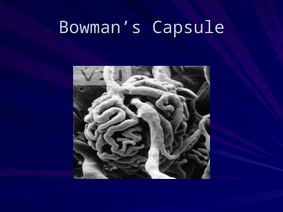
Bowman’s CapsuleBowman’s Capsule


DiffusionDiffusionSmall molecules make easily movement by diffusion in a cell
limited by the cell’s semi permeable membrane
In liquid: Equally sweet
In air: Equally smell
Coffee
Sugar
Perfume
Room

Ex. RBCEx. RBC
Isotonic=In Equilibrium
Solutes: materials-- sugar, salt.
Solvent: Liquid material that dissolve--waterSolution: A mixture of two.
Tonicity: A state of amount of dissolved material in them.
O.9% NaCl
99.1% H20
0.9% Plasma NaCl
99.9% H20

Tonicity: By NaClTonicity: By NaCl
Osmosis:From: 0.9% NaCl
99.1% H20To:
3% NaCl
97% H20
Osmosis: [H20] [H20]
99.1 H20
0.9 NaCl
0% NaCl
100% H20
Hyper tonic
Hypo tonic
So, H20 will move into RBC. So., RBC will rupture

OsmosisOsmosis
A special type of diffusion between NACl & water.
Water: Small, unchanged molecules
NaCl: Changed molecules*** Changed molecules are hard to move--- H20 move freely from a weaker solution to a more concentrated one.
NaCl: Electrolytes that regulate vascular osmotic pressure.

KidneyKidney
Purify (Filter) and reabsorb 5 inches long, level with T12 and L1~L3
Urine: collected by the pelvis
20% to 25% of the resting cardiac output (approx. 1200 mL per Min.) passes through kidney.
Liver: 27% Brain: 14%Heart: 4%
Skeletal: 15% Skin: 6% Bone: 6%
Miscellaneous: 7%
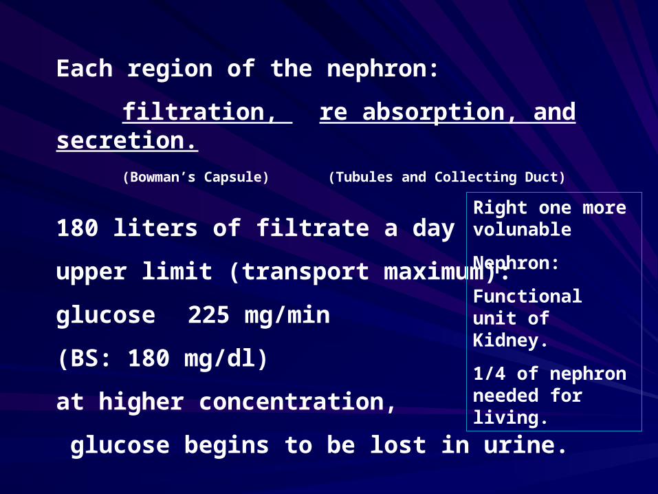
Each region of the nephron:
filtration, re absorption, and secretion.
(Bowman’s Capsule) (Tubules and Collecting Duct)
180 liters of filtrate a day
upper limit (transport maximum):
glucose 225 mg/min
(BS: 180 mg/dl)
at higher concentration,
glucose begins to be lost in urine.
Right one more volunable
Nephron:
Functional unit of Kidney.
1/4 of nephron needed for living.
Excretion of waste nitrogen (urea, uric acid, creatin)
protein--> ammonia--> harmless urea--> kidney
American food ( protein)
Detoxification of ammonia -->
¤ Liver, Kidney failure
H20 and electrolytes
Hypothalamus (Osmoreceptor)
Sense blood
¤ [ ] = H20, lytes.
A.R.FA.R.F
Is a clinical syndrome characterized by a renal shut down--> acute tubular necrosis, obstruction, acute tubular insufficiency--> Most occur in previously healthy individuals. Generally follows an identifiable trauma contact with a nephrotoxic agent. The most cause of ARF is related to surgical procedures.


Pre RenalPre Renal
Causes---> Consists of factors outside the kidneys that impair renal blood flow and lead to decreased glomerular perfusion
Problem corrected --> no ARF

PrerenalPrerenal
55-70%55-70%
Intravascular volume depletion, decreased CO, Intravascular volume depletion, decreased CO, vascular failure secondary to vasodilation or vascular failure secondary to vasodilation or obstruction---HTN, MI, severe dehydration, & obstruction---HTN, MI, severe dehydration, & shock ( not enough volume circulating).shock ( not enough volume circulating).
Reversible—can be corrected by establishing Reversible—can be corrected by establishing renal perfusion & preventing necrotic renal renal perfusion & preventing necrotic renal damage by fluid challengedamage by fluid challenge
Irreversible ischemiaIrreversible ischemia

Intra RenalIntra Renal
Conditions of actual damage to the renal tissue leading to malfunctioning of nephrons---> APN may lead ARF
Ex) acute tubular necrosis-
renal ischemia
Nephrotoxic drugs
Glomerulonephritis

IntrarenalIntrarenal
25-40%25-40%Kidney itself damaged to the kidney tissues and Kidney itself damaged to the kidney tissues and structures & includes tubular necrosis, structures & includes tubular necrosis, nephrotoxicity & alterations in renal blood flownephrotoxicity & alterations in renal blood flowInjuries @kidney glomeruli or tubules—90% due Injuries @kidney glomeruli or tubules—90% due to ATN--- glomerulonephrits, toxins, trauma, to ATN--- glomerulonephrits, toxins, trauma, crushing injuries, surgery, sepsis, CV collapse, crushing injuries, surgery, sepsis, CV collapse, MOF, ABX like aminoglycosides, street drugs, MOF, ABX like aminoglycosides, street drugs, chemo, nephrotoxic drugschemo, nephrotoxic drugsDon’t get confused w/ prerenal caused by blood Don’t get confused w/ prerenal caused by blood volume—dehydration & hypovolemiavolume—dehydration & hypovolemia

Post RenalPost Renal
Mechanical Obs. Of urinary outflow. As the flow of urine is blocked. Urine backs up into the renal pelvis. The most common causes are renal calculi, trauma, tumors.--> usually anuria rather than oliguria
Ex) Calculi, bladder tumor. Stricture (Compression)--BPH
Basement membrane is not destroyed. Trauma to back, pelvis, perineum strictures, spinal cord disease.

PostrenalPostrenal
5%5%
Obstruction of urine between the Obstruction of urine between the kidney and the urethral meatuskidney and the urethral meatus
Calculi BPH Tumors stricturesCalculi BPH Tumors strictures
B/C the obstruction, urine backflowsB/C the obstruction, urine backflows

PreventionPrevention
A. High Risk
Hospital Pt. --> Massive trauma, major surgical procedure, extensive burns, sepsis.
B. Industrial chemicals and nephrotoxic drugs.

A.R.F PhasesA.R.F PhasesA. Onset phaseA. Onset phase
begins with precipitating event--
Hypovolemia, or nephrotoxin exposure
Ends when the oliguric-anuric phase begins.
Time Span: Up to two days
urine output: 20% of normal.

A. Onset PhaseA. Onset Phase
Initial injury to the kidneyInitial injury to the kidney
ReversibleReversible
Preventable with early interventionPreventable with early intervention
UO:20% of normalUO:20% of normal
Unable to regulate electrolytesUnable to regulate electrolytes
B. Oliguric Phase
Spans the period when urine is less than 400 cc/d
Time Span: 8-14 days (1-2 wks)
urine output: 5% of normal.
Can not excrete fluid or waste products
Oliguria--> caused by reduction in the GFR

C. Diuretic Phase
Gradual increase in urine output of 1-3L up to 4-5 L/day. The high vol. Is due to osmotic diuresis from high urea concentration
and the adequate concentrating ability of tubules.
Capillary
urea H20
H20 H20 H20 H20H20 cells

Lab. Value stop rising, decreased SG:
Diluted urine and poss. Excessive diuresis when lab value stops dropping. Ends when they stabilize.
Time span: 10 days
U.O: Early 150%
Late: 200%

Diuretic PhaseDiuretic Phase
Electrolytes are lost—deficit in Electrolytes are lost—deficit in concentrating ability of tubules and concentrating ability of tubules and osmotic diuretic effect of increased osmotic diuretic effect of increased BUN, slowly increased excretion of BUN, slowly increased excretion of metabolic wastes, hypovolemia, loss metabolic wastes, hypovolemia, loss of Na, K, increased BUN initially then of Na, K, increased BUN initially then gradually return to baselinegradually return to baseline

Diuretic PhaseDiuretic Phase
S/S: postural hypotension, tachycardia, S/S: postural hypotension, tachycardia, improving mental alertness and improving mental alertness and activity, weight loss, thirsty, dry activity, weight loss, thirsty, dry mucous membrane, decreased skin mucous membrane, decreased skin turgorturgor
K replacement may requireK replacement may require

D. Recovery Phase
(convalescent phase)
Begins when lab. Values stabilize, ends when renal function returns to normal.
Time Span: 4-6 mo.
Up to 12 mo.
U.O: 100%

Recovery PhaseRecovery Phase
Increased GFRIncreased concentrating ability.Urine SG -- 1.003-1.030Urine osmolarity:Normal 300-1300Mortality Rate: 30% to 60% Most common cause of death secondary
to infection

Prevention of ARFPrevention of ARF
CHF, dehydration, shock. To minimize the risk.
1. Keep the patient hydrated (esp. before and after OR)
2. Continuously monitor the dosages and effects of
ABX and other drugs (nephrotoxic)
3. Assess renal function regularly
Treatment GoalsTreatment Goals
1. Correcting the underlying problem
2. Preventing infection
3. Treating fluid and electrolytes imbalance
4. Correcting metabolic acidosis
5. Treating clinically significant anemia.

How does one differentiate acute from chronic renal failure?
1. History-medical records.
2. Hypo calcemia, hyper phosphatemia, anemia. --> has been associated more often with CRF.
* Calcium, phosphorous, acid-base derangement are often seen in ARF as early as 48-72 hours after onset of illness.
* Anemia: nonspecific indicator.
3. Reliable indicator of CRf
Small kidney with a decreased or absence of renal cortex as assessed by UTZ (0.5 cm)
Abnormal: Kidney length of less than 9 cm
--> CRF or significant renal dz.
> 1.5 cm in renal length: unilateral/asymmetric renal dz.

CRFCRF

CRFCRF
Slow, progressive, irreversible damageSlow, progressive, irreversible damage4 stages4 stagesDiminished Renal Reserve ---50% of Diminished Renal Reserve ---50% of
nephrones are lost, asymptomatic, no S/Snephrones are lost, asymptomatic, no S/SRenal Insufficiency---75% nephrones lost, Renal Insufficiency---75% nephrones lost,
azotemia, anemia, polyuria, nocturiaazotemia, anemia, polyuria, nocturiaRenal Failure---pt needs temporary or Renal Failure---pt needs temporary or
permanent dialysispermanent dialysisEnd Stage Renal DiseaseEnd Stage Renal Disease
CRFCRF
An irreversible loss of nephrons. A symptomatic until 70-90% of the nephron is destroyed.
(Divided into four stages)
1. Diminished renal reserve: nephron loss without the loss of measured renal function. Normal BUN, CR, no sxs.
2. Renal insufficiency: a measurable decline in renal function. Loss of ability to concentrate urine --> nocturia, polyuria, often associated HTN fatigue, weak. Ha.
3. Renal failure /ESRD
4. Uremia: a clinical syndrome with severe decline in renal function, associated with dysfunction of multiple organ systems.

Etiology of CRFEtiology of CRF
1. 30%: Diabetic nephropathy
2. 26%: Hypertension
3. 14%: other urological disease ( hydronephrosis, polycystic kidney.)
4. Congenital malformations
5. Nephropathy associated with the human immunodeficiency virus.
6. Myeloma Kidney

Evaluation: To establish the degree of renal impairment.
To identify reversible factors
-infection, obstruction, volume deficit, nephrotic drugs, less than optimal cardiac output with, without HTN, uncontrolled HTN, hypercalcemia and hyperuricemia
-orthostatic Bp. Pulse
-U/A with microscope/dipstick, serum electrolyte, BUN, CR, CBC, evaluation of post void residual, UTZ

U/A: the simplest, most cost effective evaluation
Urine SG: 1.010 or less
Urine pH: less than 7.0 8.0: the question of infection
Dipstick: glucose in DM or CRF
Proteinuria: the hall mark of intrinsic renal disease.
Nephrotic-range of proteinuria is seen in glomerular lesions and 1-2 gm of protein excretion in interstitial dzs.
RBC: active renal dz of a glomerular or vascular etiology.
WBC: infection

Medical TreatmentMedical Treatment
A. Most hypervolemic--> kidney can’t eliminate amount of H20 and electrolytes. ** Hypovolemia
(Increased HCT)
B. Anemia/Bleeding
The main cause of anemia-->
Decreased production of erythropoietin by the kidney
C. Nutritional deficiency
Decreased RBC life
increased hemolysis of RBC bleeding from G I tract dialyzer may contribute to the anemic state.
Folic Acid: Essential for DNA Synthesis and normal maturation of RBC.
D. Dialysis
1) Hemodialysis: within 1-2 Hr. bring K+ to normal
2) Peritoneal dialysis: 4-8 hrs.
E. Phosphate binders
(aluminum or magnesium containing antacids)--> CA-Phos.
An inverse relationship to bind excessive CA in ECF--> Decreased serum Ca level phosphate binders--> urinary acidifier to help prevent calcium stones
F. Diet
the major goal of nutritional management is to decrease catabolism of the body’s protein. No more than 0.8-1 gm of protein/kg/day.

F. Diet
the major goal of nutritional management is to decrease catabolism of the body’s protein. No more than 0.8-1 gm of protein/kg/day.
G. HTN
1. NA-fluid restriction
2. Diuretic--Lasix
*3. Anti HTN drugs
A. Ace inhibitors-- enalapril, captopril
B. beta-adrenergic--inderal (decreased rennin released)
C. Calcium channel blockers: diltiazem, verapamil

H. Neurologic Function
No. TX. Available without dialysis
neuro Change--> renal failure progress
* contraindicated
AntiHTN: triamiterene spironolactone, amiloride
* 10 unit regular insulin + D 50 ampule. NaHCO3 bolus or 75~ 100 cc/hr- hyperkalemia tx.
* Albuterol: Potassium lowering effect

Increased nitrogenous waste products, electrolytes imbalances --> demylination of nerve fiver, axonal atrophy.
** Safety: due to weak muscle
general depression of CNS--> lethargy, fatigue, decreased concentration, dialysis dementia due to aluminum toxicity
Complications: UGI bleeding
a major Cx and 3-7% of deaths. Superficial mucosal abnormalities. Duodenitis and gastritis (10-60%)
TX: Cimetidine (H2-receptor antagonist)

Death is usually due to infection, G-I bleed, myocardiac infarction. Kidney can no longer remove toxic wastes and water from blood. Two means are mimicking the body’s lost capabilities.

Uremia is a clinical situation in which azotemia progress to systematic state.
Azotemia; an excess of urea or other nitrogenous compounds in the blood.
Fetor: offensive odors
halitosis: offensive odors of the breath.

CV: CHF, HTN, pericarditis, arrythmia, hematopoietic anemia, peripheral/systemic edema.
Neuro: Drowsy, confusion, tremor, coma, irritability, convulsion, twitching, peripheral neuropathy
Integ: pallor, yellowish color, dryness, pruritis, ecchymosis
Skeletal:
Hypocalemia, soft tissue, calcification, alteration in coagulation, increased infection
GI: Anorexia, N/V, gastrtitis, uremic halitosis, diarrhea, constipation
Resp: Pul. Edema, Pneumonia, Kussmaul Resp.

Asterixis:
A motor disturbance by sustained contraction of muscles.
Kussmaul Breathing--> Deep to rapid breathing to increase excretion of CO2
--> a compensatory mechanism of a-acidosis

Nutrition in Renal DiseaseNutrition in Renal Disease
Sodium: major ECF Caution, important in acid-base balance, fluid balance, cell permeability, and muscle action.
High NA+ Foods: Table salt, processed foods, milk, fish, poultry, meat, eggs, carrots,k some canned soup, beets, spinach, meat sauce, soy sauce, salad dressings, potassium: major ICF, important in acid-base balance, neuromuscular activity, carbohydrate metabolism, and protein synthesis.
High in K+ foods: meat, oranges, potatoes, whole grains, bananas, broccoli, beans, nuts, apricots, spinach, dried fruits, melons, peas, fruit juices, peaches, tomatoes, avocados, coffee, wine, salt substitute, antibiotics.
Proteins: build, maintain, and repair body tissues

Protein sources: eggs, milk, fish, poultry, grains, legumes (dry beans, lentils, split peas, soy beans)
Calories (carbohydrates, fats): to meet body’s need for energy and to attain or maintain ideal body weight. Fats are also important in maintaining skin integrity and forming complex lipid compounds.
Carbohydrate source: fruits, vegetables, cereals, sugar, hard candy, jelly beans, jams, jellies.

Fat sources: butter, margarine, oil, cream, bacon, meat fat, salad dressing, egg yolk, olives, nuts, avocados.
Vitamins and mineral supplements:
Vitamin C, B Complex Vitamins, calcium, phosphorus, and vitamin D.
Calcium sources: calcium carbonate (OS-call, Tums), calcium acetate (Phos-Low), supplements given between meals.

Phosphorous binding agents: Calcium acetate (Phos-Low), aluminum hydroxide gel (Amphogel), aluminum carbonate (Basalgel) binding agents given with meals.
Vitamin D source; Calciferol (Hytakerol), Calcitrol (Rocaltrol).
Iron sources: ferrous sulfate, parental iron products.

Magnesium: not usually a problem unless there is intake of magnesium containing medicines such as laxatives and antacids.

Geriatric ConsiderationGeriatric Consideration
A. Decreased RRF (Reserved Renal Factor)
Decreased GFR
Decreased Clearance
B: Aging kidney is less able to withstand changes in hydration, solute, load, cardiac output. Aging itself is the primary risk factor.
Mortality: 5-25% higher in older than younger.

HemodialysisHemodialysis
An artificial Semi permeable membrane acts like the kidney-> diffusion and osmosis
Excess fluid is removed by creating a pressure differential BTN the blood and the dialysate solution (=balanced solution of electrolytes and fluid)
With a combination of positive pressure in the blood compartment and/or negative pressure in the dialysate compartment
2.5-4 hrs 3 times/week at home or dialysis center.

Quinton Catheter;
single, double, temporary vascular access, 2-3 days femoral, subclavian, internal jugular veins.

AV AccessAV Access
AV Shunt: Temporary while internal graft is healing Machine
Artery
VeinRinse
Fistulas:Cephalic/radial Artery
Basilic vein
Thigh or forearm

Grafts (looped graft)
Bovin--> relatively resistant to infectious organism.
Antecubital vein
Brachial arteryLooped graft
NSG: 14-16 G needle A thrill/bruit can be felt by palpating
***** NO VENIPUNCTURE, BP ON THE AFFECTED ARM*****

NSG ManagementNSG Management
1. Hypovolemia/shock --due to rapid removal of vascular volume
* trendelenberg to improve cerebral blood flow
2. Analgesia: muscle cramps associated with significant discomfort and pain
: neuromuscular hypersensitivity
Peritoneal dialysis
Principle---sterile dialyzing fluid infused into peritoneal cavity through a catheter
Surgically implanted in the pt’s peritoneal cavity.
Inflow ---> dwell ---> outflow
15-30 min. 4 hr/d 15-30 min.
10 hr/n
* continuous 24 times/day
* at night 8 hrs cycler machine

TransplantationTransplantation
A living or cadaveric donor
the life expectancy for patients on dialysis is 6-7 years < 60 years.
2-3 years > 60 years or diabetic
Survival years is 90% at 3 years with cadeveric transplant
95%: Living

Ruptured AV Shunt
Subcutaneous hemorrhage--- hypovolemia--shock-- cardiac arrest
Intervention
1. Control bleeding
2. Transfusion
3. Pain relief
hematoma-arm, cold compression

To Control BleedingTo Control Bleeding
1. Tie a tourniquet above the AV or BP cuff. Don’t release until OR.
* pressure DSG, Arm.
2. IV fluid, O2 2L
3. Notify MD/surgical team
4. Type and cross match ---cont. monitor the pt for signs of shock.

5. Dopamin Drip
--Pulmonary edema secondary to fluid overload
6. Strict I & O
7. H & H Q 4-6 H
8. Monitor circulatory, motor, neurofunction below hematoma.

DefinitionDefinition
Infection of the kidney and renal pelvis.
Every PN is secondary

APNAPN
Acute infection of the kidney, characterized by acute inflammation and focal abscess, usually unilateral, often accompanied by bacteremia.
Characterized by bacteriuria (generally > 100,000 colonies/ml) and pyuria. In acute infections, a single infective pathogen usually is found.

CPNCPN
The result of repeated episodes of APN leading to progressive renal scarring.
Scars are usually asymmetric and irregular and involve the renal cortex and pelvocalyceal system. Negative urine cultures and no evidence of active infection.

PathogensPathogens
Gram-neg Bacilli: Escheria coli, proteus, Pseudomonas, Enterobacter, kebsiella, Serratia, and Citrobacter species.
Gram-pos cocci: Staphylo. S Strepto. A

Ascending InfectionAscending Infection
The most common cause of GU tract infection
Female- short urethra
altered flora d/t antibiotics, birth control
(spermicide and diaphragm)
urethral massage.

Direct extension from other organ.
-Interaperitoneal abscess
- Pelvic inflammatory disease
- GU tract fistulas.

Clinical ManifestationsClinical Manifestations
Rapidly over a few hrs. or a dayRapidly over a few hrs. or a day
Temp>39.4 C (103)FTemp>39.4 C (103)F
Shaking chillsShaking chills
N/V diarrheaN/V diarrhea
Sxs of cystitis may or may notSxs of cystitis may or may not
TachycardiaTachycardia
Flank painFlank pain
Generalized muscle tendernessGeneralized muscle tenderness
Marked tenderness on deep pressure on CVA or on Marked tenderness on deep pressure on CVA or on deep abd. Palpationdeep abd. Palpation
Significant leukocytosisSignificant leukocytosis
Pyuria with leukocyte castsPyuria with leukocyte casts
Bacteria on gram stains of unspun Bacteria on gram stains of unspun urineurine
Hematuria in acute phase of diseaseHematuria in acute phase of disease
Elderly: no classic sxs., urinary Elderly: no classic sxs., urinary incontinence (new onset), decreased incontinence (new onset), decreased appetite, confusion, lethargyappetite, confusion, lethargy
Children: not clear, low grade fever, Children: not clear, low grade fever, irritable, decreasedirritable, decreased appetite, n/v, appetite, n/v, diaper urine smells, no s/s of UTIdiaper urine smells, no s/s of UTI
Older Children: abd. Pain, Older Children: abd. Pain, frequency, flank pain, frequency, flank pain, dysuria, difficulty controlling dysuria, difficulty controlling urine.urine.Severe PN-fever subsides Severe PN-fever subsides more slowly and may not more slowly and may not disappear for several days. disappear for several days. Even after appropriate Even after appropriate antibiotic tx. antibiotic tx.

Medical ManagementMedical Management
A. Relieve obstruction prn (may be contributing to the infection)
B. C & S--> antibiotics/long term
C. Check Creatinine, CBC

Nursing Management
may treat at home!
A. patient teaching
-continue antibiotics
- 3 liters fluid/day
- check urine output
- prevent infections
- call MD

AGN
Inflammatory reaction in the glomeruliEtiology: streptococcal infection (2-3 wks after)

Pathophysiology
Antigen-antibody reaction with glomerular tissue. Inflammatory response---> increased porosity & decreased filtration---> kidney congested, swollen

Clinical Manifestations
HA, malaise, edema, flank pain, HTN, CVA tenderness, SOB
Diagnostic Evaluation
Proteinuria, hematuria, increased SG, edema, HTN, decreased UO, increased BUN & Cr.

Medical TX
Protect kidney + treat CX promptly---ABX, BR, dec.protein diet & Na diet,
anti-HTN, fluids, diuretics

Nephrotic Syndrome
Proteinuria, Hypoalbuminemia, edema, hypercholesterolemia

Etiology
Conditions that manage glomerular capillary membrane--- chronic GMN, DM, SLE, pericarditis, allergic reaction, CHF, pregnancy

Pathophysiology
Change in glom. Base membrane, inc. porosity & loss of proteins--->dec. albumin---> dec. serum osmoticpressure-->edema& dec. plasma Vol.---aldosterone--NA & H2O retention--->!
Clinical Manifestations
Edema, proteinuria, dec. albumin, inc. lipidemia, UO inc/dec-->renal failure. Dec. appetite,
fatigue

Medical Management
Dec. albuminemia, control edema, promote general health---Steroids, Diet--->protein normal or inc, inc. calories Edema---dec. NA, diuretics, check K+RUA, renal labs.

Nursing Management
Activity---bed rest--->ambulate!Fluid management-->assess/overload, diet, bpPatient education--> avoid infection, fatigue, f/u medical care Sx renal fail---MD
Nursing Management
Nutrition Na & protein control. Small frequent feedings Medication--- steroids, diureticsChecks for SEs, resp. assess patient teaching---meds, nutrition,self assessment/fluids call MD--inc. edema, DOE, fatigue, HA, infection

KidneyKidney


![RNC-A SERIES - Bakedeco RNC-210A_Manual.pdf · RNC-90A-R/L 2 RNC-120A-R/L 2 RNC-150A-R/L 3 RNC-180A-R/L 3 RNC-210A-R/L 4 [f] WATERPROOF COVER To prevent the entrance of water, the](https://img.dokumen.tips/doc/110x75/5e680bb313a66779ab666ae1/rnc-a-series-bakedeco-rnc-210amanualpdf-rnc-90a-rl-2-rnc-120a-rl-2-rnc-150a-rl.jpg)