Embed Size (px)
Citation preview

Usher syndrome (USH; OMIM 276900-2276905, 605472) is an autosomal recessive disease in which sensorineural hearing loss is associated with photoreceptor degeneration with the clinical features of retinitis pigmentosa [1-3]. USH is usually distinguished by three subtypes [4]. Individuals with USH1 are generally born completely deaf or lose most of their hearing early. Deafness is then followed by progres-sive visual impairment caused by retinitis pigmentosa, which usually becomes apparent in childhood. The patients often have difficulty maintaining their balance owing to problems in the vestibular system. USH2 is characterized by variable degrees of hearing loss and progressive visual troubles begin-ning in adolescence or early adulthood. Vestibular responses are usually normal. Patients with USH3 usually have normal hearing at birth but develop progressive and severe hearing loss over time. Retinitis pigmentosa usually develops in late
childhood or adolescence, and balance problems are present in most cases.
The prevalence of the disease has been estimated at about 3.5–8:100,000 in European populations [5] and 4.4:100,000 in the United States [6]. In a recent survey, USH represented 10.8% of inherited retinal dystrophies in southern France [7]. USH is inherited as an autosomal recessive trait, and to date, 15 USH loci have been characterized and 12 causative genes identified (A-genes, last update May 31, 2014). There are six USH1 identified genes: MYO7A (NM_000260.3, OMIM# 276903), coding the protein Myosin VIIa; USH1C (NM_153676.3; OMIM# 605242), coding Harmonin; CDH23 (ID: 64072; OMIM# 605516), coding Cadherin 23; PCDH15 (ID: 65217; OMIM# 605514), coding Protocadherin 15; USH1G (ID: 124590; OMIM# 607696), coding SANS and CIB2 (ID: 10518; OMIM# 605564) coding Ca2+-and integrin-binding protein [8]. Three genes have been associ-ated with USH2: USH2A (ID: 7399; OMIM# 608400), coding the protein Usherin; USH2C (ID: 84059; OMIM# 602851), coding GPR98 (formerly known as VLGR1b or MASS1); USH2D (ID: 25861; OMIM# 607928), coding Whirlin. Finally
Molecular Vision 2014; 20:1717-1731 <http://www.molvis.org/molvis/v20/1717>Received 22 February 2014 | Accepted 20 December 2014 | Published 23 December 2014
© 2014 Molecular Vision
1717
MYO7A and USH2A gene sequence variants in Italian patients with Usher syndrome
Andrea Sodi,1 Alessandro Mariottini,2 Ilaria Passerini,2 Vittoria Murro,1 Iryna Tachyla,1 Benedetta Bianchi,3 Ugo Menchini,1 Francesca Torricelli2
1Department of Ophthalmology, Azienda Ospedaliero-Universitaria Careggi, Florence, Italy; 2Department of Genetic Diagnosis, Azienda Ospedaliero-Universitaria Careggi, Florence, Italy; 3Department of Otolaryngology, Azienda Ospedaliero-Universitaria Careggi, Florence, Italy
Purpose: To analyze the spectrum of sequence variants in the MYO7A and USH2A genes in a group of Italian patients affected by Usher syndrome (USH).Methods: Thirty-six Italian patients with a diagnosis of USH were recruited. They received a standard ophthalmologic examination, visual field testing, optical coherence tomography (OCT) scan, and electrophysiological tests. Fluorescein angiography and fundus autofluorescence imaging were performed in selected cases. All the patients underwent an audiologic examination for the 0.25–8,000 Hz frequencies. Vestibular function was evaluated with specific tests. DNA samples were analyzed for sequence variants of the MYO7A gene (for USH1) and the USH2A gene (for USH2) with direct sequencing techniques. A few patients were analyzed for both genes.Results: In the MYO7A gene, ten missense variants were found; three patients were compound heterozygous, and two were homozygous. Thirty-four USH2A gene variants were detected, including eight missense variants, nine nonsense variants, six splicing variants, and 11 duplications/deletions; 19 patients were compound heterozygous, and three were homozygous. Four MYO7A and 17 USH2A variants have already been described in the literature. Among the novel mutations there are four USH2A large deletions, detected with multiplex ligation dependent probe amplification (MLPA) technology. Two potentially pathogenic variants were found in 27 patients (75%). Affected patients showed variable clinical pictures without a clear genotype-phenotype correlation.Conclusions: Ten variants in the MYO7A gene and 34 variants in the USH2A gene were detected in Italian patients with USH at a high detection rate. A selective analysis of these genes may be valuable for molecular analysis, combining diagnostic efficiency with little time wastage and less resource consumption.
Correspondence to: Iryna Tachyla, Department of Ophthalmology, Azienda Ospedaliero-Universitaria Careggi, Largo Brambilla 3, 50134 Florence, Italy; Phone: +39-055-411765 FAX: +39-055-4377749 ; email: [email protected]
Molecular Vision 2014; 20:1717-1731 <http://www.molvis.org/molvis/v20/1717> © 2014 Molecular Vision
1718
USH3 phenotype has been associated with the gene CLRN1 USH3A (ID: 7401; OMIM# 606397), coding the protein Clarin-1.4, and more recently with the genes ABHD12 (ID: 26090; OMIM# 613599) coding the enzyme with the same name [9] and HARS (ID: 3035; OMIM#142810) coding histidyl-tRNA synthetase [10].
Although a common phenotype has been described in the literature concerning the different USH types, the identified USH genes encode for proteins from different classes and families with different functions. Growing evidence suggests that these proteins are organized in a protein “interactome” [11-13] in the inner ear and the retina, which is critical for the development, maintenance, and correct function of the sensorineural cells.
Variants in the MYO7A gene represent the most common cause of USH1, accounting for approximately 50% of cases [14-16]. The gene is located on chromosome 11q13.5 and has 49 exons. It codes a 2,215-amino-acid protein, myosin VIIA, which is produced in the retina and in the inner ear and acts on the transport processes through the cilia of the retinal photoreceptors. In addition to USH, MYO7A variants have been associated with autosomal dominant non-syndromic sensorineural hearing impairment (DFNA11, OMIM 601317) [17] and autosomal recessive deafness (DFNB2, OMIM 60060) [17].
USH2A is the most common mutated gene in USH2 [18,19]. It is located on chromosome 1q41 and contains 72 exons. It codes the protein usherin, which can be found in a short (1,546 amino acids) and a long (5,202 amino acids) isoform, both present in the retina where they are function-ally linked with the other proteins of the interactome. USH2A gene variants have also been found in autosomal recessive retinitis pigmentosa without hearing loss (MIM 613809) [20].
To date, more than 424 variants have been identified in the coding region of MY07A, and the majority are single-base variants. In contrast, more than 648 USH2A variants have been detected, the majority are also single-base variants.
In Italian patients with USH, molecular data were previously obtained using the Asper Biotech microarray that investigated 612 already known variants using APEX technology [21]. In another study, the same group analyzed 12 patients with USH with next-generation sequencing (NGS), highlighting some limitations of this technology for USH [22]. Furthermore, ten Italian families affected by USH1 were screened for MYO7A sequence variants within a collaborative investigation that examined patients of different origin [15].
At present, no specific study has reported the complete sequence of MYO7A and USH2A, the most common USH
genes, involving a large series of Italian patients with USH. In the present study, sequence variants of the MY07A and USH2A genes were determined with direct sequencing in a group of 36 Italian patients affected by USH.
METHODS
Clinical evaluation: Thirty-six Italian patients (from 33 different families) with a clinical diagnosis of USH were recruited through the Hereditary Retinal Degenerations Referring Centre of the Eye Clinic of the University of Flor-ence. Except the visual and hearing impairment they didn’t have other health related problems. Ten were males and 26 females. The mean age was 39.7 years (±13.7 years) (range 17-72 years).
Criteria for USH phenotype included hypoacusia of various degrees associated with hemeralopia, diffused retinal dystrophy at fundus examination, visual field loss and abnormal ERG responses. The presence of typical bone spicules could be found in most of the patients but was not considered within the inclusion criteria.
The study adhered to the tenets of the Declaration of Helsinki and to the ARVO statement on human subjects. It is approved by the Local Ethics Committee. Moreover, each patient gave written informed consent.
All subjects included in the study were clinically evalu-ated with a standard ophthalmologic examination, fundus photography (Zeiss Retinograph, Carl Zeiss, Dublin, CA), optical coherence tomography (OCT) scan (Topcon 3D OCT-1000, Topcon Medical Systems, Oakland, NJ), and ERG (Electrophysiological Diagnostic Unit Retimax, Roland Consult, Brandenburg, Germany) performed according to the existing International Society for Clinical Electrophysiology of Vision (ISCEV) Guidelines [23]. Most patients under-went automated visual field testing (Humphrey Visual Field Analyzer, Carl Zeiss) while selected patients with poor vision were tested with Goldmann perimetry. In most of the cases, electrophysiological and perimetric examinations were performed in our department, but for a few patients, we accepted examinations performed during the previous year in other hospitals and included in the patient’s medical documentation. Fluorescein angiography (FA; Zeiss Reti-nograph with Image Processing Software Visupac, Carl Zeiss) was performed on three patients with sine pigmento fundus appearance to improve the visualization of the retinal dystrophy. Fundus autofluorescence imaging (FAF; Confocal SLO, HRA, Heidelberg Engineering, Heidelberg, Germany) was performed on all affected patients who agreed to collaborate.

Molecular Vision 2014; 20:1717-1731 <http://www.molvis.org/molvis/v20/1717> © 2014 Molecular Vision
1719
All patients underwent an audiologic examination for the 0.25–8,000 Hz frequencies. Vestibular function was evaluated with specific tests (caloric test, study of spon-taneous and induced nystagmus with Frenzel glasses or video-oculoscopy).
According to the ophthalmologic and audiologic pheno-type, the patients were then clinically distinguished in USH1 or USH2. Finally, all patients underwent genetic testing for the complete sequencing of the MYO7A gene in USH1 and the USH2A gene in USH2. Some patients first classified as USH2 in whom no USH2A mutation was detected later received complete sequencing of the MYO7A gene.
DNA extraction and PCR amplification: Following informed consent and a complete medical history of each family, 5 ml of peripheral blood was taken from the antecubital vein using EDTA-containing vials. DNA was extracted from 1 ml of peripheral blood using the QiaSymphony DNA Blood Midi kit on the QIAsymphony SP workstation (Qiagen, Hilden, Germany), according to the manufacturer’s protocol. Genomic DNA was isolated from peripheral leukocytes, using the QiaSymphony DNA Blood Midi kit on the QIAsymphony SP workstation (Qiagen), according to the manufacturer’s protocol. All coding exons, including intron-exon boundaries of the MYO7A and USH2A genes, were processed with the automated Core System (Beckman Coulter, Fullerton, CA). After purification, amplicons were sequenced on the 3730 DNA Analyzer (ABI, Foster City, CA). The sequences were assembled and analyzed using SeqScape software (ABI).
Variants of unknown pathogenicity were interpreted with Alamut 2.2 (Interactive Biosoftware, Rouen, France), a decision-support software application for medical molecular genetics. The software relies on web-based prediction software, such as Align-GVGD, SIFT, PolyPhen, Mutation Taster (hosted by Interactive Biosoftware). Note that Alamut 2.2 scoring systems provide a predictive evaluation only for missense variants.
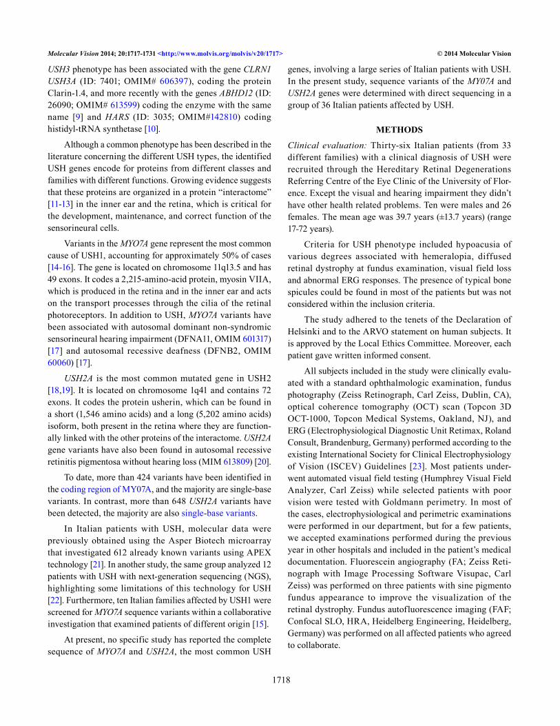
Multiplex ligation dependent probe amplification (MLPA) analysis was performed on the patients in whom no variant or only one variant was identified in the USH2A gene and on the carriers of a variant in homozygosity in which study of the parents did not confirm the Mendelian inheritance of the variant detected in the proband (Figure 1). The MLPA reaction (P361-A1/P362-A2 SALSA MLPA kit; MRC Holland, Amsterdam, The Netherlands) was performed according to the manufacturer’s recommendations. One microliter of each reaction product was separated on a POP-7 polymer with capillary electrophoresis using the 3730 DNA Analyzer (ABI). Freely available software provided by MRC Holland was used to analyze the MLPA data (Coffalyser; MRC Holland; Figure 2).
The deletion was further confirmed with real-time quan-titative PCR with a predesigned TaqMan Copy Number Assay specific for USH2A genes (Hs00961154_cn, Hs03023043_cn, Hs02691757, Applied Biosystems) using TaqMan® Universal Master Mixes with the following parameters of run : hold 95 °C for 10 min, cycle 95 °C for 15 s and 60 °C for 60 s (40 cycles). Quantitative PCR was performed using an ABI 7900HT instrument (Applied Biosystems) in a 96-well optical plate using six replicates for each sample in a final volume of 20 μl, according to the manufacturer’s protocol. Data were analyzed using CopyCaller to estimate the copy number for each sample.
RESULTS
Thirty-six Italian patients with a clinical diagnosis of USH (from 33 families) were clinically examined: ten were male and 26 female. The mean age was 39.7 years (±13.7 years; range 17–72 years). The age of onset of the symptoms (usually hemeralopia and/or perception of visual field constriction) varied, ranging from 4 to 37 years, with an average of 17.5 years (±8.8 years).
Snellen visual acuity ranged from 1.0 to light perception, with an average value of 0.56 (±0.37). In the best eye, 20
Figure 1. Pedigree of a family with two sisters with USH2. The patients carried in the USH2A gene the single sequence variant c.4144T>C (p.Trp1382Arg), which was first supposed to be present in homozygosis. Because this variant was detected in the father but not in
the mother of the patients, a deletion was then suspected. The evaluation of the patients with a new MLPA kit for the gene USH2A allowed identification in the mother and her two daughters of a heterozygous deletion of exons 17–19.
Molecular Vision 2014; 20:1717-1731 <http://www.molvis.org/molvis/v20/1717> © 2014 Molecular Vision
1720
(55.6%) patients presented normal visual acuity or mild visual loss (0.9–0.6), nine patients (25%) had moderate visual loss (05–0.2), and the other seven patients (19.4%) showed low vision (<0.2).
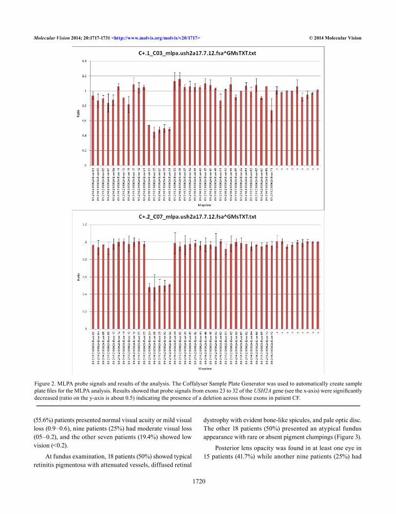
At fundus examination, 18 patients (50%) showed typical retinitis pigmentosa with attenuated vessels, diffused retinal
dystrophy with evident bone-like spicules, and pale optic disc. The other 18 patients (50%) presented an atypical fundus appearance with rare or absent pigment clumpings (Figure 3).
Posterior lens opacity was found in at least one eye in 15 patients (41.7%) while another nine patients (25%) had
Figure 2. MLPA probe signals and results of the analysis. The Coffalyser Sample Plate Generator was used to automatically create sample plate files for the MLPA analysis. Results showed that probe signals from exons 23 to 32 of the USH2A gene (see the x-axis) were significantly decreased (ratio on the y-axis is about 0.5) indicating the presence of a deletion across those exons in patient CF.

Molecular Vision 2014; 20:1717-1731 <http://www.molvis.org/molvis/v20/1717> © 2014 Molecular Vision
1721
already undergone cataract surgery in at least one eye. One (2.8%) patient presented anterior lens opacity.
OCT showed a macular edema in at least one eye in six patients (16.7%) while three cases (8.3%) presented a unilateral macular hole and four patients (11.1%) had macular atrophy.
ERG scotopic response was always bilaterally flat or severely abnormal. Photopic response was abnormal in all patients.
The Goldmann visual field was concentrically reduced in both eyes of 14 patients (38.9%) while 19 (52.7%) showed peripheral loss and pericentral scotomas leaving limited central and peripheral field remnants. In three patients (8.3%) visual field loss was severe, with only a central isle of surviving vision.
Three patients showed early and severe deafness with balance problems and onset of retinal dystrophy in child-hood and were clinically classified as USH1. Thirty-three patients were affected with a milder hearing impairment, no vestibular dysfunction, and variable retinal phenotypes and were classified as USH2.
All three patients with USH1 carried two MY07A sequence variants. Among the 33 patients clinically clas-sified as USH2, 22 (66.6%) carried two USH2A variants,
two (6.1%) carried two MYO7A variants, one (3%) carried a single MYO7A variant, two (6.1%) carried a single USH2A variant, one (3%) carried a MYO7A variant on one allele and an USH2A variant on the other allele while in the remaining five patients no MYO7A or USH2A variant was detected. The patients in whom two pathogenic MYO7A or USH2A variants were found were screened for the USH3A/Clrn1 gene, but no variant was identified.
The MYO7A and USH2A sequence variants detected in our series are summarized in Table 1. On the whole, out of the 36 patients with USH in our series, 27 (75%) carried two pathogenic variants in the same gene (five in the MYO7A gene and 22 in the USH2A gene) and therefore could be considered genetically characterized. In the MYO7A gene, ten sequence variants were identified in seven patients. Three patients were compound heterozygous, two patients were homozygous, one patient showed the variant only on one allele, and one patient carried the MYO7A variant on one allele and an USH2A variant on the other allele. All variants were missense. Each family had a specific MYO7A variant.
Six MYO7A variants have already been reported in the literature [15,22,24] and are therefore considered patho-genic. The other four variants [c.4798G>C (p.Gly1600Arg), c.5810T>C (p.Leu1937Pro), c.977T>C (p.Leu326Pro), c.4039C>A (p.Arg1347Ser)] were novel and could not be
Figure 3. Fundus pictures of two brothers of the same family, who carry the same genotype (c.2299del (p.Glu767Serfs*21) USH2A variant on one allele and c.13130C>A (p.Ser4377*) USH2A on the other allele). One patient (A, patient GF) shows a typical form of retinitis pigmentosa while the other one (B, patient GS) presents with an atypical fundus appearance with mild retinal dystrophy and rare pigment deposits. In the second case, the clinical diagnosis is supported by typical visual field and electrophysiological abnormalities. The coexistence of two different clinical pictures in the same pedigree emphasizes the poor genotype-phenotype correlation reported in our series.

Molecular Vision 2014; 20:1717-1731 <http://www.molvis.org/molvis/v20/1717> © 2014 Molecular Vision
1722
Tab
le 1
. Th
e M
YO7A
an
d U
SH2A
seq
ue
nc
e v
ar
ian
Ts d
eTe
cT
ed
in o
ur
ser
ies.
Patie
ntU
SH p
heno
type
Gen
eA
llele
1A
llele
2A
llele
1 r
efer
ence
Alle
le 2
ref
eren
ceLA
USH
1M
YO7A
c.721
C>G
(p.A
rg24
1Gly
)c.7
21C
>G (p
.Arg
241G
ly)
[15]
[15]
ZEU
SH1
MYO
7Ac.
4798
G>C
(p.G
ly16
00A
rg)
c.58
10T
>C (p
.Leu
1937
Pro)
**
TGU
SH1
MYO
7Ac.
977T
>C (p
.Leu
326P
ro)
c.56
17C
>T (p
.Arg
1873
Trp)
*[4
3]LM
aU
SH2
MYO
7Ac.
4411
T>C
(p.S
er14
71Pr
o)c.
4411
T>C
(p.S
er14
71Pr
o)[2
2][2
2]BN
USH
2M
YO7A
c.39
5C>T
(p.P
ro13
2Leu
)c.
4039
C>A
(p.A
rg13
47Se
r)[1
5]*
IPU
SH2
USH
2Ac.
2052
A>G
(p.=
)N
ot fo
und
[44]
MYO
7AN
ot fo
und
Not
foun
d
BPi
USH
2U
SH2A
c.22
76G
>T (p
.Cys
759P
he)
Not
foun
d[4
5]M
YO7A
Not
foun
dN
ot fo
und
NLu
USH
2M
YO7A
c.58
66G
>A (p
.Val
1956
Ile)
Not
foun
d[1
6]U
SH2A
Not
foun
dN
ot fo
und
EMU
SH2
USH
2Ac.7
246A
>G (p
.Asn
2416
Asp
)N
ot fo
und
*M
YO7A
Not
foun
dc.
3979
G>A
(p.G
lu13
27Ly
s)[4
6]B
GU
SH2
USH
2Ac.
8906
C>G
(p.S
er29
69*)
c.89
06C
>G (p
.Ser
2969
*)*
*Pi
FU
SH2
USH
2Ac.
2610
C>A
(p.C
ys87
0*)
c.17
29T
>C (p
.Cys
577A
rg)
[16]
*
ZKU
SH2
USH
2Ac.1
663C
>G p
.Le
u555
Val,
c.184
1–2A
>G (p
.?)c.
1175
0_11
753d
el (p
.Va
l391
7Gly
fs*1
5)*
AF
USH
2U
SH2A
c.43
9_44
5del
(p.
Ser1
47Pr
ofs*
2)c.7
595–
2144
A>G
(p.
Lys2
532T
hrfs
*56)
*[2
6]
GF
USH
2U
SH2A
c.22
99de
l (p.
Glu
767S
erfs
*21)
c.131
30C
>A (p
.Ser
4377
*)[2
5][1
6]
SFU
SH2
USH
2Ac.
9811
del (
p.M
et32
71C
ysfs
*30)
c.99
59–1
G>C
(p.?
)*
*
RA
USH
2U
SH2A
c.166
3C>G
(p.
Leu5
55V
al),
c.184
1–2A
>G (p
.?)c.
8628
G>A
(p.T
rp28
76*)
[33]
*
JPU
SH2
USH
2Ac.
8232
G>A
(p.T
rp27
44*)
c.104
50C
>T (p
.Arg
3484
*)[1
6][2
7]PB
USH
2U
SH2A
c.118
64G
>A (p
.Trp
3955
*)c.1
2067
–2A
>G (p
.?)[2
1][2
8]BV
USH
2U
SH2A
c.41
44T
>C (p
.Trp
1382
Arg
)c.
3317
-?_4
251+
?*
*BM
USH
2U
SH2A
c.41
44T
>C (p
.Trp
1382
Arg
)c.
3317
-?_4
251+
?*
*C
PU
SH2
USH
2Ac.1
841–
2A>G
(p.?)
c.184
1–2A
>G (p
.?)[3
3][3
3]
BLU
SH2
USH
2A
c.143
4G>C
(p.
Glu
478A
sp),
c.759
5–21
44A
>G (p
.Ly
s253
2Thr
fs*5
6)
c.46
28-?
_475
8+?
[26,
29]
*

Molecular Vision 2014; 20:1717-1731 <http://www.molvis.org/molvis/v20/1717> © 2014 Molecular Vision
1723
Patie
ntU
SH p
heno
type
Gen
eA
llele
1A
llele
2A
llele
1 r
efer
ence
Alle
le 2
ref
eren
ce
FTU
SH2
USH
2Ac.
1285
9_12
863d
el (p
.Pr
o428
7Arg
fs*4
)c.1
3130
C>A
(p.S
er43
77X
)*
[16]
SCU
SH2
USH
2Ac.
9811
del (
p.M
et32
71C
ysfs
*30)
c.99
59–1
G>C
(p.?
)*
*
CF
USH
2U
SH2A
c.11
831C
>A p
.Ala
3944
Asp
c.47
59-?
_632
5+?
**
FSU
SH2
USH
2Ac.
4046
del (
p.Se
r134
9Phe
fs*1
7)c.
5776
+1G
>A (p
.?)*
[33]
ML
USH
2U
SH2A
c.10
40A
>G (p
.Asp
347G
ly)
c.98
11de
l (p.
Met
3271
Cys
fs*3
0)*
*
LMo
USH
2U
SH2A
c.57
76+1
G>A
(p.?)
c.14
977_
1497
8del
(p.
Phe4
993P
rofs
*7)
[33]
*
MG
USH
2U
SH2A
c.166
3C>G
(p.
Leu5
55V
al),
c.187
C>T
(p.
Arg
63*)
c.11
918d
el (p
.Ala
3973
Valfs
*11)
[33,
47]
*
GS
USH
2U
SH2A
c.22
99de
l (p.
Glu
767S
erfs
*21)
c.131
30C
>A (p
.Ser
4377
*)[2
5][1
6]V
EU
SH2
USH
2Ac.
1119
4C>T
(p.G
ln37
32*)
c.11
194C
>T (p
.Gln
3732
*)*
*
The
sequ
ence
var
iant
s-id
entifi
ed fo
r the
firs
t tim
e in
this
stud
y ar
e m
arke
d in
bol
d on
Alle
le 1
and
/or A
llele
2 c
olum
ns a
nd w
ith a
n as
teris
k on
Alle
le 1
refe
renc
e an
d/or
Alle
le 2
re
fere
nce c
olum
ns. T
hree
pat
ient
s wer
e clin
ical
ly cl
assi
fied
as U
SH1.
Thi
rty-th
ree p
atie
nts w
ere c
lass
ified
as U
SH2.
All
the 3
USH
1 pa
tient
s car
ried
2 M
Y07
A se
quen
ce v
aria
nts.
Am
ong
the
33 p
atie
nts c
linic
ally
cla
ssifi
ed a
s USH
2, 2
2 ca
rrie
d 2
USH
2A v
aria
nts,
2 ca
rrie
d 2
MY
O7A
var
iant
s, 1
carr
ied
a si
ngle
MY
O7A
var
iant
, 2 c
arrie
d a
sing
le U
SH2A
va
riant
, 1 c
arrie
d a
MY
O7A
var
iant
on
one
alle
le a
nd a
n U
SH2A
var
iant
on
the
othe
r alle
le. I
n th
e re
mai
ning
5 p
atie
nts
no M
YO
7A o
r USH
2A v
aria
nts
coul
d be
det
ecte
d an
d th
ey w
ere
not i
nclu
ded
in th
is ta
ble.

Molecular Vision 2014; 20:1717-1731 <http://www.molvis.org/molvis/v20/1717> © 2014 Molecular Vision
1724
found in the 1000 Genomes Database. They were labeled as deleterious by the predictive software Alamut 2.2. The results of the analysis of the novel MYO7A variants are summarized in Table 2.
In the USH2A gene, 34 sequence variants were identified in 25 patients. The variants were present in compound hetero-zygosis in 19 patients, in homozygosity in three patients, as a single allele variant in two patients, and in association with a MYO7A variant on the other allele in one patient. Eight variants were missense, nine nonsense, six splicing mutations, and 11 deletions. c.1663C>G (p.Leu555Val) and c.9811del (p.Met3271Cysfs*30) were the most common allelic variants of the USH2A gene in our Italian patients with USH; these variants were found in 3/25 patients (12%), but almost all patients carried private sequence variants. Fifteen variants have already been reported in the literature [15,16,21,25-30] while the remaining 19 are novel. Among them, 14 [c.8906C>G (p.Ser2969*), c.11750_11753del (p.Val3917Glyfs*15), c.439_445del (p.Ser147Profs*2), c.9811del (p.Met3271Cysfs*30), c.9959–1G>C (p.?), c.8628G>A (p.Trp2876*), c.3317-?_4251+? (deletion of exons 17–18–19 with MLPA), c.4628-?_4758+? (deletion of exon 22 with MLPA), c.12859_12863del (p.Pro4287Argfs*4), c.4759-?_6325+? (deletion of exons 23–32 by MLPA), c.4046del (p.Ser1349Phefs*17), c.14977_14978del (p.Phe4993Profs*7), c.11918del (p.Ala3973Valfs*11), c.11194C>T (p.Gln3732*)] are nonsense, splicing variants, or deletions and can be considered pathogenic. The remaining five missense vari-ants [c.1729T>C(p.Cys577Arg), c.4144T>C (p.Trp1382Arg), c.11831C>A (p.Ala3944Asp), c.1040A>G (p.Asp347Gly), c.7246A>G (p:Asn2416Asp)] could not be found in the 1000 Genomes Database and were labeled as disease-causing by the Alamut 2.2 software. The results of the analysis of the novel USH2A variants are summarized in Table 3.
DISCUSSION
A group of 36 Italian patients with a clinical diagnosis of USH received a comprehensive ophthalmological examination and underwent molecular genetic analysis of the genes MYO7A and USH2A. The age of onset of the symptoms (usually hemeralopia and/or visual field reduction) and the clinical pictures, including best-corrected visual acuity (BCVA), perimetry, and ERG responses varied. On fundoscopy, half of the patients showed typical retinitis pigmentosa while the other half presented an atypical clinical picture with rare or absent pigment clumping. The high prevalence of atypical retinal dystrophy with poor pigment migration is not consid-ered typical of USH, even if it has been reported in other syndromic forms of retinitis pigmentosa such as Bardet-Biedl
[31]. It may be related to a peculiar genetic background of USH in Italian patients, although a previous investigation of USH in Italy did not mention this clinical feature [21].
According to the audiologic and ophthalmologic evalua-tion, the patients were distinguished in USH1 (and screened for MYO7A variants, three patients) and USH2 (and screened for USH2A variants, 33 patients). Two patients clinically classified as USH2A did not carry USH2A sequence variants and then underwent an MYO7A examination leading to the detection of biallelic potentially pathogenic variants. This finding is in agreement with previous studies [16,32-35] that reported the identification of molecular variants in a gene not corresponding to the clinical USH type and suggests that the procedure of molecular genetics should also include genes inconsistent with the traditional clinical classification of USH.
Ten MYO7A sequence variants were identified in seven patients (biallelic in five patients and on a single allele in two patients). In the three patients clinically classified with USH1, all the expected six MYO7A variants were identified, in agreement with a previous study [15] that reported a high detection rate in Italian patients with USH1, even if our USH1 series is too small to allow a general statement. They were all missense, but other types of MYO7A variants (nonsense, deletions, duplications) have been detected in larger series [15,16,32,34,36,37]. Four variants were novel but were inter-preted as pathogenic by the predictive software Alamut 2.2. In our series, all the variants were private, occurring only in isolated families, and were distributed along the gene without a clear indication of a mutational hotspot. The vari-ants c.721C>G (p.Arg241Gly) and c.4411T>C (p.Ser1471Pro) have already been reported in Italian patients with USH ([15] and [22] respectively), and the repeated detection in various small series of Italian patients suggests that these variants may be a recurrent variant in Italian patients with USH, even if c.721C>G (p.Arg241Gly) has been reported in other ethnic groups [16].
Thirty-four USH2A sequence variants were identified in 25 patients (biallelic in 22 patients and on a single allele in three patients). Among the 33 patients originally clas-sified as USH2, two potentially disease-causing USH2A variants were found in 22 (66.6%) cases while one single variant was detected in another three patients (9.1%) for a total of 47/66 (71.2%) alleles in agreement with the detection rate reported in other studies that investigated other ethnic groups [16,18,19,34,35,38]. The detected variants belonged to different subtypes (missense, nonsense, splicing, deletions). Nineteen were novel, and among them, 14 were nonsense, splicing variants, or deletions while the other four were

Molecular Vision 2014; 20:1717-1731 <http://www.molvis.org/molvis/v20/1717> © 2014 Molecular Vision
1725
Tab
le 2
. Pa
Th
og
en
iciT
y c
lue
s of
Th
e m
isse
nse
va
ria
nT f
ou
nd
in M
YO7A
ge
ne in
Th
is sT
ud
y.
DN
A L
evel
(c
DN
A) P
rote
in L
evel
(p
.)
Con
serv
ed
nucl
eotid
e (p
hylo
P:
−14.1;6.4)
Phys
ico
chem
ical
di
ffer
ence
G
rant
ham
[0–2
15]
Vari
atio
n is
in
prot
ein
dom
ain
Alig
n G
VG
DSI
FTM
utat
ion
Tast
er
c.47
98G
>C (p
.G
ly16
00A
rg)
Hig
hly
(phy
loP:
5.
86)
Mod
erat
e
(Gra
ntha
m d
ist.:
125
)FE
RM
dom
ain
C65
(GV:
0.00
- G
D:
125.
13)
Del
eter
ious
(sco
re:
0, m
edia
n: 3
.70)
dise
ase
caus
ing
(p
valu
e: 1
)
c.58
10T
>C (p
.L
eu19
37Pr
o)H
ighl
y (p
hylo
P: 4
.73)
Mod
erat
e
(Gra
ntha
m d
ist.:
98)
FER
M d
omai
n (B
and
4.1
dom
ain)
C0
(GV:
353.
86 -
GD
: 0.
00)
Del
eter
ious
(sco
re:
0, m
edia
n: 3
.69)
dise
ase
caus
ing
(p
valu
e: 1
)
c.40
39C
>A
(p.A
rg13
47Se
r)W
eakl
y (p
hylo
P:
1.50
)M
oder
ate
(Gra
ntha
m d
ist.:
110
)FE
RM
, N-te
rmin
alC
0 (G
V:24
1.31
- G
D:
21.0
4)D
elet
erio
us (s
core
: 0,
med
ian:
3.7
0)di
seas
e ca
usin
g (p
va
lue:
1)
c.97
7T>C
(p
.Leu
326P
ro)
Hig
hly
(phy
loP:
5.1
3)M
oder
ate
(Gra
ntha
m d
ist.:
98)
Myo
sin
head
, mot
or
dom
ain
GV
GD
: C
0 (G
V:23
4.72
- G
D:
50.17
)
Del
eter
ious
(sco
re:
0, m
edia
n: 3
.71)
dise
ase
caus
ing
(p
valu
e: 1
)
All
sequ
ence
var
iant
s hig
hly
cons
erve
d th
e am
ino
acid
, up
to C
.ele
gans
(con
side
ring
15 sp
ecie
s). A
lign-
GV
GD
: a g
rade
of C
0-C
65 is
giv
en w
here
C0
is b
enig
n an
d C
65 is
mos
t lik
ely
path
ogen
ic. S
IFT
(Sor
t Int
oler
ant F
rom
Tol
eran
t,) S
core
rang
es fr
om 0
to 1
. The
am
ino
acid
subs
titut
ion
is p
redi
cted
to b
e da
mag
ing
if th
e sc
ore
is <
0.05
, and
tole
rate
d if
the
scor
e is
0/>
0.05
. Mut
atio
n Te
ster
: A p
redi
ctio
n is
giv
en a
s ei
ther
‘dis
ease
-cau
sing
’ or ‘
poly
mor
phis
m’ a
long
with
a p
val
ue in
dica
ting
the
secu
rity
of th
e pr
edic
tion
(with
1
bein
g m
ost s
ecur
e). P
olyP
hen
(pol
ymor
phis
m p
heno
typi
ng):
“Pro
babl
y da
mag
ing”
(it i
s bel
ieve
d m
ost l
ikel
y to
affe
ct p
rote
in fu
nctio
n or
stru
ctur
e), “
Poss
ibly
dam
agin
g” (i
t is
belie
ved
to a
ffect
pro
tein
func
tion
or st
ruct
ure)
, “B
enig
n” (m
ost l
ikel
y la
ckin
g an
y ph
enot
ypic
effe
ct).

Molecular Vision 2014; 20:1717-1731 <http://www.molvis.org/molvis/v20/1717> © 2014 Molecular Vision
1726
Tab
le 3
. Pa
Th
og
en
iciT
y c
lue
s of
Th
e m
isse
nse
va
ria
nT f
ou
nd
in U
SH2A
ge
ne in
Th
is sT
ud
y.
DN
A L
evel
(c
DN
A) P
rote
in L
evel
(p
.)
Con
serv
ed
nucl
eotid
e (p
hylo
P:
−14.1;6.4)
Phys
ico
chem
ical
di
ffer
ence
G
rant
ham
[0–2
15]
Vari
atio
n is
in
prot
ein
dom
ain
Alig
n G
VG
DSI
FTM
utat
ion
Tast
er
c.17
29T
>C
(p.C
ys57
7Arg
)M
oder
atel
y (p
hylo
P:
3.92
)La
rge
(Gra
ntha
m d
ist.:
180
)EG
F-lik
e, la
min
inC
65 (G
V:0.
00 –
GD
: 17
9.53
)D
elet
erio
us (s
core
: 0,
med
ian:
3.8
5)di
seas
e ca
usin
g (p
val
ue:
1)
c.41
44T
>C
(p.T
rp13
82A
rg)
Mod
erat
ely
(phy
loP:
3.
76)
Mod
erat
e
(Gra
ntha
m d
ist.:
101
)Fi
bron
ectin
, typ
e II
IC
0 (G
V:26
8.54
– G
D:
82.5
4)D
elet
erio
us (s
core
: 0,
med
ian:
3.8
3)di
seas
e ca
usin
g (p
val
ue:
1)
c.11
831C
>A
p.A
la39
44A
spH
ighl
y (p
hylo
P: 5
.61)
Mod
erat
e
(Gra
ntha
m d
ist.:
126
)Fi
bron
ectin
, typ
e II
IC
65 (G
V:0.
00 –
GD
: 12
5.75
)D
elet
erio
us (s
core
: 0,
med
ian:
3.8
5)di
seas
e ca
usin
g (p
val
ue:
1)
c.10
40A
>G (p
.A
sp34
7Gly
)M
oder
atel
y (p
hylo
P:
3.35
)M
oder
ate
(Gra
ntha
m d
ist.:
94)
Lam
inin
, N-te
rmin
alG
VG
D: C
0 (G
V:21
3.16
–
GD
: 64.
73)
Del
eter
ious
(sco
re:
0, m
edia
n: 3
.85)
dise
ase
caus
ing
(p v
alue
: 1)
c.72
46A
>G
(p:A
sn24
16A
sp)
Mod
erat
ely
(phy
loP:
3.
68)
Smal
l
(Gra
ntha
m d
ist.:
23)
Fibr
onec
tin, t
ype
III
C0
(GV:
213.
69 –
GD
: 22
.66)
Del
eter
ious
(sco
re:
0, m
edia
n: 3
.85)
dise
ase
caus
ing
(p v
alue
: 1)
All
sequ
ence
var
iant
s hig
hly
cons
erve
d th
e am
ino
acid
, up
to C
hick
en (c
onsi
derin
g 10
spec
ies)
. Alig
n-G
VG
D: a
gra
de o
f C0-
C65
is g
iven
whe
re C
0 is
ben
ign
and
C65
is m
ost
likel
y pa
thog
enic
. SIF
T (S
ort I
ntol
eran
t Fro
m T
oler
ant,)
Sco
re ra
nges
from
0 to
1. T
he a
min
o ac
id s
ubst
itutio
n is
pre
dict
ed to
be
dam
agin
g if
the
scor
e is
<0.
05, a
nd to
lera
ted
if th
e sc
ore
is 0
/>0.
05. M
utat
ion
Test
er: A
pre
dict
ion
is g
iven
as
eith
er ‘d
isea
se-c
ausi
ng’ o
r ‘po
lym
orph
ism
’ alo
ng w
ith a
p v
alue
indi
catin
g th
e se
curit
y of
the
pred
ictio
n (w
ith
1 be
ing
mos
t sec
ure)
. Pol
yPhe
n (p
olym
orph
ism
phe
noty
ping
): “P
roba
bly
dam
agin
g” (i
t is
belie
ved
mos
t lik
ely
to a
ffect
pro
tein
func
tion
or s
truct
ure)
, “Po
ssib
ly d
amag
ing”
(it
is b
elie
ved
to a
ffect
pro
tein
func
tion
or s
truct
ure)
, “B
enig
n” (m
ost l
ikel
y la
ckin
g an
y ph
enot
ypic
effe
ct).
c.99
59–1
G>C
, pre
dict
ed c
hang
e at
acc
epto
r site
1 b
ps d
owns
tream
: −1
00.0
% (M
axEn
t: −1
00.0
%, N
NSP
LIC
E: −
100.
0%, H
SF: −
100.
0%).
The
cons
eque
nce
of th
is c
hang
e is
not
pre
dict
able
, but
a sk
ip o
f exo
n 51
is v
ery
likel
y.

Molecular Vision 2014; 20:1717-1731 <http://www.molvis.org/molvis/v20/1717> © 2014 Molecular Vision
1727
interpreted as pathogenic by the predictive software Alamut 2.2. The variants spread over the entire gene without a specific distribution, suggesting the absence of mutational hotspots.
Without taking into consideration the three exon dele-tions detected with MLPA-USH2A technology, 13 USH2A variants were located in the first 21 exons while the other 18 were found in the remaining 51 exons, supporting the need to sequence the whole gene. The variants c.9811del (p.Met3271Cysfs*30) and c.1663C>G (p.Leu555Val) were the most common allelic variants of the USH2A gene in our series and were found in 3/33 patients (9.1%) and then in 3/66 (4.5%) alleles. The first was a novel variant, and all three patients carrying it (two of whom were sisters) came from the area of Bari in southern Italy. The second was a novel variant already reported in Spanish [33] and Italian [21] patients with USH. In our study, the variant was always associated in cis with a pathogenic variant (a splicing variant in two patients and a nonsense variant in one patient), and therefore, the variant’s pathogenicity is questionable.
T he c om mon US H2A va r i a n t c . 229 9d el (p.Glu767Serfs*21; 25), which was the most prevalent in a group of Spanish patients with USH [19] and accounted for about 30% of all USH2 alleles in Scandinavian [18], Amer-ican [38,39], and British [16] patients, was detected only in 2/33 patients (6.1%) and then in 2/66 alleles (3%). Similarly, the variants c.4338_4339del (p.Cys1447Glnfs*29) and c.8559–2A>G (p.?), which, respectively, have a high prevalence in French Canadian [40] and Japanese [35] patients with USH was not detected in our series. The high rate of novel variants and the poor prevalence of variants common in other ethnic groups suggest a peculiar mutational spectrum of the USH2A gene in Italian patients with USH.
In our series, the majority of the USH variants (24/34, 70%) occurred only in isolated families, which confirmed the predominance of private USH2A variants in USH reported in other studies [2,3,18]. The high prevalence of novel variants and the common recurrence of private variants suggest poor utility of microarray chips, which can detect only previously identified mutations for USH molecular diagnosis. This is in agreement with the low detection rate (22.6%) obtained in a previous study in Italian patients with USH with the Asper Biotech microarray [21].
In 13 patients, MYO7A and USH2A exon sequencing did not identify biallelic mutations on the same gene (six monoal-lelic USH2A variant, one monoallelic MYO7A variant, one patient with one MYO7A and one USH2A on different alleles, five patients without potentially pathogenic variants on both genes). MLPA performed on these patients resulted in the
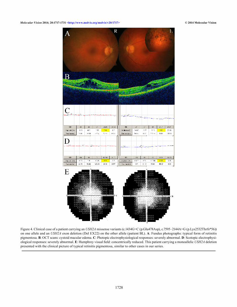
identification of three complete USH2A exon deletions in four cases (two patients were brother and sister; 4/13, which is 30.7%), always in trans with missense variants on the other allele. This result is in agreement with a recent study [41] that reported large genomic rearrangements (mainly consisting of deletions) in 35% of patients with USH previously identified as carriers of a monoallelic USH2A mutation and suggest that in USH MLPA analysis can detect a significant fraction of mutations not identified with exon sequencing. The clinical picture of one patient carrying a heterozygous USH2A dele-tion is illustrated in Figure 4.
One patient was heterozygous for mutations in both USH genes having received the USH2A variant from the unaffected father and the MYO7A variant from the unaffected mother. This finding supports the hypothesis of a possible digenic/oligogenic inheritance of the syndrome or the possible combined influence of variants of different genes on the clinical phenotype of the patient, as suggested by similar results reported in other studies [16,21,32-34,37].
In three patients, MYO7A and USH2A sequencing and MLPA analysis detected only a monoallelic variant (one MYO7A and two USH2A variants) while in five patients no variant of both genes was detected. Unidentified muta-tions could lie in the promoter, regulatory regions, and deep intronic areas, usually not analyzed during conventional mutation screening. Otherwise, they could be located in other USH genes, already known or yet to be identified. Finally, in a few cases the coexistence of non-syndromic deafness and non-syndromic retinal dystrophy cannot be excluded.
Segregation of the mutant alleles with the disease was reported in all families, particularly considering the close relatives (namely, parents, brothers, and sisters) of the proband. Figure 1 shows the pedigree of a family in whom segregation analysis first suggested the possibility of a large deletion in the USH2A gene.
In our series, the USH1 group was too small to evaluate for possible genotype-phenotype associations, and patients with USH2 showed variable clinical pictures (different ages of onset, disease progression, and fundus appearance) so it was not possible to establish reliable genotype-phenotype correlations. This phenotypic heterogeneity has already been reported in other studies [30,38,42] and is probably due to the influence of environmental factors and/or modifier genes.
In conclusion, we identified ten MYO7A and 34 USH2A sequence variants in a series of 36 patients with USH from 33 independent Italian families; to our knowledge, this is the first large study reporting complete exon sequencing of these genes in the Italian population. The high number of novel and
Molecular Vision 2014; 20:1717-1731 <http://www.molvis.org/molvis/v20/1717> © 2014 Molecular Vision
1728
Figure 4. Clinical case of a patient carrying an USH2A missense variants (c.1434G>C (p.Glu478Asp), c.7595–2144A>G (p.Lys2532Thrfs*56)) on one allele and an USH2A exon deletion (Del EX22) on the other allele (patient BL). A: Fundus photographs: typical form of retinitis pigmentosa. B: OCT scans: cystoid macular edema. C: Photopic electrophysiological responses: severely abnormal. D: Scotopic electrophysi-ological responses: severely abnormal. E: Humphrey visual field: concentrically reduced. This patient carrying a monoallelic USH2A deletion presented with the clinical picture of typical retinitis pigmentosa, similar to other cases in our series.

Molecular Vision 2014; 20:1717-1731 <http://www.molvis.org/molvis/v20/1717> © 2014 Molecular Vision
1729
private variants and the poor prevalence of variants common in other ethnic groups suggest a difference in the spectrum of USH2A sequence variants between Italian patients with USH and other populations. The overall prevalence of patients carrying two pathogenic mutations in different alleles of the same gene was 75%. This high detection rate supports the sequencing of MYO7A and USH2A genes associated with MLPA-USH2A as the first step in the molecular diagnosis of Italian patients with USH. This strategy is significantly more efficient than the microarray investigation and may represent an alternative to the NGS technology that is presently not available in all laboratories. Moreover, amplicon sequencing by NGS investigation cannot presently identify the exon deletions detected with the MLPA-USH2A method. The main limit of this diagnostic approach is the impossibility of studying other USH or still unknown genes and to detect modifying variants in additional genes that could influence the phenotype determined by the primary USH genes. Better knowledge of molecular alterations underlying USH in specific populations may lead to more efficient diagnostic strategies and future therapeutic approaches.
REFERENCES1. Keats BJ, Corey DP. The Usher syndromes. Am J Med Genet
1999; 89:158-66. [PMID: 10704190].
2. Saihan Z, Webster AR, Luxon L, Bitner-Glindzicz M. Update on Usher syndrome. Curr Opin Neurol 2009; 22:19-27. [PMID: 19165952].
3. Bonnet C, El-Amraoui A. Usher syndrome (sensorineural deafness and retinitis pigmentosa): Pathogenesis, molecular diagnosis and therapeutic approaches. Curr Opin Neurol 2012; 25:42-9. [PMID: 22185901].
4. Smith RJ, Berlin CI, Hejtmancik JF, Keats BJ, Kimberling WJ, Lewis RA, Moller CG, Pelias MZ, Tranebjaerg L. Clinical diagnosis of the Usher syndromes. Usher Syndrome Consor-tium. Am J Med Genet 1994; 50:32-8. [PMID: 8160750].
5. Grøndahl J. Estimation of prognosis and prevalence of retinitis pigmentosa and Usher syndrome in Norway. Clin Genet 1987; 31:255-64. [PMID: 3594933].
6. Boughman JA, Vernon M, Shaver KA. Usher syndrome: Defi-nition and estimate of prevalence from two high-risk popula-tions. J Chronic Dis 1983; 36:595-603. [PMID: 6885960].
7. Bocquet B, Lacroux A, Surget MO, Baudoin C, Marquette V, Manes G, Hebrard M, Senechal A, Delettre C, Roux AF, Claustres M, Dhaenens CM, Rozet JM, Perrault I, Bonnefont JP, Kaplan J, Dollfus H, Amati-Bonneau P, Bonneau D, Reynier P, Audo I, Zeitz C, Sahel JA, Paquis-Flucklinger V, Calvas P, Arveiler B, Kohl S, Wissinger B, Blanchet C, Meunier I, Hamel CP. Relative frequencies of inherited retinal dystrophies and optic neuropathies in
Southern France: Assessment of 21-year data management. Ophthalmic Epidemiol 2013; 20:13-25. [PMID: 23350551].
8. Riazuddin S, Belyantseva IA, Giese AP, Lee K, Indzhykulian AA, Nandamuri SP, Yousaf R, Sinha GP, Lee S, Terrell D, Hegde RS, Ali RA, Anwar S, Andrade-Elizondo PB, Sirmaci A, Parise LV, Basit S, Wali A, Ayub M, Ansar M, Ahmad W, Khan SN, Akram J, Tekin M, Riazuddin S, Cook T, Busch-beck EK, Frolenkov GI, Leal SM, Friedman TB, Ahmed ZM. Alterations of the CIB2 calcium- and integrin-binding protein cause Usher syndrome type 1J and nonsyndromic deafness DFNB48. Nat Genet 2012; 44:1265-71. [PMID: 23023331].
9. Eisenberger T, Slim R, Mansour A, Nauck M, Nurnberg G, Nurnberg P, Decker C, Dafinger C, Ebermann I, Bergmann C, Bolz HJ. Targeted next-generation sequencing identifies a homozygous nonsense mutation in ABHD12, the gene underlying PHARC, in a family clinically diagnosed with Usher syndrome type 3. Orphanet J Rare Dis 2012; 7:59-65. [PMID: 22938382].
10. Puffenberger EG, Jinks RN, Sougnez C, Cibulskis K, Willert RA, Achilly NP, Cassidy RP, Fiorentini CJ, Heiken KF, Lawrence JJ, Mahoney MH, Miller CJ, Nair DT, Politi KA, Worcester KN, Setton RA, Dipiazza R, Sherman EA, Eastman JT, Francklyn C, Robey-Bond S, Rider NL, Gabriel S, Morton DH, Strauss KA. Genetic mapping and exome sequencing identify variants associated with five novel diseases. PLoS ONE 2012; 7:e28936-[PMID: 22279524].
11. El-Amraoui A, Petit C. Usher I syndrome: Unravelling the mechanisms that underlie the cohesion of the growing hair bundle in inner ear sensory cells. J Cell Sci 2005; 118:4593-603. [PMID: 16219682].
12. Kremer H, van Wijk E, Marker T, Wolfrum U, Roepman R. Usher syndrome: Molecular links of pathogenesis, proteins and pathways. Hum Mol Genet 2006; 15:Spec No 2R262-70. [PMID: 16987892].
13. Reiners J, Nagel-Wolfrum K, Jurgens K, Marker T, Wolfrum U. Molecular basis of human Usher syndrome: Deciphering the meshes of the Usher protein network provides insights into the pathomechanisms of the Usher disease. Exp Eye Res 2006; 83:97-119. [PMID: 16545802].
14. Ouyang XM, Yan D, Du LL, Hejtmancik JF, Jacobson SG, Nance WE, Li AR, Angeli S, Kaiser M, Newton V, Brown SD, Balkany T, Liu XZ. Characterization of Usher syndrome type I gene mutations in an Usher syndrome patient popula-tion. Hum Genet 2005; 116:292-9. [PMID: 15660226].
15. Jaijo T, Aller E, Beneyto M, Najera C, Graziano C, Turchetti D, Seri M, Ayuso C, Baiget M, Moreno F, Morera C, Perez-Garrigues H, Millan JM. MYO7A mutation screening in Usher syndrome type I patients from diverse origins. J Med Genet 2007; 44:e71-[PMID: 17361009].
16. Le Quesne Stabej P, Saihan Z, Rangesh N, Steele-Stallard HB, Ambrose J, Coffey A, Emmerson J, Haralambous E, Hughes Y, Steel KP, Luxon LM, Webster AR, Bitner-Glindzicz M. Comprehensive sequence analysis of nine Usher syndrome genes in the UK National Collaborative Usher Study. J Med Genet 2012; 49:27-36. [PMID: 22135276].

Molecular Vision 2014; 20:1717-1731 <http://www.molvis.org/molvis/v20/1717> © 2014 Molecular Vision
1730
17. Liu X, Vansant G, Udovichenko IP, Wolfrum U, Williams DS. Myosin VIIA, the product of the Usher 1B syndrome gene, is concentrated in the connecting cilia of photoreceptor cells. Cell Motil Cytoskeleton 1997; 37:240-52. [PMID: 9227854].
18. Dreyer B, Brox V, Tranebjaerg L, Rosenberg T, Sadeghi AM, Moller C, Nilssen O. Spectrum of USH2A mutations in scan-dinavian patients with Usher syndrome type II. Hum Mutat 2008; 29:451-66. [PMID: 18273898].
19. Garcia-Garcia G, Aparisi MJ, Jaijo T, Rodrigo R, Leon AM, Avila-Fernandez A, Blanco-Kelly F, Bernal S, Navarro R, Diaz-Llopis M, Baiget M, Ayuso C, Millan JM, Aller E. Mutational screening of the USH2A gene in spanish USH patients reveals 23 novel pathogenic mutations. Orphanet J Rare Dis 2011; 6:65-78. [PMID: 22004887].
20. Rivolta C, Sweklo EA, Berson EL, Dryja TP. Missense muta-tion in the USH2A gene: Association with recessive retinitis pigmentosa without hearing loss. Am J Hum Genet 2000; 66:1975-8. [PMID: 10775529].
21. Vozzi D, Aaspollu A, Athanasakis E, Berto A, Fabretto A, Licastro D, Kulm M, Testa F, Trevisi P, Vahter M, Ziviello C, Martini A, Simonelli F, Banfi S, Gasparini P. Molecular epidemiology of Usher syndrome in Italy. Mol Vis 2011; 17:1662-8. [PMID: 21738395].
22. Licastro D, Mutarelli M, Peluso I, Neveling K, Wieskamp N, Rispoli R, Vozzi D, Athanasakis E, D’Eustacchio A, Pizzo M, D’Amico F, Ziviello C, Simonelli F, Fabretto A, Scheffer H, Gasparini P, Banfi S, Nigro V. Molecular diagnosis of Usher syndrome: Application of two different next generation sequencing-based procedures. PLoS ONE 2012; 7:e43799-[PMID: 22952768].
23. Marmor MF, Fulton AB, Holder GE, Miyake Y, Brigell M, Bach M. International Society for Clinical Electrophysiology of V. ISCEV Standard for full-field clinical electroretinog-raphy (2008 update). Doc Ophthalmol 2009; 118:69-77. [PMID: 19030905].
24. Roux AF. Molecular updates on Usher syndrome J Fr Ophtalmol 2005; 28:93-7. [PMID: 15767904].
25. Dreyer B, Tranebjaerg L, Brox V, Rosenberg T, Moller C, Beneyto M, Weston MD, Kimberling WJ, Cremers CW, Liu XZ, Nilssen O. A common ancestral origin of the frequent and widespread 2299delG USH2A mutation. Am J Hum Genet 2001; 69:228-34. [PMID: 11402400].
26. Vaché C, Besnard T, le Berre P, Garcia-Garcia G, Baux D, Larrieu L, Abadie C, Blanchet C, Bolz HJ, Millan J, Hamel C, Malcolm S, Claustres M, Roux AF. Usher syndrome type 2 caused by activation of an USH2A pseudoexon: Implica-tions for diagnosis and therapy. Hum Mutat 2012; 33:104-8. [PMID: 22009552].
27. Huang XF, Xiang P, Chen J, Xing DJ, Huang N, Min Q, Gu F, Tong Y, Pang CP, Qu J, Jin ZB. Targeted exome sequencing identified novel USH2A mutations in Usher syndrome fami-lies. PLoS ONE 2013; 8:e63832-[PMID: 23737954].
28. Auslender N, Bandah D, Rizel L, Behar DM, Shohat M, Banin E, Allon-Shalev S, Sharony R, Sharon D, Ben-Yosef T. Four USH2A founder mutations underlie the majority of Usher
syndrome type 2 cases among non-Ashkenazi Jews. Genet Test 2008; 12:289-94. [PMID: 18452394].
29. Seyedahmadi BJ, Rivolta C, Keene JA, Berson EL, Dryja TP. Comprehensive screening of the USH2A gene in Usher syndrome type II and non-syndromic recessive reti-nitis pigmentosa. Exp Eye Res 2004; 79:167-73. [PMID: 15325563].
30. Bernal S, Meda C, Solans T, Ayuso C, Garcia-Sandoval B, Valverde D, Del Rio E, Baiget M. Clinical and genetic studies in Spanish patients with Usher syndrome type II: Descrip-tion of new mutations and evidence for a lack of genotype-phenotype correlation. Clin Genet 2005; 68:204-14. [PMID: 16098008].
31. Iannaccone A, De Propris G, Roncati S, Rispoli E, Del Porto G, Pannarale MR. The ocular phenotype of the Bardet-Biedl syndrome. Comparison to non-syndromic retinitis pigmen-tosa. Ophthalmic Genet 1997; 18:13-26. [PMID: 9134546].
32. Cremers FP, Kimberling WJ, Kulm M, de Brouwer AP, van Wijk E, te Brinke H, Cremers CW, Hoefsloot LH, Banfi S, Simonelli F, Fleischhauer JC, Berger W, Kelley PM, Hara-lambous E, Bitner-Glindzicz M, Webster AR, Saihan Z, De Baere E, Leroy BP, Silvestri G, McKay GJ, Koenekoop RK, Millan JM, Rosenberg T, Joensuu T, Sankila EM, Weil D, Weston MD, Wissinger B, Kremer H. Development of a genotyping microarray for Usher syndrome. J Med Genet 2007; 44:153-60. [PMID: 16963483].
33. Jaijo T, Aller E, Garcia-Garcia G, Aparisi MJ, Bernal S, Avila-Fernandez A, Barragan I, Baiget M, Ayuso C, Antinolo G, Diaz-Llopis M, Kulm M, Beneyto M, Najera C, Millan JM. Microarray-based mutation analysis of 183 Spanish families with Usher syndrome. Invest Ophthalmol Vis Sci 2010; 51:1311-7. [PMID: 19683999].
34. Bonnet C, Grati M, Marlin S, Levilliers J, Hardelin JP, Parodi M, Niasme-Grare M, Zelenika D, Delepine M, Feldmann D, Jonard L, El-Amraoui A, Weil D, Delobel B, Vincent C, Dollfus H, Eliot MM, David A, Calais C, Vigneron J, Montaut-Verient B, Bonneau D, Dubin J, Thauvin C, Duvillard A, Francannet C, Mom T, Lacombe D, Duriez F, Drouin-Garraud V, Thuillier-Obstoy MF, Sigaudy S, Frances AM, Collignon P, Challe G, Couderc R, Lathrop M, Sahel JA, Weissenbach J, Petit C, Denoyelle F. Complete exon sequencing of all known Usher syndrome genes greatly improves molecular diagnosis. Orphanet J Rare Dis 2011; 6:21-40. [PMID: 21569298].
35. Nakanishi H, Ohtsubo M, Iwasaki S, Hotta Y, Mizuta K, Mineta H, Minoshima S. Identification of 11 novel mutations in USH2A among Japanese patients with Usher syndrome type 2. Clin Genet 2009; 76:383-91. [PMID: 19737284].
36. Riazuddin S, Nazli S, Ahmed ZM, Yang Y, Zulfiqar F, Shaikh RS, Zafar AU, Khan SN, Sabar F, Javid FT, Wilcox ER, Tsilou E, Boger ET, Sellers JR, Belyantseva IA, Riazuddin S, Friedman TB. Mutation spectrum of MYO7A and evaluation of a novel nonsyndromic deafness DFN2 allele with residual function. Hum Mutat 2008; 29:502-11. [PMID: 18181211].
37. Västinsalo H, Jalkanen R, Bergmann C, Neuhaus C, Kleemola L, Jauhola L, Bolz HJ, Sankila EM. Extended mutation

Molecular Vision 2014; 20:1717-1731 <http://www.molvis.org/molvis/v20/1717> © 2014 Molecular Vision
1731
spectrum of Usher syndrome in Finland. Acta Ophthalmol 2013; 91:325-34. [PMID: 22681893].
38. Yan D, Ouyang X, Patterson DM, Du LL, Jacobson SG, Liu XZ. Mutation analysis in the long isoform of USH2A in American patients with Usher syndrome type II. J Hum Genet 2009; 54:732-8. [PMID: 19881469].
39. Yan D, Liu XZ. Genetics and pathological mechanisms of Usher syndrome. J Hum Genet 2010; 55:327-35. [PMID: 20379205].
40. Ebermann I, Koenekoop RK, Lopez I, Bou-Khzam L, Pigeon R, Bolz HJ. An USH2A founder mutation is the major cause of Usher syndrome type 2 in Canadians of French origin and confirms common roots of Quebecois and Acadians. Euro-pean journal of human genetics Eur J Hum Genet 2009; 17:80-4. [PMID: 18665195].
41. Steele-Stallard HB, Le Quesne Stabej P, Lenassi E, Luxon LM, Claustres M, Roux AF, Webster AR, Bitner-Glindzicz M. Screening for duplications, deletions and a common intronic mutation detects 35% of second mutations in patients with USH2A monoallelic mutations on Sanger sequencing. Orphanet J Rare Dis 2013; 8:122-33. [PMID: 23924366].
42. Malm E, Ponjavic V, Moller C, Kimberling WJ, Andreasson S. Phenotypes in defined genotypes including siblings with Usher syndrome. Ophthalmic Genet 2011; 32:65-74. [PMID: 21174530].
43. Roux AF, Faugere V, Le Guedard S, Pallares-Ruiz N, Vielle A, Chambert S, Marlin S, Hamel C, Gilbert B, Malcolm
S, Claustres M. French Usher Syndrome C. Survey of the frequency of USH1 gene mutations in a cohort of Usher patients shows the importance of cadherin 23 and protocad-herin 15 genes and establishes a detection rate of above 90%. J Med Genet 2006; 43:763-8. [PMID: 16679490].
44. Weston MD, Eudy JD, Fujita S, Yao S, Usami S, Cremers C, Greenberg J, Ramesar R, Martini A, Moller C, Smith RJ, Sumegi J, Kimberling WJ. Genomic structure and identifica-tion of novel mutations in Usherin, the gene responsible for Usher syndrome type IIA. Am J Hum Genet 2000; 66:1199-210. Erratum in: Am J Hum Genet 2000 Jun;66 6 :2020. Greenburg J corrected to Greenberg J[PMID: 10729113].
45. Sandberg MA, Rosner B, Weigel-DiFranco C, McGee TL, Dryja TP, Berson EL. Disease course in patients with autosomal recessive retinitis pigmentosa due to the USH2A gene. Invest Ophthalmol Vis Sci 2008; 49:5532-9. [PMID: 18641288].
46. Vaché C, Besnard T, Blanchet C, Baux D, Larrieu L, Faugere V, Mondain M, Hamel C, Malcolm S, Claustres M, Roux AF. Nasal epithelial cells are a reliable source to study splicing variants in Usher syndrome. Hum Mutat 2010; 31:734-41. [PMID: 20513143].
47. Bernal S, Ayuso C, Antinolo G, Gimenez A, Borrego S, Trujillo MJ, Marcos I, Calaf M, Del Rio E, Baiget M. Muta-tions in USH2A in Spanish patients with autosomal recessive retinitis pigmentosa: High prevalence and phenotypic varia-tion. J Med Genet 2003; 40:e8-[PMID: 12525556].
Articles are provided courtesy of Emory University and the Zhongshan Ophthalmic Center, Sun Yat-sen University, P.R. China. The print version of this article was created on 23 December 2014. This reflects all typographical corrections and errata to the article through that date. Details of any changes may be found in the online version of the article.


![D7.02 COMMUNICATION STRATEGY [Report] · 2019-11-28 · D7.02 COMMUNICATION STRATEGY [Report] Authors: Anita Derjanecz (REHVA), Francesco Mariottini (REHVA) Co-Authors: Niels Delaere](https://img.dokumen.tips/doc/110x75/5faab8df02c45f212e430980/d702-communication-strategy-report-2019-11-28-d702-communication-strategy.jpg)


