Embed Size (px)
Citation preview

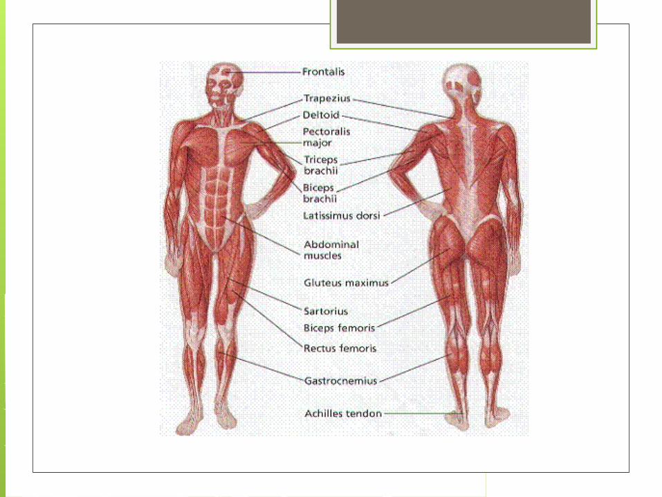
MuslcesGroup 4

Classification
Agonist: prime mover
Antagonist: reverses agonist
Synergist: prevents rotation
Fixator: stabilizes the origin of the prime
mover

Naming (LADSNOR!) Direction of the muscle fibers
Ex) transverse, rectus, oblique
Relative size of muscles Ex) major, minor
Location Number and location of origin Shape Action of the muscle
Ex) extensor, flexor

Origin and Insertion
Origin: immovable end
Insertion: movable end

Characteristics
Muscle cells are elongated
Contractions are due to the movement
of microfilaments

Structure (skeletal muscle)
Structure (cont.) From outermost to innermost
Muscle>Fascicles>Muscle fibers>Myofibril>Thick and thin filaments
Fascia>Epimysium>Perimysium>Endomysium

Tissue layers of a skeletal muscle
Fascia: covering the whole muscle
Epimysium: lies beneath the fascia
Perimysium: separates cells of fascicle
Endomysium: separates individual
muscle fibers

Fascicle Collection of muscle fibers Covered by blood vessels and axon of
motor neurons Each muscle fibers is separated by
endomysium It is surrounded by sarcolemma Contains nucleus and sarcoplasmic
reticulum Each muscle fibers is composed of
myofibril

Myofibril & Sarcomere

Sarcomere It is a repeating pattern formed by
striations

Sarcomere (cont.) Troponin: protein that works with
tropomyosin to block muscle contraction until calcium ions are present
Transverse tubule: set of membranous channels that contain extracellular fluid

Smooth muscle It is shorter than skeletal muscle, and
has single centrally located nuclei
It lacks troponin
It alternates between a state of relaxation and contraction

Cardiac muscle Composed of striated cells, containing a
single nucleus
It has a well developed sarcoplasmic reticulum
Its transverse tubule is larger than skeletal muscle’s

Contraction Acetylcholine(ACh) is the
neurotransmitter that contracts skeletal muscles
ACh binds with receptors on the motor endplate, which causes muscle impulse
Calcium ions diffuse from sarcoplasmic reticulum to sarcoplasm and binds to troponin

Contraction (cont.) Tropomyosin moves, which allows actin
and myosin to link Actin is pulled to the center of the
sarcomere, which allows muscle fibers to shorten

Threshold

Respiration
Anaerobic breaks down glucose
and releases ATP
Aerobic requires oxygen to
produce ATP

Oxygen debt
When cellular respiration is not
able to sustain the muscle, lactic
acid diffuses into the blood stream
This creates an oxygen debt, that
must be repaid later

Muscle fatigue
When a muscle loses its ability to
contract
Most likely occurs from
accumulation of lactic acid

Types of contraction