Embed Size (px)
Citation preview
MUSCLE TISSUE
• Muscle tissue is characterized by its well-developed properties of contraction.
• Muscle is responsible for the movements of the body and the various parts of the body.
• Muscle develops from embryonic mesoderm (with the exception of myoepithelium).
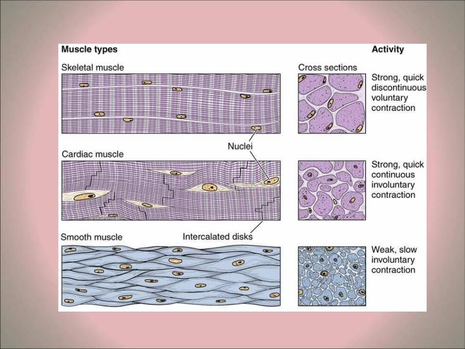
• Muscle is classified into 3 categories according to morphology and physiological function: – Skeletal Muscle – Cardiac Muscle – Smooth Muscle
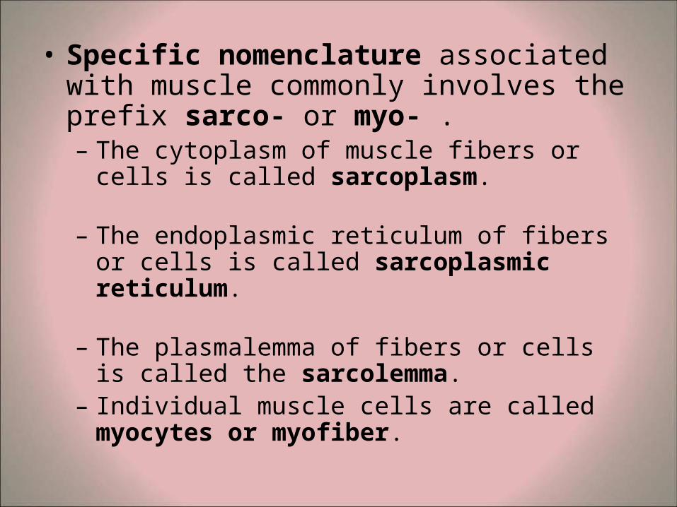
• Specific nomenclature associated with muscle commonly involves the prefix sarco- or myo- . – The cytoplasm of muscle fibers or cells is called
sarcoplasm.
– The endoplasmic reticulum of fibers or cells is called sarcoplasmic reticulum.
– The plasmalemma of fibers or cells is called the sarcolemma.
– Individual muscle cells are called myocytes or myofiber.
SKELETAL MUSCLE
• Skeletal muscle, also known as striated or voluntary muscle, comprises some 40-50% of the body mass in adults and constitutes part of the largest organ system of the body.
• During embryonic development mesodermal cells differentiate into uninuclear myoblasts, which elongate and fuse together to form myotubes, which further develop into the mature muscle fibers or myofibers.
• These myofibers are the basic units of skeletal muscle and are up to 30 cm in length.
• Myofibers possess large numbers of elongated or oval nuclei at their periphery, close to the sarcolemma.
• These myofibers are syncytia (multinucleated post-mitotic structures in which the nuclei have lost the ability to synthesize DNA).
• After regular staining myofibers are seen to have periodic cross striations (the source of the name "striated muscle").
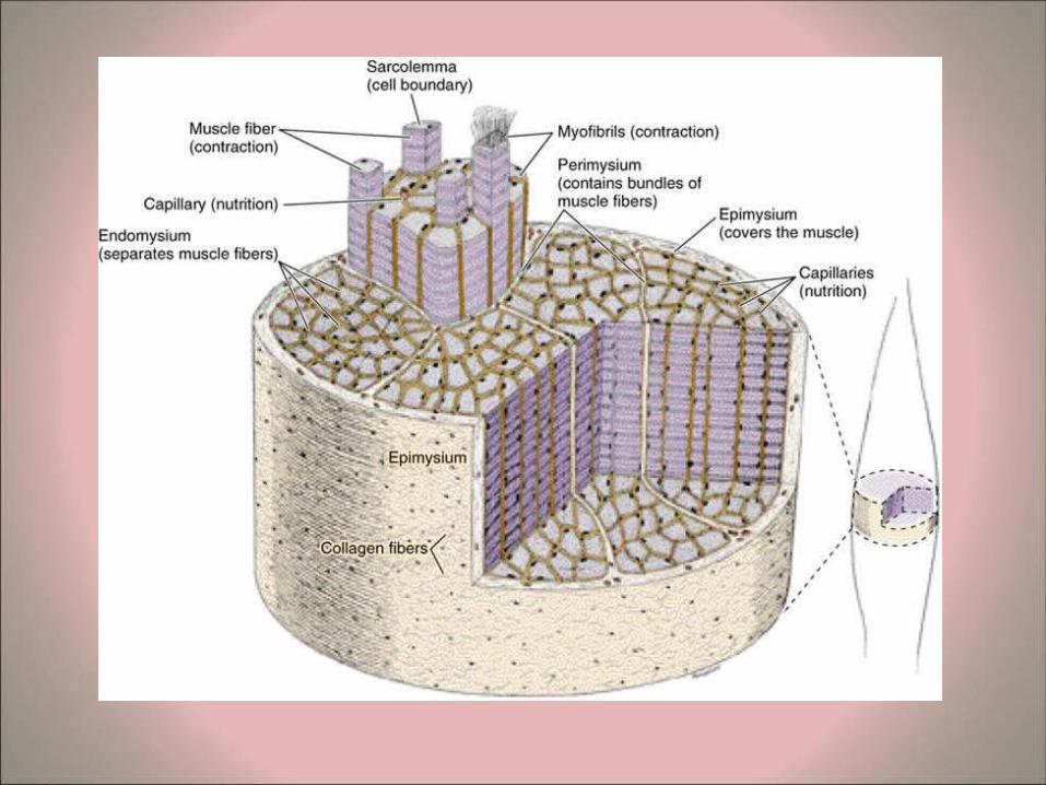
• Connective tissue arrangements of skeletal muscles – In skeletal muscles the myofibers are bound together in a
similar manner to wires in a telecommunications cable. The connective tissue in the muscle serves to bind and integrate the action of the various contractile units.
– A thin and delicate conntissue layer, known as the endomysium, surrounds each individual myofiber.
– Myofibers are grouped together in bundles or fascicles, which are also surrounded by connective tissue, known as the perimysium.
– The fascicles are surrounded and bound together by a further connective tissue coating known as the epimysium.
– All these connective tissue coatings (endomysium, perimysium and epimysium) contain collagen fibers, elastic fibers, fibroblasts and are richly vascularized.
– The ends of skeletal muscles are attached to bones, cartilage or ligaments by means of tendons.
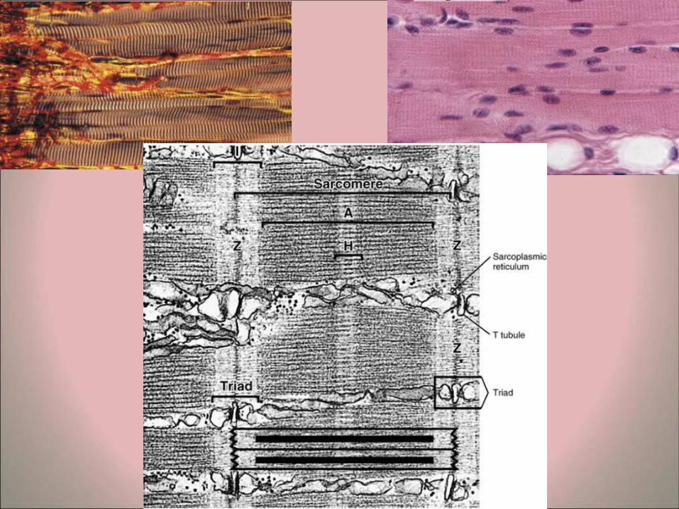
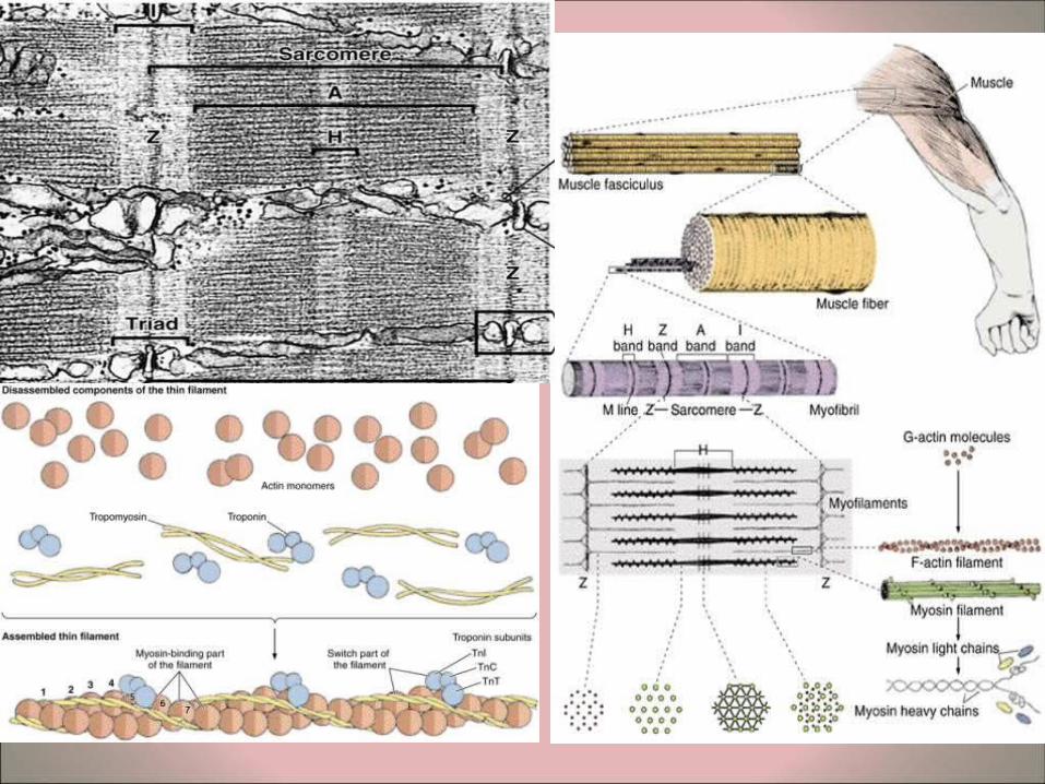
• Myofibers – Longitudinal sections of skeletal muscle fibers show repeated
cross-striations after regular staining (H&E). – The stained bands are called A-bands, and in between these
are non-stained I-bands. If the same myofiber is examined by polarizing microscopy the A-bands are seen to be birefringent or anisotropic (bright against a dark background with crossed polars), whereas the I-bands are non-birefringent or isotropic. (The origin of the nomenclature comes from these polarizing properties: A = Anisotropic, I = Isotropic).
– At higher magnifications it is possible to see a line in the middle of the I band, known as the Z line.
– Examination of a myofiber at high magnification shows that that it is composed of many parallel myofibrils..
– The unit between two Z lines is known as the sarcomere. The myofibrils consist of repeating strings of sarcomeres. The sarcomeres in adjacent myofibrils tend to be located in parallel, resulting in the overall cross-striations of the myofibers. It is also possible in some cases to distinguish a less-stained region in the middle of the A-bands, known as the H-band (Hensen's band). The sarcomeres form the basic contractile units of the fibers.
• Ultrastructure of sarcomeres – Examination of sarcomeres of myofibrils by
transmission electron microscopy reveals two sorts of myofilaments.
– The thicker myofilaments belong to the A band and are composed mainly of myosin.
– The thinner myofilaments are mainly found in the I band and are composed mainly of actin.
– These thin myofilaments are connected to the Z-line and partially extend between the thicker myofilaments. This area of overlap is important in the contraction process.
– In transverse sections in the area of overlap each thick myofilament is surrounded by six of the thinner myofilaments.
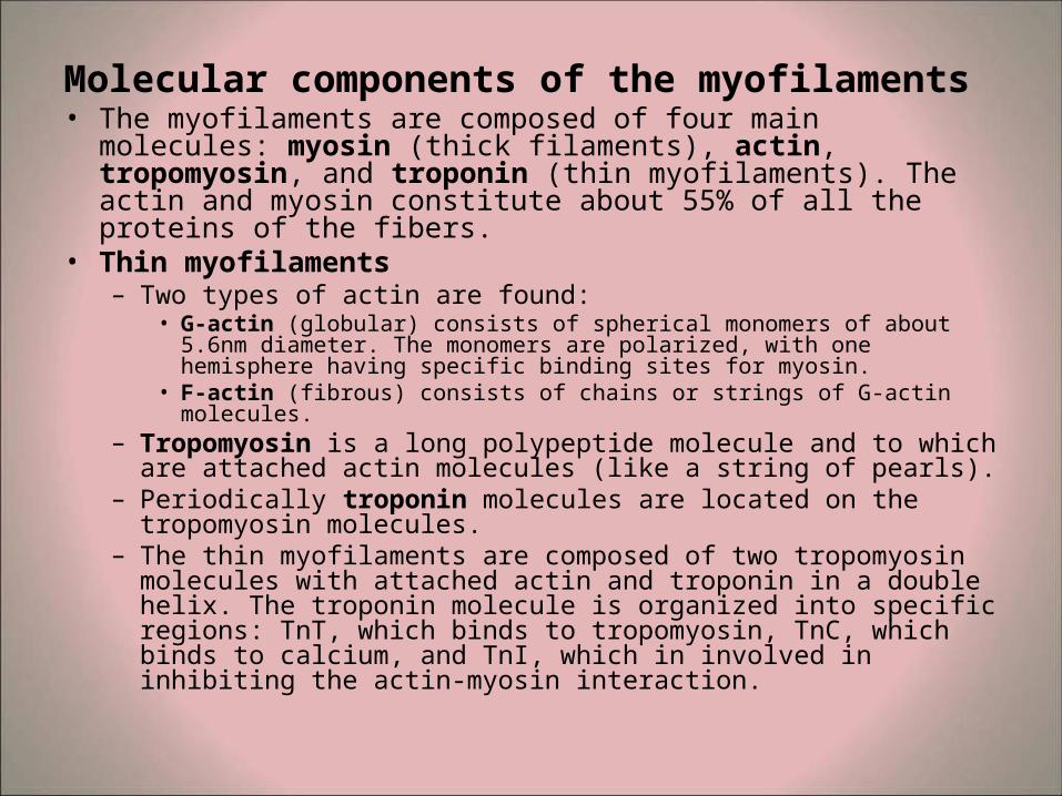
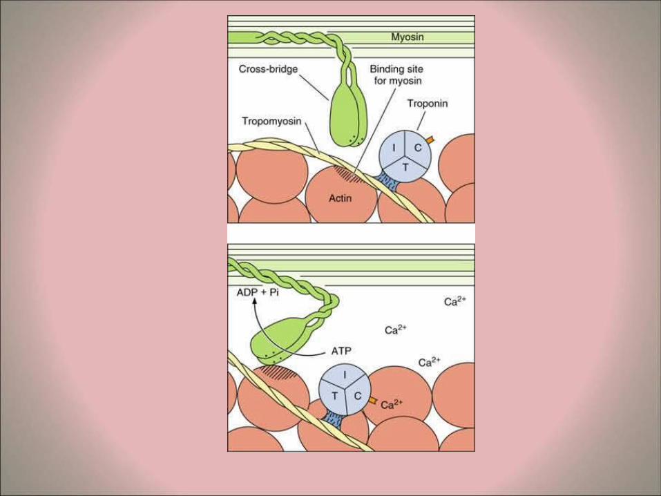
Molecular components of the myofilaments • The myofilaments are composed of four main molecules:
myosin (thick filaments), actin, tropomyosin, and troponin (thin myofilaments). The actin and myosin constitute about 55% of all the proteins of the fibers.
• Thin myofilaments – Two types of actin are found:
• G-actin (globular) consists of spherical monomers of about 5.6nm diameter. The monomers are polarized, with one hemisphere having specific binding sites for myosin.
• F-actin (fibrous) consists of chains or strings of G-actin molecules. – Tropomyosin is a long polypeptide molecule and to which are
attached actin molecules (like a string of pearls). – Periodically troponin molecules are located on the tropomyosin
molecules. – The thin myofilaments are composed of two tropomyosin
molecules with attached actin and troponin in a double helix. The troponin molecule is organized into specific regions: TnT, which binds to tropomyosin, TnC, which binds to calcium, and TnI, which in involved in inhibiting the actin-myosin interaction.

• Thick myofilaments – The myosin molecules are composed of a
rod-like portion (light meromyosin) and twin rounded heads (heavy meromyosin). These can be separated by brief hydrolysis.
– The heavy meromyosin portion contains ATP-ase activities, important in the binding of the myosin to actin during contraction process.
– The Z-lines contain the proteins α-actinin and desmin.
• T-system of tubules – Tubular invaginations of the sarcolemma
penetrate the myofibers in a transverse direction. These are known as the T-tubules (transverse tubules) and are found at the area of overlap between the A and I bands of myofibrils. Each sarcomere has two of these tubules.
– Swollen terminal cisternae or sacs of the sarcoplasmic reticulum are associated with the T-tubules. Two terminal cisternae are associated with each T-tubule to form structures (visible by transmission electron microscopy) known as triads.

• Other components of the sarcoplasm – Glycogen particles are found and serve as energy stores.
(These can be demonstrated by the PAS (periodic acid-Schiff) reaction in histological sections. At the ultrastructural level the spherical glycogen particles (b -particles) are seen individually or in small clusters).
– Many elongated mitochondria are found located between the myofibrils or in accumulations just under the sarcolemma. The numbers and activities of the mitochondria are greater in muscle fibers with high metabolic activity.
– Myoglobin is an oxygen-binding protein that gives much of the red color of muscle fibers.
– Relatively little rough endoplasmic reticulum or ribosomes are present in myofibers.
– In aged muscle fibers lipofuscin deposits (brown pigment) are common. These are now known to be large secondary lysosomes.
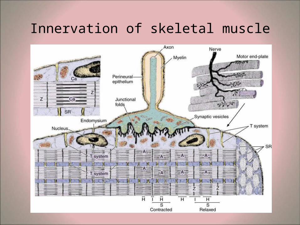
Innervation of skeletal muscle
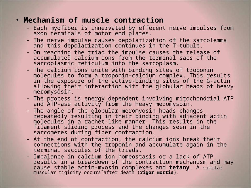
• Mechanism of muscle contraction – Each myofiber is innervated by efferent nerve impulses from axon
terminals of motor end plates. – The nerve impulse causes depolarization of the sarcolemma and this
depolarization continues in the T-tubule. – On reaching the triad the impulse causes the release of accumulated
calcium ions from the terminal sacs of the sarcoplasmic reticulum into the sarcoplasm.
– The calcium ions unite with binding sites of troponin molecules to form a troponin-calcium complex. This results in the exposure of the active-binding sites of the G-actin allowing their interaction with the globular heads of heavy meromysosin.
– The process is energy dependent involving mitochondrial ATP and ATP-ase activity from the heavy meromysoin.
– The angle of the globular meromyosin heads changes repeatedly resulting in their binding with adjacent actin molecules in a rachet-like manner. This results in the filament sliding process and the changes seen in the sarcomeres during fiber contraction.
– At the end of contraction, the calcium ions break their connections with the troponin and accumulate again in the terminal saccules of the triads.
– Imbalance in calcium ion homeostasis or a lack of ATP results in a breakdown of the contraction mechanism and may cause stable actin-myosin complexes and tetany. A similar muscular rigidity occurs after death (rigor mortis).
Classification of muscle fibers – Muscle fibers are classified into three main categories: – Red fibers (Type I) or slow-twitch high-oxidative fibers
• These have relatively small diameters, much myoglobin, many well-developed mitochondria, a rich blood supply and much ATP-ase. These type I fibers are found in muscles with very high metabolic activity involved in slow sustained contractions. The energy source is from oxidative phosphorylation.
– White fibers (Type IIa) or fast-twitch glycolytic-anaerobic fibers . These have larger diameters, less myoglobin and fewer mitochondria, relatively poorer blood supplies and less ATP-ase. These type IIa fibers are involved in rapid contraction (fast twitch) with anaerobic glycolysis.
– Intermediate fibers (Type IIb). These have structural and functional properties in between those of the other two types.
• Repair and regeneration after injury – If muscles are used intensively, trained or exercised, they
increase in mass as a result of increase in protein synthesis and sarcomere production. This results in hypertrophy of use ("Use it or lose it"). On the contrary, limb immobilization (e.g. in plaster casts, or as a result of inactivity due to hospitalization, or lack of gravity) causes loss of muscle mass (disuse myopathy or atrophy).
– Myofibers are syncytial and post-mitotic, with very limited regenerative abilities after trauma. After trauma such as muscle crush, pathological changes occur in muscle and may lead to breakdown of myofibers and release of myoglobin, which can affect renal function and be life-threatening. In the limited repair processes, satellite cells are activated, divide and can form new myotubes and myocytes. In some cases the satellite cells can fuse with existing fibers and contribute to the repair processes
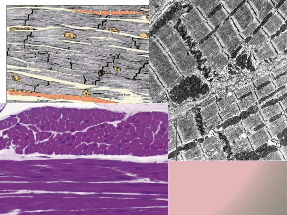
CARDIAC MUSCLE
• Cardiac muscle is also striated, but differs from the striated skeletal muscle in several respects:
• The muscle fibers branch (bifurcate) and are arranged in series to form an anastomosing network.
• Each myocyte has one or two central nuclei (unlike the many peripheral nuclei of syncytia of skeletal muscle fibers).
• The fibers have more sarcoplasm. • The mitochondria are larger and better developed. • All the fibers are Type I (red fibers, with abundant
myoglobin, high oxidative slow-twitch). • Glycogen is more common. • The myocytes have specialized areas of contact - the
intercalated disks. • Contractions are rhythmic, spontaneous and involuntary.
• The cross striations have a similar morphology and staining characteristics to those of skeletal muscle fibers, however the contractile tissue is not organized into discrete myofibrils.
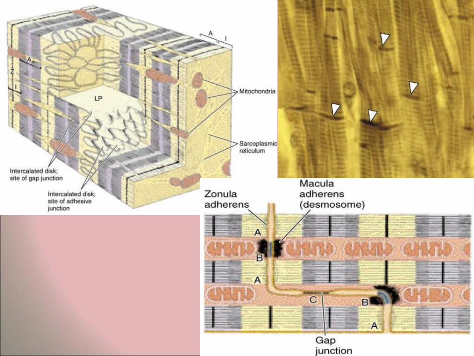
• At the ultrastructural level sarcomeres are found similar to those of skeletal muscle fibers. The large mitochondria are arranged in rows between the strings of sarcomeres.
• In aged cardiac muscle, lipofuscin is also commonly found. • Cardiac myocytes also possess a system of T-tubules. These
consist of fairly broad tubular sarcoplasmic invaginations, which terminate in the region of the Z-line of the sarcomeres. Typically these are associated with a single terminal saccule of sarcoplasmic reticulum to form diads.
• In general the sarcoplasmic reticulum of cardiac muscle fiberes is much less well developed than that of myofibers of skeletal muscle.
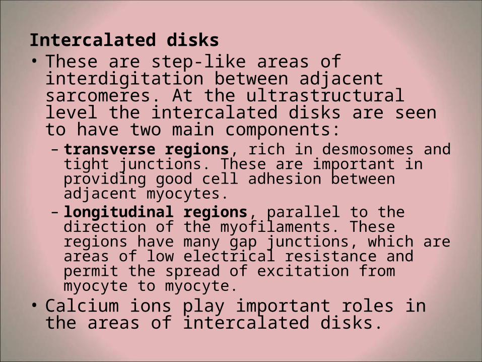
Intercalated disks • These are step-like areas of interdigitation
between adjacent sarcomeres. At the ultrastructural level the intercalated disks are seen to have two main components: – transverse regions, rich in desmosomes and
tight junctions. These are important in providing good cell adhesion between adjacent myocytes.
– longitudinal regions, parallel to the direction of the myofilaments. These regions have many gap junctions, which are areas of low electrical resistance and permit the spread of excitation from myocyte to myocyte.
• Calcium ions play important roles in the areas of intercalated disks.
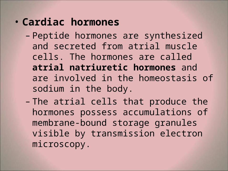
• Cardiac hormones – Peptide hormones are synthesized and
secreted from atrial muscle cells. The hormones are called atrial natriuretic hormones and are involved in the homeostasis of sodium in the body.
– The atrial cells that produce the hormones possess accumulations of membrane-bound storage granules visible by transmission electron microscopy.
Hypertrophy and regeneration of cardiac tissue • There is virtually no regeneration of cardiac tissue.
The coronary arteries supplying blood to the heart are anatomical end arteries and lack collaterals.
• In the event of blockage of coronary arteries (as a result of a blood clot or atherosclerotic blockage), the cardiac myocytes vascularized by the coronaries cannot receive essential oxygen and the result is infarct.
• Following infarcts, the remaining heart muscle undergoes compensatory hypertrophy, with subsequent enlargement of the heart.
• Hypertrophied hearts are commonly an indication of underlying pathological disorders, though they may develop in specific cases of training and overload as in athletes.
SMOOTH MUSCLE
• Smooth muscle is also known as "involuntary muscle", as contraction is not under conscious control. Smooth muscle is innervated by the autonomic nervous system.
• Smooth muscle lacks cross-striations (unlike striated and cardiac muscle).
• Smooth muscle has the ability to undergo hyperplasia and hypertrophy (as in the uterus of pregnant women).
• Smooth muscle can also regenerate, and this is important in the repair processes of injured blood vessels.
Location of smooth muscle • Smooth muscle is found in the walls of the hollow internal
organs (hollow viscera), where it plays a role in maintaining the patency of the lumen. Smooth muscle forms the contractile layers of the intestinal tract, where it is important in peristaltic contractions involved in the movement of food.
• Smooth muscle is found in the walls of the respiratory tracts.
• Smooth muscle is present in the walls of blood vessels (vascular smooth muscle, especially in arterial vessels).
• Smooth muscle is found in the dermis of the skin (arrector pili).
• Smooth muscle is found in the eye (iris diaphragm, controlling the amount of light reaching the retina).
• Smooth muscle is a major component in the wall of the uterus.
• Smooth muscle is also found in many other sites in the body
• Origin of smooth muscle • Like the other muscle types, smooth muscle is
also derived from mesoderm. – Some researchers believe that smooth muscle has
some affiliation to the connective tissue cells derived from mesenchyme, because the fibers synthesize and secrete collagen, elastin and reticulin of the sheath. They consider the smooth muscle fibers as connective tissue cells that have evolved the capacity of contractility.
• Some glands of ectodermal origin, such as sweat glands or mammary glands, possess smooth muscle cells surrounding their secretory units (myoepithelial cells). These myoepithelial cells are ectodermal in origin.
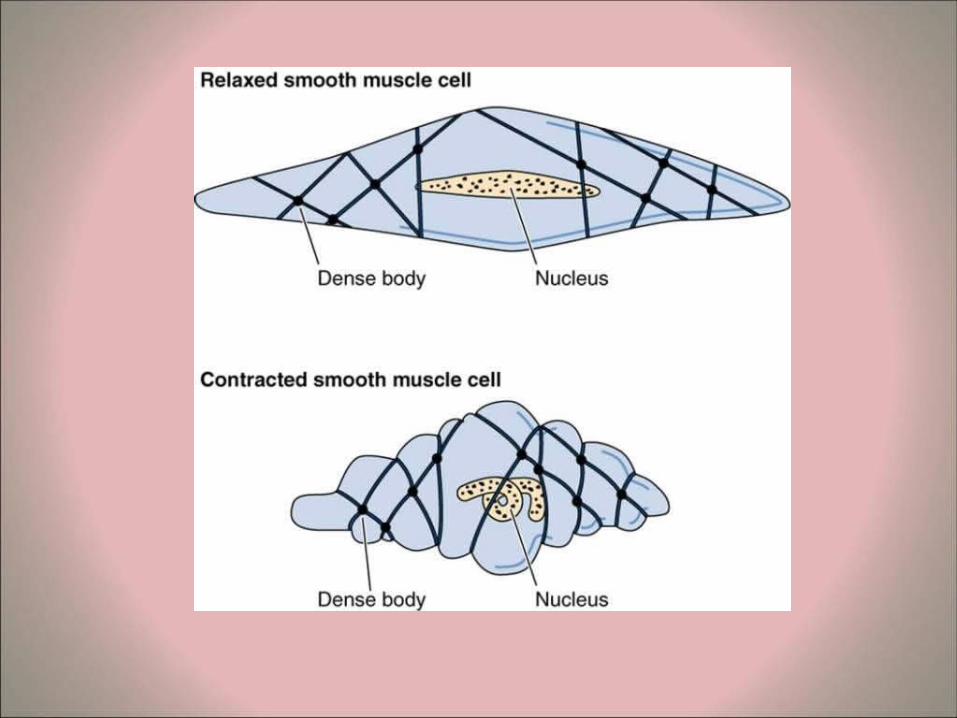
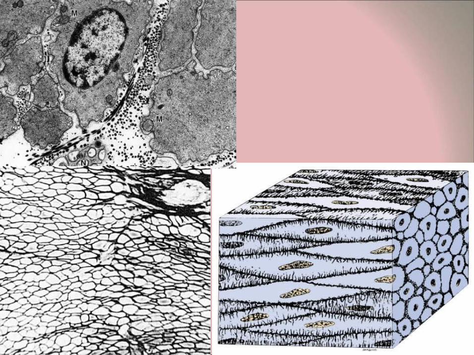
Structure of smooth muscle fibers • The smooth muscle fibers (myocytes) are spindle-
shaped (fusiform). • The nucleus is in the widest part of the fiber and is
elongated, typically with several nucleoli. In cross section, the nucleus will be evident only when the section cuts through the widest part of the myocyte.
• The length of the myocytes is very variable in different organs.
• In most organs, the smooth muscle fibers are orderly arranged in layers, strips or bundles. After regular staining (H&E) the sarcoplasm is seen to be acidophilic (stained with eosin). In sections of most of the intestinal tract, it is possible to see the two adjacent, antagonistic bands of smooth muscle (longitudinal and transverse).
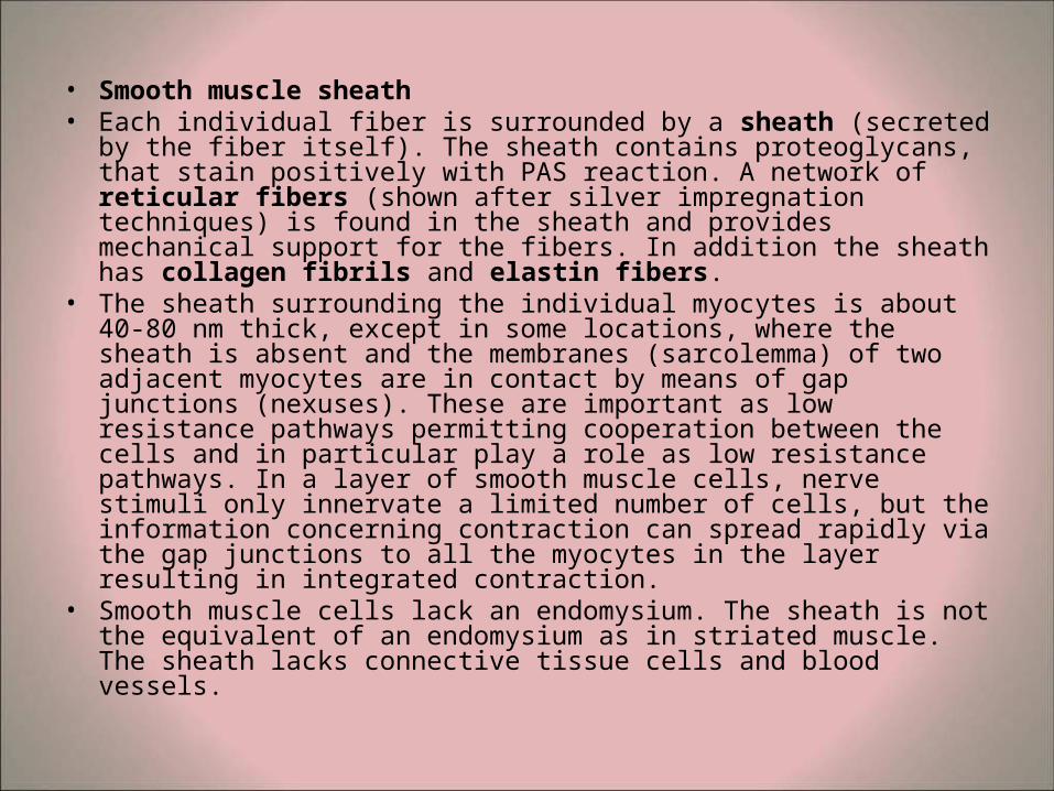
• Smooth muscle sheath • Each individual fiber is surrounded by a sheath (secreted by the
fiber itself). The sheath contains proteoglycans, that stain positively with PAS reaction. A network of reticular fibers (shown after silver impregnation techniques) is found in the sheath and provides mechanical support for the fibers. In addition the sheath has collagen fibrils and elastin fibers.
• The sheath surrounding the individual myocytes is about 40-80 nm thick, except in some locations, where the sheath is absent and the membranes (sarcolemma) of two adjacent myocytes are in contact by means of gap junctions (nexuses). These are important as low resistance pathways permitting cooperation between the cells and in particular play a role as low resistance pathways. In a layer of smooth muscle cells, nerve stimuli only innervate a limited number of cells, but the information concerning contraction can spread rapidly via the gap junctions to all the myocytes in the layer resulting in integrated contraction.
• Smooth muscle cells lack an endomysium. The sheath is not the equivalent of an endomysium as in striated muscle. The sheath lacks connective tissue cells and blood vessels.
Ultrastructure of smooth muscle• The ultrastructure of smooth muscle cells shows that the
sheath appears somewhat similar to the basal lamina of epithelial cells. The organelles are located close to the nucleus in two distinct poles. The rest of the sarcoplasm is filled with myofilaments, though these are not arranged in ordered sarcomeres as in striated muscle. Three types of myofilaments may be seen:
• thin myofilaments (5-7nm thick), which are the most common type
• thick myofilaments (about 16nm thick), which are less common
• intermediate filaments (about 10nm thick). These may be grouped as "dense bodies" and are also found in contact with the sarcolemma (attachment plaques). It is thought that these intermediate filaments provide some sort of structural support for the cells.
The contraction mechanism of smooth muscle cells
• The actin and myosin do not appear to be regularly arranged. Myosin is present in relatively low amounts. A calcium ion target protein, calmodulin. Which lead to the phosphorylation of myosin
• cAMP also regulates the contraction
• Estrogen and progesterone work on cAMP