Embed Size (px)
Citation preview

1
MUSCLE PHYSIOLOGY
Sliding Filament Model of Contraction
• Each myosin head binds and detaches several times during contraction, acting like a ratchet to generate tension and propel the thin filaments to the center of the sarcomere
• As this event occurs throughout the sarcomeres, the muscle shortens
Skeletal Muscle Contraction
• In order to contract, a skeletal muscle must:
– Be stimulated by a nerve ending– Propagate an electrical current, or action
potential, along its sarcolemma– Have a rise in intracellular Ca2+ levels, the
final trigger for contraction• Linking the electrical signal to the
contraction is excitation-contraction coupling
Nerve Stimulus of Skeletal Muscle
• Skeletal muscles are stimulated by motor neurons of the somatic nervous system
• Axons of these neurons travel in nerves to muscle cells
• Axons of motor neurons branch profusely as they enter muscles
• Each axonal branch forms a neuromuscular junction with a single muscle fiber

2
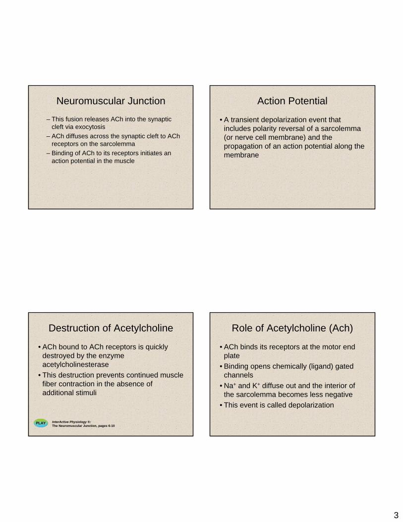
Neuromuscular Junction
• The neuromuscular junction is formed from:
– Axonal endings, which have small membranous sacs (synaptic vesicles) that contain the neurotransmitter acetylcholine(ACh)
– The motor end plate of a muscle, which is a specific part of the sarcolemma that contains ACh receptors and helps form the neuromuscular junction
Neuromuscular Junction
• Though exceedingly close, axonal ends and muscle fibers are always separated by a space called the synaptic cleft
Neuromuscular Junction
Figure 9.7 (a-c)
Neuromuscular Junction
• When a nerve impulse reaches the end of an axon at the neuromuscular junction:
– Voltage-regulated calcium channels open and allow Ca2+ to enter the axon
– Ca2+ inside the axon terminal causes axonal vesicles to fuse with the axonal membrane
PLAY InterActive Physiology ®:The Neuromuscular Junction, pages 3-5
3
Neuromuscular Junction
– This fusion releases ACh into the synaptic cleft via exocytosis
– ACh diffuses across the synaptic cleft to AChreceptors on the sarcolemma
– Binding of ACh to its receptors initiates an action potential in the muscle
Destruction of Acetylcholine
• ACh bound to ACh receptors is quickly destroyed by the enzyme acetylcholinesterase
• This destruction prevents continued muscle fiber contraction in the absence of additional stimuli
PLAY InterActive Physiology ®:The Neuromuscular Junction, pages 6-10
Action Potential
• A transient depolarization event that includes polarity reversal of a sarcolemma(or nerve cell membrane) and the propagation of an action potential along the membrane
Role of Acetylcholine (Ach)
• ACh binds its receptors at the motor end plate
• Binding opens chemically (ligand) gated channels
• Na+ and K+ diffuse out and the interior of the sarcolemma becomes less negative
• This event is called depolarization

4
Depolarization
• Initially, this is a local electrical event called end plate potential
• Later, it ignites an action potential that spreads in all directions across the sarcolemma
Action Potential: Electrical Conditions of a Polarized
Sarcolemma• The outside (extracellular) face is positive, while the inside face is negative
• This difference in charge is the resting membrane potential
Figure 9.8a
• The predominant extracellular ion is Na+
• The predominant intracellular ion is K+
• The sarcolemma is relatively impermeable to both ions
Action Potential: Electrical Conditions of a Polarized
Sarcolemma
Figure 9.8a
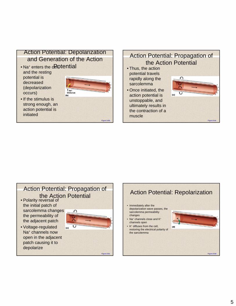
Action Potential: Depolarization and Generation of the Action
Potential• An axonal terminal of
a motor neuron releases ACh and causes a patch of the sarcolemma to become permeable to Na+ (sodium channels open)
Figure 9.8b
5
Action Potential: Depolarization and Generation of the Action
Potential• Na+ enters the cell, and the resting potential is decreased (depolarization occurs)
• If the stimulus is strong enough, an action potential is initiated
Figure 9.8b
Action Potential: Propagation of the Action Potential
• Polarity reversal of the initial patch of sarcolemma changes the permeability of the adjacent patch
• Voltage-regulated Na+ channels now open in the adjacent patch causing it to depolarize
Figure 9.8c
Action Potential: Propagation of the Action Potential
• Thus, the action potential travels rapidly along the sarcolemma
• Once initiated, the action potential is unstoppable, and ultimately results in the contraction of a muscle
Figure 9.8c
Action Potential: Repolarization
• Immediately after the depolarization wave passes, the sarcolemma permeability changes
• Na+ channels close and K+
channels open• K+ diffuses from the cell,
restoring the electrical polarity of the sarcolemma
Figure 9.8d

6
Action Potential: Repolarization
• Repolarization occurs in the same direction as depolarization, and must occur before the muscle can be stimulated again (refractory period)
• The ionic concentration of the resting state is restored by the Na+-K+ pump
Figure 9.8d
Excitation-Contraction Coupling
• Once generated, the action potential:– Is propagated along the sarcolemma– Travels down the T tubules– Triggers Ca2+ release from terminal cisternae
• Ca2+ binds to troponin and causes: – The blocking action of tropomyosin to cease– Actin active binding sites to be exposed
Excitation-Contraction Coupling
• Myosin cross bridges alternately attach and detach
• Thin filaments move toward the center of the sarcomere
• Hydrolysis of ATP powers this cycling process
• Ca2+ is removed into the SR, tropomyosin blockage is restored, and the muscle fiber relaxes
Action Potential Scan
Figure 9.9

7
Excitation-Contraction (EC) Coupling
1. Action potential generated and propagated along sarcomere to T-tubules
2. Action potential triggers Ca2+ release
3. Ca++ bind to troponin; blocking action of tropomyosin released
4. contraction via crossbridge formation; ATP hyrdolysis
5. Removal of Ca+2 by active transport
6. tropomyosin blockage restored; contraction ends
Figure 9.10
Figure 9.10
ADP
Pi
Net entry of Na+ Initiatesan action potential whichis propagated along thesarcolemma and downthe T tubules.
T tubuleSarcolemma
SR tubules (cut)
SynapticcleftSynaptic
vesicle
Axon terminal
ACh ACh ACh
Neurotransmitter released diffusesacross the synaptic cleft and attachesto ACh receptors on the sarcolemma.
Action potential inT tubule activatesvoltage-sensitive receptors,which in turn trigger Ca2+
release from terminalcisternae of SRinto cytosol.
Calcium ions bind to troponin;troponin changes shape, removingthe blocking action of tropomyosin;actin active sites exposed.
Contraction; myosin heads alternately attach toactin and detach, pulling the actin filaments towardthe center of the sarcomere; release of energy byATP hydrolysis powers the cycling process.
Removal of Ca2+ by active transportinto the SR after the actionpotential ends.
SR
Tropomyosin blockage restored,blocking myosin binding sites onactin; contraction ends andmuscle fiber relaxes.
Ca2+
Ca2+
Ca2+
Ca2+
Ca2+Ca2+
Ca2+
Ca2+
Ca2+
Ca2+
1
2
3
4
5
6
Figure 9.10
SynapticcleftSynaptic
vesicle
Axon terminal
ACh ACh ACh
Neurotransmitter released diffusesacross the synaptic cleft and attachesto ACh receptors on the sarcolemma.
Figure 9.10
Net entry of Na+ Initiatesan action potential whichis propagated along thesarcolemma and downthe T tubules.
T tubuleSarcolemma
SynapticcleftSynaptic
vesicle
Axon terminal
ACh ACh ACh
Neurotransmitter released diffusesacross the synaptic cleft and attachesto ACh receptors on the sarcolemma.
1

8
Figure 9.10
Net entry of Na+ Initiatesan action potential whichis propagated along thesarcolemma and downthe T tubules.
T tubuleSarcolemma
SR tubules (cut)
SynapticcleftSynaptic
vesicle
Axon terminal
ACh ACh ACh
Neurotransmitter released diffusesacross the synaptic cleft and attachesto ACh receptors on the sarcolemma.
Action potential inT tubule activatesvoltage-sensitive receptors,which in turn trigger Ca2+
release from terminalcisternae of SRinto cytosol.
SRCa2+
Ca2+
Ca2+Ca2+
Ca2+
Ca2+
Ca2+
Ca2+
1
2
Figure 9.10
Net entry of Na+ Initiatesan action potential whichis propagated along thesarcolemma and downthe T tubules.
T tubuleSarcolemma
SR tubules (cut)
SynapticcleftSynaptic
vesicle
Axon terminal
ACh ACh ACh
Neurotransmitter released diffusesacross the synaptic cleft and attachesto ACh receptors on the sarcolemma.
Action potential inT tubule activatesvoltage-sensitive receptors,which in turn trigger Ca2+
release from terminalcisternae of SRinto cytosol.
Calcium ions bind to troponin;troponin changes shape, removingthe blocking action of tropomyosin;actin active sites exposed.
SR
Ca2+
Ca2+
Ca2+
Ca2+Ca2+
Ca2+
Ca2+
Ca2+
Ca2+
1
2
3
Figure 9.10
Net entry of Na+ Initiatesan action potential whichis propagated along thesarcolemma and downthe T tubules.
T tubuleSarcolemma
SR tubules (cut)
SynapticcleftSynaptic
vesicle
Axon terminal
ACh ACh ACh
Neurotransmitter released diffusesacross the synaptic cleft and attachesto ACh receptors on the sarcolemma.
Action potential inT tubule activatesvoltage-sensitive receptors,which in turn trigger Ca2+
release from terminalcisternae of SRinto cytosol.
Calcium ions bind to troponin;troponin changes shape, removingthe blocking action of tropomyosin;actin active sites exposed.
Contraction; myosin heads alternately attach toactin and detach, pulling the actin filaments towardthe center of the sarcomere; release of energy byATP hydrolysis powers the cycling process.
SR
Ca2+
Ca2+
Ca2+
Ca2+Ca2+
Ca2+
Ca2+
Ca2+
Ca2+
1
2
3
4
Figure 9.10
Net entry of Na+ Initiatesan action potential whichis propagated along thesarcolemma and downthe T tubules.
T tubuleSarcolemma
SR tubules (cut)
SynapticcleftSynaptic
vesicle
Axon terminal
ACh ACh ACh
Neurotransmitter released diffusesacross the synaptic cleft and attachesto ACh receptors on the sarcolemma.
Action potential inT tubule activatesvoltage-sensitive receptors,which in turn trigger Ca2+
release from terminalcisternae of SRinto cytosol.
Calcium ions bind to troponin;troponin changes shape, removingthe blocking action of tropomyosin;actin active sites exposed.
Contraction; myosin heads alternately attach toactin and detach, pulling the actin filaments towardthe center of the sarcomere; release of energy byATP hydrolysis powers the cycling process.
Removal of Ca2+ by active transportinto the SR after the actionpotential ends.
SR
Ca2+
Ca2+
Ca2+
Ca2+
Ca2+Ca2+
Ca2+
Ca2+
Ca2+
Ca2+
1
2
3
4
5

9
Figure 9.10
ADP
Pi
Net entry of Na+ Initiatesan action potential whichis propagated along thesarcolemma and downthe T tubules.
T tubuleSarcolemma
SR tubules (cut)
SynapticcleftSynaptic
vesicle
Axon terminal
ACh ACh ACh
Neurotransmitter released diffusesacross the synaptic cleft and attachesto ACh receptors on the sarcolemma.
Action potential inT tubule activatesvoltage-sensitive receptors,which in turn trigger Ca2+
release from terminalcisternae of SRinto cytosol.
Calcium ions bind to troponin;troponin changes shape, removingthe blocking action of tropomyosin;actin active sites exposed.
Contraction; myosin heads alternately attach toactin and detach, pulling the actin filaments towardthe center of the sarcomere; release of energy byATP hydrolysis powers the cycling process.
Removal of Ca2+ by active transportinto the SR after the actionpotential ends.
SR
Tropomyosin blockage restored,blocking myosin binding sites onactin; contraction ends andmuscle fiber relaxes.
Ca2+
Ca2+
Ca2+
Ca2+
Ca2+Ca2+
Ca2+
Ca2+
Ca2+
Ca2+
1
2
3
4
5
6
Role of Ionic Calcium (Ca2+) in the Contraction Mechanism
• At low intracellular Ca2+ concentration:
– Tropomyosin blocks the binding sites on actin
– Myosin cross bridges cannot attach to binding sites on actin
– The relaxed state of the muscle is enforced
Figure 9.11a
Role of Ionic Calcium (Ca2+) in the Contraction Mechanism
• At higher intracellular Ca2+ concentrations:
– Additional calcium binds to troponin (inactive troponin binds two Ca2+)
– Calcium-activated troponin binds an additional two Ca2+
at a separate regulatory site
Figure 9.11b
Role of Ionic Calcium (Ca2+) in the Contraction Mechanism
• Calcium-activated troponin undergoes a conformational change
• This change moves tropomyosin away from actin’s binding sites
Figure 9.11c

10
Role of Ionic Calcium (Ca2+) in the Contraction Mechanism
• Myosin head can now bind and cycle
• This permits contraction (sliding of the thin filaments by the myosin cross bridges) to begin
Figure 9.11d
Sequential Events of Contraction
• Cross bridge formation – myosin cross bridge attaches to actin filament
• Working (power) stroke – myosin head pivots and pulls actin filament toward M line
• Cross bridge detachment – ATP attaches to myosin head and the cross bridge detaches
• “Cocking” of the myosin head – energy from hydrolysis of ATP cocks the myosin head into the high-energy state
PLAY InterActive Physiology ®: Sliding Filament Theory, pages 3-29
Figure 9.12
ATP
ADP
ADPATPhydrolysis
ADP
ATP
Pi
Pi
Myosin head(high-energyconfiguration)
Myosin head attaches to the actinmyofilament, forming a cross bridge.
Thin filament
As ATP is split into ADP and Pi, the myosinhead is energized (cocked into the high-energyconformation).
Inorganic phosphate (Pi) generated in theprevious contraction cycle is released, initiatingthe power (working) stroke. The myosin headpivots and bends as it pulls on the actin filament,sliding it toward the M line. Then ADP is released.
Myosin head(low-energyconfiguration)
As new ATP attaches to the myosin head, the link betweenmyosin and actin weakens, and the cross bridge detaches.
Thick filament
1
4 2
3
Figure 9.12
ADP
Pi
Myosin head(high-energyconfiguration)
Myosin head attaches to the actinmyofilament, forming a cross bridge.
1

11
Figure 9.12
ADP
ADP
Pi
Myosin head(high-energyconfiguration)
Myosin head attaches to the actinmyofilament, forming a cross bridge.
Inorganic phosphate (Pi) generated in theprevious contraction cycle is released, initiatingthe power (working) stroke. The myosin headpivots and bends as it pulls on the actin filament,sliding it toward the M line. Then ADP is released.
1
2
Figure 9.12
ADP
ADP
ATP
Pi
Myosin head(high-energyconfiguration)
Myosin head attaches to the actinmyofilament, forming a cross bridge.
1
2 Inorganic phosphate (Pi) generated in theprevious contraction cycle is released, initiatingthe power (working) stroke. The myosin headpivots and bends as it pulls on the actin filament,sliding it toward the M line. Then ADP is released.
Figure 9.12
ATP
ADP
ADP
ATP
Pi
Myosin head(high-energyconfiguration)
Myosin head attaches to the actinmyofilament, forming a cross bridge.
Myosin head(low-energyconfiguration)
As new ATP attaches to the myosin head, the link betweenmyosin and actin weakens, and the cross bridge detaches.
1
2
3
Inorganic phosphate (Pi) generated in theprevious contraction cycle is released, initiatingthe power (working) stroke. The myosin headpivots and bends as it pulls on the actin filament,sliding it toward the M line. Then ADP is released.
Figure 9.12
ATP
ADP
ADPATPhydrolysis
ADP
ATP
Pi
Pi
Myosin head(high-energyconfiguration)
Myosin head attaches to the actinmyofilament, forming a cross bridge.
Thin filament
As ATP is split into ADP and Pi, the myosinhead is energized (cocked into the high-energyconformation).
Inorganic phosphate (Pi) generated in theprevious contraction cycle is released, initiatingthe power (working) stroke. The myosin headpivots and bends as it pulls on the actin filament,sliding it toward the M line. Then ADP is released.
Myosin head(low-energyconfiguration)
As new ATP attaches to the myosin head, the link betweenmyosin and actin weakens, and the cross bridge detaches.
Thick filament
1
4 2
3

12
Figure 9.12
ATP
ADP
ADPATPhydrolysis
ADP
ATP
Pi
Pi
Myosin head(high-energyconfiguration)
Myosin head attaches to the actinmyofilament, forming a cross bridge.
Thin filament
As ATP is split into ADP and Pi, the myosinhead is energized (cocked into the high-energyconformation).
Inorganic phosphate (Pi) generated in theprevious contraction cycle is released, initiatingthe power (working) stroke. The myosin headpivots and bends as it pulls on the actin filament,sliding it toward the M line. Then ADP is released.
Myosin head(low-energyconfiguration)
As new ATP attaches to the myosin head, the link betweenmyosin and actin weakens, and the cross bridge detaches.
Thick filament
1
4 2
3
Contraction of Skeletal Muscle Fibers
• Contraction – refers to the activation of myosin’s cross bridges (force-generating sites)
• Shortening occurs when the tension generated by the cross bridge exceeds forces opposing shortening
• Contraction ends when cross bridges become inactive, the tension generated declines, and relaxation is induced
Contraction of Skeletal Muscle (Organ Level)
• Contraction of muscle fibers (cells) and muscles (organs) is similar
• The two types of muscle contractions are:– Isometric contraction – increasing muscle
tension (muscle does not shorten during contraction)
– Isotonic contraction – decreasing muscle length (muscle shortens during contraction)
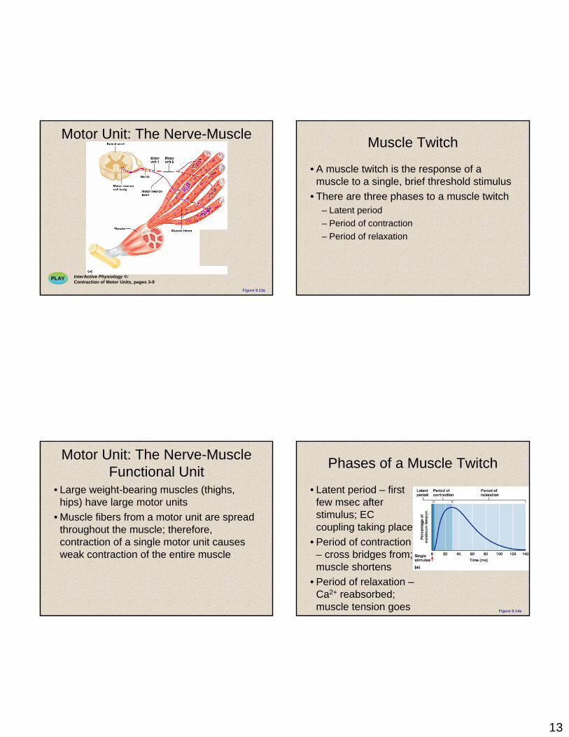
Motor Unit: The Nerve-Muscle Functional Unit
• A motor unit is a motor neuron and all the muscle fibers it supplies
• The number of muscle fibers per motor unit can vary from four to several hundred
• Muscles that control fine movements (fingers, eyes) have small motor units
13
Motor Unit: The Nerve-Muscle Functional Unit
PLAY InterActive Physiology ®:Contraction of Motor Units, pages 3-9
Figure 9.13a
Motor Unit: The Nerve-Muscle Functional Unit
• Large weight-bearing muscles (thighs, hips) have large motor units
• Muscle fibers from a motor unit are spread throughout the muscle; therefore, contraction of a single motor unit causes weak contraction of the entire muscle
Muscle Twitch
• A muscle twitch is the response of a muscle to a single, brief threshold stimulus
• There are three phases to a muscle twitch– Latent period– Period of contraction– Period of relaxation
Phases of a Muscle Twitch
• Latent period – first few msec after stimulus; EC coupling taking place
• Period of contraction – cross bridges from; muscle shortens
• Period of relaxation –Ca2+ reabsorbed; muscle tension goes t
Figure 9.14a

14
Muscle Twitch Comparisons
Figure 9.14b
Graded Muscle Responses
• Graded muscle responses are:– Variations in the degree of muscle contraction– Required for proper control of skeletal
movement• Responses are graded by:
– Changing the frequency of stimulation– Changing the strength of the stimulus
Muscle Response to Varying Stimuli• A single stimulus results in a single
contractile response – a muscle twitch• Frequently delivered stimuli (muscle does
not have time to completely relax) increases contractile force – wave summation
Figure 9.15
Muscle Response to Varying Stimuli
• More rapidly delivered stimuli result in incomplete tetanus
• If stimuli are given quickly enough, complete tetanus results
Figure 9.15

15
Muscle Response: Stimulation Strength
• Threshold stimulus – the stimulus strength at which the first observable muscle contraction occurs
• Beyond threshold, muscle contracts more vigorously as stimulus strength is increased
• Force of contraction is precisely controlled by multiple motor unit summation
• This phenomenon, called recruitment, brings more and more muscle fibers into play
Stimulus Intensity and Muscle Tension
Figure 9.16
Size Principle
Figure 9.17
Treppe: The Staircase Effect
• Staircase – increased contraction in response to multiple stimuli of the same strength
• Contractions increase because:– There is increasing availability of Ca2+ in the
sarcoplasm– Muscle enzyme systems become more
efficient because heat is increased as muscle contracts

16
Treppe: The Staircase Effect
Figure 9.18
• How do STRIATED muscles contract?
Contraction
• Each myofiber contains myofilaments.
– Thick filaments: MYOSIN
– Thin filaments: ACTIN
SARCOMERE
•Many in each striated myofiber
•Sarcomere:–Z disc to Z disc.

17
SARCOMERE Muscle Contraction
Muscle Contraction
18
Muscle Contraction
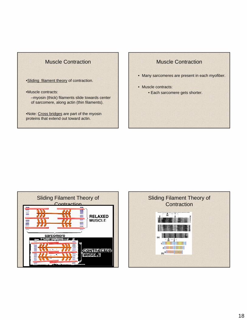
•Sliding filament theory of contraction.
•Muscle contracts:–myosin (thick) filaments slide towards center of sarcomere, along actin (thin filaments).
•Note: Cross bridges are part of the myosin proteins that extend out toward actin.
Sliding Filament Theory of Contraction
Muscle Contraction
• Many sarcomeres are present in each myofiber.
• Muscle contracts:• Each sarcomere gets shorter.
Sliding Filament Theory of Contraction

19
• How does the sliding happen?
Myosin
•Myosin is an ATPase (and motor protein).
•Each myosin head contains an ATP-binding site AND an actin binding site.
Myosin cycle
• Myosin binding ATP which splits to ADP and Pi.
• Myosin heads attach to actin.• Pi is released, causing the power stroke to
occur.• Power stroke pulls actin filament.• ADP is released, because myosin binds to a
new ATP, and releases from the actin.
Myosin cycle

20
• How is the contraction regulated?
Regulation
• ATP is present, so contractions would be continuous
• BUT– Tropomyosin lies along actin filament– Troponin is attached to tropomyosin.
• Tropomyosin is in the way, myosin can’t bind to actin.
Regulation
• Ca2+ influx releases troponin/tropomyosin block -> muscle contracts!
Regulation

21
Regulation
What’s upstream of the Ca2+ influx?
Excitation-Contraction Coupling
-- Axon of motor neuron produces AP in sarcolemma of myofiber.•- APs travel down sarcolemma and T tubules. -- SR terminal cisternae releases Ca2+
-- sarcomeres contract!
Muscle Relaxation
• Ca2+ pumped back into SR through Ca2+-ATPase pumps (always on).
• End of neuronal stimulation:– ACh-esterase degrades ACh.– Ca2+ channels close.– Choline recycled to make more ACh.

22
Sliding Filament
• ATP is energy when split into ADP+P.• Electrical impulse (action potential) travels down
the nerve and into T-tubules.• Depolarization occurs (sodium and potassium
exchange). Local and millisecond time lapse.• AP stimulates the release of calcium.• Calcium binds to troponin.• Actin and myosin then combine.
Sliding Filament cont…
• Rigor of muscle upon death?• Cross bridge cycle occurs.• Nerve impulse stops.• No calcium influx. • Allowing troponin to attach and inhibit
actin-myosin attachment.
Neural Control• Motor unit is one nerve and all fibers it
innervates.• 1:1 or 1:1,000.• Large and small, fast and slow.• Fibers may lie scattered throughout the muscle
and not all together.• Fiber diameter is related to work performed
(hypertrophy?).• When one fiber is activated all fibers are
activated.
Muscle Structure
• Muscle fibers are long• Diameter of a hair• Grouped in bundles (fasciculi)• Neuromuscular junction• Sarcoplasm contains fibers• Hundreds to thousands of myofibrils

23
Muscle Structure cont…
• Myofibrils contains protein myofilaments• Actin and myosin• Crossbridges protrude from myosin• Arranged longitudinally in sarcomere• From Z-line to Z-line• Surrounded by sarcoplasmic reticulum

24
Contractile proteins actin, myosin, troponin, and
tropomyosin
Sliding Filament Theory
• Actin slides forward on myosin filaments• Shortening the sarcomere• Many must shorten for movement• Rapid repeated contractions take place
Resting Phase
• Little calcium in the myofibril• Calcium stored in sarcoplasmic reticulum• Very few crossbridges attached• No tension in muscle
Excitation-Coupling Phase
• Calcium influx• Calcium binds with troponin• Troponin is on actin filaments• Tropomyosin shifts• Myosin crossbridge attaches to actin

25
Contraction Phase
• Energy from hydrolysis of ATP• Catalyzed by ATPase• Another ATP to detach crossbridge• Thus contraction continues• Exhaustion of ATP, ATPase and calcium
Recharge Phase
• Muscle shortening • Crossbridges work in cycle• Relax when AP stops• Calcium returns to sarcoplasmic reticulum
(ATP for pump)

26
Types of Muscle Action
• Concentric – shortening• Eccentric – lengthening (20% greater than
concentric with less energy)• Isometric – no change in length
Force Production
• Number of crossbridges dictates force• Amount of calcium regulates crossbridge
cycle• Increased frequency of AP• Number of active motor units• Increased force
– Frequency of stimulation– More motor units
Cross Sectional Area (CSA)
• Maximum force is related to CSA• Larger CSA equals larger force• Sarcomeres must be parallel• More potential crossbridges• Thicker muscles apply force
Velocity of Shortening
• Sarcomeres in series increase velocity• Sarcomeres shorten simultaneously• Longer muscles produce velocity• Force production is inversely related to
velocity• Fewer crossbridges in contact• Pennation angle affects force and velocity

27
Length-Tension Relationship
• Potential crossbridges depends on muscle length
• Percentage of contraction• Long or short reduces force• Resting length is optimal
Stretch-Shortening Cycle
• Pre stretch of muscle• Concentric preceded by eccentric• Force is increased• Stretch reflex potentiation• Elastic energy
DOMS
• Occurs 24-72 hours post exercise• Muscle damage leads to inflammation• Increase in muscle fluid• Reduces strength• Reduces oxidative process
Older Muscle
• Sarcopenia is loss of muscle mass• Older adults especially• Pronounced in lower limb extensors• Predominately type II fibers• Inactivity related

28
Concentric and eccentric.
0
Torque
Min
Max
240 750 d/s
Force
Power

29
Muscle force is proportional to physiologic cross-sectional area (PCSA). (mass?)
Muscle velocity is proportional to muscle fiber length.
Hypertrophy results primarily from the growth of each muscle cell, rather than an increase in the number of cells.
The first measurable effect is an increase in the neural drive stimulating muscle contraction.
30
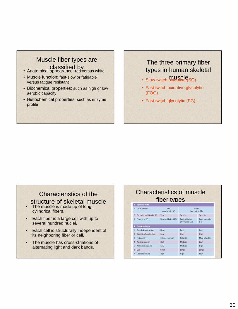
Muscle fiber types are classified by
• Anatomical appearance: red versus white• Muscle function: fast-slow or fatigable
versus fatigue resistant• Biochemical properties: such as high or low
aerobic capacity• Histochemical properties: such as enzyme
profile
Characteristics of the structure of skeletal muscle
• The muscle is made up of long, cylindrical fibers.
• Each fiber is a large cell with up to several hundred nuclei.
• Each cell is structurally independent of its neighboring fiber or cell.
• The muscle has cross-striations of alternating light and dark bands.
The three primary fiber types in human skeletal
muscle• Slow twitch oxidative (SO)
• Fast twitch oxidative glycolytic (FOG)
• Fast twitch glycolytic (FG)
Characteristics of muscle fiber types

31
Structure of the myofibril• Sarcomere
– functional unit– composed of two types of parallel myofilaments
• Myosin• Actin
• Z-line– membrane that separates sarcomeres
• A band– dark band seen as part of striation
• H zone– amount by which the two ends of the thin filaments
fail to meet• I band
– area between the ends of the myosin– light band in the striation
Significance of fiber-type composition for athletes
• High percentage of SO fibers—candidate for distance running or other endurance sports
• High percentage of FT fibers—candidate for power or sprint events
• Percentage is genetically determined
Series of events that lead to muscle contraction in the sliding filament model
1. Neural stimulation causes the sarcoplasmic reticulum to release calcium.
2. Calcium binds to troponin, which removes the inhibitory effect of tropomyosin and actin-myosin bind.
3. Myosin cross-bridges swivel, pulling the actin and z-lines.
4. Fresh ATP binds to the myosin cross-bridges, leading to cross-bridge recycling.
5. Neural stimulation ceases and relaxation occurs.
Contraction
Theory 11. Actin-myosin bind Activates myosin ATPaseBreakdown of ATP molecule liberates energy
2. Energy causes myosin cross-bridge to swivel to center of sarcomere Myosin is bound to actin, which is bound to Z-lines Z-lines pulled closer together Sarcomere shortened
3. Fresh ATP molecule binds to myosin cross-bridge Actin-myosin binding released Myosin cross-bridge stands back up
4. Actin-myosin rebinds at new siteActivates myosin ATPaseBreakdown of ATP molecule liberates energy
5. And process repeats

32
Theory 21. Myosin cross-bridge stores energy
from breakdown of ATP by myosin ATPase Actin-myosin binding releases stored energy
2. Causes myosin cross-bridge to swivel ADP and Pi are released from the myosin cross-bridge
3. Fresh ATP molecule binds to myosin cross-bridge Actin-myosin binding released ATP is broken down and energy causes myosin cross-bridge to stand back up (re-energized)
4. Fresh ATP molecule is broken down Energy reenergizes myosin cross-bridge
5. Process repeats
Comparing the two theories• Both theories state that fresh ATP binds to
the myosin cross-bridge to release it from actin during cross-bridge recycling.
• Theory 2 states that after the myosin-actin binding is released, the fresh ATP molecule is broken down and the energy released is used to reenergize the myosin cross-bridge.
• According to Theory 1, energy is not needed to cause the myosin cross-bridge to stand back up.
Next Class
• Collect velocity spectrum data• Make Excel graphs of force/velocity and
power/velocity curves
Muscle Tone• Muscle tone:
– Is the constant, slightly contracted state of all muscles, which does not produce active movements
– Keeps the muscles firm, healthy, and ready to respond to stimulus
• Spinal reflexes account for muscle tone by:– Activating one motor unit and then another– Responding to activation of stretch receptors
in muscles and tendons

33
Isotonic Contractions
• In isotonic contractions, the muscle changes in length (decreasing the angle of the joint) and moves the load
• The two types of isotonic contractions are concentric and eccentric
– Concentric contractions – the muscle shortens and does work
– Eccentric contractions – the muscle contracts as it lengthens
Isotonic Contractions
Figure 9.19a
Isometric Contractions
• Tension increases to the muscle’s capacity, but the muscle neither shortens nor lengthens
• Occurs if the load is greater than the tension the muscle is able to develop
Isometric Contractions
Figure 9.19b

34
Muscle Metabolism: Energy for Contraction
• ATP is the only source used directly for contractile activity
• As soon as available stores of ATP are hydrolyzed (4-6 seconds), they are regenerated by:
– The interaction of ADP with creatinephosphate (CP)
– Anaerobic glycolysis – Aerobic respiration
Muscle Metabolism: Energy for Contraction
Figure 9.20
Muscle Metabolism: Anaerobic Glycolysis
• When muscle contractile activity reaches 70% of maximum:
– Bulging muscles compress blood vessels– Oxygen delivery is impaired– Pyruvic acid is converted into lactic acid
Muscle Metabolism: Anaerobic Glycolysis
• The lactic acid:– Diffuses into the bloodstream– Is picked up and used as fuel by the liver,
kidneys, and heart– Is converted back into pyruvic acid by the liver

35
Muscle Fatigue
• Muscle fatigue – the muscle is in a state of physiological inability to contract
• Muscle fatigue occurs when:– ATP production fails to keep pace with ATP
use– There is a relative deficit of ATP, causing
contractures– Lactic acid accumulates in the muscle– Ionic imbalances are present
Muscle Fatigue
• Intense exercise produces rapid muscle fatigue (with rapid recovery)
• Na+-K+ pumps cannot restore ionic balances quickly enough
• Low-intensity exercise produces slow-developing fatigue
• SR is damaged and Ca2+ regulation is disrupted
Oxygen Debt
• Vigorous exercise causes dramatic changes in muscle chemistry
• For a muscle to return to a resting state:– Oxygen reserves must be replenished– Lactic acid must be converted to pyruvic acid– Glycogen stores must be replaced– ATP and CP reserves must be resynthesized
Oxygen Debt
• Oxygen debt – the extra amount of O2 needed for the above restorative processes

36
Heat Production During Muscle Activity
• Only 40% of the energy released in muscle activity is useful as work
• The remaining 60% is given off as heat • Dangerous heat levels are prevented by
radiation of heat from the skin and sweating
Force of Muscle Contraction
• The force of contraction is affected by:– The number of muscle fibers contracting –
the more motor fibers in a muscle, the stronger the contraction
– The relative size of the muscle – the bulkier the muscle, the greater its strength
– Degree of muscle stretch – muscles contract strongest when muscle fibers are 80-120% of their normal resting length
Force of Muscle Contraction
Figure 9.21a–c
Length Tension Relationships
Figure 9.22

37
Table 9.2
Muscle Fiber Type: Functional Characteristics
• Speed of contraction – determined by speed in which ATPases split ATP
– The two types of fibers are slow and fast• ATP-forming pathways
– Oxidative fibers – use aerobic pathways– Glycolytic fibers – use anaerobic glycolysis
• These two criteria define three categories –slow oxidative fibers, fast oxidative fibers, and fast glycolytic fibers
PLAY InterActive Physiology ®:Muscle Metabolism, pages 16-22
Muscle Fiber Type: Speed of Contraction
• Slow oxidative fibers contract slowly, have slow acting myosin ATPases, and are fatigue resistant
• Fast oxidative fibers contract quickly, have fast myosin ATPases, and have moderate resistance to fatigue
• Fast glycolytic fibers contract quickly, have fast myosin ATPases, and are easily fatiguedPLAY InterActive Physiology ®:
Muscle Metabolism, pages 24-29
Load and Contraction
Figure 9.23

38
Effects of Aerobic Exercise
• Aerobic exercise results in an increase of:– Muscle capillaries– Number of mitochondria– Myoglobin synthesis
Effects of Resistance Exercise
• Resistance exercise (typically anaerobic) results in:
– Muscle hypertrophy– Increased mitochondria, myofilaments, and
glycogen stores
Proportion and Organization of Myofilaments in Smooth Muscle
Figure 9.26
Contraction of Smooth Muscle
• Whole sheets of smooth muscle exhibit slow, synchronized contraction
• They contract in unison, reflecting their electrical coupling with gap junctions
• Action potentials are transmitted from cell to cell

39
Contraction of Smooth Muscle
• Some smooth muscle cells: – Act as pacemakers and set the contractile
pace for whole sheets of muscle– Are self-excitatory and depolarize without
external stimuli
Contraction Mechanism
• Actin and myosin interact according to the sliding filament mechanism
• The final trigger for contractions is a rise in intracellular Ca2+
• Ca2+ is released from the SR and from the extracellular space
• Ca2+ interacts with calmodulin and myosin light chain kinase to activate myosin
Role of Calcium Ion
• Ca2+ binds to calmodulin and activates it• Activated calmodulin activates the kinase
enzyme• Activated kinase transfers phosphate from
ATP to myosin cross bridges• Phosphorylated cross bridges interact with
actin to produce shortening• Smooth muscle relaxes when intracellular
Ca2+ levels drop
Special Features of Smooth Muscle Contraction
• Unique characteristics of smooth muscle include:
– Smooth muscle tone– Slow, prolonged contractile activity– Low energy requirements– Response to stretch
40
Response to Stretch
• Smooth muscle exhibits a phenomenon called stress-relaxation response in which:
– Smooth muscle responds to stretch only briefly, and then adapts to its new length
– The new length, however, retains its ability to contract
– This enables organs such as the stomach and bladder to temporarily store contents
Sliding Filaments• All the sarcomeres in a fiber will contract
together. This contracts the fiber itself. The number of fibers contracting will determine the force of the contraction of the whole muscle.
• We can actually divide the whole process of muscle contraction into 4 steps:– Excitation– Excitation-contraction coupling – Contraction– Relaxation
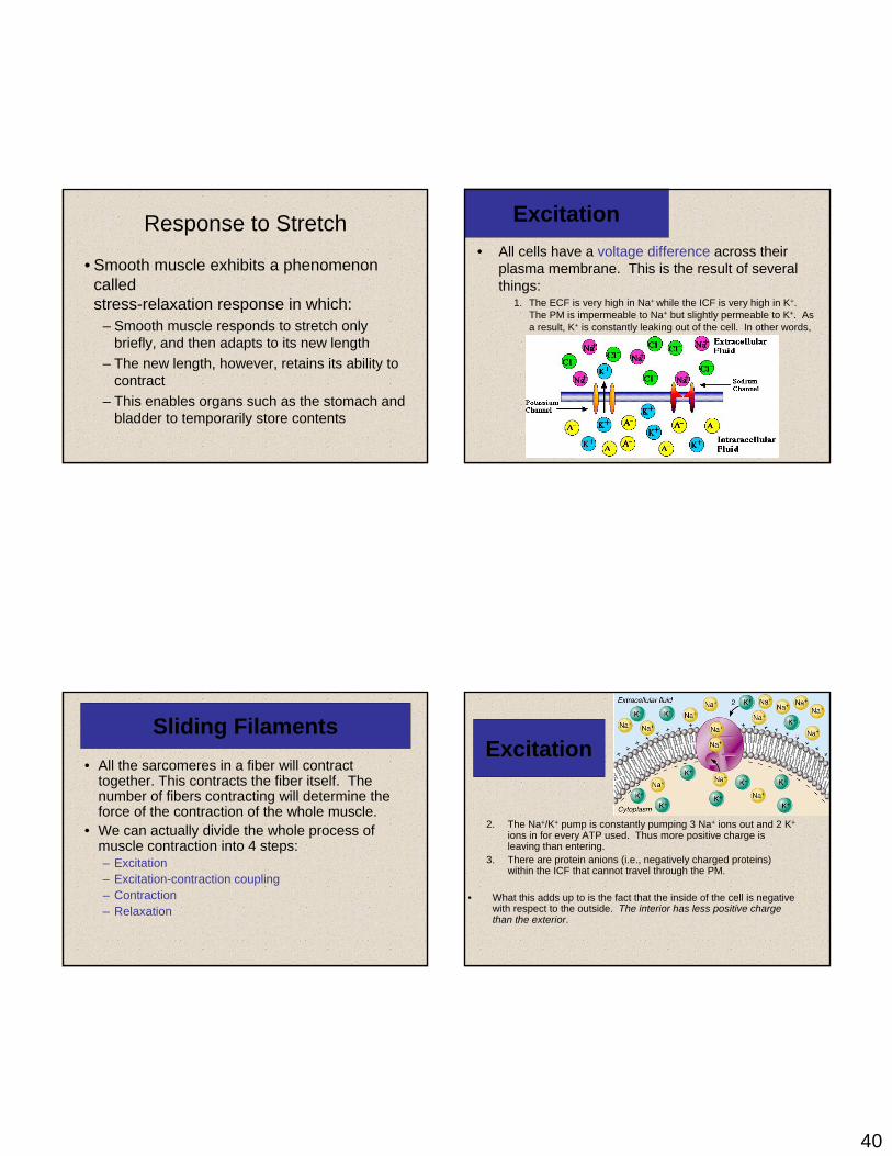
Excitation • All cells have a voltage difference across their
plasma membrane. This is the result of several things:
1. The ECF is very high in Na+ while the ICF is very high in K+. The PM is impermeable to Na+ but slightly permeable to K+. As a result, K+ is constantly leaking out of the cell. In other words, positive charge is constantly leaking out of the cell.
Excitation
2. The Na+/K+ pump is constantly pumping 3 Na+ ions out and 2 K+
ions in for every ATP used. Thus more positive charge is leaving than entering.
3. There are protein anions (i.e., negatively charged proteins) within the ICF that cannot travel through the PM.
• What this adds up to is the fact that the inside of the cell is negative with respect to the outside. The interior has less positive charge than the exterior.

41
Excitation• This charge separation is known as a membrane
potential (abbreviated Vm).
• The value for Vm in inactive muscle cells is typically btwn –80 and –90 millivolts.
• Cells that exhibit a Vm are said to be polarized. – Why do you suppose that is?
• Vm can be changed by influx or efflux of charge.
Excitation• The PM has integral proteins that act as gated ion channels. These
are channels that are normally closed, but in response to a certain signal, they will open and allow specific ions to pass through them.
• Ion channels may be:– Ligand-gated the binding of an extracellular molecule (e.g.,
hormone, neurotransmitter) causes these channels to open.– Voltage-gated ΔVm causes these channels to open.– Mechanically-gated stretch or mechanical pressure opens
these channels.• When a channel is open, its specific ion(s) will enter or exit
depending on their electrochemical gradient.
Excitation• In general each muscle is
served by one nerve – a bundle of axons carrying signals from the spinal cord to the muscle.
• W/i the muscle, each axon will go its own way and eventually branch into multiple small extensions called telodendria. Each telodendrium ends in a bulbous swelling known as the synaptic end bulb.
The site of interaction btwn a neuron and any other cell is known as a synapse. The synapse btwn a neuron and a muscle is known as the neuromuscular junction.
ExcitationThe minute space between the synaptic end bulb and the sarcolemma is known as the synaptic cleft. There is a depression in the sarcolemma at the synaptic cleft known as the motor end plate.
The synaptic end bulb is filled w/ vesicles that contain the neurotransmitter, acetylcholine.
The motor end plate is chock full of acetylcholine receptors.

42
Excitation1. A nerve signal will arrive at the synaptic end bulb and this
will cause the ACh-containing vesicles to undergo exocytosis.
2. ACh will diffuse across the synaptic cleft and bind to the ACh receptors. These receptors are actually ligand-gated Na+ channels. The binding of ACh causes them to open.
3. Na+ will rush into the cell, making the local cell interior more positive. This is known as depolarization. It is a local event!
Excitation• Adjacent to the motor end plate, the sarcolemma
contains voltage-gated ion channels. In order for these channels to open, the Vm must depolarize from its resting value of –90mV to approximately –50mV. This is the threshold. Vmmust become this positive for the voltage-gated channels to open.
• The degree of depolarization depends on how much Na+ influx occurred which in turn depends on how many Na+ channels were opened by binding ACh.
Excitation• If the Vm fails to depolarize to threshold,
nothing will happen. The Vm will soon return to normal and no muscle contraction will occur.
• If the Vm does reach threshold, 2 types of voltage-gated ion channels will open:– Fast Na+ channels– Slow K+ channels
• If Vm reaches threshold, fast Na+ channels open and Na+
rushes in causing the Vm to depolarize to +30mV. The depolarization stops when the Na+ channels become inactivated.
• At this point, slow K+ channels have opened & K+ efflux occurs. This returns Vm back to its resting level. This is repolarization.
• If we were to graph this change in Vm over time, it would look somewhat like the animation below.
• This is known as an action potential.
43
• An AP can propagate itself across the surface of the PM.
• The depolarization caused by the Na+ influx in one particular area of the sarcolemma causes voltage-gated channels in the adjacent membrane to open. The resulting ionic influx then causes voltage-gated channels to open in the next patch of membrane and so on and so on. Thus the AP propagates itself.
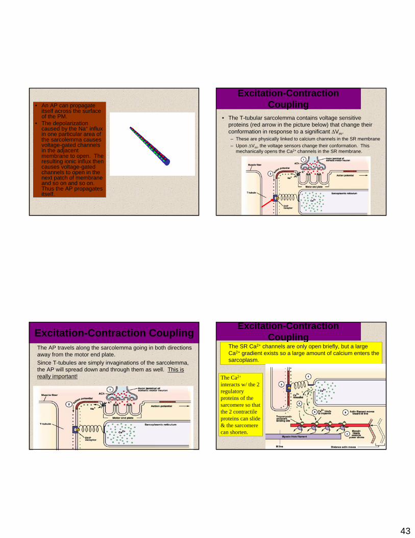
Excitation-Contraction CouplingThe AP travels along the sarcolemma going in both directions away from the motor end plate. Since T-tubules are simply invaginations of the sarcolemma, the AP will spread down and through them as well. This is really important!
Excitation-Contraction Coupling
• The T-tubular sarcolemma contains voltage sensitive proteins (red arrow in the picture below) that change their conformation in response to a significant ΔVm.– These are physically linked to calcium channels in the SR membrane – Upon ΔVm, the voltage sensors change their conformation. This
mechanically opens the Ca2+ channels in the SR membrane.
Excitation-Contraction Coupling
The SR Ca2+ channels are only open briefly, but a large Ca2+ gradient exists so a large amount of calcium enters the sarcoplasm.
The Ca2+
interacts w/ the 2 regulatory proteins of the sarcomere so that the 2 contractile proteins can slide & the sarcomere can shorten.

44
Let’s backtrack for just a moment…
• Now that we know what an action potential is, it should be noted that the exocytosis of the ACh vesicles is caused by the arrival of an AP at the synaptic end bulb.
• The AP causes the opening of voltage-gated Ca2+ channels in the synaptic end bulb plasma membrane. The resulting calcium influx causes the exocytosis of the vesicles.
Contraction• Normally,
tropomyosin obstructs the myosin binding site on the G-actin subunits.
• Calcium binds to the troponin-C polypeptide of the troponin triad. This changes the conformation of troponin which changes the conformation of tropomyosin which exposes the myosin binding site on actin.
Contraction
• Once actin’s myosin binding site is exposed, myosin will attach to it.– At this point myosin has just hydrolyzed ATP into ADP and Pi –
however both molecules are still bound to the myosin.– The ATP hydrolysis provides the energy for the “cocking” of the
myosin head• Once myosin is bound to actin, the myosin head will
release the ADP and Pi which will cause it change conformation. This results in the thin filament sliding along the thick filament.
• Myosin then remains bound to actin until it binds to another ATP. Myosin then hydrolyzes the new ATP and the cycle can begin again.
45
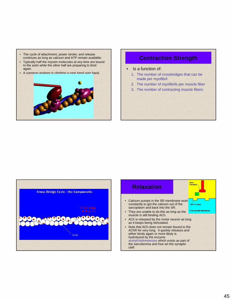
• The cycle of attachment, power stroke, and release continues as long as calcium and ATP remain available.
• Typically half the myosin molecules at any time are bound to the actin while the other half are preparing to bind again.
• A common analogy is climbing a rope hand over hand.
Contraction Strength
• Is a function of:1. The number of crossbridges that can be
made per myofibril2. The number of myofibrils per muscle fiber3. The number of contracting muscle fibers
Relaxation
• Calcium pumps in the SR membrane work constantly to get the calcium out of the sarcoplasm and back into the SR.
• They are unable to do this as long as the muscle is still binding ACh.
• ACh is released by the motor neuron as long as it keeps being stimulated.
• Note that ACh does not remain bound to the AChR for very long. It quickly releases and either binds again or more likely is hydrolyzed by the enzyme acetylcholinesterase which exists as part of the sarcolemma and free w/i the synaptic cleft

46
Relaxation• When the muscle ceases being
stimulated, the calcium pumps “win” and sarcoplasmic [Ca2+] drops. – Calcium stops being
available for troponin and tropomyosin shifts back into its inhibitory position.
• The muscle then returns back to its original length via the elasticity of the connective tissue elements, plus the contraction of antagonistic muscles, and gravity. This animation shows another way to
induce muscle relaxation. Does it make sense?
QUICK THOUGHT QUESTION: In this sculpture, why are the lion’s back legs paralyzed even though they were not injured?
Rigor Mortis• Upon death, muscle cells are unable to
prevent calcium entry. This allows myosin to bind to actin. Since there is no ATP made postmortem, the myosin cannot unbind and the body remains in a state of muscular rigidity for almost the next couple days.
Muscle Metabolism• The chemical energy released
by the hydrolysis of ATP is necessary for both muscle contraction and muscle relaxation.
• Muscles typically store limited amounts of ATP – enough to power 4-6s of activity.– So resting muscles must
have energy stored in other ways.

47
Resting Muscle and the Krebs
Cycle• Resting muscle fibers typically
takes up fatty acids from the blood stream. – How might they enter the cell? – Inside the muscle fiber, the FA’s are
oxidized to several molecules of a compound called Acetyl-CoA. This oxidation will also produce several molecules of NADH and FADH2.
– Acetyl-CoA will then enter a cyclical series of reactions known as the Krebs cycle or Tricarboxylic Acid cycle.
– In the Krebs cycle, acetyl-CoA combines with the compound oxaloacetate and then enters a series of rxns. The end product of these rxns is CO2, ATP, NADH, FADH2, and oxaloacetate (thus we call it a cycle)
Krebs Cycle Products
Oxaloacetate will simply combine with another molecule of acetyl-CoA and reenter the cycle.
NADH and FADH will enter another series of rxns known as the Electron Transport Chain. These rxns occur along the inner membrane of the mitochondrion and they basically consist of the passing of electrons from compound to compound with energy being released each time and used to drive the synthesis of ATP. The final electron acceptor is oxygen when it combines with 2 hydrogen atoms to yield water.
Krebs Cycle Products
• CO2 will diffuse out of the mitochondria, out of the muscle fiber, and into to the blood stream which will take it to the lungs.
• The ATP made in the Krebs cycle plus the ATP made during the ETC will be used in many ways.– See if you can list at least 5!
ATP Use in the Resting Muscle Cell
• ATP is necessary for cellular housekeeping duties.
• ATP powers the combination of glucose monomers (which have been taken up from the blood stream) into the storage polymer glycogen.
• ATP is used to create another energy storage compound called creatine phosphate or phosphocreatine:
ATP + Creatine ADP + Creatine-Phosphatethis rxn is catalyzed by the enzyme creatine kinase

48
Working Muscle• As we begin to exercise, we almost immediately
use our stored ATP.• For the next 15 seconds or so, we turn to the
phosphagen system, a.k.a., the energy stored in creatine-phosphate.Creatine-P + ADP Creatine Kinase Creatine + ATP– The ATP is then available to power contraction and
relaxation: myosin ATPase, Ca2+ ATPase in the SR membrane, and Na+/K+ ATPase in the sarcolemma.
– The phosphagen system dominates in events such as the 100m dash or lifting weights.
Working Muscle• After the phosphagen system is depleted, the
muscles must find another ATP source.• The process of anaerobic metabolism can
maintain ATP supply for about 45-60s.• Anaerobic means “without air,” and it is the
breakdown of glucose without the presence of oxygen. – It usually takes a little time for the respiratory and
cardiovascular systems to catch up with the muscles and supply O2 for aerobic metabolism.
Anaerobic Metabolism• Glucose is supplied by the breakdown of
glycogen or via uptake from the bloodstream.• Glucose is broken down into 2 molecules of
pyruvic acid, with the concomitant of 2 ATP and the conversion of 2 molecules of NAD+
into NADH. This process is known as glycolysis and it occurs in the sarcoplasm.– Unfortunately, w/o O2, we cannot use the NADH in
the ETC.– In order for more glycolysis to proceed, the muscle
cell must regenerate the NAD+. It does this by coupling the conversion of pyruvic acid into lactic acid with the conversion of NADH into NAD+

49
Anaerobic Metabolism
• Lactic acid typically diffuses out of muscles into the blood stream and is taken to the liver, kidneys, or heart which can use it as an energy source.
• Anaerobic metabolism is inefficient. Large amounts of glucose are used for very small ATP returns. Plus, lactic acid is a toxic end product whose presence contributes to muscle fatigue.
• Anaerobic metabolism dominates in sports that requires bursts of speed and activity, e.g., basketball.
Aerobic Metabolism
• Occurs when the respiratory and cardiovascular systems have “caught up with” the working muscles.– Prior to this, some aerobic respiration will occur
thanks to the muscle protein, myoglobin, which binds and stores oxygen.
• During rest and light to moderate exercise, aerobic metabolism contributes 95% of the necessary ATP.
• Compounds which can be aerobically metabolized include:– Pyruvic acid (made via glycolysis), fatty acids, and
amino acids.
Aerobic Metabolism
• It occurs in the mitochondria.
• Pyruvic acid from glycolysis is the primary substrate. The cell also utilizes fatty acids and amino acids.
• Aerobic respiration typically yields 36 ATP per molecule of glucose. Compare this to anaerobic metabolism.
Muscle Fatigue
• Physiological inability to contract• Results primarily from a relative deficit
of ATP.• Other contributing factors include the
decrease in sarcoplasmic pH (what causes this?), increased sarcoplasmic [ADP], and ionic imbalances.

50
Oxygen Debt• Refers to the fact that post-exercise breathing
rate >>> resting breathing rate• This excess oxygen intake serves many tasks:
– Replenish the oxygen stored by myoglobin and hemoglobin
– Convert remaining lactic acid back into glucose– Used for aerobic metabolism to make ATP which is
used to:• Replenish the phosphagen system• Replenish the glycogen stores• Power the Na+/K+ pump so as to restore resting ionic
conditions within the cell.
Whole Muscle Contraction
• Why can you electrically stimulate a muscle to contract? (HINT: what kind of channels could an electric current open?)
• A sub-threshold stimulus would not cause contraction because no AP would be produced!
• The response of a muscle to a single supra-threshold stimulus would be a twitch – the muscle quickly contracts and then relaxes.
• Let’s take a look at a measurement of a neuron’s AP, a muscle fiber’s AP, and the tension developed by that muscle fiber.
Phases of the Muscle Twitch1. Latent Period
– Time btwn stimulus and generation of tension
– Includes all time required for excitation, excitation-contraction coupling, and stretching of the series elastic components.
2. Contraction3. Relaxation
Now, let’s look at various types of muscle twitches

51
The black arrows signify stimulation
Here we have multiple twitches separated by ample time. Notice that the previous twitch has no effect on a new twitch and that these twitches are similar in size. This is why we can say that muscle contraction – at least on the level of a single fiber – is an all-or-none event.
Here, we have an initial stimulation and resulting twitch all by itself. Then we have 2 stimuli in somewhat rapid succession. The 2nd twitch has added on to the first. This is known as wave or temporal summation. It occurs because there is still calcium from the 1st twitch in the sarcoplasm at the time of the 2nd twitch.
Here, we have wave summation until maxtension is achieved.
– Maximum tension is known as tetanus
– Do not confuse this w/ the disease caused by the bacterium Clostridium tetani. Its toxins prevent the normal inhibition of muscle contractions as mediated in the spinal cord. This leads to uncontrolled, unwanted muscle contraction and ultimately respiratory arrest.
Btwn stimulations, only the tiniest bit of relaxation occurs. Since some relaxation does occur, we say the tetanus is unfused or incomplete.Most muscle actions occur as a result of muscle fibers undergoing asynchronous, unfused tetanus
Here, the stimuli are close enough to one another so that tetanus is complete and no relaxation occurs until fatigue sets in.

52
Here we have the phenomenon known as treppe(German for staircase). Notice that the subsequent contractions grow stronger. There 2 reasons for this:1. Slight increase in sarcoplasmic [Ca2+]2. Heat liberated by working muscle increases the rate and efficiency of enzyme function within the muscle fiber.
Motor Units• A motor unit is defined as a somatic motor neuron and all the skeletal muscle fibers it innervates.
• When this neuron is stimulated, all the muscle fibers it synapses upon will be stimulated and will contract as a unit
• The # of muscle fibers per motor unit may be as high as several hundred or as few as four.– The smaller the motor unit, the
finer and more delicate the movements.
– Extraocular muscles typically have small motor units while the large postural muscles have large motor units
Notice that the muscle fibers of a single unit are not clustered together but are spread out. What’s the advantage to this?
Graded Responses
• It should be obvious that you can contract a muscle at just about any rate and with any force you desire.
• How does this fact concur with the quickness of a single muscle twitch.– We achieve smooth contractions of the whole
muscle by varying the frequency of stimuli sent to the muscle fibers and by recruitment – varying the number and size of the motor units involved
Thought problem: compare the act of picking up a pencil with the act of picking up a desk
Internal vs. External Tension
• When a skeletal muscle contracts, the myofibrils inside the muscle fibers generate internal tension. This internal tension is transferred to the series elastic components of the muscle – the fibers of the endomysium, perimysium, epimysium, and tendons. The tension of the SEC is known as external tension.• The SEC behaves like fat rubber bands. They stretch easily at first, but as they elongate they become stiffer and more effective at transferring the external tension to the resistance.– Attach a rubber band to a weight and then try to pick it up.
What happens?

53
Types of Contractions
• Contractions can be:1. Isometric
• Iso= same, metr=measure
2. Isotonic• Iso=same, ton=tension
Isotonic Contraction• Tension reaches a plateau and then the muscle
shortens. Consider the following experiment:1. A skeletal muscle 1cm2 in cross-sectional area can
develop roughly 4kg of force in complete tetanus.2. If we hang a 3kg weight from that muscle and stimulate it,
the muscle will shorten.3. Before the muscle can shorten, the cross-bridges must
produce enough tension to overcome the resistance – in this case the 3kg weight. Over this period, internal tension in the muscle fibers rises until the external tension in the tendon exceeds the amount of resistance.
4. As the muscle shortens, the internal and external tensions in the muscle remain constant at a value that just exceeds the resistance.
Resistance and Speed of Contraction
• There is an inverse relationship between the amount of resistance and the speed of contraction.
• The heavier the load, the longer it takes for the movement to begin because muscle tension, which increases gradually, must exceed the resistance before shortening can occur– More cross-bridges must be
formed, more fibers involved. This takes more time.

54
Isometric Contractions
• The muscle as a whole does not change length and the tension produced never exceeds the resistance.
• Consider the following:– To the same muscle as before, we attach a 6kg weight.– Although cross-bridges form and tension rises to peak
values, the muscle cannot overcome the resistance of the weight and cannot shorten.
– Although the muscle as a whole does not shorten, the individual fibers shorten until the tendons are taut and the external tension equals the internal tension. The muscle fibers cannot shorten further because the external tension does not exceed the resistance.
Muscle Tone
• Some of the motor units w/i particular muscle are always active, even when the muscle is not contracting.– Their contractions do not produce enough tension to
cause movement, but they do tense and firm the muscle.
– This resting tension in a skeletal muscle is called tone.– The identity of the motor units involved changes
constantly. • Why do you suppose this is?
• Resting muscle tone stabilizes the position of bones and joints.
Muscle Fiber Types
2 main types:1. Slow fibers2. Fast fibers

55
Slow Fibers• Contract slowly because its myosin
ATPases work slowly.• Depends on oxygen delivery and
aerobic metabolism.• Is fatigue resistant and has high
endurance.• Is thin in diameter – large amt of
cytoplasm impedes O2 and nutrient diffusion.• Cannot develop high tension – small diameter means few myofibrils.
• Has rich capillary supply and lots of mitochondria.• Contains lots of the O2-storing protein, myoglobin which gives it
a red color.• Uses lipids, carbs, and amino acids as substrates for it aerobic
metabolism.• Best suited for endurance type activities.• A.k.a. red fibers, slow oxidative fibers, type I fibers.
Fast Fibers• So named because they can
contract in 0.01 seconds or less after stimulation.
• Fast fibers are large in diameter; they contain densely packed myofibrils, large glycogen reserves, and relatively few mitochondria.
• Able to develop a great deal of tension b/c they contain a largenumber of sarcomeres.
• Use ATP in massive amounts. Supported by anaerobic metabolism. Fatigue rapidly.
• A.k.a., fast fatigue (FF) fibers, fast glycolytic (FG) fibers, white fibers.
• Best suited for short term, power activities.
Thought questions: why do chickens have white breast meat and dark leg meat? What does this say about the activities of the associated muscles? Why do ducks have dark breast meat?