Embed Size (px)
Citation preview

J. Embryol. exp. Morph. Vol. 19, 2, pp. 193-202, April 1968 ] 9 3With 2 plates
Printed in Great Britain
Muscle proteins in the chick myotome examinedby the immunofluorescent method
By AKIRA IKEDA1, RANA L. ABBOTT1
& JAN LANGMAN1
From the Department of Anatomy, University of Virginia, Charlottesville
INTRODUCTION
In previous studies on the development of the somite (Langman & Nelson,1968), the majority of the cells of the myotome appeared to arise from thedermatome and not, as previously suggested, from the cells of the so-calleddorso-medial somite lip (Williams, 1910; Hamilton, 1952; Boyd, 1960). Oncethe myotome cells, characterized by a pale, round to irregular, nucleus and darklystained nucleolus, are formed, they fail to synthesize DNA. During subsequentdevelopment, the myotome appears to extend in ventro-lateral direction by theaddition of new cells originating primarily from the dermatome. After thedermatome has lost its epithelial structure, the myotome, in transverse section,consists of a tissue band with an abundance of cytoplasm and a large number ofround to spindle-shaped nuclei. Though DNA synthesis was rarely observedamong the cells of the extended myotome, considerable proliferative activitywas observed in the adjacent tissues.
To determine at which stage of development the cells of the myotome beginto synthesize muscle proteins, Holtzer, Marshall & Finck (1957) used thefluorescent antibody technique on glycerol-extracted, squashed, and teasedsamples of myotome cells obtained from Hamburger & Hamilton (1951) stage14-29 embryos. According to the authors, two types of cells can be distinguishedin muscle differentiation, one type consisting of elongated, spindle-shaped cellsand the other of round to irregular cells similar to mesenchyme cells. The elon-gated cells, referred to as ' myoblasts', begin to synthesize myosin at stages 15-16;they have then lost their ability to synthesize DNA. The mesenchyme-like cells,referred to as 'presumptive myoblasts', will differentiate into myoblasts, butcannot histochemically be differentiated from surrounding mesenchyme cells.Unlike the myoblasts they are capable of synthesizing DNA (Stockdale &Holtzer, 1961).
In contrast to the work of Holtzer et ah (1957), who stated that myosin is thefirst specific muscle protein to appear during development, Ogawa (1962) found
1 Authors' address: Department of Anatomy, School of Medicine, University of Virginia,Charlottesville, Virginia, 22901, U.S.A.
194 A. IKEDA, R. L. ABBOTT & J. LANGMAN
by means of precipitin tests that actin appears in the muscle fibers at the 72 hstage and myosin approximately 24 h later.
When the data obtained with the immunofluorescent and precipitin techniquesare compared with information from electron-microscopic studies, considerableagreement about the stage of development at which the muscle proteins appearis noted. Przybylski & Blumberg (1966) state that at stages 15-16 the cells of themyotome, dermatome and adjacent mesenchyme are ultrastructurally similarto each other. By stages 16-17, however, some of the myotome cells assume anelongated shape and the first myofilaments, 50-70 A in diameter, appear.Slightly later dense filaments 100-120 A in diameter become visible. Duringsubsequent stages the 100 A filaments are separated by smaller 50 A filaments,suggesting an early A and I band configuration. Allen & Pepe (1965), Dessouky& Hibbs (1965), and Obinata, Yamamoto & Maruyama (1966), who likewisestudied muscle differentiation in the chick, come to only slightly differentconclusions and it is now generally accepted that the thin filaments, believed torepresent actin, coincide with or precede slightly the appearance of the thickfilaments thought to be myosin (Huxley, 1957).
Since the fluorescent studies have been carried out mainly with squashed orteased cells, thereby disturbing the interrelationship of the presumptive myo-blasts, myoblasts and myotubes, and the ultrastructural investigations arehandicapped by their limited field of vision, the precise interrelationship of thecells in the developing myotome remains unknown. Hence, this work wasundertaken to examine by the immunofluorescent antibody technique at whichstage and at what place in the myotome plate the first specific muscle proteinsarise. In addition, by using transverse sections we hoped to obtain a betterinsight into the spatial interrelationship of presumptive myoblasts, myoblastsand myotubes.
MATERIALS AND METHODS
Preparation of myosin antiserum. Chicken myosin was prepared according tothe Szent-Gyorgyi method (1951) with some minor modifications as previouslydescribed (Ikeda, Abbott & Langman, 1968). One gram of myosin was dissolvedin 1 ml 0-6 M Tris/sodium EDTA/boric acid (TEB) buffer (pH 8-4) and to thissolution was added an equal volume of Freund's complete adjuvant (Difco).Two ml of the emulsion was injected subcutaneously into each of a number ofalbino rabbits. The injections were repeated with 7-day intervals for 2-3 months.Serum was collected repeatedly throughout the course of immunization andtested for its specificity by the agar-gel immunoelectrophoretic technique (Ikeda& Zwaan, 1967). If satisfactory results were obtained, the serum was stored insmall aliquots at - 20 °C. In order to examine the specificity of the myosinantiserum, it was tested against total muscle extract, actin, myosin, adenylicacid deaminase, chicken serum and liver extract. Actin and adenylic acid deami-nase were prepared as previously described (Ikeda et al. 1968).

Muscle proteins in the myotome ' 195Tissue preparation. Fertilized White Leghorn eggs were incubated at 38 °C and
the embryos removed 52 h to 5 days after the beginning of incubation. Theembryos were then staged according to Hamburger & Hamilton (1951) and fixedin either Carnoy's solution, acetone or 95 % ethyl alcohol. Since the latter twofixatives damage the histological structure of the myotome area, Carnoy'ssolution was finally selected as the best fixative for our experiments. It gave aminimum of autofluorescence and a maximum specific reaction. After the tissuewas fixed for 2 h in Carnoy's solution at 3 °C, it was dehydrated and embeddedin paraffin in the usual way. Sections were cut at 3-5 /i and paraffin removed intwo changes of xylene for 15 min, followed by three changes of 95 % ethanol for15 min. Finally, they were rinsed twice for 10 min in buffered saline. To deter-mine whether myosin was denatured by the heat of the embedding processmyosin extract was heated at 60 °C for 15 min (Locker, 1956). In subsequentimmunoelectrophoretic tests the heated myosin reacted similarly to the unheatedcontrol.
Jmmunofluorescent method. The indirect fluorescent method ('sandwichtechnique') was selected for our experiments because of its great sensitivity(Coons, 1956). Goat gamma-globulin solution, prepared against rabbit gamma-globulin and conjugated with fluoresceinisothiocyanate, was obtained com-mercially (Difco). To reduce the non-specific staining, the solutions wereabsorbed with mouse tissue powder (Difco).
The tissue sections were exposed to chicken myosin antiserum for 20 min in amoisture chamber at 20 °C. They were then washed twice for 10 min withbuffered saline and covered with fluorescent goat globulin for 20 min. Afterrinsing twice in buffered saline, the sections were mounted in a buffered aqueoussolution of polyvinyl alchohol (Elvanol, grade 5105; Dupont ElectrochemicalsDepartment, Wilmington, Delaware) (Rodriguez & Deinhardt, 1960). Controlexperiments were performed as described previously (Ikeda & Zwaan, 1967).
The slides were examined under a Reichert Zetopan fluorescence microscopeequipped with a high-pressure mercury vapor lamp HBO-200 with primaryfilters (Schott) UG 1 and BG 12 and barrier filter GG 9. Photographs weremade on 35 mm Tri-X Pan film (Kodak), which was developed with Rodinol(Agfa) 1:100 for 13 min at 20 °C.
RESULTS
Immunoelectrophoretic experiments. When myosin extract is tested againsttotal muscle antiserum, one distinct and one small precipitin band appear(Text-fig. 1A). When, however, total muscle extract is tested against myosinantiserum, a minimum of 3-4 precipitin bands appear, indicating that themyosin antiserum to be used in our further experiments contains antibodies notonly against myosin but also against several other fractions (Text-fig. 1B). Sinceone of these bands has a position corresponding to that formed by adenylic aciddeaminase when tested against total muscle antiserum (Text-fig. 1C), and
196 A. IKEDA, R. L. ABBOTT & J. LANGMAN
another band corresponds to that formed by actin (Text-fig. 1D), it is evidentthat the myosin antiserum contains antibodies against adenylic acid deaminase,actin and possibly some other antigens. It does not contain as many antibodiesas total muscle antiserum, as is apparent from the precipitin pattern formed bytesting total muscle extract against its own antiserum (Text-fig. 1E).
Immunofluorescent experiments. The first fluorescent reaction over the cells
oo
Text-fig. 1. Drawing of the immunoelectrophoretic precipitin pattern formed bytesting the following. A, Myosin against total muscle antiserum: note the denseprecipitin band and the small band close to the antigen well. B, Total muscle extractagainst myosin antiserum: in comparison with A it is evident that the myosin anti-serum contains antibodies against several proteins. C, Adenylic acid deaminaseagainst total muscle antiserum: compare the position of this band with those foundin B. D, Actin against total muscle antiserum: compare the position of the band withthose found in B. E, Total muscle extract against total muscle antiserum.
PLATE 1
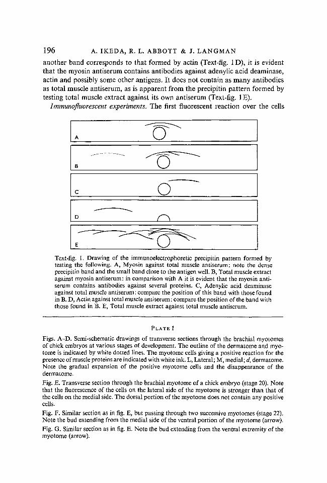
Figs. A-D. Semi-schematic drawings of transverse sections through the brachial myotomesof chick embryos at various stages of development. The outline of the dermatome and myo-tome is indicated by white dotted lines. The myotome cells giving a positive reaction for thepresence of muscle proteins are indicated with white ink. L, Lateral; M, medial; d, dermatome.Note the gradual expansion of the positive myotome cells and the disappearance of thedermatome.Fig. E. Transverse section through the brachial myotome of a chick embryo (stage 20). Notethat the fluorescence of the cells on the lateral side of the myotome is stronger than that ofthe cells on the medial side. The dorsal portion of the myotome does not contain any positivecells.Fig. F. Similar section as in fig. E, but passing through two successive myotomes (stage 22).Note the bud extending from the medial side of the ventral portion of the myotome (arrow).Fig. G. Similar section as in fig. E. Note the bud extending from the ventral extremity of themyotome (arrow).

/. Embryol. exp. Morph., Vol. 19, Part 2
> Stages" 17-18 Stage 19 Stage 20
A. IKEDA, R. L. ABBOTT & J. LANGMAN facing p. 196

J. Embryol. exp. Morph., Vol. 19, Part 2 PLATE 2
A. IKEDA, R. L. ABBOTT & J. LANGMAN
Muscle proteins in the myotome 197
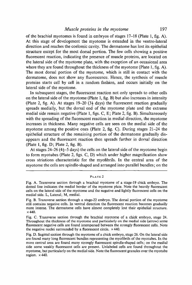
of the brachial myotomes is found in embryos of stages 17-18 (Plate 1, fig. A).At this stage of development the myotome is extended in the ventro-lateraldirection and reaches the coelomic cavity. The dermatome has lost its epithelialstructure except for the most dorsal portion. The few cells showing a positivefluorescent reaction, indicating the presence of muscle proteins, are located onthe lateral side of the myotome plate, with the exception of an occasional areawhere they are found throughout the thickness of the myotome (Plate 1, fig. A).The most dorsal portion of the myotome, which is still in contact with thedermatome, does not show any fluorescence. Hence, the synthesis of muscleproteins starts cell by cell in a random fashion, and occurs initially on thelateral side of the myotome.
In subsequent stages, the fluorescent reaction not only spreads to other cellson the lateral side of the myotome (Plate 1, fig. B) but also increases in intensity(Plate 2, fig. A). At stages 19-20 (3^ days) the fluorescent reaction graduallyspreads medially, but the dorsal end of the myotome plate and the extrememedial side remain negative (Plate 1, figs. C, E; Plate 2, fig. B). Simultaneouslywith the spreading of the fluorescent reaction in medial direction, the myotomeincreases in thickness. Many negative cells are seen on the medial side of themyotome among the positive ones (Plate 2, fig. C). During stages 21-24 theepithelial structure of the remaining portion of the dermatome gradually dis-appears and the fluorescent reaction then spreads further in dorsal direction(Plate 1, fig. D; Plate 2, fig. B).
At stages 24-26 (4^-5 days) the cells on the lateral side of the myotome beginto form myotubes (Plate 2, figs. C, D) which under higher magnification showcross striations characteristic for the myofibrils. In the central area of themyotome the cells are spindle-shaped and arranged into parallel bundles; on the
PLATE 2
Fig. A. Transverse section through a brachial myotome of a stage-19 chick embryo. Thedotted line indicates the medial border of the myotome plate. Note the heavily fluorescentcells on the lateral side of the myotome and the negative and lightly fluorescent cells on themedial side. L, Lateral; M, medial.Fig. B. Transverse section through a stage-22 embryo. The dorsal portion of the myotomestill contains negative cells. In ventral direction the fluorescent reaction becomes graduallymore intense. The dermatome cells have almost completely lost their epithelial structure.x440.Fig. C. Transverse section through the brachial myotome of a chick embryo, stage 24.Throughout the thickness of the myotome and particularly on the medial side (arrow) somefluorescent negative cells are found interspersed between the strongly fluorescent cells. Notethe negative nuclei surrounded by a fluorescent circle, x 440.Fig. D. Sagittal section through the myotome of a chick embryo, stage 26. On the lateral sideare found many long fluorescent bundles representing the myofibrils of the myotubes. In themore central area are found many strongly fluorescent spindle-shaped cells; on the medialside some weakly fluorescent cells are present. Unlabeled cells are found throughout themyotome, but particularly on the medial side. Note the fluorescent granules over the myotuberegion. x440.
198 A. IKEDA, R. L. ABBOTT & J. LANGMAN
medial side some cells are weakly fluorescent while others are negative. Fluo-rescent granules appear in the region of the myotubes, and are seen only aftermyotube formation has started (Plate 2, fig. D).
DISCUSSION
One of the difficulties of the fluorescent antibody technique is that the preciselocalization of a protein can only be undertaken with success after the specificityof the antibody has been thoroughly tested. Our immunoelectrophoretic studieson the purity of the myosin antiserum, indeed, demonstrated the presence of anumber of contaminating antibodies. Among these were antibodies againstactin and adenylic acid deaminase and possibly some other components.Holtzer et al. (1957) likewise reported that their myosin antiserum containedone major antibody and one or two trace components. Samuels (1961) andFinck (1965) suggested that the myosin fractions were contaminated withadenylic acid deaminase, actomyosin or nucleoprotein. Despite repeated frac-tionation of the chemically prepared myosin with ammonium sulfate, thecontaminating components could not be completely removed (Holtzer et al.1957; Finck, 1965). Since our fluorescent antibody experiments were performedwith impure myosin antiserum, we have refrained from stating that the positivereaction is caused by myosin, but rather by the presence of muscle proteins.
In our work the first fluorescent cells appeared at Hamburger & Hamiltonstage-17 embryos. This is slightly later than indicated by the work of Holtzeret al. (1957), who found the first reaction at stages 15-16. Initially the cause ofthe minor discrepancy was thought to be a difference in the specificity andstrength of our antiserum in comparison with that of Holtzer. Considering,however, that our antiserum demonstrated the presence of muscle proteins inwidely separated, individual cells and that immunoelectrophoretically it notonly reacted with myosin but also with actin, it seems more likely that thedifference is caused by our experimental technique. Holtzer et al. (1957) usedglycerol extracted, squashed and teased tissues, while we used 3/i transversesections. The amount of muscle proteins capable of reacting with the antiserumwas undoubtedly, in our work, considerably less than in the case of whole cells.Whether the first reaction found in our work is caused by actin or myosin orpossibly by both remains to be studied with more highly purified antibodies.Allen & Pepe (1965) found that thin filaments (50-70 A in diameter), in alllikelihood representing actin molecules, appear at stage 16 and are initiallyrandomly dispersed through the cytoplasm. This observation was confirmed byObinata et al. (1966) and Przybylski & Blumberg (1966). Dessouky & Hibbs(1965), on the contrary, found that the thin and the thick filaments, representingmyosin, appear approximately at the same time. Hence, it is impossible atpresent to state which of the two proteins is synthesized first, or whether bothappear at the same time.

Muscle proteins in the myotome 199In previous work (Langman & Nelson, 1968) the cells of the myotome were
found to appear first in the dorsal region and from this position to extend inventro-lateral direction by the addition of new cells formed by the overlyingdermatome. Hence we expected that the muscle proteins would appear first inthe dorsal region of the myotome. On the contrary the first fluorescent cells,indicating synthesis of muscle proteins, appear in the more central regions of themyotome. Muscle protein synthesis in the most dorsal portion of the myotomedoes not start until the adjacent dermatome cells have lost their epithelial struc-ture. Only then does the fluorescent reaction gradually extend in dorsal direction.Two possible explanations of this observation must be considered: (1) the cellsof the dermatome inhibit the synthesis of muscle proteins in the adjacent myo-tome cells; (2) the first formed myotome cells migrate from their initial positionin a ventro-lateral direction and are continuously replaced dorsally by new cellsformed by the dermatome. Since the latter possibility seems more probable tous, this would mean that the ventro-lateral extension of the myotome is partiallycaused by migration of cells from dorsal to ventral and partially by the additionof new cells.
Holtzer et al. (1957) and Stockdale & Holtzer (1961) called the cells whichsynthesized muscle proteins 'myoblasts', while the cells destined to becomemyoblasts, but not yet synthesizing muscle proteins, are referred to as ' presump-tive myoblasts'. Though the myoblasts do not synthesize DNA, the latter cellsstill maintain this ability. Our observations indicate that differentiation frompresumptive myoblast to myoblast and from myoblast to myotome occursfirst on the lateral side of the myotome and subsequently in the central andmedial areas. Initially the myotome cells formed by the dermatome synthesizeneither muscle proteins nor DNA. With the disappearance of the dermatomethe first muscle proteins become evident in the cells along the lateral border ofthe myotome. These cells we will call myoblasts. The cells on the medial side ofmyotome plate still synthesize neither muscle protein nor DNA. Since theyform an intermediate stage between the myoblast and presumptive myoblast, wewill refer to them as 'primitive myoblasts'. In preliminary experiments in whichthe fluorescent technique was combined with the radioautographic technique,it was noted that adjacent to the medial side of the original myotome many cellswere synthesizing DNA. Since these cells, according to our observations, willlater start to synthesize muscle proteins, we will call them presumptive myoblasts.The progressive differentiation from presumptive myoblasts to myotubes becomesparticularly evident by stage 24. At this stage myotubes with distinct crossstriations are visible on the lateral side of the myotome. In the central area (ormedial side of the original myotome) are found spindle-shaped myoblasts,sometimes arranged in parallel bundles, sometimes as individual cells. On themedial side some cells have just started to synthesize muscle proteins, whileothers are still negative.
Simultaneously with the formation of the myotubes, we noted the appearance

200 A. IKEDA, R. L. ABBOTT & J. LANGMAN
of fine granular fluorescent droplets. These droplets, localized over the myotubearea and immediately adjacent to it, were not seen over the spindle-shapedmyoblasts or on the medial side of the myotome. Though the granules may beartefacts, Przybylski & Blumberg (1966) reported that during the fusion ofmyoblasts into myotubes, the cell membranes break down and some cytoplasmicmaterial and ribosomes appear outside the cells. It can be argued that theseparticles are much too small to show a fluorescent reaction, but keeping in mindthat in our experiments the sensitive indirect 'sandwich' method was used, itseems possible that some of the fluorescent granules contain muscle proteinswhich leaked out of the myoblasts during myotube formation.
Considering the progressive differentiation from presumptive myoblasts tomyotube as a process which begins initially on the lateral side of the myotome,which factor or factors initiate the synthesis of muscle proteins ? When Holtzer& Detwiler (1954) grafted tissue strips of spinal cord and somites obtained fromsalamanders into similarly aged hosts, the somite cells formed muscle andcartilage. When they did not include spinal cord in the graft, the somites failedto differentiate. In similar experiments performed in the chick embryo, thespinal cord was likewise found to be of primary importance for the differentiationand growth of muscle cells (Lash, Holtzer & Holtzer, 1957). According toHoltzer and co-workers, the influence of the spinal cord appears to be mediatedby a factor which is transmissable through mesenchyme. Whether this factor istransmissible through such a thick layer of mesenchyme, as is present in stage17-20 embryos, remains, in our opinion, questionable.
SUMMARY
This work was undertaken to examine by the immunofluorescent antibodytechnique at which stage of development of the chick embryo the first muscleproteins are formed. In addition the spatial interrelationship of presumptivemyoblasts, myoblasts and myotubes was studied. Chick embryos ranging in agefrom 52 h to 5 days were fixed in Carnoy and transverse sections cut at 3-4 fi.The indirect fluorescent method (sandwich technique) was applied using fluor-scent goat gamma-globulin and myosin antiserum.
1. When the specificity of the myosin antiserum was tested by the immuno-electrophoretic technique, antibodies against myosin as well as against actin,adenylic acid deaminase and some other fractions were detected. Consequently,the fluorescent reactions found in the embryo do not specifically indicate theprescence of myosin, but rather that of muscle proteins.
2. The first fluorescent reaction over the myotome was found at stages 17-18,and was restricted to a few cells on the lateral side of the central myotomeregion. From here the reaction spread rapidly to other cells on the lateral sidein a ventral and dorsal direction. Simultaneously with the increase in thicknessof the myotome, the fluorescent reaction spread in a medial direction. The cells
Muscle proteins in the myotome 201of the myotome, particularly those in the most dorsal portion, did not begin toshow fluorescense until the adjacent dermatome cells had lost their epithelialstructure.
3. At stages 24-26 the lateral side of the myotome is formed by myotubeswith fluorescent myofibrils. In a medial direction are found subsequently heavilyfluorescent spindle cells, either in bundles or as individual cells, which in turnare followed by irregular cells either slightly or not at all fluorescent. The lattercells sometimes showed DNA synthesis. Hence, the differentiation from pre-sumptive myoblast to myoblast and from myoblast to myotube proceeds fromlateral to medial.
RESUME
Examen des proteines musculaires du myotome de poulet par lamethode dHmmunofluorescence
Ce travail a ete entrepris pour examiner, a l'aide de la technique des anticorpsimmunofluorescents, a quel stade du developpement de l'embryon de poulet sontformees les premieres proteines musculaires. On a etudie en outre les relationsspatiales mutuelles entre myoblastes presomptifs, myoblastes et myotubes. Desembryons de poulet d'un age compris entre 52 h et 5 jours ont ete fixes auCarnoy et debites en coupes transversales de 3 a 4 pt,. On a applique la methodede fluorescence indirecte (technique du 'sandwich') en utilisant de la gamma-globuline fluorescente de chevre et un serum anti-myosine.
1. Quand la specificite du serum anti-myosine a ete eprouvee par la techniqued'immunoelectrophorese, on a decele des anticorps anti-myosine ainsi queanti-actine, anti-adenylacidodesaminase et quelques autres fractions. Par con-sequent, les reactions fluorescentes trouvees chez l'embryon n'indiquent passpecifiquement la presence de myosine mais plutot celle de proteines musculaires.
2. La premiere reaction fluorescent sur le myotome a ete trouvee aux stades17-18; elle est restreinte a quelques cellules sur la face laterale de la regioncentrale du myotome. A partir de la, la reaction s'etend rapidement aux autrescellules de la face laterale, dans les directions ventrale et dorsale. En meme tempsque s'accroit l'epaisseur du myotome, la reaction fluorescente s'etend en direc-tion mediane. Les cellules du myotome, en particulier celles de la partie la plusdorsale, n'ont pas commence a presenter de fluorescence avant que les cellulesadjacentes du dermatome aient perdu leur structure epitheliale.
3. Aux stades 24-26, la face laterale du myotome est formee de myotubesavec des myofibrilles fortement fluorescentes. Dans la direction mediane, onensuite des cellules fusiformes fortement fluorescentes, soit en faisceaux, soitisolees, qui sont a leur tour suivies par des cellules irregulieres, soit legerementfluorescentes, soit pas du tout. Ces dernieres cellules ont parfois montre unesynthese d'ADN. De la, la differentiation des myoblastes presomptifs enmyoblastes et des myoblastes en myotubes a lieu depuis la region laterale versla region mediane.

202 A. IKEDA, R. L. ABBOTT & J. LANGMAN
This work was supported by Grant GB-3237 of the National Science Foundation and by aPostdoctoral Research Fellowship of the National Council to Combat Blindness, Inc., NewYork City, to Dr A. Ikeda.
REFERENCES
ALLEN, R. R. & PEPE, F. A. (1965). Ultrastructure of developing muscle cells in the chickembryo. Am. J. Anat. 116, 115-48.
BOYD, J. D. (1960). Development of striated muscle. In The Structure and Function of Muscle(ed. G. H. Bourne), vol. 1, pp. 63-85. New York: Academic Press.
COONS, A. H. (1956). Histochemistry with labelled antibody. Int. Rev. Cytol. 5, 1-24.DESSOUKY, D. A. & HIBBS, R. G. (1965). An electron microscope study of the development of
the somatic muscle of the chick embryo. Am J. Anat. 116, 523-66.FINCK, H. (1965). lmmunochemical studies on myosin. I. Effects of different methods of
preparation on the immunochemical properties of chicken skeletal muscle myosin. Biochim.biophys. Acta 111, 208-20.
HAMBURGER, V. & HAMILTON, H. L. (1951). A series of normal stages in development of thechick embryo. / . Morph. 88, 49-92.
HAMILTON, H. L. (1952). Lillie's Development of the Chick. New York: Holt, Rinehart andWinston.
HOLTZER, H. & DETWILER, S. R. (1954). The dependence of somitic differentiation on theneural axis. Anat. Rec. 118, 390.
HOLTZER, H., MARSHALL, J. M. & FINCK, H. (1957). An analysis of myogenesis by the use offluorescent antimyosin. J. biophys. biochem. Cytol. 3, 705-24.
HUXLEY, H. E. (1957). The double array of filaments in cross-striated muscle. / . biophys.biochem. Cytol. 3, 631^8.
IKEDA, A., ABBOTT, R. L. & LANGMAN, J. (1968). Immunoelectrophoretic analysis of muscleproteins. Biochim. biophys. Acta (in the Press).
IKEDA, A. & ZWAAN, J. (1967). The changing cellular localization of a-crystallin in the lensof the chicken embryo, studied by immunofluorescence. Devi Biol. 15, 348-67.
LANGMAN, J. & NELSON, G. R. (1968). A radioautographic study of the development of thesomite in the chick embryo. / . Embryol. exp. Morph. 19, 217-26.
LASH, J., HOLTZER, H. & HOLTZER, S. (1957). An experimental analysis of the developmentof the spinal column. VI. Aspects of cartilage induction. Expl Cell Res. 132, 292-303.
LOCKER, R. J. (1956). The dissociation of myosin by heat coagulation. Biochim. biophys. Acta20, 514-21.
OBINATA, T., YAMAMOTO, M. & MURUYAMA, K. (1966). The identification of randomly formedthin filaments in differentiating muscle cells of the chick embryo. Devi Biol. 14, 192-213.
OGAWA, Y. (1962). Synthesis of skeletal muscle proteins in early embryos and regeneratingtissue of chick and Triturus. Expl Cell Res. 26, 269-74.
PRZYBYLSKI, R. J. & BLUMBERG, J. M. (1966). Ultrastructural aspects of myogenesis in thechick. Lab. Invest. 15, 836-63.
RODRIGUEZ, J. & DEINHARDT, F. (1960). Preparation of a semi-permanent mounting mediumfor fluorescent antibody studies. Virology 12, 316-7.
SAMUELS, A. (1961). The immuno-enzymology of muscle proteins. I. General features ofmyosin and 5'-adenylic acid deaminase. Archs Biochem. Biophys. 92, 497-506.
STOCKDALE, F. E. & HOLTZER, H. (1961). DNA synthesis and myogenesis. Expl Cell Res. 24,508-20.
SZENT-GYORGYI, A. (1951). Chemistry of Muscular Contraction. New York: Academic Press.WILLIAMS, L. W. (1910). The somites of the chick. Am. J. Anat. 11, 55-100.
{Manuscript received 29 August 1967)