Embed Size (px)
Citation preview
4
c** * - - .
NASA Technical Memorandum 102227
Muscle Changes With Eccentric Exercise: Implications on Earth and in Space Alan R. Hargens, Scott Parazynski, Michael Aratow, and Jan Friden
August 1989
( N A S A - T M - 1 0 2 2 2 7 ) MUSCLE CHANG€S U1Ti-i N39-29016
ECCENTRIC EXERCISE: IPPLXCATIONS ON EARTH AND IN SPACE ( N A S A , Ames Research Center) 1-f P C Y C L 0 6 P Uncl dS
6 3 / 5 2 0232569
NASA National Aeronautics and Space Administration
https://ntrs.nasa.gov/search.jsp?R=19890019645 2018-11-18T06:03:59+00:00Z
-
NASA Technical Memorandum 102227
Muscle Changes With Eccentric Exercise: Implications on Earth and in Space Alan R. Hargens, Scott Parazynski, and Michael Aratow Ames Research Center, Moffett Field, California Jan Friden, Umea University Hospital, UmeA, Sweden
August 1989
NASA National Aeronautics and Spa= Administration
Ames Research Center Moffett Field, California 94035
, * ' * ,
Muscle Changes With Eccentric Exercise: Implications on Earth and in Space
Alan R. Hargens, Scott Parazynski, Michael Aratow, and Jan Fridh*
Space Physiology Laboratory(239- 17), Life Science Division, NASA Ames Research Center, Moffett Field, CA 94035, USA and *Department of Hand and Reconstructive Surgery, UmeA University Hospital, S-901 85 UmeA, SWEDEN
ABSTRACT In this paper, we review recent investigations of fluid pressure, morphology, and enzyme activities of skeletal muscle exercised eccentrically or concentrically in normal human subjects. Intramuscular pressures were measured before, during, and after submaximal exercise and correlated with subjective muscle soreness, fiber size, water content, and blood indices of muscle enzymes. High-intensity eccentric exercise is characterized by post- exercise pain, elevated intramuscular pressures, and swelling of both type 1 and 2 fibers as compared to concentric exercise. Thus, long periods of unaccustomed, high-level eccentric contraction may cause muscle injury, fiber swelling, fluid accumulation, elevated intramuscular pressure, and delayed muscle soreness. Training regimens of progressively increasing eccentric exercise, however, cause less soreness and are extremely efficacious in increasing muscle mass and strength. It is proposed that on Earth, postural muscles are uniquely adapted to low levels of prolonged eccentric contraction that are absent during weightlessness. The almost complete absence of eccentric exercise in space may be an important contributor to muscle atrophy and therefore equipment should be designed to integrate eccentric contractions into exercise protocols for long-term spaceflight.
INTRODUCTION Eccentric contraction occurs when a given muscle elongates (Le., fibers lengthen) while active tension is produced. Conversely, concentric contraction is defined as muscle shortening during active tension. Eccentric contractions in everyday activities are uniquely related to the presence of a normal gravitational field in that the muscle counteracts the force of gravity while it is lengthening. For example, walking downstairs, bending, and jumping are common activities that include significant amounts of eccentric work (Davies &
Barnes, 1972). As a consequence of these activities, eccentric contraction
level of submaximal speed (Abbott et al., 1952; Abbott & Bigland, 1953; Asmussen, 1952). Thus, work is being performed on the eccentrically contracting muscle by stretching (Abbott et al., 1950).
I subjects the muscle to higher tensions than concentric contraction at a given
Muscle stiffness and soreness can occur one or two days following unaccustomed exercise. The initial episode of soreness is related to muscle fiber damage, edema, and elevated intramuscular pressure (FridCn et al., 1986; FridCn et al., 1988). Associated with post-eccentric exercise soreness is an elevation of muscle enzymes in serum (Armstrong et al., 1983; Friden et al., 1989; Newham et al., 1983a; Tiidus & Ianuzzo, 1983). Although this delayed muscle soreness is noted at the beginning of an eccentric contraction training program, the muscle increases its eccentric work capacity and the delayed soreness diminishes as an adaptation to prolonged eccentric training (FridCn et al., 1983a).
The purpose of this paper is to review our recent physiologic comparisons of eccentric versus concentric exercise and to recommend suitable exercises for long-term spaceflight based upon present knowledge in this field.
MUSCLE INJURY, SWELLING, AND SORENESS ASSOCIATED WITH ECCENTRIC EXERCISE In an attempt to determine whether delayed muscle soreness is associated with swelling and increased intramuscular pressure, the anterior muscles of the lower leg of eight volunteers were subjected to repetitive eccentric and concentric contractions. By use of the slit-catheter technique (Hargens & Mubarak, 1981), tissue fluid pressure in the anterior compartment was recorded before, during, immediately after, and two days after exercise.
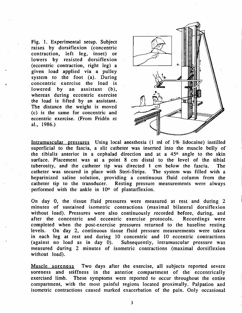
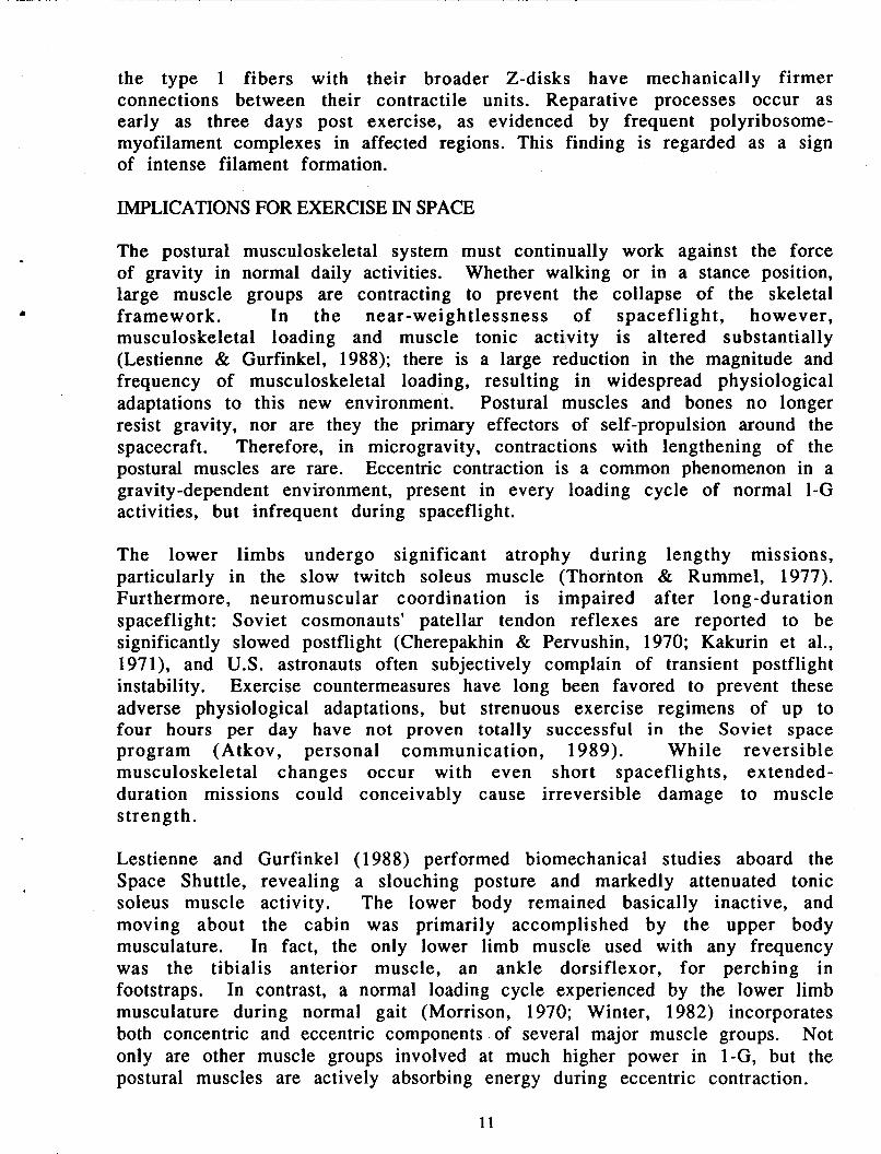
Exercise D rotocol The anterior compartments of the lower legs were exercised eccentrically and concentrically through the use of an exercise apparatus (Brutus 1000 System, Excel) (Fig. 1). The subjects were positioned supine on a padded physical therapy table, and a padded strap was taped in place on the left foot 10 cm from the medial malleolus. The subject raised a known weight (15% of the subject's maximal dorsiflexion torque) with ankle dorsiflexion. At the point of full dorsiflexion, the weight was removed, allowing the patient to return his foot to a full plantarflexed position without lowering the weight. This maneuver was then repeated at a rate of once every 3 seconds for a total of 20 minutes (i.e., 400 contractions) and constituted the concentric exercise part of the protocol. The eccentric exercise protocol was initiated with the right foot being positioned in full dorsiflexion. The same weight was applied and the foot was then lowered to a full plantarflexed position. This maneuver was also repeated at the same frequency and duration (including 1-minute rest periods every 4 minutes) as before.
2
c
Fig. 1. Experimental setup. Subject raises by dorsiflexion (concentric contraction, left leg, inset) or lowers by resisted dorsiflexion (eccentric contraction, right leg) a given load applied via a pulley system to the foot (a). During concentric exercise the load is lowered by an assistant (b), whereas during eccentric exercise the load is lifted by an assistant. The distance the weight is moved (c) is the same for concentric and eccentric exercise. (From Fr idh et al., 1986.)
Intramuscular Dressu res Using local anesthesia (1 ml of 1% lidocaine) instilled superficial to the fascia, a slit catheter was inserted into the muscle belly of the tibialis anterior in a cephalad direction and at a 450 angle to the skin surface. Placement was at a point 8 cm distal to the level of the tibial tuberosity, and the catheter tip was directed 1 cm below the fascia. The catheter was secured in place with Steri-Strips. The system was filled with a heparinized saline solution, providing a continuous fluid column from the catheter tip to the transducer. Resting pressure measurements were always performed with the ankle in 100 of plantarflexion.
On day 0, the tissue fluid pressures were measured at rest and during 2 minutes of sustained isometric contractions (maximal bilateral dorsiflexion without load). Pressures were also continuously recorded before, during, and after the concentric and eccentric exercise protocols. Recordings were completed when the post-exercise pressures returned to the baseline resting levels. On day 2, continuous tissue fluid pressure measurements were taken in each leg at rest and during 10 concentric and 10 eccentric contractions (against no load as in day 0). Subsequently, intramuscular pressure was measured during 2 minutes of isometric contractions (maximal dorsiflexion without load).
Muscle so reness Two days after the exercise, all subjects reported severe soreness and stiffness in the anterior compartment of the eccentrically exercised limb. These symptoms were reported to occur throughout the entire compartment, with the most painful regions located proximally. Palpation and isometric contractions caused marked exacerbation of the pain. Only occasional
5
stiffness was perceived from the concentrically exercised muscles. Our subjects reported that the preponderance of their symptoms was in the right (eccentrically exercised) leg 48 hours after exercise. This correlated with an increase in resting pressure and the peak tissue fluid pressure during isometric contraction 48 hours later.
Muscle pain occurring 24-48 hours after unaccustomed exercise is a phenomenon familiar to most individuals. Eccentric contractions are particularly associated with delayed muscle soreness and stiffness (Armstrong, 1984; Asmussen, 1952; FridCn et al., 1983b; Newham et al., 1983b). Various distributions of symptoms have been reported. The musculotendinous junction appears to be the primary site of palpable tenderness, whereas the muscle belly is relatively spared (Edwards et al., 1981; Newham et al., 1983b). We found this to be true with our subjects, where the muscle origins and insertions were the principal sites of point tenderness. However, delayed muscle soreness, a symptom that was easily aggravated when isometrically contracting the previously eccentrically exercised compartmen t, was reported to be distributed equally throughout the muscle.
Muscle f ibers are oriented most obl iquely just proximal to the musculotendinous junction, diminishing their ability to withstand the high levels of tension reportedly produced within a muscle undergoing eccentric contractions. This may help explain the higher degree of muscular discomfort seen after performing eccentric exercise (Newham et al., 1983b). Also, muscle pain receptors are most concentrated in the regions of tendinous and connective tissue (Kumazawa account for our finding that maximal point tenderness.
& Mizumura, 1977; Stacey, 1969), which may the musculotendinous junctions are the site of
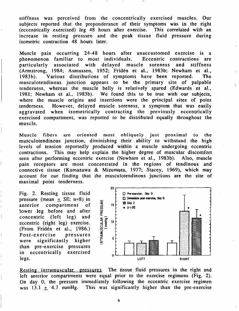
Fig. 2. Resting tissue fluid pressure (mean 2 SE; n=8) in anterior compartment of lower leg before and after concentric (left leg) and eccentric (right leg) exercise. (From FridCn et al., 1986.) Pos t -exerc ise pressures were significantly higher than pre-exercise pressures in eccentrically exercised legs.
0 Presrercise, Do) 0 13 Imncdkte post-exercise, Do) 0 m DOY 2
pcD5
W
v, v,
a 3 15
LEFT RIGHT
Resting intramuscular pressures The tissue fluid pressures in the right and left anterior compartments were equal prior to the exercise regimens (Fig. 2). On day 0, the pressure immediately following the eccentric exercise regimen was 13.1 2 4.7 mmHg. This was significantly higher than the pre-exercise
4
i
250
W ct 3 200 v) v) W
Fig. 3. Peak intramuscular pressure (mean 2 SE; n=8)
resting pressure of 3.6 0.7 mmHg (p<0.05). The resting pressure before exercise in the eccentrically exercised leg was significantly higher on day 2 compared to day 0 (10.5 2.5 vs. 3.6 0.7 mmHg, P<0.05). This was not the case for the concentrically exercised muscles of the left anterior compartmen t, where no significant elevation in tissue fluid pressure occurred immediately or 48 hours after exercise.
- 0 Day 0
I 0 DOY 2 - it* D < 0 0 1
p<m -
The pre-exercise pressures are well within the normal range of 0-10 mmHg reported previously (Mubarak, 198 1). The elevated compartment pressures that occurred 48 hours after the eccentric exercise regimen (10.5) are still below what is considered a borderline resting pressure (15 mmHg) seen in patients with chronic compartment syndrome (Mubarak, 198 1). The significant elevation of the immediate post-exercise pressures following the eccentric exercise regimen (13.1 mmHg) is far lower than the post-exercise pressures (30 mmHg) seen in patients with the reproducible symptoms of chronic compartment syndrome. Apparently compartment compliance is decreased after eccentric exercise, but not to the extent hypothesized in chronic compartment syndrome. No subject experienced "ischemic" pain after exercise, as the pressures generated are not sufficient to compromise the microvascular circulation.
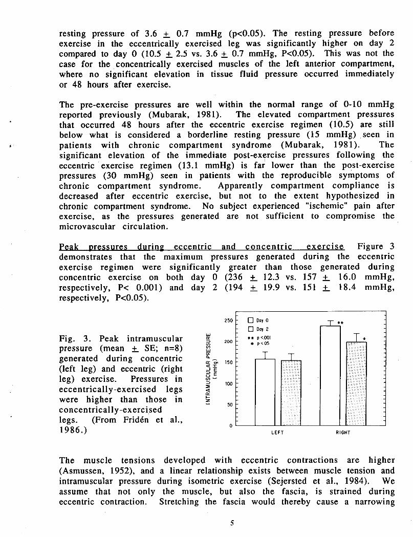
Peak pressu res during ecce ntric a nd co ncentric exercise Figure 3 demonstrates that the maximum pressures generated during the eccentric exercise regimen were significantly greater than those generated during concentric exercise on both day 0 (236 & 12.3 vs. 157 & 16.0 mmHg, respectively, P< 0.001) and day 2 (194 19.9 vs. 151 +. 18.4 mmHg, respectively, Pe0.05).
legs. (From FridCn et al., 1986.)
0 I L E F T
T I
RIGHT
The muscle tensions developed with eccentric contractions are higher (Asmussen, 1952), and a linear relationship exists between muscle tension and intramuscular pressure during isometric exercise (Sejersted et al., 1984). We assume that not only the muscle, but also the fascia, is strained during eccentric contraction. Stretching the fascia would thereby cause a narrowing
5
of the cylindrical anterior compartment. This can further reduce compliance, raise the intramuscular pressure, and explain the greater tension per load during eccentric contractions. Intramuscular pressure elevations have been reported to be associated with extremes of passive ankle plantarflexion and dorsiflexion with a fully extended knee, causing significant stretch of the anterior compartment (Gershuni et al., 1984).
250 - 0 Re-exercise. Day 0 - Post-exercise, Day 0 T
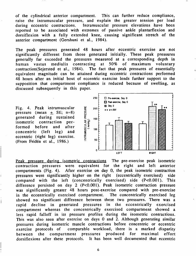
Fig. 4. Peak intramuscular pressure (mean & SE; n=8) generated during sustained isometric contraction per- formed before and after concentric (left leg) and eccentric (right leg) exercise. (From Fridtn et al., 1986.)
150
100
50
I a- * LEFT RIGHT
Peak messure - during isometric contractions The pre-exercise peak isometric contraction pressures were equivalent for the right and left anterior compartments (Fig. 4). After exercise on day 0, the peak isometric contraction pressures were significantly higher on the right (eccentrically exercised) side compared with the left (concentrically exercised) side (P<O.OOl). This difference persisted on day 2 (P<O.OOl). Peak isometric contraction pressure was significantly greater 48 hours post-exercise compared with pre-exercise in the eccentrically exercised compartment. The concentrically exercised leg showed no significant difference between these two pressures. There was a rapid decline in generated pressures in the eccentrically exercised compartment whereas the concentrically exercised compartment showed a less rapid falloff in its pressure profiles during the isometric contractions. This was also seen after exercise on days 0 and 2. Although generating similar pressures during isometric muscle contractions before concentric or eccentric exercise protocols of comparable workload, there is a marked disparity between the compartment pressures produced for maximal effort dorsiflexions after these protocols. It has been well documented that eccentric
1
6
exercise produces much more muscle fiber damage than concentric exercise of equal power (Fridkn et al., 1983b). We found that 2 days after eccentric exercise, the muscles produce a significantly higher compartment pressure during an isometric contraction despite the well-documented degradation of these muscles and their impaired ability to generate forces (Fridkn et al., 1986). Therefore, we believe that the increased tissue fluid pressure is due to the swelling of the compartment and a decrease of its compliance.
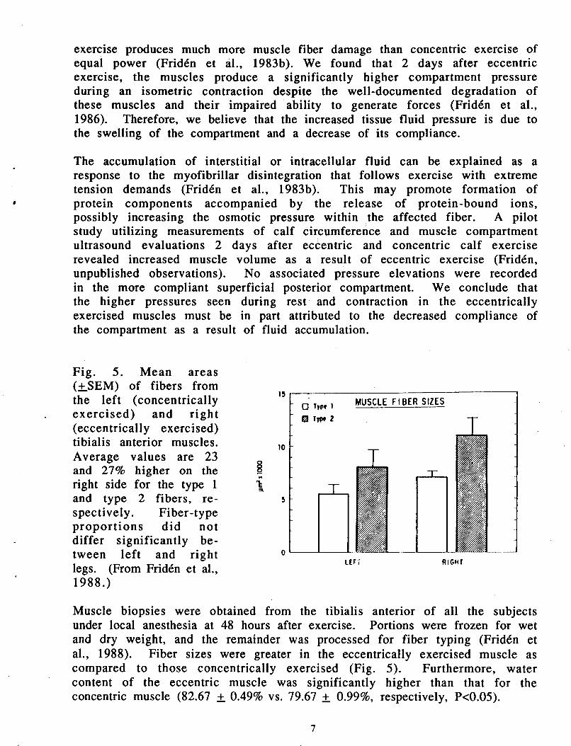
15 r MUSCLE F I B E R SIZES - 0 f l P I 1
the left (concentrically
(eccentrically exercised) tibialis anterior muscles. Average values are 23 and 27% higher on the right side for the type 1 and type 2 fibers, re- spectively. Fi ber-type proportions did not differ significantly be-
, exercised) and right a TlW 2
0- -
The accumulation of interstitial or intracellular fluid can be explained as a response to the myofibrillar disintegration that follows exercise with extreme tension demands (Fridkn et al., 1983b). This may promote formation of protein components accompanied by the release of protein-bound ions, possibly increasing the osmotic pressure within the affected fiber. A pilot study utilizing measurements of calf circumference and muscle compartment ultrasound evaluations 2 days after eccentric and concentric calf exercise revealed increased muscle volume as a result of eccentric exercise (Fridkn, unpublished observations). No associated pressure elevations were recorded in the more compliant superficial posterior compartment. We conclude that the higher pressures seen during rest and contraction in the eccentrically exercised muscles must be in part attributed to the decreased compliance of the compartment as a result of fluid accumulation.
Muscle biopsies were obtained from the tibialis anterior of all the subjects under local anesthesia at 48 hours after exercise. Portions were frozen for wet and dry weight, and the remainder was processed for fiber typing (FridCn et al., 1988). Fiber sizes were greater in the eccentrically exercised muscle as compared to those concentrically exercised (Fig. 5). Furthermore, water content of the eccentric muscle was significantly higher than that for the concentric muscle (82.67 2 0.49% vs. 79.67 2 0.99%, respectively, Pc0.05).
7
I MUSCLE ENZYME ACTIVITIES WITH ECCENTRIC EXERCISE
Before the onset of the three exercise protocols and 48 hours later, 5 ml blood was drawn via antecubital venipuncture for assaying muscle enzymes. Serum enzyme analyses were completed within 2 hours after blood sampling. Serum glutamic oxaloacetic transaminase (SGOT) and lactic dehydrogenase (LDH) were measured on a Technicon SMAC System continuous flow analyzer. Serum creatine kinase (CK) was measured on a Cobas Bio centrifugal analyzer. We selected 48 hours post-exercise for blood sampling in an attempt to determine whether the serum levels of muscle enzymes may reliably reflect soreness .
250
1 ' 200 c 2
F 1 5 0 - > 0
w a
f 100- N z w
50
0.
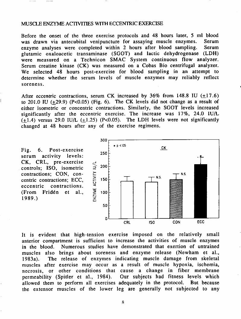
After eccentric contractions, serum CK increased by 36% from 148.8 IU (k17.6) to 201.0 IU (229.9) (Pc0.05) (Fig. 6) . The CK levels did not change as a result of either isometric or concentric contractions. Similarly, the SGOT levels increased significantly after the eccentric exercise. The increase was 17%. 24.0 IU/L (- +1.4) versus 29.0 IU/L (k1.25) (P<0.05). The LDH levels were not significantly
L
-
1
-
changed at 48 hours after
Fig. 6. Post-exercise serum activity levels: CK, CRL, pre-exercise controls; ISO, isometric contractions; CON, con- centric contractions; ECC, eccentric contractions. (From FridCn et al., 1989.)
any of the exercise regimens.
300 r
CRL
'i
N.S.
IS0 CON ECC
It is evident that high-tension exercise imposed on the relatively small anterior compartment is sufficient to increase the activities of muscle enzymes in the blood. Numerous studies have demonstrated that exertion of untrained muscles also brings about soreness and enzyme release (Newham et al., 1983a). The release of enzymes indicating muscle damage from skeletal muscles after exercise may occur as a result of muscle hypoxia, ischemia, necrosis, or other conditions that cause a change in fiber membrane
allowed them to perform all exercises adequately in the protocol. But because the extensor muscles of the lower leg are generally not subjected to any
I permeability (Spitler et al., 1984). Our subjects had fitness levels which
8
+
significant lengthening contractions during "normal" exercise, it is not surprising that a sensation of soreness developed.
Serum CK has been reported to pass its peak 8-24 hours post-exercise (Tiidus & Ianuzzo, 1983), yet at 48 hours, it was increased most (as a percentage of the activity) of all the enzymes assayed in this study. These elevated levels probably indicate fiber injury (Armstrong et al., 1983). Previous studies reporting CK elevations have primarily focused on the intensity or duration of the exercise rather than the mode of contraction. Armstrong & co-workers (1 983) reported increased CK activity after eccentric-biased treadmill exercise. Those CK increments, however, were caused by fiber damage occurring in several muscles in the four legs of a rat, whereas our results concentrate on significant changes caused by eccentric exercise of a single compartment. Thus, we conclude that the mechanical stress on the ankle dorsiflexors during the eccentric exercise regimen exceeded their normal daily stress loads.
SGOT elevations have been reported in association with high-intensity physical exercise among well-trained athletes (Metivier et al., 1980), although other studies have failed to demonstrate any relationship between dynamic exercise and SGOT levels (Tiidus & Ianuzzo, 1983). Here we present a clear correlation to the eccentric mode of exercise. It is likely that this represents the delayed effect of subcellular injuries to the contractile material subjected to unaccustomed tension demands and/or possible disruption of sarcolemma. Both these mechanisms can be involved in enzyme efflux into the blood (Armstrong et al., 1983). An increase in SGOT has also been attributed to its increased synthesis during times of hypoxia (Highman & Altland, 1963).
No significant elevations of LDH were found 48 hours after either of the regimens. This is in accordance with the findings of Schwane & co-workers (1983). The specific time course of LDH serum levels after muscle injury (Le., peak level occurring 8-16 hours post-exercise) may be a plausible explanation for the absence of increased LDH activity, at least following the high-tension eccentric protocol.
We assume that the low compliance of the lower leg anterior compartment (Hargens et al., 1978) makes the muscles more susceptible to strain-induced damage, swelling, and pressure elevation, which further increases the likelihood of structural injury. Based on these findings, we conclude that post- exercise serum levels of muscle enzymes at the time of maximal soreness are qualitatively reliable markers of muscle damage caused by high muscle tension. Whether the serum levels of muscle enzymes quantitatively reflect the degree of structural injury is presently unknown.
MUSCLE MORPHOLOGY WITH ECCENTRIC EXERCISE Light microscopy The first light microscopic evidence of post-eccentric exercise changes was obtained in toluidine blue stained longitudinal sections
9
(FridCn et al., 1981). These sections displayed a distorted sarcomeric band I pattern. The degenerative changes were focal (involving a few sarcomeres and
myofibrils) and were extensively distributed.
Using immunofluorescence microscopy, disorganized intermediate filament architecture was revealed in sore muscle following eccentric exercise (FridCn et al., 1984). The specimens displayed frequent transverse fluorescent striations between 2-disks, not observed in normal muscle samples. These changes may reflect mechanical disturbance of the intermediate filament system caused by the high, damaging tensions developed during eccentric contraction or distension of the cytoskeleton caused by edema. An increased intracellular pressure due to the release of osmotic components caused by the disruption of Z-disks may distribute forces unevenly on the cytoskeleton attachments. A second possible explanation for the cytoskeletal abnormalities is that degeneration is a secondary response to the extensive myofibrillar lesions.
Increased lysosomal enzyme activity has been demonstrated in several animal studies in relation to strenuous physical exercise. Microscopically, this activity is indirectly detected by lipofuscin granules which are regarded as the indigestible residue of protein degradation. Increased density of lipofuscin generally relates to ischemic fiber injuries, but also occurs after a single bout of eccentric exercise (Fr idh et al., 1984). This is interpreted as a sign of acutely increased protein degradation following exercise with tensions exceeding a certain critical level.
Electron microscoDv At the ultrastructural level, the most prominent finding following eccentric exercise was disorganization of the contractile material, particularly the myofibrillar Z-disks. The degree of alteration varied considerably from minor streaming of the 2-disks to total disruption of many sarcomeres. While in some cases only one single 2-disk of one myofibril was affected, there was evidence for involvement of several sarcomeres and myofibrils in others. The myofilamentous material in sarcomeres adjacent to the affected Z-disks was either supercontracted or disorganized and out of
pattern, even in areas where neither streaming nor broadening was seen. Loss of thick myofilaments was apparent. Mitochondria were seldom seen in or close to the abnormal area. Transverse sections through A-bands confirmed the disturbance arrangement of filaments. A correlation between the degree of structural deviation and the time from eccentric exercise to the point of biopsy has been described (Fridh et al., 1983b). It is of interest to note that the ultrastructurally defined type 2 fibers (fast-twitch) seem to be more susceptible to damage than the type 1 fibers; a 3: l damage ratio was seen three days post-exercise. The high tension demands during eccentric exercise may preferentially recruit type 2 fibers. On the other hand, type 2 fibers are designed primarily to contribute to high tension development, so this interpretation may be unlikely. A more probable explanation is the fact that
I register. The 2-disks showed frequent gaps in their normally regular lattice
I
10
the type 1 fibers with their broader Z-disks have mechanically firmer connections between their contractile units. Reparative processes occur as early as three days post exercise, as evidenced by frequent polyribosome- myofilament complexes in affected regions. This finding is regarded as a sign of intense filament formation.
IMPLICATIONS FOR EXERCISE IN SPACE
The postural musculoskeletal system must continually work against the force of gravity in normal daily activities. Whether walking or in a stance position, large muscle groups are contracting to prevent the collapse of the skeletal framework. In the near-weightlessness of spaceflight, however, musculoskeletal loading and muscle tonic activity is altered substantially (Lestienne & Gurfinkel, 1988); there is a large reduction in the magnitude and frequency of musculoskeletal loading, resulting in widespread physiological adaptations to this new environment. Postural muscles and bones no longer resist gravity, nor are they the primary effectors of self-propulsion around the spacecraft. Therefore, in microgravity, contractions with lengthening of the postural muscles are rare. Eccentric contraction is a common phenomenon in a gravity-dependent environment, present in every loading cycle of normal 1-G activities, but infrequent during spaceflight.
The lower limbs undergo significant atrophy during lengthy missions, particularly in the slow twitch soleus muscle (Thornton & Rummel, 1977). Furthermore, neuromuscular coordination is impaired after long-duration spaceflight: Soviet cosmonauts' patellar tendon reflexes are reported to be significantly slowed postflight (Cherepakhin & Pervushin, 1970; Kakurin et al., 1971), and U.S. astronauts often subjectively complain of transient postflight instability. Exercise countermeasures have long been favored to prevent these adverse physiological adaptations, but strenuous exercise regimens of up to four hours per day have not proven totally successful in the Soviet space program (Atkov, personal communication, 1989). While reversible musculoskeletal changes occur with even short spaceflights, extended- duration missions could conceivably cause irreversible damage to muscle strength.
Lestienne and Gurfinkel (1 988) performed biomechanical studies aboard the Space Shuttle, revealing a slouching posture and markedly attenuated tonic soleus muscle activity. The lower body remained basically inactive, and moving about the cabin was primarily accomplished by the upper body musculature. In fact, the only lower limb muscle used with any frequency was the tibialis anterior muscle, an ankle dorsiflexor, for perching in footstraps. In contrast, a normal loading cycle experienced by the lower limb musculature during normal gait (Morrison, 1970; Winter, 1982) incorporates both concentric and eccentric components of several major muscle groups. Not only are other muscle groups involved at much higher power in 1-G, but the postural muscles are actively absorbing energy during eccentric contraction.
11
Eccentric muscular contraction is clearly an attractive spaceflight countermeasure for several reasons. Owing to the lack of gravity and normal loading of the postural musculature in space, exercise regimens that incorporate eccentric contraction will better simulate and maintain 1 -G patterns of motor neuron firing during spaceflight. High-intensity eccentric resistance exercise is extremely effective in maintaining or building muscle mass (Fridkn et al., 1983a). Added benefits include improved reflex times (Tipton & Karpovich, 1966; Francis & Tipton, 1969) and +Gz tolerance (Jacobs et al., 1987), which is important during reentry. While heavy resistance exercise equipment is not currently available for spaceflight, several investigators advocate its use inflight (Convertino, 1987; Parazynski et al., 1989; Whalen et al., 1988).
With this in mind, a self-contained, variable-resistance exercise device has been designed that could exercise the astronaut through the full range of motion of a high stepping gait with significantly higher force levels than experienced during normal 1-G gait (Parazynski et al., 1989). Other lower and upper extremity exercises built within the system should allow maintenance of musculoskeletal integrity during long-duration spaceflight.
Abbott, B.C. & Bigland, B. (1953): The effects of force and speed changes on the rate of oxygen consumption. J. Physiol. Lond. 120, 319-325.
Abbott, B.C., Aubert, X.M. & Hill, A.V. (1950): The absorption of work when a muscle is stretched. J. Physiol. 1 1 1 , 41 -42.
Abbott, B.C., Bigland, B. & Ritchie, J.M. (1952): The physiological cost of negative work. J. Physiol. Lond. 117, 380-390.
Armstrong, R.B. (1984): Mechanisms of exercise-induced delayed onset muscle soreness: a brief review. Med. Sci. Sports Exercise. 16, 529-538.
Armstrong, R.B., Ogilvie, R.W. & Schwane, J.A. (1983): Eccentric exercise- induced injury to rat skeletal muscle. J. Appl. Physiol. 54, 80-93.
Asmussen, E. (1952): Positive and negative muscular work. Acta Physiol. Scand. 28, 364-382.
Cherepakhin, M.A. & Pervushin, V.I. (1970): Space flight effect on neuromuscular system of cosmonauts.
Convertino, V.A. (1987): Potential benefits of maximal exercise just prior to return from weightlessness. Aviat. Space Environ. Med. 58 , 568-572.
Davies, C.T.M. & Barnes, C. (1972): Negative (eccentric) work. I. Effects of repeated exercise. Ergonomics 15, 3-14.
Edwards, R.H.T., Mills, K.R. & Newham, D.J. (1981): Measurement of severity
Space Biol. Med. 4(6), 64-69.
and distribution of experimental muscle tenderness. J.
Francis, P.R. & Tipton, C.M. (1969): Influence of training on 317, 1P- 2P.
time. Med Sci. Sports 1 , 91-94.
Physiol. Lond.
quadricep reflex
12
r
Fridtn, J., Sjostrom M. & Ekbolm, B. (1981): A morphological study of delayed muscle soreness. Experentia 37, 506-507.
FridCn, J., Seger, J., Sjostrom, M. & Ekbolm, B. (1983a): Adaptive response in human skeletal muscle subjected to prolonged eccentric training. Sports Med. 4, 177-183.
FridCn, J., Sjostrom, M. & Ekbolm, B. (1983b): Myofibrillar damage following intense eccentric exercise in man. Int. J . Sports Med. 4, 170-176.
FridCn, J., Kjorell, U.& Thornell, L.E. (1984): Delayed muscle soreness and cytoskeletal alterations: An immunocytological study in man. Int. J . Sports Med. 5,1518.
FridCn, J., Sfakianos, P.N. & Hargens, A.R. (1986): Muscle soreness and intramuscular fluid pressure: comparison between eccentric and concentric load. J . Appl. Physiol. 61, 2175-2179.
muscle swelling after repetitive eccentric contractions.
Int. J .
FridCn, J., Sfakianos, P.N., Hargens, A.R. & Akeson, W.H. (1988): Residual J . Orthop. Res. 6,
493-498. FridCn, J., Sfakianos, P.N. & Hargens, A.R. (1989): Blood indices of muscle
injury associated with eccentric muscle contractions. J . Orthop. Res. 7, 142-145.
Gershuni, D.H., Yaru, N.C., Hargens, A.R., Lieber, R.L., O'Hara, R.C. & Akeson, W.H. (1984): Ankle and knee position as a factor modifying intracompartmental pressure in the human leg. J . Bone Joint Surg. 66A,
Hargens, A.R., Akeson, W.H., Mubarak, S.J., Owen, C.A., Evans, K.L., Garetto, L.P., 14 15- 1420.
Gonsalves, M.R. & Schmidt, D.A. (1978): Fluid balance within the canine arterolateral compartment and its relationship to compartment syndromes. J . Bone Joint Surg. 60A, 499-505.
compartment syndromes. In Compartment Syndromes and Volkmann's Contracture, ed. by S.J. Mubarak & A.R. Hargens, pp.106-122 Philadelphia, PA: W.B. Saunders.
Highman B. & Altland, P.D. (1963): Effects of exercise and training on serum enzyme and tissue changes in rats. Am. J . Physiol. 205, 162-166.
Jacobs, I., Bell, D.G., Pope, J. & Lee, W. (1987): Effects of hydraulic resistance circuit training on physical fitness components of potential relevance to +Gz tolerance. Aviat. Space Environ. Med. 5 8 , 754-760.
Kakurin, L.I., Cherepakhin, M.A. & Pervushin, V.I. (1971): Effect of spaceflight factors on human muscle tone. Space Biol. Med. 5(2), 91-99.
Kumazawa, T. & Mizumura, K. (1977): Thin-fibre receptors responding to mechanical, chemical, and thermal stimulation in the skeletal muscle of the dog. J . Physiol. Lond. 273, 179-194.
dual process underlying adaptation to an unusual environment. in NeuroSciences. 11, 359-363.
Metivier, G., Poortmans, J., Vanroux, R. & Gauthier, R. (1980): Serum glutamic oxaloacetic acid transaminase changes during exercise of various intensities in trained athletes. J . Sports Med. Phys. Fitness 20, 152-157.
Hargens, A.R. & Mubarak, S.J. (1981): Laboratory diagnosis of acute
Lestienne, F.G. & Gurfinkel, V.S. (1988): Postural control in weightlessness: a Trends
13
Morrison, J.B. (1970): The mechanics of muscle function in locomotion. J.
Mubarak, S.J. (198 1): Exertional compartment syndromes. In Compartment Biomech. 3, 431-451.
Syndromes and Volkmann's Contracture, ed. by S.J. Mubarak & A.R. Hargens, pp. 209-226. Philadelphia, PA: W.B. Saunders.
Newham, D.J., Jones, D.A.& Edwards, R.H.T. (1983a): Large delayed plasma creatine kinase changes after stepping exercise. 385.
fatigue after concentric and eccentric muscle contractions. Lond. 64, 55-62.
( 1 989): Development of an exercise device to prevent musculoskeletal deconditioning during human spaceflight.
Schwane, J.A., Johnson, S.R., Vandenakker, C.B.& Armstrong, R.B. (1983) Delayed-onset muscular soreness and plasma CPK and LDH activities after downhill running. Med. Sci. Sports Exercise 15, 51-56.
Hermansen, L. ( 1 984): Intramuscular fluid pressure during isometric contraction of human skeletal muscle. J. Appl. Physiol. 56, 287-295.
Haptoglobin and serum enzymatic response to maximal exercise in relation to physical fitness. Med Sci. Sports Exercise 16, 366-370.
Stacey, M.J. (1969): Free nerve endings in skeletal muscle of the cat. J. Anat.
Thornton, W. & Rummel, J. ( 1 977): Muscle deconditioning and its prevention
Tiidus, P.M. & Ianuzzo, L.D. (1983): Effects of intensity and duration of
Muscle & Nerve 6, 380-
Newham, D.J., Mills, K.R., Quigley, B.M. & Edwards, R.H.T. (1983b): Pain and Clin. Sci.
Parazynski, S.E., Schwandt, D.F., Whalen, R.T., Aratow, M. & Hargens, A.R.
Clin. Res. 37, 220A.
Sejersted, O.M., Hargens, A.R., Kardel, K.R., Blom, P., Jensen, 0. &
Spitler, D.L., Alexander, W.C., Hoffler, G.W., Doerr, D.F. & Buchanan, P. (1984):
105, 23 1-254.
in space flight. Biomedical Results from Skylab, NASA SP-377, 191-197.
muscular exercise on delayed soreness and serum enzyme activities. Med. Sci. Sports Exercise 15, 461-465.
Phys io l . 21, 15-18.
the regulation of bone density. J. Biomech. 21, 825-837.
Biomech. 16:91-97.
Tipton, C.M. & Karpovich, P.V. (1966): Exercise and the patellar reflex. J. Appl.
Whalen, R.T., Carter, D.R. & Steele, C.R. (1988): Influence of physical activity on
Winter, D.A. (1982): Moments of force and mechanical power in jogging. J.
Acknowledgement: This work was supported by NASA, the Veterans Administration, USPHS/NIH grant AM-25501, the Swedish Medical Research Council, the Tore Nilsson Foundation, and the Swedish Sports Research Council.
14
NASA 1. Report No.
NASA TM- 102227
Report Documentation Page 2. Government Accession No.
21. No. of Pages
16
7. Author(s)
Alan R. Hargens, Scott Parazynski, Michael Aratow, and Jan FridCn* *UmeA Univ. Hospital; UmeA, Sweden
22. Price
A02
~ ~~~~
9. Performing Organization Name and Address
Ames Research Center Moffett Field, CA 94035
19. Security Classif. (of this report)
Unclassified
12. Sponsoring Agency Name and Address
20. Security Classif. (of thi
Unclassified
National Aeronautics and Space Administration Washington, DC 20546-0001
3. Recipient's Catalog No.
5. Report Date
August 1989 6. Performing Organization Code
8. Performing Organization Report No.
A-89224
10. Work Unit No.
1 1. Contract or Grant No.
13. Type of Report and Period Covered
Technical Memorandum
14. Sponsoring Agency Code
15. Supplementary Notes
Point of Contact: Alan R. Hargens, Ames Research Center, MS 239-17, Moffett Field, CA 94035 (415) 694-5746 or FI'S 464-5746
(Presented at Int. Soc. for Myochemistry mtg.)
16. Abstract
In this paper, we review recent investigations of fluid pressure, morphology, and enzyme activities of skeletal muscle exercised eccentrically or concentrically in normal human subjects. Intramuscular pressures were measured before, during, and after submaximal exercise and correlated with subjective muscle soreness, fiber size, water content, and blood indices of muscle enzymes. High-intensity eccentric exercise is characterized by post-exercise pain, elevated intramuscular pressures, and swel- ling of both type l and 2 fibers as compared to concentric exercise. Thus, long periods of unaccus- tomed, high-level eccentric contraction may cause muscle injury, fiber swelling, fluid accumulation, elevated intramuscular pressure, and delayed muscle soreness. Training regimens of progressively increasing eccentric exercise, however, cause less soreness and are extremely efficacious in increasing muscle mass and strength. It is proposed that on Earth, postural muscles are uniquely adapted to low levels of prolonged eccentric contraction that are absent during weightlessness. $,The? almost complete absence of eccentric exercise in space may be an important contributor to muscle atrophy and there- fore equipment should be designed to integrate eccentric contractions into exercise protocols for long- term spaceflight.
Weightlessness, Microgravity, Exercise space, Muscle, Eccentric
17. Key Words (Suggested by Author(s)) 18. Distribution Statement
Unclassified-Unlimited
Subject Category - 52
I
ASA FORM 1626 OCTM For sale by the National Technical Information Service, Springfield. Virginia 22161


![Muscle Physiology - Home Pagemuscle.ucsd.edu/More_HTML/papers/pdf/Lieber_PT_1993.pdf · 2006-08-19 · muscle mechanics. implications for rehabilitation. Phys Then 1993, 73844—856.]](https://img.dokumen.tips/doc/110x75/5f3e1a028466116ebe1bab52/muscle-physiology-home-2006-08-19-muscle-mechanics-implications-for-rehabilitation.jpg)


![Brachialis Muscle Rupture and Hematoma brachialis muscle is also responsible for main-taining the stability of the elbow throughout concentric and eccentric contraction [9]. Kulig](https://img.dokumen.tips/doc/110x75/5afda6037f8b9a814d8dcb54/brachialis-muscle-rupture-and-hematoma-brachialis-muscle-is-also-responsible-for.jpg)