Embed Size (px)
Citation preview

r Human Brain Mapping 0000:000–000 (2011) r
Multivariate Patterns of Brain–CognitionAssociations Relating to Vulnerability and ClinicalOutcome in the At-Risk Mental States for Psychosis
Nikolaos Koutsouleris,1* Christian Gaser,2 Katja Patschurek-Kliche,1
Johanna Scheuerecker,1 Ronald Bottlender,1 Petra Decker,1
Gisela Schmitt,1 Maximilian Reiser,3Hans-Jurgen Moller,1
and Eva M. Meisenzahl1
1Department of Psychiatry and Psychotherapy, Ludwig-Maximilian-University, Munich, Germany2Department of Psychiatry, Friedrich-Schiller-University, Jena, Germany
3Department of Radiology, Ludwig-Maximilian-University, Munich, Germany
r r
Abstract: Background: Neuropsychological deficits are a core feature of established psychosis andhave been previously linked to fronto-temporo-limbic brain alterations. Both neurocognitive andneuroanatomical abnormalities characterize clinical at-risk mental states (ARMS) for psychosis. How-ever, structure–cognition relationships in the ARMS have not been directly explored using multivari-ate neuroimaging techniques. Methods: Voxel-based morphometry and partial least squares wereemployed to study system-level covariance patterns between whole-brain morphological data andprocessing speed, working memory, verbal learning/IQ, and executive functions in 40 ARMS sub-jects and 30 healthy controls (HC). The detected structure–cognition covariance patterns were testedfor significance and reliability using non-parametric permutation and bootstrap resampling. Results:We identified ARMS-specific covariance patterns that described a generalized association of neuro-cognitive measures with predominantly prefronto-temporo-limbic and subcortical structures as wellas the interconnecting white matter. In the conversion group, this generalized profile particularlyinvolved working memory and verbal IQ and was positively correlated with limbic, insular and sub-cortical volumes as well as negatively related to prefrontal, temporal, parietal, and occipital cortices.Conversely, the neurocognitive profiles in the HC group were confined to working memory, learningand IQ, which were diffusely associated with cortical and subcortical brain regions. Conclusions:These findings suggest that the ARMS and prodromal phase of psychosis are characterized by a con-vergent mapping from multi-domain neurocognitive measures to a set of prefronto-temporo-limbicand subcortical structures. Furthermore, a neuroanatomical separation between positive and negativebrain–cognition correlations may not only point to a biological process determining the clinical riskfor disease transition, but also to possible compensatory or dysmaturational neural processes. HumBrain Mapp 00:000–000, 2011. VC 2011 Wiley-Liss, Inc.
Additional Supporting Information may be found in the onlineversion of this article.
Contract grant sponsor: Ludwig-Maximilian-University.
*Correspondence to: Nikolaos Koutsouleris, Clinic of Psychiatryand Psychotherapy, Ludwig-Maximilian-University, Nussbaumstr.7, 80336 Munich, Germany.E-mail: [email protected]
Received for publication 12 November 2010; Revised 20 March2011; Accepted 12 April 2011
DOI: 10.1002/hbm.21342Published online in Wiley Online Library (wileyonlinelibrary.com).
VC 2011 Wiley-Liss, Inc.
Keywords: at-risk mental state for psychosis; brain–cognition correlations; voxel-based morphometry;multivariate analysis; partial least squares
r r
INTRODUCTION
From a very early stage, schizophrenia entails deficits inthe executive, mnemonic, and perceptual domains ofneurocognitive functioning [Frommann et al., 2010;Heinrichs and Zakzanis, 1998]. A direct link between thesedeficits and an underlying brain pathology has long beenposited based on the concurrent evidence of neurocogni-tive and neuroanatomical abnormalities. This hypothesiswas first supported by magnetic resonance imaging (MRI)studies [see Antonova et al., 2004; Crespo-Facorro et al.,2007a, for review] that mainly detected altered relation-ships between neuroanatomical and neuropsychologicalmeasures, e.g., attenuated correlations between prefrontalvolumes and processing speed as well as reversed correla-tions between verbal memory performance and hippocam-pal volume in schizophrenic patients vs. healthy controls[Sanfilipo et al., 2002]. In summary, these investigationspointed to a distributed neural circuitry subserving dis-ease-specific brain–cognition associations.
Abbreviations
ARMS at-risk mental state for psychosisARMS-E/-L ‘‘Early’’ ARMS/‘‘Late’’ ARMSARMS-NT/-T non-transitions/transitions to psychosisDS digit span testDSM-IV diagnostic and statistical manual of mental
disorders, 4th editionDST digit-symbol testGM(V) gray matter (volume)HC healthy controlsICD-10 international classification of diseases, 10th
editionLNS letter-number span testLV(s) latent variable(s)MADRS Montgomery-Asberg depression rating scaleMPRAGE magnetization prepared rapid acquisit ion
gradient echoMRI magnetic resonance imagingMWT-B Mehrfach-Wortschatz test BPANSS positive and negative symptom scalePLS partial least squaresRAVLT-IR rey auditory verbal learning test—immediate recallRAVLT-DR rey auditory verbal learning test—delayed recallTMT-A trail-making test, part ATMT-B trail-making test, part BSOPT self-ordered pointing taskSPM statistical parametric mappingVBM voxel-based morphometryWM(V) white matter (volume)
Overlapping but milder cognitive abnormalities havealso been found in subjects at genetic risk for schizophre-nia, such as the patients’ offspring and unaffected relatives[Erlenmeyer-Kimling et al., 2000; Faraone et al., 1999; Hanset al., 1999; Owens and Johnstone, 2006]. Recently, thesedata have been complemented by clinical high-risk studiesfollowing either the Melbourne ‘‘ultra-high risk’’ approach[Yung et al., 1998] or a combination of predictive basicsymptoms [Klosterkotter et al., 2001] and ultra-high riskcriteria [Frommann et al., 2010; Pukrop et al., 2006; Simonet al., 2006]. These studies showed that clinically definedat-risk mental states for psychosis (ARMS) are associatedwith deficits in processing speed [Brewer et al., 2005;Niendam et al., 2006; Simon et al., 2007], sustained atten-tion [Francey et al., 2005], verbal learning/memory [Lenczet al., 2006; Niendam et al., 2006; Pukrop et al., 2006;Simon et al., 2007] and executive functions [Hawkinset al., 2004; Pukrop et al., 2006; Simon et al., 2007]. Further-more, recent voxel-based morphometry (VBM) studiesrevealed distributed brain abnormalities in the ARMS,which predominantly covered prefrontal, opercular, lim-bic, and paralimbic structures [Borgwardt et al., 2007; Jobet al., 2003, 2005; Koutsouleris et al., 2009a,b; Meisenzahlet al., 2008b; Pantelis et al., 2003] similar to the establisheddisease [Honea et al., 2005; Koutsouleris et al., 2008; Mei-senzahl et al., 2008a]. These cross-sectional neurocognitiveand neuroanatomical alterations may particularly relate toan ultra-high risk state for the disease as defined by thepresence of subclinical psychotic symptoms [Borgwardtet al., 2007; Frommann et al., 2010; Koutsouleris et al.,2009b; Pukrop et al., 2007]. Moreover, longitudinal neuro-psychological and morphometric studies revealed inde-pendently from each other (1) a deterioration of cognitiveabilities, i.e., executive functioning [Wood et al., 2007], aswell as a (2) progressive reduction of prefrontal, temporal,and cerebellar volumes in subsequent converters to psy-chosis [Borgwardt et al., 2008; Job et al., 2005; Koutsouleriset al., 2010a; Pantelis et al., 2003; Sun et al., 2009]. Takentogether, these concurrent structural and neuropsychologi-cal findings point to an active biological process affectingboth the neuroanatomical and neurocognitive dimensionsas the disease unfolds during the transition from adoles-cence to adulthood [Pantelis et al., 2005].
In keeping with this hypothesis, Hurlemann et al. [2008]were the first to observe a direct link between reducedhippocampal volumes and verbal learning deficits inclinical ARMS subjects, which was most pronounced inthe ultra-high risk state. Furthermore, our recentVBM analysis detected correlations between cognitive set-shifting impairments and prefronto-callosal regions, as
r Koutsouleris et al. r
r 2 r

well as a volumetric network linking these regions withfurther prefrontal, cerebellar and parietal areas [Koutsoule-ris et al., 2010b]. However, regarding the multifaceted be-havioral and morphological alterations in the ARMS, theseunivariate approaches may have unveiled only a smallfraction of the risk-specific associations between neuroan-atomy and neurocognition. The greater portion of theseassociations may have been missed so far due the biologi-cal complexity of brain–behavior correlations, meaningthat (1) a single brain structure may be involved acrossmultiple cognitive functions, whereas (2) different sets ofbrain structures may contribute to a single cognitive pro-
cess. This multiplicity of overlapping mappings constitutesa high-dimensional analytical problem [Davatzikos, 2004]that can only be adequately resolved using multivariatetechniques, like Partial Least Squares (PLS), which are ca-pable of revealing the hidden structure underlying thecomplexity of brain–cognition associations [Gilboa et al.,2005; Kawasaki et al., 2007; McIntosh and Lobaugh, 2004;Menzies et al., 2007; Nestor et al., 2002; Tura et al., 2008].We used PLS to explore system-level covariance patternsbetween whole-brain structural imaging data and a com-prehensive neuropsychological test battery obtained froma previously described population of clinical ARMS and
TABLE I. Inclusion/exclusion criteria
ARMS-E: ARMS subjects without APS and/or BLIPS : : :
(1) : : : having one or more of the following basic symptoms appeared first at least 12 months prior tostudy inclusion and several times per week during the last 3 months.� Thought interferences� Thought perseveration� Thought pressure� Thought blockages� Disturbances of receptive language, either heard or read� Decreased ability to discriminate between ideas and perception, fantasy, and true memories� Unstable ideas of reference (subject-centrism)� Derealization� Visual perception disturbances� Acoustic perception disturbances
and/or
(2) : : : showing a reduction in the Global Assessment of Functioning Score (DSM IV) of at least 30points (within the past year) combined with at least one of the following trait markers:� First-degree relative with a lifetime-diagnosis of schizophrenia or a schizophrenia spectrumdisorder� Pre- or perinatal complications
ARMS-L: ARMS subjects with/without basic symptoms, with/without global functioning and trait
markers : : :
(1) : : : having at least one of the following Attenuated Psychotic Symptoms (APS) within the last threemonths, appearing several times per week for a period of at least 1 week:� Ideas of reference� Odd beliefs or magical thinking� Unusual perceptual experiences� Odd thinking and speech� Suspiciousness or paranoid ideation
and/or
(2) : : : having at least one of the following Brief Limited Intermittent Psychotic Symptoms (BLIPS),defined as the appearance of one of the following psychotic symptoms for less than 1 week (intervalbetween episodes at least 1 week), resolving spontaneously:� Hallucinations� Delusions� Formal thought disorder� Gross disorganized or catatonic behavior
Exclusion Criteria
� Disease transition as defined by Yung et al.� A past or present diagnosis of schizophrenia spectrum and bipolar disorders, as well as delirium,dementia, amnestic, or other cognitive disorders, mental retardation, and psychiatric disorders dueto a somatic factor, following the DSM-IV criteria� Alcohol or drug abuse within three months prior to examination, following the DSM-IV criteria� A past or present inflammatory, traumatic or epileptic diseases of the central nervous system� Any previous treatment with antipsychotics prior to neurocognitive assessment� Healthy controls: positive familial history of schizophrenic or affective psychoses in the first-degree relatives
r Multivariate Patterns of Brain–Cognition Associations r
r 3 r

healthy control subjects [Koutsouleris et al., 2010b]. Basedon the existing brain–cognition literature in schizophrenia[Antonova et al., 2004; Crespo-Facorro et al., 2007a], weexpected that cross-domain neurocognitive performance inthe ARMS would be linked to specific patterns of prefronto-temporo-limbic and subcortical regions not observed inhealthy controls and (1) that physiological brain-cognitionrelationships found in healthy controls would be attenuatedor absent in the ARMS. Furthermore, we hypothesized thatthese patterns would be particularly present in an ultra-highrisk state compared to a milder ARMS, which is primarilydefined by the presence of basic symptoms.
METHODS
Study Participants
Forty individuals in an ARMS for psychosis (Table III)and 30 healthy controls (HC) matched group-wise for age,gender, handedness, and premorbid verbal IQ wererecruited at the Early Detection and Intervention Center forMental Crises of the Clinic of Psychiatry and Psychotherapy,Ludwig-Maximilian-University, Germany, for MRI scan-ning and neuropsychological testing using an establishedoperationalized recruitment protocol [Table I; Frommannet al., 2008, 2010; Koutsouleris et al., 2009a,b]. This protocolwas based on a two-stage concept of the ARMS, distinguish-ing between an ‘‘early,’’ or non-psychotic ARMS (ARMS-E),with an increased risk for psychosis, and a ‘‘late,’’ or psy-chotic ARMS (ARMS-L), characterized by an imminent riskfor disease transition. Exclusion criteria (Table I) were care-fully assessed by evaluating the personal and familial his-tory using a semi-structured clinical interview and theStructured Clinical Interview for DSM-IV [American Psychi-
atric Association, 1994]. In particular, candidate individualswith a present or past abuse of drugs (e.g., cannabis, opiates,and amphetamines) and/or alcohol (according to DSM-IV)were excluded from the study. Recruited ARMS individualswere rated using the Global Assessment of FunctioningScale of the DSM-IV, the Positive and Negative SymptomScale (PANSS, Kay et al. [1987]) and the Montgomery-Asberg Depression Rating Scale (MADRS, Montgomery andAsberg [1979]).
ARMS subjects were regularly followed over 4 years todetect possible disease transitions. Subjects meeting thetransition criteria of Yung et al. [1998] were diagnosedwith a schizophrenia spectrum disorder using the ICD-10research criteria at transition and after one year. Follow-upinformation could be obtained from 27 subjects after anaverage interval of 3.7 (SD: 1.1) years, consisting of 11 con-verters (ARMS-T: n ¼ 8, schizophrenia, 3, schizoaffectivepsychosis), and 16 non-converters (ARMS-NT: n ¼ 14 nopsychiatric diagnosis, 2 major depression). Ten convertershad been initially assigned to the ARMS-L and 1 to theARMS-E subgroup. Out of the 13 subjects without follow-up, 6 could not be contacted or refused to participate,whereas 7 had not completed the follow-up. No antipsy-chotics were prescribed prior to MRI scanning and neuro-psychological testing. All subjects provided their writteninformed consent before study inclusion. The study wasapproved by the Local Research Ethics Committee of theLudwig-Maximilian-University.
Neuropsychological Testing
At the time of MRI scanning, nine standardized neuro-psychological tests (Table II) were administered to all sub-jects by trained master-level neurophysiologists (K.K., J.S.,
TABLE II. Neuropsychological test battery
Cognitive domain Variables
Premorbid verbal IQMehrfach-Wortschatztest B (MWT-B)(Lehrl, 2005) Raw score correct
Processing speedTrail-making test, part A (TMT-A)(Reitan, 1992) Time to completion [s]Digit symbol test (DST, [WAIS-III; Wechsler, 1997]) Raw score correct
Working memoryDigit span test (DS, [WAIS-III; Wechsler, 1997]) Raw score correctLetter number span test (LNS) [Gold et al., 1997] Raw score correctSubject-ordered pointing task (SOPT) [Petrides, 1995] Error score
Verbal learning and memoryRey auditory verbal learning test (RAVLT) [Lezak, 1995] Sum of raw score correct
after trials 1–5 (RAVLT-IR)Raw score correct after
delayed recall (RAVLT-DR)Executive functionsTrail-making test, part B (TMT-B) [Reitan, 1992] Time to completion [s]Verbal Fluency (letters) (VF) [Aschenbrenner et al., 2001] Sum of correct responses
Cognitive domains were defined according to Schultze-Lutter et al. (2007b).
r Koutsouleris et al. r
r 4 r
P.D.) to assess cross-domain cognitive functioning, includ-ing premorbid verbal IQ, processing speed, working mem-ory, verbal and visual memory, as well as executivefunctions [Schultze-Lutter et al., 2007b]. From these data,10 test variables were computed (Table II) and adjustedfor the effects of age and gender using partial correlations.The adjusted scores were z-transformed based on therespective HC data and entered analyses of variance thatassessed between-group differences in (1) HC vs. ARMS,(2) HC vs. ARMS-E vs. ARMS-L, and (3) HC vs. ARMS-NT vs. ARMS-T. Significant between-group effects wereexamined for pairwise group differences using post-hocBonferroni tests. Adjustment for multiple comparisons wasperformed using Holm’s sequential method [Holm, 1979].Significance was defined at P < 0.05.
MRI Data Acquisition and Preprocessing
MR images were obtained on a 1.5 T Magnetom Visionscanner (Siemens, Erlangen, Germany) using a T1-weighted 3D-MPRAGE sequence (TR, 11.6 ms; TE, 4.9 ms;field of view, 230 mm; matrix, 512 � 512; 126 contiguousaxial slices of 1.5 mm thickness; voxel size, 0.45 � 0.45 �1.5 mm3). All images were first carefully checked for MRIscanner artifacts and gross anatomical abnormalities bytrained clinical neuroradiologists and then processed usingthe VBM8 toolbox [Gaser, 2008] and Statistical ParametricMapping (SPM8, Wellcome Trust Centre for Neuroimag-ing [2009]) by following exactly the protocol described inKoutsouleris et al. [2010b]. In summary, the toolboxextends the unified segmentation model of SPM8 [Ash-burner and Friston, 2005] by the (1) application of theOptimized Blockwise Nonlocal-Means Filter to increasethe signal-to-noise ratio of the data [Coupe et al., 2006], (2)segmentation into gray matter (GM), white matter (WM)and cerebrospinal fluid using an adaptive maximum aposteriori approach [Rajapakse et al., 1997] extended by apartial volume estimation model [Manjon et al., 2008], (3)postprocessing using a hidden Markow Random Fieldmodel [Bach-Cuadra et al., 2005], and (4) high-dimensionalregistration to MNI space using the Diffeomorphic Ana-tomical Registration Through Exponentiated Lie Algebratoolbox [Ashburner, 2009, 2007; Bergouignan et al., 2009;Klein et al., 2009]. The normalized GM and WM mapswere modulated to compare GM and WM volumes(GMV/WMV) across groups and smoothed with a 5-mmGaussian kernel. The considerably improved anatomicaloverlap of individual tissue maps obtained using the high-dimensional normalization procedure allowed the use of asmall kernel width, and thus facilitated a high spatial reso-lution of the multivariate statistical analysis.
Multivariate Statistical Analysis
We investigated system-level covariance patternsbetween neuroanatomy and neurocognition using PLS
[Fujiwara et al., 2008; Giessing et al., 2007; Krishnan et al.,2010; Menzies et al., 2007] as implemented in the PLSguitoolbox (http://www.rotman-baycrest.on.ca). PLS is amultivariate, data-driven method that is well suited tocapture multicollinear interactions between brain andbehavior because it reduces high-dimensional brain–behavior correlations into a small set of latent variables(LVs) [Krishnan et al., 2010]. Each LV describes a distinctbrain–behavior correlation pattern, which consists (1) of asingular image of volumetric effects covarying with the be-havioral variables, and (2) of a profile of covariancesbetween the behavioral measures and the singular image.Both these behavioral and the volumetric covariances,which describe the LV, are referred to as saliences. Fur-thermore, the expression of the singular image in each par-ticipant’s brain is characterized by a global brainscore, thesummed product of the singular image with the partici-pant’s GMV/WMV map. The set of LVs is sorted accord-ing to the singular values dLV, which express the strengthof association between volumetric and behavioral saliencesin each LV.
A random effects model based on a non-parametric per-mutation test decides which of the LVs represent general-izable covariance patterns [Krishnan et al., 2010]. EachLVs’ significance is determined at the whole-brain level byrandomly reassigning the observations to the experimentalpredictors and recomputing the dLV of the permuted PLSmodels. We performed 5,000 permutations to estimate thepermutation distribution of dLV and rejected the null hy-pothesis that the observed dLV were obtained by chance ata ¼ 0.05. Furthermore, the stability of covariance elementswas assessed by estimating the standard errors of the sali-ences on the LVs using 1,000 bootstrap resamplings [Efronand Tibshirani, 1986; Krishnan et al., 2010; McIntosh andLobaugh, 2004]. Voxels with an absolute ratio of salienceto standard error �2, corresponding to 95% confidencelimits, were considered reliable as they showed little varia-tion of their experimental effects [Krishnan et al., 2010;McIntosh and Lobaugh, 2004; Sampson et al., 1989]. Reli-able pattern elements of significant LVs were mapped toanatomical regions using Automated Anatomical Labeling[Tzourio-Mazoyer et al., 2002] (see SupportingInformation).
The following strategy was employed to investigatebrain–cognition covariance patterns across groups (seeFig. 1). Initially, an omnibus test of between-group effectsassessed multivariate neurocognition x tissue type(smoothed GMV/WMV maps) x group (HC/ARMS) inter-actions. Therefore, we created a behavioral design matrixby (1) group-wise sorting the 10 z-transformed, unadjustedneurocognitive predictors, as well as age and gender and(2) replicating each group’s predictor matrix across theGMV/WMV tissue conditions. Then, we computed the co-variance between this design matrix and the smoothed tis-sue maps stacked across the HC and ARMS groups. Thiscovariance matrix was decomposed into a series of LVs bymeans of singular value decomposition [Krishnan et al.,
r Multivariate Patterns of Brain–Cognition Associations r
r 5 r

2010]. Permutation testing revealed that 10 of the 48 LVs inthis between-group model (12 predictors � 2 tissue types �2 groups) were significant (Table V). To evaluate whetherour study groups were differentially or conjointly involvedin these 10 brain–cognition patterns, we employed a post-hoc analysis, by first conducting two within-group PLSanalyses for the HC and ARMS samples, respectively.
Then, we assessed how strongly the within-group brain–cognition covariance patterns contributed to the between-group effects. Therefore, we evaluated the correlations ofsignificant within-group to significant between-group LVsby computing the inner products of the respective singularimages (see Fig. 1). This procedure resulted in a correlationmatrix, from which within-group LVs explaining �25% of
Figure 1.
Inner product analyses of between-group LVs to within-group
LVs. The correlation matrices represent the pairwise inner prod-
ucts computed between the singular images of significant
between-group LVs (A: Behavioral PLS analysis of HC vs. ARMS;
B: Behavioral PLS analysis of ARMS-NT vs. ARMS-T) and the sin-
gular images of significant within-group LVs (A: Behavioral PLS
analyses of HC, ARMS, ARMS-E, ARMS-L, ARMS-NT, and ARMS-
T; B: Behavioral PLS analyses of ARMS-NT and ARMS-T). Abso-
lute correlations coefficients �0.5 were highlighted as the respec-
tive within-group LVs were further detailed in the present study.
r Koutsouleris et al. r
r 6 r

the common variance (correlation �0.5) were further exam-ined. This cutoff was chosen to focus the analysis on themost informative covariance patterns.
Additionally, we performed within-group PLS analysesfor each of the ARMS-E, ARMS-L, ARMS-NT, and ARMS-T subgroups and computed the inner products betweenthe significant LVs of these models and the significantbetween-group LVs of HC vs. ARMS. These analysesaimed at assessing whether the brain–cognition covariancepatterns observed in HC vs. ARMS (1) were particularlyexpressed in an ultra-high risk for psychosis (ARMS-L vs.ARMS-E) and (2) were mainly driven by the transition vs.the non-transition group. Furthermore, brain–cognition co-variance patterns specifically associated with illness transi-tion were explored in a separate omnibus ARMS-NT vs.ARMS-T test and further examined using the post-hocframework described above. Again, within-group LVswith a correlation �0.5 were further examined.
RESULTS
Sociodemographic, Clinical, and Global
Anatomical Variables
No significant differences in the sociodemographicvariables were detected in any group comparison,except for age in the ARMS-T compared to the othergroups (Table III). Furthermore, the genetic risk for schiz-ophrenic or affective psychoses did not differ betweenthe ARMS subgroups. More pronounced psychopatho-logical abnormalities were observed in ARMS-L vs.ARMS-E regarding the PANSS positive score and inARMS-T vs. ARMS-NT regarding the PANSS total, posi-tive and negative score. ARMS-NT scored higher in theMADRS compared to ARMS-T.
Neurocognitive Test Battery
Significant between-group differences were identified
primarily in the processing speed, executive functioning,
visual working memory and verbal learning domains
(Table IV). The ARMS group performed worse in the
TMT-B and SOPT vs. HC. Further neurocognitive deficits
involving the DST, TMT-A, TMT-B, SOPT, RAVLT-IR, and
RAVLT-DR were identified in the HC vs. ARMS-E vs.
ARMS-L subgroup analysis, which were mainly driven by
ARMS-L who scored significantly below HC and ARMS-E
across these tests, with the exception of the SOPT, which
was almost equally reduced in ARMS-E and ARMS-L.
Similar neurocognitive deficits were observed in HC vs.
ARMS-NT vs. ARMS-T, consisting of (1) significant TMT-
B, SOPT, RAVLT-IR and RAVLT-DR deficits in ARMS-T
vs. HC, (2) TMT-B, SOPT and RAVLT-IR impairments in
ARMS-NT vs. HC, and (3) pronounced, but non-signifi-
cantly reduced performances in ARMS-T vs. ARMS-NT,
particularly in the TMT-B and RAVLT-DR.
Brain–Cognition PLS Analyses
HC vs. ARMS
Inner product analysis. Ten LVs were significant in the om-nibus test, accounting for 56.3% of the covariance betweenbrain structure, neurocognition, age, and gender (Table V,Fig. 1A). The permutation test of the within-group PLSmodels detected three significant LVs in the HC and four inthe ARMS group. As shown in the inner product matrix ofFigure 1A, a strong correlation existed between the singularimages of between-group LV1 and the LV1 of the within-group ARMS model (rLV1 ¼ 0.90), which was weaker or notpresent in the HC model (rLV1 ¼ �0.22; rLV2 ¼ �0.48; rLV5 ¼0.04). This effect was driven by the ARMS-L group becausethe singular images of between-group LV1 and LV1 ofARMS-L were highly correlated (rLV1 ¼ 0.72), while no suchcorrelation was found in the ARMS-E model (rLV1 ¼ �0.06).However, the between-group LV1 covariance pattern wasnot specifically associated with transition to psychosis asthe LV1 of both the ARMS-NT and ARMS-T models weresimilarly correlated to between-group LV1 (ARMS-NT: rLV1¼ 0.58; ARMS-T: rLV1 ¼ 0.52).
A strong correlation (r ¼ �0.78) existed between the sin-gular images of between-group LV7 and the LV3 of theARMS model. This correlation was not specifically drivenby the ARMS-L (rLV3 ¼ �0.50) or ARMS-E (rLV1 ¼ �0.53)groups and was absent/weak in the significant LVs of theHC model (rLV1 ¼ 0.17, rLV2 ¼ �0.16, rLV5 ¼ �0.22) or theARMS-NT (rLV1 ¼ �0.33, rLV3 ¼ �0.31) and ARMS-T mod-els (rLV1 ¼ �0.19). In contrast, HC-specific correlationswere found between the singular images of between-groupLV2, LV4, and LV8 and within-group LV1 (r ¼ �0.85),LV2 (r ¼ 0.54) and LV5 (r ¼ �0.80), respectively. Thesebetween-group LVs were not correlated to the LVs of theARMS model or the ARMS subgroup analyses.
Within-group HC analysis. The profile of LV1 (P ¼ 0.012,13.7% covariance; Table V, Fig. 2A, and Supporting Infor-mation Table I) consisted of reliable positive correlationsbetween the HC individuals’ GM/WM brainscores andpremorbid verbal IQ, (visual) working memory andverbal learning as well as age. This profile was present inthe positive GM saliences, located predominantly in (1)the temporal pole, inferior temporal and fusiform gyrus,with extensions to the olfactory and parahippocampalgyri as well as the inferior occipital cortex, (2) the rightsuperior parietal GMV, and (3) the thalamus, cerebellumand vermis. Furthermore, positive brain–age and brain–cognition correlations were also present in the positiveWM saliences, which mapped mainly to the fornix, theright corticospinal tract and the middle cerebellarpeduncle. In GM/WM structures showing negative sali-ences (occipital, parietal cortices, corpus callosum) the
r Multivariate Patterns of Brain–Cognition Associations r
r 7 r

TABLEIII.Analysisofso
ciodemographic,clinical,andglobalanatomicalvariables
HC
ARMS
T/v2
PARMS-E
ARMS-L
F/v2
PARMS-N
TARMS-T
F/v2
P
Sociodemographic
variables
N30
4017
2316
11Age:
mean(SD)[years]
26.0
(2.7)
24.5
(5.9)
1.51
n.s.
25.5
(5.6)
23.7
(5.9)
1.57
n.s.
26.0
(6.8)
21.6
(3.3)
4.64
<0.05
Gen
der:male/
female(%
)18/12
(60/
40)
27/13
(67.5/
32.5)
1.27
n.s.
10/7
(58.8/
41.2)
17/6
(73.9/
26.1)
1.39
n.s.
11/5
(68.8/
31.3)
9/2
(81.8/
18.2)
0.58
n.s.
Han
ded
ness:
right/left/
ambidextruous(%
)29/1/
0(96.7/
3.3/
0)33/4/
3(82.5/
10.0/7.5)
0.42
n.s.
14/2/
1(82.4/
11.8/5.9)
19/2/
2(82.6/
8.7/
8.7)
4.02
n.s.
12/3/
1(75.0/
18.8/6.3)
11/0/
0(100/0/
0)3.23
n.s.
Sch
ooled
ucation:mean
(SD)[years]
12.4
(1.2)
11.9
(1.2)
2.51
n.s.
12.2
11.6
1.99
n.s.
11.8
(1.3)
11.6
(1.2)
1.28
n.s.
Verbal
IQ(M
WT-B):mean(SD)
109.7(8.3)
107.0(14.4)
1.00
n.s.
110.1(13.8)
104.7(14.7)
1.41
n.s.
111.3(14.1)
104.2
(17.3)
1.19
n.s.
No.(%
)offirst-deg
reerelatives
withschizophrenic
psych
oses
—6(15.0)
——
2(11.8)
4(17.4)
0.24
n.s.
3(18.8)
2(18.2)
0.001
n.s.
No.(%
)offirst-deg
reerelatives
withaffectivepsych
oses
—7(17.5)
——
5(29.4)
2(8.7)
2.91
n.s
4(25.0)
2(18.2)
0.18
n.s.
Clinicalvariables:
mean(SD)
FP
FP
GAFscore
—58.6
(11.6)
——
61.0
(8.8)
56.8
(13.5)
0.49
n.s.
59.1
(11.9)
60.0
(15.4)
0.16
n.s.
PANSStotalscore
—60.1
(18.6)
——
56.8
(14.0)
64.7
(22.5)
1.64
n.s.
48.2
(9.1)
65.3
(21.3)
5.48
<0.05
PANSSpositivescore
—12.2
(4.2)
——
9.9(2.6)
14.6
(4.5)
13.4
<0.001
9.6(2.2)
14.5
(3.8)
9.16
<0.01
PANSSneg
ativescore
—15.7
(7.9)
——
14.9
(6.7)
16.8
(8.8)
0.34
n.s.
11.2
(4.3)
18.5
(9.4)
5.53
<0.05
PANSSgen
eral
score
—32.2
(9.4)
——
32.0
(7.9)
33.3
(11.2)
0.42
n.s.
27.5
(5.5)
32.3
(11.2)
1.70
n.s.
MADRSscore
—15.2
(8.9)
——
18.6
(8.3)
12.5
(8.6)
1.25
n.s
16.0
(4.8)
6.4(3.5)
12.59
<0.01
GlobalAnatomicalParameters
[ml]:mean(SD)
FP
FP
FP
Graymattervolume
610.5(36.9)
635.1(63.3)
1.67
n.s.
623.9(52.5)
643.4(70.2)
0.78
n.s.
619.3(58.7)
676.1(57.7)
2.72
n.s.
Whitemattervolume
613(63.4)
621.8(70.3)
0.52
n.s.
627.6(75.6)
617.5(67.4)
0.49
n.s.
625.0(83.0)
629.6(54.9)
0.45
n.s.
Cereb
rosp
inal
fluid
volume
199.8(22.9)
199.5(28.1)
0.05
n.s.
206.1(27.0)
194.6(28.5)
1.11
n.s.
199.0(34.2)
200.5(22.0)
0.04
n.s
Totalintracranialvolume
1423.9
(106.7)
1456.4
(126.8)
0.74
n.s.
1457.6
(128.2)
1455.5
(128.6)
0.69
n.s.
1443.0
(144.2)
1506.0
(102.1)
0.99
n.s.
Abbreviations:
ARMSAt-RiskMen
talState
forpsych
osis,
ARMS-E
earlyARMSsu
bgroup,ARMS-L
late
ARMSsu
bgroup,ARMS-N
Tnon-conversionsu
bgroup,ARMS-T
con-
versionsu
bgroup,GAFGlobal
Assessm
entofFunctioning,HC
HealthyControlsu
bjects,
PANSSPositivean
dNeg
ativeSymptom
Scale,Fmaineffect’s
Fvalue,
TStuden
t’st
test
value,
v2Pearsonv2value.
Sch
oolingyears,clinical
andglobal
anatomical
variableswereassessed
usingANCOVA
designs,
withgroupen
teredas
maineffect
andag
ean
dgen
der
defi
ned
ascovariatesofnointerest.AllPvalues
aretw
o-sided
andexactin
case
ofnonparam
etrictests.
r Koutsouleris et al. r
r 8 r

TABLEIV.Statisticalanalysisofbetw
een-gro
updifferencesin
the10neuro
cognitivetest
measu
res
HC
vs.
ARMS:
ttest
HC
vs.
ARMS-E
vs.
ARMS-L:ANOVA
andPost-hocan
alyses
HC
vs.
ARMS-N
Tvs.
ARMS-T:ANOVA
andPost-hocan
alyses
ARMS:
mean
(SD)
TP
ARMS-E:
mean
(SD)
ARMS-L:
mean
(SD)
FP
HC
vs.
ARMS-E
HC
vs.
ARMS-L
ARMS-E
vs.
ARMS-L
ARMS-N
T:
mean
(SD)
ARMS-T:
mean
(SD)
FP
HC
vs.
ARMS-N
T
HC
vs.
ARMS-T
ARMS-N
Tvs
ARMS-T
MWT-B
�0.33
(1.73)
1.00
0.323
0.04
(1.66)
�0.61(1.76)
1.41
0.252
0.18
(1.69)
�0.67(2.07)
1.19
0.311
DST
�0.66
(1.13)
0.69
0.495
�0.08
(1.03)
�1.1
(1.01)
8.65
0.000*
1.000
0.001*
0.007*
�0.67(1.25)
�1.03(0.7)
4.87
0.011
DS
�0.18
(1.13)
0.34
0.734
0.11
(1.17)
�0.39(1.08)
1.32
0.273
0.28
(0.89)
�0.54(1.26)
2.11
0.131
LNS
�0.8
(2.9)
0.03
0.974
�0.12
(2.57)
�1.3
(3.08)
2.41
0.098
�0.37(3.61)
�0.92(2.02)
0.71
0.494
TMT-A
�0.58
(1.48)
2.30
0.025
0.04
(1.1)
�1.04(1.58)
5.55
0.006*
1.000
0.011*
0.025*
�0.27(1.65)
�0.77(1.35)
1.47
0.239
TMT-B
�1.56
(1.83)
4.25
0.000*
�0.78
(1.48)
�2.13(1.88)
13.95
0.000*
0.241
0.000*
0.016*
�1.54(2.04)
�2.56(1.3)
15.26
0.000*
0.003*
0.000*
0.213
SOPT
�2.13
(2.26)
5.69
0.000*
�2.1
(2.73)
�2.15(1.91)
11.34
0.000*
0.001*
0.000*
1.000
�2.53(2.58)
�2.6
(1.96)
14.99
0.000*
0.000*
0.000*
1.000
RAVLT-IR
�1.43
(1.63)
1.44
0.153
�0.75
(1.29)
�1.94(1.69)
13.86
0.000*
0.200
0.000*
0.021*
�1.24(1.67)
�1.71(1.72)
8.21
0.001*
0.014*
0.002*
1.000
RAVLT-D
R�1
.77
(2.38)
2.10
0.040
�0.93
(1.62)
�2.39(2.69)
10.88
0.000*
0.305
0.000*
0.049*
�1.34(2.6)
�2.57(2.61)
7.90
0.001*
0.084
0.001*
0.320
VF
�0.19
(1.44)
0.61
0.541
0.21
(1.54)
�0.47(1.33)
1.62
0.205
0.003(1.23)
�0.57(1.64)
1.01
0.371
Neu
rocognitivetest
scoresweread
justed
fortheeffectsofag
ean
dgen
der
and
stan
dardized
accord
ingto
resp
ectivemeansan
dstan
dard
dev
iationsoftheHC
data.
For
each
neu
rocognitivetest
variable,statisticalcomparisonswereconducted
toev
aluategroup-lev
eldifferencesbetweenHC
vs.
ARMS(t
test)as
wellas
HC
vs.
ARMS-E
vs.
ARMS-L
and
HC
vs.
ARMS-N
Tvs.
ARMS-T
(ANOVA).
TheHolm
-Bonferronicorrection
was
employed
tocorrecttheP
values
formultiple
comparisonsan
dsignificant
between-groupdifferenceswereflag
ged
withan
asterisk.In
thesecases,
aBonferronipost-hocan
alysiswas
carriedoutto
determinethesignificance
ofpairw
isegroupdif-
ferences.
Abbreviationsofneu
ropsych
ological
test
variablesaredetailedin
Tab
le2an
din
theAbbreviationslist.
r Multivariate Patterns of Brain–Cognition Associations r
r 9 r

brain–age and brain–cognition correlations describedabove were reversed.
The profile of LV2 (P ¼ 0.001, 12.7% covariance) involvedpositive correlations between GM/WM brainscores and age,sex and premorbid verbal IQ (Table V, Fig. 2B, and Support-ing Information Table I). Positive correlations were alsofound between GM brainscores and processing speed, whilenegative correlations were detected between WM brainscoresand working memory. This correlation profile mapped topositive GM saliences mainly located in (1) the medial, lateraland orbital prefrontal cortices with extensions to the cingulateand supplementary motor cortices, bilaterally, (2) the opercu-lar region (ventromedial prefrontal cortex, insula, angulargyrus), (3) the lateral parietal regions, and (4) in the medialportions of the cerebellar hemispheres and the vermis. More-over, this correlation profile was present in positive WM sali-ences mainly observed in the left sagittal stratum, the inferiorfronto-occipital fascicle and the external capsule. The correla-
tion profile was reversed in voxels with negative GM salien-
ces, involving the (1) premotor and motor cortices, bilaterally,
(2) opercular structures (ventromedial, insular and superior
temporal cortices), (3) inferior temporal and fusiform regions
with extensions to the medial occipital cortex, and (4) cerebel-
lum and vermis. Negative WM saliences were observed in
the corona radiata, the fornix, and the cerebellar WMV.The neurocognitive profile of LV5 (P ¼ 0.040, 7.0%
covariance) consisted of positive GM/WM brainscore corre-lations with immediate verbal learning and negative corre-lations with verbal fluency. Differential effects within theGM condition involved positive/negative correlations withprocessing speed/working memory. No reliable age andgender covariation was detected (Table V, Fig. 2C). This cor-relation profile mapped to positive GM saliences in the lat-eral prefrontal, the left supramarginal and the bilateraloccipital cortices as well as to positive WM saliences in theleft anterior corona radiata. It was reversed in negative GMsaliences found in the limbic and perisylvian structures andnegative WM saliences observed in the corona radiata, cing-ulum/fornix, sagittal stratum, and internal capsule.
Within-group ARMS analysis. The profile of LV1 (Table V:P < 0.001, 18.8% covariance) consisted of positive correla-
tions between all neurocognitive measures (except for the
SOPT) and the GM/WM brainscores (Table V, Fig. 3, and
Supporting Information Table I). Within this pattern, the
strongest correlations were observed in the executive func-
tions and verbal learning domains, while working memory
and premorbid verbal IQ showed the weakest associations.
Furthermore, we identified reliable brainscore correlations
for age and gender. This correlation profile mapped to posi-
tive GM saliences within (1) the ventromedial prefrontal
and orbitofrontal cortices, (2) the inferior frontal gyrus, left
insula and supramarginal gyrus, (3) the hippocampus, par-
ahippocampus and posterior cingulate cortex, (4) the cau-
date nuclei and right thalamus, and (5) the right occipitalcortex. Positive WM saliences were left-pronounced and
involved the corona radiata, corpus callosum, fornix andcingulum, uncinate fascicle, superior fronto-occipital fasci-cle, and the internal capsule. The profile of brain–cognition,brain–age, and brain–sex correlations was reversed in thenegative GM saliences, including (1) portions of the lateraland inferior temporal cortices, bilaterally, (2) the Rolandic
TABLE V. Random effects analysis of between-group
and within-group PLS models
LV# P Covariance (%)
HC vs ARMS 1 <0.001 9.12 <0.001 7.93 0.014 7.04 0.031 6.95 0.001 6.17 <0.001 4.98 <0.001 4.19 0.001 3.7
10 0.015 3.411 <0.001 3.2
Sum: 56.3
HC 1 0.012 13.72 0.001 12.75 0.040 7.0
Sum: 33.4
ARMS 1 <0.001 18.83 <0.001 11.14 0.001 8.86 0.005 5.6
Sum: 44.3
ARMS-E 1 <0.001 18.2
ARMS-L 1 0.015 30.03 <0.001 9.85 0.05 6.0
Sum: 35.8
ARMS-NT vs ARMS-T 1 <0.001 19.62 0.021 13.63 <0.001 9.84 0.047 7.75 <0.001 5.06 0.004 4.27 0.002 4.08 0.05 3.0
10 0.018 2.912 0.016 2.5
Sum: 72.3
ARMS-NT 1 <0.001 22.83 0.020 10.5
Sum: 33.3
ARMS-T 1 0.028 33.2
Abbreviations: LV # No. of the significant (P < 0.05) latent vari-able, P significance as determined by non-parameteric permuta-tion testing, Covariance (%) percentage of the total brain–behaviorcovariance explained by the respective latent variable.
r Koutsouleris et al. r
r 10 r

opercula, left angular gyrus, right supramarginal gyrus,and (3) the medial and superior occipital cortices. NegativeWM saliences were detected in the left tapetum.
Similar to LV1, the profile of LV3 (P < 0.001, 11.1% co-variance) was characterized by (1) an opposite effectbetween age and neurocognitive correlations in both tissueconditions and (2) a neurocognitive involvement restrictedto processing speed, (visual) working memory and verballearning. This correlation profile was mainly associatedwith left-lateralized positive GM saliences, covering (1) theprefrontal and cingulate cortices, (2) the lateral and infe-rior temporal regions, (3) the olfactory and parahippocam-pal cortices, and (4) the cerebellum. Positive WM salienceswere confined to the anterior corona radiata, corpus cal-losum, right sagittal stratum, and cerebellar peduncles.The correlation profile of LV3 was reversed in the negativeGM saliences found in the (1) left perisylvian region, (2)thalamus, basal ganglia and mesencephalic structures, (3)occipital cortex, and (4) cerebellum. Negative WM salien-ces involved the right superior and inferior fronto-occipitalfascicle, right internal capsule, and brainstem.
ARMS-NT vs. ARMS-T
Inner product analysis. Ten LVs were significant in
the omnibus test, explaining 72.3% covariance. The per-
mutation analysis of the ARMS-NT and ARMS-T models
detected two significant LVs in the former and one in the
latter group (Table V, Fig. 1B). A pronounced correlation
between the singular images of between-group LV1 and
LV1 of the ARMS-T model (r ¼ 0.99) was detected in the
inner product analysis (Fig. 1B), which was not present in
the significant LVs of the ARMS-NT model (rLV1 ¼ 0.02; rLV3¼ 0.09). Conversely, specific correlations between the singu-
lar images of the omnibus test and the ARMS-NT model
were found for between-group LV3/LV5 and within-group
LV1 (r ¼ �0.99)/LV3 (r ¼ �0.85), respectively.
Within-group ARMS-NT analysis. The profile of LV1(P < 0.001, 22.8% covariance) involved positive correla-tions between GM/WM brainscores and processingspeed, executive functioning, verbal learning, and age(Table V, Fig. 4, and Supporting Information Table I).This correlation profile mapped to positive GM saliences,located within the (1) prefrontal, anterior cingulate andolfactory regions, (2) caudate nucleus, and (3) cerebellum.Positive WM saliences were identified in the anterior co-rona radiata, bilaterally, with left-lateralized extensions tothe corpus callosum, fornix and uncinate fascicle, as wellas in the left superior longitudinal and fronto-occipitalfascicle, internal capsules, and right corticospinal tract.The brain–cognition and brain–age correlations werereversed in negative GM saliences covering portions ofthe dorsomedial prefrontal, middle and inferior temporaland occipital cortices as well as the putamen. Further
left-lateralized negative GM saliences were detected inthe thalamus and the perisylvian region, while right-later-alized saliences were detected in the parietal areas. Nega-tive WM saliences were observed in the left externalcapsule, as well as in the right cingulum, inferior fronto-occipital fasciculus, intenal and external capsules, as wellas the cerebellar and pontine WMV.
LV3 was significant at P ¼ 0.020, accounting for 10.5%of the covariance (Table V). Similar to LV1, we observedpositive correlations between GM/WM brainscores andage, premorbid verbal IQ and working memory, whereasnegative correlations for gender, visual working memory,and verbal learning measures (Fig. 4A). This correlationprofile involved positive GM saliences within the prefron-tal and middle temporal cortices, as well as the supple-mentary motor/premotor areas, perisylvian regions,posterior cingulate cortex, and the cerebellum. PositiveWM saliences were confined to the left uncinate fascicleand internal capsule, as well as to the right cerebralpeduncle. The correlation profile of LV3 was reversed innegative GM saliences located mainly in the left fusiformand angular gyrus, as well as the right dorsomedial pre-frontal and cingulate cortex and the pallidum. We identi-fied negative WM saliences within the (1) corona radiata,(2) bilateral cingulum, (3) right superior longitudinal fasci-cle and left sagittal stratum, (4) left internal and rightexternal capsule, and (5) the left thalamic radiation.
Within-group ARMS-T analysis. The profile of LV1 (P ¼0.028, 33.2% covariance) consisted of positive brainscorescorrelations across all predictors in both tissue conditions(Table V, Fig. 5, and Supporting Information Table I).The highest correlations were observed in the (visual)working memory domain, the lowest in the verbal learn-ing domain. This correlation profile mainly involved posi-tive GM saliences in the basal ganglia, medial temporallobe structures, insular cortices and left STG. PositiveWM saliences were detected in the uncinate fascicle, for-nix and cingulum, internal and external capsules, supe-rior and inferior fronto-occipital fascicles, right sagittalstratum, superior and posterior corona radiata and thebrainstem WMV. Brainscore correlations were reversed innegative GM saliences found in the (1) middle, inferiorand fusiform cortices, (2) Rolandic opercula and supra-marginal gyri, (3) prefrontal, orbitofrontal and olfactorycortices, and (4) the cerebellum. Negative WM salienceswere confined to the right cingulum.
DISCUSSION
This study employed state-of-the-art analysis tools toreveal multivariate associations between neuroanatomy andneurocognition that specifically marked an elevated risk fordeveloping psychosis. These findings were obtained in aneuroleptic-naıve ARMS population recruited using estab-lished operationalized high-risk criteria [Frommann et al.,2008, 2010; Hurlemann et al., 2008; Koutsouleris et al.,
r Multivariate Patterns of Brain–Cognition Associations r
r 11 r

Figure 2.
r Koutsouleris et al. r
r 12 r

2009a,b; Meisenzahl et al., 2008b; Quednow et al., 2008;Ruhrmann et al., 2003, 2010; Schultze-Lutter et al., 2007a].The sociodemographic and clinical characteristics of ourpopulation are in line with previous investigations employ-ing the combined basic symptoms-UHR approach to studyneurocognitive and/or neuroanatomical abnormalities inthe ARMS [Hurlemann et al., 2008; Pukrop et al., 2007, 2006;Schultze-Lutter et al., 2007b]. Moreover, the transitionrate of 41% in the subgroup of 27 ARMS subjects withavailable clinical follow-up is in keeping with the literature,supporting that our sample is representative of an elevatedvulnerability for psychosis [Cannon et al., 2008; Larsen,2002; Miller et al., 2002; Yung et al., 2003].
Profiles of Neurocognitive Deficits in the
ARMS for Psychosis
The entire ARMS group was impaired in the executivefunctioning and visual working memory domains, rangingon average 1.5–2.0 standard deviations below the perform-ance of healthy controls. The considerable heterogeneity ofneurocognitive data reported by previous ARMS studiesregarding the type and degree of affected neuropsycholog-ical measures makes it difficult to exactly refer to the liter-ature within the scope of this study [see Pukrop andKlosterkotter, 2010, for review]. Nonetheless, the profile ofneurocognitive deficits observed in our ARMS subjectspartly overlaps with previous findings of impaired neuro-psychological test measures in (1) ARMS vs. normativedata [Hawkins et al., 2004; Niendam et al., 2006; Schallet al., 2003] or (2) ARMS vs. HC [Lencz et al., 2006; Seid-man et al., 2010]. A broader spectrum of neurocognitivedeficits involving processing speed, verbal learning/mem-ory, and executive functioning was associated with anultra-high risk for psychosis as expressed by the ARMS-Lgroup. Particularly, the latter two domains also differenti-ated the conversion from the non-conversion group, albeitnot to a level reaching statistical significance. In contrast,
the ARMS-E individuals were unimpaired in processingspeed and showed only non-significant deficits in verbalmemory/learning. These findings are consistent with
recent cross-sectional and longitudinal studies reporting a
deterioration and broadening of neuropsychological defi-cits across subsequent ARMS stages, meaning that these
deficits are initially confined to circumscribed domainsand subsequently intensify/generalize across multipleneurocognitive dimensions in parallel with the onset of
overt psychosis [Frommann et al., 2010; Pukrop et al.,2006, 2007; Simon et al., 2007; Wood et al., 2007]. Con-
versely, the stability of pronounced visual working mem-ory deficits across ARMS-E and ARMS-L, ARMS-NT, and
ARMS-T may suggest that the SOPT marks an elevatedvulnerability for psychosis that may not be linked to the
ultimate illness transition. This observation, however, con-trasts with previous ARMS investigations that reported
SOPT deficits in ARMS-L vs. ARMS-E [Frommann et al.,2010; Pukrop et al., 2006] and ARMS-T vs. ARMS-NT indi-viduals [Pukrop et al., 2007]. These inconsistencies may
result from the prevailing cross-sectional design in the lit-erature. Therefore, larger longitudinal studies are needed
to clarify the trajectory of neuropsychological deficits inemerging psychosis.
Brain–Cognition Covariance Patterns
in the ARMS
To the best of our knowledge, this is the first study toreport on brain–cognition covariance patterns (1)extracted from whole-brain, structural MRI data and neu-ropsychological measures obtained across different cogni-tive domains and (2) related to a clinically defined riskfor the development of schizophrenic psychosis. In sum-mary, PLS revealed qualitatively different brain–cognitionassociations in HC vs. ARMS subjects consistent with ourfirst hypothesis and previous MRI studies investigatingthe relationships between brain structure and neurocogni-tion in established psychosis [see Antonova et al., 2004;Crespo-Facorro et al., 2007a, for review]. These studiesdemonstrated a disease-specific disruption/reversal ofphysiological brain–cognition relationships in schizo-phrenia. In this context, Sanfilipo et al. [2002] reportedattenuated correlations between prefrontal volumes
Figure 2.
Latent variables 1, 2, and 5 of the within-group HC analysis.
Left: For the LVs described in part A (LV1), B (LV2), and C
(LV5) of the figure, the correlations between the GM (green)/
WM (dark red) brainscores and the age, gender, and neurocog-
nitive data of the HC subjects were depicted as bar graphs.
Whiskers indicate the 95% confidence intervals of the correla-
tion coefficients as determined by the PLS bootstrapping proce-
dure. Correlations with zero-crossing confidence intervals were
considered unreliable and hence were painted in light gray to
facilitate the interpretation of the covariance patterns repre-
sented by each LV. Abbreviations of neuropsychological test vari-
ables are detailed in Table II. Right: The slice images of A, B, and
C show the neuroanatomical mapping of reliable brain saliences
with an absolute bootstrap ratio �2, corresponding to 95% con-
fidence intervals. Using the software package MRIcron (C. Rohr-
den, http://www.sph.sc.edu/comd/rorden/mricron/), the positive
(warm color scale) and negative (cool color scale) saliences
were overlaid on the average normalized and skull-stripped T1-
image computed from the data of all study participants. Brain
regions with negative brain saliences express inversely the pat-
tern of neurocognitive, age and gender loadings described on
the left side of the figure.
r Multivariate Patterns of Brain–Cognition Associations r
r 13 r

and processing speed as well as reversed correlationsbetween hippocampal volume and verbal memory inschizophrenic patients (SZ) vs. HC. Moreover, Salgado-Pineda et al. [2003] detected correlations between sus-tained attention and GM density in frontal, thalamic, andtemporo-parietal regions of SZ, but not HC subjects.
Finally, Antonova et al. [2005] found a positive associa-tion between precuneus volume and verbal memory inSZ, whereas a positive association between inferior fron-tal volumes and mnemonic functions in HC.
In keeping with our previous univariate analysis ofneuroanatomical correlates of executive dysfunction in
Figure 3.
Latent variables 1 and 3 of the within-group ARMS analysis. See the legend of Figure 2 for a
description of the left and right parts of the figure and Table II for the abbreviations of neuropsy-
chological tests.
r Koutsouleris et al. r
r 14 r
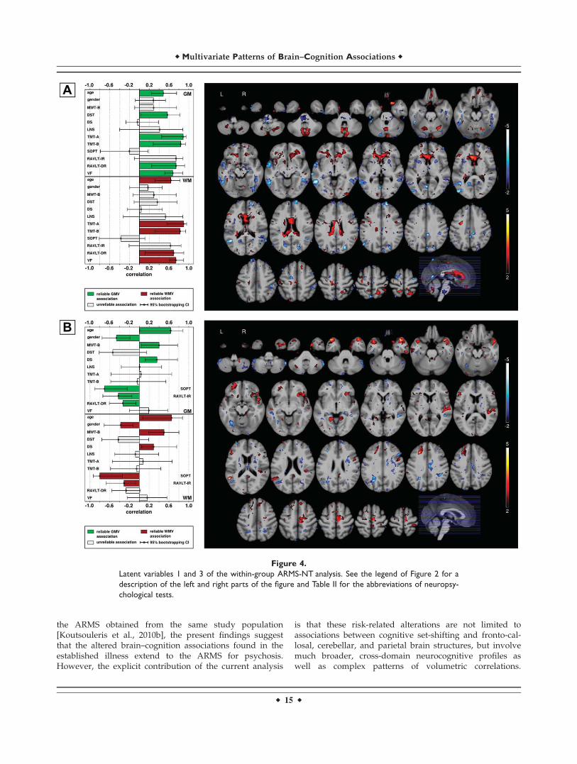
the ARMS obtained from the same study population[Koutsouleris et al., 2010b], the present findings suggestthat the altered brain–cognition associations found in theestablished illness extend to the ARMS for psychosis.However, the explicit contribution of the current analysis
is that these risk-related alterations are not limited toassociations between cognitive set-shifting and fronto-cal-losal, cerebellar, and parietal brain structures, but involvemuch broader, cross-domain neurocognitive profiles aswell as complex patterns of volumetric correlations.
Figure 4.
Latent variables 1 and 3 of the within-group ARMS-NT analysis. See the legend of Figure 2 for a
description of the left and right parts of the figure and Table II for the abbreviations of neuropsy-
chological tests.
r Multivariate Patterns of Brain–Cognition Associations r
r 15 r

Furthermore, the PLS method extended our previousresults by revealing that our HC group’s neurocognitiveprofiles were mainly confined to verbal measures (Fig.2A–C). In contrast, the ARMS group showed a broaderneurocognitive profile, including also processing speedand executive functions (Fig. 3A). This cross-domaininvolvement was even more pronounced in the ARMS-Tgroup; in that it affected the whole range of neurocogni-tive measures (see Fig. 5). Moreover, the current analysisrevealed that the HC group’s neuroanatomical salienceswere rather diffusely distributed across cortical and sub-cortical structures (Fig. 2A–C). Conversely, the ARMSgroup’s neuroanatomical patterns primarily mapped toprefrontal, limbic, temporal, perisylvian, and subcorticalstructures, including cortico-cortical and subcortico-corti-cal WM tracts (Fig. 3A,B). This localization of neuroana-tomical loadings to these brain regions was mostexpressed in the ARMS-T group.
More specifically, LV1 of the ARMS model expressed aprofile of broad, cross-domain neuropsychologicalinvolvement. This profile correlated (1) positively withprefrontal, limbic and paralimbic volumes, the intra- andinterhemispheric cortico-cortical WM tracts (superior lon-gitudinal fascicle, corpus callosum) and (2) negativelywith occipito-temporo-parietal GMV. Furthermore, LV1showed a reliable age- and gender covariation, meaningthat low-performing, younger males had less volume than
high-performing, older female subjects in voxels withpositive loadings. This relationship was reversed in vox-els with negative loadings. In keeping with our secondhypothesis, the inner product analysis (Fig. 1A) revealedthat this pattern was largely driven by the ARMS-Lgroup, suggesting that LV1 was linked to an ultra-highrisk for psychosis.
A similar neurocognitive profile was observed in theLV1 of the ARMS-T model (see Fig. 5) consisting of gen-eralized, cross-domain neurocognitive involvement withan emphasis on working memory/verbal IQ and an evenstronger age/gender covariation effect. Furthermore, therespective singular image expressed a spatial separationof positive and negative saliences similar to the LV1 sin-gular image of the ARMS model. However, the LV1 sin-gular image of ARMS-T consisted of highly reliablepositive saliences particularly in the insular and limbicstructures, the basal ganglia and neighboring/associatedWM tracts, as well as of highly reliable negative saliencesdistributed across the temporal, prefrontal, parietal, andoccipital cortices. As shown by the inner product analy-sis, this singular image specifically predicted the differen-tial effect of between-group LV1 in the ARMS-NT vs.ARMS-T omnibus analysis (Fig. 1B). However, it did notsolely drive the differences between HC and ARMS sub-jects as the respective singular image of the nonconver-sion group showed an almost equal correlation with
Figure 5.
Latent variable 1 of the within-group ARMS-T analysis. See the legend of Figure 2 for a
description of the left and right parts of the figure and Table II for the abbreviatons of neuro-
psychological tests.
r Koutsouleris et al. r
r 16 r

between-group LV1 in the HC vs. ARMS omnibus test(Fig. 1A). This observation suggests that brain–cognitionassociations specifically linked to disease transition maybe differentiated from covariance patterns related to avulnerability for psychosis-like experiences [Cornblattet al., 1997; Lencz et al., 2006].
Taken together, three conclusions may be drawn. First,a convergent mapping from a broad profile of neurocog-nitive functions to a specific set of prefrontal, perisylvian,temporal, and subcortical structures distinguished theARMS from HC subjects. This neurocognitive-neuroana-tomical convergence was particularly expressed in theconversion group that showed a strong positive associa-tion between cross-domain neuropsychological measuresand subcortical, limbic, and paralimbic structures. Thisobservation agrees with several lines of evidence, includ-ing (1) the established involvement of these brain struc-tures in a pattern of volumetric abnormalitiescharacterizing the ARMS [Borgwardt et al., 2007, 2008;Job et al., 2005; Koutsouleris et al., 2009b; Meisenzahlet al., 2008b; Pantelis et al., 2003] as well as overt psycho-sis [Honea et al., 2005; Pantelis et al., 2005], (2) correla-tions between hippocampal volume and delayed verbalrecall in ARMS-L, but not ARMS-E or HC subjects [Hur-lemann et al., 2008], (3) strong positive correlationsbetween thalamic volumes and RAVLT-IR performancein genetic high-risk individuals with a subsequent diseasetransition (Lymer et al., 2006), (4) disruptions of fronto-temporo-limbic connectivity [Nakamura et al., 2005] andstructural abnormalities of the caudate nuclei [Levittet al., 2002, 2004] relating to cognitive dysfunction inschizotypal personality disorder, and (5) altered associa-tions between prefronto-temporo-limbic and subcorticalvolumes (and interconnecting WMV) and executive/memory functions in schizophrenia [Bonilha et al., 2008;Cocchi et al., 2009; Crespo-Facorro et al., 2007b; Guret al., 2000; Laywer et al., 2006; Nakamura et al., 2008;Nestor et al., 2002; Premkumar et al., 2008; Perez-Iglesiaset al., 2010; Rusch et al., 2007; Sanfilipo et al., 2002;Szeszko et al., 2002]. In particular, our findings are con-sistent with the brain–cognition study of Nestor et al.[2002], which was the first to use PLS for the analysis ofmultivariate mappings from neurocognitive to neuroana-tomical measures in chronic schizophrenic patients. TheirPLS analysis revealed associations between prefronto-temporal regions of interest and neurocognitive variablesmeasuring categorization abilities (temporal and paralim-bic structures) as well as working memory and mentalset-shifting functions (frontal lobes).
Second, the neuroanatomical separation of positive andnegative saliences in the ARMS-specific brain–cognitionpatterns, which was particularly expressed by the conver-sion group, suggests a differential neurocognitive involve-ment of neural structures. In the light of the considerableneural plasticity observed in early adulthood [Panteliset al., 2005; Rapoport and Gogtay, 2008; Shaw et al.,2008], one speculative interpretation may be that positive
brain–cognition correlations reflect the biological proc-esses associated with the risk for conversion to psychosis,while negative associations result from continuous com-pensatory processes, which lead to an augmentation ofGMV and WMV in the associated brain structures, e.g.,through an increase in synaptic density [Murray et al.,2010; Ragland et al., 2004; Rusch et al., 2007]. This inter-pretation may be further supported by findings of volu-metric increments within paralimbic, inferior temporal,parietal and occipital brain regions of converters vs. non-converters [Borgwardt et al., 2007] and first-episodepatients vs. HC [Cocchi et al., 2009], as well as by reportsof ‘‘counterintuitive’’ negative brain–cognition correlationsin schizophrenic patients vs. HC [Cocchi et al., 2009;Rusch et al., 2007; Sanfilipo et al., 2002]. Alternatively,the spatial separation of positive and negative brain–cog-nition correlations may result from an abnormal matura-tional trajectory leading to distinct patterns of excessiveand defective synaptic pruning during different criticalperiods of brain development [Harris et al., 2004; Kesha-van et al., 1994; Lacerda et al., 2007; Pantelis et al., 2005;Rapoport and Gogtay, 2008].
Third, these conclusions have to be interpreted withrespect to the age and gender dependencies of the risk-specific brain–cognition associations. This finding of adouble covariation agrees with (1) reports of sexuallydimorphic brain abnormalities in the ARMS and estab-lished psychosis [Davatzikos et al., 2005; Goldstein et al.,2002; Koutsouleris et al., 2009b; Narr et al., 2003], with aparticular involvement of younger, male compared toolder, female patients [Narr et al., 2003] and (2) studiesshowing a stronger cognitive impairment of male vs.female patients [Goldstein et al., 1998, 1994; Walderet al., 2007].
These observations should be further explored in futurestudies of larger samples that prospectively combine neu-roanatomical and neuropsychological measurements inorder to clarify the trajectories of brain–behavior associa-tions in emerging psychosis. Finally, we demonstrated thatmultivariate statistical methods have the potential tounveil complex links between brain and behavior by dis-secting their associations into interpretable covariancecomponents. Therefore, these techniques may be ofbroader interest to the field as they may allow deconstruct-ing the multifaceted psychiatric phenotypes into their dis-tinct neural components.
REFERENCES
American Psychiatric Association (1994): Diagnostic and StatisticalManual for Mental Disorders, 4th ed. Washington, DC: Amer-ican Psychiatric Association.
Antonova E, Sharma T, Morris R, Kumari V (2004): The relation-ship between brain structure and neurocognition in schizo-phrenia: A selective review. Schizophr Res 70:117–145.
Antonova E, Kumari V, Morris R, Halari R, Anilkumar A, Mehro-tra R, Sharma T (2005): The relationship of structural altera-tions to cognitive deficits in schizophrenia: A voxel-basedmorphometry study. Biol Psychiatry 58:457–467.
r Multivariate Patterns of Brain–Cognition Associations r
r 17 r

Aschenbrenner S, Tucha O, Lange K (2001): RegensburgerWortflussigkeits-Test (RWT). Gottingen, Germany: HogrefeVerlag.
Ashburner J (2007): A fast diffeomorphic image registration algo-rithm. Neuroimage 38:95–113.
Ashburner J (2009): Computational anatomy with the SPM soft-ware. Magn Reson Imaging 27:1163–1174.
Ashburner J, Friston KJ (2005): Unified segmentation. Neuroimage26:839–851.
Bach Cuadra M, Cammoun L, Butz T, Cuisenaire O, Thiran JP(2005): Comparison and validation of tissue modelization andstatistical classification methods in T1-weighted MR brainimages. IEEE Trans Med Imaging 24:1548–1565.
Bergouignan L, Chupin M, Czechowska Y, Kinkingnehun S,Lemogne C, Bastard GL, Lepage M, Garnero L, Colliot O,Fossati P (2009): Can voxel based morphometry, manualsegmentation and automated segmentation equally detect hip-pocampal volume differences in acute depression? Neuro-image 45:29–37.
Bonilha L, Molnar C, Horner MD, Anderson B, Forster L, GeorgeMS, Nahas Z (2008): Neurocognitive deficits and prefrontalcortical atrophy in patients with schizophrenia. Schizophr Res101:142–151.
Borgwardt SJ, Riecher-Rossler A, Dazzan P, Chitnis X, Aston J,Drewe M, Gschwandtner U, Haller S, Pfluger M, RechsteinerE, D’souza M, Stieglitz RD, Rad EW, McGuire PK (2007): Re-gional gray matter volume abnormalities in the at risk mentalstate. Biol Psychiatry 61:1148–1156.
Borgwardt SJ, McGuire PK, Aston J, Gschwandtner U, PflugerMO, Stieglitz RD, Radue EW, RiecherRossler A (2008): Reduc-tions in frontal, temporal and parietal volume associated withthe onset of psychosis. Schizophr Res 106:108–114.
Brewer WJ, Francey SM, Wood SJ, Jackson HJ, Pantelis C, PhillipsLJ, Yung AR, Anderson VA, McGorry PD (2005): Memoryimpairments identified in people at ultra-high risk for psycho-sis who later develop first-episode psychosis. Am J Psychiatry162:71–78.
Cannon TD, Cadenhead K, Cornblatt B, Woods SW, Addington J,Walker E, Seidman LJ, Perkins D, Tsuang M, McGlashan T,Heinssen R (2008): Prediction of psychosis in youth at highclinical risk: A multi-site longitudinal study in North America.Arch Gen Psychiatry 65:28–37.
Cocchi L, Walterfang M, Testa R, Wood SJ, Seal ML, Suckling J,Takahashi T, Proffitt TM, Brewer WJ, Adamson C, Soulsby B,Velakoulis D, McGorry PD, Pantelis C (2009): Grey and whitematter abnormalities are associated with impaired spatialworking memory ability in first-episode schizophrenia. Schiz-ophr Res 115:163–172.
Cornblatt B, Obuchowski M, Schnur DB, O’Brien JD (1997): Atten-tion and clinical symptoms in schizophrenia. Psychiatr Q68:343–359.
Coupe P, Yger P, Barillot C (2006): Fast non local means denoisingfor 3D MR images. Med Image Comput Comput Assist Interv9:33–40.
Crespo-Facorro B, Barbadillo L, Pelayo-Teran JM, Rodrıguez-San-chez JM (2007a): Neuropsychological functioning and brainstructure in schizophrenia. Int Rev Psychiatry 19:325–336.
Crespo-Facorro B, Roiz-Santianez R, Pelayo-Teran JCCM,Rodrıguez-Sanchez JCCM, Perez-Iglesias R, Gonzalez-BlanchC, Tordesillas-Gutierrez D, Gonzalez-Mandly A, Dez C, Mag-notta VA, Andreasen NC, Vazquez-Barquero JCCL (2007b):Reduced thalamic volume in first-episode non-affective psy-
chosis: Correlations with clinical variables, symptomatologyand cognitive functioning. Neuroimage 35:1613–1623.
Davatzikos C (2004): Why voxel-based morphometric analysisshould be used with great caution when characterizing groupdifferences. Neuroimage 23:17–20.
Davatzikos C, Shen D, Gur RC, Wu X, Liu D, Fan Y, Hughett P,Turetsky BI, Gur RE (2005): Whole-brain morphometric studyof schizophrenia revealing a spatially complex set of focalabnormalities. Arch Gen Psychiatry 62:1218–1227.
Efron B, Tibshirani R (1986): Bootstrap methods for standarderrors, confidence intervals and other measures of statisticalaccuracy. Stat Sci 1:54–77.
Erlenmeyer-Kimling L, Rock D, Roberts SA, Janal M, KestenbaumC, Cornblatt B, Adamo UH, Gottesman II (2000): Attention,memory, and motor skills as childhood predictors of schizo-phrenia-related psychoses: The New York High-Risk Project.Am J Psychiatry 157:1416–1422.
Faraone SV, Seidman UJ, Kremen WS, Toomey R, Pepple JR,Tsuang MT (1999): Neuropsychological functioning among thenon-psychotic relatives of schizophrenic patients: A 4-year fol-low-up study. J Abnorm Psychol 108:176–181.
Francey SM, Jackson HJ, Phillips UJ, Wood SJ, Yung AR, McGorryPD (2005): Sustained attention in young people at high risk ofpsychosis does not predict transition to psychosis. SchizophrRes 79:127–136.
Frommann I, Brinkmeyer J, Ruhrmann S, Hack E, Brockhaus-Dumke A, Bechdolf A, Wolwer W, Klosterkotter J, Maier W,Wagner M (2008): Auditory P300 in individuals clinically atrisk for psychosis. Int J Psychophysiol 70:192–205.
Frommann I, Pukrop R, Brinkmeyer J, Bechdolf A, Ruhrmann S,Berning J, Decker P, Riedel M, Moller HJ, Wolwer W, GaebelW, Klosterkotter J, Maier W, Wagner M (2010): Neuropsycho-logical profiles in different at-risk states of psychosis: Execu-tive control impairment in the early-and additional memorydysfunction in the late-prodromal state. Schizophr Bull .
Fujiwara E, Schwartz MU, Gao F, Black SE, Uevine B (2008): Ven-tral frontal cortex functions and quantified MRI in traumaticbrain injury. Neuropsychologia 46:461–474.
Gaser C (2008): Voxel-based morphometry toolbox, version 5(VBM5). Available at: http://dbm.neuro.uni-jena.de.
Giessing C, Fink GR, Rosler F, Thiel CM (2007): fMRI data predictindividual differences of behavioral effects of nicotine: A par-tial least square analysis. J Cogn Neurosci 19:658–670.
Gilboa A, Ramirez J, Kohler S, Westmacott R, Black SE, Mosco-vitch M (2005): Retrieval of autobiographical memory in Alz-heimer’s disease: Relation to volumes of medial temporal lobeand other structures. Hippocampus 15:535–550.
Gold JM, Carpenter C, Randolph C, Goldberg TE, Weinberger DR(1997): Auditory working memory and Wisconsin Card SortingTest performance in schizophrenia. Arch Gen Psychiatry54:159–165.
Goldstein JM, Seidman UJ, Santangelo S, Knapp PH, Tsuang MT(1994): Are schizophrenic men at higher risk for developmentaldeficits than schizophrenic women? Implications for adult neu-ropsychological functions. J Psychiatr Res 28:483–498.
Goldstein JM, Seidman UJ, Goodman JM, Koren D, Uee H, Wein-traub S, Tsuang MT (1998): Are there sex differences in neuro-psychological functions among patients with schizophrenia?Am J Psychiatry 155:1358–1364.
Goldstein JM, Seidman UJ, O’Brien UM, Horton NJ, Kennedy DN,Makris N, Caviness VS, Faraone SV, Tsuang MT (2002): Impactof normal sexual dimorphisms on sex differences in structural
r Koutsouleris et al. r
r 18 r

brain abnormalities in schizophrenia assessed by magnetic res-onance imaging. Arch Gen Psychiatry 59:154–164.
Gur RE, Cowell PE, Uatshaw A, Turetsky BI, Grossman RI,Arnold SE, Bilker WB, Gur RC (2000): Reduced dorsal and or-bital prefrontal gray matter volumes in schizophrenia. ArchGen Psychiatry 57:761–768.
Hafner H, Maurer K, Ruhrmann S, Bechdolf A, Klosterkotter J,Wagner M, Maier W, Bottlender R, Moller HJ, Gaebel W,Wolwer W (2004): Early detection and secondary prevention ofpsychosis: Facts and visions. Eur Arch Psychiatry Clin Neuro-sci 254:117–128.
Hans SL, Marcus J, Nuechterlein KH, Asarnow RF, Styr B, Auer-bach JG (1999): Neurobehavioral deficits at adolescence inchildren at risk for schizophrenia: The Jerusalem infant devel-opment study. Arch Gen Psychiatry 56:741–748.
Harris JM, Whalley H, Yates S, Miller P, Johnstone EC, LawrieSM (2004): Abnormal cortical folding in high-risk individuals:A predictor of the development of schizophrenia? Biol Psychi-atry 56:182–189.
Hawkins KA, McGlashan TH, Quinlan D, Miller TJ, Perkins DO,Zipursky RB, Addington J, Woods SW (2004): Factorial struc-ture of the scale of prodromal symptoms. Schizophr Res68:339–347.
Heinrichs RW, Zakzanis KK (1998): Neurocognitive deficit inschizophrenia: A quantitative review of the evidence. Neuro-psychology 12:426–445.
Holm S (1979): A simple sequentially rejective multiple test proce-dure. Scand J Stat 6:65–70.
Honea R, Crow TJ, Passingham D, Mackay CE (2005): Regionaldeficits in brain volume in schizophrenia: A meta-analysis ofvoxel-based morphometry studies. Am J Psychiatry 162:2233–2245.
Hurlemann R, Jessen F, Wagner M, Frommann I, Ruhrmann S,Brockhaus A, Picker H, Scheef L, Block W, Schild HH, Moller-Hartmann W, Krug B, Falkai P, Klosterkotter J, Maier W(2008): Interrelated neuropsychological and anatomical evi-dence of hippocampal pathology in the at-risk mental state.Psychol Med 38:843–851.
Job DE, Whalley HC, McConnell S, Glabus M, Johnstone EC, Law-rie SM (2003): Voxel-based morphometry of grey matter den-sities in subjects at high risk of schizophrenia. Schizophr Res64:1–13.
Job DE, Whalley HC, Johnstone EC, Lawrie SM (2005): Grey mat-ter changes over time in high risk subjects developing schizo-phrenia. Neuroimage 25:1023–1030.
Kawasaki Y, Suzuki M, Kherif F, Takahashi T, Zhou SY, Naka-mura K, Matsui M, Sumiyoshi T, Seto H, Kurachi M (2007):Multivariate voxel-based morphometry successfully differenti-ates schizophrenia patients from healthy controls. Neuroimage34:235–242.
Kay SR, Fiszbein A, Opler LA (1987): The positive and negativesyndrome scale (PANSS) for schizophrenia. Schizophr Bull13:261–276.
Keshavan MS, Anderson S, Pettegrew JW (1994): Is schizophreniadue to excessive synaptic pruning in the prefrontal cortex? TheFeinberg hypothesis revisited. J Psychiatr Res 28:239–265.
Klein A, Andersson J, Ardekani BA, Ashburner J, Avants B,Chiang MC, Christensen GE, Collins DL, Gee J, Hellier P, SongJH, Jenkinson M, Lepage C, Rueckert D, Thompson P, Vercau-teren T, Woods RP, Mann JJ, Parsey RV (2009): Evaluation of14 nonlinear deformation algorithms applied to human brainMRI registration. Neuroimage 46:786–802.
Klosterkotter J, Hellmich M, Steinmeyer EM, Schultze-Lutter F(2001): Diagnosing schizophrenia in the initial prodromalphase. Arch Gen Psychiatry 58:158–164.
Koutsouleris N, Gaser C, Jager M, Bottlender R, Frodl T, Hol-zinger S, Schmitt GJE, Zetzsche T, Burgermeister B, Scheuer-ecker J, Born C, Reiser M, Moller HJ, Meisenzahl EM (2008):Structural correlates of psychopathological symptom dimen-sions in schizophrenia: A voxel-based morphometric study.Neuroimage 39:1600–1612.
Koutsouleris N, Meisenzahl E, Davatzikos C, Bottlender R, FrodlT, Scheuerecker J, Schmitt G, Zetzsche T, Decker P, Reiser M,Moller HJ, Gaser C (2009a): Neuroanatomical pattern classi-fication identifies subjects in at-risk mental states of psychosisand predicts disease transition. Arch Gen Psychiatry 66:700–712.
Koutsouleris N, Schmitt G, Gaser C, Bottlender R, Scheuerecker J,McGuire P, Burgermeister B, Born C, Reiser M, Moller HJ,Meisenzahl E (2009b): Neuroanatomical correlates of differentvulnerability states of psychosis in relation to clinical outcome.Br J Psychiatry 195:218–226.
Koutsouleris N, Gaser C, Bottlender R, Davatzikos C, Decker P,Jager M, Schmitt G, Reiser M, Moller HJ, Meisenzahl EM(2010a): Use of neuroanatomical pattern regression to predictthe structural brain dynamics of vulnerability and transition topsychosis. Schizophr Res 123:175–187.
Koutsouleris N, Patschurek-Kliche K, Scheuerecker J, Decker P,Bottlender R, Schmitt G, Rujescu D, Giegling I, Gaser C, ReiserM, Moller HJ, Meisenzahl EM (2010b): Neuroanatomical corre-lates of executive dysfunction in the at-risk mental state forpsychosis. Schizophr Res 123:160–174.
Krishnan A, Williams LJ, McIntosh AR, Abdi H (2010): Partialleast squares (PLS) methods for neuroimaging: A tutorial andreview. Neuroimage .
Lacerda ALT, Hardan AY, Yorbik O, Vemulapalli M, Prasad KM,Keshavan MS (2007): Morphology of the orbitofrontal cortex infirst-episode schizophrenia: Relationship with negative symp-tomatology. Prog Neuropsychopharmacol Biol Psychiatry 31:510–516.
Larsen TK (2002): The transition from premorbid period topsychosis: How can it be described? Acta Psychiatr Scand106:10–11.
Laywer G, Nyman H, Agartz I, Arnborg S, Jnsson EG, SedvallGC, Hall H (2006): Morphological correlates to cognitive dys-function in schizophrenia as studied with Bayesian regression.BMC Psychiatry 6:31.
Lehrl S (2005): Mehrfachwahl-Wortschatz-Intelligenztest MWT-B,5th ed. Balingen: Spitta Verlag.
Lencz T, Smith CW, McLaughlin D, Auther A, Nakayama E,Hovey L, Cornblatt BA (2006): Generalized and specific neuro-cognitive deficits in prodromal schizophrenia. Biol Psychiatry59:863–871.
Levitt JJ, McCarley RW, Dickey CC, Voglmaier MM, NiznikiewiczMA, Seidman LJ, Hirayasu Y, Ciszewski AA, Kikinis R, JoleszFA, Shenton ME (2002): MRI study of caudate nucleus volumeand its cognitive correlates in neuroleptic-naive patientswith schizotypal personality disorder. Am J Psychiatry 159:1190–1197.
Levitt JJ, Westin CF, Nestor PG, Estepar RSJ, Dickey CC, Voglma-ier MM, Seidman LJ, Kikinis R, Jolesz FA, McCarley RW,Shenton ME (2004): Shape of caudate nucleus and its cognitivecorrelates in neuroleptic-naive schizotypal personality disor-der. Biol Psychiatry 55:177–184.
r Multivariate Patterns of Brain–Cognition Associations r
r 19 r

Lezak MD (1995): Neuropsychological Assessment, 3rd ed. NewYork: Oxford University Press.
Lymer GKS, Job DE, William T, Moorhead J, McIntosh AM,Owens DGC, Johnstone EC, Lawrie SM (2006): Brain-behaviorrelationships in people at high genetic risk of schizophrenia.Neuroimage 33:275–285.
Manjon JV, Tohka J, Garcıa-Martı G, Carbonell-Caballero J, Lull JJ,Martı-Bonmatı L, Robles M (2008): Robust MRI brain tissue pa-rameter estimation by multistage outlier rejection. Magn ResonMed 59:866–873.
McIntosh AR, Lobaugh NJ (2004): Partial least squares analysis ofneuroimaging data: Applications and advances. Neuroimage23(Suppl 1):S250–S263.
Meisenzahl EM, Koutsouleris N, Bottlender R, Scheuerecker J,Jager M, Teipel SJ, Holzinger S, Frodl T, Preuss U, Schmitt G,Burgermeister B, Reiser M, Born C, Moller HJ (2008a):Structural brain alterations at different stages of schizophre-nia: A voxel-based morphometric study. Schizophr Res 104:44–60.
Meisenzahl EM, Koutsouleris N, Gaser C, Bottlender R, SchmittGJE, McGuire P, Decker P, Burgermeister B, Born C, Reiser M,Moller HJ (2008b): Structural brain alterations in subjects athigh-risk of psychosis: A voxel-based morphometric study.Schizophr Res 102:150–162.
Menzies L, Achard S, Chamberlain SR, Fineberg N, Chen CH, delCampo N, Sahakian BJ, Robbins TW, Bullmore E (2007): Neu-rocognitive endophenotypes of obsessive-compulsive disorder.Brain 130:3223–3236.
Miller TJ, McGlashan TH, Rosen JL, Somjee L, Markovich PJ, SteinK, Woods SW (2002): Prospective diagnosis of the initialprodrome for schizophrenia based on the structured interviewfor prodromal syndromes: Preliminary evidence of interraterreliability and predictive validity. Am J Psychiatry 159:863–865.
Montgomery SA, Asberg M (1979): A new depression scaledesigned to be sensitive to change. Br J Psychiatry 134:382–389.
Murray GK, Corlett PR, Fletcher PC (2010): The neural underpin-nings of associative learning in health and psychosis: How canperformance be preserved when brain responses are abnor-mal? Schizophr Bull 36:465–471.
Nakamura M, McCarley RW, Kubicki M, Dickey CC, Niznikie-wicz MA, Voglmaier MM, Seidman LJ, Maier SE, Westin CF,Kikinis R, Shenton ME (2005): Fronto-temporal disconnectivityin schizotypal personality disorder: A diffusion tensor imagingstudy. Biol Psychiatry 58:468–478.
Nakamura M, Nestor PG, Levitt JJ, Cohen AS, Kawashima T,Shenton ME, McCarley RW (2008): Orbitofrontal volume deficitin schizophrenia and thought disorder. Brain 131:180–195.
Narr KL, Sharma T, Woods RP, Thompson PM, Sowell ER, Rex D,Kim S, Asuncion D, Jang S, Mazziotta J, Toga AW (2003):Increases in regional subarachnoid CSF without apparent corti-cal gray matter deficits in schizophrenia: Modulating effects ofsex and age. Am J Psychiatry 160:2169–2180.
Nestor PG, O’Donnell BF, McCarley RW, Niznikiewicz M, Bar-nard J, Shen ZJ, Bookstein FL, Shenton ME (2002): A new sta-tistical method for testing hypotheses of neuropsychological/MRI relationships in schizophrenia: Partial least squares analy-sis. Schizophr Res 53:57–66.
Niendam TA, Bearden CE, Johnson JK, McKinley M, Loewy R,O’Brien M, Nuechterlein KH, Green MF, Cannon TD (2006):Neurocognitive performance and functional disability in thepsychosis prodrome. Schizophr Res 84:100–111.
Owens DGC, Johnstone EC (2006): Precursors and prodromata ofschizophrenia: Findings from the Edinburgh High Risk Studyand their literature context. Psychol Med 36:1501–1514.
Pantelis C, Velakoulis D, McGorry PD, Wood SJ, Suckling J, Phil-lips LJ, Yung AR, Bullmore ET, Brewer W, Soulsby B, Des-mond P, McGuire PK (2003): Neuroanatomical abnormalitiesbefore and after onset of psychosis: A cross-sectional and lon-gitudinal MRI comparison. Lancet 361:281–288.
Pantelis C, Yucel M, Wood SJ, Velakoulis D, Sun D, Berger G,Stuart GW, Yung A, Phillips L, McGorry PD (2005): Structuralbrain imaging evidence for multiple pathological processes atdifferent stages of brain development in schizophrenia. Schiz-ophr Bull 31:672–696.
Perez-Iglesias R, Tordesillas-Gutierrez D, McGuire PK, Barker GJ,Roiz-Santianez R, Mata I, de Lucas EM, Rodrıguez-SanchezJM, Ayesa-Arriola R, Vazquez-Barquero JL, Crespo-Facorro B(2010): White matter integrity and cognitive impairment infirst-episode psychosis. Am J Psychiatry 167:451–458.
Petrides M (1995): Impairments on nonspatial self-ordered andexternally ordered working memory tasks after lesions of themid-dorsal part of the lateral frontal cortex in the monkey.J Neurosci 15:359–375.
Premkumar P, Fannon D, Kuipers E, Simmons A, Frangou S,Kumari V (2008): Emotional decision-making and its dissoci-able components in schizophrenia and schizoaffective disorder:A behavioral and MRI investigation. Neuropsychologia 46:2002–2012.
Pukrop R, Klosterkotter J (2010): Neurocognitive indicators of clin-ical high-risk states for psychosis: A critical review of the evi-dence. Neurotox Res 18:272–286.
Pukrop R, Schultze-Lutter F, Ruhrmann S, Brockhaus-Dumke A,Tendolkar I, Bechdolf A, Matuschek E, Klosterkotter J (2006):Neurocognitive functioning in subjects at risk for a firstepisode of psychosis compared with first- and multiple-episode schizophrenia. J Clin Exp Neuropsychol 28:1388–1407.
Pukrop R, Ruhrmann S, Schultze-Lutter F, Bechdolf A, Brockhaus-Dumke A, Klosterkotter J (2007): Neurocognitive indicators fora conversion to psychosis: Comparison of patients in a poten-tially initial prodromal state who did or did not convert to apsychosis. Schizophr Res 92:116–125.
Quednow BB, Frommann I, Berning J, Kuhn KU, Maier W, Wag-ner M (2008): Impaired sensorimotor gating of the acousticstartle response in the prodrome of schizophrenia. Biol Psychi-atry 64:766–773.
Ragland JD, Gur RC, Valdez J, Turetsky BI, Elliott M, Kohler C,Siegel S, Kanes S, Gur RE (2004): Event-related fMRI of fronto-temporal activity during word encoding and recognition inschizophrenia. Am J Psychiatry 161:1004–1015.
Rajapakse JC, Giedd JN, Rapoport JL (1997): Statistical approachto segmentation of single-channel cerebral MR images. IEEETrans Med Imaging 16:176–186.
Rapoport JL, Gogtay N (2008): Brain neuroplasticity in healthy,hyperactive and psychotic children: Insights from neuroimag-ing. Neuropsychopharmacology 33:181–197.
Reitan RM (1992): TMT, Trail Making Test A & B. South Tucson,AR: Reitan Neuropsychology Laboratory.
Ruhrmann S, Schultze-Lutter F, Klosterkotter J (2003): Early detec-tion and intervention in the initial prodromal phase of schizo-phrenia. Pharmacopsychiatry 36 (Suppl 3):S162–S167.
Ruhrmann S, Schultze-Lutter F, Salokangas RKR, Heinimaa M,Linszen D, Dingemans P, Birchwood M, Patterson P, Juckel G,
r Koutsouleris et al. r
r 20 r

Heinz A, Morrison A, Lewis S, von Reventlow HG, Klosterkot-ter J (2010): Prediction of psychosis in adolescents and youngadults at high risk: Results from the prospective European pre-diction of psychosis study. Arch Gen Psychiatry 67:241–251.
Rusch N, Spoletini I, Wilke M, Bria P, Paola MD, Iulio FD, Marti-notti G, Caltagirone C, Spalletta G (2007): Prefrontal-thalamic-cerebellar gray matter networks and executive functioning inschizophrenia. Schizophr Res 93:79–89.
Salgado-Pineda P, Baeza I, Perez-Gomez M, Vendrell P, Junque C,Bargallo N, Bernardo M (2003): Sustained attention impairmentcorrelates to gray matter decreases in first episode neuroleptic-naive schizophrenic patients. Neuroimage 19:365–375.
Sampson PD, Streissguth AP, Barr HM, Bookstein FL (1989): Neu-robehavioral effects of prenatal alcohol: Part II. Partial leastsquares analysis. Neurotoxicol Teratol 11:477–491.
Sanfilipo M, Lafargue T, Rusinek H, Arena L, Loneragan C, Lau-tin A, Rotrosen J, Wolkin A (2002): Cognitive performance inschizophrenia: Relationship to regional brain volumes and psy-chiatric symptoms. Psychiatry Res 116:1–23.
Schall U, Halpin S, Hunt S (2003): Neurocognitive profiles ofyoung people at high risk versus first-episode psychosis: A fol-low-up study. Schizophr Res 60(Suppl):156.
Schultze-Lutter F, Ruhrmann S, Picker H, von Reventlow HG,Brockhaus-Dumke A, Klosterkotter J (2007a): Basic symptomsin early psychotic and depressive disorders. Br J PsychiatrySuppl 51:s31–s37.
Schultze-Lutter F, Ruhrmann S, Picker H, von Reventlow HG,Daumann B, Brockhaus-Dumke A, Klosterkotter J, Pukrop R(2007b): Relationship between subjective and objective cogni-tive function in the early and late prodrome. Br J PsychiatrySuppl 51:s43–s51.
Seidman LJ, Giuliano AJ, Meyer EC, Addington J, Cadenhead KS,Cannon TD, McGlashan TH, Perkins DO, Tsuang MT, WalkerEF, Woods SW, Bearden CE, Christensen BK, Hawkins K, Hea-ton R, Keefe RSE, Heinssen R, Cornblatt BA, Group NAPLSN(2010): Neuropsychology of the prodrome to psychosis in theNAPLS consortium: Relationship to family history and conver-sion to psychosis. Arch Gen Psychiatry 67:578–588.
Shaw P, Kabani NJ, Lerch JP, Eckstrand K, Lenroot R, Gogtay N,Greenstein D, Clasen L, Evans A, Rapoport JL, Giedd JN, WiseSP (2008): Neurodevelopmental trajectories of the human cere-bral cortex. J Neurosci 28:3586–3594.
Simon AE, Dvorsky DN, Boesch J, Roth B, Isler E, Schueler P,Petralli C, Umbricht D (2006): Defining subjects at risk for
psychosis: A comparison of two approaches. Schizophr Res81:83–90.
Simon AE, Cattapan-Ludewig K, Zmilacher S, Arbach D, GruberK, Dvorsky DN, Roth B, Isler E, Zimmer A, Umbricht D(2007): Cognitive functioning in the schizophrenia prodrome.Schizophr Bull 33:761–771.
Sun D, van Erp TGM, Thompson PM, Bearden CE, Daley M,Kushan L, Hardt ME, Nuechterlein KH, Toga AW, Cannon TD(2009): Elucidating a magnetic resonance imaging-based neuro-anatomic biomarker for psychosis: Classification analysis usingprobabilistic brain atlas and machine learning algorithms. BiolPsychiatry 66:1055–1060.
Szeszko PR, Strous RD, Goldman RS, Ashtari M, Knuth KH, Lie-berman JA, Bilder RM (2002): Neuropsychological correlates ofhippocampal volumes in patients experiencing a first episodeof schizophrenia. Am J Psychiatry 159:217–226.
Tura E, Turner JA, Fallon JH, Kennedy JL, Potkin SG (2008): Mul-tivariate analyses suggest genetic impacts on neurocircuitry inschizophrenia. Neuroreport 19:603–607.
Tzourio-Mazoyer N, Uandeau B, Papathanassiou D, Crivello F,Etard O, Delcroix N, Mazoyer B, Joliot M (2002): Automatedanatomical labeling of activations in SPM using a macroscopicanatomical parcellation of the MNI MRI single-subject brain.Neuroimage 15:273–289.
Walder DJ, Seidman UJ, Makris N, Tsuang MT, Kennedy DN,Goldstein JM (2007): Neuroanatomic substrates of sex differen-ces in language dysfunction in schizophrenia: A pilot study.Schizophr Res 90:295–301.
Wechsler D (1997): Wechsler Adult Intelligence Scale, 3rd ed. SanAntonio, TX: Psychological Cooperation.
Wellcome Trust Centre for Neuroimaging (2009): Statistical Para-metric Mapping, version 8. Available at: http://www.fil.ion.ucl.ac.uk/spm/software/spm8/.Wood SJ, Brewer WJ, Koutsouradis P, Phillips UJ, Francey SM,
Proffitt TM, Yung AR, Jackson HJ, McGorry PD, Pantelis C(2007): Cognitive decline following psychosis onset: Data fromthe pace clinic. Br J Psychiatry Suppl 51:s52–s57.
Yung AR, Phillips UJ, McGorry PD, McFarlane CA, Francey S,Harrigan S, Patton GC, Jackson HJ (1998): Prediction of psy-chosis: A step toward indicated prevention of schizophrenia.Br J Psychiarty Suppl 172:14–20.
Yung AR, Phillips UJ, Yuen HP, Francey SM, McFarlane CA, Hallg-ren M, McGorry PD (2003): Psychosis prediction: 12-month followup of a high-risk (‘‘prodromal’’) group. Schizophr Res 60:21–32.
r Multivariate Patterns of Brain–Cognition Associations r
r 21 r