Embed Size (px)
Citation preview
Genes, Brain and Behavior (2010) doi: 10.1111/j.1601-183X.2010.00662.x
Persistent spatial working memory deficits in ratsfollowing in utero RNAi of Dyx1c1
C. E. Szalkowski†,∗, J. R. Hinman†, S. W.Threlkeld‡, Y. Wang§, A. LePack¶, G. D. Rosen∗∗,J. J. Chrobak†, J. J. LoTurco§ and R. H. Fitch†
†Department of Psychology/Behavioral Neuroscience,University of Connecticut, Storrs, CT, ‡Department ofPsychology, Rhode Island College, Providence, RI,§Department of Physiology and Neurobiology, University ofConnecticut, Storrs, CT, ¶Division of Molecular Psychiatry,Abraham Ribicoff Research Facilities, Connecticut MentalHealth Center, Yale University School of Medicine, New Haven,CT, and **Department of Neurology, Beth Israel DeaconessMedical Center, Boston, MA, USA*Corresponding author: C. E. Szalkowski, University of Connecti-cut, 406 Babbidge Road, Unit 1020, Storrs, CT 06269, USA.E-mail: [email protected]
Disruptions in the development of the neocortex are
associated with cognitive deficits in humans and other
mammals. Several genes contribute to neocortical devel-
opment, and research into the behavioral phenotype
associated with specific gene manipulations is advanc-
ing rapidly. Findings include evidence that variants in
the human gene DYX1C1 may be associated with an
increased risk of developmental dyslexia. Concurrent
research has shown that the rat homolog for this gene
modulates critical parameters of early cortical develop-
ment, including neuronal migration. Moreover, recent
studies have shown auditory processing and spatial
learning deficits in rats following in utero transfection
of an RNA interference (RNAi) vector of the rat homolog
Dyx1c1 gene. The current study examined the effects
of in utero RNAi of Dyx1c1 on working memory perfor-
mance in Sprague–Dawley rats. This task was chosen
based on the evidence of short-term memory deficits in
dyslexic populations, as well as more recent evidence
of an association between memory deficits and DYX1C1
anomalies in humans. Working memory performance
was assessed using a novel match-to-place radial water
maze task that allows the evaluation of memory for a
single brief (∼4–10 seconds) swim to a new goal location
each day. A 10-min retention interval was used, followed
by a test trial. Histology revealed migrational abnormal-
ities and laminar disruption in Dyx1c1 RNAi-treated rats.
Dyx1c1 RNAi-treated rats exhibited a subtle, but signif-
icant and persistent impairment in working memory as
compared to Shams. These results provide further sup-
port for the role of Dyx1c1 in neuronal migration and
working memory.
Keywords: Brain development, dyslexia, neuronal migration,RNA interference, working memory
Received 28 June 2010, revised 18 August 2010, acceptedfor publication 17 October 2010
Neocortical migrational anomalies have been associated withlearning, language and other cognitive deficits in humans(Boscariol et al. 2009; Casanova et al. 2004; Galaburda et al.1985; 1994; Hage et al. 2006). Studies from our laboratoryhave shown that rodents with cortical malformations exhibitdeficits in rapid auditory processing (RAP) abilities, which aresimilar to those observed in humans with language impair-ment (Rosen et al. 1995; Clark et al. 2000; Peiffer et al.2002; 2004a; Threlkeld et al. 2006, 2007). In fact, longitu-dinal assessment of infants, as well as studies of olderpopulations, suggest that the RAP difficulties may representone facet of the constellation of deficits associated withlearning disorders such as developmental dyslexia (Benasichet al. 2006; Choudhury et al. 2007; Farmer et al. 1995; Tallal& Benasich 2002; Tallal et al. 1993; Wright et al. 1997).
Twin studies first suggested that developmental dyslexiawas a genetic disorder and early genetic linkage studies laidthe groundwork for recent molecular genetic studies (seeFisher & DeFries 2002 for review). Recent genetic asso-ciation analyses in human populations have revealed genevariants (ROBO1, DCDC2, KIAA0319 and DYX1C1) that areassociated with increased risk of developmental dyslexia(Brkanac et al. 2007; Chapman et al. 2004; Cope et al. 2005;Dahdouh et al. 2009; Francks et al. 2004; Hannula-Jouppiet al. 2005; Harold et al. 2006; Ludwig et al. 2008; Marinoet al. 2007; Massinen et al. 2009; Meng et al. 2005; Parac-chini et al. 2006; Schumacher et al. 2006; Taipale et al. 2003;Wigg et al. 2004; Wilcke et al. 2009). DYX1C1, which islocated on chromosome 15, was the first candidate dyslexiarisk gene reported, with variants linked to reading-relatedphenotypes in a dyslexic family in Finland. A linkage dise-quilibrium analysis linked the same variants of the gene toclinical disability in a larger population of Finnish dyslexics(Taipale et al. 2003).
Recent reports have linked variants in DYX1C1 to impair-ments in short-term memory in dyslexic individuals (Marinoet al. 2007). Verbal and non-verbal short-term memorydeficits – including visuospatial memory deficits – are oftencomorbid with the phonological impairments that character-ize developmental dyslexia and specific language impairment(Archibald & Gathercole 2006; Baddeley & Hitch 1974; Gath-ercole et al. 2006; Smith-Spark & Fisk 2007).
Animal research employing embryonic (in utero) RNAinterference (RNAi) has shown that the knockdown ofthe rodent homolog for DYX1C1 (Dyx1c1) in rats resultsin aberrant neuronal migration, including the formation ofheterotopic and ectopic clusters of neurons throughout the
© 2010 The Authors 1Genes, Brain and Behavior © 2010 Blackwell Publishing Ltd and International Behavioural and Neural Genetics Society
Szalkowski et al.
cortex (Rosen et al. 2007; Wang et al. 2006). Behavioralassays on rats following in utero RNAi of Dyx1c1 haverevealed deficits in RAP. Spatial learning deficits were alsopresent in a subset of animals with neuronal heterotopias inthe hippocampus (Threlkeld et al. 2007).
This study sought to explore the possibility that earlycortical disruption via interference with the rodent homologof DYX1C1 may lead to higher order memory and learningimpairments paralleling those observed in developmentaldyslexia. Specifically, we employed a model using E14 inutero ventricular RNAi against Dyx1c1 to characterize theworking memory abilities of embryonically transfected rats.
Methods
A total of 61 male Sprague–Dawley rats (Charles River Labo-ratory, Wilmington, MA, USA) were used in these experiments(Dyx1c1 RNAi = 33; Sham = 28). All rats were pair housed in tubswith same-sex littermates and were maintained on a 12 h/12 hlight/dark cycle in a temperature-controlled room. Water was avail-able ad libitum. All procedures were performed in accordance withguidelines set forth by the National Institutes of Health and wereapproved by the University of Connecticut’s Institutional Care andUse Committee.
TransfectionTransfection of in utero RNAi of Dyx1c1 was performed by Y. W.at the University of Connecticut in accordance with the procedureoutlined in Bai et al. 2003. Two batches of surgeries were per-formed. In all Dyx1c1 treatments, plasmids encoding short hairpin(pU6DyxHPB) RNA (Dyx1c1 RNAi) were transfected into the fetal(embryonic day 14 or E14) ventricular zone (VZ) by in utero electro-poration, following externalization of the uterine horn. In Batch 1, theDyx1c1 shRNA plasmid was co-transfected with a plasmid encodingenhanced green fluorescent protein (eGFP), as well as fast green dye(a benign dye used to confirm injection into the lateral ventricles dur-ing the procedure). In Batch 1, Shams subjects received injections ofa solution of fast green dye only. In Batch 2, Dyx1c1 subjects receivedinjection of the Dyx1c1 shRNA plasmid and fast green dye. Sham sub-jects received transfection with plasmids (pCAGGS-RFP) encodingmonomeric red fluorescent protein (mRFP) and fast green dye.
Briefly, time-mated Sprague–Dawley dams (Charles River Labo-ratory) were palpated to confirm gestational age as predicted bytimed mating. At E14, dams were anesthetized (ketamine/xylazine;100/10 mixture, 0.1 mg/g, intraperitoneally) and abdominal incisionswere made through the skin and muscle. The uterine horns wereexposed and injections were made as follows: Batch 1, Dyx1c1 RNAiplasmids (1.5 μg/μl) + eGFP plasmids (0.5 μg/μl) + fast green dye,Shams received injections of fast green dye; Batch 2, Dyx1c1 RNAiplasmids (1.5 μg/μl) + fast green dye, Shams received mRFP plas-mids (0.4 μg/μl) + fast green dye. All solutions were microinjectedinto fetal ventricles by pressure (General Valve Picospritzer, PineBrook, NJ, USA). This was performed directly through the uterinewall. One randomly chosen lateral ventricle (left or right) of eachembryo was transfected, using a pulled glass capillary (DrummondScientific, Broomall, PA, USA). Electroporation was achieved by dis-charge of a 500-mV capacitor charged to 50–100 mV. A pair ofcopper alloy plates (1 × 0.5 cm) pinching the head of each embryowas the conduit for the voltage pulse and the voltage current wasdischarged through both sides of the brain, transfecting the cor-tex bilaterally. Since embryos were unable to be sexed at this ageand only males were used for testing, equal numbers of Sham andDyx1c1 RNAi injections were made, at roughly double the numbersneeded. (Transfected females were used in other studies). Note thatonly males were used in this study based on previous research thathas demonstrated that male rodents exhibit more robust functionaldeficits than females following early disruption of neuronal migration
(Fitch et al. 1997; Peiffer et al. 2002, 2004b). Additionally, there is ahigher incidence of dyslexia in males than in females (Flannery et al.2000; Katusic et al. 2001; Rutter et al. 2004).
Sixty-one male subjects were weaned on postnatal Day 21 (P21)and received right or left ear marking. Treatment could not beidentified at weaning; therefore subjects were housed in same-sexlittermate pairs. (Treatment was later identified post-mortem via flu-orescence of GFP in Batch 1 and via fluorescence of RFP in Batch2). Note that the numbers of Sham and RNAi-treated subjects wereexpected to be roughly equal, since equal numbers of Sham and RNAiinjections were made and only males were used. Histology later con-firmed Dyx1c1 treatment (n = 33) and Sham (n = 28) for a total of 61.
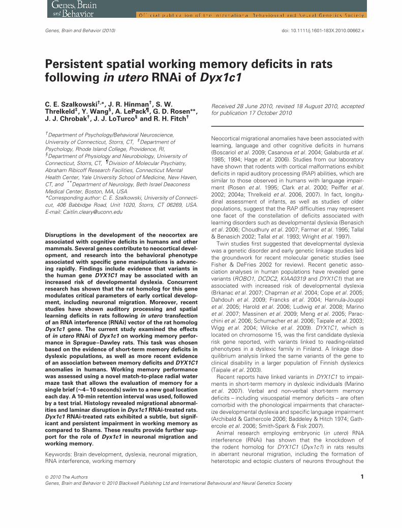
ApparatusThe radial arm water maze was housed in a black Plexiglas pool(140 cm in diameter and 40-cm deep) filled with cool water [22(±2)◦C]. The maze consisted of eight removable stainless steel armspainted flat black that could be attached to a central octagonal hub(50 cm across). Each corridor was 14-cm wide and extended 36 cmto the edge of the pool (Fig. 1). A removable black plastic platform(10 cm diameter) served as the escape platform and was submerged4 cm beneath the surface of the water. The entire apparatus was ina large room with two empty walls, a long table and the cage rackforming the boundaries around the maze. During testing, light wasprovided by a desk lamp in the northeast corner of the room, foradditional spatial cues.
Figure 1: Diagram of the delayed match-to-sample radial
water maze task, including sample (left) and test (right)
trials. Exemplars for the first and second days of testing aregiven. S = start arm [location changes between sample (S1)and test trial (S2)] and G = goal arm (location remains fixed).Each day of testing rats were given one forced-choice sampletrial in which all arms were blocked except for the start armand goal arm. A plastic escape platform was submerged at theend of the goal arm. During the test trial (10 min later), all thearms were open and a new start arm (S2) was used to test theanimal’s memory of the spatial location of the goal arm. Eachanimal received one sample trial and one test trial each day oftesting. The sequences of start arms and goal locations usedeach day varied systematically among 48 patterns that regulatedthe sequence of start and goal arms, the turn angles and therelationship between the start and goal arms across trials.
2 Genes, Brain and Behavior (2010)
Memory deficits following Dyx1c1 RNAi
Delayed match-to-sample testingBeginning on P33, subjects were handled 5 min a day for the weekprior to testing. On the initial testing day (P40), subjects had no priorexposure to the testing room, the water maze or the platform. Allrats were assessed on an initial acclimation trial and were foundto be capable of navigating the maze and mounting the platform.Subsequent testing of animals consisted of four daily sessions perweek (four sessions per week, one session per day, one sample andone test trial per session). Each session consisted of a forced-choicesample trial and a test trial in which all arms were open (see Chrobaket al. 2008 for additional details). During the sample trial, all corridorswere blocked at the intersection of the arm and central hub, exceptfor the start and goal arms. Each rat swam out of the start arm,navigated to the only open corridor (the goal arm) and mountedthe escape platform. The subject was removed immediately aftermounting the platform, gently dried with a towel and returned tothe home cage. Subjects took ∼4–20 seconds to complete thesample trial. This study employed a 10-min delay, so the test trialwas administered 10 min after the sample. A new start arm wasused during the test trial, but the goal location remained the same.(The start arm was changed to insure navigation based on spatialmemory rather than memory of turn angle). During the test trial, allmaze arms were open. Subjects were tested once a day each day ofa 5-day work week, over a period of 12 weeks, using a different startand goal arm each day. Sequences of start arms and goal locationswere varied systematically among 48 patterns. This regulated thesequence of the start and goal arms and the relationship betweenthem, across trials. The goal location was restricted to arms 90◦ (twoarms) or more away from the prior (i.e. yesterday’s) goal location.
At the end of testing, all subjects were transcardially perfused forassessment of brain tissue and analysis of experimental treatment(Sham or Dyx1c1 RNAi), such that the behavioral data could beanalysed as a function of treatment. Importantly, before post-mortem analyses, all behavioral assessments were performed blindto treatment.
Control trialsTo assess the possibility that subjects might use intramaze cues (i.e.visual, olfactory or somatosensory cues) to find the platform, weexamined the performance on periodic ‘control’ trials. During thesetrials, no forced-choice sample trial was given and rats had to seekand find the platform in a random new location. These trials provideda measure of ‘chance’ performance and consistently revealed a rangeof mean errors at 4.4–4.7 errors per day for both groups for all controlsample trials. There were no differences between the Control Trialperformance of Dyx1c1 RNAi and Sham animals.
Dependent measuresDependent measures assessed included the number of incorrectarm entries (errors) during the test trial, mean latency per arm choiceduring the sample and test trials (total latency to reach the platformdivided by the number of arms entered during the trial) and the typeof the first error made during the test trial (when an error was made).First error types were divided into three groups: prior goal errors (inwhich the subject’s first entry was into the prior day’s goal arm),adjacent arm errors (in which the subject’s first entry was into oneof the two arms adjacent to the goal arm) and other errors (whichdescribes random entry into an arm that was not the prior goal noran adjacent arm).
Histological analysisUpon the completion of testing, subjects were weighed, deeplyanesthetized with ketamine/xylazine (100 and 15 mg/kg, respec-tively) and transcardially perfused with phosphate buffered salinefollowed by chilled 4% paraformaldehyde. Heads were removed andbrains were extracted, bottled in paraformaldehyde and shipped toGlenn D. Rosen at Beth Israel Deaconess Medical Center for histo-logical preparation. Brains were placed into a 30% sucrose bufferprior to being cut in the coronal plane at 40 μm section thickness.A 1-in-10 series of sections were mounted and stained with Thionin
for Nissl substance, while an adjacent series of free-floating sectionswere mounted and screened using fluorescence microscopy for thepresence of GFP or RFP. Another series of sections was immunohis-tochemically processed for visualization of RFP or GFP (Chemicon,1:200) using ABC protocols. Light microscopic analysis was used tovisualize the disposition of transfected cells and identify dysplasia inRNAi-treated and Sham subjects.
Data analysesMultivariate analyses of variance (ANOVA) were used to analyze botherror and latency data for order and trend. The pattern of error typeswas analyzed using a χ2 analysis on the frequency distribution ofprior, adjacent and other errors. All reported P-values are two-tailed.All statistical analyses were conducted using SPSS or MicrosoftExcel.
Results
Histology
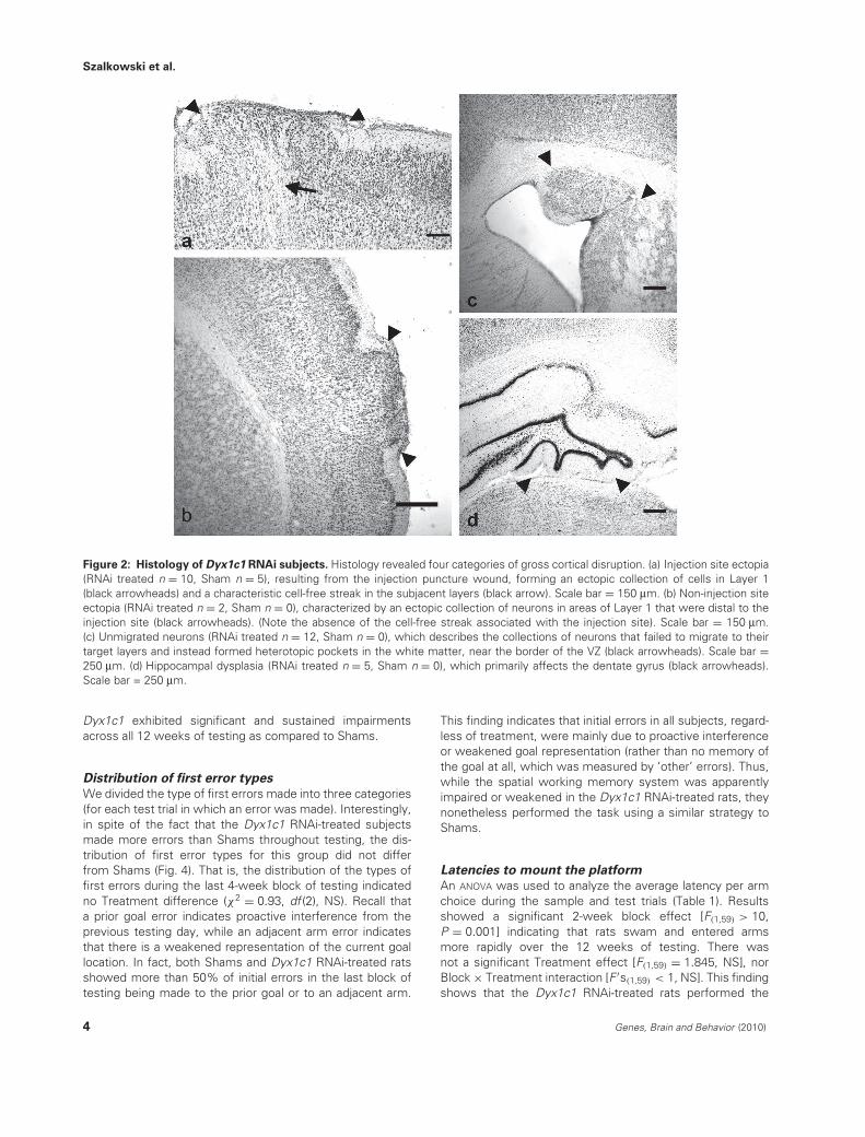
Fluorescence microscopy and immunohistochemical stainingwere used to confirm the presence or absence of GFPand/or RFP. Analysis revealed 33 experimental (RFP-negativeand/or GFP-positive) and 28 control (RFP-positive and/or GFP-negative) subjects. Further histological examination revealedfive categories of cortical characterization: (1) no visiblemalformations (RNAi treated n = 18, Sham n = 23), whichdescribes any subject whose brain tissue was free ofthe gross malformations defined in the other categories,(2) injection site ectopia (RNAi treated n = 10, Sham n = 5),resulting from the injection puncture wound, forming anectopic collection of cells in Layer 1, (3) non-injection siteectopia (RNAi treated n = 2, Sham n = 0), an ectopiccollection of neurons in areas of Layer 1 that were distalto the injection site, (4) unmigrated neurons (RNAi treatedn = 12, Sham n = 0), in which collections of neurons failed tomigrate to their target layers and instead formed heterotopicpockets in the white matter near the border of the VZ and(5) hippocampal dysplasia (RNAi treated n = 5, Sham n = 0)or unmigrated neocortical neurons that primarily disruptedthe dentate gyrus (Fig. 2). The various malformations variedin size and number and some subjects had multiple typesof disruption. It is also worth noting that, other than focalinjection site ectopia (n = 5), Sham animals did not displayany cortical disruption. Note that in addition to being analyzedas a function of treatment (Dyx1c1 RNAi vs. Sham), allbehavioral results were analyzed as a function of thesehistological subgroups.
Errors to find the platform (test trials)
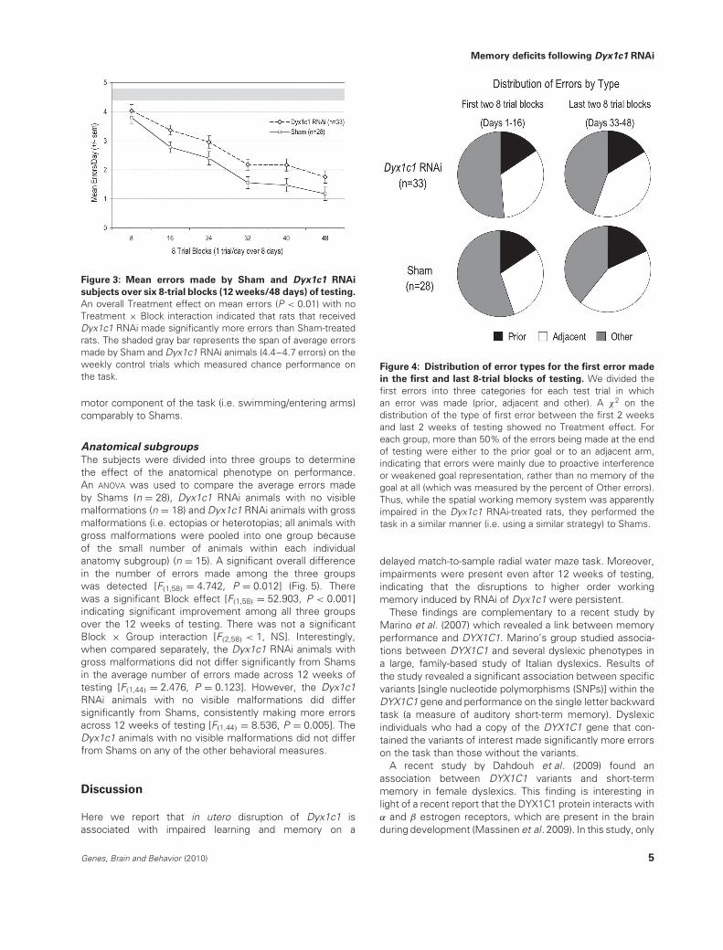
Analyses of the overall errors revealed an overall significanteffect of Treatment, with Dyx1c1 RNAi-treated subjects(n = 33) showing impaired acquisition and performance ofthe delayed match-to-sample radial water maze task ascompared to Shams [F(1,59) = 7.826, P = 0.007] (Fig. 3). Wealso found a significant effect of 2-week block (referringto 2-week blocks of testing) [F(1,59) > 20, P < 0.001], withimproved performance (fewer errors) over testing. Therewas no 2-week block × Treatment interaction [F(1,59) < 1,not significant (NS)], indicating learning for both groups. Yetthese data show that rats that received RNAi targeted against
Genes, Brain and Behavior (2010) 3
Szalkowski et al.
Figure 2: Histology of Dyx1c1 RNAi subjects. Histology revealed four categories of gross cortical disruption. (a) Injection site ectopia(RNAi treated n = 10, Sham n = 5), resulting from the injection puncture wound, forming an ectopic collection of cells in Layer 1(black arrowheads) and a characteristic cell-free streak in the subjacent layers (black arrow). Scale bar = 150 μm. (b) Non-injection siteectopia (RNAi treated n = 2, Sham n = 0), characterized by an ectopic collection of neurons in areas of Layer 1 that were distal to theinjection site (black arrowheads). (Note the absence of the cell-free streak associated with the injection site). Scale bar = 150 μm.(c) Unmigrated neurons (RNAi treated n = 12, Sham n = 0), which describes the collections of neurons that failed to migrate to theirtarget layers and instead formed heterotopic pockets in the white matter, near the border of the VZ (black arrowheads). Scale bar =250 μm. (d) Hippocampal dysplasia (RNAi treated n = 5, Sham n = 0), which primarily affects the dentate gyrus (black arrowheads).Scale bar = 250 μm.
Dyx1c1 exhibited significant and sustained impairmentsacross all 12 weeks of testing as compared to Shams.
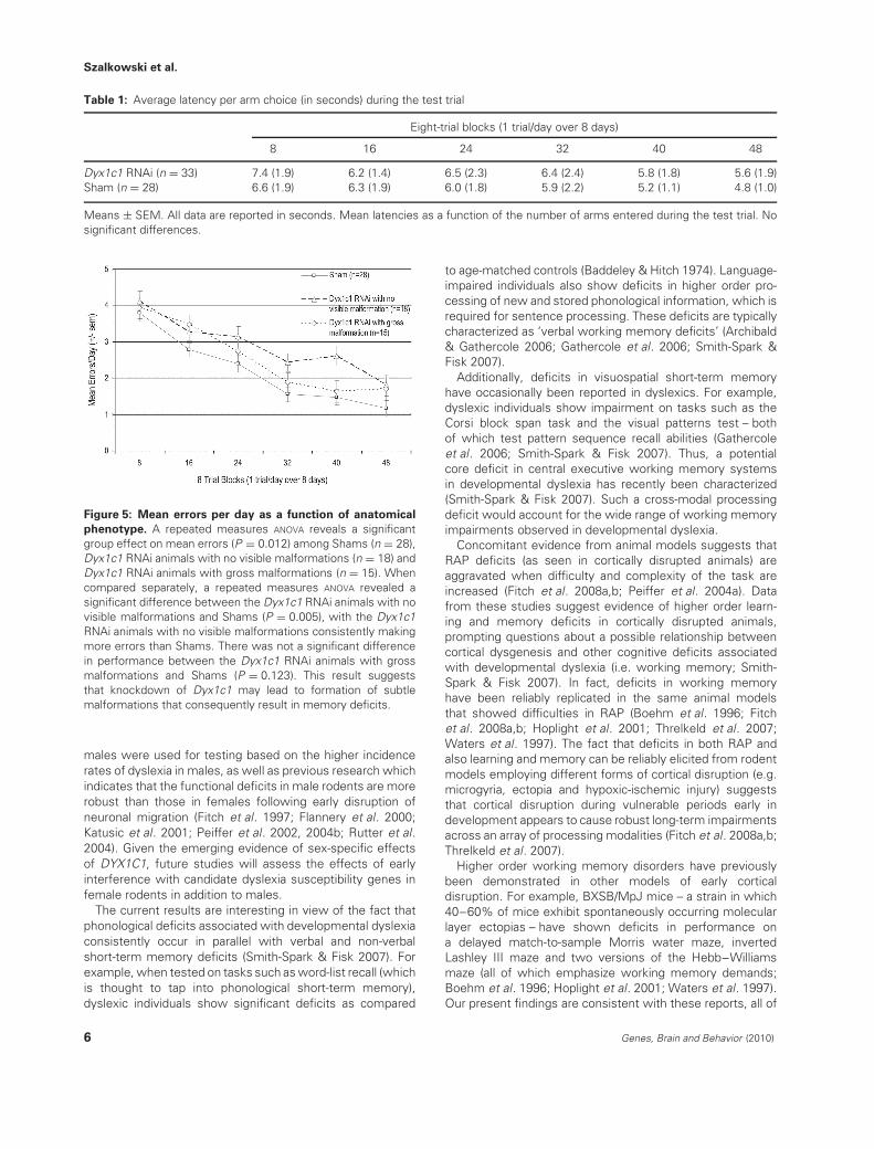
Distribution of first error types
We divided the type of first errors made into three categories(for each test trial in which an error was made). Interestingly,in spite of the fact that the Dyx1c1 RNAi-treated subjectsmade more errors than Shams throughout testing, the dis-tribution of first error types for this group did not differfrom Shams (Fig. 4). That is, the distribution of the types offirst errors during the last 4-week block of testing indicatedno Treatment difference (χ2 = 0.93, df (2), NS). Recall thata prior goal error indicates proactive interference from theprevious testing day, while an adjacent arm error indicatesthat there is a weakened representation of the current goallocation. In fact, both Shams and Dyx1c1 RNAi-treated ratsshowed more than 50% of initial errors in the last block oftesting being made to the prior goal or to an adjacent arm.
This finding indicates that initial errors in all subjects, regard-less of treatment, were mainly due to proactive interferenceor weakened goal representation (rather than no memory ofthe goal at all, which was measured by ‘other’ errors). Thus,while the spatial working memory system was apparentlyimpaired or weakened in the Dyx1c1 RNAi-treated rats, theynonetheless performed the task using a similar strategy toShams.
Latencies to mount the platform
An ANOVA was used to analyze the average latency per armchoice during the sample and test trials (Table 1). Resultsshowed a significant 2-week block effect [F(1,59) > 10,P = 0.001] indicating that rats swam and entered armsmore rapidly over the 12 weeks of testing. There wasnot a significant Treatment effect [F(1,59) = 1.845, NS], norBlock × Treatment interaction [F ′s(1,59) < 1, NS]. This findingshows that the Dyx1c1 RNAi-treated rats performed the
4 Genes, Brain and Behavior (2010)
Memory deficits following Dyx1c1 RNAi
Figure 3: Mean errors made by Sham and Dyx1c1 RNAi
subjects over six 8-trial blocks (12 weeks/48 days) of testing.
An overall Treatment effect on mean errors (P < 0.01) with noTreatment × Block interaction indicated that rats that receivedDyx1c1 RNAi made significantly more errors than Sham-treatedrats. The shaded gray bar represents the span of average errorsmade by Sham and Dyx1c1 RNAi animals (4.4–4.7 errors) on theweekly control trials which measured chance performance onthe task.
motor component of the task (i.e. swimming/entering arms)comparably to Shams.
Anatomical subgroups
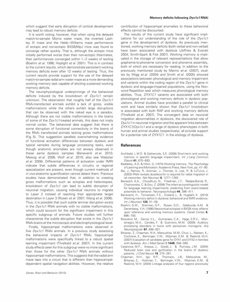
The subjects were divided into three groups to determinethe effect of the anatomical phenotype on performance.An ANOVA was used to compare the average errors madeby Shams (n = 28), Dyx1c1 RNAi animals with no visiblemalformations (n = 18) and Dyx1c1 RNAi animals with grossmalformations (i.e. ectopias or heterotopias; all animals withgross malformations were pooled into one group becauseof the small number of animals within each individualanatomy subgroup) (n = 15). A significant overall differencein the number of errors made among the three groupswas detected [F(1,58) = 4.742, P = 0.012] (Fig. 5). Therewas a significant Block effect [F(1,58) = 52.903, P < 0.001]indicating significant improvement among all three groupsover the 12 weeks of testing. There was not a significantBlock × Group interaction [F(2,58) < 1, NS]. Interestingly,when compared separately, the Dyx1c1 RNAi animals withgross malformations did not differ significantly from Shamsin the average number of errors made across 12 weeks oftesting [F(1,44) = 2.476, P = 0.123]. However, the Dyx1c1RNAi animals with no visible malformations did differsignificantly from Shams, consistently making more errorsacross 12 weeks of testing [F(1,44) = 8.536, P = 0.005]. TheDyx1c1 animals with no visible malformations did not differfrom Shams on any of the other behavioral measures.
Discussion
Here we report that in utero disruption of Dyx1c1 isassociated with impaired learning and memory on a
Figure 4: Distribution of error types for the first error made
in the first and last 8-trial blocks of testing. We divided thefirst errors into three categories for each test trial in whichan error was made (prior, adjacent and other). A χ2 on thedistribution of the type of first error between the first 2 weeksand last 2 weeks of testing showed no Treatment effect. Foreach group, more than 50% of the errors being made at the endof testing were either to the prior goal or to an adjacent arm,indicating that errors were mainly due to proactive interferenceor weakened goal representation, rather than no memory of thegoal at all (which was measured by the percent of Other errors).Thus, while the spatial working memory system was apparentlyimpaired in the Dyx1c1 RNAi-treated rats, they performed thetask in a similar manner (i.e. using a similar strategy) to Shams.
delayed match-to-sample radial water maze task. Moreover,impairments were present even after 12 weeks of testing,indicating that the disruptions to higher order workingmemory induced by RNAi of Dyx1c1 were persistent.
These findings are complementary to a recent study byMarino et al. (2007) which revealed a link between memoryperformance and DYX1C1. Marino’s group studied associa-tions between DYX1C1 and several dyslexic phenotypes ina large, family-based study of Italian dyslexics. Results ofthe study revealed a significant association between specificvariants [single nucleotide polymorphisms (SNPs)] within theDYX1C1 gene and performance on the single letter backwardtask (a measure of auditory short-term memory). Dyslexicindividuals who had a copy of the DYX1C1 gene that con-tained the variants of interest made significantly more errorson the task than those without the variants.
A recent study by Dahdouh et al. (2009) found anassociation between DYX1C1 variants and short-termmemory in female dyslexics. This finding is interesting inlight of a recent report that the DYX1C1 protein interacts withα and β estrogen receptors, which are present in the brainduring development (Massinen et al. 2009). In this study, only
Genes, Brain and Behavior (2010) 5
Szalkowski et al.
Table 1: Average latency per arm choice (in seconds) during the test trial
Eight-trial blocks (1 trial/day over 8 days)
8 16 24 32 40 48
Dyx1c1 RNAi (n = 33) 7.4 (1.9) 6.2 (1.4) 6.5 (2.3) 6.4 (2.4) 5.8 (1.8) 5.6 (1.9)Sham (n = 28) 6.6 (1.9) 6.3 (1.9) 6.0 (1.8) 5.9 (2.2) 5.2 (1.1) 4.8 (1.0)
Means ± SEM. All data are reported in seconds. Mean latencies as a function of the number of arms entered during the test trial. Nosignificant differences.
Figure 5: Mean errors per day as a function of anatomical
phenotype. A repeated measures ANOVA reveals a significantgroup effect on mean errors (P = 0.012) among Shams (n = 28),Dyx1c1 RNAi animals with no visible malformations (n = 18) andDyx1c1 RNAi animals with gross malformations (n = 15). Whencompared separately, a repeated measures ANOVA revealed asignificant difference between the Dyx1c1 RNAi animals with novisible malformations and Shams (P = 0.005), with the Dyx1c1RNAi animals with no visible malformations consistently makingmore errors than Shams. There was not a significant differencein performance between the Dyx1c1 RNAi animals with grossmalformations and Shams (P = 0.123). This result suggeststhat knockdown of Dyx1c1 may lead to formation of subtlemalformations that consequently result in memory deficits.
males were used for testing based on the higher incidencerates of dyslexia in males, as well as previous research whichindicates that the functional deficits in male rodents are morerobust than those in females following early disruption ofneuronal migration (Fitch et al. 1997; Flannery et al. 2000;Katusic et al. 2001; Peiffer et al. 2002, 2004b; Rutter et al.2004). Given the emerging evidence of sex-specific effectsof DYX1C1, future studies will assess the effects of earlyinterference with candidate dyslexia susceptibility genes infemale rodents in addition to males.
The current results are interesting in view of the fact thatphonological deficits associated with developmental dyslexiaconsistently occur in parallel with verbal and non-verbalshort-term memory deficits (Smith-Spark & Fisk 2007). Forexample, when tested on tasks such as word-list recall (whichis thought to tap into phonological short-term memory),dyslexic individuals show significant deficits as compared
to age-matched controls (Baddeley & Hitch 1974). Language-impaired individuals also show deficits in higher order pro-cessing of new and stored phonological information, which isrequired for sentence processing. These deficits are typicallycharacterized as ‘verbal working memory deficits’ (Archibald& Gathercole 2006; Gathercole et al. 2006; Smith-Spark &Fisk 2007).
Additionally, deficits in visuospatial short-term memoryhave occasionally been reported in dyslexics. For example,dyslexic individuals show impairment on tasks such as theCorsi block span task and the visual patterns test – bothof which test pattern sequence recall abilities (Gathercoleet al. 2006; Smith-Spark & Fisk 2007). Thus, a potentialcore deficit in central executive working memory systemsin developmental dyslexia has recently been characterized(Smith-Spark & Fisk 2007). Such a cross-modal processingdeficit would account for the wide range of working memoryimpairments observed in developmental dyslexia.
Concomitant evidence from animal models suggests thatRAP deficits (as seen in cortically disrupted animals) areaggravated when difficulty and complexity of the task areincreased (Fitch et al. 2008a,b; Peiffer et al. 2004a). Datafrom these studies suggest evidence of higher order learn-ing and memory deficits in cortically disrupted animals,prompting questions about a possible relationship betweencortical dysgenesis and other cognitive deficits associatedwith developmental dyslexia (i.e. working memory; Smith-Spark & Fisk 2007). In fact, deficits in working memoryhave been reliably replicated in the same animal modelsthat showed difficulties in RAP (Boehm et al. 1996; Fitchet al. 2008a,b; Hoplight et al. 2001; Threlkeld et al. 2007;Waters et al. 1997). The fact that deficits in both RAP andalso learning and memory can be reliably elicited from rodentmodels employing different forms of cortical disruption (e.g.microgyria, ectopia and hypoxic-ischemic injury) suggeststhat cortical disruption during vulnerable periods early indevelopment appears to cause robust long-term impairmentsacross an array of processing modalities (Fitch et al. 2008a,b;Threlkeld et al. 2007).
Higher order working memory disorders have previouslybeen demonstrated in other models of early corticaldisruption. For example, BXSB/MpJ mice – a strain in which40–60% of mice exhibit spontaneously occurring molecularlayer ectopias – have shown deficits in performance ona delayed match-to-sample Morris water maze, invertedLashley III maze and two versions of the Hebb–Williamsmaze (all of which emphasize working memory demands;Boehm et al. 1996; Hoplight et al. 2001; Waters et al. 1997).Our present findings are consistent with these reports, all of
6 Genes, Brain and Behavior (2010)
Memory deficits following Dyx1c1 RNAi
which suggest that early disruption of cortical developmentmay lead to robust memory deficits.
It is worth noting, however, that when using the delayedmatch-to-sample Morris water maze, the inverted Lash-ley III maze and the Hebb–Williams maze performanceof ectopic and non-ectopic BXSB/MpJ mice was found toconverge rather quickly. That is, although the ectopic miceinitially performed worse than their non-ectopic littermates,their performances converged within 1–2 weeks of testing(Boehm et al. 1996; Hoplight et al. 2001). This is in contrastto the current results, which demonstrate persistent workingmemory deficits even after 12 weeks of testing. Thus, thecurrent results provide support for the use of the delayedmatch-to-sample radial arm water maze as a more demandingworking memory task capable of eliciting sustained workingmemory deficits.
The neurophysiological underpinnings of the behavioraldeficits induced by the knockdown of Dyx1c1 remainunknown. The observation that roughly half of the Dyx1c1RNAi-transfected animals exhibit a lack of gross, visiblemalformations while the others exhibit large disruptionsthat can be observed with the naked eye is a paradox.Although there are not visible malformations in the brainsof some of the Dyx1c1-treated animals, this does not implynormal cortex. The behavioral data suggest that there issome disruption of functional connectivity in the brains ofthe RNAi transfected animals lacking gross malformations(Fig. 5). This suggestion parallels overwhelming evidenceof functional activation differences between dyslexic andtypical samples during language processing tasks, eventhough anatomic anomalies are not always observed inthese same dyslexic samples (Beneventi et al. 2010;Maisog et al. 2008; Wolf et al. 2010; also see Websteret al. 2008). Differential patterns of activation under fMRIindicate that subtle differences in circuitry or regionalspecialization are present, even though current methods ofin vivo anatomic quantification cannot detect them. Previousstudies have demonstrated that, in addition to creatinggross malformations such as ectopias and heterotopias,knockdown of Dyx1c1 can lead to subtle disruption ofneuronal migration, causing individual neurons to migrateto Layer 2 instead of reaching their appropriate targetdestination in Layer 3 (Rosen et al. 2007; Wang et al. 2006).Thus, it is possible that such subtle laminar disruption existsin the Dyx1c1 RNAi animals with no visible malformations,which could account for the significant impairment in thisspecific subgroup of animals. Future studies will furthercharacterize the subtle disruption that exists in the Dyx1c1RNAi brains at the microscopic and electrophysiological level.
Finally, hippocampal malformations were observed infive Dyx1c1 RNAi animals. In a previous study assessingthe behavioral impacts of Dyx1c1 RNAi, hippocampalmalformations were specifically linked to a robust spatiallearning impairment (Threlkeld et al. 2007). In the currentstudy effects seen for this subgroup were no more significantthan those for the other Dyx1c1 RNAi animals withouthippocampal malformations. This suggests that the radial armmaze taps into a circuit that is different than hippocampal-dependent spatial navigation abilities. However, the relative
contribution of hippocampal anomalies to these behavioraleffects cannot be discounted.
The results of the current study have significant impli-cations for our understanding of the role of the Dyx1c1gene in the development of dyslexia. As previously men-tioned, working memory deficits (both verbal and non-verbal)have been associated with dyslexia (Jeffries & Everatt2004; Smith-Spark & Fisk 2007). Working memory is impli-cated in the storage of relevant representations that allowgrapheme-to-phoneme conversion and phoneme assembly,both of which are necessary for reading. In addition to thepreviously mentioned study by Marino et al. (2007), stud-ies by Wigg et al. (2004) and Smith et al. (2005) showedassociations between phonological and memory impairmentand variants within the coding region of the Dyx1c1 gene indyslexic and language-impaired populations, using the Non-word Repetition task which measures phonological memoryabilities. Thus, DYX1C1 variants are associated with bothphonological and working memory deficits in dyslexic pop-ulations. Animal studies have provided a parallel to clinicalwork and have similarly shown that Dyx1c1 knockdownis associated with both RAP and working memory deficits(Threlkeld et al. 2007). The convergent data on neuronalmigration abnormalities in dyslexics, the discovered role ofDyx1c1 in neuronal migration and the apparent links betweenDYX1C1/Dyx1c1 and a range of dyslexic phenotypes in bothhuman and animal studies (respectively), all provide supportfor a potential role of DYX1C1 in the etiology of dyslexia.
References
Archibald, L.M.D. & Gathercole, S.E. (2006) Short-term and workingmemory in specific language impairmtent. Int J Lang CommunDisord 41, 675–693.
Baddeley, A.D. & Hitch, G. (1974) Working memory. The Psychologyof Learning and Motivation. Academic Press, New York, pp. 47–90.
Bai, J., Ramos, R., Ackman, J., Thomas, A., Lee, R. & LoTurco, J.(2003) RNAi reveals doublecortin is required for radial migration inrat neocortex. Nat Neurosci 6, 1277–1283.
Benasich, A.A., Choudhury, N., Friedman, J.T., Realpe-Bonila, R.,Chojnowska, C. & Gou, Z. (2006) The infant as a prelinguistic modelfor language learning impairments: predicting from event-relatedpotentials to behavior. Neuropsychologia 44, 396–411.
Beneventi, H., Tonnessen, F.E., Ersland, L. & Hugdahl, K. (2010)Working memory deficit in dyslexia: behavioral and fMRI evidence.Int J Neurosci 120, 51–59.
Boehm, G.W., Sherman, G.F., Rosen, G.D., Galaburda, A.M. &Denenberg, V.H. (1996) Neocortical ectopias in BXSB mice: effectsupon reference and working memory systems. Cereb Cortex 6,696–700.
Boscariol, M., Garcia, V.L., Guimaraes, C.A., Hage, S.R.V., Mon-tenegro, M.A., Cendes, F. & Guerreiro, M.M. (2009) Auditoryprocessing disorders in twins with perisylvian microgyria. ArqNeuropsiquiatr 67, 499–501.
Brkanac, Z., Chapman, N.H., Matsushita, M.M., Chun, L., Nielsen, K.,Cochrane, E., Berninger, V.W., Wijsman, E.M. & Raskind, W.H.(2007) Evaluation of candidate gene for DYX1 and DYX2 in familieswith dyslexia. Am J Med Genet B 144B, 556–560.
Casanova, M.F., Araque, J., Giedd, J. & Rumsey, J.M. (2004)Reduced brain size and gyrification in the brains of dyslexicpatients. J Child Neurol 19, 275–281.
Chapman, N.H., Igo, R.P., Thomson, J.B., Matsushita, M.,Brkanac, Z., Holzman, T., Berninger, V.W., Wijsman, E.M. &Raskind, W.H. (2004) Linkage analyses of four regions previously
Genes, Brain and Behavior (2010) 7
Szalkowski et al.
implicated in dyslexia: confirmation of a locus on chromosome15q. Am J Med Genet B 131B, 67–75.
Choudhury, N., Leppanen, P., Leevers, H. & Benasich, A.A. (2007)Infant information processing and family history of specificlanguage impairment: converging evidence for RAP deficits fromtwo paradigms. Dev Sci 10, 213–236.
Chrobak, J.J., Hinman, J.R. & Sabolek, H.R. (2008) Revealing pastmemories: proactive interferences and ketamine. J Neurosci 28,4512–4520.
Clark, M., Sherman, G.F., Bimonte, H. & Fitch, R.H. (2000) Percep-tual auditory gap detection deficits in male BXSB mice withcerebrocortical ectopias. Neuroreport 11, 693–696.
Cope, N., Harold, D., Hill, G., Moskvina, V., Stevenson, J., Hol-mans, P., Owen, M., O’Donovan, M. & Williams, J. (2005) Strongevidence that KIAA0319 on chromosome 6p is a susceptibilitygene for developmental dyslexia. Am J Hum Genet 76, 581–591.
Dahdouh, F., Anthoni, H., Tapia-Paez, I., Peyrard-Janvid, M., Schulte-Korne, G., Warnke, A., Remschmidt, H., Ziegler, A., Kere, J.,Muller-Myhsok, B., Nothen, M.M., Schumacher, J. & Zucchelli, M.(2009) Further evidence for DYX1C1 as a susceptibility factor fordyslexia. Psychiatr Genet 19, 59–63.
Farmer, M.E. & Klein, R.M. (1995) The evidence for a temporalprocessing deficit linked to dyslexia: a review. Psychon Bull Rev2, 460–493.
Fisher, S.E. & DeFries, J.C. (2002) Developmental dyslexia: geneticdissection of a complex cognitive trait. Nat Rev Neurosci 3,767–780.
Fitch, R.H., Brown, C., Tallal, P. & Rosen, G.D. (1997) Effects ofsex and MK-801 on auditory-processing deficits associated withdevelopmental microgyric lesions in rats. Behav Neurol 111,404–412.
Fitch, R.H., Breslawski, H., Rosen, G.D. & Chrobak, J.J. (2008a)Persistent spatial working memory deficits in rats with bilateralcortical microgyria. Behav Brain Funct 4, 45.
Fitch, R.H., Threlkeld, S.W., McClure, M.M. & Peiffer, A.M. (2008b)Use of a modified prepulse inhibition paradigm to assess complexauditory dscirimination in rodents. Brain Res Bull 76, 1–7.
Flannery, K.A., Liederman, J., Daly, L. & Schultz, J. (2000) Maleprevalence for reading disability is found in a large sample of Blackand White children free from ascertainment bias. J Int NeuropsychSoc 6, 433–442.
Francks, C., Paracchini, S., Smith, S.D., Richardson, A.J., Scerri, T.S.,Cardon, L.R., Marlow, A.J., MacPhie, L., Walter, J., Penning-ton, B.F., Fisher, S.E., Olson, R.K., DeFries, J.C., Stein, J.F. &Monaco, A.P. (2004) A 77-kilobase region of chromosome 6p22.2is associated with dyslexia in families from the United Kingdomand from the United States. Am J Hum Genet 75, 1046–1058.
Galaburda, A.M., Sherman, G.F., Rosen, G.D., Aboitiz, F. &Geschwind, N. (1985) Developmental dyslexia: four consecutivepatients with cortical anomalies. Ann Neurol 18, 222–233.
Galaburda, A.M., Menard, M. & Rosen, G.D. (1994) Evidence foraberrant auditory anatomy in developmental dyslexia. Proc NatlAcad Sci USA 91, 8010–8013.
Gathercole, S.E., Alloway, T.P., Willis, C. & Adams, A. (2006) Work-ing memory in children with reading disabilities. J Expert ChildPsychol 93, 265–281.
Hage, S.R.V., Cendes, F., Montenegro, M.A., Abramides, D.V.,Guimaraes, C.A. & Guerreiro, M.M. (2006) Specific languageimpairment: linguistic and neurobiological aspects. Arq Neurop-siquiatr 64, 173–180.
Hannula-Jouppi, K., Kaminen-Ahola, N., Taipale, M., Eklund, R.,Nolpola-Hemmi, J., Kaariainen, H. & Kere, J. (2005) The axon guid-ance receptor gene ROBO1 is a candidate gene for developmentaldyslexia. PLoS Genet 1, e50.
Harold, D., Paracchini, S., Scerri, T., Dennis, M., Cope, N., Hill, G.,Moskvina, V., Walter, J., Richardson, A.J., Owen, M.J., Stein, J.F.,Green, E.D., O’Donovan, M.C., Williams, J. & Monaco, A.P. (2006)Further evidence that the KIAA0319 gene confers susceptibility todevelopmental dyslexia. Mol Psychiatr 11, 1085–1091.
Hoplight, B.J., Sherman, G.F., Hyde, L.A. & Denenberg, V.H. (2001)Effects of neocortical ectopias and environmental enrichment onHebb-Williams maze learning in BXSB mice. Neurobiol Learn Mem76, 33–45.
Jeffries, S. & Everatt, J. (2004) Working memory: its role in dyslexiaand other specific learning difficulties. Dyslexia 10, 196–214.
Katusic, S.K., Colligan, R.C., Barbaresi, W.J., Schaid, D.J. & Jacobsen,S.J. (2001) Incidence of reading disability in a population-based bincohort. Mayo Clin Proc 76, 1081–1092.
Ludwig, K.U., Roeske, D., Schumacher, J., Schulte-Korne, G., Konig,I.R., Warnke, A., Plume, E., Ziegler, A., Remschmidt, H., Muller-Myhsok, B., Nothen, M.M. & Hoffmann, P. (2008) Investigationof interaction between DCDC2 and KIAA0319 in a large Germandyslexia sample. J Neural Transm 115, 1587–1589.
Maisog, J.M., Einbinder, E.R., Flowers, D.L., Turkeltaub, P.E. &Eden, G.F. (2008) A meta-analysis of functional neuroimagingstudies of dyslexia. Ann NY Acad Sci 1145, 237–259.
Marino, C., Citterio, A., Giorda, R., Facoetti, A., Menozzi, G.,Vanzin, L., Lorusso, M.L., Nobile, M. & Molteni, M. (2007) Asso-ciation of short-term memory with a variant within DYX1C1 indevelopmental dyslexia. Genes Brain Behav 6, 640–646.
Massinen, S., Tammimies, K., Tapia-Paez, I., Matsson, H., Hokka-nen, M.E., Soderberg, O., Landegren, U., Castren, E., Gustafsson,J.A., Treuter, E. & Kere, J. (2009) Functional interaction of DYX1C1with estrogen receptors suggests involvement of hormonal path-ways in dyslexia. Hum Mol Genet 18, 2802–2812.
Meng, H., Smith, S., Hager, K., Held, M., Liu, J., Olson, R., Penning-ton, B., DeFries, J., Gelernter, J., O’Reilly-Pol, T., Somlo, S., Skud-larski, P., Shaywitz, S., Shaywitz, B., Marchione, K., Wang, Y.,Paramasviam, M., LoTurco, J., Page, G. & Gruen, J. (2005) DCDC2is associated with reading disability and modulates neuronal devel-opment in the brain. Proc Natl Acad Sci USA 102, 17053–17058.
Paracchini, S., Thomas, A., Castro, S., Lai, C., Paramasivam, M.,Wang, Y., Keating, B., Taylor, J., Hacking, D., Scerri, T., Francks, C.,Richardson, A., Wade-Martins, R., Stein, J., Knight, J., Copp, A.,LoTurco, J. & Monaco, A. (2006) The chromosome 6p22 haplotypeassociated with dyslexia reduces the expression of KIAA0319, anovel gene involved in neuronal migration. Hum Mol Genet 15,1659–1666.
Peiffer, A.M., Rosen, G.D. & Fitch, R.H. (2002) Sex differences inrapid auditory processing deficits in ectopic BXSB/MpJ mice.Neuroreport 13, 2277–2280.
Peiffer, A.M., Friedman, J., Rosen, G.D. & Fitch, R.H. (2004a)Impaired gap detection in juvenile microgyric rats. Dev Brain Res152, 93–98.
Peiffer, A.M., Rosen, G.D. & Fitch, R.H. (2004b) Sex differences inrapid auditory processing deficits in microgyric rats. Dev Brain Res148, 53–57.
Rosen, G.D., Waters, N., Galaburda, A.M. & Denenberg, V.H. (1995)Behavioral consequences of neonatal injury of the neocortex. BrainRes 681, 177–189.
Rosen, G.D., Bai, J., Wang, Y., Fiondella, C., Threlkeld, S.W.,LoTurco, J. & Galaburda, A.M. (2007) Disruption of neuronal migra-tion by RNAi of Dyx1c1 results in neocortical and hippocampalmalformations. Cereb Cortex 17, 2562–2572.
Rutter, M., Avshalom, C., Fergusson, D., Horwood, L.J., Good-man, R., Maughan, B., Moffitt, T.E., Meltzer, H. & Carroll, J. (2004)Sex differences in developmental reading disability: new findingsfrom 4 epidemiological studies. JAMA 291, 2007–2012.
Schumacher, J., Anthoni, H., Dahdouh, F., Konig, I.R., Hillmer, A.M.,Kluck, N., Manthey, M., Plume, E., Warnke, A., Remschmidt, H.,Hulsman, J., Cichon, S., Lindgren, C.M., Propping, P.,Zucchelli, M., Zeigler, A., Peyrard-Janvid, M., Schulte-Korne, G.,Nothen, M.M. & Kere, J. (2006) Strong genetic evidence of DCDC2as a susceptibility gene for dyslexia. Am J Hum Genet 78, 52–62.
Smith, S.D., Pennington, B.F., Boada, R. & Shriberg, L.D. (2005)Linkage of speech sound disorder to reading disability loci. J ChildPsychol Psyc 46, 1057–1066.
Smith-Spark, J.H. & Fisk, J.E. (2007) Working memory functioning indevelopmental dyslexia. Memory 15, 34–56.
8 Genes, Brain and Behavior (2010)
Memory deficits following Dyx1c1 RNAi
Taipale, M., Kaminen, N., Nopola-Hemmi, J., Haltia, T., Myllylu-oma, B., Lyytinen, H., Muller, K., Kaaranen, M., Lindsberg, P.,Hannula-Jouppi, K. & Kere, J. (2003) A candidate gene for develop-mental dyslexia encodes a nuclear tetratricopeptide repeat domainprotein dynamically regulated in brain. Proc Natl Acad Sci USA 100,11553–11558.
Tallal, P. & Benasich, A.A. (2002) Developmental language learningimpairments. Dev Psychopathol 14, 559–579.
Tallal, P., Miller, S. & Fitch, R.H. (1993) Neurobiological basis ofspeech: a case for the preeminence of temporal processing. AnnNY Acad Sci 682, 27–47.
Threlkeld, S.W., McClure, M.M., Rosen, G.D. & Fitch, R.H. (2006)Developmental timeframes for induction of microgyria and rapidauditory processing deficits in the rat. Brain Res 1109, 22–31.
Threlkeld, S.W., McClure, M.M., Bai, J., Wang, Y., Rosen, G.D.,LoTurco, J., Galaburda, A.M. & Fitch, R.H. (2007) Developmentaldisruptions and behavioral impairments in rats following in uteroRNAi of Dyx1c1. Brain Res Bull 71, 508–514.
Wang, Y., Paramasivam, M., Thomas, A., Bai, J., Kaminen-Ahola, N.,Kere, J., Voskuil, J., Rosen, G.D., Galaburda, A.M. & LoTurco, J.(2006) Dyx1c1 functions in neuronal migration in developingneocortex. Neuroscience 143, 515–522.
Waters, N.S., Sherman, G.F., Galaburda, A.M. & Denenberg, V.H.(1997) Effects of cortical ectopias on spatial delayed match-to-sample performance in BXSB mice. Behav Brain Res 84, 23–29.
Webster, R.I., Erdos, C., Evans, K., Majnemer, A., Saigal, G.,Kehayia, E., Thordardottir, E., Evans, A. & Shevell, M.I. (2008) Neu-rological and magnetic resonance imaging findings in children withdevelopmental language impairment. J Child Neurol 23, 870–877.
Wigg, K.G., Couto, J.M., Feng, Y., Anderson, B., Cate-Carter, T.D.,Macciardi, F., Tannock, R., Lovett, M.W., Humphries, T.W. &Barr, C.L. (2004) Support for EKN1 as the susceptibility locusfor dyslexia on 15q21. Mol Pyschiatr 9, 1111–1121.
Wilcke, A., Weissfuss, J., Kirsten, H., Wolfram, G., Boltze, J. &Ahnert, P. (2009) The rold of gene DCDC2 in German dyslexics.Ann of Dyslexia 59, 1–11.
Wolf, R.C., Sambataro, F., Lohr, C., Steinbrink, C., Martin, C. &Vasic, N. (2010) Functional brain network abnormalities duringverbal working memory performance in adolescents and youngadults with dyslexia. Neuropsychologia 48, 309–318.
Wright, B., Lombardino, L., King, W., Puranik, C., Leonard, C. &Merzenich, M. (1997) Deficits in auditory temporal and spectralresolution in language-impaired children. Nature 387, 176–178.
Acknowledgements
This work was supported by NIH Grant P01HD57853.
Genes, Brain and Behavior (2010) 9