Embed Size (px)
Citation preview

Available online at www.sciencedirect.com
ScienceDirectActa Materialia 82 (2015) 22–31
www.elsevier.com/locate/actamat
Multiple twins of a decagonal approximant embedded in S-Al2CuMgphase resulting in pitting initiation of a 2024Al alloy
J. Wang,1 B. Zhang,1 Y.T. Zhou and X.L. Ma⇑
Shenyang National Laboratory for Materials Science, Institute of Metal Research, Chinese Academy of Sciences,
Wenhua Road 72, 110016 Shenyang, People’s Republic of China
Received 17 June 2014; revised 27 August 2014; accepted 1 September 2014
Abstract—The pitting of Al–Cu–Mg alloy is believed to originate from the local dissolution of S-Al2CuMg particles, and the dissolution activitydiffers from one particle to another. Nevertheless, the initial site where the dissolution of the S phase preferentially occurs and the cause of the het-erogeneity in the electrochemical dissolution activity remain unknown, hindering our understanding of pitting initiation of Al–Cu–Mg alloys. In thiswork, we have applied in situ ex-environmental transmission electron microscopy and identified a large number of nanosized Al20Cu2Mn3
approximants of the decagonal quasicrystal embedded in the S phase. We find that the S phase with Al20Cu2Mn3 inclusions is more active than thosefree of the approximants. Such a preference is clarified to result from the decomposition of Al20Cu2Mn3 approximant prior to the dissolution of the Sphase. In addition, we also find that the electrochemical behavior of Al20Cu2Mn3 approximants is different. The approximants with multiple twinsare more active than those with few planar defects.� 2014 Acta Materialia Inc. Published by Elsevier Ltd. All rights reserved.
Keywords: S phase; Pitting; Al20Cu2Mn3; 2024Al; Multiple twins
1. Introduction
Al–Cu–Mg alloys (2xxx series commercial alloys) arewidely used in aerospace and other industrial applicationsbecause of their combination of strength, damage toler-ance, formability and density. Al–Cu–Mg alloys derivetheir strength from a heterogeneous microstructure thatconsists of hardening precipitates and dispersoids [1,2].However, they are prone to localized corrosion, and thuspremature breakdown may occur when they are used in achloride-containing environment.
In the past few decades, researchers have been striving tostudy the pitting of Al–Cu–Mg alloy using various tech-niques [3–8]. Among various second-phase particles presentin Al–Cu–Mg alloy, attention is usually paid to the influ-ence of the S-Al2CuMg phase, which is believed to dissolvepreferentially as the initiation sites of pitting [9–15]. The Sphase is one of the key strengthening precipitates inhigh-strength Al–Cu–Mg alloys and has an orthorhombicstructure with space group Cmcm [16–21]. In previous stud-ies, the mechanism of pitting corrosion occurring at the Sphase was believed to be as follows. The S phase iselectrochemically active with respect to the matrix and itis dissolved initially as an anode with Mg and Al selective
http://dx.doi.org/10.1016/j.actamat.2014.09.0011359-6462/� 2014 Acta Materialia Inc. Published by Elsevier Ltd. All rights
⇑Corresponding author; e-mail: [email protected] These authors contributed equally to this work.
dissolution. With the development of Mg and Al dealloy-ing, the remnant becomes noble and tends to behave as acathode, which results in the dissolution of the matrixaround the S phase and consequently pitting occurrence[10,12,22]. In addition, the S phase particles are known tofeature a great heterogeneity in electrochemical activity.In the chloride-containing electrolyte, some S particles aresusceptible to dissolution, while others are electrochemi-cally inactive. Moreover, the dissolution of a so-calledactive S particle usually occurs in an inhomogeneous man-ner. The intrinsic mechanism has not yet been clarified,which limits our understanding of pitting initiation inAl–Cu–Mg alloys [7,23]. Such an unpredictable phenomenonmust result from the poor knowledge of microstructuralinformation which is derived from the relatively lower spa-tial resolution of analysis tools or modes, such as scanningelectron microscopy (SEM), bright-field transmissionelectron microscopy (TEM), energy dispersive X-ray spec-troscopy (EDXS), standard electrochemical measurements,scanning vibrating electrode (SVE) and scanning Kelvinprobe force microscopy (SKPFM), as well as secondaryion mass spectroscopy (SIMS) [8–13,23].
In this work, we have applied an in situ ex-environmen-tal TEM technique [24] to clarify the pitting initiation of an2024Al alloy. We provide atomic-scale information on theinitial site of S phase dissolution and show a novel interpre-tation of the classical problem, which has been unresolvedfor decades.
reserved.

J. Wang et al. / Acta Materialia 82 (2015) 22–31 23
2. Experimental procedure
2.1. Sample preparation
2024 aluminum alloy with nominal composition ofAl–4.35Cu–1.55Mg–0.53Mn–0.11Fe–0.13Si–0.03Ti (wt.%)was chosen for the investigation. The alloy was subjectedto solid-solution treatment (SST) at 768 K and waterquenching. After SST, the alloy was cold-drawn into a tubeform at room temperature with shrinkage of 8–15%, andthen was annealed at 380 �C for 1 h. The sample was cutinto sections perpendicular to the deformation directionusing a linear precision saw with a thickness of 600 lmand then ground to �100 lm thick by 1000 grit silicon car-bide papers. Disks with a diameter of 3 mm were preparedby die-cutting, then ground using variant grit silicon car-bide papers, polished with diamond paste to 20 lm andfinally thinned by ion-milling. After the first-round TEMobservation, some of the specimens were plasma-cleanedand then immersed in the 0.5 mol l�1 NaCl solution atroom temperature for various periods (the duration rangedfrom 8 to 60 min). The TEM specimens which experiencedthe immersion tests were cleaned in distilled water, dried,plasma-cleaned and then transferred into the TEM forfurther investigation, and this was designated as the in situex-environmental TEM method.
2.2. SEM and TEM characterizations
An FEI field emission scanning electron microscope wasused to observe the morphology of precipitates. A TecnaiG2 F30 transmission electron microscope, with a point res-olution of 0.19 nm, equipped with a high-angle angular-dark-field (HAADF) detector and X-ray energy-dispersivespectrometer (EDS) systems, was used at 300 kV for elec-tron diffraction, HAADF imaging, high-resolution electronmicroscopy (HREM) imaging and composition analysis.
3. Results and discussion
3.1. Heterogeneous dissolution of S phase
Fig. 1a is an SEM image showing the distribution of thesecond phase in the present 2024Al alloy. A combination ofelectron diffraction and EDS analysis (not shown here)indicates that the sample is mainly composed of three kindsof second-phase particles, e.g. S-Al2CuMg (at.%: Al 50.5,Cu 28.4, Mg 21.1), h-Al2Cu (at.%: Al 60.7, Cu 39.3) andAl–Cu–Fe–Mn–Si (at.%: Al 62.8, Cu 7.6, Mn 10.4, Fe14.5, Si 4.7) phase. The at.% of each element in everysecond phase is the average value from ten particles. TheS phase particles account for �60% of the whole secondphases.
An in situ ex-environmental TEM method and HAADFtechnique in the TEM are employed in this study. TheHAADF mode provides incoherent images and uses high-angle scattering, leading to strong atomic number (Z) con-trast. Therefore the image contrast in such a mode isstrongly associated with the local variety of chemical com-position and/or thickness contribution [25]. By repeatedobservation of a fixed TEM specimen of 2024Al alloybefore and after immersion for varying durations (severaltens of min) in 0.5 mol l�1 NaCl solution, we monitor thelocal microstructural evolution of a settled S phase particle.
The structural characteristics of the S phases before andafter dissolution are particularly focused, and a typicalexample is shown in Fig. 1. Fig. 1b is the HAADF imageof an S phase particle while Fig. 1c is the same particle thathas been immersed in 0.5 mol l�1 NaCl for 15 min. Thedissolution of the S phase results in a darker contrast(thickness contribution), from which the dissolution withinthe S phase is seen to be strongly localized with some smallpits. They are distributed randomly and always feature ananocore with a different contrast. Fig. 1d(I–XII) ande(I–XII) are the zoom-in images of the local area inFig. 1b and c, showing the same areas in specimens before(Fig. 1d(I–XII)) and after immersion (Fig. 1e(I–XII). It isof great interest to find that each core appears to corre-spond to one nanoparticle embedded in the S phase. Thesenanoparticles show slightly brighter contrast than the Sphase matrix before immersion, while they show darkercontrast than the dissolved S phase after immersion inNaCl, which indicates that the nanoparticles are dissolvedmore severely than the S phase particle. It seems that thelocal dissolution of the S phase is strongly correlated withthe nanoparticle and the initial site of dissolution is at theperiphery of the nanoparticle.
A number of HAADF images of S phase particles aftersuffering local dissolution are displayed in Fig. 2 with thesame magnification. These pits and S phase particles showvarious dimensions and geometric projections, but each pitforms around a nanoparticle, no matter whether it islocated at the inner part of or at the edge of S phase. Itis seen that the dissolution of the S phase around most ofthe nanoparticles seems to concentrically propagate intothe S phase.
Various TEM techniques are applied to identify crystal-lographic characteristics of the nanoparticles embedded inthe S phase. The lattice type of these fine particles isdetermined on the basis of electron diffraction experiments.By observing dozens of S phase particles in TEM, it is con-firmed that most S phase particles feature nanoparticleinclusions. Such nanoparticles are also dispersed in the Almatrix with a similar size to those in the S phase. Abright-field TEM image of an S phase is shown inFig. 3a. The S phase is distinguished by combining EDSand electron diffraction patterns (EDPs). Fig. 3b is a seriesof EDPs obtained from S phase particles. The space groupCmcm with lattice parameters of a = 0.40 nm, b = 0.92 nmand c = 0.71 nm is derived based on the extinction rules inthe EDPs, which agrees with previous studies [16–21]. InFig. 3a, four nanoparticles with sizes of 20–200 nm embed-ded in the S phase are arrowed and labeled as I, II, III andIV. It is seen from the zoom-in images of the nanoparticlesin Fig. 3c that multiple twins are present in these nanopar-ticles and they are distinguished in two types of configura-tion: parallel shaped twin plates (Fig. 3c(I and II)) andprism shaped twin plates (Fig. 3c(III and IV)).
One typical nanoparticle with prism shaped plates (asshown in Fig. 4a) is selected for detailed TEM characteriza-tion. The darker contrast of the particle in this bright-fieldmode indicates that they are parallel or close to a low-indexed zone axis. The EDP (as seen in Fig. 4b) shows apseudo-ten-fold symmetry and the EDS analysis (seen inFig. 4c) indicates that the nanoparticle is composed of Al,Cu and Mn. To further identify the nanoparticle, aHRTEM image (Fig. 4d) is acquired. The nanoparticle iscomposed of twins with a rotation of nearly 36�. Thecrystal which has such a pseudo-tenfold symmetry is an

Fig. 1. In situ ex-environmental TEM observation showing the local dissolution of S phase. (a) An SEM image of the as-received 2024Al showingcoarse intermetallic particles: S(Al2CuMg), h(Al2Cu) and Al–Cu–Mn–Fe phases. (b) A HAADF image showing an S phase particle. (c) The sameparticle as that in (b) but after being immersed in 0.5 mol l�1 NaCl for 15 min. (d) Zoom-in images showing 12 nanoparticles embedded in the S phasein (b). (e) Zoom-in images showing local dissolution around these nanoparticles in (c).
24 J. Wang et al. / Acta Materialia 82 (2015) 22–31
approximant of a decagonal quasicrystal (DQC) [26,27].The DQC and its crystalline approximants share similarstructural units, which are stacked aperiodically in the for-mer but periodically in the latter. The approximant of theDQC in 2024Al alloy is Al20Cu2Mn3 with space groupBbmm and lattice parameters of a = 2.42 nm, b = 1.25 nmand c = 0.775 nm [1,28–31]. The HRTEM micrographand the EDP in Fig. 4d and b of Al20Cu2Mn3 are obtainedalong the crystallographic [010] direction and the twinplanes are all {101}. The Al20Cu2Mn3 is formed duringthe homogenization process of Al alloy and cannot be dis-solved in subsequent heat treatment [1]. Such uniformly dis-tributed fine Al20Cu2Mn3 dispersoids are frequentlyobserved and they are present in many other aluminumalloys. Owing to the fine dimension and complex crystallo-graphic structure, the dispersoids are known to improve themechanical properties of aluminum alloys [32,33]. How-ever, the electrochemical effect of Al20Cu2Mn3 has been lit-tle understood, probably due to the lack of high-resolutiontechniques for the direct observation of local corrosion. Itis not straightforward to observe the nanosize Al20Cu2Mn3
particles embedded in the S phase since the contrast
difference between the S phase and Al20Cu2Mn3 is subtlein bright-field TEM and even in the HAADF mode.
After observing dozens of S phases in specimens, we findthat the dissolution activity between S phase particles ishighly heterogeneous. Such a difference in activity hasalready been found in previous studies [7,23], but the sub-stantial mechanism has not been clarified yet. In the speci-men immersed in 0.5 mol l�1 NaCl solution for 45 min,most S phase particles are dissolved severely, while someremain unchanged. This indicates that S phase particlesare electrochemically different. We find that the S phase par-ticles free of or with few Al20Cu2Mn3 nanoparticles usuallydo not suffer local dissolution. Fig. 5 shows two typical sam-ples of undissolved S phase particles and their EDS mappingimages, from which the two particles are seen to be free ofAl20Cu2Mn3. Although several Al–Cu–Mn–Fe containingparticles exist at the boundary, they do not induce any dis-solution of the S phase. Therefore, it is the Al20Cu2Mn3
approximant that makes the difference in S phase activity.In other words, the S phases with Al20Cu2Mn3 approximantare more active, while those free of or with fewnanoparticles are inactive at the early stage of corrosion.

Fig. 3. TEM observation of S phase particle. (a) Bright-field TEM image in which four nanoparticles embedded in an S phase are arrowed. (b) EDPsof S phase with zone axis of [010], [100] and [001]. (c) Zoom-in bright-field TEM images of the nanoparticles in (a) labeled as I, II, III and IV.
Fig. 2. HAADF images of S phase particles in which local dissolution occurred and pits formed around the nanoparticles. These images have thesame magnification and were obtained from the specimens immersed in 0.5 mol l�1 NaCl for 8–25 min.
J. Wang et al. / Acta Materialia 82 (2015) 22–31 25
3.2. Development process of localized corrosion
The HAADF images shown in Fig. 6 illustrate thecontinuous development of local dissolution of the S phaseand the resultant dissolution of the Al matrix. First, theAl20Cu2Mn3 phase rather than the S phase appears activeand is dissolved initially. As shown in Fig. 6a, threeAl20Cu2Mn3 particles (arrowed) embedded in an S phase
are dissolved locally and small pits are left behind, whilethe surrounding S phase remains unchanged. Second, thesmall pit formed by the dissolution of Al20Cu2Mn3 isenlarged by the dissolution of the S phase at the peripheryof dissolved Al20Cu2Mn3, and Al20Cu2Mn3 is subsequentlydissolved severely, as shown with the darker contrast inFig. 6b. Third, when the pit reaches the boundary of theS phase/matrix, the dissolution develops along the

Fig. 5. HAADF images showing two undissolved S phase particles in 2024Al after being immersed in 0.5 mol l�1 NaCl for 45 min and their EDSmapping images.
Fig. 4. Identification of the nanoparticle. (a) Bright-field TEM image of the nanoparticle. (b) EDP obtained from the nanoparticle in (a). (c) EDSanalysis of the particle is composed of Al, Mn and Cu. (d) HRTEM image along the same axis in (b).
26 J. Wang et al. / Acta Materialia 82 (2015) 22–31
boundary quickly (shown in Fig. 6c) as a result of the cou-pling effect between the S phase (anode) and Al matrix(cathode), while the extending around the Al20Cu2Mn3
remnant becomes minor. The dissolved S phase, with Mgand Al dealloying, tends to change from anodic to cathodicwith respect to the Al matrix and the local pH near thematrix becomes more alkaline because of the cathodic reac-tion, resulting in the dissolution of the adjacent Al matrix(shown in Fig. 6c). Finally, based on the characters ofcorrosion morphology shown in Fig. 6d, the most severecorrosion is identified in zone I, mild corrosion in zone II
and no corrosion in zone III. Such morphology implies thatonce the dissolution of the S phase reaches and subse-quently develops along the boundary, the dissolution isextended from its periphery to the interior. Meanwhile,the dissolution in the Al matrix further extends outward.In other words, the cathodic S phase remnant ring, whichis formed by the dissolution of the S phase particle alongthe boundary, both causes the Al matrix to dissolve andcontinues accelerating the S phase dissolution to extendinto the interior. The dissolution of Al20Cu2Mn3 locatedat or near the boundary results in the extending of S phase

Fig. 6. Observation of the continuous development of local dissolution of S phase and the resulting dissolution of Al matrix. The Al20Cu2Mn3
particles are arrowed. (a) A HAADF image showing that slight local dissolution occurs and that fine pits form at the Al20Cu2Mn3. (b) Pits enlarge tothe S phase at the periphery of dissolved Al20Cu2Mn3. (c) When the pits are extended to reach the boundary of S phase/matrix, the dissolutiondevelops along the boundary quickly and results in the dissolution of the adjacent Al matrix. (d) The cathodic S-phase remnant causes the Al matrixto dissolve and also to continue accelerating the S-phase dissolution to extend into the interior.
J. Wang et al. / Acta Materialia 82 (2015) 22–31 27
dissolution to quickly reach the boundary of the S phase/Almatrix, and thus results in the rapid dissolution of the adja-cent Al matrix. Therefore, the Al20Cu2Mn3 nanoparticleslocated at or near the boundary lead to the occurrence ofAl matrix dissolution more effectively than when they arein the interior.
As the strengthening dispersoids in 2024Al, most ofthe Al20Cu2Mn3 particles disperse in the Al matrix, butthe electrochemical behavior of Al20Cu2Mn3 in thematrix is totally different from those in the S phase.By observing the images shown in Figs. 1 and 2, a largenumber of Al20Cu2Mn3 dispersoids are seen to dispersein the matrix. When the Al20Cu2Mn3 particles in the Sphase are dissolved severely, it is hard to find the disso-lution of the Al20Cu2Mn3 particles in the matrix. At ahigh resolution, only slight dissolution is found in someof them. Fig. 7 is an HAADF animage showing threelocally dissolved Al20Cu2Mn3 particles in the Al matrixwith variant corroded degrees. The dissolution occursin the interior randomly and it does not propagatefurther, which indicates that the local dissolution isretarded greatly. Although some trace-dissolution dotsare seen to distribute in the adjacent matrix, evidentlyit is not due to the dissolved nanoparticle but a minorprocess triggered by the nearby cathodic S phase rem-
nant. So, the Al20Cu2Mn3 remnant, different from theS phase, does not seem to switch from anode to cathodewith respect to the Al matrix.
To fully understand the effect of locally dissolvedAl20Cu2Mn3 in the early corrosion stage, we analyze thechemical composition of a single pit in the S phase. Fig. 8shows an S phase with local dissolution and its EDSmapping on a pit. The corroded core is composed of Al,Cu, Mn and O and the contrast of the O element is brighterthan that in the corroded S phase, which indicates that theAl20Cu2Mn3 nanoparticle is dissolved more severely thanthe S phase and an oxide is proposed to form. The enrich-ment of Cu in the corroded core is slight and much of theAl and Mn elements still exist in the corroded Al20Cu2Mn3
nanoparticle, which means that Al20Cu2Mn3 is onlydissolved partially. Meanwhile, the S phase is alreadylocally dissolved at the periphery. In the dissolved zonethere is a lack of Al and Mg but it is enriched with Cuand O, which indicates that the S phase is decomposed bythe selective dissolution of Al and Mg while Cu remainsin the form of an oxide. From the images of Figs. 1, 2and 6, it is seen that Al20Cu2Mn3 is dissolved almostcompletely when the S phase is dissolved severely, fromwhich we can deduce that the dissolution of Al20Cu2Mn3
is continued parallel to the S phase dissolution.

Fig. 8. HAADF image of a locally dissolved S phase and its EDS mapping on a pit.
Fig. 7. HAADF images showing three local-dissolved Al20Cu2Mn3 particles distributed in Al matrix with varying degrees of corrosion.
28 J. Wang et al. / Acta Materialia 82 (2015) 22–31
Based on the discussion above, we hypothesize thatlocal corrosion proceeds as follows: the S phase, Al20C-u2Mn3 phase and Al matrix consist of a multi-galvaniccouple. The S phase and Al20Cu2Mn3 act as the anodeand Al matrix cathode. The Al20Cu2Mn3 phase is moreactive and dissolved initially. Although Cu enrichmentoccurs, the dissolution does not make Al20Cu2Mn3
switch to cathodic, while it can create a local acid envi-ronment due to the hydrolysis of metal cations. The Sphase anode, triggered by the local acid environment,becomes dissolved and enlarged. The local dissolved areain the S phase turns cathodic, which in turn acceleratesthe further dissolution of the adjacent Al20Cu2Mn3.However, the Al20Cu2Mn3 particles in the matrix haveno chance to experience such a “self-catalyzing” like pro-cess and thus they are corroded slightly as an anodewith respect to the Al matrix. When the local dissolutionof the S phase extends to the boundary, the effect of theS phase (anodic)/Al matrix (cathodic) galvanic couple isprominent. Thus the dissolution develops along theboundary quickly and a Cu-rich remnant ring forms.When dealloying develops further, the Cu-rich ringchanges from an anode to a cathode, which results ina local dissolution of the adjacent Al matrix and a par-allel extending of S phase dissolution. Thus the promi-nent effect of the Al20Cu2Mn3 phase is to indirectlyinfluence the localized corrosion behavior of Al alloythrough increasing the electrochemical activity of the Sphase, other than directly impacting the Al matrix,which is one of the main reasons why the Al20Cu2Mn3
phase has been neglected in electrochemistry in the pastfew decades.
3.3. Heterogeneous dissolution of Al20Cu2Mn3
By observing numerous Al20Cu2Mn3 particles, an inho-mogeneous dissolution in the Al20Cu2Mn3 particles is iden-tified. In order to determine the detailed dissolution, weshorten the immersion duration to monitor the local disso-lution at an initial stage. Fig. 9a is an HAADF image show-ing an S phase particle with some Al20Cu2Mn3 embeddingand Fig. 9b is the same particle which has been immersed in0.5 mol l�1 NaCl for 8 min. By comparing these images, adarker contrast is seen at the inner part of an Al20Cu2Mn3
nanoparticle (marked with arrow) after immersion, whichindicates that the Al20Cu2Mn3 particle is dissolved slightlywhile the S phase remains unchanged. Fig. 9c shows bright-field TEM images of the same Al20Cu2Mn3 particle as inFig. 9a. In contrast to the HAADF mode, which is moresensitive to chemical composition and/or thickness contri-bution, the bright-field mode shows more crystallographicdetails. It is seen that the Al20Cu2Mn3 particle features mul-tiple twins with a configuration of prism shaped plates andthe initial site of preferential dissolution is in the vicinity ofthe intersected zone. Similarly, the dissolution activity ofAl20Cu2Mn3 particles is also found to be highly heteroge-neous from one to another. As shown in Fig. 10a and b,in an S phase particle immersed in NaCl for 25 min, someAl20Cu2Mn3 particles marked with black arrows are dis-solved severely, around which the S phase is also decom-posed. Moreover, the Al matrix at the S phase/Alinterface is dissolved as well. Some other Al20Cu2Mn3
particles labeled as I, II, III and IV, nevertheless, werenot attacked, which indicates that Al20Cu2Mn3 particlesbehave differently. Fig. 10c(I–IV) shows the zoom-in
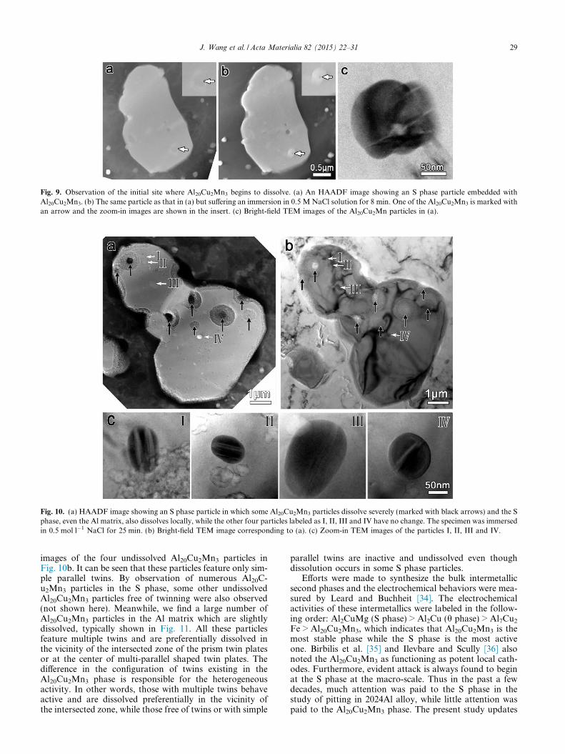
Fig. 10. (a) HAADF image showing an S phase particle in which some Al20Cu2Mn3 particles dissolve severely (marked with black arrows) and the Sphase, even the Al matrix, also dissolves locally, while the other four particles labeled as I, II, III and IV have no change. The specimen was immersedin 0.5 mol l�1 NaCl for 25 min. (b) Bright-field TEM image corresponding to (a). (c) Zoom-in TEM images of the particles I, II, III and IV.
Fig. 9. Observation of the initial site where Al20Cu2Mn3 begins to dissolve. (a) An HAADF image showing an S phase particle embedded withAl20Cu2Mn3. (b) The same particle as that in (a) but suffering an immersion in 0.5 M NaCl solution for 8 min. One of the Al20Cu2Mn3 is marked withan arrow and the zoom-in images are shown in the insert. (c) Bright-field TEM images of the Al20Cu2Mn particles in (a).
J. Wang et al. / Acta Materialia 82 (2015) 22–31 29
images of the four undissolved Al20Cu2Mn3 particles inFig. 10b. It can be seen that these particles feature only sim-ple parallel twins. By observation of numerous Al20C-u2Mn3 particles in the S phase, some other undissolvedAl20Cu2Mn3 particles free of twinning were also observed(not shown here). Meanwhile, we find a large number ofAl20Cu2Mn3 particles in the Al matrix which are slightlydissolved, typically shown in Fig. 11. All these particlesfeature multiple twins and are preferentially dissolved inthe vicinity of the intersected zone of the prism twin platesor at the center of multi-parallel shaped twin plates. Thedifference in the configuration of twins existing in theAl20Cu2Mn3 phase is responsible for the heterogeneousactivity. In other words, those with multiple twins behaveactive and are dissolved preferentially in the vicinity ofthe intersected zone, while those free of twins or with simple
parallel twins are inactive and undissolved even thoughdissolution occurs in some S phase particles.
Efforts were made to synthesize the bulk intermetallicsecond phases and the electrochemical behaviors were mea-sured by Leard and Buchheit [34]. The electrochemicalactivities of these intermetallics were labeled in the follow-ing order: Al2CuMg (S phase) > Al2Cu (h phase) > Al7Cu2
Fe > Al20Cu2Mn3, which indicates that Al20Cu2Mn3 is themost stable phase while the S phase is the most activeone. Birbilis et al. [35] and Ilevbare and Scully [36] alsonoted the Al20Cu2Mn3 as functioning as potent local cath-odes. Furthermore, evident attack is always found to beginat the S phase at the macro-scale. Thus in the past a fewdecades, much attention was paid to the S phase in thestudy of pitting in 2024Al alloy, while little attention waspaid to the Al20Cu2Mn3 phase. The present study updates

Fig. 11. Twins in Al20Cu2Mn3 provide the initial dissolution sites. (a) Bright-field TEM images of Al20Cu2Mn3 particles showing the feature of twinsin Al20Cu2Mn3. (b) HAADF images of the same particles showing the initial site of dissolution in Al20Cu2Mn3. The specimen was immersed in0.5 mol l�1 NaCl for 25 min.
30 J. Wang et al. / Acta Materialia 82 (2015) 22–31
the activity order of the main second phase as: Al20Cu2Mn3
(with multiple twins) > S phase > Al20Cu2Mn3 (withouttwins or with simple parallel twins), which is remarkablydifferent from the previous viewpoint. However, the Al20C-u2Mn3 which was used for investigation in the aboveresearch was synthesized artificially, rather than using a dis-persoid precipitated in alloys. As a result, the compositionand microstructural configuration in the as-synthesizedAl20Cu2Mn3 are proposed to be different from the disper-soid precipitations in the present 2024Al alloy, which may sig-nificantly influence the electrochemical properties. Thus, thecathodic property of as-synthesized Al20Cu2Mn3 cannot berepresentative of that precipitated in industrial Al alloys.
The phenomenon that Al20Cu2Mn3 with multiple twinsis more active than those free of twins implies that a specialstructure or chemical composition may exist at the inter-sected zone of twins, which changes the electrochemicalactivity. The microstructure of Al20Cu2Mn3 is similar withthe T–Al–Pd–Mn and Al–Pd–Fe phases in which the meta-dislocations were observed in previous studies [37,38]. Met-adislocations are known to be very complex defects incrystals as they usually involve several hundreds of atomsin their core. The metadislocations are proposed to existin the present Al20Cu2Mn3 approximant and probably theyprefer to form at the intersected zone of twin plates. Theheterogeneous dissolution activity of Al20Cu2Mn3 particlesin the present study might be more or less associated withthe metadislocations to be confirmed further.
4. Conclusions
With the application of in situ ex-environmental TEM,we have identified the initial site, at an atomic scale, of pit-ting corrosion in an Al–Cu–Mg alloy. The S-Al2CuMgphase is compositionally and structurally inhomogeneous.
A large number of nanosized approximants of the DQC,which is determined to be Al20Cu2Mn3, are embedded inmost S particles. TEM studies indicate that the embeddedAl20Cu2Mn3 approximants are responsible for the activityof the S phase, which clarifies the well-known phenomenonthat the activities of the S phase particles are electrochem-ically different from one to another. In addition, we havedirectly observed that the dissolution of Al20Cu2Mn3
approximant is the initial stage prior to the S phase disso-lution. Moreover, we also find the initial site of the Al20C-u2Mn3 dissolution and the electrochemical activity ofdifferent Al20Cu2Mn3 particles is varied. The Al20Cu2Mn3
approximants with multiple twins are more active thanthose with few planar defects.
Acknowledgements
This work is supported by the National Natural ScienceFoundation of China (51101157) and the National Basic ResearchProgram of China (2009CB623705). We are grateful for the tech-nical assistance of Mr B. Wu of this lab in the TEM experiments.
References
[1] S.C. Wang, M.J. Starink, Int. Mater. Rev. 50 (2005) 193.[2] S. Cheng, Y.H. Zhao, Y.T. Zhu, E. Ma, Acta Mater. 55
(2007) 5822.[3] A. Hughes, T.H. Muster, A. Boag, A.M. Glenn, C. Luo, X.
Zhou, et al., Corros. Sci. 52 (2010) 665.[4] R. Wei, C.-M. Liao, M. Gao, Metall. Mater. Trans. A 29
(1998) 1153.[5] W. Zhang, G.S. Frankel, Electrochim. Acta 48 (2003) 1193.[6] C. Blanc, A. Freulon, M.-C. Lafont, Y. Kihn, G. Mankowski,
Corros. Sci. 48 (2006) 3838.[7] L. Lacroix, L. Ressier, C. Blanc, G. Mankowski, J.
Electrochem. Soc. 155 (2008) C131.

J. Wang et al. / Acta Materialia 82 (2015) 22–31 31
[8] C. Augustin, E. Andrieu, C. Blanc, G. Mankowski, J.Delfosse, J. Electrochem. Soc. 154 (2007) C637.
[9] A. Boag, A.E. Hughes, A.M. Glenn, T.H. Muster, D.McCulloch, Corros. Sci. 53 (2011) 17.
[10] R.G. Buchheit, R.P. Grant, P.F. Hlava, B. McKenzie, G.L.Zender, J. Electrochem. Soc. 144 (1997) 2621.
[11] V. Guillaumin, G. Mankowski, Corros. Sci. 41 (1998) 421.[12] D. Zhu, W.J. van Ooij, Corros. Sci. 45 (2003) 2177.[13] N. Birbilis, R.G. Buchheit, D.L. Ho, M. Forsyth, Electro-
chem. Solid State Lett. 8 (2005) C180.[14] M. Bethencourt, F.J. Botana, M.J. Cano, M. Marcos, J.M.
Sanchez-Amaya, L. Gonzalez-Rovira, Corros. Sci. 51 (2009) 518.[15] A. Boag, R.J. Taylor, T.H. Muster, N. Goodman, D.
McCulloch, C. Ryan, et al., Corros. Sci. 52 (2010) 90.[16] V. Radmilovic, R. Kilaas, U. Dahmen, G.J. Shiflet, Acta
Mater. 47 (1999) 3987.[17] J. Majimel, G. Molenat, F. Danoix, O. Thuillier, D. Blavette,
G. Lapasset, et al., Philos. Mag. 84 (2004) 3263.[18] S.C. Wang, M.J. Starink, Acta Mater. 55 (2007) 933.[19] Z.R. Liu, J.H. Chen, S.B. Wang, D.W. Yuan, M.J. Yin, C.L.
Wu, Acta Mater. 59 (2011) 7396.[20] H. Perlitz, A. Westgten, Arkiv. Kemi. Mineral. Geol. 16B
(1943) 13.[21] R. Kilaas, V. Radmilovic, Ultramicroscopy 88 (2001) 63.[22] H.M. Obispo, L.E. Murr, R.M. Arrowood, E.A. Trillo, J.
Mater. Sci. 35 (2000) 3479.[23] L. Lacroix, L. Ressier, C. Blanc, G. Mankowski, J. Electro-
chem. Soc. 155 (2008) C8.[24] S.J. Zheng, Y.J. Wang, B. Zhang, Y.L. Zhu, C. Liu, P. Hu,
et al., Acta Mater. 58 (2010) 5070.
[25] S.J. Pennycook, Structure determination through Z-contrastmicroscopy, in: P.W. Hawkes, V.-A. Marco (Eds.), Advancesin Imaging and Electron Physics, vol. 123, Elsevier, Amster-dam, 2002, p. 173.
[26] J.D. FitzGerald, R.L. Withers, A.M. Stewart, A. Calka,Philos. Mag. B58 (1988) 15.
[27] R.C. Hudd, W.H. Taaylor, Acta Crystallogr. 15 (1962) 271.[28] K. Robinson, Philos. Mag. 43 (1952) 775.[29] K. Robinson, Acta Crystallogr. 7 (1954) 494.[30] S.C. Wang, C.Z. Li, M.G. Yan, Mater. Res. Bull. 24 (1989)
1267.[31] X.Z. Li, K.H. Kuo, Philos. Mag. B 66 (1992) 117.[32] Y. Li, Z. Liu, L. Lin, J. Peng, A. Ning, J. Mater. Sci. 46 (2011)
3708.[33] A.R. Toleuova, N.A. Belov, D.U. Smagulov, A.N. Alabin,
Met. Sci. Heat Treat. 54 (2012) 402.[34] R.R. Leard, R.G. Buchheit, Electrochemical characterization
of copper-bearing intermetallic compounds and localizedcorrosion of Al–Cu–Mg–Mn alloy 2024, in: P.J.H.S.J. Greg-son (Ed.), Aluminum Alloys 2002: Their Physical andMechanical Properties, Pts. 1–3, vol. 396–4, Trans TechPublications Ltd, Switzerland, 2002, p. 1491.
[35] N. Birbilis, M.K. Cavanaugh, R.G. Buchheit, Corros. Sci. 48(2006) 4202.
[36] G.O. Ilevbare, J.R. Scully, Corrosion 57 (2001) 134.[37] M. Heggen, L. Houben, M. Feuerbacher, Nat. Mater. 9
(2010) 332.[38] M. Heggen, L. Houben, M. Feuerbacher, Philos. Mag. 88
(2008) 2333.


![A complicated quasicrystal approximant [epsilon]16 predicted by](https://img.dokumen.tips/doc/110x75/613d3a34736caf36b75ad4c8/a-complicated-quasicrystal-approximant-epsilon16-predicted-by.jpg)