Embed Size (px)
Citation preview

Multimodality Evaluation of
Gastric Pathology with
Endoscopic Correlation: Part 2, Neoplastic Disease Entities
SF MOHAMMAD, MD 1; C MA, MD 2; M NGUYEN, MD 1; M DESHMUKH, MD 1; R MASAMED, MD 2; DJ MARGOLIS MD 2;
MK PATEL, MD 1
1 Olive View-UCLA Medical Center, Department of Radiology 2 UCLA Medical Center, Department of Radiological Sciences
David Geffen School of Medicine at UCLA
No Relevant Financial Disclosures
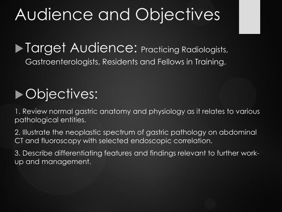
Audience and Objectives
Target Audience: Practicing Radiologists,
Gastroenterologists, Residents and Fellows in Training.
Objectives: 1. Review normal gastric anatomy and physiology as it relates to various
pathological entities.
2. Illustrate the neoplastic spectrum of gastric pathology on abdominal
CT and fluoroscopy with selected endoscopic correlation.
3. Describe differentiating features and findings relevant to further work-
up and management.

Stomach Normal Anatomy
Normal stomach appearance on (A -B) double
contrast UGI, (C -D) contrast enhanced CT
(CECT) , and (E -F)
Esophagogastroduodenoscopy (EGD).
A
F E
D C
B
UGI
EGD
CT
• DISTENSIBILITY: Easily distensible
• WALL: Homogeneous enhancement. Normal
gastric wall thickness is site specific:
• <10 mm in thickness for an under
distended stomach.
• <5 mm for a distended gastric body
• The distal gastric antrum often exceeds
the threshold of 5 mm. Some studies
suggest that irregular or eccentric
antral thickening >12 mm is abnormal.
• Pseudo-thickening is also noted at the
fundus.
• Gastric wall thickening is a nonspecific
finding. Wall thickness of >1 cm has a
high sensitivity but low specificity in
detecting malignant or potentially
malignant lesions on CT, therefore
further diagnostic evaluation may be
required.
• Assess enhancement pattern, if
thickening is focal, eccentric or
irregular.
• Imaging Evaluation:
• Upper Gastrointestinal Fluoroscopy
(UGI)
• Contrast enhanced CT (CECT)
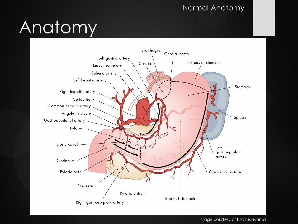
Anatomy
Image courtesy of Lisa Nishiyama
Normal Anatomy

Abnormalities of the Stomach Wall
• Thickening
• Circumferential
• Gastritis (A)- i.e. NSAIDS, H. pylori, radiation, hypertrophic gastropathy
• Tumor
• Eccentric
• Tumor – Primary or metastatic
• Serpentine
• Varices
Lumen
• Foreign body
• Polyps (B)
• Leiomyoma
• Lipoma
• Gastrointestinal Stromal Tumor (GIST)
• Bezoar/Retained food
• Ectopic pancreatic rest
• Malignant Neoplasms
Distensibility
• Extrinsic mass effect
• Annular pancreas
• Regional adenopathy or other mass effect
• Inherent mural indistensibility (C )
• Malignancy
• Peptic scarring
• Corrosive ingestion
• Granulomatous disease
Position
• Hiatal hernia (D)
• Volvulus
Integrity
• Ulcer (E)
• Perforation
• Emphysematous gastritis
• Gastric emphysema
A E D C B
Abnormal Stomach

UGI Evaluation
Gastric
Evaluation (radiologically or endoscopically):
- Rugal folds - Mucosa - Distensibility - Anatomy/
position - Presence and
degree of reflux
C B A D
D A C B E
Abnormal UGI:
Normal UGI:
Normal (A) rugal folds, (B) mucosa with areae gastricae pattern and GE junction, (C)
distensibility(D) and posterior duodenal sweep.
(A) Thickened rugal folds, (B) polyps, (C) persistent indistensibility, (D) hiatal hernia with esophageal reflux and (E)
ulcerations.
Imaging of the Stomach: UGI

Normal esophagus Normal rugal folds
Esophagitis Blood
Retained food
Erythema Ulceration
Normal mucosa
Esophagogastroduodenoscopy (EGD)
Polyps Rugal fold thickening Luminal narrowing
Imaging of the Stomach: EGD
Normal duodenum

Gastric Adenocarcinoma Background
Most common gastric malignancy, > 95% of all
malignant tumors of the stomach
Overall 5-year survival rates < 20%
Prognosis correlated to stage: CT is the staging
modality of choice
Peak incidence: 50 and 70 years
Early stage gastric cancers are curable lesions,
with 5-year survival rates of more than 90%.
Predisposing conditions: atrophic gastritis,
pernicious anemia, gastric polyps, partial
gastrectomy, and Ménétrier disease
30% of cancers are located in the antrum, 30% in
the body, and 30% in the fundus or cardia region.
The remaining 10% are diffusely infiltrating lesions
that involve the entire stomach
Pathology
Most gastric cancers are adenocarcinomas of
mucinous cell origin
Signet-ring cell carcinomas account for 5%–15%
scirrhous infiltration of the gastric wall. Scirrhous
carcinomas frequently involve the distal half of the
stomach, arise near the pylorus, and gradually extend
upward from the antrum into the body and fundus.
Imaging features
Focal area of mural thickening with or without
Polypoid lesion
Generalized mural thickening
In early gastric cancers, malignant invasion is limited to
the mucosa or submucosa. Advanced gastric cancer
invades the muscularis propria.
Signet-ring cell cancer usually manifests as a scirrhous
tumor of the stomach that leads to obliteration of
gastric folds and diffuse thickening of the gastric wall
(linitis plastica)
Gastric Adenocarcinoma
Gastric adenocarcinoma on CT and EGD

• CT is the staging modality of choice because it
can identify the primary tumor and assess for
extragastric disease, which is vital to determine
treatment, palliative versus curative gastric
surgery.
• EUS (endoscopic ultrasound) is the diagnostic
modality of choice for the preoperative staging
of early gastric cancer.
• Extragastric disease:
• Direct extension:
• Pancreas via lesser sac.
• Transverse colon via gastrocolic ligament.
• Liver via gastrohepatic ligament.
• Longitudinally, ie esophagus
• Lymph nodes: Local, regional or distant
• Distant Metastases
CT Evaluation Gastric
Adenocarcinoma A
B
(A) Axial and (B) coronal CECT of a patient with
gastric cancer demonstrates extensive
extragastric spread with regional
lymphadenopathy (yellow arrows) and
periportal extension of tumor (blue arrow) to the
liver with narrowing of the portal vein .
Gastric Adenocarcinoma

Ulceration
(A- C) Abnormal persistent area of ulceration (yellow arrows) in the gastric antrum on UGI. (D) CECT demonstrates antral mural edema and thickening (blue arrow).
(E- G) Corresponding EGD demonstrates prominent gastric folds (green arrows) in the antrum which did not distend easily.
Gastric Adenocarcinoma
F G
E
D A C B

Scirrhous adenocarcinoma: (A-B) UGI
demonstrates a rigid mid-distal
gastric body (yellow arrows) with
distorted rugal pattern, but no gastric
obstruction.
(C-E) Corresponding CT
demonstrates irregular thickening
(green arrows) of the gastric body
and antrum.
Infiltration: Focal Indistensibility
Gastric Adenocarcinoma
E D
A
C
B

(A-B) UGI demonstrates
persistent gastric
indistensibility (yellow
arrows).
(C-D ) CECT
demonstrates
corresponding diffuse
gastric wall thickening
(green arrows)
(E-G) EGD confirms
diffuse irregular
thickening with
infiltration of the folds
(blue arrows) extending
from the cardia to the
peri-pyloric region in
the antrum. Irregular
and minimally stenotic
lumen (magenta
arrows).
Pathology confirmed
signet ring cell cancer,
with a background of
chronic gastritis.
Linitis Plastica- Diffuse Indistensibility Gastric Adenocarcinoma
G F E
C B A
Linitis Plastica Differential:
• Scirrhous Adenocarcinoma
• Metastatic disease (breast)
• Lymphoma
• Granulomatous disease
D

Patient with a history of H. pylori gastritis presents with early satiety and abdominal pain. (A-B) Axial MRI and (C )
axial and (D) coronal CECT demonstrates prior gastrojejunostomy (yellow arrow) with irregular circumferential
thickening (blue arrows), loss of normal rugal fold pattern (green arrow) and luminal narrowing (magenta arrows) of
the gastric antrum as well as multiple prominent lymph nodes (red arrow). (E -G) EGD demonstrates an indurated
(orange arrows) circumferential mass (blue arrows) involving the antrum with narrowing of the gastric lumen.
Loss of Normal Rugal Folds Gastric Adenocarcinoma
C B A
F
D
E G

Focal wall thickening
(A ) axial and (B) coronal CECT demonstrates marked irregular thickening (blue arrows) of the gastric antrum. (C )
Corresponding EGD demonstrated a mass (magenta arrows) extending from the pre-pyloric antrum to the duodenal
bulb with diffuse ulceration and friable appearing mucosa.
(A) Coronal CECT demonstrates more focal thickening (green arrow) and hyperenhancement (yellow arrow) of the
greater curvature. (B -C) Corresponding EGD demonstrates an erythematous ulcerating mass (orange arrows).
Gastric Adenocarcinoma
C B A
B A C

(A) Axial and (B) coronal CECT demonstrates thickening of the stomach (yellow arrows), particularly the greater
curvature. (C -D) Corresponding EGD demonstrates a deep friable gastric ulcer (blue arrows) with irregular borders
at the angularis, worrisome for malignancy. Pathology confirmed poorly-differentiated adenocarcinoma, diffuse
type with signet ring cell features. - H. pylori.
Diffuse wall thickening
(A -B) Axial CECT demonstrates irregular marked gastric wall thickening (green arrows). (C-D) Corresponding
EGD with erosive, friable, edematous and nodular gastric mucosa (red arrow). Pathology confirmed signet ring cell
cancer, ulcer and H. pylori.
Gastric Adenocarcinoma
C B A
B
D
A C D

(A) Coronal and (B) axial CECT
demonstrates an enhancing polypoid
mass (green arrows) in the stomach. (C
) UGI confirms a polypoid filling defect
(blue arrow) in the stomach. (D )
Corresponding EGD demonstrates an
approximately 3 cm ulcerated fundal
mass (yellow arrows) . Pathology
confirmed gastric adenocarcinoma.
Polypoid
Gastric Adenocarcinoma
C
B A
D
Gastric Outlet Obstruction
Gastric Adenocarcinoma
C B A
F D E G
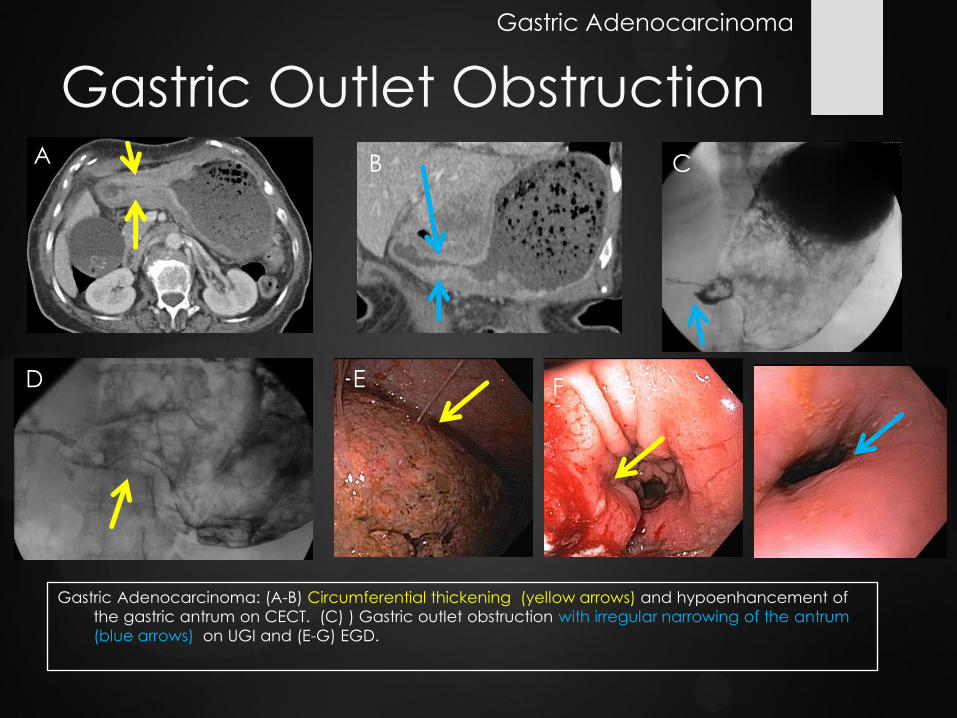
Gastric Adenocarcinoma: (A-B) Circumferential thickening (yellow arrows) and hypoenhancement of
the gastric antrum on CECT. (C) ) Gastric outlet obstruction with irregular narrowing of the antrum
(blue arrows) on UGI and (E-G) EGD.

Virchow Nodes (A) Axial and (B) coronal CECT
demonstrates gastric
outlet obstruction (yellow
arrow) due to an antral
mass (blue arrow).
(C ) Corresponding EGD
demonstrated a friable
malignant appearing antral
mass.
(D -E) Staging CT Chest
demonstrates left
supraclavicular (orange
arrows) and left axillary
adenopathy (green arrows)
which has corresponding (F)
abnormal FDG activity on PET .
A
F E D
C B
Pattern of Gastric Metastases
Lymph Node Evaluation:
Greater than 6 mm is suspicious
Local (N1): prepyloric, gastrocolic ligament and gastrohepatic
ligament. Usually removed with gastrectomy.
Regional (N2): Portahepatic, hepatoduodenal ligament,
peripancreatic and celiac.
Distant (N3): left axillary and left supraclavicular (Virchow nodes).

Gastric Adenocarcinoma. (A) Coronal
and (B) axial CECT demonstrates
omental nodules (blue arrows), ascites
and focal gastric enhancement with
thickening of the gastric folds (yellow
arrow). (C and D) EGD with thickened
gastric folds (magenta arrow) and mild
erythema (green arrow).
Omental Carcinomatosis
Pattern of Gastric Metastases
C
B A
D Intraperitoneal and
omental metastases
are common in
advanced gastric
cancer and may
manifest as nodules,
ascites, irregular
thickening of the
mesentery and
omentum.

Krukenberg tumor
25 yo female presents to ED with pelvic pain and hard abdomen. (A) CECT demonstrates ascites, large pelvic mass ( yellow arrow) and (B) a small
focal area of gastric thickening (blue arrow). Given the degree of pelvic disease
findings were felt to represent an ovarian primary with metastatic disease. Frozen
section during oophorectomy and exploratory laparoscopy confirmed bilateral
Krukenberg tumors with gastric adenocarcinoma as the primary.
(C ) Status post partial gastrectomy(green arrow) and oophorectomy.
Gastric Cancer is the most common primary to metastasize to the
ovaries. Usually bilateral and are known as Krukenberg tumors.
Pattern of Gastric Metastases
C B A

Osseous and dural
metastases
(A) CECT in a patient with known gastric cancer (blue arrows) develops (B -C) multiple enhancing vertebral
bodies compatible with thoracolumbar osseous (yellow arrow) metastases with epidural drop metastases
(green arrow) and paravertebral extension.
A
C B
• Liver is the most common site for hematogenous metastases because the portal vein drains the stomach.
• Lungs, adrenals and kidneys are less common. • Bone and cerebral metastases are uncommon in gastric cancer.
Pattern of Gastric Metastases

Gastric Lymphoma 1-5% of gastric malignancies
Stomach is the most common location of
extranodal lymphoma
B Cell type non-Hodgkin lymphoma- aggressive
May cause Linitis plastica
Low grade mucosa-associated lymphoid tissue (MALT)- indolent clinical course, better prognosis than gastric carcinoma
Associated with Helicobacter pylori
Gastric wall thickening, usually minimal
Adenopathy or extragastric extension is uncommon
Transpyloric spread of tumor into the
duodenum occurs in 30% of patients;
more common than with
adenocarcinoma. Since the stomach
remains pliable and does not obstruct,
patients may present with perforation.
A B
C D
Gastric MALT: (A) Axial CECT demonstrates a
homogeneously enhancing solid gastric fundal mass
(blue arrow) . Further evaluation with EGD demonstrates
(B - C) a smooth-walled firm mass without overlying
mucosal abnormalities. (D) Follow up CT after treatment
demonstrates interval improvement without a discrete
residual mass identified.
Gastric Lymphoma

Gastric Lymphoma Imaging Findings:
Segmental or diffuse bulky gastric wall thickening due to submucosal spread, >1 cm. Nodular, disorganized wall thickening. Less commonly may be a polypoid mass or ulcerative.
Less likely to cause gastric outlet obstruction than gastric adenocarcinoma.
Infiltration of the stomach with
preservation of the perigastric fat planes.
Perigastric adenopathy
Adenopathy that extends below the renal hila favor
lymphoma over adenocarcinoma
Sandwich sign: presence of lymph nodes on either side of the mesenteric vessels.
Gastric Lymphoma
C
B A
D
(A) Coronal and (B-C) axial CECt demonstrates
circumferential gastric thickening(yellow arrows) with
more focal prominence in the distal body with regional
lymphadenopathy (blue arrow).
(D) Corresponding hypermetabolic activity of the gastric
wall and lymphadenopathy is noted on FDG-PET

Metastases Hematogenous metastases to
the stomach: melanoma,
breast, lung, ovarian,
esophageal, hepatic
Contiguous tumor invasion into
stomach from neighboring
organs: pancreas, esophagus,
gallbladder, liver, colon, and
kidney Ovarian metastases to the stomach: (A) Coronal CECT
demonstrates nodular gastric wall thickening (green arrow) and a
large abdominal wall mass (orange arrow). (B-C) Diffuse, polypoid,
ulcerated and friable gastric mass involving the proximal stomach to
the antrum is demonstrated on corresponding EGD .
Metastases to the Stomach
C
B A
B A C
(A-C) Melanoma
metastases to the
stomach

Gastrointestinal Stromal
Tumor (GIST) Most common mesenchymal neoplasm of
the GI tract and most common submucosal gastric tumor.
Most frequently found in the stomach (60-70%)
1% of all gastric tumors
10-30% of GISTs are malignant and the risk of malignancy increases with diameter > 5 cm, and extension into adjacent organs
Malignant GIST featurers: Large, heterogeneous, central necrosis, ulceration, calcifications and metastases
Most common in gastric antrum and body
CT features: solid smooth bordered mass without areas of necrosis, submucosal lesion with preserved mucosal lining, mucosal ulceration in central portions of the tumor
Adenopathy is uncommon
Defining feature: c-KIT (tyrosine kinase growth factor expression
Extent of the GIST may be under estimated on endoscopy
Gastrointestinal Stromal Tumor
C
B A
D
(A-B) CECT coronal and axial images demonstrate a
homogeneous submucosa (blue arrows)l gastric mass
(C ) Axial CECT images demonstrate a larger,
heterogeneous submucosal GIST
(D) EGD appearance of a submucosal mass (yellow
arrow).

GIST
Metastatic GIST: large, submucosal gastric lesion with areas of necrosis and calcifications and metastatic disease to the
liver.
Malignant GIST
(A-B) EGD of a GIST underestimates the size of the mass; better delineated on (C-D) corresponding CECT
Gastrointestinal Stromal Tumor
C B A
B
D
A C D

Carcinoid Rare
Prevalence: ~0.3%
3 subtypes
Type 1
Associations: enterochromaffin-like cell hyperplasia, hypergastrinemia, and chronic atrophic gastritis, with or without pernicious anemia
Generally benign
Type 2: least common, MEN type 1 association
Hypergastrinemic states of Zollinger-Ellison syndrome in association with MEN type 1
30% of patients with MEN 1 have gastric carcinoid tumors
Imaging features: Multiple masses in the setting of diffuse gastric wall thickening
Tumor-related death and carcinoid syndrome are rare
Type 3: sporadic tumors
Not associated with hypergastrinemic state
Imaging features: Large, solitary tumors that may show ulceration and are more likely to be invasive with distant metastases
Carcinoid syndrome may be seen in patients with hepatic metastases.
Poor prognosis; 5 year survival rates of 20%
Pearls: In a patient with chronic atrophic gastritis with polyps, possibility of type 1 gastric carcinoid tumor is raised.
(A and B) large gastric cardia mass
(C and D) ulcerated friable gastric cardia mass on EGD
(E and F) Follow up CECT in 6 months demonstrates
metastatic lymphadenopathy and hepatic lesions
Neuroendocrine Gastric Tumor
C
B A
F
D
E

Uncommon Gastric Tumors Leiomyoma Lipoma
Plasmacytoma Sarcoma
Uncommon Gastric Tumors
B A A
B A B A
Lipoma may be indistinguishable from GIST on UGI.
Solitary intraluminal mass within the antrum.
CT is diagnostic.

Conclusion
Gastric pathology is commonly encountered on
abdominal CT and fluoroscopy, which are
complimentary in evaluating the stomach
mucosa and surrounding structures.
The radiologist plays an essential role in
characterizing the lesions and guiding further endoscopic work-up and management.

References • Brittenden, J., & Tolan, D. J. (2012). Radiology of the Post Surgical Abdomen. London: Springer.
• Dai HX. CT differentiation for benign from malignant stomach wall thickening. Biomed Imaging Interv J. 2007; 3 (1):e12-415.
• Fishman EF, Urban BA, & Hruban RH . CT of the Stomach: Spectrum of Disease. RadioGraphics . 1996; 16: 1035-1054.
• Ghai S, Pattison J, Ghai S, O’Malley ME, Khalili K, Stephens M. Primary Gastrointestinal Lymphoma: Spectrum of Imaging Findings with Pathological Correlation. RadioGraphics . 2007; 27:1371-1388.
• Grayson DE, Abbott RM, Levy AD, Sherman PM. Emphysematous Infections of the Abdomen and Pelvis: A Pictorial Review. RadioGraphics . 2002; 22 :543-561.
• Horton K, Fishman EK. Current Role of CT in Imaging of the Stomach. RadioGraphics . 2003; 23: 75-87.
• Johnson, CD (1993). Alimentary Tract Imaging: A Teaching File. St. Louis: Mosby.
• Johnson CD, Schmit GD (2005). Mayo Clinic Gastrointestinal Imaging Review. Rochester: Mayo Clinic Scientific Press.
• Kim JH, Eun HW, Goo DE, Shim CS, Auh YH. Imaging of Various Gastric Lesions with 2D MPR and CT Gastrography Performed with Multidetector CT. RadioGraphics .2006; 26: 1101-1118.
• Meyers M, Charnsangavej C, Oliphant M (2005). Meyers’ Dynamic Radiology of the Abdomen: Normal and Pathological anatomy. New York: Springer.
• Pickhardt PJ, Asher DB. Wall Thickening of the Gastric Antrum as a Normal Finding: Multidetector CT with Cadaveric ComparisonAmerican Journal of Roentgenology. 2003;181: 973-979.
• Rakita D, Hines JJ, Davidoff S, Sideridis K, Yacobozzi M, Friedman B . CT Imaging of Endoscopy-confirmed Gastric Pathology. Applied Radiology Nov 2013: 18-28.
Contact information: [email protected]