Embed Size (px)
Citation preview
Multimodality Evaluation of
Gastric Pathology with
Endoscopic Correlation: Part
1, Non-Neoplastic Disease Entities
SF MOHAMMAD, MD 1; C MA, MD 2; M DESHMUKH, MD 1; A PAHWA, MD 1 ; R MASAMED, MD 2; DJ MARGOLIS MD 2;
MK PATEL, MD 1
1 Olive View-UCLA Medical Center, Department of Radiology 2 UCLA Medical Center, Department of Radiological Sciences
David Geffen School of Medicine at UCLA
No Relevant Financial Disclosures

Audience and Objectives
Target Audience: Practicing Radiologists,
Gastroenterologists, Residents and Fellows in Training.
Objectives: 1. Review normal gastric anatomy and physiology as it relates to various
pathological entities.
2. Illustrate the non-neoplastic spectrum of gastric pathology on
abdominal CT and fluoroscopy with selected endoscopic correlation.
3. Describe differentiating features and findings relevant to further work-
up and management.

Stomach Normal Anatomy
Normal stomach appearance on (A -B) double
contrast UGI, (C -D) contrast enhanced CT
(CECT) , and (E -F)
Esophagogastroduodenoscopy (EGD).
A
F E
D C
B
UGI
EGD
CT
• DISTENSIBILITY: Easily distensible
• WALL: Homogeneous enhancement. Normal
gastric wall thickness is site specific:
• <10 mm in thickness for an under
distended stomach.
• <5 mm for a distended gastric body
• The distal gastric antrum often exceeds
the threshold of 5 mm. Some studies
suggest that irregular or eccentric
antral thickening >12 mm is abnormal.
• Pseudo-thickening is also noted at the
fundus.
• Gastric wall thickening is a nonspecific
finding. Wall thickness of >1 cm has a
high sensitivity but low specificity in
detecting malignant or potentially
malignant lesions on CT, therefore
further diagnostic evaluation may be
required.
• Assess enhancement pattern, if
thickening is focal, eccentric or
irregular.
• Imaging Evaluation:
• Upper Gastrointestinal Fluoroscopy
(UGI)
• Contrast enhanced CT (CECT)

Anatomy
Image courtesy of Lisa Nishiyama
Normal Anatomy

Abnormalities of the Stomach Wall
• Thickening
• Circumferential
• Gastritis (A)- i.e. NSAIDS, H. pylori, radiation, hypertrophic gastropathy
• Tumor
• Eccentric
• Tumor – Primary or metastatic
• Serpentine
• Varices
Lumen
• Foreign body
• Polyps (B)
• Leiomyoma
• Lipoma
• Gastrointestinal Stromal Tumor (GIST)
• Bezoar/Retained food
• Ectopic pancreatic rest
• Malignant Neoplasms
Distensibility
• Extrinsic mass effect
• Annular pancreas
• Regional adenopathy or other mass effect
• Inherent mural indistensibility (C )
• Malignancy
• Peptic scarring
• Corrosive ingestion
• Granulomatous disease
Position
• Hiatal hernia (D)
• Volvulus
Integrity
• Ulcer (E)
• Perforation
• Emphysematous gastritis
• Gastric emphysema
A E D C B
Abnormal Stomach

UGI Evaluation
Gastric
Evaluation (radiologically or endoscopically):
- Rugal folds - Mucosa - Distensibility - Anatomy/
position - Presence and
degree of reflux
C B A D
D A C B E
Abnormal UGI:
Normal UGI:
Normal (A) rugal folds, (B) mucosa with areae gastricae pattern and GE junction, (C)
distensibility(D) and posterior duodenal sweep.
(A) Thickened rugal folds, (B) polyps, (C) persistent indistensibility, (D) hiatal hernia with esophageal reflux and (E)
ulcerations.
Imaging of the Stomach: UGI

Normal esophagus Normal rugal folds
Esophagitis Blood
Retained food
Erythema Ulceration
Normal mucosa
Esophagogastroduodenoscopy (EGD)
Polyps Rugal fold thickening Luminal narrowing
Imaging of the Stomach: EGD
Normal duodenum

• Gastric abnormalities can commonly be encountered on routine CT evaluation, although a contracted stomach may mask or mimic many gastric lesions.
• CT gastrography can prospectively optimize gastric distention in patients with suspected gastric disease
• Methods of distention:
• Effervescent granules with a small amount of water to distend the stomach prior to CT.
• 750 mL of water approximately 15 minutes prior to scanning and an additional 250 mL immediately prior to scanning.
• May use a combination of supine and prone imaging to ensure adequate gastric distention.
CT Gastric Evaluation
Imaging of the Stomach: CT
A
B
Normal Stomach: (A) Axial CECT images of the decompressed
stomach. (B) Axial images at the same level from CT
Enterography in the same patient with gastric distention.

Gastritis Most common site of involvement: Antrum
CT features: Thickened gastric folds, wall thickening with soft tissue attenuation
Risk factor: Helicobacter pylori infection, alcohol, aspirin, nonsteroidal anti-inflammatory drugs, stress, viral or fungal infection, chemotherapy agents and radiation
H. pylori gastritis is identified in nearly 80% of patients with gastric ulcers and in nearly 100% of patients with chronic gastritis. It is a significant risk factor for developing adenocarcinoma and lymphoma. Diagnosed with serologic tests and endoscopic biopsies. Treatment: acid blocker and antibiotics.
Radiation gastritis may be seen 1 month to 2 years after therapy. Gastric thickening is demarcated by boundaries that correspond to radiation ports.
Often in patients with pancreatic cancer who undergo radiation therapy after a Whipple operation. Thickening is usually seen in the area of gastrojejunostomy which corresponds to the site receiving peak radiation dose.
Biopsy is required in some cases, as polypoid and lobulated folds are difficult to distinguish from cancer
Wall: Gastritis
A
E
D C
B
Imaging appearance of
gastritis : (A - B) gastric
wall thickening on axial
CECT. EGD
demonstrating (C)
erythema (D) luminal
narrowing and (E)
nodular gastric wall
thickening.

(A-C) CT and (D) UGI demonstrate diffuse gastric wall thickening. Pathology confirmed chronic gastritis
and + H. pylori
Localized gastric wall thickening
Wall: Gastritis
Diffuse gastric wall thickening
(A) Coronal and (B) axial CT and (C-D)UGI demonstrate localized wall thickening along the greater
curvature. Pathology confirmed moderate to severe chronic gastritis and + H. pylori
A B C D
A B C D
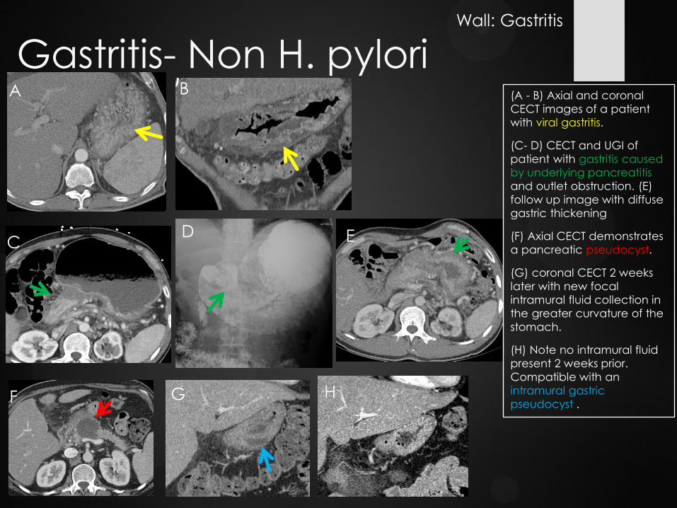
G F H
(A - B) Axial and coronal
CECT images of a patient
with viral gastritis.
(C- D) CECT and UGI of
patient with gastritis caused
by underlying pancreatitis
and outlet obstruction. (E)
follow up image with diffuse
gastric thickening
(F) Axial CECT demonstrates
a pancreatic pseudocyst.
(G) coronal CECT 2 weeks
later with new focal
intramural fluid collection in
the greater curvature of the
stomach.
(H) Note no intramural fluid
present 2 weeks prior.
Compatible with an
intramural gastric
pseudocyst .
D E C
B A
Gastritis- Non H. pylori Wall: Gastritis

Varices Imaging features
CT:
Gastric: Well-defined clusters of rounded or tubular enhancing structures within body and fundus of the stomach.
Antrum is typically spared.
Perigastric: Collateral vessels are commonly seen in the region of the gastrohepatic ligament, near the lesser omentum, along the course of the coronary vein
Can be mistaken for gastric wall thickening, gastric cancer, or perigastric adenopathy on both endoscopy and CT, particularly if intravenous contrast material is not administered for the CT
UGI:
Multiple serpentine filling defects that change in size and shape during fluoroscopic observation.
“Downhill” varices: Upper 1/3 of esophagus, results from superior vena cava obstruction. Often asymptomatic.
“Uphill” varices: Lower aspect of esophagus, result from portal hypertension. May present with GI bleeding
Differential: Varicoid carcinoma appears fixed, often with shouldered margins and noncompliant wall.
(A-D) UGI demonstrates
serpentine filling defects
that change in size and
shape between images
(E) Corresponding CECT
demonstrates
esophageal mucosal
varices.
Wall: Varices

Commonly associated with splanchnic
obstruction or portal hypertension
Gastric varices in association with
esophageal varices: underlying cirrhosis
and portal hypertension
Gastric varices without esophageal
varices: Underlying splenic vein
thrombosis or occlusion
Most commonly secondary to
pancreatitis or pancreatic carcinoma
Gastric Varices
Wall: Varices
Portal hypertension: (A-D) Gastric and perigastric
varices on CT.
Splenic vein thrombosis: (E) Isolated gastric cardia
varices on CT with (F) corresponding appearance of
the (F) gastric cardia varices on EGD.
A
D C
B
E F
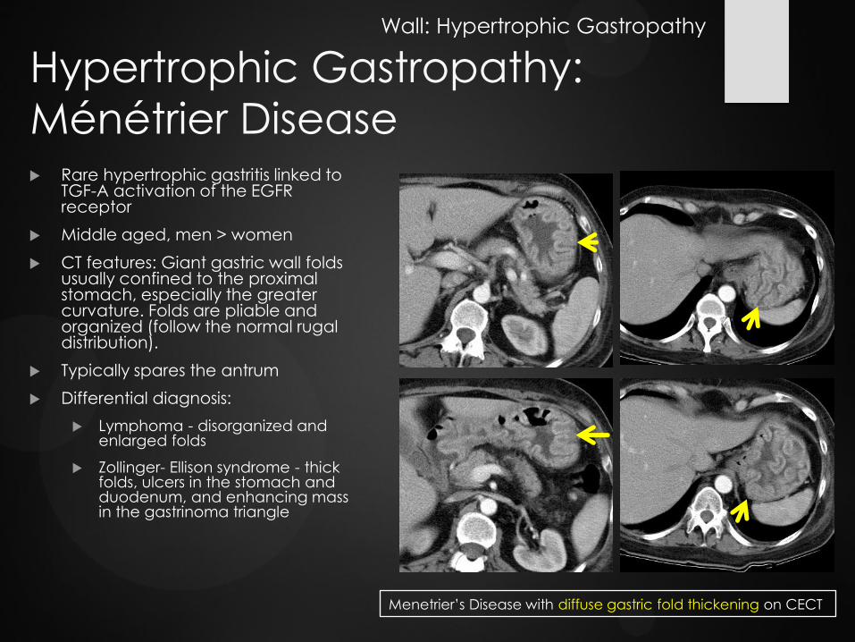
Rare hypertrophic gastritis linked to TGF-A activation of the EGFR receptor
Middle aged, men > women
CT features: Giant gastric wall folds usually confined to the proximal stomach, especially the greater curvature. Folds are pliable and organized (follow the normal rugal distribution).
Typically spares the antrum
Differential diagnosis:
Lymphoma - disorganized and enlarged folds
Zollinger- Ellison syndrome - thick folds, ulcers in the stomach and duodenum, and enhancing mass in the gastrinoma triangle
Hypertrophic Gastropathy:
Ménétrier Disease
Wall: Hypertrophic Gastropathy
Menetrier’s Disease with diffuse gastric fold thickening on CECT

Hypertrophic Gastropathy:
Zollinger-Ellison Syndrome
Wall: Hypertrophic Gastropathy
(A) Coronal enhanced T1W MRI demonstrates marked gastric wall
thickening with hypervascular peripancreatic mass in the
gastrinoma triangle. (B) Endoscopic ultrasound demonstrates
corresponding solid mass. (C ) EGD demonstrating rugal fold
thickening. (D) In- 111 Octreotide scan demonstrates
corresponding abnormal tracer accumulation, consistent with a
neuroendocrine tumor (gastrinoma).
Hypersecretion of gastric acid secondary to a
gastrinoma. 80-90% of gastrinomas are found in
the gastrinoma triangle. At least half of the
tumors are malignant.
Elevated hormone gastrin ( even after secretin
injection) gastric acid hypersecretion
recurrent peptic ulcer disease, diarrhea, reflux,
pain, anemia.
Imaging findings: hypersecretions, thickened
rugal and duodenal folds, multiple ulcers,
recurrent duodenal and jejunal ulcers,
~25% have MEN 1
Differential: gastritis from other causes, gastric
lymphoma
*
*
*
Gastrinoma triangle: Junction of the cystic and common bile duct, junction of the head/neck of the pancreas and junction
of the 2nd/3rd portion of the duodenum

Gastric Polyps • Types; Hyperplastic, adenomatous, and
hamartomatous polyps
• Most polyps are either hyperplastic or
adenomatous.
• Gastric polyps in Peutz-Jeghers syndrome,
juvenile polyposis and Cronhkhite –Canada
syndrome are hamartomatous polyps.
• Gastric polyps in Lynch syndrome are
adenomatous.
• May be difficult to see on CT, especially when
exam is not tailored for gastric evaluation.
• Polyps are better visualized on EGD and UGI
than on routine CECT.
Lumen: Polyps
A
D c
B
(A) CECT demonstrates subtle polyps along
the anterior gastric wall, indistinguishable from
rugal folds. (B) Corresponding EGD clearly
delineates innumerable polyps in the same
patient.
(C ) Polyps and (D) familial adenomatous
polyposis (FAP) syndrome visualized on UGI in
additional patients.

Hyperplastic polyps
75-90% of all gastric polyps
Seen in the setting of chronic gastritis, atrophic gastritis, or bile reflux gastritis
No malignant potential, but increased risk for coexisting gastric carcinomas.
Smooth, sessile, round, or oval lesions, ranging from 5 to 10 mm in diameter
Usually multiple lesions of similar size, clustered in the gastric body or fundus on the posterior gastric wall
Innumerable hyperplastic polyps in the gastric polyposis syndrome (fundic gland polyposis).
Gastric polyps in FAP are usually hyperplastic, whereas polyps in the remaining intestines are adenomatous.
Gastric Polyps A
E
D C
B
Multiple gastric polyps
noted on (A-C) UGI with
corresponding (D - E)
polyps on EGD.
Pathology confirmed
hyperplastic polyps with
underlying gastritis
Lumen: Polyps

Adenomatous polyps
Rare
Larger than hyperplastic polyps (>1 cm), more commonly pedunculated
Often solitary
Occur adjacent to antrum
40% of cases contain or will develop carcinoma, generally among the larger lesions (>2 cm)
Gastric Polyps
C
B A
(A) Noncontrast CT
demonstrates a polypoid
mass along the greater
curvature. (B) Axial and
(C) coronal contrast
enhanced CT
demonstrates avid
enhancement of this
polypoid mass.
Lumen: Polyps

Foreign Bodies
Lumen: Foreign Bodies
A
E D
C B
Foreign bodies in the stomach: (A-B)Scout
and CT demonstrate a fork handle in the
stomach. (C) Scout demonstrates ileus and a
necklace in the stomach. (D-E) Axial and
coronal CT demonstrate a bag of drugs in
the stomach in a drug mule.

Incomplete Distensibility Extrinsic pathology
Lymphadenopathy
Annular pancreas
Bowel
Intrinsic pathology
Linitis plastic (metastases or scirrhous adenocarcinoma)
Scarring or stricture
Granulomatous disease
(A) Indistensible gastric antrum on UGI due to adjacent dilated
bowel loops better seen on (B) CT
A D C B
(A) Coronal CECT demonstrates gastric distention and gastric outlet obstruction. (B) CECT demonstrating annular
pancreas at the site extrinsic compression. (C) Corresponding EGD demonstrates luminal narrowing from extrinsic
compression, no intraluminal mass. (D) UGI following gastrojejunostomy as definitive treatment.
Distensibility

Hiatal Hernia • Sliding and Paraesophageal
types
• May be seen on UGI and CT
• Identify supradiaphragmatic
location of the
gastroesophageal junction
• May be potential risk for
volvulus
(A) Small hiatal hernia on an UGI. (B -C) Larger
paraesophageal hernia with (D) evidence of reflux
on UGI. (E) Retrocardiac density on chest radiograph
corresponds to a large paraesophageal hernia as
seen on (F) axial and (G) coronal CECT.
A
C
G
D
B
E F
Position: Hiatal Hernia

Gastric Volvulus Rare
180˚ torsion of the stomach with intrathoracic location of the stomach:
Organoaxial
Torsion along the longitudinal axis (upside down stomach). Greater curvature is located superiorly and the body is cranial to the fundus.
More common
Often associated with a long standing hiatal hernia, paraesophageal hernia or diaphragmatic eventration.
Mesenteroaxial – torsion about the gastric mesentery
Antrum and pylorus torse anteriorly and superiorly
Often causes obstruction in the region of the pylorus/antrum.
Ischemia and gangrene may develop in either subtype.
Treatment: surgical intervention
Position: Volvulus
Organoaxial gastric volvulus:
(A-C) Coronal CECT and (D) sagittal CECT
images demonstrate intrathoracic location of
the stomach with gastric distention and the
greater curvature cranial to the lesser
curvature.
A B
C D

Mucosal defect that extends to the muscularis mucosa and beyond
Usually occur as a solitary lesion
Important cause of acute abdomen: bleeding, perforation, obstruction or penetration
Associations: Aspirin, nonsteroidal anti-inflammatory drugs, alcohol, coffee, corticosteroids, and stress
Imaging findings may include:
Mucosal defect extending beyond the expected contour of the stomach
Regional edema
Gastric outlet obstruction
Perforation
Average time to heal is 8 weeks.
Gastric Ulcers
A D C B
Giant gastric ulcer (>3 cm): (A) Axial and (B) coronal CECT demonstrates a large ulcer along the lesser curvature of
the stomach in a patient presenting with melena. (C ) A 4 x 3 cm clean based ulcer on corresponding EGD along the
lesser curvature without active bleeding. (D) Follow up EGD following 8 weeks of proton pump inhibitor treatment
demonstrates healing of the ulcer.
Integrity: Peptic Ulcer Disease
Benign ulcer: round or oval crater, smooth & symmetric ulcer
collar (submucosal edema surrounding the ulcer cavity),
Hampton line- thin line of nonulcerated mucosa surrounding
the ulcer. Folds cross the mound of surrounding edema and
extend up to the ulcer crater. Most are along lesser curvature
(posterior wall of the antrum or body of the stomach).
Malignant ulcer: Nodular ulcer collar, abrupt transition
between surrounding tissue and normal gastric wall. Crater
does not project beyond the expected location of the gastric
wall. Radiating folds do not extend to the ulcer crater. Carman
meniscus ulcer straddling the lesser curvature looks like a
crescent on compression views with nodular tumor surrounding
the periphery of the ulcer

Peptic Ulcer Disease
(A) Axial and (B) coronal CT demonstrates gastric outlet obstruction with edema and ulceration of the antrum and
pylorus. Corresponding EGD demonstrates (C) retained food, (D) large deep ulceration at the pylorus with (E)
narrowing of the pylorus channel. Pathology confirmed gastritis and ulceration with presence of H. pylori and fungi.
Gastric Outlet Obstruction
Peptic Ulcer Disease: (A - B) coronal CECT demonstrates irregular edema and ulceration of the antrum with
suspected microperforation. (C - D) UGI demonstrates multiple ulcers of the gastric antrum.
D B C
Extensive Edema and Wall Thickening
Integrity: Peptic Ulcer Disease
D C B A E
A

Causes:
Infection by a gas- producing organism, typically Escherichia coli
Caustic ingestion
Alcohol abuse
Ischemia
Life-threatening condition with a high mortality rate
Acute sepsis
Gastric hemorrhage
Imaging features: Gastric wall thickening, air within layers of gastric wall
Endoscopic features: “cobblestone” appearance of the gastric mucosa from submucosal blebs of air
Emphysematous Gastritis Integrity: Emphysematous Gastritis
A B
(A) Patient with lap band presents with acute abdominal
pain. Axial CECT demonstrates a malpositioned gastric
band and air within the gastric wall due to resulting
ischemia.
(B) UGI demonstrates subsequent removal of the band.

Benign condition
More commonly seen than emphysematous gastritis
CT appearances of gastric emphysema and emphysematous gastritis can be similar
Patients with benign gastric emphysema are asymptomatic and condition resolves spontaneously
Causes: Infection, ischemia, increased intraluminal pressure, severe vomiting, spontaneous or traumatic rupture of a pulmonary bulla, nasogastric tube placement
Imaging features: Linear distribution of mural air without associated wall thickening and inflammation
Gastric Emphysema
A B
C (A) CT demonstrates air
within the gastric wall
noted incidentally in an
asymptomatic patient with
(B) large underlying bullae.
Findings are most
consistent with benign
gastric emphysema.
(C ) Follow up NECT
demonstrates spontaneous
resolution of the gastric
emphysema.
Integrity: Gastric Emphysema

Differential Diagnosis Wall Thickening
Gastritis (H. pylori)
Peptic ulcer disease Zollinger-Ellison syndrome
Menetrier disease
Varices
Malignancy
Filling Defect
Polyps
Foreign bodies
Bezoar/ Retained food
GIST
Lipoma
Leiomyoma
Ectopic pancreatic rest
Malignancy
Indistensibility
Extrinsic mass effect
Peptic scarring
Corrosive ingestion
Granulomatous disease
Malignancy
Ulcerations Peptic ulcer disease Medication-induced ulcers
Malignancy
• Antrum spared: Varices and Menetrier’s disease
• Greater curvature predominantly: Menetrier’s disease; thickened organized folds.
• Lesser curvature: benign ulcers (Hampton line)
• Antrum predominantly: H. pylori gastritis, lipoma
• Stomach and duodenal ulcerations and rugal fold thickening: Zollinger- Ellison syndrome look for tumor in gastrinoma triangle
• Lymphoma: thickened disorganized folds
Overview

Conclusion
Gastric pathology is commonly encountered on
abdominal CT and fluoroscopy, which are
complimentary in evaluating the stomach
mucosa and surrounding structures.
The radiologist plays an essential role in
characterizing the lesions and guiding further endoscopic work-up and management.

References • Brittenden, J., & Tolan, D. J. (2012). Radiology of the Post Surgical Abdomen. London: Springer.
• Dai HX. CT differentiation for benign from malignant stomach wall thickening. Biomed Imaging Interv J. 2007; 3 (1):e12-415.
• Fishman EF, Urban BA, & Hruban RH . CT of the Stomach: Spectrum of Disease. RadioGraphics . 1996; 16: 1035-1054.
• Ghai S, Pattison J, Ghai S, O’Malley ME, Khalili K, Stephens M. Primary Gastrointestinal Lymphoma: Spectrum of Imaging Findings with Pathological Correlation. RadioGraphics . 2007; 27:1371-1388.
• Grayson DE, Abbott RM, Levy AD, Sherman PM. Emphysematous Infections of the Abdomen and Pelvis: A Pictorial Review. RadioGraphics . 2002; 22 :543-561.
• Horton K, Fishman EK. Current Role of CT in Imaging of the Stomach. RadioGraphics . 2003; 23: 75-87.
• Johnson, CD (1993). Alimentary Tract Imaging: A Teaching File. St. Louis: Mosby.
• Johnson CD, Schmit GD (2005). Mayo Clinic Gastrointestinal Imaging Review. Rochester: Mayo Clinic Scientific Press.
• Kim JH, Eun HW, Goo DE, Shim CS, Auh YH. Imaging of Various Gastric Lesions with 2D MPR and CT Gastrography Performed with Multidetector CT. RadioGraphics .2006; 26: 1101-1118.
• Meyers M, Charnsangavej C, Oliphant M (2005). Meyers’ Dynamic Radiology of the Abdomen: Normal and Pathological anatomy. New York: Springer.
• Pickhardt PJ, Asher DB. Wall Thickening of the Gastric Antrum as a Normal Finding: Multidetector CT with Cadaveric Comparison. American Journal of Roentgenology. 2003;181: 973-979.
• Rakita D, Hines JJ, Davidoff S, Sideridis K, Yacobozzi M, Friedman B . CT Imaging of Endoscopy-confirmed Gastric Pathology. Applied Radiology Nov 2013: 18-28.
Contact information: [email protected]


![Multimodality imaging of adult gastric emergencies: … fundus, body, antrum, and pylorus [Figure 1A]. The cardia surrounds the gastroesophageal (GE) junction. Gastric fundus is the](https://img.dokumen.tips/doc/110x75/5ad16b9b7f8b9a482c8b64c7/multimodality-imaging-of-adult-gastric-emergencies-fundus-body-antrum-and.jpg)