Embed Size (px)
Citation preview
Multi-Frame Super-Resolution with QualitySelf-Assessment for Retinal Fundus Videos
Thomas Kohler1,2, Alexander Brost1, Katja Mogalle1, Qianyi Zhang1,Christiane Kohler3, Georg Michelson2,3, Joachim Hornegger1,2,
Ralf P. Tornow3
1 Pattern Recognition Lab, Friedrich-Alexander-Universitat Erlangen-Nurnberg2 Erlangen Graduate School in Advanced Optical Technologies (SAOT)
[email protected] Department of Ophthalmology, Friedrich-Alexander-Universitat Erlangen-Nurnberg
Abstract. This paper proposes a novel super-resolution framework toreconstruct high-resolution fundus images from multiple low-resolutionvideo frames in retinal fundus imaging. Natural eye movements duringan examination are used as a cue for super-resolution in a robust max-imum a-posteriori scheme. In order to compensate heterogeneous illu-mination on the fundus, we integrate retrospective illumination correc-tion for photometric registration to the underlying imaging model. Ourmethod utilizes quality self-assessment to provide objective quality scoresfor reconstructed images as well as to select regularization parametersautomatically. In our evaluation on real data acquired from six humansubjects with a low-cost video camera, the proposed method achievedconsiderable enhancements of low-resolution frames and improved noiseand sharpness characteristics by 74 %. In terms of image analysis, wedemonstrate the importance of our method for the improvement of au-tomatic blood vessel segmentation as an example application, where thesensitivity was increased by 13 % using super-resolution reconstruction.
1 Introduction
Fundus imaging is one of the most routinely used modalities in clinical practice todiagnose retinal diseases. High-end fundus cameras provide color photographs ofhigh spatial resolution captured from the background of the human eye. Despitetheir broad application for diagnostic purposes, e. g. for diabetic retinopathy orglaucoma, fundus cameras are limited to the acquisition of single or stereo im-ages. In this context, novel video camera systems provide a complementary tech-nology that enables the acquisition of fast temporal changes for new applicationssuch as time course measurement of fundus reflections to examine the cardiaccycle [1]. However, inherent limitations for diagnostic applications are the lowerspatial resolution as well as the inferior conditions in terms of signal-to-noiseratio (SNR) and image contrast due to technological or economical constraints.
Methods used for image enhancement in fundus video imaging include de-noising techniques, e. g. temporal averaging schemes [2]. Additionally, blind de-

2 T. Kohler et al.
convolution has been proposed for image restoration [3]. However, this tech-nique is applied to pairs of photographs acquired in a longitudinal examinationrather than video data and does not increase the spatial resolution in terms ofpixel sampling. To overcome this issue, multi-frame super-resolution algorithms[4] reconstruct a high-resolution (HR) image with improved SNR from multiplelow-resolution (LR) frames by exploiting sub-pixel motion in an image sequence.Established methods formulate super-resolution from a Bayesian perspective asmaximum a-posteriori (MAP) estimation [4] or employ marginalization to recon-struct HR images [5]. As super-resolution is an ill-posed problem and sensitiveto the accuracy of the motion estimate, robust algorithms have been introduced,e. g. in the work of Farsiu et al. [6]. Super-resolution methods have also beenutilized for various medical imaging modalities [7]. In terms of retinal imaging,Murillo et al. [8] have presented a first super-resolution approach for scanninglaser ophthalmoscopes. However, to the best of our knowledge, this method hasnot been investigated for fundus video imaging. In particular, it does not considerspecific aspects of fundus images such as heterogeneous illumination.
This paper proposes a novel super-resolution framework to reconstruct HRimages from LR video sequences in retinal imaging. In our approach, natural eyemovements during an examination are used as a cue for super-resolution. Themajor contribution of our work is threefold. First, we incorporate retrospectiveillumination correction for photometric registration to the underlying imagingmodel to compensate spatially and temporally heterogeneous illumination on thefundus. Second, we utilize no-reference quality assessment for fundus images toprovide objective image quality scores and to select reconstruction parametersautomatically. Finally, our experimental evaluation demonstrates the importanceof our method towards diagnostic applicability of fundus video cameras.
2 Proposed Method
2.1 Multi-Frame Super-Resolution Reconstruction
We exploit LR frames denoted as y(1), . . . ,y(K) where the luminance channelof the k-th frame (k = 1 . . .K) is reorganized into a vector y(k) ∈ RM . Dueto eye motion during image acquisition, each frame y(k) is warped with respectto the unknown HR image x ∈ RN according to a geometric transformation.Each warped y(k) is a blurred and downsampled version of x due to the camerapoint spread function (PSF) and the finite pixel size. Furthermore, spatially andtemporally heterogeneous illumination is a common issue in retinal imaging andresults in photometric differences between x and y(k). Finally, each frame isaffected by additive noise ε(k). We utilize a generative model [6] extended witha photometric transformation to define the relation between x and each y(k):
y(k) = γ(k)m �DB(k)M (k)x+ γ(k)
a 1 + ε(k), (1)
where D, B(k) and M (k) models sub-sampling, blur and the geometric transfor-
mation of x for the k-th frame, respectively. γ(k)m represents the bias field which

Multi-Frame Super-Resolution for Retinal Fundus Videos 3
affects the k-th frame in a multiplicative illumination model, where � denotes
the element-wise vector product. The additive term γ(k)a 1 for the all-one vector 1
models varying brightness over time. Assuming a fixed and space invariant PSFK(u) resulting in a fixed blur kernel B = B(k), the different transformationsD, B and M (k) are combined to a sparse system matrix W (k) [5]:
W (k) = DBM (k) with Wij = K(||ui −M (k)(vj)||2
), (2)
where ui are the coordinates of the i-th pixel in x and M (k)(vj) are the coor-dinates of the j-th pixel vj in y(k) warped to x using the transformation M (k).
Geometric and Photometric Registration. Image registration is de-composed into two stages for photometric and geometric transformations. The
photometric transformation is modeled by the bias field γ(k)m which is assumed
to be spatially smooth and temporal changes in brightness modeled by γ(k)a . To
estimate γ(k)m , we employ a retrospective correction based on a B-spline approx-
imation [9] of y(k). Once the bias field γ(k)m is determined, the associated illumi-
nation corrected frame y(k) is obtained by inverting the illumination model:
y(k) = γ(k)−1m � y(k), (3)
where γ(k)−1m denotes the pixel-wise inverted bias field. Then, the illumination
corrected frames y(1), . . . , y(K) are photometrically registered up to an offset
γ(k)a which is determined by the temporal changes of the median brightness:
γ(k)a = Median(y(k))−Median(y(r)). (4)
For geometric registration, we focus on steady acquisitions, where eye motion isgiven by small random movements excluding saccades occurring in wider inter-vals. Therefore, eye motion is modeled by a 2-D homography inM (k) as perspec-tive distortions caused by the retina curvature are negligible. The homographyis estimated by means of affine registration [10] in a robust coarse-to-fine scheme
from the photometrically registered frame y(k), where y(1) is used as reference.Image Reconstruction. After geometric and photometric registration, the
system matrices W (k) are assembled from the transformation parameters ac-cording to Eq. (2). Multi-frame super-resolution is formulated as unconstrainedminimization problem using the Lp norm as data fidelity measure:
x = arg minx
{K∑k=1
∣∣∣∣∣∣y(k) − γ(k)m �W (k)x− γ(k)
a
∣∣∣∣∣∣pp
+ λ ·R(x)
}, (5)
where R(x) weighted by λ regularizes the HR estimate x to enforce smoothness.In order to make super-resolution robust to the registration uncertainty, wechose p = 1 and adopted L1 norm minimization [6], which corresponds to aMAP estimate for x if ε(k) is Laplacian noise. For R(x), the edge preservingbilateral total variation (BTV) with window size L and weight α is employed:
R(x) =
L∑m=−L
L∑n=−L
α|m|+|n| ||x− Smv Snhx||1 , (6)
4 T. Kohler et al.
Image Quality Self-Assessment
PhotometricRegistration
SR ImageReconstruction
GeometricRegistration
Low-resolutionimage sequence
Spatial & Temporalvarying illumination
Super-resolved imageSub-pixel eye motion
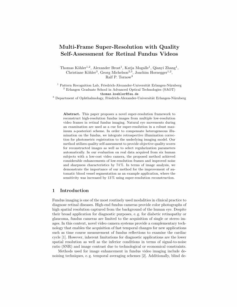
Fig. 1. Flowchart of the proposed multi-frame super-resolution framework.
which compares x to its shifted versions in vertical and horizontal direction de-fined in matrix notation by Smv and Snh, respectively. The objective function inEq. (5) is minimized employing iterative Scaled Conjugate Gradient (SCG) opti-mization to enhance the convergence compared to steepest descent minimization[6]. The temporal median of the geometrically and photometrically registered LRsequence bicubic upsampled to the HR grid is used as initial guess for SCG.
2.2 Image Quality Self-Assessment and Parameter Selection
Super-resolution relies on the initialization of the regularization weight λ and isaffected by residual noise in case of too small λ, whereas a large λ leads to over-smoothing. Parameter selection typically involves cross validation proceduresbased on simple measures such as the mean squared error [5]. However, thesemeasures do not correlate with visual perception for diagnostic purposes. In thispaper, the content-based no-reference quality metric Qv [11] for fundus imagesis utilized. Qv quantifies noise and sharpness for an image x according to:
Qv(x) =∑
pi∈P(x)
σi · q(pi), (7)
where q(pi) measures the local quality for an anisotropic patch pi, which iscombined to Qv(x) based on spatially adaptive weights σi. The set of patchesP(x) is indicated by a dominant intensity gradient orientation determined bystatistical significance testing and σi denotes the local variance of a vessel prob-ability map in pi estimated via blood vessel segmentation. To obtain unbiasedscores, all patches pi and weights σi are computed for the temporal medianof the registered sequence y(1), . . . , y(K). As Qv(x) depends on the number ofpatches, quality assessment is normalized by the reference frame y(r) accord-ing to Qv(x) = (Qv(x) − Qv(y(r)))/Qv(y
(r)) to quantify the relative improve-ment. We combine super-resolution with a data-driven selection of the regularizerweight according to:
λ = arg maxλ
Qv(xλ), (8)
where xλ denotes the super-resolved image reconstructed according to Eq. (5)with weight λ. In order to find an optimal weight, we perform a grid search withequidistant step size ∆ log λ in the interval [log λl, log λu] of the log-transformed

Multi-Frame Super-Resolution for Retinal Fundus Videos 5
Table 1. Peak-signal-to-noise ratio (PSNR) along with sensitivity and specificity ofvessel segmentation for LR frames, temporal median and super-resolution.
LR frame Median image SR image Ground truth
PSNR (in dB) 31.09 ± 3.10 31.41 ± 3.28 31.92 ± 3.39 -
Sensitivity (%) 57.59 ± 6.01 67.83 ± 4.96 70.37 ± 5.00 72.85 ± 6.70Specificity (%) 94.31 ± 1.40 94.80 ± 1.19 93.99 ± 1.26 94.57 ± 1.34
range of λ chosen as initialization. For a fixed λ, a few SCG iterations are per-formed to check whether it improves the super-resolved image. For the selectedλ, a super-resolved image is estimated according to Eq. (5) by running SCG untilconvergence. The overall flowchart of our framework is outlined in Fig. 1.
3 Experiments and Results
We adjusted all parameters experimentally based on real fundus video data usedin our experiments1. For BTV regularization, we chose α = 0.7 and L = 1 withlog λl = −2.0, log λu = 0 and step size ∆ log λ = 0.2 to select an optimal weight.For quality self-assessment, anisotropic patches of size 8×8 were analyzed.
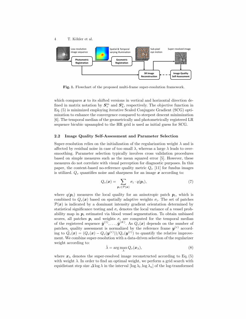
Synthetic Data. We generated synthetic image sequences with K = 16frames for 40 images taken from the DRIVE database [12], by applying our modeldefined in Eq. (1) in forward direction. The frames were related to the referenceframe by a uniformly distributed random translation (−2 to +2 pixels) to simu-late eye motion, affected by Gaussian noise (σn = 0.01), blurred by an isotropicGaussian PSF (σ = 1.0) and sub-sampled by a factor of 2. For super-resolution,we considered the green color channel as in fundus imaging the red and blueones are typically over- and under-saturated, respectively. Super-resolved im-ages were assessed using the peak-signal-to-noise ratio (PSNR). Additionally,we investigated blood vessel segmentation [13] as application of our methodto compare an automatic segmentation to a manually created gold standard.Quantitative measures are summarized in Table 1 and the associated qualitativeresults are presented in Fig. 2. Our framework improved the mean PSNR by0.8 dB compared to LR images. In terms of vessel segmentation, the sensitivitywas enhanced by 13 % as fine vessels were reconstructed by our method. Bothincreases achieved by super-resolution compared to LR frames and the temporalmedian were statistically significant (p < 0.05) based on a Wilcoxon signed-ranktest. The specificity was comparable to segmentation on the ground truth.
Real Data. We acquired monochromatic fundus video data with a low-costcamera prototype developed by Ralf P. Tornow, FAU Erlangen-Nurnberg, Ger-many. The system is based on a CCD camera (640×480 px) equipped with LEDillumination and covers a field of view (FOV) of 20◦. As frame rate we chose12.5 Hz. The left eye from six healthy subjects was examined. Additionally, weexamined the subjects with a Kowa nonmyd camera (1600×1216 px, 25◦ FOV)
1 Supplementary material is available online http://www5.cs.fau.de/research/software/
6 T. Kohler et al.
(a) LR frame (b) Median image (c) SR image (d) Ground truthPSNR: 32.6 dB PSNR: 32.9 dB PSNR: 33.6 dB
(e) Se: 0.57, Sp: 0.96 (f) Se: 0.65, Sp: 0.96 (g) Se: 0.67, Sp: 0.96 (h) Se: 0.68, Sp: 0.95
Fig. 2. Synthetic images with peak-signal-to-noise ratio (PSNR) for LR data (a), tem-poral median (b) and super-resolved data (c) in comparison to the ground truth (d).We evaluated sensitivity (Se) and specificity (Sp) for vessel segmentation where true-positive and false-positive pixels shown in (e) - (h) are color-coded in green and red.
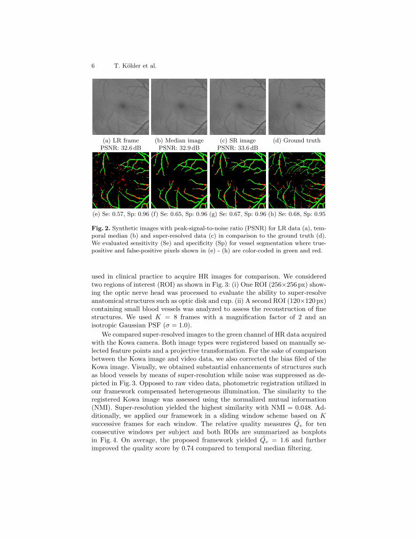
used in clinical practice to acquire HR images for comparison. We consideredtwo regions of interest (ROI) as shown in Fig. 3: (i) One ROI (256×256 px) show-ing the optic nerve head was processed to evaluate the ability to super-resolveanatomical structures such as optic disk and cup. (ii) A second ROI (120×120 px)containing small blood vessels was analyzed to assess the reconstruction of finestructures. We used K = 8 frames with a magnification factor of 2 and anisotropic Gaussian PSF (σ = 1.0).
We compared super-resolved images to the green channel of HR data acquiredwith the Kowa camera. Both image types were registered based on manually se-lected feature points and a projective transformation. For the sake of comparisonbetween the Kowa image and video data, we also corrected the bias filed of theKowa image. Visually, we obtained substantial enhancements of structures suchas blood vessels by means of super-resolution while noise was suppressed as de-picted in Fig. 3. Opposed to raw video data, photometric registration utilized inour framework compensated heterogeneous illumination. The similarity to theregistered Kowa image was assessed using the normalized mutual information(NMI). Super-resolution yielded the highest similarity with NMI = 0.048. Ad-ditionally, we applied our framework in a sliding window scheme based on Ksuccessive frames for each window. The relative quality measures Qv for tenconsecutive windows per subject and both ROIs are summarized as boxplotsin Fig. 4. On average, the proposed framework yielded Qv = 1.6 and furtherimproved the quality score by 0.74 compared to temporal median filtering.

Multi-Frame Super-Resolution for Retinal Fundus Videos 7
(a) LR frame (b) Median image (c) SR image (d) Kowa nonmydNMI = 0.013 NMI = 0.044 NMI = 0.048
Fig. 3. Results obtained from the low-cost camera: Low-resolution frame (a), temporalmedian used as initial guess (b), final super-resolved image (c) and green channelof Kowa nonmyd image for the same subject (d). We assessed the similarity to theregistered Kowa image using the normalized mutual information (NMI).
1 2 3 4 5 60
0.5
1
1.5
2
2.5
3
Subject
Qv
Temporal medianSuper−resolved
1 2 3 4 5 60
0.5
1
1.5
2
2.5
3
Subject
Qv
Temporal medianSuper−resolved
Fig. 4. Boxplot of Qv for temporal median filtering and super-resolution in imageregions showing the optic nerve (left) and blood vessels (right) as depicted in Fig. 3.
4 Conclusion and Future Work
This paper proposes a novel super-resolution framework for fundus video imag-ing. Multi-frame super-resolution exploits natural eye movements during an ex-

8 T. Kohler et al.
amination by means of affine registration to reconstruct a motion-compensatedHR image from LR video data. The underlying model considers photometricregistration to account for heterogeneous illumination. We also employ qualityself-assessment for automatic parameter selection and to provide an objectivequality score for reconstructed images. Our method is able to achieve an imagequality for super-resolved images generated with a low-cost fundus camera thatis comparable to a high-resolution commercially available camera. The investiga-tion of super-resolution for an analysis of disease-specific anomalies to improvethe reliability of medical diagnoses is ongoing research. We will also study theimpact of the proposed method in large-scale studies, e. g. in glaucoma screening.
Acknowledgments. The authors gratefully acknowledge funding of the Erlan-gen Graduate School in Advanced Optical Technologies (SAOT) by the GermanNational Science Foundation (DFG) in the framework of the excellence initiative.
References
1. Tornow, R., Kopp, O., Schultheiss, B.: Time course of fundus reflection changesaccording to the cardiac cycle. Invest. Ophthalmol. Vis. Sci. 44 (2003) 1296
2. Kohler, T., Hornegger, J., Mayer, M., Michelson, G.: Quality-guided denoising forlow-cost fundus imaging. In: Proceedings BVM 2012. (2012) 292–297
3. Marrugo, A.G., Sorel, M., Sroubek, F., Millan, M.S.: Retinal image restoration bymeans of blind deconvolution. J Biomed Opt 16(11) (2011) 116016
4. Milanfar, P.: Super-resolution imaging. CRC Press (2010)5. Pickup, L.C., Capel, D.P., Roberts, S.J., Zisserman, A.: Overcoming Registration
Uncertainty in Image Super-Resolution: Maximize or Marginalize? EURASIP JAdv Signal Process 2007 (2007) 1–15
6. Farsiu, S., Robinson, M.D., Elad, M., Milanfar, P.: Fast and robust multiframesuper resolution. IEEE Trans Image Process 13(10) (2004) 1327–1344
7. Greenspan, H.: Super-Resolution in Medical Imaging. Comput J 52(1) (2008)43–63
8. Murillo, S., Echegaray, S., Zamora, G., Soliz, P., Bauman, W.: Quantitative andqualitative image quality analysis of super resolution images from a low cost scan-ning laser ophthalmoscope. In: Proc. SPIE Medical Imaging 2011. (2011) 79624T
9. Kolar, R., Odstrcilik, J., Jan, J., Harabis, V.: Illumination Correction and ContrastEqualization in Colour Fundus Images. In: Proc. EUSIPCO 2011. (2011) 298–302
10. Evangelidis, G.D., Psarakis, E.Z.: Parametric image alignment using enhancedcorrelation coefficient maximization. IEEE Trans Pattern Anal Mach Intell 30(10)(2008) 1858–65
11. Kohler, T., Budai, A., Kraus, M.F., Odstrcilik, J., Michelson, G., Hornegger, J.:Automatic no-reference quality assessment for retinal fundus images using vesselsegmentation. In: Proceedings CBMS 2013. (2013) 95–100
12. Staal, J., Abramoff, M.D., Niemeijer, M., Viergever, M.A., van Ginneken, B.:Ridge-based vessel segmentation in color images of the retina. IEEE Trans MedImaging 23(4) (2004) 501–509
13. Budai, A., Bock, R., Maier, A., Hornegger, J., Michelson, G.: Robust vessel seg-mentation in fundus images. Int J Biomed Imaging 2013 (2013) 154860