Embed Size (px)
Citation preview
Analysis of an Universal Joint: Movements and Muscles of the Eye
Thomas P. Langer
1

Model of Free Eye Movements
Introduction to the Basic Concepts and the Elements of the Model
The eye is an excellent motor system in which to study movements of joints, because it is comparatively simple, with only six extraocular muscles that move the eye, a nearly massless, nearly spherical body, embedded in an enclosing, but not restricting, socket (Williams, Bannister et al. 1995). The eye is able to, and does, move in all available directions, so it is a universal joint. We can model it with some simplification that does not change the basic structure and organization of the eye, using actual measurements of the eye, its muscle attachments, and its fascia to study the implications of its anatomy. It is comparatively easy to incorporate head movements, so that the vestibular sensory system provides a drive and to introduce concatenated joint movements.
The analysis of eye movements will be considered in two parts. First, we will examine the geometry and movements on the assumption that the muscles extend directly from their origins to their insertions except as they are displaced by passing over the surface of the globe. This is the anatomy with which most people are familiar, because it is the version generally taught. It will be called the free muscle model, to distinguish it from the other model, which will be called the restricted muscle model. Recent studies have found that the fascia of the orbit restrains the movements of the eye muscles, forming “pulleys” that hold the muscle bellies in a nearly constant position relative to the orbit (Demer, Miller et al. 1995; Clark, Miller et al. 1997; Demer, Poukens et al. 1997; Clark, Miller et al. 2000; Demer, Oh et al. 2000). The muscle tendons pass over the slings of the pulleys and to their insertions. The effect of this arrangement is to move the functional origins of the muscles anteriorly, to positions a short distance posterior to the eye’s equator in neutral gaze. This arrangement has consequences for the movements of the eye. However, to fully appreciate the implications of
2
Model of Free Eye Movements
this anatomy it helps to first consider the situation where the restraints do not apply. We will consider the free muscle situation first and then the restrained muscle situation.
The Geometrical Anatomy of the Eye
We will be looking at the anatomy of the eye from a special point of view, with the aim of representing the various components in terms of mathematical structures. Such an approach will be called geometrical anatomy, because we are interested in the spatial relations between the anatomical elements and their influence on spatial movements.
The questions that will be addressed are primarily anatomical, but the answers frequently have functional implications. We will not address the neural control of eye movements, as such, but such considerations enter into the statement of the initial constraints on allowable gaze directions and orientations. The results of the analysis may have implications for the nature of the neural control.
The Globe of the Eye is Modeled as a Sphere
The eye is actually slightly oblong, generally longest along the visual axis, and there is a slight elevation at the cornea relative to the sagittal axis of the eye, but there is little lost if we model the globe of the eye as a sphere with the line of sight being a radius of the sphere. The deviations from a sphere are small enough that they would normally be less than the natural variation from individual to individual. The types of questions addressed here will not depend critically upon the eccentricity of the globe. The magnitudes of the deviations will be considered below.
If the eye is spherical, then it is reasonable to have all eye movements occur about a point at the center of the globe, the center of the sphere in the model. It also simplifies the calculations if we use the radius of the globe as the unit of measurement. Doing so makes the insertion vectors for
3

Model of Free Eye Movements
the muscles all unit vectors and allows one to express movements of the globe in radians or degrees. There is no loss of generality in scaling the system to the radius of the globe.
The Line of Sight is a Framed Vector
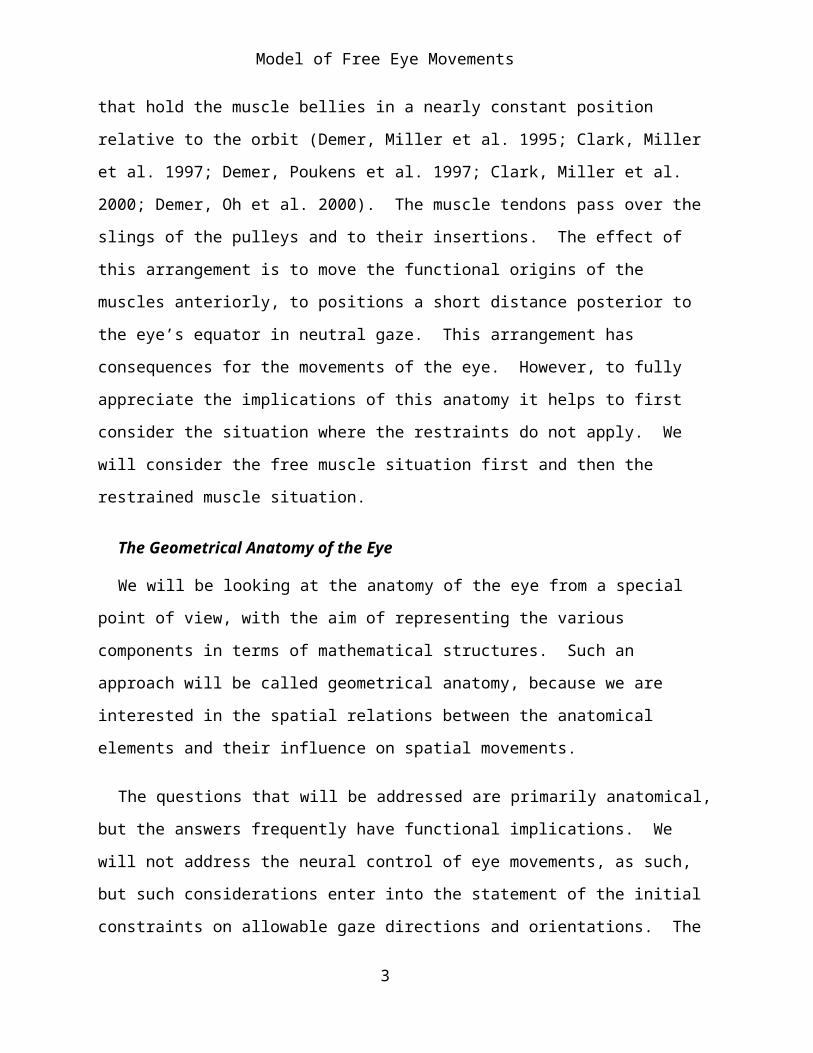
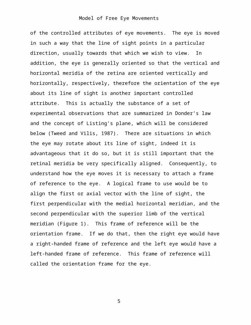
The line of sight is an important attribute to consider when modeling eye movements, because it is one of the most important of the controlled attributes of eye movements. The eye is moved in such a way that the line of sight points in a particular direction, usually towards that which we wish to view. In addition, the eye is generally oriented so that the vertical and horizontal meridia of the retina are oriented vertically and horizontally, respectively, therefore the orientation of the eye about its line of sight is another important controlled attribute. This is actually the substance of a set of experimental observations that are summarized in Donder’s law and the concept of Listing’s plane, which will be considered below (Tweed and Vilis, 1987). There are situations in which the eye may rotate about its line of sight, indeed it is advantageous that it do so, but it is still important that the retinal meridia be very specifically aligned. Consequently, to understand how the eye moves it is necessary to attach a frame of reference to the eye. A logical frame to use would be to align the first or axial vector with the line of sight, the first perpendicular with the medial horizontal meridian, and the second perpendicular with the superior limb of the vertical meridian (Figure 1). This frame of reference will be the orientation frame. If we do that, then the right eye would have a right-handed frame of reference and the left eye would have a left-handed frame of reference. This frame of reference will called the orientation frame for the eye.
4
Model of Free Eye Movements
Figure 1. An eyeball with an orientation frame of reference. A three dimensional model of the right eye with an orientation frame of reference indicated by a set of three orthogonal vectors. Because the vectors are considered to occur in the order: first, the line of sight, second, the medial perpendicular to the line of sight, and, third, the vertical perpendicular to the line of sight, the orientation frame of reference is right-handed. Note that that the frame of reference is an abstraction of the orientation of the globe and while we place it in a particular position relative to the globe, it may occur at any position as long as the vectors continue to point in the same directions.
The line of sight may be viewed as a framed vector, taking origin from the center of the globe, extending from the center of the eye to the center of the pupil, with the orientation frame being its frame of reference. For many purposes we will deal with only the orientation frame of reference.
5

Model of Free Eye Movements
The Muscles
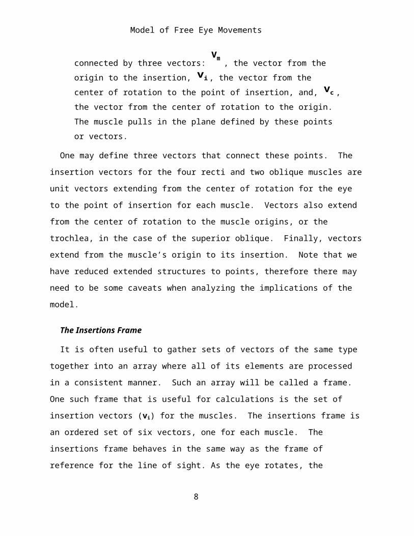
For each eye muscle there are three reference points. The first is their origin, at the common tendinous ring from which most of the oculomotor muscles take origin, but at the trochlea for the superior oblique, and in the inferior medial rim of the eye socket for the inferior oblique. The second reference point is the center of rotation for the eye. The third is their insertions, where the muscles attach to the globe (Figure 2).
Figure 2. Anatomical vectors of the eye. The eye muscles are defined by three reference points, which define three vectors. The three points are the point of origin of the muscle, the point of insertion of the muscle, and the center of rotation. These are connected by three vectors:
vm , the vector from the origin to the insertion, v i , the vector from the center of rotation to the point of insertion, and, v c , the vector from the center of rotation to the origin. The muscle pulls in the plane defined by these points or vectors.
One may define three vectors that connect these points. The insertion vectors for the four recti and two oblique muscles are unit vectors extending from the center of rotation for the eye to the point of insertion for each muscle. Vectors also extend from the center of rotation to the muscle origins, or the trochlea, in the case of the superior oblique. Finally, vectors extend from the muscle’s origin to its insertion. Note that we have reduced
6

Model of Free Eye Movements
extended structures to points, therefore there may need to be some caveats when analyzing the implications of the model.
The Insertions Frame
It is often useful to gather sets of vectors of the same type together into an array where all of its elements are processed in a consistent manner. Such an array will be called a frame. One such frame that is useful for calculations is the set of insertion vectors (vi) for the muscles. The insertions frame is an ordered set of six vectors, one for each muscle. The insertions frame behaves in the same way as the frame of reference for the line of sight. As the eye rotates, the insertions frame rotates in the same manner, because the muscles are attached to the globe.
We can form similar frames for the vectors to the origins of the muscles and those between the origins and insertions. These frames do not move in the same manner as the globe of the eye. However, they are important in computing the pulling directions of the muscles.
The Pulling Direction of a Muscle
The pulling direction for each muscle occurs in the plane that contains the muscle’s insertion vector and the vector from the center of the globe to the point of origin for the muscle. That is, a muscle’s two endpoints and the center of rotation for the eye define the plane of the muscle’s pull. For the purposes of calculation, the origin of the superior oblique is taken to be the trochlea, rather than its actual origin immediately superior to the common tendinous ring, with the four recti. When we consider the restrained muscle model, their pulleys will be substituted for the actual origins of the muscles.
If vm is the vector for muscle M, vo is the vector from the center of rotation to the point of origin of the muscle, and v i is the vector from the center of rotation to the muscle insertion, then .
7
Model of Free Eye Movements
The attachment of the eye muscles is such as to exert tangential forces upon the eye. If this were not so, then the muscles would tend to drag the entire eye in the direction opposite to the direction of the eye muscle vector, vm . In fact that is what would happen if the eye were not resting upon a fat pad, in the space posterior to the globe and between the extraocular muscles, that resists retraction of the eye. In addition, the eye is suspended in a dense web of fascia about the level of its equator, which extends from the globe to the orbital walls. This penumbral fascia will be critical to understanding the dynamics of the eye.
We can recast these concepts into arrays. Let be the array of vectors between the point of origin of the muscle and the point of insertion, the
array of vectors from the point of origin to the center of rotation, with the usual caveat for the superior oblique, and is the array of vectors from the
center of rotation to the muscle insertions, then it follows that -
which is equivalent to the set of equations:
The Set of Muscle Lengths Determines Eye Position and Orientation
The controlled variable for the extrinsic oculomotor muscles is primarily muscle length. To move to a particular eye position and orientation it would be sufficient to set the muscle lengths and let the system come to equilibrium. The actual controlled variables are probably steady state muscle tensions, however, there is a unique solution if we use muscle lengths and an infinite set of solutions if we use muscle tensions. Actual measurements of the applied neural signals indicates that, at least for rapid eye movements, a pulse-step driver is used since it gives quicker responses and shorter times to equilibrium. However, the main point at this junction is
8

Model of Free Eye Movements
that the six muscle lengths completely and uniquely specify the eye’s position and orientation.
The word ‘gaze’ will be used to signify the combined eye position and orientation. Donder’s law is an observational summation that states that there is a unique eye orientation for every eye position. In other words, once you specify the direction that the eye is looking, the orientation of the eye is also determined. This is not an anatomical constraint. At every eye position there is a wide range of orientations that the eye might assume, but the neural control over natural eye movements specifies that the actual orientation will be one particular orientation.
Consequently, there are six variables to be set to establish a unique eye position/orientation. There are six extrinsic eye muscles. Since there is no redundancy in the muscles, a particular set of eye muscle lengths establishes a unique gaze. In summation, six eye muscles are both necessary and sufficient to establish a unique one-to-one mapping between the set of muscle lengths and the set of gazes.
If one had only one eye, then the orientation would be less critical. One could control eye movement only to establish eye position and orientation would be determined by eye position. In such a hypothetical system only three non-coplanar extrinsic oculomotor muscles would be sufficient. Actually, since we need to control only two variables, azimuth and elevation, two non-coplanar pairs of muscles would suffice. However, in most systems that are actually viewing the external world it is desirable to also control horizontal and vertical orientation, therefore orientation of the eye is also important and six muscles are required.
The reason it is necessary to control orientation is that if the eye moves on any trajectory that is not a great circle, there is a spin imparted to the eye and the amount of spin depends on the trajectory followed. This can be easily seen in a simple, if artificial, example. Imagine the eye looking
9

Model of Free Eye Movements
straightforward with a marker directly above the iris on the eye’s vertical meridian. Now, the eye swings up 90°, so that it is looking straight up, then swings 90° to the right, so that it is looking straight to the right, and finally swings back to neutral. If you examine the eye, you will see that the vertical marker is directly to the right of the iris. The eye movements caused the eye to rotate 90° about the line of sight even though it did not rotate about the line of sight during any of the movements. Similar conjoint spin will happen whenever the eye moves out of the plane of the great circle that it was on when it left neutral position and orientation, that is neutral gaze. This rotation with movements that do not follow a great circle is an intrinsic property of three-dimensional space.
The Muscle Rotation Frame
When it contracts, a muscle will rotate the eye about an axis of rotation which is perpendicular to the plane defined by the insertion vector, v i , the muscle axis, vm , and the vector from the center of rotation to the muscle’s origin, v c . These axes of rotation will be called muscle rotation vectors and they can be grouped into a frame, much like the muscle insertion frame. To compute the rotation vector frame, the vector from the center of rotation to the muscle origin is replaced with a unit vector that extends in the same direction. If the insertion frame element is and the origin vector is , then
the corresponding element of the rotation frame would be the unit vector with the same direction as the vector of the quaternion that expresses the rotation from to ,
.
As the eye shifts it will change the insertion vectors and therefore the rotation frame’s elements. The rotation frame can be used to compute the consequences of changing the length of a muscle, or a combination of muscles, by adding the contributions of each muscle’s action.
10
Model of Free Eye Movements
For an illustrative instance, consider the medial rectus. If one carries the muscle insertion vector into the vector pointing from the center of rotation towards the common tendinous ring, then the vector of the rotation quaternion will point superiorly or in the positive k direction. In neutral position, . When we consider the restricted muscle model it will be found that this is only an approximately correct statement, because the medial rectus pulls from slightly below the vertical meridian of the eye in neutral position.
Changes in the Insertion Vectors with Eye Rotations
If is the location of the muscle insertion for muscle ‘M’ at time ‘t’ and is the rotation of the globe during the interval starting at time ‘t’, then -
More generally, in array format -
Generalizing this relationship -
The following simplistic examples are introduced to assist the assimilation of this concept since it is central to all that follows.
Examples: If r = k and v = i, that is the rotation vector is orthogonal to the insertion vector, then -
11

Model of Free Eye Movements
The movements described by this expression are confined to a single plane, the plane perpendicular to the rotation vector, therefore all the movements are pure swings.
If r = k and v = k, that is the rotation vector is aligned with the insertion vector, then -
12

Model of Free Eye Movements
Φ =k∗cos2φ + kk − kk( )∗cosφsinφ − kkk∗sin2φ
= k∗cos2φ + 0∗cosφsinφ + k∗sin2φ
= k∗ cos2φ + sin2φ( )= k .
13
Model of Free Eye Movements
The radial insertion vector, v, is not changed by the movement, no matter what its angular excursion, therefore all movements of the radial vector are pure spins.
From these last two calculations, it is clear that if the muscle is pulling in the plane of the frame element, then the element moves with pure swing. If the muscle is pulling in a plane orthogonal to the frame element, then the element experiences a pure spin. Finally, if the muscle pulls in any other plane relative to the frame element, then the element will experience a combination of swing and spin. The precise movement of the element can be calculated from the derived formula, given the axis of the element, v, and the rotational axis of the muscle pull, r.
14

Model of Free Eye Movements
may be read as “the value of v at time
15

Model of Free Eye Movements
T + ΔT16

Model of Free Eye Movements
, conditional upon the transformation of the frame , , is -”. The
multiplication of vectors is quaternion multiplication.
Actual Anatomical Measurements of the Eye
We can obtain measurements of the eye and muscle insertions from atlases, photographs, and x-ray and MRI images of the eye. The following measurements come from Grays Anatomy (‘96) and they are in accord with measurements taken from several different atlases and photographs.
Measurement Value (mm)vertical diameter of the eye 23.5transverse diameter of the eye 24anterior-posterior diameter of the eye 24 (20 - 29)vertical diameter of the cornea 10.6horizontal diameter of the cornea 11.7rectus medialis insertion -> corneal rim 5.5rectus inferioris insertion -> corneal rim 6.5rectus lateralis insertion -> corneal rim 6.9rectus superioris insertion -> corneal rim 7.7
We can rough out an approximate set of values for the basic structures of the eye. Taking the radius of the cornea to be about 5.5 mm then the angle
subtended by the cornea is about twice sin-1(5.5/12) = 27.39° or about 55°. Assume the inset distance for a rectus muscle is about 6.5 mm beyond the rim of the cornea then it would be 6.5/80 * 2π radians = 0.51 rad ~ 0.5 rad = 28.66° ~ 30°. Therefore, the insertion would attach about 55° to 60° away from the line of sight or about 30° to 35° anterior to the mid-coronal plane of the eye.
Therefore, one might expect the eye to have an excursion of about 35° with the muscular force being exerted as a torque on the eye, directed perpendicular to a line from the center of the eye through the insertion (torque radius), in the plane determined by the line of pull of the eye muscle and the line from the muscle origin to the center of rotation for the eye.
17

Model of Free Eye Movements
The vector of the rotation quaternion would be perpendicular to that plane. Let the convention be to let the direction of the positive rotation be the movement that takes the torque radius into the line from the center of rotation to the muscle origin. During normal vision, the eye can actually rotate about 30° medial, about 50° laterally, about 20° up, and 30° down (Williams, Bannister et al. 1995). Normal excursions are probably less than these figures. A possible reason that lateral rotation is so much greater than medial rotation is that the origins for the recti lie about 23° medial to the sagittal plane of the eye, therefore more of the lateral rectus lies upon the surface of the eye. However, there are other possible reasons, which will be discussed when we consider the restrained muscle model. These have to do with the rectus lateralis pulley being much more posteriorly placed.
Computation of the Insertions, Origins, and Orientation Frames of Reference
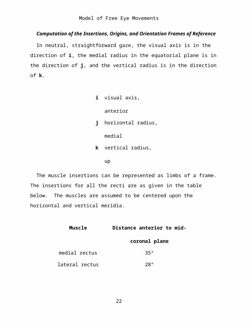
In neutral, straightforward gaze, the visual axis is in the direction of i, the medial radius in the equatorial plane is in the direction of j, and the vertical radius is in the direction of k.
i visual axis, anteriorj horizontal radius,
medialk vertical radius, up
The muscle insertions can be represented as limbs of a frame. The insertions for all the recti are as given in the table below. The muscles are assumed to be centered upon the horizontal and vertical meridia.
Muscle Distance anterior to mid-coronal
18

Model of Free Eye Movements
plane
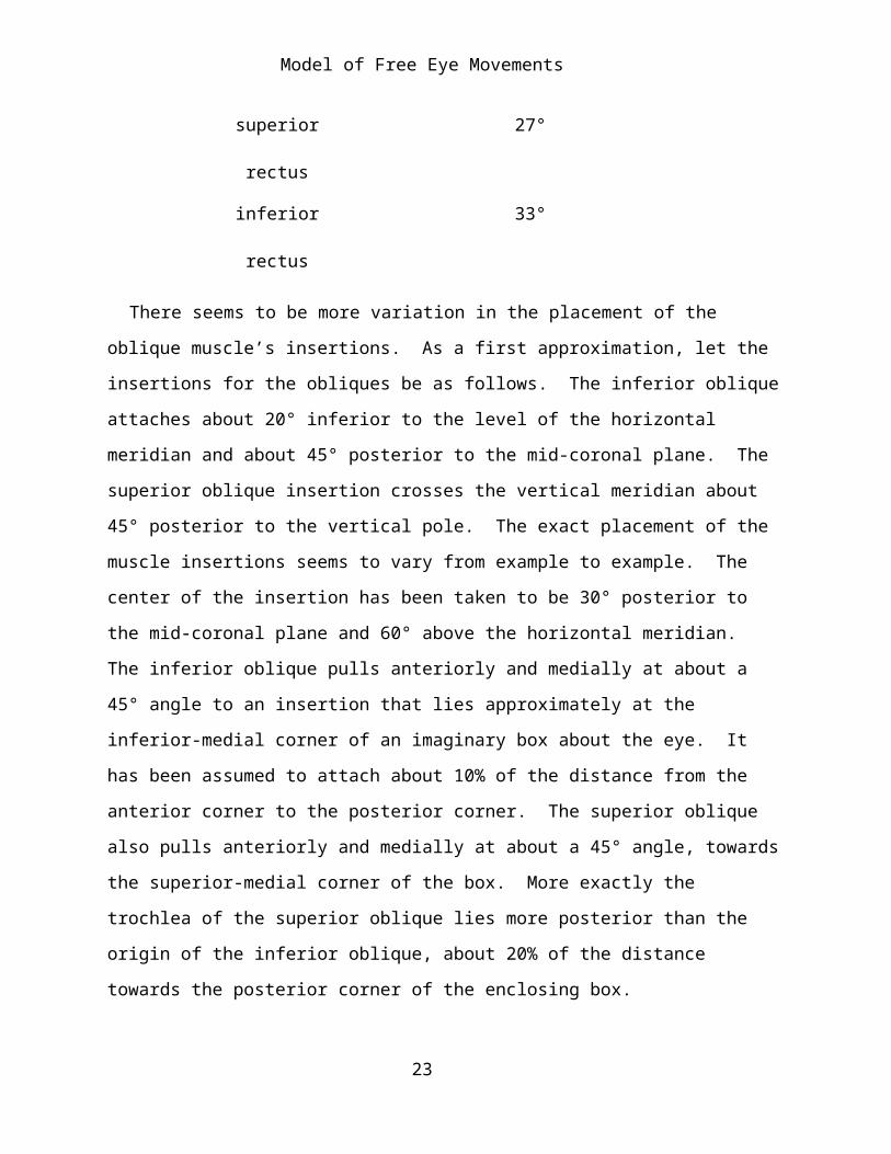
medial rectus 35°lateral rectus 28°
superior rectus 27°inferior rectus 33°
There seems to be more variation in the placement of the oblique muscle’s insertions. As a first approximation, let the insertions for the obliques be as follows. The inferior oblique attaches about 20° inferior to the level of the horizontal meridian and about 45° posterior to the mid-coronal plane. The superior oblique insertion crosses the vertical meridian about 45° posterior to the vertical pole. The exact placement of the muscle insertions seems to vary from example to example. The center of the insertion has been taken to be 30° posterior to the mid-coronal plane and 60° above the horizontal meridian. The inferior oblique pulls anteriorly and medially at about a 45° angle to an insertion that lies approximately at the inferior-medial corner of an imaginary box about the eye. It has been assumed to attach about 10% of the distance from the anterior corner to the posterior corner. The superior oblique also pulls anteriorly and medially at about a 45° angle, towards the superior-medial corner of the box. More exactly the trochlea of the superior oblique lies more posterior than the origin of the inferior oblique, about 20% of the distance towards the posterior corner of the enclosing box.
If we take the eye to be a sphere with a diameter of 24 mm, then it has a circumference of about 75.36 mm. A quarter of that would be about 18.84 mm. Taking the vertical radius of the cornea to be about 5.3 mm then the vertical angle subtended by the cornea is arcsine (5.3/12.0) = 26.21°. Assume the inset distance for the vertical recti to be about 6.5 mm below the rim of the cornea and 7.7 mm. above the cornea, then they would be 6.5/75.36 * 360° = 31.05° below the corneal rim and 7.7/75.36 * 360° =
19

Model of Free Eye Movements
36.78° above the corneal rim. Therefore, the insertions would attach about 57.26° below and 62.99° above the line of sight or about 33° and 27°, respectively, anterior to the mid-coronal plane of the eye.
Taking the horizontal radius of the cornea to be about 5.85 mm then the angle subtended by the cornea is about sin-1(5.85/12.0) = 29.18°. Assume the inset distance for the horizontal recti to be about 5.5 mm medial to the rim of the cornea and 6.9 mm. lateral to the cornea, then they would be 5.5/75.36 * 360 = 26.27° medial to the corneal rim and 6.9/75.36 * 360 = 32.96° lateral to the corneal rim. Therefore, the insertions would attach about 55.45° medial to and 62.14° lateral to the line of sight or about 35° and 28°, respectively, anterior to the mid-coronal plane of the eye.
These approximations will lead to the following vectors for the insertions of the six extraocular muscles. The radius of the eye is taken to be 1.0.
Muscle i j k
Medial rectus 0.57 0.82 0Lateral rectus 0.47 - 0.88 0
Superior rectus 0.45 0 0.89Inferior rectus 0.54 0 - 0.84
Superior
oblique
- 0.5 -0.43 0.75
Inferior oblique - 0.87 - 0.5 0.0
The muscle origins for the six extraocular muscles are approximately as follows:
Muscle i j k
Medial rectus - 3.0 1.27 0
20
Model of Free Eye Movements
Lateral rectus - 3.0 1.27 0Superior rectus - 3.0 1.27 0Inferior rectus - 3.0 1.27 0
Superior
oblique
0.6 1.0 1.0
Inferior oblique 0.8 1.0 - 1.0
The radius of the eye is 1.0 and the origin of the coordinate system is at the center of the globe.
The Point of Tangency
The point where the muscle ceases to be tangential to the scleral surface occurs posterior to the coronal meridian (Figure 2). Roughly, the common tendinous ring lies one eyeball diameter posterior to the posterior pole of the eye. Therefore, the line from the center of the eye to the tendinous origins is about 3 eye radii long and the point of tangency is one eye radius from the eye’s center and perpendicular to the scleral boundary. It follows that the angle between the radius of tangency and the equatorial plane is arcsine (1/3), which is 19.47° ~ 20° plus the offset due to the muscle origin arising from a point about 23° medial to the line of sight. The axis from the common tendinous ring to the center of the eye is at a 23° angle to the sagittal axis of the orbit.
For the lateral rectus, the distance of the point of tangency posterior to the coronal equator is added to the angular excursion from the equatorial plane to its insertion, which was computed above to be about 28°. The available excursion for the lateral rectus is about 70°. For the medial rectus, it approximately compensates for the 23° medial rotation of the eye relative to the orbit, therefore the available excursion for the medial rectus is about 30°. For the superior and inferior recti the available excursion is
21
Model of Free Eye Movements
about 45° and 51°, respectively. There is more than enough angular excursion to allow the full range of eye movements by contraction of the recti. The oblique muscles, because of the their insertions adjacent to the globe, are even more adequate for the observed excursion of the eye. Based on these observations, we can compute the consequences of eye movements without worrying if they are within the available range of the eye muscles. The muscular forces are always tangential to the scleral surface because the muscles lie on the surface of the eyeball.
Two Models of the Eye
We found it useful to use two models for computation. The first is a good approximation of the actual situation in the human orbit. The origins of the recti lies about three units posterior to the center of the eye and about 23° medial to the line of sight when the eye is looking straight forward. The obliques are either attached to the medial anterior rim of the eye socket (inferior rectus) or pass through a pulley attached to the rim (superior oblique). The attachments of the muscles to the globe are good approximations to the actual anatomical insertions.
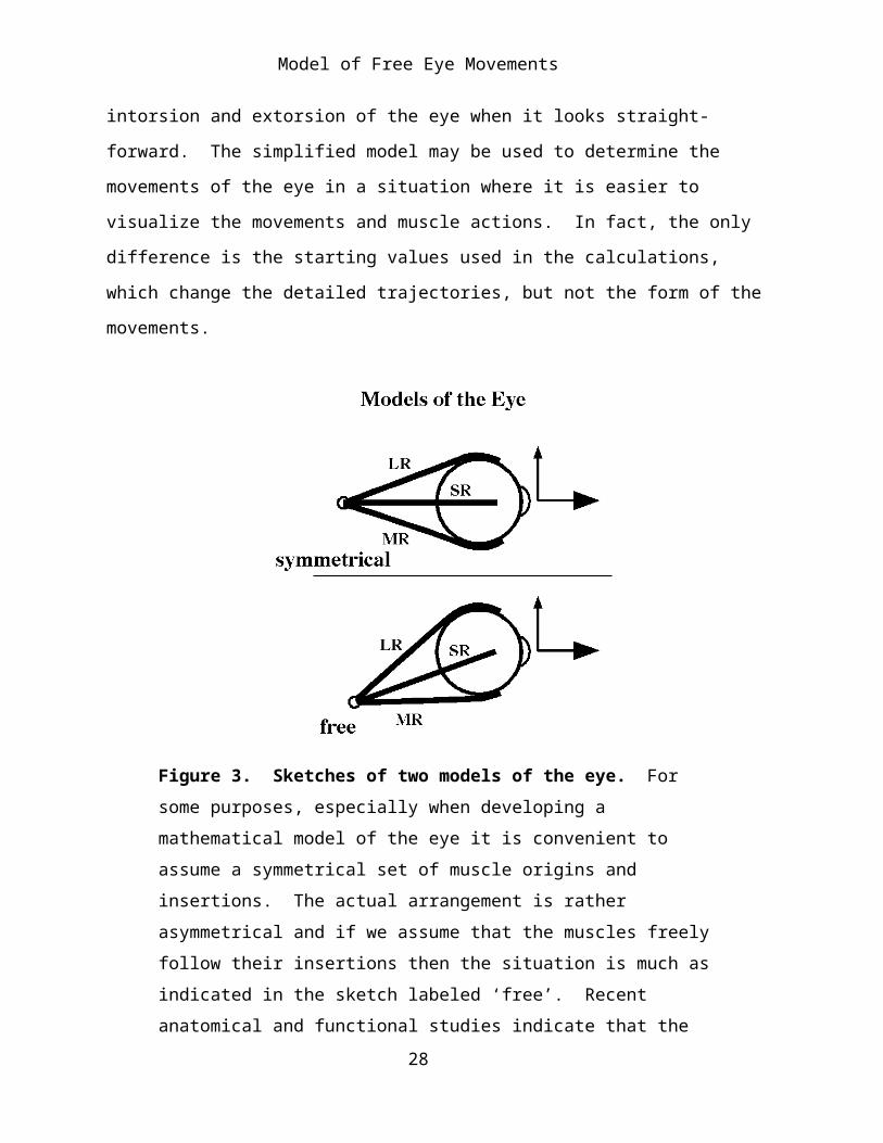
The second model, which will be called the symmetrical model, has the recti taking origin from a point three units directly posterior to the center of the globe and attaching to the globe 45° anterior to the frontal meridian of the eye. The oblique muscles are similarly simplified so that they produce pure intorsion and extorsion of the eye when it looks straight-forward. The simplified model may be used to determine the movements of the eye in a situation where it is easier to visualize the movements and muscle actions. In fact, the only difference is the starting values used in the calculations, which change the detailed trajectories, but not the form of the movements.
22
Model of Free Eye Movements
Figure 3. Sketches of two models of the eye. For some purposes, especially when developing a mathematical model of the eye it is convenient to assume a symmetrical set of muscle origins and insertions. The actual arrangement is rather asymmetrical and if we assume that the muscles freely follow their insertions then the situation is much as indicated in the sketch labeled ‘free’. Recent anatomical and functional studies indicate that the rectus muscles are restricted by pulleys that lie in the region just caudal to the globe’s equator and so there is a third model, called the ‘restricted’ muscle model. The restricted model is treated in a separate paper.
It was seen that once we moved away from the neutral position, where the eye looks straightforward, the same quirks and complications were observed in the simplified model as in the more realistic model, therefore the more realistic and natural model is used in the following analysis. Curiously, the restrained model turns out to have many of the features of the symmetrical model.
23
Model of Free Eye Movements
Geodesic and Non-Geodesic Eye Movements
Virtually all eye movements are conical rotations, therefore have both swing and spin components. However, a movement can be classified only in relation to some reference vector. The reference vector for eye movements, unless stated otherwise is neutral straightforward gaze. The only rotations of the eye, that would not have both swing and spin attributes would be when the axis of rotation is aligned with the axis of the line of sight, which is pure spin, and those in which the extension vector of the line of sight sweeps out a great circle, which is pure swing.
We can interpret these principles in terms of the eye and its movements as follows. If a rotation occurs about the line of sight, that is pure intorsion or extorsion and the eye is considered to undergo a pure spin. If the eye sweeps from the center of gaze radially to another eye position, then the extension vector remains in a single plane and its tip follows a great circle for the eye. Consequently, there is no spin and the eye undergoes a pure swing. Adduction is medially directed swing, abduction laterally directed swing. Superiorly directed swings are elevations and inferiorly directed swings are depressions of the eye.
Any trajectory that causes the extension vector of the line of sight to sweep out a great circle will be is a geodesic movement and therefore a pure swing. All other trajectories have a combination of spin and swing.
Note that unique geodesic trajectories exist between any two eye positions that are not coincident or opposite to each other. These potential geodesic movements are important in determining how much spin and swing occur during an actual eye movement or set of eye movements.
Donder’s Law and Listing’s Plane
It has been observed that for actual eye movements the eye orientation for a given line of sight is independent of the actual trajectory followed in
24

Model of Free Eye Movements
achieving that position. This observation is called Donder’s Law (Tweed. and Vilis, 1987). Listing observed that the rotation axis that would move the eye from neutral position, with the line of sight directed straightforward, to the position and orientation dictated by Donder’s Law is always contained in the same plane. The plane that contains all the rotation vectors can be deduced from the fact that rotation of the eye in pure elevation or depression from neutral position will place the rotation vector along the horizontal axis of the eyeball and pure abduction or adduction will have a rotation axis along the vertical axis of the eyeball. Two lines determine a plane, therefore, the plane containing the set of rotation axes, called Listing’s plane, is orthogonal to the line of sight in neutral and the orientation associated with each line of sight, as dictated by Donder’s Law, is simply the orientation that results if the eye moves along the geodesic between neutral position and orientation and the offset line of sight. Stated another way, the orientation of the eye at any particular position is the orientation that would result from a geodesic trajectory from neutral position and orientation. An interesting corollary to this characteristic of eye movements is that any rotation of the eye from one position to another is effectively along a geodesic to neutral gaze and then along a second geodesic from neutral sight to the final position, since all rest positions have no spin relative to neutral position and orientation. At first sight, one might think that eye movements between different visual targets would generally be the shortest distance between the two targets, that is geodesics, but it easy to show that such trajectories would rapidly degrade the alignment of the eye and would not be consistent with Donder’s Law.
The Computation of Swing and Spin
A substantial portion of the analysis in this essay involves the swing and spin components of a conical rotation, therefore, let us pause to quickly outline how such a calculation is performed. The details of the calculation
25
Model of Free Eye Movements
require a basic understanding of quaternion algebra, but the essence of the calculation may be appreciated without the algebra.
Supposing that the eye started from the neutral position, looking straightforward, and followed a trajectory in which the extension vector of the line of sight does not stay in a single plane, the amount of spin and swing can be computed as follows. Start by specifying the primary frame vector, x, which is generally equal to the extension vector, E, before the movement, , and after the movement, . There are three frames that have
to be calculated; the initial orientation frame, , the final orientation frame, ,
and the orientation frame, , that would have resulted if the movement had
swept the extension vector along a great circle connecting the initial and final positions. The variables were generated as the trajectory occurred,
but the fifth variable, , does not actually occur, therefore has to be
computed from the other variables.
The intermediate frame, , is computed as follows: Compute the rotation quaternion for the equivalent great circle trajectory. This done by dividing the initial line of sight, , by the final line of sight, . The result is a
quaternion, RG .
The amount of swing in moving from to is the angle of the rotation quaternion for the great circle trajectory, . To compute the frame if the eye
had swung through the same excursion along the equivalent great circle, transform the initial frame using the great circle rotation quaternion.
Once we know the value of we can compute the amount of pure swing and spin which occurred along the actual trajectory. The amount of pure
26
Model of Free Eye Movements
swing is the angle of the quaternion . To compute the amount of spin one
must compute the rotation quaternion that transforms into . The
intermediate frame has a line of sight that points in the same direction as the final line of sight, but the medial (yF and yG ) and vertical (zF and zG ) perpendiculars to the line of sight are rotated relative those in the final eye orientation. The spin is the ratio of the perpendicular in the final orientation to the same perpendicular in the intermediate orientation.
The amount of pure spin is the angle of the quaternion RSp . The single rotation that would transform the initial frame into the final frame is the product of the half-angle quaternions for pure swing and spin.
Note that the rotation is a conical rotation, therefore one must use Euler’s formula.
FF =rC∗ΦI ∗rC−1
27
Model of Free Eye Movements
Calculation of the Model
The Defining Arrays
The model starts with three frames, that is, sets of vectors:
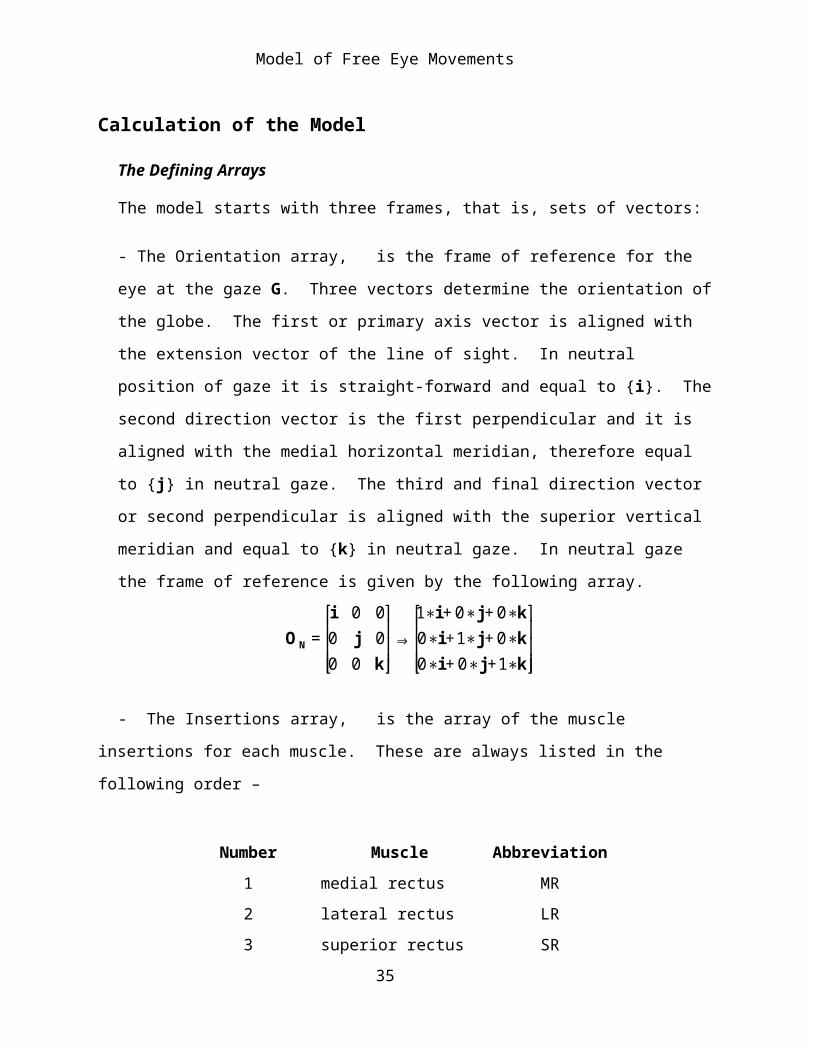
- The Orientation array, is the frame of reference for the eye at the gaze G. Three vectors determine the orientation of the globe. The first or primary axis vector is aligned with the extension vector of the line of sight. In neutral position of gaze it is straight-forward and equal to {i}. The second direction vector is the first perpendicular and it is aligned with the medial horizontal meridian, therefore equal to {j} in neutral gaze. The third and final direction vector or second perpendicular is aligned with the superior vertical meridian and equal to {k} in neutral gaze. In neutral gaze the frame of reference is given by the following array.
€
ON =i 0 00 j 00 0 k
⎡
⎣
⎢ ⎢ ⎢
⎤
⎦
⎥ ⎥ ⎥⇒
1∗i + 0∗j + 0∗k0∗i +1∗j + 0∗k0∗i + 0∗j +1∗k
⎡
⎣
⎢ ⎢ ⎢
⎤
⎦
⎥ ⎥ ⎥
- The Insertions array, is the array of the muscle insertions for each muscle. These are always listed in the following order –
Number Muscle Abbreviation1 medial rectus MR2 lateral rectus LR3 superior rectus SR4 inferior rectus IR5 superior oblique SO6 inferior oblique IO
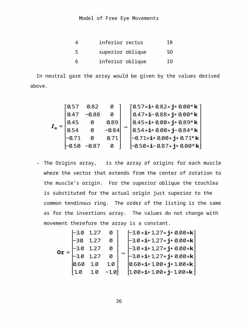
In neutral gaze the array would be given by the values derived above.
28

Model of Free Eye Movements
€
IN =
0.57 0.82 00.47 −0.88 00.45 0 0.890.54 0 −0.84−0.71 0 0.71−0.50 −0.87 0
⎡
⎣
⎢ ⎢ ⎢ ⎢ ⎢ ⎢ ⎢
⎤
⎦
⎥ ⎥ ⎥ ⎥ ⎥ ⎥ ⎥
⇒
0.57∗i + 0.82∗j + 0.00* k0.47∗i − 0.88∗j + 0.00* k0.45∗i + 0.00∗j + 0.89* k0.54∗i + 0.00∗j − 0.84 * k−0.71∗i + 0.00∗j + 0.71* k−0.50∗i − 0.87∗j + 0.00* k
⎡
⎣
⎢ ⎢ ⎢ ⎢ ⎢ ⎢ ⎢
⎤
⎦
⎥ ⎥ ⎥ ⎥ ⎥ ⎥ ⎥
- The Origins array, is the array of origins for each muscle where the vector that extends from the center of rotation to the muscle’s origin. For the superior oblique the trochlea is substituted for the actual origin just superior to the common tendinous ring. The order of the listing is the same as for the insertions array. The values do not change with movement therefore the array is a constant.
€
Or =
−3.0 1.27 0−30. 1.27 0−3.0 1.27 0−3.0 1.27 00.60 1.0 1.01.0 1.0 −1.0
⎡
⎣
⎢ ⎢ ⎢ ⎢ ⎢ ⎢ ⎢
⎤
⎦
⎥ ⎥ ⎥ ⎥ ⎥ ⎥ ⎥
⇒
−3.0∗i +1.27∗j + 0.00∗k−3.0∗i +1.27∗j + 0.00∗k−3.0∗i +1.27∗j + 0.00∗k−3.0∗i +1.27∗j + 0.00∗k0.60∗i +1.00∗j +1.00∗k1.00∗i +1.00∗j −1.00∗k
⎡
⎣
⎢ ⎢ ⎢ ⎢ ⎢ ⎢ ⎢
⎤
⎦
⎥ ⎥ ⎥ ⎥ ⎥ ⎥ ⎥
Muscle Lengths in Neutral Gaze
- The muscle lengths are computed for each muscle with the eye in neutral gaze. This is done by calculating the angle of the quaternion that rotates the muscle’s insertion vector into its origins vector and subtracting the angle between the origins vector and the vector to the point of tangency. This gives the amount of the muscle that lies upon the surface of the eye, which is the part that changes with eye movements. The result is added to the distance from the point of tangency to the origin. This is the distance from the origin to the eyeball, which remains constant. In the case of the superior oblique the distance from the common tendinous ring to the trochlea is also added. For many calculations the change in muscle length is the relevant variable, in which case we subtract the muscle lengths in neutral gaze from the muscle lengths at the offset eye gaze.
29

Model of Free Eye Movements
Geodesic Rotations to a Regular Array of Offset Positions
The eye is moved to a regular array of vertical and horizontal offset positions along geodesic trajectories, so that there will be no spin relative to the neutral gaze. The rotation along a geodesic is expressed by the quaternion that turns the neutral line of sight into the line of sight at the offset position.
where and are the extension vectors of the line of sight in neutral and offset gaze, respectively.
The use of the geodesic rotation was initially chosen because it preserves the orientation of the eye so that vertical remains vertical and horizontal remains horizontal at the offset position. It also accords with Listings Law in that all the rotation quaternions have their vector components in Listing’s Plane, the plane orthogonal to the extension vector for the line of sight in neutral gaze.
The grid used to determine the points of offset is one that is like the latitude and longitude lines on a globe of the earth, with the poles being the superior and inferior poles of the eye. This is not the only way that one might compute a regular array of offset points. For instance, one might select the points so that they are projections of the intersects of a distant square array. The calculations were done both ways and the basic patterns are very similar. The choice of the array of offsets is largely arbitrary.
Variables Computed at Each Offset
At each offset position a number of variables are computed. The computed variables are as follows:
The new orientation frame in the offset position, , where ‘m’ is the horizontal offset and ‘n’ is the vertical offset. The zero inside the brackets indicates that there is no muscular perturbation. We subsequently compute
30

Model of Free Eye Movements
the effect of individual muscle contractions at the offset position and the muscle’s number is placed in the brackets for the orientation frame. If no number is specified then the unperturbed orientation frame is meant.
The neutral orientation frame, , has experienced a pure swing to yield . It is necessary to compute its new frame elements for comparison with the neutral position and with the orientation frames that result after standard size muscle contractions in each of the oculomotor muscles, starting from the offset position.
The new insertions frame, . As the eye moves it carries the muscle insertions along the same trajectory. The new insertion frame is compared with the insertions frame in neutral gaze to determine the change in muscle length for each muscle. It is also used to compute the new rotation vector for each of the oculomotor muscles.
The muscle lengths are computed in the offset position, using the same protocol as outlined for the calculations in neutral gaze, and compared with the muscle lengths in the neutral gaze position, . So the new muscle
lengths are computed and the muscle lengths in neutral gaze are subtracted to generate the change in muscle length, .
The orientation frame, , is computed after the eye is moved by a standard size contraction of each muscle, Ex, where M stands for each of the muscles in turn.
31
Model of Free Eye Movements
Om,n M[ ] = Sm,n M[ ]∗Om, n ∗Sm,n−1 M[ ] ,32

Model of Free Eye Movements
If one muscle contracts, then the others must also change length to compensate and allow the eye to move, but we are ignoring the compensatory changes for the purposes of this and the subsequent calculations, because the purpose of the calculations is more akin to computing a differential then an actual movement. The muscle contraction is a small twitch. We are primarily concerned with the pulling direction of each muscle at each offset.
The direction of pull, , for each oculomotor muscle is computed at each offset position. The direction that a muscle pulls may change dramatically as its insertion moves relative to the center of rotation.
If we allow a muscle to shorten a standard amount, then there will be a movement of the eye at the point of attachment along the line of pull. Depending on the geometry of the muscle’s origin and insertion relative to the center of rotation and the line of sight, the movement may impart swing and/or spin to the globe.
The final calculations determine the swing, , and spin, , introduced by each muscle contracting a standard amount. These are computed by rotating the orientation frame at the offset position into the orientation frame at the end of the muscle contraction.
33

Model of Free Eye Movements
In words, we first align the first component or axis component of the
frame before and after the muscle contraction, which is the swing. The swing is the ratio of the final frame’s axis component to the initial frame’s axis component. If we then swing the frame prior to the contraction through the calculated rotation, we obtain an intermediate orientation frame that is spun relative to the frame after the muscle contraction, but which is aligned with its primary axis. We can determine the magnitude of the spin by dividing either the second or third component of the final orientation frame by the corresponding component of the intermediate frame of reference.
Finally, the swing and spin are normalized by dividing them by the excursion, Ex, which is the amount of the muscle contraction. Because most eye movements are conical rotations with respect to the line of sight the swing and spin are less than the amount of the muscle contraction. These normalized swings and spins are called and .
Methods
The results described below are based upon calculations using the model just presented based upon the measurements tabulated in the Introduction. The actual calculations have been done several times with several versions of programs in different programming languages, but the data presented here is based upon programs written in Mathematica. Most of the figures are taken directly from those programs with some additional labeling added in Canvas 9. The program that codifies the model is taken directly from the analysis given immediately above. All the computation and data processing were performed on a Power Mac G4.
34
Model of Free Eye Movements
The Results of the Calculations
Changes in Muscle Length as a Function of Horizontal and Vertical Offset
The length of the six extrinsic oculomotor muscles determines the alignment of the eye, that is, the direction of the line of sight and the orientation of its frame of reference. There must be an unique set of muscle lengths for each alignment. Conversely, if one sets the lengths of all six muscles, then the alignment is uniquely determined. From these observations it follows that it is anatomically interesting to determine how muscle length depends on alignment. For this analysis the alignments have been constrained to those with null spin relative to neutral gaze. That is they are in accord with Donder’s law.
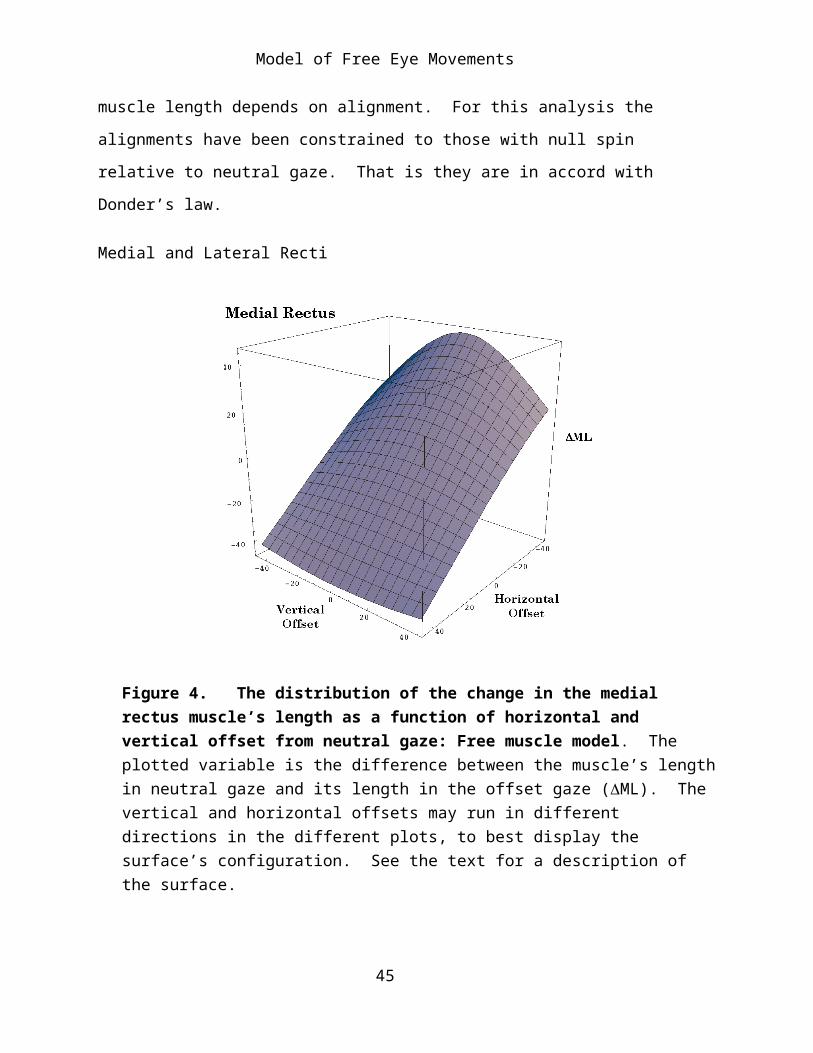
Medial and Lateral Recti
Figure 4. The distribution of the change in the medial rectus muscle’s length as a function of horizontal and vertical offset from neutral gaze: Free muscle model. The plotted variable is the difference between the muscle’s length in neutral gaze and its length
35
Model of Free Eye Movements
in the offset gaze (ML). The vertical and horizontal offsets may run in different directions in the different plots, to best display the surface’s configuration. See the text for a description of the surface.
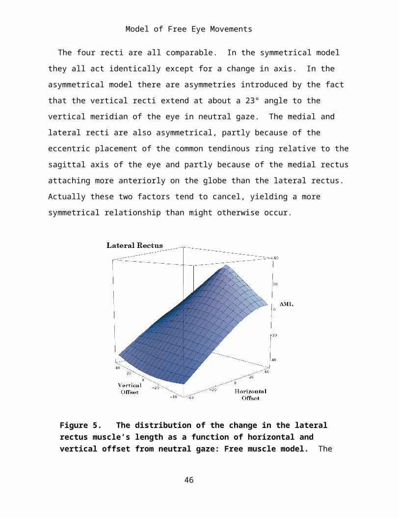
The four recti are all comparable. In the symmetrical model they all act identically except for a change in axis. In the asymmetrical model there are asymmetries introduced by the fact that the vertical recti extend at about a 23° angle to the vertical meridian of the eye in neutral gaze. The medial and lateral recti are also asymmetrical, partly because of the eccentric placement of the common tendinous ring relative to the sagittal axis of the eye and partly because of the medial rectus attaching more anteriorly on the globe than the lateral rectus. Actually these two factors tend to cancel, yielding a more symmetrical relationship than might otherwise occur.
Figure 5. The distribution of the change in the lateral rectus muscle’s length as a function of horizontal and vertical offset from neutral gaze: Free muscle model. The conventions are the same as for the medial rectus muscle figure.
The variable that is plotted in the figures is the difference in muscle length between the eye in neutral gaze and in offset gazes. The computed
36

Model of Free Eye Movements
points lie in a curvilinear surface. The horizontal recti are longest when the line of sight is along the horizontal meridian, where they are linearly related to the horizontal offset (Figures 4 and 5). For most of the surface, as gaze shifts vertically, either superiorly or inferiorly the muscles become relatively shorter. The change with vertical offset is curvilinear. The amount of curvature increases as the muscle becomes longer. At the shortest muscle lengths the direction of the curvature reverses and the muscle becomes longer as gazes shifts vertically. This is a very small curvature and it probably does not occur for normal gaze positions. The reversal in curvature is more pronounced for the lateral rectus muscle.
Despite the curvilinear shape of the surfaces, they are comparatively flat surfaces, except for the lateral rectus at the most medial gaze positions.
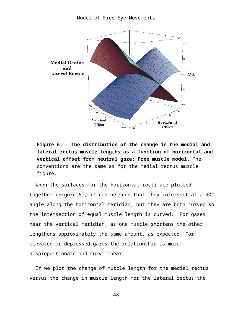
Figure 6. The distribution of the change in the medial and lateral rectus muscle lengths as a function of horizontal and vertical offset from neutral gaze: Free muscle model. The conventions are the same as for the medial rectus muscle figure.
37
Model of Free Eye Movements
When the surfaces for the horizontal recti are plotted together (Figure 6), it can be seen that they intersect at a 90° angle along the horizontal meridian, but they are both curved so the intersection of equal muscle length is curved. For gazes near the vertical meridian, as one muscle shortens the other lengthens approximately the same amount, as expected. For elevated or depressed gazes the relationship is more disproportionate and curvilinear.
If we plot the change of muscle length for the medial rectus versus the change in muscle length for the lateral rectus the result is that the relation becomes more nonlinear as one moves to more superior or inferior gaze directions.
Taken alone, the two horizontal recti are unstable in that any deviation of the eye from the horizontal meridian will tend to be amplified by the tension in the horizontal recti drawing the eye further in the same direction. Of course, other extraocular eye muscles will tend to act against that tendency.
Superior and Inferior Recti
The relationships for the vertical recti are similar to those for the horizontal recti if we switch the vertical and horizontal labels (Figures 7 and 8). The main difference is that the surface is not symmetrical about neutral gaze, but about a laterally displaced gaze. The line of symmetry is a vertical meridian 23° lateral to neutral gaze. This is obviously due to the medial location of the annulus of Zinn. As with the horizontal recti, the vertical recti are longest when they are pulling directly superiorly or inferiorly. Their length is proportional to the amount of elevation or depression, when the eye’s line of sight is approximately 23° lateral to neutral gaze.
38

Model of Free Eye Movements
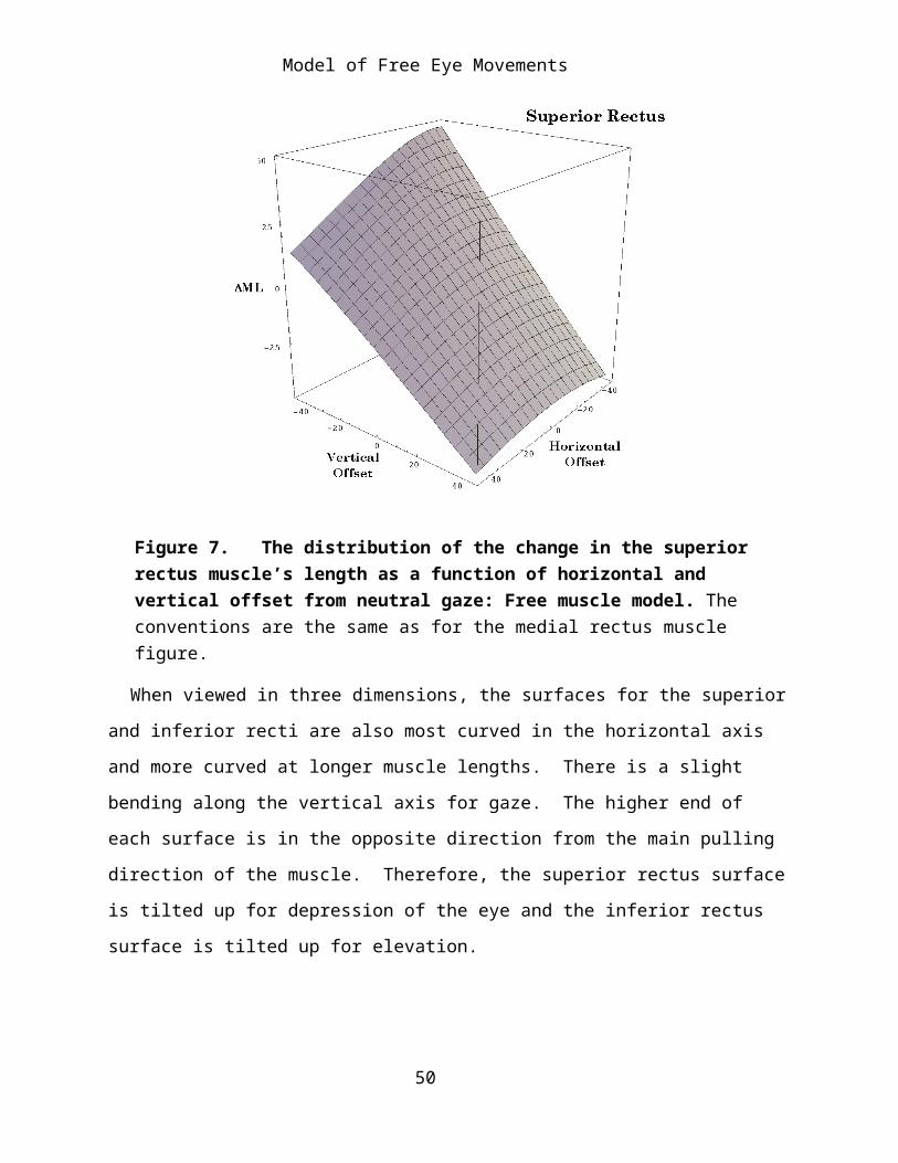
Figure 7. The distribution of the change in the superior rectus muscle’s length as a function of horizontal and vertical offset from neutral gaze: Free muscle model. The conventions are the same as for the medial rectus muscle figure.
When viewed in three dimensions, the surfaces for the superior and inferior recti are also most curved in the horizontal axis and more curved at longer muscle lengths. There is a slight bending along the vertical axis for gaze. The higher end of each surface is in the opposite direction from the main pulling direction of the muscle. Therefore, the superior rectus surface is tilted up for depression of the eye and the inferior rectus surface is tilted up for elevation.
39

Model of Free Eye Movements
Figure 8. The distribution of the change in the inferior rectus muscle’s length as a function of horizontal and vertical offset from neutral gaze: Free muscle model. The conventions are the same as for the medial rectus muscle figure.
When plotted together (Figure 9), the two surfaces intersect at an approximately right angle and, as with the horizontal recti, as one muscle lengthens the other contracts approximately proportionately. The ratio of their length changes deviates from 1.0 as one moves away from the vertical meridian at 23° lateral to neutral gaze.
Superior and Inferior Obliques
The changes in muscle lengths for the oblique muscles have different relations to gaze direction (Figures 10. 11, and 12). The distribution of the data is not symmetrical about neutral alignment and it is skewed or rotated relative to the horizontal and vertical meridia. The changes in muscle length are substantially less than the change in eye excursion, because the
40

Model of Free Eye Movements
muscle is pulling at an eccentric location on the globe. A substantial part of the muscle action is to roll the eye into intorsion or extorsion. There are not strongly differing projections of the surfaces onto the horizontal and vertical offset axes. However, if we were to choose our axes appropriately, the surface would be much like the surfaces for the recti. The differences are largely due to the different orientation of the oblique muscle attachments to the line of sight.
Figure 9. The distribution of the change in the superior and inferior rectus muscle lengths as a function of horizontal and vertical offset from neutral gaze: Free muscle model. The conventions are the same as for the medial rectus muscle figure.
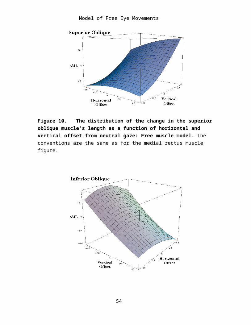
When viewed in three dimensional plots it is apparent that the surfaces for the oblique muscles are hyperbolic or saddle shaped and they are inclined so that the main axis is directed towards increasing muscle length for elevation and adduction (superior oblique) or increasing muscle length with depression and adduction (inferior oblique). Because we can not see
41
Model of Free Eye Movements
torsion in this type of plot the superior oblique appears to be an eye depressor and the inferior oblique appears to be an eye elevator.
Figure 10. The distribution of the change in the superior oblique muscle’s length as a function of horizontal and vertical offset from neutral gaze: Free muscle model. The conventions are the same as for the medial rectus muscle figure.
42

Model of Free Eye Movements
Figure 11. The distribution of the change in the inferior oblique muscle’s length as a function of horizontal and vertical offset from neutral gaze: Free muscle model. The conventions are the same as for the medial rectus muscle figure.
Figure 12. The distribution of the change in the superior and inferior oblique muscle lengths as a function of horizontal and vertical offset from neutral gaze: Free muscle model. The conventions are the same as for the medial rectus muscle figure.
Summary
When viewed in three dimensions the surface for each eye muscle is similar. Each is a modestly saddle-shaped or hyperbolic surface with an axis that is nearly linear and an orthogonal axis that is modestly curved, therefore non-linear. The pairs of horizontal and vertical recti are orthogonal to each other in the plane of their major pull and the surfaces for the vertical recti are orthogonal to the surfaces for the horizontal recti. The surfaces for the obliques are skewed relative to each other, because the muscles do not lie in the same plane for any eye position, however, their surfaces are roughly orthogonal to each other. Their linear axes are at oblique angles to the linear axes of the surfaces for the recti.
43
Model of Free Eye Movements
If we plot all the surfaces together, it is apparent that there is a complex relationship between the various surfaces. The details are beyond analysis at this point and they shift with changes in the models assumptions about the locations of the origins and insertions of the extraocular muscles. However, it is clear that if we draw a perpendicular to the offset coordinates, the line will always penetrate all six surfaces and the relationships between the values of the various intersects is constantly shifting. It is these sets of six intersects that determine and are determined by the gaze direction. If we plot the six intersects versus gaze direction then we have a two dimensional surface in an eight dimensional space. This surface will be called the static muscle length surface. For each horizontal and vertical offset there is a six dimensional vector for the set of muscle lengths. Each of the figures above is a section of that surface taken orthogonal to the other five axes. Much like what happens when one sections a three dimensional cone with a two dimensional plane and obtains a circle, parabola, hyperbola, or intersecting straight lines depending on which plane of section is viewed. In this case we are also seeing only a small portion of the static muscle length surface, because the eye has a relatively small excursion.
44
Model of Free Eye Movements
Figure 13. The distribution of the change in all of the extraocular muscle lengths as a function of horizontal and vertical offset from neutral gaze: Free muscle model. The conventions are the same as for the medial rectus muscle figure.
On the other hand, these are the relationships that apply for the particular muscle attachments that exist for the eye. There is not a simple relationship between the alignment of the eye and the set of muscle lengths. The relationship is definitely nonlinear and asymmetrical about neutral gaze.
45
Model of Free Eye Movements
Muscle Pulling Direction as a Function of Horizontal and Vertical Offset
The individual eye muscles pull the globe with tangentially applied forces applied at the point of insertion. In the following analysis it is assumed that the muscle applies all of its force at a single point, which is clearly not correct since their attachments are linear and approximately perpendicular to the long axis of the muscle. However, as a first approximation these calculations give an indication of the types of movements that each muscle might produce in isolation if it is caused to produce a small twitch.
For almost all gaze directions, contraction of each muscle produces both a swing and a spin. The gaze direction shifts, but there is also a torsion of the eye. Most of what follows is concerned with the swing component, but, in general, the smaller the swing the greater the spin that is produced concurrently. The relationship between these two components of the movements will be addressed below.
As stated above, the lengths of all six muscles together determine eye position and orientation, therefore the action of individual muscles gives us very limited information about eye movements. However, by knowing how each muscle pulls we may come to better understand how lesions that affect individual muscles might alter the movements of the eyes.
The six oculomotor muscles tend to be considered in terms of three pairs, because of the similarities of their anatomy. The horizontal recti reach around the horizontal meridian of the eye in neutral gaze and tend to adduct (MR) and abduct (LR). The vertical recti have insertions that cross the vertical meridian in neutral gaze and tend to elevate (SR) and depress (IR) the eye, but since they take origin medial to the center of rotation they also tend to pull their insertions medially unless the eye is abducted more than about 23°. Finally, the two oblique muscles extend from points anterior and medial to the center of rotation and attach posteriorly, therefore, when the eye is in neutral gaze, they tend to pull the posterior
46

Model of Free Eye Movements
lateral portion of the eye superiorly and medially (SO) and inferiorly and medially (IO). Because of they pull from points medial to the globe these muscles have a stronger tendency to rotate the eye about its line of sight, causing intorsion (SO) and extorsion (IO).
The analysis just outlined is based upon the geometry of the eye’s anatomy when it sits in neutral gaze. As it moves away from that alignment the muscle insertions also move and cause the lines of pull to change. These changes may cause substantial changes in the actions of the muscles. The following analysis addresses these possibilities.
Each muscle pulling direction figure is organized in the same way, to facilitate comparisons between muscles and comparisons of the same muscle when the muscle is free to follow its insertions and when it is restricted by pulleys (Langer, 2004). The pulling directions are plotted as vectors having their origin at the gaze position at which they are pulling. This calculation has been done for every muscle at an array of gaze directions ranging from 45° of abduction to 45° of adduction and from 45° of depression to 45° of elevation. As noted above, the normal range of eye movement is substantially less than this. The approximate set of gaze directions that normally occur has been indicated by a circular shaded region with a radius of about 30°. The length of the vector’s shaft is proportional to the magnitude of the swing produced by the muscle twitch. The actual twitch size used in the calculations was 2.5°, but the vectors have been set to a maximal length of 5° to make them more visible. The arrowheads are a constant size. Sometimes the shaft is so short that it lies within the arrowhead (see the plot for the superior oblique).
47
Model of Free Eye Movements
Medial Rectus and Lateral Rectus
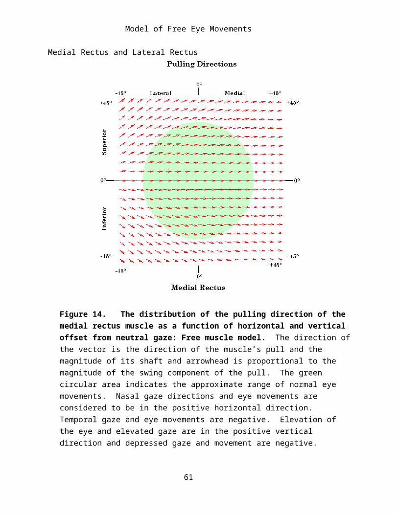
Figure 14. The distribution of the pulling direction of the medial rectus muscle as a function of horizontal and vertical offset from neutral gaze: Free muscle model. The direction of the vector is the direction of the muscle’s pull and the magnitude of its shaft and arrowhead is proportional to the magnitude of the swing component of the pull. The green circular area indicates the approximate range of normal eye movements. Nasal gaze directions and eye movements are considered to be in the positive horizontal direction. Temporal gaze and eye movements are negative. Elevation of the eye and elevated gaze are in the positive vertical direction and depressed gaze and movement are negative.
Since the horizontal recti extend along the horizontal meridian it is expected that their action is to pull the eye directly medially and laterally from neutral alignment. If the eye is elevated, their insertions move
48

Model of Free Eye Movements
superiorly and the line of pull is directed superior to the horizontal meridian, therefore both recti will elevate the eye as well as abduct or adduct. Conversely, with depression of the eye they further depress the eye as well as abduct and adduct. The tendency to elevate or depress the eye is more pronounced when the direction of the line of sight is in the part of the range which most elongates the eye muscle. This effect is more pronounced for the lateral rectus than for the medial rectus. When the eye is moved into the more distal quadrants, the elevation and depression components may become a major component of the muscle’s action.
Figure 15. The distribution of the pulling direction of the lateral rectus muscle as a function of horizontal and vertical offset from neutral gaze: Free muscle model. The conventions are as given for the medial rectus figure.
49
Model of Free Eye Movements
When the pulling directions are plotted together one can see that the two horizontal recti together have a pronounced tendency to pull the eye away from the horizontal meridian when it is at more eccentric positions of elevation or depression.
For positions near neutral alignment the great majority of the muscle’s action is expressed in swing, as one might expect, however in the more eccentric alignments the length of the line of pull vector becomes shorter, indicating that more of the muscle’s action is being expressed in potential spin about the line of sight (see below for more detailed analysis of this tendency).
Superior Rectus and Inferior Rectus
50

Model of Free Eye Movements
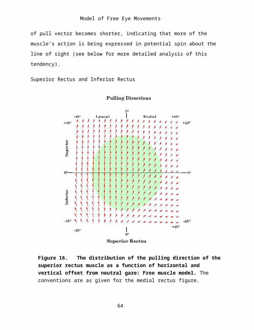
Figure 16. The distribution of the pulling direction of the superior rectus muscle as a function of horizontal and vertical offset from neutral gaze: Free muscle model. The conventions are as given for the medial rectus figure.
The vertical recti are distributed so as to pull the anterior portion of the globe superiorly and inferiorly. Therefore, their dominant actions in neutral alignment is elevation and depression, but because their origins lying medial to the center of rotation for the eye they also pull the anterior eye medially. If the eye is rotated about 23° laterally, then the muscles will lie along the vertical meridian and the vertical recti will bring about pure elevation and depression. If the eye rotates beyond that point then their action when the muscle is elongated is to pull it further into abduction, assisting the lateral rectus, rather than the medial rectus. When the muscle is most contracted, it still draws the eye nasally. For extreme medial gaze the actions of the vertical recti are as much adduction as elevation or depression. With medial gaze the lines of pull for both muscles also become shorter indicating that they are tend to impart a substantial potential spin about the line of sight.
51

Model of Free Eye Movements
Figure 17. The distribution of the pulling direction of the inferior rectus muscle as a function of horizontal and vertical offset from neutral gaze: Free muscle model. The conventions are as given for the medial rectus figure.
Superior Oblique and Inferior Oblique
The pulling directions for the obliques near neutral gaze is unexpected, they pull the posterior lateral aspect of the eye either superiorly and medially (SO) or inferiorly and medially (IO) they should therefore cause the line of sight to move inferiorly and medially (SO) or superiorly and medially (IO). The model says that both tend to pull the anterior eye primarily vertically and to a lesser extend laterally. If the gaze is directed medially, then the predominant direction tends to be depression (SO) or elevation
52

Model of Free Eye Movements
(IO). Consequently, the initial movement of the line of sight when these muscles contract is down and lateral (SO) and up and lateral (IO).
Figure 18. The distribution of the pulling direction of the superior oblique muscle as a function of horizontal and vertical offset from neutral gaze: Free muscle model. The conventions are as given for the medial rectus figure.
The outcome is sensitive to the details of the muscle’s insertions and origins. If the insertions are closer to the coronal plane then the initial pulling directions tend to be medial and if the insertions are more posterior, then the initially pulling direction is lateral. Given the measurements used in the model, which are based on a survey of several atlases the results are as indicated.
53
Model of Free Eye Movements
Figure 19. The distribution of the pulling direction of the inferior oblique muscle as a function of horizontal and vertical offset from neutral gaze: Free muscle model. The conventions are as given for the medial rectus figure.
Leaving aside whether the obliques pull the line of sight medially or laterally, it is clear that for a laterally aligned eye they produce primarily intorsion and extorsion and for a medially aligned eye the obliques are also significant elevators and depressors. When the eye is elevated the superior oblique is more of a depressor and when it is depressed the superior oblique produces relatively more intorsion. The inferior oblique has the opposite pattern, extorsion is relatively more prominent in an elevated alignment and elevation is relatively more prominent in a depressed alignment.
54

Model of Free Eye Movements
All of the Muscles Together
When all the muscle pulls are superimposed there are a number of features that become apparent. Near neutral gaze the muscles are aligned so as to produce mostly horizontal or vertical pulls. The vertical recti pull a bit in the nasal direction, but the obliques pull about the same amount temporally. Oblique movements are accomplished by combining a horizontal rectus with a vertical rectus and one of the obliques. Movements towards neutral vision use the complementary set of muscles.
Near neutral alignment the majority of the pull generated by the muscles goes into generating swing of the line of sight. When the eye is aligned peripherally in the orbit there is more pull directed into spin. All the muscles introduce torsion or spin at most eye positions. The amount of spin tends to be least for the horizontal recti, moderate for the vertical recti and greatest for the obliques. The vertical recti produce the most spin when gaze is nasal and the obliques produce the most spin when gaze is temporal.
When all the pulling directions are plotted together it may be noted that at the more eccentric gazes, all the muscles are pulling away from neutral gaze. There is little or no restoring force, drawing the eye centrally. It is likely that this is compensated for by the fact that the fascia of the orbit is strained by the movement of the eye into eccentric positions, therefore, it becomes a substantial passive restoring force that tends to draw the eye back towards neutral gaze.
The sum of the active and passive forces for all the muscles must be zero, otherwise the eye would move from the initial alignment. The muscle’s active contractile forces and their passive restoring forces add vectorially and their torques upon the eye sum to zero. However, we know that if their lengths are appropriate, then all the linear and rotatory forces must cancel out for any particular position. Remember that the attribute plotted in the
55

Model of Free Eye Movements
above figures is pulling direction. The force being generated to maintain the appropriate muscle length is a different attribute.
The lengths of the lines in the above figures are proportional to the relative amount of swing generated by the muscle in that eye alignment. Conversely, the relative amount of spin imparted by the muscle may be appreciated by the relative shortness of the pulling direction line. While one decreases, the other increases. The amount of spin and swing imparted by a muscle are not linearly or reciprocally related. It will be shown below, they are orthogonally related in the manner of the sine and cosine functions.
Swing, Spin, SwEx, and SpEx
Spin versus Swing
At each gaze direction, each eye muscle imparts movement to the eye by contracting along its line of pull. The interaction between the location of the muscle’s attachment, as determined by the gaze, and the muscle’s line of pull at that gaze determines the action of the muscle, that is, how much swing and spin would be imparted to the eye by that muscle working in isolation. The same muscle may have quite different effects depending on the gaze direction. The action of a muscle will often be broken into two components, pure swing and pure spin. These are the two extremes of a continuum of movements, which may be called conical swing or conical rotations.
To resolve a conical rotation into spin and pure swing is artificial, but it allows one to have a better appreciation of the relative amounts of two qualities the movements has. Pure spin tends to be seen as the object in some way remaining stationary and twisting about its axis. Pure swing is seen as the body moving in a planar arc through space. A bit of reflection will reveal that this is an illusion because if the body is experiencing pure
56

Model of Free Eye Movements
spin about one axis, then it is experiencing pure swing in the plane orthogonal to that axis. All other parts of the object are experiencing a combination of pure swing and pure spin that are equally valid and are called conical rotation. Therefore, the relative amounts of spin and swing are not a property of the object, but are contingent upon the axis one chooses as a reference. For the eye, the reference axis is usually the line of sight and the frame of reference is the eye’s orientation in neutral gaze.
In the model, the action of each muscle was computed by calculating the frame of reference at a given gaze position with the orientation that has null spin relative to neutral alignment. That is the eyeball’s orientation prior to the muscle’s contraction. The shift in that gaze after a small muscle contraction is then computed for twitches of each muscle. To align the frame prior to the muscle contraction with the frame after the contraction the initial frame must undergo a conical rotation. The conical rotation may be expressed as the product of a pure swing and a pure spin. Consequently, these component movements are a natural consequence of computing the conical swing.
SwEx and SpEx
When the calculations are performed we arbitrarily choose small standard change in the muscle’s length by selecting a magnitude of muscle contraction. This is the magnitude of the eye’s movement in the plane that contains the muscle’s direction of pull and the center of rotation for the eye. Since the swing and spin components depend linearly upon the muscle excursion, it is reasonable to divide them by the excursion to obtain values that do not depend on which particular value of excursion was chosen for the actual calculation. These new variables have been denoted by SwEx and SpEx for the normalized swing and spin, respectively.
SwEx and SpEx have a very nice property, which illustrated by Figure 20, which plots SwEx versus SpEx for all of the computed movements of the
57

Model of Free Eye Movements
horizontal rectus muscle. The points lie on a circular arc. They behave as if
58

Model of Free Eye Movements
for any conical rotation there were a angle,
59

Model of Free Eye Movements
φ
60
Model of Free Eye Movements
such that
61
Model of Free Eye Movements
SwEx = sinφ and SpEx = cosφ .62

Model of Free Eye Movements
In the next figure (Figure 21), all the computed movements for all the eye muscles at all the gaze directions have been similarly plotted and one can see that the property is not special to one muscle.
If we consider the lateral rectus muscle, SwEx is 1.0 for contractions with the direction of the line of sight along the horizontal meridian. As anticipated, there is no swing imparted by such contractions. When the eye is elevated or depressed relative to the horizontal meridian there is substantial spin imparted to the eye by contraction of the lateral rectus. For instance, with the eye elevated 10° in the vertical meridian the amount of spin imparted is over 20% of the magnitude of the muscle contraction. For more eccentric eye positions the spin may be over 80% of the magnitude of the muscle contraction.
63
Model of Free Eye Movements
Figure 20. The distribution of spin (SpEx) as a function of swing (SwEx) for the horizontal rectus muscles. See the text for a description of the figure.
The superior rectus has a comparable pattern with the horizontal and vertical axes switched and the center of the distribution placed about 23° laterally. As with the horizontal recti, there is an unexpectedly great spin imparted by the vertical recti when the eye is eccentrically placed.
The obliques are notably different in that there is a much greater range of both SpEx and SwEx. Spin is a more pronounced component of the conical rotations produced by muscle contractions, largely because of the manner in which the oblique muscles attach and pull in a plane that strongly inclined relative to the axes of the orientation frame for the line of sight. The line of sight is our reference for eye movements, therefore contractions of the oblique muscles produce eye movements that appear complex in that frame of reference.
64

Model of Free Eye Movements
Figure 21. The distribution of spin (SpEx) as a function of swing (SwEx) for all of the extraocular muscles. See the text for a description of the figure.
As pointed out above, spin and swing are always relative to a particular frame of reference, therefore, they are not absolute indices of a movement. Still, they convey a quality of the movement to which we intuitively relate.
The Distribution of Swing and Spin as a Function of Gaze Direction
Since spin and swing for any particular conical swing are related as the sine and cosine, it is convenient to plot them as radial vectors. The horizontal and vertical components of the vector are the swing and spin respectively. These vectors have been calculated and plotted at every point in the sampling array for every muscle. Since spin and swing are always in
65

Model of Free Eye Movements
the direction of the excursion, they are positive and the vectors are between 0° and 90°.
Figure 22. The distribution of swing (SwEx) and spin (SpEx) for the medial rectus muscle as a function of horizontal and vertical gaze: Free muscle model. The vectors are the vector sum of the swing (SwEx: horizontal) and the spin (SpEx: vertical), times 5°, to increase visibility.
Medial Rectus
There is no spin when the medial rectus is pulling from a gaze in the horizontal meridian (Figure 22), but as soon as the gaze moves either superiorly or inferiorly there begins to be a spin component. The size of the
66

Model of Free Eye Movements
spin component is greater for gazes that are more lateral and more elevated or depressed. There is a remarkable amount of spin for some gazes.
Lateral Rectus
The distribution of spin and swing with lateral rectus twitches is similar to that for medial rectus (Figure 23). It differs in having a greater spin for the same amount of elevation or depression and having greater spin for more medial gaze directions. For some gaze directions within the normal range of eye movements, the spin component is as big as the swing component. About 40° medial, the lateral rectus is pulling almost directly into elevation and spin is the dominant component of the movement.
67
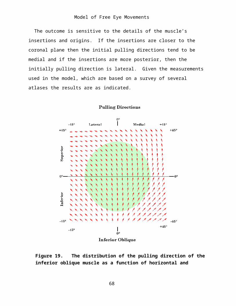
Model of Free Eye Movements
Figure 23. The distribution of swing (SwEx) and spin (SpEx) for the lateral rectus muscle as a function of horizontal and vertical gaze: Free muscle model. The conventions are the same as for the medial rectus figure.
Superior Rectus
The vertical recti are substantially more apt to impart a spin to the eyeball when they twitch (Figure 24). Gazes from a vertical line about 23° lateral to neutral gaze tend to be pure swing in elevation. To either side of that meridian there is increasing spin, as with the horizontal recti, but since the meridian is so far displaced into the lateral quadrants, the more medial gaze positions develop a strong spin component. At the medial extreme of normal eye movements, the spin component is about equal to the swing component.
The Inferior Rectus
The inferior rectus has an almost identical distribution of swing and spin with respect to gaze direction (Figure 25). For any gaze direction medial to neutral, there is a substantial spin component.
conventions are the same as for the medial rectus figure.
Superior Oblique
The oblique eye muscles have substantial spin components for their twitches at all gaze directions (Figure 26). Generally, the spin component is larger, but for most gazes the two components are of comparable size. The spin is greatest for gaze in the inferior lateral quadrant and smallest for the upper medial quadrant.
68

Model of Free Eye Movements
Inferior Oblique
The ratio of the spin to the swing for twitches of the inferior oblique is remarkably consistent in all quadrants. It is the most consistent of all the extraocular muscles even though it is most variable in its pulling directions.
The consistency with which the oblique muscles pull with spin and swing is apparently due to the short distances between their functional origins and their insertions. The same consistency occurs when the rectus muscles are restricted by pulleys much closer to the eyeball than their anatomical origins.
69
Model of Free Eye Movements
Figure 24. The distribution of swing (SwEx) and spin (SpEx) for the superior rectus muscle as a function of horizontal and vertical gaze: Free muscle model. The conventions are the same as for the medial rectus figure.
Discussion of Observations
The Considered Model is a Simplification of the Full Eye Movement Model
This paper examines the movements of the eye in the orbit as an example of an anatomical joint that is very general in certain ways, but comparatively simple in others. The eye is very general in that it is essentially an universal joint. It may experience movement in any direction about its center of rotation. The model has been simplified for present purposes because the eye moving in the orbit does not experience notable translations and all rotations occur about a single point at the center of the globe. Movements about other axes do occur when the head moves and will have to be dealt with in other analytic studies, but the intent of the current analysis was to focus on those particular aspects of the eye’s movements that occur when the eye is scanning the visual world from a stable head and body.
70
Model of Free Eye Movements
Figure 25. The distribution of swing (SwEx) and spin (SpEx) for the inferior rectus muscle as a function of horizontal and vertical gaze: Free muscle model. The conventions are the same as for the medial rectus figure.
For purposes of the analysis, the eye has been represented by a framed vector, which means that we consider its locus, its extension, its direction, and its orientation. Because all the eye movements considered have been rotations about a single point, at the center of the eye, its locus can be ignored in the analysis, by placing it at the origin of the coordinate system. The eye does not change shape during the movements, therefore its extension is a constant magnitude, which has been set equal to unity. The analysis concentrates on the direction and orientation of the eye, attributes
71
Model of Free Eye Movements
that are grouped together into the concept of gaze. It is assumed that the line of sight is the controlled parameter of eye movements and orientation is automatically determined for any given gaze direction.
Figure 26. The distribution of swing (SwEx) and spin (SpEx) for the superior oblique muscle as a function of horizontal and vertical gaze: Free muscle model. The conventions are the same as for the medial rectus figure.
72

Model of Free Eye Movements
Figure 27. The distribution of swing (SwEx) and spin (SpEx) for the inferior oblique muscle as a function of horizontal and vertical gaze: Free muscle model. The conventions are the same as for the medial rectus figure.
Alignment is a Unique Function of Extrinsic Eye Muscle Lengths
Since movement of the eye in the orbit normally occurs only by changes in the lengths of the six extraocular muscles, the analysis of changes in the line of sight must consider the possible and actual sets of muscle lengths. Gaze is a function of six parameters, three for its direction and three for its orientation. There are six extraocular muscles, therefore there should be a one-to-one mapping between the set of muscle lengths and gaze directions. Note though that the muscles occur in three roughly antagonistic pairs. The
73

Model of Free Eye Movements
gaze direction is not the domain of three muscles and the orientation the domain of the other three. Rather, all six muscles participate inextricably in the determination of both attributes of gaze, simultaneously. Still, given the anatomy of the eye in the orbit, it should be possible to compute the unique set of muscle lengths that hold the eye in a particular gaze. Such calculations have been done and they are the basis of this analysis.
Gaze Orientation is a Function of Gaze Direction
While a wide range of gaze orientations are in theory possible for every gaze direction, it is observed that a comparatively small subset of the possible combinations are actually realized. It is assumed that the eye prefers those alignments that place vertical elements in the visual image along the vertical axis of the retina and horizontal elements in the visual image along the horizontal axis of the retina. We take the vertical and horizontal axes of the eye to be those that occur when the eye is in neutral gaze.
In order for the alignment of the eye to remain the same as the alignment of the visual world, assuming that the visual world does not move, there must be no spin about the line of sight as the eye moves from neutral gaze to its new gaze direction. To simplify the nomenclature let us say that the gaze is spin neutral if it has no spin relative to some reference alignment, usually neutral gaze.
There will be null spin relative to neutral gaze only for eye movements that occur along great circles of the globe that pass through neutral gaze. For movements that occur along trajectories that are not geodesics through neutral gaze, the eye must continually undergo both swing and spin such that its orientation is continually adjusted to be always appropriate to its direction. The manner in which it affects the transformation depends on the trajectory that it follows. This problem is considered elsewhere.
74

Model of Free Eye Movements
Spin Neutrality Implies That Eye Muscle Length is a Controlled Parameter
Spin neutrality of eye movements is equivalent to saying that there is a particular set of muscle lengths for each direction of gaze. If the set of muscle lengths is specified, then the direction and orientation of the eye are determined and only certain combinations of muscle lengths are allowed with normal eye movements. These specific sets of muscle lengths can be computed by computing the allowed alignments, that is the alignments that have null spin relative to neutral gaze.
The simplest way to maintain spin neutrality would be to set the movement control system up so that the orientation of a line of sight for a given gaze direction is a unique function of gaze direction. This might be done by specifying the set of muscle lengths for that direction of gaze. This type of control is probably largely due to learning in the manner that neural networks learn. Trying specific eye movements and then making corrections based upon the direction of the error between the desired gaze and the actual gaze.
Listing’s Plane is Equivalent to Null Spin Relative to Neutral Gaze
Alignments that have null spin relative to neutral gaze are alignments that occur as the line of sight moves along geodesics though neutral gaze. If the line of sight were swept up along the vertical meridian, the axis of rotation would lie along a line from the center of rotation along the transverse axis. Similarly, if the eye were swept along the horizontal meridian, the axis of rotation would be along a vertical line through the center of rotation. In fact, it is easily seen that all the axes of rotation would be in a single plane perpendicular to the anterior-posterior axis of the eye. This experimentally observed plane is called Listing’s plane. The assumption that normally allowed eye alignments have null spin relative to neutral gaze is equivalent to stating that all the rotation axes for the displacements of the eye from neutral gaze lie in Listing’s plane.
75

Model of Free Eye Movements
Spin Neutrality Is Not Transitive
Note that alignments that have null spin relative to neutral gaze do not necessarily have null spin relative to each other. In fact, they generally will not, unless they lie on the same great circle through neutral gaze. This can be seen by a simple, if unrealistic example. Visualize the effect of elevating the line of site 90° from neutral gaze, along the vertical meridian, and the effect of adducting the eye 90°, along the horizontal meridian. Both movements were along great circles, therefore there was no spin introduced by either movement. Now, elevate the 90° adducted eye through 90°, until the direction of gaze coincides with that for the 90° elevated eye. There is no spin introduced here because we are again following the geodesic path between the two alignments, however, the orientation of the eye after the two step path is rotated 90° relative to the eye after the one step path, which is 90° of pure spin since the gaze directions are identical. Both the 90° elevated eye and the 90° adducted eye have lines of sight that have null spin relative to neutral gaze yet have 90° of spin relative to each other. Spin neutrality is not transitive.
Coplanar Movements Are Interchangeable, Spin Neutral, and Transitive
As long as a sequence of eye movements lie in a single plane, that is the gaze direction remains in a single plane during all of the movements, the movements are interchangeable in that the final eye alignment is always the same independent of the order in which the movements are performed and all intermediate alignments are null spin relative to each other and relative to the original and final alignments. As soon as one of the eye movements passes out of that plane the order of the movements relative to the extra-planar one becomes critical and there is a net non-null spin for the sequence of movements, except in very unusual instances.
76

Model of Free Eye Movements
Eye Movements Remain Spin Neutral Relative to Neutral Gaze
Curiously, despite the improbability of having a sequence of non-coplanar movements remaining spin neutral relative to the any particular alignment, that is exactly what is observed for actual eye movements. Eye alignment at the beginning and end of an eye movement is spin neutral relative to neutral gaze.
For smooth pursuit eye movements and other eye movements in which clear vision is maintained throughout the movement, one would expect that alignment would always be spin neutral relative to neutral gaze. Parenthetically, one would also expect that if neutral gaze were changed, say by tilting the head, then the reference for spin neutrality would also change in the same direction. However, if the control of eye movements is ‘hard-wired’, then the eyes might continue to move as if the eyes were moving from neutral gaze.
SwEx and SpEx
While it is arbitrary to divide a conical rotation into a pure spin and pure swing component. They do accord with our intuitions about eye movements. Spin causes the visual image to rotate on the retina. It is generally minimized by the neural control systems, but it is absolutely critical because movements of the eye in three dimensions tend to impart spin to the eye as a consequence of the structure of space and the properties of rotations. As the eye rotates there is a continuous adjustment of the eye’s spin to keep the vertical and horizontal meridia of the retina aligned with the vertical and horizontal axes of the visual world. Therefore, spin is to a large extent a hidden component of eye movements.
For all but a small fraction of individual muscle contractions, there is a potential conical rotation of the line of sight. SwEx and SpEx express the relative amounts of swing and spin imparted to the line of sight. In actual
77

Model of Free Eye Movements
fact they are an abstraction in several ways. Spin and swing are contingent upon the point of view. A conical eye movement is generally neither a pure spin nor a pure swing, but something between, having qualities of each and differing from each.
The Balance of Forces
If the eye muscle contracts and the eye moves there is also a change in all the other muscle lengths and pulling directions. Therefore, the eye will not actually follow the trajectory that one would compute based upon an isolated twitch contraction.
Gaze is uniquely and completely determined by the set of muscle lengths. The balance between the active and passive components of the muscle and the forces acting upon the muscle determines muscle length. At rest the forces acting on the muscle are the forces exerted by the other muscles pulling on the globe and the other soft tissues of the eye and the orbit. It is difficult to assess these other passive forces in normal preparation, because of the active and passive muscle forces. However, in the brain dead, where the brainstem drive has been lost it is noted that the eyes rest in an approximately neutral gaze (Adams and Victor 1993, p. 303). This is also true in the recently dead.
The eye is suspended in the orbit, by periorbital fat and a complex arrangement of fascial membranes (Williams, Bannister et al. 1995, p.1359). Some of these, which extend from the external surfaces of the extraocular muscles to the walls of the orbit, are called check ligaments. Their role is uncertain, but their arrangement is appropriate to draw the eye back towards neutral gaze when it is displaced in any direction. These fascial membranes have been studied in some detail in the last few years and it is thought that specialized regions within the fascia form slings that restrict the movements of the extraocular muscles, possibly altering the dynamics of eye movements (Demer et al., 1995; Clark, 2000). It is also possible that
78
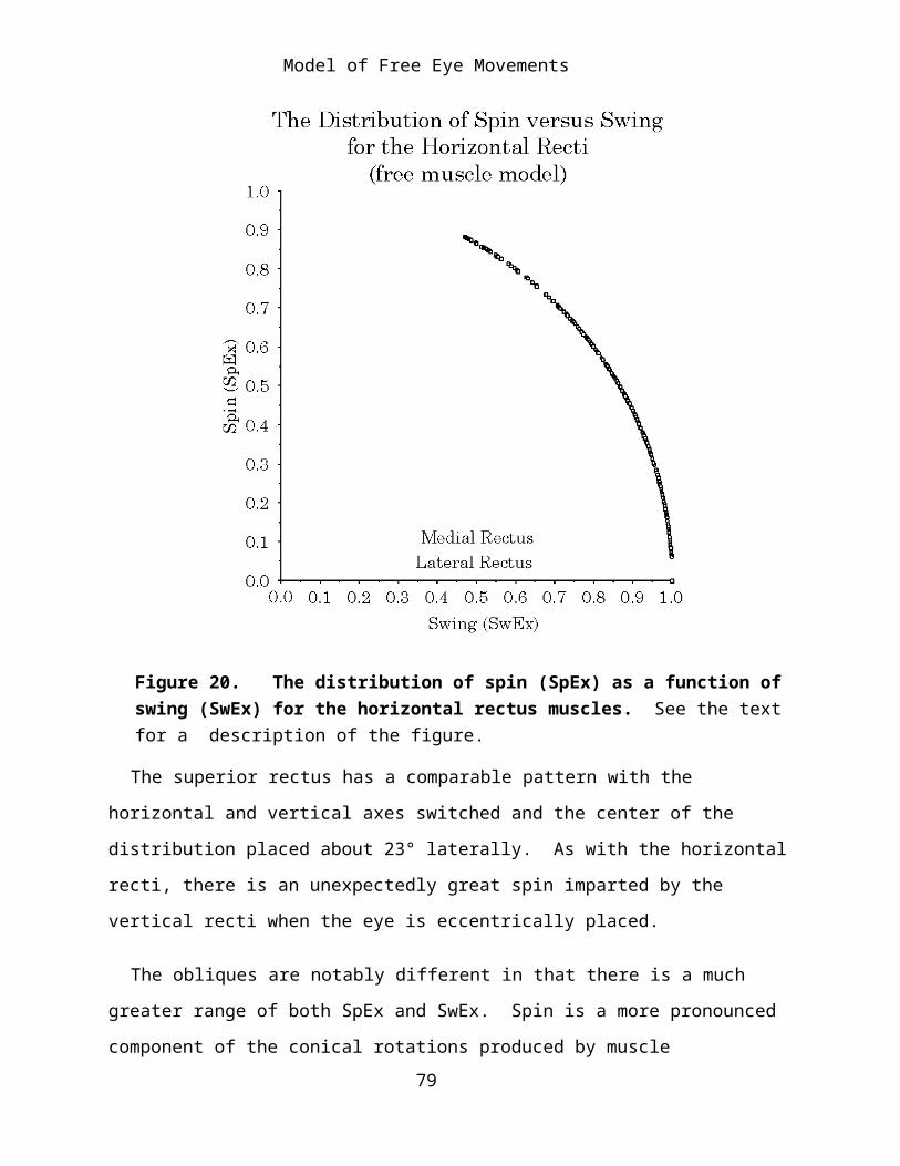
Model of Free Eye Movements
the penumbra of fascial tissues that surround the eyeball at the level of its equator and for some distance posteriorly, may also provide a passive restoring force to bring the eye back towards neutral and to help maintain spin neutral gaze at gazes displaced from neutral position.
In addition, numerous thin septa extend from the fascia of the scleral wall (vagina bulbi or Tenon’s capsule) into the periorbicular fat and form septa that segregate the fat into lobules. Such as network of closed distensible compartments might well act as a highly damped restoring spring that would draw the eye towards neutral gaze. Once the eye is near neutral gaze, there is a well distributed array of muscle pulls that can fine-tune the gaze alignment and orientation.
Summary
In this analysis the anatomy of the eye in the orbit and particularly the anatomy of the extraocular muscles has been examined from the point of view of their geometry. That geometry has been expressed in a set of mathematical expressions that allow one to manipulate the information in their description in a quantitative fashion. By such manipulations we were able to examine the changes in muscle length that occur as gaze is shifted. In this analysis we assumed that the controlled variable is what we have called gaze, meaning the line of sight and the orientation of the eye about that line of sight. It was assumed that the eye is generally oriented so that the visual world is projected as nearly as possible to the appropriate alignment. Vertical tends to be aligned with the vertical meridia and horizontal with the horizontal meridia. From that assumption, it follows that there is a unique one-to-one mapping of the set of muscle lengths upon the set of gazes. Further it follows that for the eye’s gaze to be completely determined by the sets of muscle lengths it is necessary and sufficient for there to be six extraocular muscles. The mapping between the gaze and the sets of muscle lengths was computed and many of the relationships
79

Model of Free Eye Movements
illustrated. The distribution of muscle length with respect to eye position is a curvilinear two-dimensional surface in an eight dimensional space, two for elevation/depression and medial/lateral displacement and one for each muscle’s length.
Because of the geometry of the muscle origins and insertions, it follows that when the muscles contract, they will draw the eye in particular directions. The second part of the analysis was an examination of how those pulling directions shift with gaze. It was observed that the action of a particular muscle might vary considerably as a function of gaze. A single muscle may cause the eye to move laterally in certain gazes and vertically in others. Because of the changing role of the eye muscles at different gazes, the nervous system must organize its control is a manner that does not give an eye muscle a fixed action. It is argued that a logical way to control eye movements is to generate a neural map of the two dimensional surface that relates the gaze to the appropriate set of muscle lengths.
In addition to the pulling direction, which is a pure swing, the extraocular muscles also impart a spin to the eye in almost every gaze. It is more difficult to illustrate the spin over a range of gazes, but it was noted that there was an elegant relationship between the amount of spin and the amount of swing generated by a muscle contraction. For small angular excursions of muscle pull the sum of the squares of the swing and the spin is square of the excursion. As swing becomes a greater proportion to the angular excursion, spin decreases, and vice versa. However, the relationship is not linear, so the total amount of swing and spin may greatly exceed the angular excursion.
The geometrical analysis of eye movements can address a number of other attributes of eye movements, some of which are addressed elsewhere. However, it should be clear from this initial foray, that it offers a means of asking quantitative questions and obtaining both quantitative and
80

Model of Free Eye Movements
qualitative answers. By being quantitative it often exposes areas where we do not understand the function of the anatomical system and need to look more closely, possibly quantitatively, at the anatomy.
Bibliography
Adams, R. D. and M. Victor (1993). Principles of Neurology. New York, McGraw-Hill.
Clark, R. A., J. M. Miller, et al. (1997). “Location and stability of rectus muscle pulleys. Muscle paths as a function of gaze.” Invest Ophthalmol Vis Sci 38(1): 227-40.
Clark, R. A., J. M. Miller, et al. (2000). “Three-dimensional location of human rectus pulleys by path inflections in secondary gaze positions.” Invest Ophthalmol Vis Sci 41(12): 3787-97.
Clement, R.A. (1991) "Natural coordinates for specification of eye movements." Vision Res. 31(11): 2029-32.
Demer, J. L., J. M. Miller, et al. (1995). “Evidence for fibromuscular pulleys of the recti extraocular muscles.” Invest Ophthalmol Vis Sci 36(6): 1125-36.
Demer, J. L., S. Y. Oh, et al. (2000). “Evidence for active control of rectus extraocular muscle pulleys.” Invest Ophthalmol Vis Sci 41(6): 1280-90.
Demer, J. L., V. Poukens, et al. (1997). “Innervation of extraocular pulley smooth muscle in monkeys and humans.” Invest Ophthalmol Vis Sci 38(9): 1774-85.
Hore, J., S. Watts, et al. (1992). “Constraints on arm position when pointing in three dimensions: Donders' law and the Fick gimbal strategy.” J Neurophysiol 68(2): 374-83.
81

Model of Free Eye Movements
Kuipers, J. B. (1999). Quaternions and Rotation Sequences: A Primer with Applications. Princeton, New Jersey, Princeton University Press.
Radau, P., D. Tweed, et al. (1994). “Three-dimensional eye, head, and chest orientations after large gaze shifts and the underlying neural strategies.” J Neurophysiol 72(6): 2840-52.
Raphan, T. (1998). “Modeling control of eye orientation in three dimensions. I. Role of muscle pulleys in determining saccadic trajectory.” J Neurophysiol 79(5): 2653-67.
Schnabolk, C. and T. Raphan (1994). “Modeling three-dimensional velocity-to-position transformation in oculomotor control [published erratum appears in J Neurophysiol 1994 May; 71: 5; following table of contents].” J Neurophysiol 71(2): 623-38.
Straumann, D., T. Haslwanter, et al. (1991). “Listing's law for eye, head and arm movements and their synergistic control.” Exp Brain Res 86(1): 209-15.
Tweed, D., H. Misslisch, et al. (1994). “Testing models of the oculomotor velocity-to-position transformation.” J Neurophysiol 72(3): 1425-9.
Tweed, D. and T. Vilis (1987). “Implications of rotational kinematics for the oculomotor system in three dimensions.” J Neurophysiol 58(4): 832-49.
Tweed, D. and T. Vilis (1990). “Geometric relations of eye position and velocity vectors during saccades.” Vision Res 30(1): 111-27.
Williams, P. L., L. H. Bannister, et al. (1995). Gray's Anatomy. The Anatomical Basis of Medicine and Surgery. New York, Churchill Livingstone
82