Embed Size (px)
Citation preview

This article was downloaded by: [81.109.181.181]On: 05 May 2014, At: 14:54Publisher: Taylor & FrancisInforma Ltd Registered in England and Wales Registered Number: 1072954 Registered office: Mortimer House,37-41 Mortimer Street, London W1T 3JH, UK
Italian Journal of ZoologyPublication details, including instructions for authors and subscription information:http://www.tandfonline.com/loi/tizo20
Morphology of the feeding apparatus in nestlings ofMeropsAndrea Brusaferro a & Alberto Mario Simonetta ba Dipartimento di Biologia Molecolare, Cellulare e Anímale , Università di Camerino , viaCamerini 2, Camerino (MC) , I‐62032 , Italyb Dipartimento di Biologia Animale e Genetica “Leo Pardi”; , Università di Firenze , viaRomana 17, Firenze , I‐50125 , ItalyPublished online: 22 Oct 2009.
To cite this article: Andrea Brusaferro & Alberto Mario Simonetta (1998) Morphology of the feeding apparatus in nestlings ofMerops , Italian Journal of Zoology, 65:3, 249-259, DOI: 10.1080/11250008809386755
To link to this article: http://dx.doi.org/10.1080/11250008809386755
PLEASE SCROLL DOWN FOR ARTICLE
Taylor & Francis makes every effort to ensure the accuracy of all the information (the “Content”) contained in thepublications on our platform. However, Taylor & Francis, our agents, and our licensors make no representationsor warranties whatsoever as to the accuracy, completeness, or suitability for any purpose of the Content. Anyopinions and views expressed in this publication are the opinions and views of the authors, and are not theviews of or endorsed by Taylor & Francis. The accuracy of the Content should not be relied upon and should beindependently verified with primary sources of information. Taylor and Francis shall not be liable for any losses,actions, claims, proceedings, demands, costs, expenses, damages, and other liabilities whatsoever or howsoevercaused arising directly or indirectly in connection with, in relation to or arising out of the use of the Content.
This article may be used for research, teaching, and private study purposes. Any substantial or systematicreproduction, redistribution, reselling, loan, sub-licensing, systematic supply, or distribution in anyform to anyone is expressly forbidden. Terms & Conditions of access and use can be found at http://www.tandfonline.com/page/terms-and-conditions

Irai. J. Zool., 65: 249-259 (1998)
Morphology of the feeding apparatusin nestlings of Merops
ANDREA BRUSAFERRODipartimento di Biologia Molecolare, Cellulare e Anímale,Università di Camerino,via Camerini 2, I-62032 Camerino (MC) (Italy)
ALBERTO MARIO SIMONETTADipartimento di Biologia Animale e Genetica "Leo Pardi",Università di Firenze,via Romana 17, I-50125 Firenze (Italy)
ABSTRACT
The morphological comparison of the facial skeleton andtongue between young nestlings and subadults has shown thatMerops desmognatism is due to the ossification of the processusprenasalis and solum nasi. The maxillo-palatines are not fusedalong the midline and the pterigoid is fused to the vomer. Theheterochrony in the development of the skull of Merops was sug-gested to be due to protracted parental care so that only whenthe bird captures its prey, do the stresses produced in the killingactions induce ossification of the solum nasi and of the processusprenasalis.
KEY WORDS: Aves - Coraciiformes - Merops - Skull morpholo-gy.
ACKNOWLEDGEMENTS
For part of the material used in this investigation, we are in-debted to Or. Robert Prys-Jones of the British Museum (NaturalHistory) who loaned two heads of nestlings (Merops viridis andMerops ornatiis) and to Dr. Annamaria Nistri of the "La Specola"Museum in Florence (Italy) for one subadult skull of Merops cipi-aster. Thanks to the British Museum (Natural History) of Tringand to the "La Specola" Museum of Florence (Italy), it was possi-ble to examine some nestlings of different families of the Coraci-iformes.
(Received 1 April 1998 - Accepted 4 May 1998)
INTRODUCTION
The morphology of the feeding apparatus is highly. varied and strongly adaptive in birds. The heterogeneity
of the feeding apparatus of the Coraciiformes representsan excellent example because it is very difficult to iden-tify any palatal character common to all the families.The few common characters, such as the desmognathiccondition of the palate and the strong medial ligamentof the quadrate (this is not true for the Alcedinidae;Burton, 1984) are insufficient to lead to an evolutionaryinterpretation or to assess systematic relationships. Thecrucial point is whether the investigation of develop-mental pathways leading to the desmognathic conditionare or are not important with respect to the varieties ofdesmognatism (Parker, 1876). The desmognathism ofMerops is different with respect to that of Nyctiornis(Meropidae), and to that of Alcedinidae as well.
The cranial morphology of the Meropidae and that ofthe Coraciiformes has been studied since the last centu-ry by several anatomists (Huxley, 1867; Sharpe, 1868-71;Murie, 1872b, c, 1873; Garrod, 1874, 1878; Shufeldt,1884, 1903; Fürbringer, 1888, 1902; Gadow, 1892; Bed-dard, 1893; Stresemann, 1934, 1959; Wetmore, 1934,I960; Lowe, 1946, 1948; Verheyen, 1955; Rawal, 1970;Cracraft, 1971; Rawal & Bhatt, 1974; Soliman et al.,1975; Burton, 1984; Bhattacharyya, 1987), but the avail-able knowledge concerns only the adult skull and so farwe know nothing about the nestlings and the post-nataldevelopment of Coraciiformes except for that of Upupaepops (Soliman et al, 1975).
Indeed the study of post-natal development deservesmore attention. During the nestling stage, while the car-tilages are already well developed, the dermal bonesare incompletely developed and they are neither fusednor completely ossified, so that it is possible to seeclearly the connections between the different structures.Comparison with the adult stage, and as far as possiblewith an intermediate stage, help us in learning theadaptive changes of the entire feeding apparatus, aswell as identifiyng homologous structures that are mostimportant factors for understanding systematic relation-ships.
The present paper concerns the morphology of theMeropidae and is part of a systematic attempt to clarifythe adaptive pathways of the feeding apparatus and con-tribute to the debate on the systematics of Coraciiformes.
MATERIALS AND METHODS
Four specimens of Merops apiaster, one of M. viridis, and oneof M. ornatus were studied. Of the specimens of M. apiaster,three were nestlings collected by one of us (A. M. Simonerta) in1977 from Afgoi (Somalia) and one was a subadult on loan fromthe Museo "La Specola" of Florence (collection No. 3017). Thespecimen of M. viridis was a nestling (unregistered) loaned by theBritish Museum (Natural History) of Tring and the specimen of M.ornatus was a subadult (collection No. A\ 1969.15.163) loaned bythe British Museum (Natural History) of Tring.
Dow
nloa
ded
by [
81.1
09.1
81.1
81]
at 1
4:54
05
May
201
4

250 A. BRUSAFERRO, A. M. SIMONETTA
With the exception of the skull of one nestling of M. apiaster,that was stained in toto by the Wassersug method (1976), theskulls were prepared for microscopical analysis after décalcifica-tion in "Top Decal" (Pabish) and embedding in paraffin-celloidin.
The skulls of the nestlings were microtomized in 12-H-thick se-rial section and the subadult skulls in sections of 14 u.m. Thepreparations were stained with Mallory's triple stain method(1900) and the graphics reconstructions were made with thePC3D recstruction software of Jandel Scientific Corporation.
RESULTS
Topography of palatal bones
In the nestling of Merops the corpora of the fusedpremaxillaries form the anterior tip of the beak. Eachcorpus premaxillaris extends backwards in a dorsalprocessus nasalis (processus frontalis, Marinelli, 1936;processus prenasalis, Erdmann, 1940; processus ascen-dens, de Beer, 1937), in a ventral processus palatinus(palato-maxillary, W. K. Parker, 1866) and in a ventrolat-eral processus maxillaris (processus labialis, Jollie, 1957;processus palatinus, Webb, 1957; processus alveolaris, deBeer, 1937). The proc. maxillaris extends backwardsand forms the lateral edge of the beak just behind thecorpora of the fused premaxillaries and in front of theproc. zygomaticus of the maxillary bone. The proc. max-illaris for the whole of its length .has a syndesmoticallymedial contact with the maxillary bone. The proc. palat-inus does not fuse medially with its corresponding con-trolateral process and extends backwards ventro-medial-ly to the palatine and medially to the maxillary (Fig. 1).
The anterior tip, as well as the anterior portion of thecorpus maxillaris is syndesmotically united with the lat-eral proc. maxillaris of the premaxillary bone, with theventral palatine bone and with the medio-ventral proc.palatinus of the premaxillary bone. The greater portionof corpus maxillaris is ventral to the atrioturbinal regionof the nasal capsules; the corpus is located dorsally tothe palatine and medio-ventrally to the proc. maxillarisof the premaxillary bone. More posteriorly, each maxil-lary bone thins off into two posteriorly directedprocesses: the processus jugalis (processus labialis, Jol-lie, 1957; zygomatic process, Müller, 1963) and theprocessus maxillopalatinus (palatine process, Gadow &Selenka, 1891, de Beer, 1937, Hofer, 1945, Jollie, 1957).The palatine process of the maxillary is a short processthat originates from the median surface of the maxillaryand is directed medially and dorsally to the palatine;each maxillopalatine is separated from the other oneand its posterior portion leaves a broad fissure (thechoanae) between the two structures (Fig. 1).
Each palatine consists of a narrow anterior part in thenasal region of the skull, while in the orbital region itbroadens to a flat plate-like structure. From its anteriortip, the palatine continues backwards ventrally to themaxillary and laterally to the proc. palatinus ossis pre-maxillaris; where the maxillary thins off into a proc.maxillapalatinus and proc. zygomaticus, the palatinebends inwards ventrally to the maxillapalatinus. There-
fore the palatine has a simple morphology in the nasalregion: it is a bony rod with an elliptical cross section.Behind the choanae, the palatine becomes more com-plex and it broadens to a flat plate-like structure; herethe medial edge of the palatine is produced medially toform a dorsal processus ethmopalatinus and a processusventralis. The medial edge of the ethmopalatine liesagainst the anterior tip of the parasphenoid (Fig. 2);more posteriorly, the distal edge of the ethmopalatine iswedged between the parasphenoid and the anterior tipsof the vomers. Posteriorly the ethmopalatine becomeslarger and it forms the most developed part of the pala-tine. The proc. ventralis palatini remains unchanged forits whole length, but, for a short length, its distal edgebends inwards approaching the respective counterpart.Between these two processes, the medial surface of thepalatine has a concavity; in the orbital region a proces-sus medialis palatini develops from the concavity andits distal edge is located ventral to the pterygoid. Morecaudally the process is in syndesmosis with the respec-tive process of the corresponding bone.
The vomers are a paired structures placed ventrally tothe ethmopalatine; short and kidney shaped in crosssection (Fig. 2), they continue backwards and are syn-ostotically fused with the pterygoids.
The pterygoid reaches from the palatine to thequadrate to which it is articulated by means of a syn-ovial joint ventral to the proc. orbitoquadratus (proc. or-bitalis) of the quadrate. This joint is of a ball-and-sockettype and the posterior tip of the pterygoid bears a cup-shaped concavity with no articular cartilage; this fits in-to a cartilaginous protuberance on the medial surface ofthe quadrate. Anteriorly, the pterygoid overlaps the pos-terior end of the palatine dorsally and its anterior end issynostotically fused with the vomer. Dorsally the ptery-goids are separated from the rostrum by a narrow fis-sure V-shaped in cross section. More posteriorly eachpterygoid is articulated with a short secondary basiptery-goid process of the basisphenoid; the basipterygoid jointis synovial (Fig. 3) and the articular surfaces are cov-ered by cartilage and located in an articular cavity en-closed by a synovial membrane.
The proc. jugalis ossis maxillaris is a long, slenderprocess tapering posteriorly; it overlaps the jugal andthe anterior half of the quadratojugal and the bones arein syndesmosis.
The jugal is a short bony rod; its anterior portion isdorsal to the processus jugalis ossis maxillaris and theposterior portion is dorsal to the quadratojugal.
The quadratojugal is a slender bony rod; its posteriorend fits into a cup-shaped fossa of the quadrate and nosynovial cavity is present. A limited amount of move-ment between the two elements is possible, but there isno real diarthrosis.
The nasal capsules
In the nestling of Merops, the nasal capsule is almostentirely cartilaginous. A certain amount of ossification
Dow
nloa
ded
by [
81.1
09.1
81.1
81]
at 1
4:54
05
May
201
4

FEEDING APPARATUS OF MEROPS 251
POP
TN PNAS fN PP
BOMT SN PP
POP
Fig. 1 - Graphic reconstruction of theskull of a young nestling of Merops apt-aster. A, dorsal view, above chondro-cranium; B, lateral view, nasal capsulewithout parietotectal cartilage; C, ven-tral view, below chondrocranium. Ab-breviations BO, basioccipital; BTP, ba-sitemporal plate; EC, ectethmoid; EO,exoccipital; FC, fenestra craniofacialis;FM, foramen magnum; FN, fenestra nar-ina; FR, frontal; IS, interobital septum;L, lacrimal; MA, maxillary; MP, maxil-lopalatine; MT, maxilloturbinal; NA,nasal; NT, nasal septum; OC, occipitalcondyle; PAR, parietal; BPTA,basipterigoid articulation; PL, palatine;PLS, pleurosphenoid; PM, premaxillary;P.MAX, processus maxiltaris of the pre-maxillary; P.NAS, processus nasalis ofthe premaxillary; POP, postorbitalprocess; PP, processus prenasalis P.PAL,processus palatinus of the premaxillary;PS, parasphenoid; PSR, parasphenoidrostrum; PT, pterygoid; Q, quadrate; QJ,quadratojugal; SN, solum nasi; SO,supraoccipital; SQ, squamosal; TN, tec-tum nasi; V, vomers. Roman numeralsindicate foramen for cranial nerves. Bar,2 mm.
has taken place in the septum and in the roof of thecapsule posteriorly. The anterior end of the nasal cap-sule is produced into a very prominent azygous proces-sus prenasalis (Fig. 1); the premaxillary bones surroundthe anterior end of this process. Posteriorly the proc.prenasalis merges into the septum nasi lengthening dor-
sally to assume a drop-shape in cross section (Fig. 4A);more posteriorly, the processus fuses dorsally with nasalcartilages forming a clear crest on the ventral surface ofthe nasal capsule (Fig. 4B). Therefore the proc. pre-nasalis does not arise from the anterior end of the nasalcapsule but from its ventral surface and the posterior
Dow
nloa
ded
by [
81.1
09.1
81.1
81]
at 1
4:54
05
May
201
4

252 A. BRUSAFERRO, A. M. SIMONETTA
Fig. 2 - Transverse section showing the anterior end of thevomers and the palatine in a young nestling of Merops viridis. Ab-breviations: IS, interorbital septum; PL, palatine; PSR, parasphe-noidal rostrum; V, vomers. Bar, 0.5 mm.
BSPH
SEC.C
Fig. 3 - Transverse section showing the basipterigoid articulationin a young nestling of Merops aptaster. Abbreviations: BSPH, ba-sisphenoid; PT, pterigoyd; S.CAV, synovial cavity; SEC.C, sec-ondary cartilage. Bar, 0.5 mm.
end of the process appears to be the swollen ventralsurface of the septum nasi. In this region, each nasalvestibulum is separated by connective tissue coveredwith epithelium. Behind the fenestra internasalis, theseptum nasi increases in height fusing with the parieto-tectal cartilage. The cupola anterior is absent becausethe fenestra narina opens anteriorly (Figs 1-4B).
The anterior two-thirds of the septum in front of thefissura craniofacialis are cartilaginous and thin, but inthe atrioturbinal region its ventral edge is swollen. Moreposteriorly, in the region of maxilloturbinal cartilage,the ventral edge of septum nasi has two cartilaginousbulges, the dorsal one being fused with the solum nasi.
In front of the fissura, the septum nasi is again thin withno ventral bulge.
The roof of the nasal capsule is complete and theparietotectal cartilage continues laterally downwards toform the side wall of the capsule. Posterior to the fenes-tra narina the parietotectal cartilage decreases inheight, so that the lower portion of the atrioturbinal (deBeer, 1937) may be seen laterally. At the posterior endof the atrioturbinal, however, the side wall is extendedventrally again to cover the maxilloturbinal laterally (deBeer, 1937).
The floor of the nasal capsule is complete and carti-laginous in the region of the atrioturbinal and maxtllo-turbinal cartilage, but it completely disappears in frontof coanae.
The atrioturbinal extends from a distance behind thefenestra narina up to some distance in front of the an-terior tip of the maxilloturbinal. For almost its entirelength it is attached to the side wall of the nasal capsuleand projects freely into the vestibulum of the nasal cap-sule. The anterior portion consists of a thin primarylamella bearing two secondary lamellae: a well-devel-oped ventrally direct one and a smaller dorsal one. Pos-teriorly, the primary lamella becomes shorter and thick-er, the secondary ventral one lengthens and the dorsalone is reduced to a low cartilaginous ridge, which dis-appears near the hinder end of the turbinai (Fig. 4C-D).The posterior end of the atrioturbinal is a bulge locatedon the floor of the nasal capsule.
The maxilloturbinal is cartilaginous and is fused to thedorsal surface of the side wall of the nasal capsule. Theanterior end is located immediately behind the posteriorend of the atrioturbinal cartilage and consists of a shortcartilaginous lamella. More posteriorly, the maxillo-turbinal presents a peculiar arched shape with the con-vexity directed toward the septum nasi. In the region ofthe concha nasalis (de Beer, 1937), the maxilloturbinalis located on the medial surface of the side wall of thenasal capsule and its distal end borders the choanae(Fig. 4E-F). Wedged in between the tectum nasi and thedorsal surface of the maxilloturbinal cartilage is a welldeveloped concha nasalis; it is thin and entirely carti-laginous (Fig. 4G-H).
In the posterior region of the nasal capsule, the nasalcavity is smaller and protected dorsally by the tectumnasi, while laterally and ventrally it is surrounded byconnective tissue. More posteriorly, each vestibulum issubdivided into two smaller cavities; the medial oneopens into the orbital region thus forming a paranasalsinus, while the lateral one is a caecum close to thelacrimal duct.
The planum antorbitale is a cartilaginous plate ex-tending ventrolaterally from the septum-, it does notclose the posterior wall of the nasal capsule, but is athin cartilage strip that points the passage from thenasal to the orbital region. Dorsomedially, it is closelyconnected to the lateral surfaces of the septum, whilethe distal edge is syndesmotically connected to the
Dow
nloa
ded
by [
81.1
09.1
81.1
81]
at 1
4:54
05
May
201
4

FEEDING APPARATUS OF MEROPS 253
B
H
22
22
-^ V
25
17-
Fig. 4 - Outline of the series of anterior-posterior transverse sections through different regions of nasal capsule in a young nestling. 1,processus nasalis ossispremaxillaris-, 2, processus maxillaris ossispremaxillaris; 3, maxillary; 4, palatine; 5, processuspalatinus ossispre-maxillaris; 6, processusprenasalis-, 7, fenestra narina; 8, septum nasi; 9, parietotectal cartilage; 10, atrioturbinal cartilage; 11, solum nasi;12, processus maxillaris ossis nasalis-, 13, processus premaxillaris ossis nasalis; 14, maxilloturbinal cartilage; 15, concha nasalis; 16,processus maxillopalatinus; 17, processus zygomaticus; 18, nasal; 19, planum antorbitale; 20, fenestra craniofacialis; 21, lacrimal; 22, ju-gal; 23, frontal; 24, parasphenoid; 25, vomers. Bar, 2 mm.
Dow
nloa
ded
by [
81.1
09.1
81.1
81]
at 1
4:54
05
May
201
4

254 A. BRUSAFERRO, A. M. SIMONETTA
lacrimal. The planum tapers rapidly to form a shortprocess closely adjacent to the septum (Fig. 4I-J).
The membrane bones of the nasal capsules
The premaxillaries are fused to form a single bonystructure strengthening the tip of the beak; each pre-maxillary possesses three backward directed processes:a proc. frontalis, a proc. palatalis and a proc. alveolaris.The latter two have been dealt with in the descriptionof the palate. The proc. frontalis ossis premaxillaris con-tinues backwards, dorsal to the nasal capsule, for theentire length of the bill, while the caudal tip of theproc. frontalis is placed at the level of the nasal-frontalhinge (Fig. 1).
The nasal is a thin bony plate lateral to the posteriorthird of the proc. frontalis ossis premaxillaris; at its foreend, the nasal is bifurcated to form a median proc. pre-maxillaris and a lateral proc. maxillaris. The proc. max-illaris ossis nasalis is a ventrally directed, long, slender,tapering process extending forwards dorsally to themaxillary and mediodorsally to the proc. lateralis ossispremaxillaris (Fig. 1).
The fenestra narina is bounded anteriorly by the pre-maxillaries and dorsally by the proc. frontalis ossis pre-maxillaris and by the proc. premaxillaris ossis nasalis;posteriorly it is bounded by the nasal and ventrally bythe proc. maxillaris ossis nasalis and the proc. lateralisossis premaxillaris.
The lacrimal is well developed and is formed by adorsal portion (head of the lacrimal), joining with thenasal and the frontal and by a descending process. Thedistal end of the descending process widens into asmall basal plate that adjoins the jugal. The foot of thedescending process overlaps for a short length with thedistal edge of the planum antorbitale.
The frontals are in syndemosis along the dorsal mid-line, except for their anteriorly diverging ends; anterior-ly, the frontal is overlapped by the nasal but the meso-kinetic hinge is not restricted to this region; the dorsalbending is located at the level of the nasal immediatelydorsally to the fissura craniofacialis, and here bothnasals are very thin forming a restricted line of bending.
The lower jaw
The following dermal bones occur in the lower jaw ofMerops: dentary, splenial (opercular; Gadow, 1892), go-nial (complementary; Suschkin, 1899; Gaupp, 1911), an-gular and surangular (Fig. 5). The different dermalbones surround a central canal in which lies Meckel'scartilage (Fig. 6A). In the nestling of Merops, the differ-ent bony elements are in syndemosis, except the gonialand the surangular which show an incipient synostosis.
The left and the right dentaries join anteriorly be-tween the dentary internal surface. The dentary extendsbackwards for half the length of the mandible and has aventral process reaching far backwards and lying lateralto the anterior portion of the angular. The dentary cov-
RLD SA CMF ART
ART
MC ANG
Fig. 5 - Graphics reconstruction of the mandible in a youngnestling of Merops apiaster. A, lateral view; B, medial view; C,medial view without the splenial and gonial bones. Abbreviations:ANG, angular; ART, articular; CMF, caudal mandibular fenestra; D,dentary; GON, gonial; MC, Meckel's cartilage; RLD, ramus later-alis of the dentary; RMD, ramus medialis of the dentary; SA,surangular; SPL, splenial; V.PROC.D, ventral processus of the den-tary. Bar, 2 mm.
ers the anterior half of Meckel's cartilage dorso-laterallyand ventrally and it is thicker in the dorsal surface.More posteriorly, the dorsal edge of the dentary dis-plays a groove in which lies the anterior end of thesurangular. Going backwards, the groove becomesdeeper and turns into a fissure which separates the den-tary in the ramus lateralis and the ramus medialis. Be-hind to the anterior end of the surangular the ramuslateralis separates in dorsal and ventral processes (Fig.6B). The dorsal process of the ramus lateralis and theramus medialis of the dentary continue backwards dor-sally to Meckel's cartilage, their tapering distal ends halfway along the lower jaw. The ventral process is longerand continues backwards lying laterally to the Meckel'scartilage. Posteriorly, the ventral process of the dentarybecomes thin and it ends beyond the angular and thesurangular.
The anterior end of the surangular lies wedged be-tween the dorsal processes of the dentary (Fig. 6A-B).The surangular broadens posteriorly and covers dorso-laterally the posterior half of the Meckel's cartilage. It isbordered ventrally by the angular.
The splenial is a thin bony plate that medially coversMeckel's cartilage (Fig. 6A-B). The anterior half of thesplenial is bordered dorsally by the ramus medialis ofthe dentary and ventrally by the ventral process of thedentary. The posterior half of the splenial is bordereddorsally by the surangular and ventrally by the angular.Posteriorly the splenial displays synostosis with the an-gular.
Dow
nloa
ded
by [
81.1
09.1
81.1
81]
at 1
4:54
05
May
201
4

FEEDING APPARATUS OF MEROPS 255
MC
SPL
SPL
R.MAND. V
MC
VPROC.D
MC
VPROC.D
Fig. 6 - Series of anterior-posterior transverse sections through dif-ferent regions of the lower jaw in a young nestling of Meropsviridis. Abbreviations: ANG, angular; D, dentary; GON, gonial;MC, Meckel's cartilage; RLD, ramus lateralis of the dentary; RMD,ramus meclialis of the dentary; SA, surangular; SPL, splenial;V.PROC.D, ventral processus of the dentary. Bar, 2 mm.
The gonial (Fig. 6C) medially covers the posterior halfof Meckel's cartilage. Its anterior end lies between theramus meclialis and the splenial, medio-dorsal to thecartilage. Posteriorly, it is fused with the angular. For its
entire length the gonial is bordered dorsally by thesurangular and ventrally by the splenial.
The angular is a bony plate ventral to Meckel's carti-lage. Its anterior end lies immediately in front of theanterior end of the gonial. The angular broadens poste-riorly between the dorsal Meckel's cartilage and theventral splenial and dentary bones. Behind the posteriorends of the dentary and splenial bones, the angular is alarge bony plate ventral to Meckel's cartilage. Here it isbordered laterally by the surangular and medio-dorsallyby the gonial.
The posterior end of Meckel's cartilage is covered me-dially by the gonial, ventrally by the angular and dorso-laterally by the surangular. The posterior portion ofMeckel's cartilage is not yet ossified to form the articu-lar; it just shows signs of pericordal ossification. The ar-ticular part of the cartilage shows a short lateral processand a medial process. Approaching the basitemporalbone, the medial process extends medially and its me-dial edge fits into a cup-shaped concavity limited dor-sally by the lateral process of the basitemporal bone.Each articular surface is covered by a thin layer of con-nective tissue.
The hyoid skeleton
The hyoid of birds consists of an unpaired corpus (.cop-ula) made up usually by fused copula I and copula II;the copula articulates with paired cornua branchialia,each consisting of a proximal hypobranchial (cerato-branchial; Suschkin, 1899) and a distal ceratobranchial(epibranchial; Suschkin, 1899).
The hyoid of Merops is typically avian (Fig. 7). Theparaglossum supports the tongue as generally in Aves,and before Reichert (1837) it was called either an os lin-guale or an os entoglossale. According to Owen (1866),T. J. Parker (1892) and Pycraft (1898-1901), the para-glossum is a Y-shaped cartilage and the posteriorprongs correspond with the ceratohyals, while the ante-rior slender unpaired structure is the basihyal. All au-thors except Kallius (1905) believed this; Crompton(1953) corroborates the original claim of Kallius (1905)and states that "...the anläge of the paraglossal cartilage,is situated dorsally to the anterior region of the copula.On either side if this plate a centre of condensation ofthe tissue is observable. These indicate the paired na-ture of the paraglossal cartilage".
In the nestling of Merops, the only evidence of itspaired origin is the two anteriorly directed prongs. In adorsal view, the paraglossum is an A-shaped cartilage.In fact, each prong converges anteriorly and fuses in aslender cartilaginous unpaired structure. More posterior-ly, the paraglossum articulates with the dorsal surface ofthe copula, and in this region the two prongs of para-glossum are connected by a cartilaginous bridge.
The copula consists of a fused copula I and copula IIand is flattened laterally. Its anterior portion lies be-tween the two prongs of the paraglossum, and midwayalong its length the copula is constricted laterally, form-
Dow
nloa
ded
by [
81.1
09.1
81.1
81]
at 1
4:54
05
May
201
4

A. BRUSAFERRO. A. M. SIMONETTA
PAR.
COR I
COPII
HPB.
CTB.
Ify. 7 - Graphics reconstruction of the ventral aspect of the hyoidcomplex in a young nestling of Merops apiaster. Abbreviations:COM, copula I; COP.II, copula II; CTB, ceratobranchial; HTB, hy-pobranchial; PAR, paraglossa. Bar, 1 mm.
ing an articular fossa for the hypobranchial on eitherside. Posteriorly the copula is flattened laterally againand has a long posteriorly directed process.
The hypobranchial and the ceratobranchial are articu-lated to form a long, slender, rod-like cornu branchialef. In the stages investigated, the cornu branchiale I is inan advanced stage of perichondral ossification whereasits terminal portions are cartilaginous. The cornuabranchialia extend backwards and outwards from thecopula in the form of a V. The posterior third of thecornu branchiale curves upwards and its distal endslightly inwards.
DISCUSSION
Cranial kinesis
The kinetic mechanism in the skull of Merops is typi-cally prokinetic, the most common form of kinesis inbirds, in which the upper jaw maintains its shape as itpivots about the craniofacial hinge (Hérissant, 1752;Uoecher, 1951; Bock, 1964; Bühler, 1981; Zusi, 1993).
Rotation upwards of the upper jaw begins by the con-traction of the Musculus protractor pterygoidei. Accord-ing to Burton (1984), the M. protractor of Meropidae islarge, its insertion is over the entire medial surface ofthe orbital process, and the muscle shows no differenti-ation into two parts. The study of the transverse sec-tions in the nestlings corroborates Burton's statements(1984).
The quadrate is pulled forward and slightly inwardsby the contraction of M. protractor pterygoidei. Forwardmovements of the quadrate are transferred to the maxil-lary and the premaxillary by way of the palatine whichattaches in syndesmosis with these bones, and by thelower temporal arch. The pterygoid and the palatine arein syndesmosis and they can slide along the rostrum.
The beak is rotated dorsally but the amount of dorsalrotation of the beak in the nestling cannot be very con-siderable, as a true mesokinetic joint is absent. In thenestlings investigated, there is no 'real fracturing' of theseptal apparatus, but the bending of the nasal septum isperhaps possible to a very limited extent as the septumis still cartilaginous. In addition bending is facilitated bythe presence of a large fenestra septi nasi posterior (Fig.1) which is filled with connective tissue. The ventralborders of the fenestra septi nasi posterior and that offenestra septi nasi anterior are formed by the fused tra-beculae and in front of the nasal capsule this trabecularbar is elongated into a very prominent processus pre-nasalis. In the subadult skull, the ventral border of thefenestra septi nasi posterior is still cartilaginous and par-tially resorbed. In the adult skull, the nasal septum hasbecome ossified but the bending is possible becausethe ventral border of the fenestra septi nasi posterior istotally resorbed (it is now called fissura craniofacialis)so that there is a real 'fracturing' of a septal apparatus(de Villiers, 1946) and there is a dorsal line of bending.Both in the nestling and adult skull, bending is limitedto the nasal and to the processus frontalis ossis premax-illaris. The frontals do not take any active part in thedorsal hinge.
The movements of the upper jaw are also limited, inthe nestling, by the presence of a true basipterygoid ar-ticulation (Fig. 3). Given the proportions of the skull,the mesokinetic flex line lies cranially with respect tothe basipterygoid articulation so that this not only can-not work as a pivot, but actually restrains kinesis, as itis clear that in Merops an efficient kinetic mechanism in-volves the rotation of the pterygoid away from the ba-sisphenoid. The résorption or modification of thebasipterygoid articulation is therefore necessary. In thenestling of Merops, the basipterygoid joint, probably inrelation with the comparative shortness of the beak, issynovial with small articular surfaces, and this implieslittle movement of the upper jaw. In the subadult skull,the basipterygoid process is reduced to a small spine ofa basisphenoid (Fig. 8) and the joint is substituted by adelicate ligament allowing an increase in the amount ofkinetic movements. However the structural continuity ofthe septal apparatus is still an impediment to the move-ments of the jaw.
To sum up: the nestling and the subadult of Meropstechnically have a kinetic upper jaw, but the move-ments of the jaws are limited and apparently they arebasically passive and function as a sort of shock ab-sorber. An efficient kinetic mechanism is obtained inthe adult skull with active food-getting.
2%
Dow
nloa
ded
by [
81.1
09.1
81.1
81]
at 1
4:54
05
May
201
4
FEEDING APPARATUS OF MEROPS 257
BSPH
PT
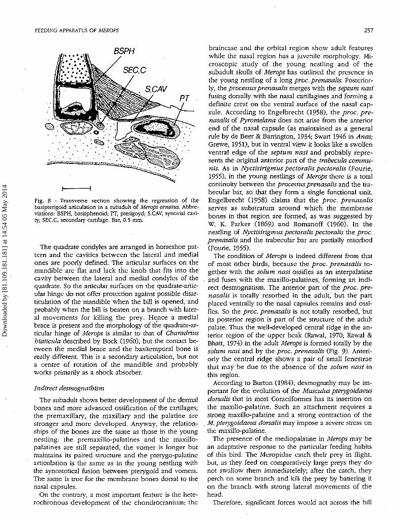
Fig. 8 - Transverse section showing the regression of thebasipterigoid articulation in a subadult of Merops ornatus. Abbre-viations: BSPH, basisphenoid; PT, pterigoyd; S.CAV, synovial cavi-ty; SEC.C, secondary cartilage. Bar, 0.5 mm.
The quadrate condyles are arranged in horseshoe pat-tern and the cavities between the lateral and medialones are poorly defined. The articular surfaces on themandible are flat and lack the knob that fits into thecavity between the lateral and medial condyles of thequadrate. So the articular surfaces on the quadrate-artic-ular hinge do not offer protection against possible disar-ticulation of the mandible when the bill is opened, andprobably when the bill is beaten on a branch with later-al movements for killing the prey. Hence a medialbrace is present and the morphology of the quadrate-ar-ticular hinge of Merops is similar to that of Charadriushiaticula described by Bock (I960), but the contact be-tween the medial brace and the basitemporal bone isreally different. This is a secondary articulation, but nota centre of rotation of the mandible and probablyworks primarily as a shock absorber.
Indirect desmognathism
The subadult shows better development of the dermalbones and more advanced ossification of the cartilages;the premaxillary, the maxillary and the palatine arestronger and more developed. Anyway, the relation-ships of the bones are the same as those in the youngnestling: the premaxillo-palatines and the maxillo-palatines are still separated, the vomer is longer butmaintains its paired structure and the pterygo-palatinearticulation is the same as in the young nestling withthe synostotical fusion between pterygoid and vomers.The same is true for the membrane bones dorsal to thenasal capsules.
On the contrary, a most important feature is the hete-rochronous development of the chondrocranium; the
braincase and the orbital region show adult featureswhile the nasal region has a juvenile morphology. Mi-croscopic study of the young nestling and of thesubadult skulls of Merops has outlined the presence inthe young nestling of a long proc. prenasalis. Posterior-ly, the processus prenasalis merges with the septum nasifusing dorsally with the nasal cartilagines and forming adefinite crest on the ventral surface of the nasal cap-sule. According to Engelbrecht (1958), the proc. pre-nasalis of Pyromelana does not arise from the anteriorend of the nasal capsule (as maintained as a generalrule by de Beer & Barrington, 1934; Swart 1946 in Anas;Grewe, 1951), but in ventral view it looks like a swollenventral edge of the septum nasi and probably repre-sents the original anterior part of the trabecula commu-nis. As in Nyctisirigmus pectoralis pectoralis (Fourie,1955), in the young nestlings of Merops there is a totalcontinuity between the processus prenasalis and the tra-becular bar, so that they form a single functional unit.Engelbrecht (1958) claims that the proc. prenasalisserves as substratum around which the membranebones in that region are formed, as was suggested byW. K. Parker (1869) and Romanoff (I960). In thenestling of Nyctisirigmus pectoralis pectoralis the proc.prenasalis and the trabecular bar are partially resorbed(Fourie, 1955).
The condition of Merops is indeed different from thatof most other birds, because the proc. prenasalis to-gether with the solum nasi ossifies as an interpalatineand fuses with the maxillo-palatines, forming an indi-rect desmognatism. The anterior part of the proc. pre-nasalis is totally resorbed in the adult, but the partplaced ventrally to the nasal capsules remains and ossi-fies. So the proc. prenasalis is not totally resorbed, butits posterior region is part of the structure of the adultpalate. Thus the well-developed central ridge in the an-terior region of the upper beak (Rawal, 1970; Rawal &Bhatt, 1974) in the adult Merops is formed totally by thesolum nasi and by the proc. prenasalis (Fig. 9). Anteri-orly the central ridge shows a pair of small fenestraethat may be due to the absence of the solum nasi inthis region.
According to Burton (1984), desmognathy may be im-portant for the evolution of the Musculus pterygoidaeusdorsalis that in most Coraciiformes has its insertion onthe maxillo-palatine. Such an attachment requires astrong maxillo-palatine and a strong contraction of theM. pterygoidaeus dorsalis may impose a severe stress onthe maxillo-palatine.
The presence of the mediopalatine in Merops may bean adaptative response to the particular feeding habitsof this bird. The Meropidae catch their prey in flight,but, as they feed on comparatively large preys they donot swallow them immediatelely; after the catch, theyperch on some branch and kill the prey by battering iton the branch with strong lateral movements of thehead.
Therefore, significant forces would act across the bill
Dow
nloa
ded
by [
81.1
09.1
81.1
81]
at 1
4:54
05
May
201
4

258 A. BRUSAFERRO, A. M. SIMONETTA
MA
PL
Fig. 9 - The indirect desmognatism in the Merops adult skull. MA,maxillary; MP, maxillopalatine; PL, palatine; SN, solum nasi; V,vomer. Bar, 1 mm.
axis and they include upward forces and lateral ones.Simple upward forces meet a resistance from the well-developed maxillo-palatines, but these will not neces-sarily require fusion. The technique of beating the preyagainst the perch may well, instead, have been an im-portant selective factor: the lateral forces involved arenecessarily large and they may well require a solidbridge across the middle part of the beak (Burton,1984). Burton' claim (1984) is corroborated by the factthat the subadult skull of Merops shows a delayed de-velopment of the entire nasal region, while the carti-lages of the braincase display a good state of pericordaland endocordal ossification. The differences betweenthe nestling and the subadult skull are irrelevant in thenasal region and only the dermal bones are more devel-oped. Indeed, in the juvenile, the maxillo-palatines arewell developed but not fused in the midline. In thespecies of the genus Merops, parental care is very pecu-liar: the parents do not directly feed their offspring, butsimply carry the food to their subterranean nest wherethe juveniles pick it by themselves. This practice maybe protracted for a long time and it appears that onlywhen the bird begins to capture and kill its prey, do thestresses of the killing action induce the rapid ossifica-tion of the solum nasi and of the processus prenasalis.
REFERENCES
Beddard F. E., 1893 - General characters and anatomy of theCoraciidae. In: H. E. Dresser, A monograph of the Coraciidae.Publ. by the author, Farnborough, Kent.
Beecher W. J., 1951 - Adaptations for food-getting in the Ameri-can blackbirds. The Auk, 68: 411-440.
Bhattacharyya B. N., 1987 - On the structural adaptations of thebill, skull-elements, tongue and hyoid of some indian insect-eating birds. Gegenbaurs morphol. Jahrb., 133: 311-351.
Bock W. J., 1960 - Secondary articulation of the avian mandible.The Auk, 77: 19-55.
Bock W. J., 1964 - Kinetics in the avian skull. J. Morph., 114: 1-41.Bühler P., 1981 - Functional anatomy of the avian jaw apparatus.
In: A. S. King & J. McLelland (eds), Form and function in birds.Academic Press, London, vol. 2, pp. 439-468.
Burton P. J. K., 1984 - Anatomy and evolution of the feeding ap-paratus in the avian orders Coraciiformes and Piciformes. Bull.Br. Mus. nat. Hist. (Zool.), 47: 331-443.
Cracraft J., 1971 - The relationships and evolution of the rollers:families Coraciidae, Brachypteraciidae, and Leptosomatidae.The Auk, 88: 723- 752.
de Beer G. R. 1937 - The development of the vertebrate skull.Clarendon Press, Oxford,
de Beer G. R., Barrington E. J. W., 1934 - The segmentation andchondrification of the skull of the duck.. Philos. Trans, r. Soc.Lond., 223: 411-467.
de Villiers C. G. S., 1946 - The relations of the vomer and palato-quadrate bar to the cranial rostrum in the Tinamou (CypturellusSpecies). Ann. Univ. Stellenbosch, 24 (A), 21 pp.
Engelbrecht D v. Z., 1958 - The development of the chondrocra-nium of Pyromelana orix crix. Acta Zool., 39: 115-199.
Erdmann K., 1940 - Zur Entwicklungsgeschichte der Knochen imSchädel des Huhnes bis zum Zeitpunktdes Ausschlüpfens ausdem Ei. Z. Morph. Ökol. Tiere, 36. 315-400.
Fourie S., 1955 - A contribution to the cranial morphology of Nyc-tisirigmus pectoralis pectcralis with special reference to thepalate and cranial kinesis. Ann. Univ. Stellenbosch, 31: 178-215.
Fürbringer M., 1888 - Untersuchungen zur Morphologie und Sys-tematik der Vögel. Vols.l, 2. Von Holkema, Amsterdam, 1751pp.
Fürbringer M., 1902 - Zur vergleichenden Anatomie desBrustschulterapparates und der Schultermuskeln. Part.5. Z.Naturwiss., Jena, 36: 289-736.
Gadow H., 1892 - On the classification of birds. Proc. zool. Soc.Lond., 1892: 229-256.
Gadow H., Selenka E., 1891 - Vögel. Anatom. Teil 6 (4). In:Bronn's Klassen und Ordnungen des Tierreichs, Leipzig, pp.920-983.
Garrod A. H., 1874 - On certain muscles of birds and their valuein classification. Part II. Prcc. zool. Soc. Lond., 1874: 111-124.
Garrod A. H., 1878 - On the systematic position of the Momoti-dae. Proc. zool. Soc. Lond., 1878: 100-102.
Gaupp E., 1911 - Beiträge zur Kenntnis des Unterkiefers derWirbeltiere. Anat. Anz., 34: 1-433.
Grewe F. J., 1951 - Nuwe gegewens aangaande die ontogenesevan die neuskliere, die orgaan van Jacobson en die dekbenevan die skedel by die genus Anas. Ann. Univ. Stellenbosch, 27:69-99.
Hérissant F. D., 1752 - Observations anatomiques sur les mouve-mens du bec des oiseaux. Mém. Acad. Inst. France, Paris, Sci.math, phys., pp. 345-386.
Hofer H., 1945 - Untersuchungen über den Bau des Vogelschädel,besonders über den der Spechte und der SteiBhüner. Zool.Jahrb., Abt. Anat. Ontolog., 69. 1-158.
Huxley T. H., 1867 - On the classification of birds, and on the tax-onomic value of the modification of certain of the cranial bonesobservable in that class. Proc. zool. Soc. Lond., 1867, pp. 415.
Jollie M. T., 1957 - The head skeleton of the chicken and remarkson the anatomy of this region in other birds. J. Morph., 100.389-436.
Kallius E., 1905 - Beiträge zur Entwicklung der Zunge. II Teil:Vögel. Anat. Hefte. Bd. 28 H. 2/3, pp. 307-586.
Lowe P. R., 1946 - On the systematic position of the woodpeckers(Pici), honey-guides (Indicator), hoopoes" and others. Ibis, 88:103-127.
Lowe P. R., 1948 - What are the Coraciiformes? Ibis, 90. 572-582.
Dow
nloa
ded
by [
81.1
09.1
81.1
81]
at 1
4:54
05
May
201
4

FEEDING APPARATUS OF MEROPS 259
Mallory F. B., 1900 - A Contribution to staining methods; 1. A dif-ferential stain for connective-tissue fibrillae and reticulum. J.exp. Med., 5. 15-20.
Marinelli W., 1936 - Kranium und Viszeralskelett der Sauropsiden.2. Vögeln. In: Handbuch des vergleichende Anatomie derWirbeltiere, herausgegeg, 4: 809-838.
Müller H.J., 1963 - Die Morphologie und Entwicklung des Crani-ums von Rhea americana Linné. Anat. Inst. Univ. Frankfurt amMain., 168:35-118.
Murie J., 1872a - On the motmots and their affinities. Ibis, 15.383-412.
Murie J., 1872b - On the skeleton of Todus, with remarks as to itsallies. Proc. zool. Soc. Lond., 1872, 664-680.
Murie J., 1873 - On the Upupidae and their relationships. Ibis, 15:181-211.
Owen R., 1866 - On the anatomy of vertebrates. Vol. 1: Fishes andReptiles. London.
Parker T. J., 1892 - Observations on the anatomy and develop-ment of Apterix. Philos. Trans., 182, 25 pp
Parker W. K., 1866 - On the structure and development of theskull in the ostrich Tribe. Philos. Trans, r. Soc. Lond., 156: 113-184.
Parker W. K., 1869 - On the strucutre and development of theskull of the common fowl (Gathis domesticus). Philos. Trans, r.Soc. Lond., 159: 755-807.
Parker W. K., 1876 - On the structure and development of thebird's skull (Part II). Trans, linn. Soc. Lond., 1: 99-154.
Pycraft W. P., 1898-1901 - On the morphology and phylogeny ofthe Paleognathae (Ratitae and Crypturi) and Neognathae (Cari-natae). Trans, zool. Soc. Lond., 15:
Rawal U. M., 1970 - Adaptations for food getting in green bee-eater. Vidya, J. Gujarat Univ. 13: 164-178.
Rawal U. M., Bhatt P. L., 1974 - Comparative morphology of thefeeding apparatus in a few representative birds from orderCoraciiformes. Vidya, J. Gujarat Univ., 17: 1-21.
Reichert C, 1837 - Über die Visceralbögen der Wirbeltiere im all-gemeinen und deren Metamorphosen dei den Vögeln undSaugetieren. Arch. Anat. Physiol. Lpz., 120 pp.
Romanoff A. L., 1960 - The avian embryo. Macmillan & Co., NewYork.
Sharpe R. B., 1868-1871. A monograph of the Alcedinidae: or,family of kingfishers. Publ. by author, London.
Shufeldt R. W., 1884 - Osteology of Ceryle alcyon. J. Anat. Physi-ol., 18: 279-294.
Shufeldt R. W., 1903 - On the osteology and systematic position ofthe kingfishers (Halcyones). Am. Nat., 37. 697-725.
Soliman M. A., Hammovda H. G., Mokhter, F. M. 1975 - The de-velopment of the osteocranium of a young nestling of Upupaepops major (Egyptian hoopoe). I. The membrane bones andthe articular. Bull. Fac. Sci. Univ. Cairo, 45: 239-272.
Stresemann E., 1934 - Aves. In: W. Kukenthal & T. Krumbach(eds), Handbuch der Zoologie, 7 (2). Walter de Gruyter, Berlin,899 pp. '
Stresemann E., 1959 - The status of avian systematics and its un-solved problems. The Auk, 76: 269-280.
Suschkin P. P., 1899 - Zur Morphologie des Vogelskelets. I Schädelvon Tinnunculs. Nouv. Mem. Soc. Nat. Moscou., 16.
Swart P. J., 1946 - Die ontogenese van die "Desmognate" mond-daktipe by Anas. Ann. Univ. Stellenbosch, 24: 1-20.
Verheyen R., 1955. Analyse du potentiel morphologique et consid-érations sur la systématique des Coraciiformes (Wetmore 1934).Bull. Inst. r. Sci. nat. Belgique, 31 (92-92): 1-51.
Wassersug R. J., 1976 - A procedure for differential staining of car-tilage and bone in whole formalin-fixed vertebrates. Stain Tech-nol., 51: 131-134.
Webb M., 1957 - The ontogeny of the cranial bones, cranial pe-ripheral and cranial parasympathetic nerves, together with a
. study of the visceral muscles of Struthio. Acta zool., 38: 81-203.Wetmore A., 1934 - A Systematic classification for the birds of the
world, revised and amended. Smithson. Misc. Coll., 89 (13): 1-11.
Wetmore A., 1960 - A classification for the birds of the world.Smithson. Misc. Coll., 139 (11): 1-37.
Zusi R. L., 1993 - Patterns of diversity in the avian skull. In: Theskull. Patterns of structural and systematic diversity. Universityof Chicago Press, vol. 2, pp. 391-437.
Dow
nloa
ded
by [
81.1
09.1
81.1
81]
at 1
4:54
05
May
201
4