Embed Size (px)
Citation preview
65
MORPHOLOGICAL DETAILS USEFUL TO IDENTIFY THE BONES OR BONE FRAGMENTS BELONGING OF CARCASS
IN SHEEP OR HOMOLOGOUS REGIONS IN DOG
Gabriel PREDOI, Cristian BELU, Iulian DUMITRESCU, Anca ȘEICARU, Petronela ROȘU, Cătălin MICȘA
University of Agronomic Sciences and Veterinary Medicine of Bucharest, Faculty of Veterinary Medicine,
Splaiul Independenței nr. 105 District 5, 050097, Bucharest, Romania
Corresponding author email: [email protected]
Abstract Very often, veterinary professionals are faced with directly and quickly identify the bodies of animals, carcasses or carcass portions. This operation is based on morphological characters highlighting defining species, sex and even approximate age. They are very frequent cases when the soft parts are damaged bodies and carcasses of animals are partially or totally boned. In view of this, it shows the importance of examining the skeleton as a whole or its constituent parts. Operation animal identification by morphological features of the skeleton is more difficult as are younger animals (presence of cartilage growth, strengthening bones insufficient, incomplete formation of characteristic details, enable scattering and fragmentation of the bones). In the domestic mammals there is possibility of occurrence of confusion, especially if bones and body parts belonging to the same class animals close. Only perfect knowledge of bone morphology allows the veterinarian to determine what species undoubtedly stems from the housing or housing part, without the need for additional tests. Detailed analysis presented in this paper aims to provide the most important clues so that identification of species belonging specifically to be made, even if some bones which, at first glance, seem indistinguishable. These two species can be distinguished some bones relatively easy: lumbar vertebrae, sacrum, most limb bones. However, some characters are less distinct bones, cervical vertebrae II-VII, some thoracic vertebrae, tibia, etc. However the study also insisted on the possibility of identifying all bones, because we often available only bone fragments, which prevents taking into account the most important element, namely the general appearance of the bone. The study revealed, in an original manner, details that may constitute criteria for determining the species from which the bones or fragments analyzed, largely completing a series of data described under "classical osteology" Key words: sheep, dog, bones, morphological details.
INTRODUCTION
Examination of bone has a great importance in forensic medicine, both human and domestic animals. Most often, over time, the soft parts of the bodies are damaged and the only elements that can constitute evidence of analysis are bones. Bones are extremely useful in establishing the identity of the individual of origin. Bones help in this regard because they may be used to establish the following characteristics of the individual of origin: race, gender, age at death, dimensions and direct identification clue. Currently, an accurate identification is based on DNA analysis (Ciobotaru, 2013, Savu, Petcu, 2008). If a body is found in burnt stage, it will be very difficult to identify physically. DNA fingerprinting comes to the rescue in such scenarios, but from where will we collect DNA on a burnt body? Depending on the level of
burn, teeth or bones act as sources for DNA. Teeth have got pulp that may be protected from fire by the intrinsic properties of teeth. Similarly, bone can provide bone marrow from which DNA extraction is possible (Georgescu, 2013, Savu, 2013). Although in some food control or forensic medicine works there are data of compared osteology, useful in identifying the species and their specific features, we believe that this detailed study, based on the methods of comparative anatomy is useful to those interested in the above areas (Ganță et al., 2008, Gudea et.al, 2011, Predoi et al, 2011). MATERIALS AND METHODS Bones were from 20 sheep and 15 dog bodies. The animals were designed for dissection and research activities in the Anatomy Laboratory of the Faculty of Veterinary Medicine,
Scientific Works. Series C. Veterinary Medicine. Vol. LXI (1)ISSN 2065-1295; ISSN 2343-9394 (CD-ROM); ISSN 2067-3663 (Online); ISSN-L 2065-1295

66
Bucharest. Both sheep and dogs were of different races, ages and sexes. The bones were cleaned of organic debris and subjected of maceration process. The identification, description and homologation of formation were performed according to Nomina Anatomica Veterinaria - 2005. RESULTS AND DISCUSSIONS The first two cervical vertebrae, atlas and axis, are relatively typical for the two species, difficult to be to confused. Problems may occur for the identification of other cervical vertebrae . We consider that the most important character, on which we can make a difference, is the presence of the muscular tubercles on the dorsal part of the caudal articular processes in dog (Fig. 1). The other elements are relative and not always helpful in identification.
Figure 1. The 3-th cervical vertebra in dog (A) and the 5-th
cervical vertebra in sheep (B) (lateral view) (original) 1-spinous process; 2-cranial articular process; 3-
transverse processes; 4-caudal areticular processes; 5-muscular tubercles.
By focusing on the thoracic vertebrae, will analyze the spinous process appearance, which is narrower, thicker and finished tuberous for the first 10 vertebrae in dogs. The lateral orientation of the transverse processes articular surfaces in the dog, is different from the ventro-cranial orientation in sheep (Fig. 2). In dog, the last 4-5 vertebrae have, as a defining element, accessories processes. Number of lumbar vertebrae is not absolutely characteristic. For this reason it will look spinous, transverse, articular and accesory processes, totally different, enough elements to easily identify the species (Fig. 3). In all, the sacrum is easily recognizable. Examination of the cranial part allows identifying species after cranial articular
processes, with concave surfaces from top to bottom in sheep and plane in dog. At the caudal extremity, transverse processes of the last sacral vertebra are characteristic, directed caudally, long and sharp in dog, exceeding the terminal face of the bone.
Figure 2. The 10-th thoracic vertebra in dog (A) and sheep (B) (lateral view) (original)
1-spinous process; 2 – the costotransversal articular surface; 3- vertebral body.
Figure 3. The 3-th lumbar vertebra in sheep (A) and dog (B) (cranial view) (original)
1-transverse process; 2-cranial articular processes; 3- spinous processes; 4- accessories processes.
In dog, ribs have a high degree of curvature, cylindroid aspect of the head and tuberous
67
aspect of the distal end. In sheep, the ribs are widened and the end are not tuberous. When analyzing fragments belonging to the dorsal edge of the scapula, in dog found to be missing the suprascapular cartilage and cervical angle is rounded (Fig. 4). In the glenoid angle, tuber infraspinos is well circumscribed in dog and elongated, extended to neck of the scapula in sheep (Fig. 5).
Figure 4. Aspect of the cervical angle of the scapula in (A) dog and (B) sheep (lateral view) (original)
1-supraspinous fossa; 2 infraspinous fossa; 3-scapular spine.
Figure 5. The appearance of the glenoidal angle in sheep (A) and dog (B) (ventral view) (original)
1-glenoid cavity; 2- infraspinous tuberosity; 3- supraglenoidal tuberosity; 4- glenoid notch.
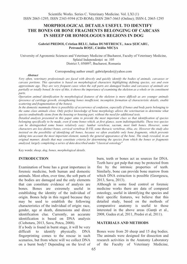
Humerus is more difficult to identify if there is only the distal extremity. Supratrochlear hole is a characteristic element of the dog. When there is only the articular surface will be analyzed the medial lip of trochlea which is wide in sheep. In this species the condyle appears as a cylinder
segment. In dogs medial lip of throchlea is sharp and it’s groove is well defined. The condyle is triangular (Fig. 6).
Figure 6 The appearance of distal extremity of the humerus in sheep (A) and dog (B) (cranial view)
(original) 1-medial lip of the trochleea; 2-lateral lip of the
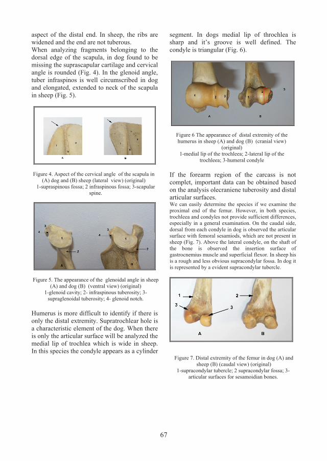
trochleea; 3-humeral condyle If the forearm region of the carcass is not complet, important data can be obtained based on the analysis olecraniene tuberosity and distal articular surfaces. We can easily determine the species if we examine the proximal end of the femur. However, in both species, trochleea and condyles not provide sufficient differences, especially in a general examination. On the caudal side, dorsal from each condyle in dog is observed the articular surface with femoral sesamiods, which are not present in sheep (Fig. 7). Above the lateral condyle, on the shaft of the bone is observed the insertion surface of gastrocnemius muscle and superficial flexor. In sheep his is a rough and less obvious supracondylar fossa. In dog it is represented by a evident supracondylar tubercle.
Figure 7. Distal extremity of the femur in dog (A) and sheep (B) (caudal view) (original)
1-supracondylar tubercle; 2 supracondylar fossa; 3- articular surfaces for sesamoidian bones.

68
If we examine the tibia, first will identify the fibular styloid apophysis in sheep. However, it is useful to identify other specific elements. The sheep tibial crest ends slowly in sheep while the dog stops suddenly, tibial groove is deeper in sheep than in dog and lateral distal end of the tibia in sheep is provided with bone malleolar articular surface (Fig. 8).
.
Figure 8. Distal extremity of the tibia in dog (A) and sheep (B) (lateral view) (original)
CONCLUSIONS On the identification of other cervical vertebrae we consider the most important character, on which we can make a difference, is the presence of the muscular tubercles on the dorsal part of the caudal articular processes in dog. By focusing on the thoracic vertebrae, the lateral orientation of the transverse processes articular surfaces in the dog, is different from the ventro-cranial orientation in sheep and in dog, the last 4-5 vertebrae have, as a defining element, accessories processes. In dog, ribs have a high degree of curvature, cylindroid aspect of the head and tuberous aspect of the distal end but in sheep, the ribs are widened and the end are not tuberous. In the glenoid angle, tuber infraspinos is well circumscribed in dog and elongated, extended to neck of the scapula in sheep. If we examine the proximal end of the femur, on the caudal side, dorsal from each condyle in dog is observed the articular surface with femoral sesamiods, which are not present in sheep. Above the lateral condyle, on the shaft of the femur the insertion surface of gastrocnemius
muscle and superficial flexor on the sheep is a rough and less obvious supracondylar fossa, and in dog it is represented by a evident supracondylar tubercle. If we examine the tibia, first will identify the fibular styloid apophysis in sheep and the sheep tibial crest ends slowly while the dog stops suddenly, tibial groove is deeper in sheep than in dog and lateral distal end of the tibia in sheep is provided with bone malleolar articular surface REFERENCES Ciobotaru, Emilia, 2013, Medicină legală veterinară. Ed.
Ceres, București, p. 13-22. Ganță, Vanda Carmen, M. Pentea, C. Pop, Maria Moț, S.
Jivcov, 2008. Osteologie veterinară. Ed. Mirton, Timișoara, p. 23-64, 77-117, 122-142.
Georgescu, B., G. Predoi, C. Belu, 2013. Anatomia omului. Ed. Ceres, București, p. 53-56.
Gudea A., Stan F., 2011, The Discriminative Macroscopical Identification of the Bones of Sheep (Ovis aries), Goat (Capra hircus) and Roe Deer (Capreollus capreollus). 1. Elements of the Forelimb Skeleton, in Bulletin of University of Agricultural Sciences and Veterinary Medicine Cluj Napoca- Veterinary Medicine, Volume 68(1)/2011, p. 171-178.
Predoi, G., B. Georgescu, C. Belu, I. Dumitrescu, Anca Seicaru. Petronela Rosu, 2011. Anatomia comparata a animalelor domestice (osteologie, artrologie, miologie). Ed. Ceres, Bucureşti, p. 21-23, 52-59, 66-74-76, 87-88.
Savu, C., Carmen Petcu, 2008. Igiena și controlul produselor de origine animală. Ed. Semne, București, p. 132-138.