Embed Size (px)
Citation preview
Morphological and Morphometrical Study of Human Lens in
Senile Cataract
Pages with reference to book, From 141 To 144 Muhammed Roshan Shaikh, Muhammed Zahoor Janjua ( Department of Anatomy, Basic Medical Sciences Institute, Jinnah
Postgraduate Medical Centre, Karachi. )
Abstract
Histological changes were studied in 34 senile cataractous lenses removed surgically from patients
aged 50 to 78 years. Sixty-eight percent had nuclear sclerosis, 44% swollen cells and morgagnian
globular changes, 23% calcium deposition, 15% migration of epithelial cells beneath posterior capsule
and villous projection in posterior in 7%. Several cases had more than one histological abnormality.
There was significant reduction in the diameter of epithelial cells of the cataract and insignificant
change in capsular thickness (JPMA 47:141,1997).
Introduction
The lens is one of the components of ocular refractive media which transmit and refract light and is the
only component whose refractive powercanbe varied. The normal lens is soft elastic and perfectly
transparent due to uniform arrangement of cells in the axial portion of the lens, the consistent thickness
of their cell membranes, the finely granular and evenly dense cytoplasm and the paucity of organelles.
Cataract is an opacificationorloss of transparency in the crystalline lens of the eye. The senile cataract
that constitutes a public health problem is the age related opacification of the lens that impairs vision to
such an extent that occupational pursuits or the activities of daily living are severely restricted
whichleads to economic and psychological deprivation that adversely affect the quality of life and
occurs in persons above 50 years. Cataract is a major cause of blindness in the world being 400 times
more in Asia than Europe1. The risk of occurrance of cataract in differentparts of the world, its
causative factors, and biochemical aspects2,3 have been studied thoroughly. This study reports the
morphological and morphometric changes in the senile cataract.
Materials and Methods
Three hundred and fifty cases of senile cataract admitted in ophthalmological ward of Jinnah
Postgraduate Medical Centre, Karachi for extraction during 1991 to 1994 were followed up for general
and clinical examination. Fifty one patients undergoing intracapsular cataract extraction without histoiy
of ocular trauma, glaucoma, use of corticosteroids and suffering from any systemic disease (e.g.,
diabetes mellitus, hypertension etc) were selected and cataracts from any other cause were excluded.
Seven lenses being clinically normal were obtained with cooperation of Eye Bank Society of Pakistan
from different eye centres of Karachi during 1991 to 1994 and 51 senile intracapsular cataractous
lenses from Eye Theatre of Jinnah Postgraduate Medical centre, Karachi were processed immediately
to avoid any morphological change due to tissue death. On histological examination, only 4 out of
7(ages 51,58,65 and 72 years) were absolutely normal and 34 out of 51(50 to 78 years) were
intracapsular senile cataracts. Evety lens (nonnal orcataractous) was left forfixation in 10% buffered
neutral formaline4 for 24 hours then cut into two halves and fixed in fresh fixative for another 24 hours.
The tissues were then dehydrated in ascending grades of alcohol from 70% to absolute alcohol. Tissues
were cleaned in xylene and embedded in paraffm after paraffm infiltration. Three micron thick sections

stained with haemotoxylin and eosin, Masson’s trichrome and PAS were examined for morphological
and morphometrical changes in lens capsule, epithelium, fibers and nucleus.
Results
The control lenses showed almost normal appearance on gross examination. They were almost
transparent, non-vascular, soft in consistency elastic in nature and bi-convex in shape being some what
flatter anteriorly, surfaces were smooth with an average diameter of 9mm and thickness of 3-4 mm. The
cataractous lenses were opaque yellow to brown in colour, hard in consistency and had uneven surfaces
and diameters. Histologically the transverse sections of control lenses showed biconvex body
enveloped by capsule with lens nucleus in the centre separated posteriorly from capsule by posterior
capsule only, while anteriorly and at equators by subcapsular monolayered sheet of epithelial cells
alongwith anterior cortex composed of parallel and mendionally arranged lamellae of lens cells or
fibers whose nuclei defined the bent arrangement known as bow configuration (Figure 1).
The nucleus showed antero-posterior arrangement of primaiy lens fibers,
surrounded meriodonally by secondaiy fibers.

A decrease in the thickness of the capsule in cataracts at all planes was recorded when compared with
the normal lenses. The mean diameter of subcapsular single sheeted cuboidal epithelial cells (8.98±0.36
urn) was significantly reduced when compared with that of normal lenses (11.86±0.97 um)as shown in
Table I.
Thecortexofcataractous lenses lost the normal lameller arrangement and the bow configuration and
showed following degenerative changes (Table II).

The cellular swelling (bladder cell change): The intracellular degenerative change was observed in the
lens cortex in 15 cases of between 50 and 65 years of age. The swollen cell diameter ranged between
18 and 84 um(Figure 2).

The retention of broken lens cells debris and water in the form of tiny globules (morgagman globules)
with diameterof 1.3 to 17 urn was noted in 12 cases of 60 to 70 years of age (Figure 3).
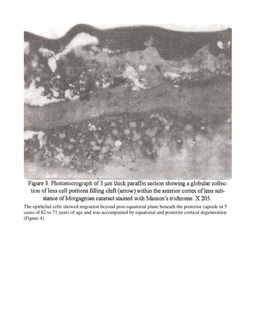
The epithelial cells showed migration beyond post-equatorial plane beneath the posterior capsule in 5
cases of 62 to 71 years of age and was accompanied by equatorial and posterior cortical degeneration
(Figure 4).

Calcarious change (calcium deposition) was noted in 8 cases of 65 to 75 yeats age (Figure 5).

The nuclear lens cells (fibers) lost their usual regular concenteric laminations and whole mass took a
uniform stain. This nuclear sclerotic change was observed in 23 cases of 65 to 78 yeats age (Figure 6).

Villous projection of posterior processes of the lens fibers in posterior capsule was noted in two cases
of 77 and 78 yeats.
Discussion
The significant decrease 8.98 j.tm±0.25 um was note in the mean epithelial cell diameter of the 34
cataractous lenses range 6 to 13 um when compared with the age matched control lenses where it was
11.86±1.37 um (range 10 to 14 um). This may be attributed to the decreased metabolic activity in
cataractous lenses. Brown and Bron6 measured the epithelial cell diameter 12.7 um (range 8 to 21 um)
biomicroscopically of 100 subjects ranging between 11 to 75 years including 20 diabetics. Our fmdings
onthe epithelial size do notcorrespond withtheir observations as they have included cases withawide
range of age, normal and diseased from a different race. Bladdercell orswollen cell, a degenerative
change might have resulted from decreased metabolic activity and the distuthed normal cell function in
old age. Uga et al7 observed similar change in 24 weeks old addy strain albino mice and suggested as
an age related change. Similar changes were reported in Nakano Strain mice on 20th post-natal day8
(hereditaiy cataract). These changes may be due to deficiency in the Na-K-ATPase leading to

electrolyte disturbances resulting in osmotio swelling. Galactose feeding also resulted in similar
change9.
Morgagnian Globular formation might have resulted by fragments of swollen cell wall. Gorthy10
observed large globularbodies incataractous rat lens and postulated that these might have resulted from
detachment of degraded massive fiber cell portions. Similar changes have been reported in a
cataractous patient with retinitis pigmentosa11 . Nuclear sclerosis recorded in maximum number of
cases might have resulted from denaturation and coagulation of soluble lens protein. Same change
occurred in the philly mouse lens (congenital cataract) due to the presence of abnormal lens proteins in
attenuated lens12. Calcium deposition on the damaged lens fibers mightbe due to altered permeability
ofthe lens resulted from decreased metabolism and energy because
of old age. Lenticulardepositsofcalciumoxalate has also been observed by Johnson13 who assumed that
the calcium oxalate are fonnedin and by the lens probably as a result of altered biochemistry.
Posterior migration of epithelial cells beneath posterior capsule might be stimulated.by prior equatorial
and posterior cortical degeneration. Similar changes had been recorded in a hereditary cataract of philly
mouse12. The reduced proliferative activity in the germinative zone might induce the posterior
migration of epithelium. More than one changes were observed in many cataracts. Present study
revealed that the decreased energy available for normal functioning of lens in old age resulted in
degenerative and sclerotic changes.
Acknowledgements
The help of doctors and staff at the OPDs and Eye Operation Theater of Jirmah Postgraduate Medical
Centre in selection of cases, Mr. Zia H. Zuberi, Secretary to Eye Bank Society of Pakistan in arranging
control lenses, staff of Anatomy Department in laboratory work and of Mr. Muhammad Akhter Anwer,
Medical Statistician, Jinnah Postgraduate Medical Centre in statistical analysis of the results are
gratefully acknowledged.
References
1. Weale, R.A. The age variation of ’senile’ cataract in various parts of the world. Br. J. Opthalmol.,
1982;66:31-34.
2. Ready, D.V. N. Distribution offtee aminoacidsand related compoundsinocular fluids, lens and
plasma. Invest. Ophthalmol., 1967;6:478.
3. Satoh, K. Age related changes in the structural protein of the human lens. Exp. Eye. Res.,
1972;14:53-57.
4. Lee, G. and Luna, H.T. Manual of histologic staining methods of the Armed Forces Institute of
Pathology, 3rd edition. New York, Toronto. London and Sydney. McGraw Hill Book Company, 1968,
p. 3.
5. Bland, M. An introduction to medical statistics. 1st ad. Oxford, Oxford University Press, 1987.
6. Brown, N.A. and Bron, A.G. An estimate of the human lens epithelial cell size in vivo. Exp. Eye
Res,, 1987;44:899-906.
7. Uga. S.. Kohara. M. and Ishikawa, S. Morphological study of age related changes inmouse lens. Jpn.
J. Ophthalmol., 1983;27:157-65.
8. Sakuragawa, M., Kuwabara, T., Kinoshita, J.H. Swelling of lens fibers. Exp. EyeRes., 1975;21:381-
94.
9. Von Salmann, L. The lens epithelium in the pathogenesis of cataract. Am.J. Ophthalmol., 1957;44:
159.70.

10. Gorthy, W.C. Cataracts in the aging rat lens. Exp. Eye Res., I 978;27:301 -22.
11. Dilley, K.J., Bron, A.J. and Habgood, JO. Anterior polar and posterior subcapsular cataracts in a
patient with retinitispigmentose: A light microscopic and ultrastructure study. Exp. Eye Res., 1976;22:
155-67.
12. Uga, S.. Kador, P.F. and Kuwabara, T. Cytological study of philly mouse cataract. Exp. Eye Res.,
1980;30:79-92.
13. Zimmerman, L.E. and Johnson, F.B. Calcium oxalate crystals within ocular tissues. A
clinicopathologic and histochemical study. Arch. Ophthalmol., 1958;60:372-83.
14. Worgul, BY., Merium, G.R., Szechter, A. et al. Lens epithelium and radiation cataract 1.
Preliminary study. Arch. Ophthalmol., 1976;94:996-99.