Embed Size (px)
Citation preview

MOLECULAR STUDIES OF LIPOPROTEIN LIPASE
by
Zhang Shuhua
A thesis submitted to the faculty of The University of Utah
in partial fulfillment of the requirements for the degree of
Master of Science
in
Medical Laboratory Science
Department of Pathology
The University of Utah
June 1991
THE UNIVERSITY OF UTAH GRADUATE SCHOOL
SUPERVISORY COMMITTEE APPROVAL
of a thesis submitted by
Zhang Shuhua
This thesis has been read by each member of the following supervisory committee and
by majority vote has been found to be satisfactory.
Chair: arc Lalouel
Roger R. Williams
THE UNIVERSITY OF UTAH GRADUATE SCHOOL
FIN AL READING APPROV AL
To the Graduate Council of the University of Utah:
I have read the dissertation of Zhang Shuhua in its final form and have found that (1) its format, citations and bibliographic style are consistent and acceptable; (2) its illustrative materials including figures, tables, and charts are in place; and (3) the final manuscript is satisfactory to the supervisory committee and is ready for submission to The Graduate School. � __ _
5/1'1191 � :=> .
Jean-Marc Lalouel Chair, Supervisory Committee
Approved for the Major Department
Approved for the Graduate Council
B. Gale Dick Dean of TIle Graduate School

ABSTRACT
Molecular defects in the lipoprotein lipase (LPL) gene account for a significant
proportion of the occurrence of the massive hypertriglyceridemia and other clinical
manifestations of the chylomicronemia syndrome. Furthermore, some published reports
suggest that the heterozygous state for LPL deficiency leads to a reduced capacity to
clear triglyceride-rich lipoproteins from plasma; when this clearance is saturated through
dietary or hormonal factors, moderate hypertriglyceridemia may be observed. The LPL
gene of four hypertriglyceridemic subjects was examined for the presence of molecular
variants. In one subject with the classical presentation of the chylomicronemia syndrome
and documented deficiency of LPL activity in postheparin plasma, we have identified a
mutation leading to the substitution of glutamic acid for glycine at residue 195 of the
mature enzyme. The patient was homozygous for this mutation. By in vitro mutagenesis
and transient expression in cultured mammalian cells, it was shown that the protein
encoded by this mutated sequence lacked catalytic activity, and data on the homologous
enzyme pancreatic lipase indicate that this mutation occurred within the catalytic domain
of the enzyme. By contrast, we identified two other mutations in two of the three other
hypertriglyceridemic subjects with moderate hypertriglyceridelnia that did not appear of
functional significance. Therefore, we have found no evidence of a role of molecular
defects of LPL in these milder cases.

TABLE OF CONTENTS Page
ABSTRACT ...................................................................................................... IV
LIST OF FIGURES .......................................................................................... VII
LIST OF TABLES . .............. .......... .......... .......... ........ .......................... ...... ...... viii
ACKNOWLEDGEMENT ........ ................ .......... .... .......... ............ .... .......... ...... IX
INTRODUCTION ............................................................................................ 1
Lipid Transport in Plasma ............. ...... .......... ......................................... 1 Disorders of Lipid Metabolism ........ .......... ....... ................. ...................... 1 Lipoprotein Lipase : General Properties ... ........ .......... ........ .......... ........... 5 Lipoprotein Lipase: Functional Domains .... ........ ........ ...... ........ .... .......... 6 Lipoprotein Lipase: Gene Structure ..................... ...... .... .......................... 8 Lipoprotein Lipase Deficiency ................................................................. 10 Hypertriglyceridemia ................................................................................ 12 Rationale and Significance of the Present Study.................................... 15
MATERIALS AND METHODS .................................................................... 16
Subjects ..................................................................................................... 16 Southern Blot Analysis ..... ........ ........ ............ ...... ...... ........ .......... ............. 16 Detection of Molecular Variants as Confonnational Polymorphisms..... 18 DNA Sequencing ...................................................................................... 20 Allele-Specific Oligonucleotide Hybridization ....................................... 22 In Vitro Mutagenesis ................................................................................ 24 Transient Expression in Cultured Mammalian Cells ........... .... .......... ..... 27 Assay of LPL Mass and Activity..... ........ .......... ........ .......... ...... ............. 27 Affinity Chromatography on Heparin-Sepharose ............... ............ ......... 28
RESULTS ........................................................................................................ 29
Probing Total Genomic DNA for Major Rearrangements ..................... 29 Detection of Single Nucleotide Substitutions ......................................... 31 Identification of Molecular Variants by DNA Sequencing .................... 32 The 839A Mutation Encodes a Functionally Inactive Enzyme .............. 37

Page
DISCUSSION .................................................................................................. 41
CONCLUSION ............................................................................................... 46
REFERENCES .................................................................................................. 47
VI

LIST OF FIGURES
Figure Page
1. Lipoprotein transport.................................................... ... ................... ........ ..... 3
2. Physical properties of lipoprotein lipase....... ...... ................ .......................... 7
3. The homology of human lipoprotein lipase and pancreatic lipase................ 9
4. Structure of the human LPL gene..................................................... .............. 11
5. DNA constructions ........................................................................................... 25
6. Oligonucleotide-directed mutagenesis of 195 (Gly~Glu) mutation ............... 26
7. Experimental results from Southern-Blot hybridization .................................. 30
8. Experimental results from conformational polymorphism............. ................ 33
9. Comparison of normal nucleotide sequence with mutant sequences found in exons 1, 5, 9, of the LPL gene........................................................ 36
10. Experimental results from allele-specific oligonucleotide hybridization..... 38

LIST OF TABLES
Table Page
1. Physical and Chemical Features of Human Plasma Lipoprotein Classes .... 2
2. Classification of Hyperlipoproteinemias........................................................ 4
3. Subjects Used in the Present Study................................................................ 17
4. Synthetic Oligonucleotides Used for Enzymatic Amplification of Exons 1-9 from Total Genomic DNA...... ...... ................ ........ ......................... 19
5. Synthetic Oligonucleotides Used as Probes for Slot-Blot Hybridization ................................................................................................. 23
6. Analysis of Conformational Polymorphism and Experimental Results ...... 34
7. LPL Activity and Mass in the Medium and Lysate of Transfected COS-1 Cells................................................................................ 40

ACKNOWLEDGEMENTS
In any research endeavor there are many individuals to be acknowledged for their
various contributions. To begin with, the author wishes to express her most sincere
appreciation to Professor Jean-Marc Lalouel for his assistance and guidance. His timely
suggestions and constant encouragement contributed immensely to this investigation.
Grateful appreciation is extended to professors Roger R. Williams and James T.
Wu for their insightful advice and cooperation during our discussion of my research
results.
The author express heartfelt thanks to Dr. Akira Hata who served as her direct
laboratory supervisor and is responsible for her successful research results.
The author appreciates the cooperation extended by all the personnel in Professor
Lalouel's laboratory and colleges in Howard Hughes Medical Institute for providing
invaluable help in class work, research and experimental work.
Also, the author derives extreme pleasure in acknowledging her husband and
daughter for their understanding and support during many years consumed in this
academic achievements.
Finally, thanks are extended to the countless many who are not listed here but
whose help has been instrumental in this work.

INTRODUCTION
Lipid Transport in Plasma
Long-chain fatty acids of intestinal or hepatic origin transit in plasma after
esterification with glycerol to form triacylglycerols. The transport of this hydrophobic
material in an aqueous environment is achieved by associating the rather insoluble lipids
with more polar ones, such as phospholipids, and combining them with cholesterol and
protein to form hydrophilic lipoprotein particles. Synthesized by the intestine and the
liver, lipoproteins undergo a series of catabolic steps before they are cleared by specific
cellular receptors (Havel and Kane, 1989). Heterogeneity in size and density has led to
the definition of discrete classes of lipoproteins, as summarized in Table 1. The
characterization of their tissue of origin, lipid and protein composition, and metabolic
fate has led to the model of plasma lipid transport and metabolism depicted in Figure 1.
Disorders of Lipid Metabolism
A variety of pathological states are associated with either abnormal concentration
or altered composition of some lipoproteins, referred to as hypolipidemias,
hyperlipidemias and dyslipidemias. A classification of the hyperlipidemias (Table 2) has
been introduced by Fredrikson and Levy (1967). Although this classification was based
on an analysis of observed lipid profiles within families with several hyperlipidemic
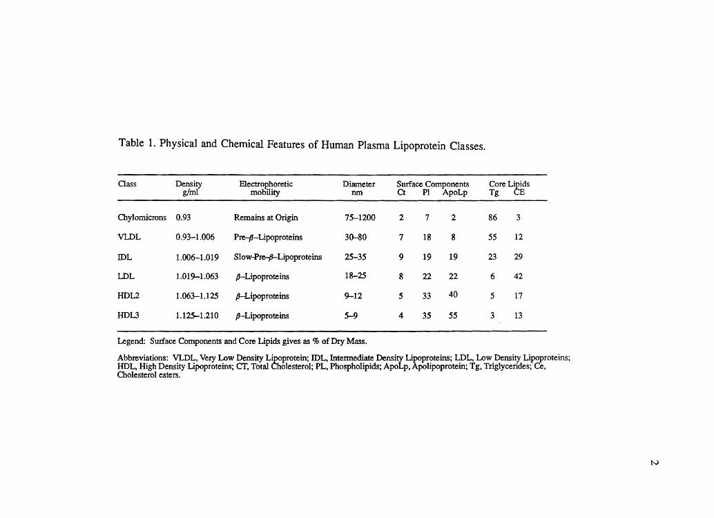
Table 1. Physical and Chemical Features of Human Plasma Lipoprotein Classes.
Class Density EleCtrophoretic Diameter Surface Components Core L~idS g/ml mobility nm Ct PI ApoLp Tg E
Chylomicrons 0.93 Remains at Origin 75-1200 2 7 2 86 3
VLDL 0.93-1.006 Pre-P-Lipoproteins 30-80 7 18 8 55 12
IDL 1.006-1.019 Slovv~L~proteins 25-35 9 19 19 23 29
LDL 1.019-1.063 p-Lipoproteins 18-25 8 22 22 6 42
HDL2 1.063-1.125 p-Lipoproteins 9-12 5 33 40 5 17
HDL3 1.125-1.210 p-Lipoproteins 5-9 4 35 55 3 13
Legend: Surface Components and Core Lipids gives as % of Dry Mass.
Abbreviations: VLDL, Very Lovv Density L~protein; IDL, Intennediate Density ~proteins; LDL, Low Density Lipoproteins; HDL, High Density Lipoproteins; cr, Total ole sterol; PL, Phospholipids; ApoLp, polipoprotein; Tg, TrigJycerides; Ce, Cholesterol esters.
tv

Figure 1.
Cholesterol +
1r191yceles
Exogenous
Bile acids + -----.
cholestero~
Endogenous
~L rLptor I·· LDL [ptor • r-. -----.
.---L......Jiv'--er-...... 1 ~ Extrahepatic "-sc-a-ve-ng-e-r --. ~ cells cells
Intestine
+ ~r /~~
~ B Chole~erol I Cholesterol
I / phospholipid ~ ~~ ~
LPL LPL ~ ~ Extrahepatic capillary beds I , Cholesteryl ~~
I •
esters ~
Lipoprotein transport.
Both the exogenous and endogenous cycles begin with the secretion of triglyceride-rich particles (chylomicrons and VLDL) that are converted to cholesteryl ester-rich particles (remnants, IDL, and LDL) through interaction with LPL. Abbreviations: LPL, lipoprotein lipase; VLDL, very low density lipoproteins; IDL, intermediate density lipoproteins; LDL, low density lipoproteins; HDL, high density lipoproteins; LCAT, lecithin cholesterol acyltransferase.
w

Table 2. Classification of Hyperlipoproteinemias.
Li12012roteins LiQids Type Chy VLDL LDL CH TO
1 +++ Nor t N t ttt
2a 0 N tt tt N
2b 0 t t t t
3 0 tt Nor ..1- tt tt (abnonnal)
4 0 tt N N tt
5 ++ tt ' N t ttt
Note: Chy, chylomicrons; VLDL, very low density lipoproteins; LDL, low density lipoproteins; CH, cholesterol; TO, triglycerides; N, normal
4

5
subjects, it does not correspond to well defined genetic entities. Furthennore, clustering
of different types of hyperlipidemias within families is rather common. This is
particularly the case for the hyperlipidemias characterized by increased plasma
concentration of triglycerides, where Type I, Type V, and Type IV profiles are often seen
among close relatives.
High plasma concentrations of triglycerides may result from increased synthesis,
impaired plasma catabolism, or decreased clearance of triglyceride-rich lipoproteins. By
hydrolyzing triglycerides from chylomicrons and very low density lipoproteins (VLDL),
the enzyme lipoprotein lipase plays a central role in their remodeling and eventual
clearance. This graduate research work has consisted of the molecular analysis of the
lipoprotein lipase gene in four hypertriglyceridemic subjects. Pertinent infonnation
regarding the biochemistry and gene structure of LPL, LPL deficiency states, and outline
the rationale of the study will be sumarized.
Lipoprotein Lipase : General Properties
Lipoprotein lipase (LPL; triacylglyceroprotein acylhydrolase, EC 3.1.1.34) is an
extracellular enzyme synthesized by a variety of parenchymal cells including adipocytes,
skeletal and cardiac muscle cells, the mammary gland and activated macrophages. After
secretion from such producing cells, the enzyme diffuses through the vascular tree and
becomes anchored to the surface of the capillary endothelium of extrahepatic tissues by
an ionic interaction with the glycosaminoglycan heparan sulfate. The enzyme, which
requires the specific cofactor apolipoprotein C-II for efficient catalytic activity,
hydrolyses triacylglycerols by binding to the surface of chylomicrons and very low

6
density lipoproteins (VLDL), thereby releasing free fatty acids for uptake into the tissues
where they can either be used immediately as fuel or reesterified for storage. General
properties of the enzyme are presented in Figure 2. Thus the enzyme plays a key role
in the metabolism of large triglyceride-rich lipoproteins to smaller particles, and
detennines the distribution of fatty acid energy among various tissues. General reviews
summarize a vast literature on the biochemistry of this enzyme, such as Smith and
Pownall (1984), Garfmkel and Schotz (1987), or Olivecrona and Bengtsson-Olivecrona,
(1987).
Hormonal regulation of LPL has mainly been studied in adipose tissue or isolated
adipocytes. Stimulation of adipose tissue enzyme activity has been demonstrated by
insulin, glucocorticoids, adenosine analogues, gastrin and pancreozymin. Inhibitory
effects on LPL activity in adipose tissue have been observed with catecholamines,
dibutyryl cAMP and estrogens. Aspects of hormonal regulation of LPL activity are
reviewed in Weinberg (1987).
Lipoprotein Lipase: Functional Domains
Similar to other lipases, it is assumed that LPL requires intact catalytic as well
as interfacial lipid binding sites in order to hydrolyze lipid emulsions and lipoprotein
particles. Although LPL readily binds to lipid-water interfaces, the enzyme also requires
a protein cofactor, apolipoprotein C-II, to become fully active. The cofactor requirement
of LPL disappears when soluble substrates are hydrolyzed. The striking homology that
has been demonstrated between LPL, hepatic triglyceride lipase, and pancreatic lipase,

Serine Esterase (Ser-Asp-His Catalytic triad)
Active Enzyme:
Monomer:
Substrates:
pH Optimum:
homodimer
native 475 aa, mature 448 aa, 55 kDa, 8% carbohydrates glycosylated at Asn 43 and Asn 359
triacylglycerols of lipoproteins, DAG, phospholipids, p-nitrophenylesters
8-9
Activated by Apo C-II
Figure 2. Physical properties of lipoprotein lipase.
.....,J

8
may actually hint at the structure of some of the functional domains of LPL. In these
enzymes there is a large region of homology, from residue 105 to residue 209 in human
LPL. This region includes a putative sequence for interfacial lipid binding (human
residues 126-135). References can be found in the general reviews quoted in the previous
section.
Some work on bovine LPL and canine pancreatic lipase suggested that the active
serine of human LPL may be located at residue 132. Recently, the three-dimensional
structure of the human pancreatic lipase gene has been solved (Winkler et aI., 1990),
confirming that the catalytic site of pancreatic lipase (PL) consists of the Ser-Asp-His
catalytic triad commonly found in serine proteases. The specificity of the enzyme for
lipid substrates appears to be related to the buried localization of the active serine in a
pocket with a high density of hydrophobic residues. The homology of human LPL and
PL becomes manifest when both sequences are optimally aligned (Figure 3).
Other functional domains of LPL, such as the cofactor binding site and the
heparin-binding site, remain to be identified. Furthermore, although the noncovalent
homodimer structure of the active enzyme has been demonstrated, the sequence elements
involved in this interaction are unknown. Consequently, the identification of molecular
variants of LPL may provide an opportunity to identify such functional domains.
Lipoprotein Lipase: Gene Structure
The human LPL complementary DNA (cDNA) has been cloned from adipose
tissue (Wion et aI, 1987). It encodes a protein of 475 an1ino acids including an apparent

HUMAN LIPOPROTEIN (LPL) AND PANCREATIC (PL) LIPASES
70 80 90 100 110 1 YESWVPKLVAALYKREPDSNVIVVDWLSRAQEHYPVSAGYTKLVGQDVARFINWMEEE
I I I I I I I III I II II I 2 EENWLANVCKNLFKVE-SVNClCVDWKGGSRTGYTQASQNIRlVGAEVAYFVEFLQSA
90 100 110 120 130 -
120 130 140 150 160 170 FNYPLDNVHLLGYS LGAHAAG lAGS LTNKKVNRlTGLDPAGPNFEYAEAPSRLS PDDA I I III I 11111111 II" 11111111 I I II I II FGYSPSNVHVlGHSLGAHAAGEAGRRTNGTIGRlTGLDPAEPCFQGTPELVRLDPSDA
140 150 * 160 170 * 180 190
180 190 200 210 220 230 DFVDVLHT-FTRGSPGRSlGlQKPVGHVDlYPNGGTFQPGC--NlGEAlRVlAERGLG I1II II I I III I I11I III II I I I
KFVDVlHTDGAPlVPNLGFGMSQVVGHLDFFPNGGVEMPGCKKNlLSQlVDlDGlWEG 200 210 220 230 240 250
240 250 260 DVDQLVKCSHERSlHLFlDSLLNEENPSKAYRCS 265
I I I II II I I TRD-FAACNHLRSYKYYTDSlVNPDGFAGFPCAS 287
260 * 270 280
Catalytic triad PL LPL
Ser 152 Asp 176 His 263
Ser 132 Asp 156 His 241
Surface loop PL LPL .
237 - 261 216 - 239
Figure 3. The homology of human lipoprotein lipase and pancreatic lipase: central region of homology.
9
10
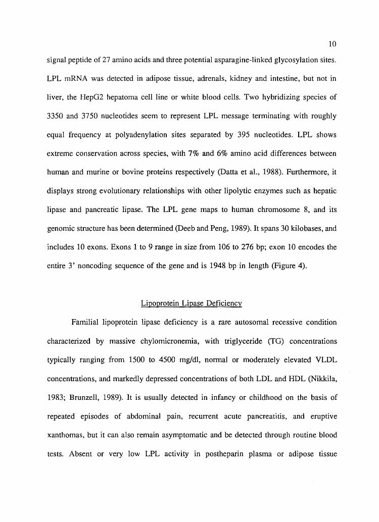
signal peptide of 27 amino acids and three potential asparagine-linked glycosylation sites.
LPL mRNA was detected in adipose tissue, adrenals, kidney and intestine, but not in
liver, the Hep02 hepatoma cell line or white blood cells. Two hybridizing species of
3350 and 3750 nucleotides seem to represent LPL message terminating with roughly
equal frequency at polyadenylation sites separated by 395 nucleotides. LPL shows
extreme conservation across species, with 7% and 6% amino acid differences between
human and murine or bovine proteins respectively (Datta et aI., 1988). Furthermore, it
displays strong evolutionary relationships with other lipolytic enzymes such as hepatic
lipase and pancreatic lipase. The LPL gene maps to human chromosome 8, and its
genomic structure has been determined (Deeb and Peng, 1989). It spans 30 kilobases, and
includes 10 exons. Exons 1 to 9 range in size from 106 to 276 bp; exon 10 encodes the
entire 3' noncoding sequence of the gene and is 1948 bp in length (Figure 4).
Lipoprotein Lipase Deficiency
Familial lipoprotein lipase deficiency is a rare autosomal recessive condition
characterized by massive chylomicronemia, with triglyceride (TO) concentrations
typically ranging from 1500 to 4500 mg/dl, normal or moderately elevated VLDL
concentrations, and markedly depressed concentrations of both LDL and HDL (Nikkila,
1983; Brunzell, 1989). It is usually detected in infancy or childhood on the basis of
repeated episodes of abdominal pain, recurrent acute pancreatitis, and eruptive
xanthomas, but it can also remain asymptomatic and be detected through routine blood
tests. Absent or very low LPL activity in postheparin plasma or adipose tissue

GENE
Exon 1
5'
mRNA
5'
Figure 4.
\ \
\ \
\
\ \
\
276
\ \ \ \ \ \ \ \ \ \ \ \
2 3 4 5 6 7
I I I I II I / / / 1/ / r , \ II I I I I I I, I I 1\ /1 / '/ / / 1// I I , " I
I I I 'I/ \ / I I I ' 1 '/ ", I I
I / I / II I \ ' I I I' I I ' '/ f I I J
I I I /' I I ", I I I I I I 1/1/ /1 \ I I I
I I I I I II ' , \ , I I
1 I I I I '/ " \ I I I I I I /, /1 II , I I I
I I / '.1 1/ II \ I I I I I I II /1 " 'I I I
I , I I I 1/ II \ / I I I I I / I /1 ' , l I I ,
I I I 1/ II II \ / II / I I /1 /1 I' , I ,I
I I I II I . r' \ I .1 "' I \,
Structure of the human LPL gene.
8 9
1\ ,
I \ I t I , I , I \ , \ " , \ I \ , \ " , \ I \ , 'I' , 'I \ , I I , ,
'I \ , II , , , I \ I
" " \I \ I II , , . \ , . ~, ,
10
3'
1 kb
1948 3'
100 bp
The length of LPL gene is approximately 30 kb (the top portion of the figure). The 10 exons are represented by the filled boxes. Solid portions of exons represent none-coding sequences. The cDNA is interrupted by 9 introns giving rise to 10 exons. Exons 1-9 are of average size (105-276 bp) whereas exon 10 ,which codes for all of the 3' untranslated region of the mRNA, is 1948 bp in length (lower portion of the figure).
I-" I-"

12
establishes the diagnosis. A recent study of LPL-deficient, unrelated probands revealed
a major genomic rearrangement in 4 of 19 alleles (Langlois et aI., 1989) resulting from
an internal duplication disrupting the normal structure of the gene (Devlin et aI., 1990).
Other mutations of the gene, including several amino acid substitutions, have been
reported (Emi et aI., 1990; Hata et al., 1990; Beg et al., 1990; Dichek et al., 1991). Less
often, a similar syndrome can be ascribed to functional deficiency or absence of the LPL
cofactor, apo C-II, which can be diagnosed by assaying cofactor activity of the patient's
plasma (Nikkila, 1983; Brunzell, 1989).
H ypertri glyceride n1ia
The heterozygous state for LPL deficiency (LP~) remains poorly characterized.
Obligate heterozygotes are either normal or exhibit mild hyperlipidemia, and multiple
lipoprotein phenotypes have been observed in some pedigrees. Measuring both mass and
activity of LPL, Babirak et aI. (1989) suggested that Hypertriglyceridemia in relatives of
LPL deficient probands, possibly of the type seen in familial combined hyperlipidemia
(HLP), segregated with LP~; only about half of the subjects thus classified as
heterozygotes exhibited HTG. Identifying carriers at the molecular level in a large
pedigree, Wilson et al. were able to characterize the phenotypic expression of LP~
(Wilson et aI, 1990), with delayed expression of HTG, high VLDL cholesterol,
subnormal LDL cholesterol and reduced HDL cholesterol concentrations.
Familial forms of HTG may result from LP~, at least in some families. Four
familial lipid disorders featuring HTG have been defined (Havel and Kane, 1989): (1)
in familial hypertriglyceridemia (FHTG), affected relatives have elevated plasma VLDL

13
but normal or low LDL concentrations (type IV); (2) in primary type V HLP,
chylomicronemia and/or elevated VLDL are observed in family members (types V and
IV); (3) in familial combined HLP (FCHL), multiple lipoprotein phenotypes are observed
among relatives, including isolated elevation of VLDL (type IV), isolated elevation of
LDL cholesterol (type IIa), or elevation of both (type IIb); (4) in type III HLP, a rare
condition associated with premature atherosclerosis and usually homozygosity for the E2
electrophoretic variant of apolipoprotein E, high cholesterol and triglyceride
concentrations result from the accumulation of abnormal B-VLDL particles. Although
this purely phenotypic classification is unlikely to be of much etiological significance,
it has served as a basis for clinical investigations over the past two decades.
The mechanisms underlying increased plasma VLDL remain poorly understood.
Extrinsic factors such as obesity, diabetes, excessive alcohol intake, or medication with
exogenous steroids contribute to the etiology of HTG. Some metabolic investigations in
various HTG subjects (type IV or type IIb) would support a predominant role for VLDL
overproduction, while others suggest that defective VLDL removal is of greater
significance (Grundy, 1984; Grundy and Vega, 1988). Compositional differences of
VLDL particles have been reported between type IV and type lIb pro bands. In type V
subjects, turnover studies best support a combination of both overproduction and
impaired clearance of VLDL. Assessment of the published evidence is further
complicated by differences in patient selection and experimental procedures. At present,
it is reasonable to conclude that both factors are at play to varying degrees in HTG
patients. Whether overproduction or decreased clearance account for familial aggregation
can also be debated. In the only large series where kinetic parameters were investigated

14
among type IV relatives of FHTG or FCHL probands (Sane and Nikkila, 1987), it was
found that reduced clearance, but not overproduction, followed a familial pattern, and
that no particular feature of VLDL triglyceride kinetics could distinguish type IV subjects
in FHTG from type IV subjects in FCHL.
A common, saturable removal system for which both endogenous and exogenous
triglycerides depend and compete was revealed by dietary manipulations in HTG subjects
and appears related to LPL (Brunzell et ai., 1973). An inverse relationship exists between
fasting plasma TG concentration and postheparin plasma or adipose tissue LPL activities,
the latter being negatively correlated with obesity. The relationship between HTG and
LPL activity is blurred by variations in assay procedures. Furthermore, measurement of
total LPL activity does not distinguish between differences in specific activity and
differences in total pool of the enzyme; the latter is subject to a host of hormonal
influences (Weinberg, 1987). Consequently, LPL activity in plasma collected after an
heparin injection cannot serve as a predictor of the heterozygous state for LPL
deficiency.
A combination of both overproduction and decreased clearance appears to be
involved in most HTG subjects, and the phenotypic definition of familial syndromes must
admit substantial etiological heterogeneity. Observations in such families, as well as on
LP~, support a role for an inherited defect of LPL in an as yet undefined subset of
familial HTG. Interactions with other factors, genetic or environmental, account for
variation in phenotypic expression. This is clearly the picture emerging from studies of
familial type V HLP (Grundy and Vega, 1988). Overproduction may result from obesity,
alcohol, estrogen use, or diabetes, while decreased clearance may result either from down

15
regulation or froln inherited, partial deficiency in LPL.
Rationale and Significance of the Present Study
Genetics, nutrition, and metabolic or hormonal disturbances influence plasma TG
concentrations. Mutations leading to quasi-complete deficiency lead to type I HLP in the
homozygous state, with a prevalence in the order of 10-6• Even such patients, however,
express type IV HTG when adhering to a stringent low fat diet (Nikkila, 1983; Brunzell,
1989). LP4, a much more prevalent genotype, represents a latent lipolytic defect leading
to delayed expression of HTG (type IV or type V) when triggered by nutritional,
metabolic or hormonal factors.
Consequently, current study has examined the hypothesis that molecular variants
of the LPL gene may account for HTG in 4 subjects referred to us by Dr Gerald Luc,
from the Hospital Hotel Dieu in Paris, France. In subject K2042, the quasi-absence of
LPL activity in post-heparin plasma suggested LPL deficiency in the homozygous state.
Other subjects examined presented plasma triglycerides in excess of 1000 mg/dl, with
either normal or elevated VLDL cholesterol (Type IV or Type V hyperlipidemia). The
hypothesis was that these subjects could carry mutations of the LPL gene in the
heterozygous state.
MATERIALS AND METHODS
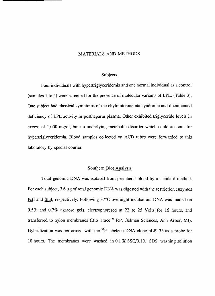
Subjects
Four individuals with hypertriglyceridemia and one normal individual as a control
(samples 1 to 5) were screened for the presence of molecular variants of LPL. (fable 3).
One subject had classical symptoms of the chylomicronemia syndrome and documented
deficiency of LPL activity in postheparin plasma. Other exhibited triglyceride levels in
excess of 1,000 mgldl, but no underlying metabolic disorder which could account for
hypertriglyceridemia. Blood samples collected on ACD tubes were forwarded to this
laboratory by special courier.
Southern Blot Analysis
Total genomic DNA was isolated from peripheral blood by a standard method.
For each subject, 3.6 Jlg of total genomic DNA was digested with the restriction enzyn1es
PstI and StuI, respectively. Following 37°C overnight incubation, DNA was loaded on
0.5% and 0.7% agarose gels, electrophoresed at 22 to 25 Volts for 16 hours, and
transferred to nylon membranes (Bio Trace™ RP, Gelman Sciences, Ann Arbor, MI).
Hybridization was performed with the 32p labeled cDNA clone pLPL35 as a probe for
10 hours. The membranes were washed in 0.1 X SSC/0.1 % SDS washing solution

Table 3. Subjects Used in the Present Study.
Subject Source
France
Sample #
1 2 3 4 5
Pedigree ID
K 2039 K2040 K 2041 K 2042* Control
Clinical Dianosis
Hypertriglyceridemia Hypertriglyceridemia Hypertrigl yceridemia LPL Deficiency
* The offspring of a consanguineous marriage.
17

18
(Ix SSC= 0.15M NaCV15 mM Na citrate, ph 7.0) for 30 minutes at 52°C and submitted
to autoradiography.
Detection of Molecular Variants as Conformational Polymorphisms
Each of the nine translated exons of the human LPL gene was enzymatically
amplified (Mullis, et al., 1987) from genomic DNA on a Thermal Cycler (Perkin Elmer
Cetus, Norwalk, Cf) basically following the method of Orita et al. (1989).
Oligonucleotide sequences used as primers are shown in Table 4. The reaction mixture
contained 5.2 pmol of each primer, 2 nmol of each dNTP, 0.1 Jlg of genomic DNA, 1
JlCi of [a-32P]dCfP (3000Ci/mmol, 10 mCi/ml, Amershan) and 0.05 units of Tag
polymerase (perkin Elmer-Cetus, Norwalk, Cf) in 10 JlI of amplification buffer. This
mixture was subjected to the iterative application of 10 reaction cycles each including
denaturation at 95°C for 1 minute, primer-annealing (at 48°C for exon 1, at 43°C for
exons 8 and 9, and at 54°C for other exons) for 1 minute, and primer-extension at 72°C
for 1 minute. For all exons, 20 subsequent cycles were applied with primer-annealing
performed at 60°C. After amplification, 50 Jll of 0.1 % SDS/10 mM EDT A was added
in the mixture. An aliquot was withdrawn and mixed with equal volume of 95%
formamide dye, boiled for 2 minutes and applied (2 Jll/lane) to a 5% polyacrylamide gel
containing 0.5 X TBE, with or without 10% glycerol. Electrophoresis was performed at
300 to 500V for 16 to 20 hours at room temperature or at 4°C. The gel was dried and
autoradiographed with or without intensifying screen for 3 to 7 hours.
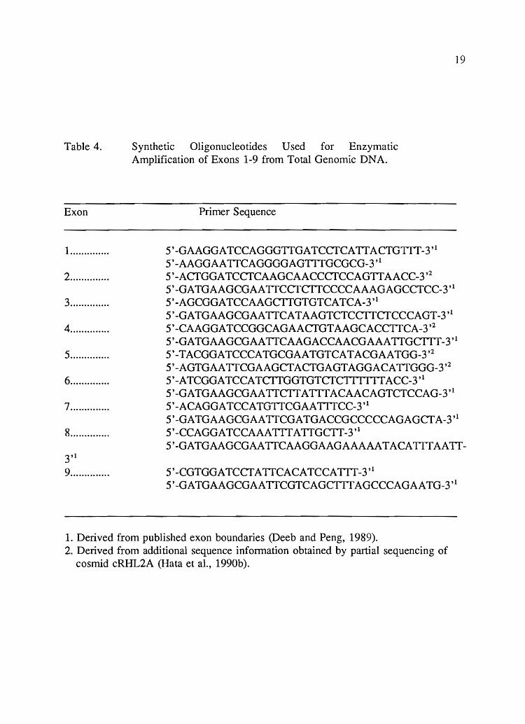
Table 4.
Exon
1 ............. .
2 ............. .
3 ............. .
4 ............. .
5 ............. .
6 ............. .
7 ............. .
8 ............. .
3'1 9 ............. .
Synthetic Oligonucleotides U sed for Enzymatic Amplification of Exons 1-9 from Total Genomic DNA.
Primer Sequence
5'-GAAGGATCCAGGGTTGATCCTCATTACTGTIT-3,1 5'-AAGGAATTCAGGGGAGTTTGCGCG-3,1 5' -ACTGGA TCCTCAAGCAACCCTCCAGTT AACC-3,2 5'-GATGAAGCGAATTCCTCTTCCCCAAAGAGCCTCC-3,1 5' -AGCGGATCCAAGCTTGTGTCA TCA-3 ,1 5'-GATGAAGCGAATTCATAAGTCTCCTTCTCCCAGT-3,1
19
5' -CAAGGATCCGGCAGAACTGTAAGCACCTTCA-3,2 5'-GATGAAGCGAATTCAAGACCAACGAAATTGcrrT-3,1 5'-TACGGATCCCATGCGAATGTCATACGAATGG-3,2 5'-AGTGAATTCGAAGCTACTGAGTAGGACATTGGG-3,2 5'-ATCGGATCCATCTTGGTGTCTCrl III IACC-3,1 5'-GATGAAGCGAATTCTTATTTACAACAGTCTCCAG-3,1 5' -ACAGGA TCCATGTTCGAA TTTCC-3,1 5'-GATGAAGCGAATTCGATGACCGCCCCCAGAGCTA-3,1 5'-CCAGGATCCAAATTTATTGCTT-3,1 5'-GATGAAGCGAATTCAAGGAAGAAAAATACATITAATT-
5' -CGTGGA TCCT A TTCACATCCA TTT _3'1 5' -GATGAAGCGAA TTCGTCAGcrrT AGCCCAGAA TG-3,1
1. Derived from published exon boundaries (Deeb and Peng, 1989). 2. Derived from additional sequence information obtained by partial sequencing of
cosmid cRHL2A (Rata et al., 1990b).

20
DNA Sequencing
All translated exons for which a mobility shift was identified were sequenced by
one or more of several methods:
(1) Direct sequencing from genomic DNA using single-stranded DNA
template generated by asymmetrical amplification. A segment spanning
exon 5 of the LPL gene was enzymatically amplified by the polymerase
chain reaction and the resulting product was purified by spin-dialysis on
a Centricon 100 column (Amicon, Danvers, MA). An aliquot (1 ).11) was
used to generate single-stranded DNA by asymmetric amplification (Saiki
et aI., 1988). Sequencing was performed by application of a standard
chain-termination protocol (Sanger and Coulson, 1975) with the enzyme
Sequenase Version 2.0 (U.S. Biochemicals).
(2) Direct sequencing of double-stranded DNA excised from gels by way
of a Thermocycling protocol. Bands of altered mobility revealed by
electrophoresis of amplified DNA fragments spanning exons 1 and 9 of
human LPL were excised from the dried gel, suspended in 100 ).11 of H20
and vortexed. After incubation at 37°C for 1 hour, 1 ).11 of each aliquot
was subjected to enzymatic amplification. The sense- and antisense
primers included at their 5'-end a sequence complementary to the M13
universal (5'-TGTAAAACGACGGCCAGT-3') and reverse (5'-CAGGA
AACAGCT ATGACC-3') priming sites respectively. The reaction mixture
was subjected to 30 cycles of denaturation at 95°C for 1 minute,
annealing at 60°C for 1 minute, and extension at 72°C for 1 minute. Each

amplified product was spin-dialyzed on a Centricon 100 column (Amicon,
Danvers, MA).
Direct sequencing of double-stranded DNA was performed on an
ABI 373A DNA sequencer (Applied Biosystems, Foster City, CA) using
fluorescent M13 primers, Tag polymerase, and a thermocycling protocol
supplied by the manufacturer (ABI protocol, 1990). In brief, 12 JlI of a 40
JlI volume of amplified product were submitted to iterative extensions on
a DNA thermocycler. Ten cycles were applied, defined by the following
conditions: denaturation at 95°C for 30 seconds, annealing at 60°C for 1
second and extension at 70°C for 1 minute. This was followed by 10
additional cycles of denaturation (95°C for 30 seconds) and extension
(70°C for 1 minute). The repeated application of several rounds of
denaturation, reannealing and extension should increase the frequency
with which termination occurs with incorporation of ddNTP rather than
dNTP as would occur when template reannealing prevents further
extension.
(3) Sequencing of cloned DNA segments. In some instances, DNA
segments were sequenced after cloning into the M13mp18 vector. These
segments were generated by enzymatic amplification of genomic DNA
using 100 pmol of each pair of primers and 2.5 units of Taq polymerase
in 100 JlI of amplification buffer, with conditions as reported earlier.
Since the primers used for amplification were augmented with recognition
sites for the endonucleases BamHI and BcoRI, each amplified fragment
could be directionally cloned into the M13mp18 vector after restriction
21
enzyme digestion. After bacterial transformation and plating, 6
independent clones were isolated, and single-stranded DNA was produced
directly from plaques suspended in distilled water by asymmetric
enzymatic amplification (Saiki et al., 1988), using the M13 universal and
reverse primers. Automated analysis of DNA sequence was performed by
means of an ABI 373A DNA sequencer (Applied Biosystems, Foster City,
CA).
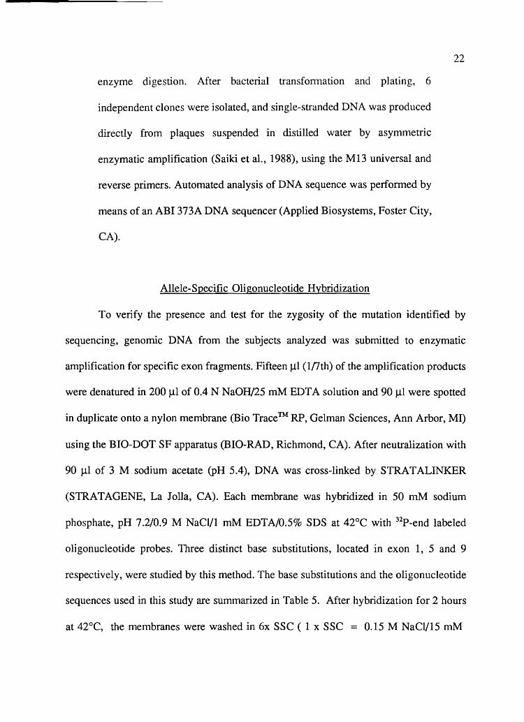
Allele-Specific Oligonucleotide Hybridization
22
To verify the presence and test for the zygosity of the mutation identified by
sequencing, genomic DNA from the subjects analyzed was submitted to enzymatic
amplification for specific exon fragments. Fifteen Jll (Inth) of the amplification products
were denatured in 200 J.tl of 0.4 N NaOH/25 mM EDTA solution and 90 J.ll were spotted
in duplicate onto a nylon membrane (Bio Trace™ RP, Gelman Sciences, Ann Arbor, MI)
using the BIO-DOT SF apparatus (BIO-RAD, Richmond, CA). After neutralization with
90 J.lI of 3 M sodium acetate (PH 5.4), DNA was cross-linked by STRA T ALINKER
(STRATAGENE, La Jolla, CA). Each membrane was hybridized in 50 mM sodium
phosphate, pH 7.2/0.9 M NaCl/l mM EDTA/0.5% SDS at 42°C with 32P_end labeled
oligonucleotide probes. Three distinct base substitutions, located in exon 1, 5 and 9
respectively, were studied by this method. The base substitutions and the oligonucleotide
sequences used in this study are summarized in Table 5. After hybridization for 2 hours
at 42°C, the membranes were washed in 6x SSC ( 1 x SSC = 0.15 M NaCl/15 mM

Table 5.
Exon
1
5
9
Synthetic Oligonucleotides Used as Probes for Slot-Blot Hybridization.
Probe
Wild Type Mutant
Wild Tyep Mutant
Wild Type Mutant
Oligonucleotide Sequence
5'-AGTGAATTTAGGTCCCf-3' T
5'-AGGGACCTAAATTCACT-3' 5'-AGTGAATTTAAGTCCCf-3'
Ser He Gly lie Glu Lys 5'-AGC ATT GGA ATC CAG AAA-3'
GAA Glu
5' -TTTCTGGA TTCCAA TGCfT-3' 5'-AAGCATTGAAATCCAGAAA-3'
Lys Lys Ser Gly 5'-AAG AAG TCA GGC TGG-3'
TGA stop
5'-TAAGAAGTCAGGCfGGT-3' 5'-TAAGAAGTGAGGCfGGT-3'
23

24
Na citrate, pH 7.0) at 1 to 2°C below the melting temperature computed for each probes,
and submitted to autoradiography.
In Vitro Mutagenesis
Mutagenesis was carried out as described by Kunkel (1985) with some
modification, using a Muta-Gene M13 in vitro mutagenesis kit, version 2 (BIO-RAD).
At first, wild type LPL cDNA was released from the clone pLPL35 after digestion with
SaIl (Wion et al., 1987), and the SaIl sites were converted to EcoRI sites using EcoRI
linkers. After digestion with EcoRI, this fragment was cloned into the EcoRI site of
M13mp18 (Figure 5). Oligonucleotide containing the 839 nucleotide mutation (5' -TCGA
AGCATTGAAATCCAGAAACCAG-3') (exon 5) and 1595 nucleotide mutation (5'-TA
AGAAGTGAGGCfGAAAC-3') (exon 9) were used as mutagenic primers.
The template for in vitro mutagenesis was prepared by propagation on the dut,
ung double mutant bacterium CJ236, leading to the incorporation of Uracil at positions
normally occupied by Thymine. A complementary strand was synthesized with the T7
DNA polymerase and closed circles were formed by the T4 DNA ligase. This DNA was
transformed into E. coli DH5aF', a strain proficient in uracil N-glycosylase activity,
leading to efficient inactivation of the uracil-containing strands, and thereby yielding the
non-uracil-containing survivor available for replication. Mutant plaques were selected by
oligonucleotide hybridization (Figure 6). DNA of M13 phage in the replicative form (RF)
was isolated by an alkaline lysis minipreparation method (Birnboim, 1983), digested with
EcoRI and inserted into the EcoRI site of the eukaryotic expression vector pMT2
(Kaufman et aI., 1989). After transformation of E. coli DH5a, 6 to 12 colonies were
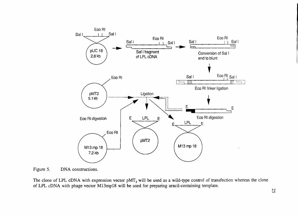
Figure 5.
EcoRI Sail Sail
>
Sail ---. c::; Sail Eco RI b II Sail u:y
Eco RI
Sail fragment of LPLcDNA
Conversion of Sal I end to blunt
+ Sail Eco RI Sal I
"fU'UU I I I "' ... -- no
L.::::J .. I II IIllu::::~ .. J
Eco Rllinker ligation Ligation
----------------.-r:-----T----:--=i-r E + • L......,
,~!. ..... -...... E
Eco RI digestion E LPL E 7 LPL E
EcoRI
DNA constructions.
The clone of LPL cDNA with expression vector pMT2 will be used as a wild-type control of transfection whereas the clone of LPL eDNA with phage vector M13mp18 will be used for preparing uracil-containing template.
tv Ul
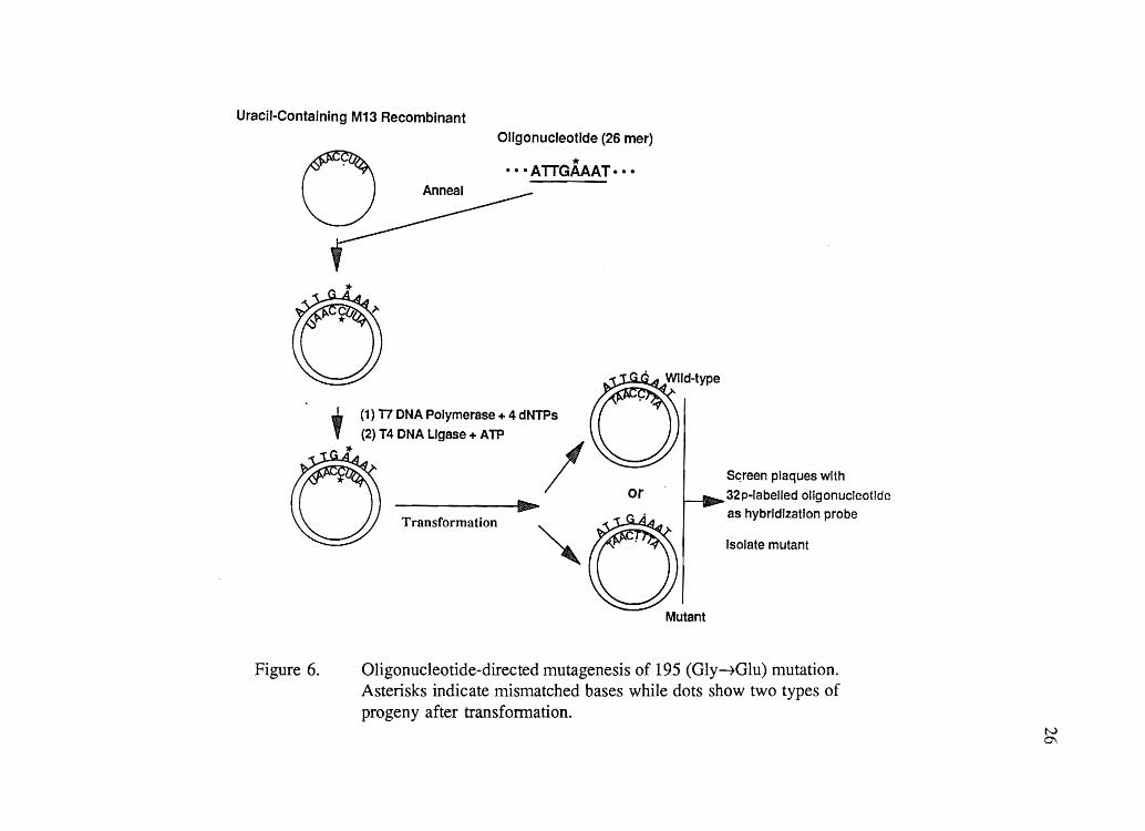
Uracil-Containing M13 Recombinant
Oligonucleotide (26 mer)
o * •.• ATTGAAAT··· Anneal
Figure 6.
IIIIh...... ~
Transformation
Mutant
Sc;reen plaques with
32p-labelled oligonucleotide
as hybrIdization probe
Isolate mutant
Oligonucleotide-directed mutagenesis of 195 (Gly~Glu) mutation. Asterisks indicate mismatched bases while dots show two types of progeny after transformation.
tv 0\

27
picked for culturing to provide DNA. Proper orientation of insert was determined by
digestion with the restriction enzyme Pvull. Sequencing analysis of the inserts confirmed
that either the G to A substitution at position 839 or the C to G substitution at position
1595 were the only change introduced in the mutant construct.
Transient Expression in Cultured Mammalian Cells
COS-l cells were plated at an initial density of 1.0 x 10⁶ per 10 cm dish and
maintained in 5 ml of Dulbecco's modified Eagle's medium with 10% FCS before
transfection. They were transfected in triplicate with plasmid DNA (6 Jig/dish), following
the DEAE-dextran method described by Selden(21) with some modification. One hour after
DNA addition, chloroquine was added to the medium at a final concentration of 80 JiM.
After 4 hours of incubation at 37°C, the cells were treated with dimethyl sulfoxide
(DMSO). The 5 ml of 10% DMSOIPBS was added to each plate and incubated for 2
minutes at room temperature. After aspirating the DMSO, each plate was then washed
with 5 ml of Ix PBS. Afterwards, 5 ml of complete medium was added to each plate
and incubation was carried out at 37°C. Twenty-four hours after transfection, serum
free, heparin-containing (5 U/ml) medium was substituted and the culture was allowed
to continue for another 48 hours. Conditioned medium and cells were collected to assay
LPL activity and immunoreactivity.
Assay of LPL Mass and Activity
LPL mass was detennined by a sandwich enzyme-linked immunoadsorbent assay
using purified bovine LPL as the standard (Iverius and Ostlund-Lindqvist, 1976). LPL

28
activity in conditioned medium or cell lysates was determined by monitoring the
hydrolysis of triacylglycerols in an emulsion composed of a radiolabeled triolein
substrate and Intralipid as an emulsifier, as described by Iverius et al. (1985,1986). Both
assays were performed in the laboratory of Dr P.H. Iverius at the Veterans Affairs
Hospital, Salt Lake City.
Affinity Chromatography on Heparin-Sepharose
Analytical heparin-Sepharose chromatography was performed at O°C using an
FPLC system (Pharmacia LKB Biotechnology, Inc., Piscataway, NJ ) equipped with a
FRAC-lOO fraction collector, a HR 5110 column, and a 10 ml Superloop for sample
loading. The column (2 ml) was packed with heparin-Sepharose CL-6B, prepared as
described previously (Iverius et al., 1971, 1976), submerged in a cylinder of ice-water
and equilibrated with 0.15 M NaCl/O.l % (v/v) Triton X-lOOIO.Ol M Na-phosphate buffer
(pH 7.5). Samples were loaded from a loop at a flow rate of 0.2 ml/min, after which the
column was washed with 19.5 ml of the equilibration buffer. Elution was performed with
an 8 mllinear gradient of 0.15 to 1.5M NaCI in 0.1% (v/v) Triton X-lOOIO.Ol M Na
phosphate buffer (pH 7.5) at a rate 0.05 ml/min. Finally, the column was stripped with
6 ml of 2 M NaCl in the same buffer at a rate of 0.2 ml/min. Fractions of 0.5 ml were
collected throughout the procedure in tubes chilled by packing ice into the fraction
collector carousel.

RESULTS
It is hypothetically proposed that molecular variants of the LPL gene can account
for either LPL deficiency, as documented by lack of LPL activity in the postheparin
plasma of subjects K2042, or for a subset of more common forms of primary
hypertriglyceridemia, as observed in the other three subjects under study. Mutational
events that might affect the gene range from major genomic rearrangements, such as
large insertions or deletions, to single nucleotide substitutions. Both of these extremes
have been previously observed in the LPL gene of LPL deficients subjects.
Probing Total Genomic DNA for Major Rearrangements
Major rearrangenlents in a gene can be detected by probing genomic DNA after
digestion with restriction endonucleases. This is of particular relevance here, given the
report of a common internal duplication in the LPL gene of LPL deficient patients
(Devlin et aI., 1990).
Total genomic DNA was digested with the restriction endonucleases PstI and StuI,
electrophoresed, transferred to nylon membranes, and hybridized to a radiolabelled probe
containing the entire coding region of the LPL cDNA (pLPL35, a gift from Dick Lawn;
Wion et aI., 1987). The results are presented in Figure 7. None of the 5 subjects
exhibited any obvious molecular defect.

Figure 7.
1 234 5 1 234 5
kb kb
8·2 11
5·6 6
3·8
2·9
2·2 2·5
1-6 2·1
Stu I Pst I
Experimental results from Southern-Blot hybridization. The total genomic DNA of 5 study subjects was digested with the restriction enzyme Stu I and Pst 1. None of the subjects has major gene rearrangement. Lane 1: K2039, Lane 2: K2040, Lane 3: K2041 Lane 4: K2042 , Lane 5: control
30
31
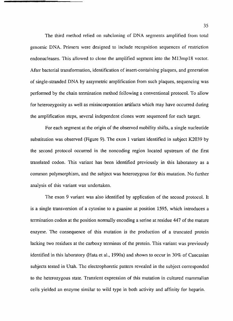
Detection of Single Nucleotide Substitutions
Minimal alterations such as nucleotide substitutions eventually require DNA
sequencing for their characterization. Given the total size of the LPL gene, approximately
30,000 nucleotides, direct examination of the entire gene is not practical. There are,
however, a variety of procedures which allow to screen segments of genomic DNA for
the presence of such variants.
The approach followed in this work relies on the detection of molecular variants
through mobility shifts revealed upon electrophoresis of single-stranded DNA under
nondenaturing conditions. Single-stranded DNA is known to present ordered structures
in solution, although such structures are much less stable than those adopted by double
stranded DNA. It has been shown that even a single nucleotide substitution can induce
a conformational change of single-stranded DNA which can be detected as a mobility
shift upon electrophoresis under nondenaturing conditions (Kanazawa et aI., 1986). This
was developed into a method, called by the authors "single-stranded conformation
polymorphism (SSCP)," for the direct detection of base substitutions in DNA segments
by Orita et al. (1989). A given DNA segment is amplified by the polymerase reaction,
labelled during this process through incorporation of a radioactive nucleotide, denatured
by heat in the presence of formamide, electrophoresed on a polyacrylamide gel under
nondenaturing conditions, and the latter is subjected to autoradiography.
This method has been used to examine the nine coding exons and corresponding
splice junctions of the LPL gene in five study subjects. The primers used for this purpose
are presented in Table 3. As the conformations adopted are quite sensitive to conditions
such as pH, ionic strength, temperature and solvent, four distinct electrophoretic

32
conditions were tested. Results obtained are summarized in Figure 8 and Table 6.
Three distinct mobility shifts were observed. In subject K2039, a mobility shift
was observed in exon 1 when electrophoresis was performed in the cold room in the
presence of glycerol. In subject K2041, another mobility shift was observed in exon 9
under two experimental conditions. Subject K2042 presented a third electrophoretic
variant in exon 5 of the gene.
Identification of Molecular Variants by DNA Sequencing
To identify the sequence variation at the origin of the observed mobility shifts,
each observed electrophoretic variant was subjected to DNA sequencing. Three distinct
protocols were applied.
In the fIrst method, direct sequencing by the chain termination method is
performed on DNA segments produced by enzymatic amplification from total genomic
DNA. A drawback of this method, however, is that it may be difficult to identify
molecular variants present in the heterozygous state.
The second method, developed in this laboratory by Hata et al. (1990a), exploits
the electrophoretic separation to purify and sequence an individual allele. Each band
representing a mobility shift was excised from the gel, resuspended in distilled water, and
subjected to enzymatic amplification with unique primers extended with universal M13
direct and reverse primers. After purifIcation of the amplified DNA segment on a spin
column, an aliquot of double-stranded DNA was sequenced on an automated instrument
(ABI373A) by the chain termination method using a newly developed thermocycling
protocol (ABI protocol, 1990).
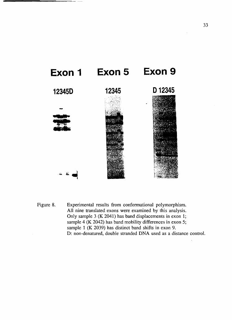
Exon 1 Exon5 Exon9
123450 12345 D 12345
Figure 8. Experimental results from conformational polymorphism. All nine translated exons were examined by this analysis. Only sanlple 3 (K 2041) has band displacements in exon 1; sample 4 (K 2042) has band mobility differences in exon 5; sample 1 (K 2039) has distinct band shifts in exon 9.
33
D: non-denatured, double stranded DNA used as a distance control.

Table 6. Analysis of Conformational Polymorphism and Results.
Gly(+) Gly(-) I Gly(+) Gly(-) I Gly(+) Gly(-) I Gly(+) Gly(-) Exonl4C 22C 4C 22C 4C 22C 4C 22C 4C 22C 4C 22C 4C 22C 4C 22C
1 2 3 4 5 6 7 8 9 + +
Gly (+) -- Polyacrylamide gel with 10% glycerol Gly (-) -- Polyacrylamide gel without glycerol + : Mobility shift detected - : No mobility shift
+
+
22C
VJ ~
35
The third method relied on subcloning of DNA segments amplified from total
genomic DNA. Primers were designed to include recognition sequences of restriction
endonucleases. This allowed to clone the amplified segment into the M13mp18 vector.
After bacterial transformation, identification of insert-containing plaques, and generation
of single-stranded DNA by assymetric amplification from such plaques, sequencing was
perfonned by the chain termination method following a conventional protocol. To allow
for heterozygosity as well as misincorporation artifacts which may have occurred during
the amplification steps, several independent clones were sequenced for each target.
For each segment at the origin of the observed mobility shifts, a single nucleotide
substitution was observed (Figure 9). The exon 1 variant identified in subject K2039 by
the second protocol occurred in the noncoding region located upstream of the first
translated codon. This variant has been identified previously in this laboratory as a
common polymorphism, and the subject was heterozygous for this mutation. No further
analysis of this variant was undertaken.
The exon 9 variant was also identified by application of the second protocol. It
is a single transversion of a cytosine to a guanine at position 1595, which introduces a
termination codon at the position nonnally encoding a serine at residue 447 of the mature
enzyme. The consequence of this mutation is the production of a truncated protein
lacking two residues at the carboxy terminus of the protein. This variant was previously
identified in this laboratory (Hata et aI., 1990a) and shown to occur in 30% of Caucasian
subjects tested in Utah. The electrophoretic pattern revealed in the subject corresponded
to the heterozygous state. Transient expression of this mutation in cultured mammalian
cells yielded an enzyme similar to wild type in both activity and affinity for heparin.

Exon 1
Exon5
Exon 9
WILD TYPE
t
Serite Gly lie GluLys t
CGAAGCAT TGG AATCCAGAAACCA
Asn Lys Lys Ser Gly * t
MUTANT
t
Ser lie Glu lie Glu Lys t
CGAAGCA T T GAAATCCA GAAACCA
Asn Lys Lys * t
A A T A A G A A G T GAG GC T G G T G
Figure 9. Comparison of normal nucleotide sequence with mutant sequences found in exons 1, 5, 9, of the LPL gene. Arrows point the base change from normal to mutant. Asterisks indicate the stop codon.
36

37
Individual K2042, who presented typical LPL deficiency, was the offspring of a
consanguineous marriage. Furthennore, the electrophoretic pattern exhibited by this
subject, with an apparent absence of bands of characteristic wild type mobilities,
suggested that he might be homozygous for a mutation in exon 5 of the LPL gene.
Consequently, direct sequencing was fIrst applied. A guanine-to-adenine transition at
nucleotide position 839 was observed. This leads to a missense mutation with the
substitution of glutamic acid for glycine at residue 195 of the mature enzyme. The
presence of this mutation was confIrmed when two clones were sequenced by the third
method: both clones contained this mutation. Hybridization of total genomic DNA of the
patient with synthetic oligonucleotides specific for wild type and mutant sequences at this
position established that the patient was homozygous for this mutation (Figure 10).
The 839A Mutation Encodes a Functionally Inactive Enzyme
The functional significance of the amino acid substitution observed in subject
K2042 cannot be inferred from the sequence alone. Therefore, the mutation was
reproduced in vitro, expressed in cultured cells, and cell lysates and conditioned media
were assayed for several criteria of LPL.
The experimental protocols used were presented in the previous section, and
therefore they are only briefly outlined here. As LPL is a glycoprotein, a mammalian
expression system was used. Mutagenesis was performed by second-strand synthesis on
a wild type template initiated by a mutagenic synthetic oligonucleotide which differed
from the wild type sequence only with respect to the observed mutation. The wild type
strand had been prepared in a deficient bacterial strain, which resulted in the

Figure 10.
123
WILD TYPE I I.
MUTANT I
Experimental results from allele-specific oligonucleotide hybridization. Allele-specific oligonucleotide hybridization after enzymatic amplification of a segment of exon 5 from the LPL gene. Column 1 corresponds to sample 4 (K 2042) (homozygous for this mutation). Columns 2 and 3 are normal controls (homozygous for wild type).
38

39
incorporation of uracil instead of thymine at a number of positions. As a consequence,
when double-stranded heteroduplexes generated at the mutagenic step were propagated
in a wild type bacterial strain, the uracil-containing strand was hydrolyzed, yielding a
high proportion of plaques containing only mutant phages. Several plaques were selected
and the presence of the desired mutation was confirmed by DNA sequencing. The LPL
insert of one such phage was released and transferred into the expression vector pMT2,
where the LPL gene is placed under the control of the major promoter of Adenovirus.
After transfection of cultured monkey kidney cells (COS-l cells), cell lysates and
conditioned media were collected and analyzed by Dr P.H. Iv erius, at the Veterans
Affairs Hospital. A positive control was provided by parallel transfection with a vector
containing the wild type sequence.
The results of such assays are presented in Table 7. Immunoreactive LPL material
was detected with a monoclonal antibody for human LPL in celllysates and conditioned
media obtained after transfections with both wild-type and mutant sequences. However,
while transfection with the wild-type sequence led to the recovery of active enzyme in
both celllysates and conditioned medium, no such activity could be detected in cells and
media of transfection experiments performed with the vector containing the 839A
mutation. Preliminary results indicates that the immunoreactive material detected in cells
transfected with pMTI-LPL839A has slightly reduced affinity for heparin compared to
wild-type enzyme when analyzed by heparin-Sepharose chromatography.
In conclusion, data obtained demonstrate that the 839A mutation leads to the
production of functionally inactive enzyme, which accounts for the enzymatic deficiency
noted in subject K2042.

40
Table 7. LPL Activity and Mass in the Medium and Lysate of Transfected COS-1 Cells.
Sample
Wild Type 195 Mutant
Wild Type 447 Mutant
Mass nglml
151 24
200 183
Medium
Activity nmoVmin/ml
73 o
58 55
Mass nglm1
643 629
Cell Lysate
Activity nmoVmin/ml
100 o

DISCUSSION
Four hypertriglyceridemic subjects for the possible presence of mutations in the
LPL gene have been examined. No major genomic rearrangement was revealed when
total genomic DNA digested with selected enzymes was resolved by electrophoresis,
transferred to nylon membranes, and hybridized to a cDNA probe spanning the entire
coding region of the gene. Subsequently, genomic segments of DNA enzymatically
amplified by the polymerase reaction were submitted to electrophoresis on
polyacrylamide gels in an attempt to detect mobility shifts induced by nucleotide
substitutions. Three distinct electrophoretic variants were detected, and the sequences of
the corresponding DNA segments were determined.
A nucleotide substitution identified in ex on 1 of the gene occurs upstream from
the translated region. This variant was previously identified in a number of other subjects
in this laboratory, and it is likely to represent a common polymorphism. Calculations
based on estimates of nucleotide substitution and the neutral theory of molecular
evolution predict that one such polymorphism is expected for every 100 to a few
hundreds nucleotides in regions devoid of functional significance. Without evidence for
any function of the sequence involved, it is reasonable to assume that this common
polymorphism is without biochemical significance.
Another subject presented a nucleotide substitution in exon 9 of the gene which
introduces a premature stop codon. As a consequence, this molecular variant encodes a

42
truncated protein lacking the last two amino acids normally observed at the carboxy-
terminal end of the protein. This variant had also been identified previously in this
laboratory, occurring among 30% of Utah subjects of Northern European descent (Hata
et aI., 1990a). The mutation was reproduced in vitro and expressed in cultured
mammalian cells. The truncated protein was similar to the wild type enzyme with respect
to the properties examined: enzymatic activity towards a triolein lipid emulsion,
immunoreactivity with a monoclonal antibody, and affinity for heparin when analyzed
by heparin-sepharose chromatography.
The third variant identified corresponded to a nucleotide substitution in exon 5
of the LPL gene, leading to a single amino acid substitution at position 195 of the mature
enzyme, with the presence of glutamic acid instead of glycine. This mutation was present
in the homozygous state in the patient. When expressed in cultured cells, this sequence
yielded immunoreactive but inactive LPL. Preliminary data indicated that affinity for
heparin, as examined by chromatography on an heparin-Superose column, was not
impaired in a major way. This patient presented classical LPL deficiency, with no
significant LPL activity in post heparin plasma, and he was the offspring of a
consanguineous marriage. Therefore, it can be concluded that the mutation identified is
responsible for this enzyme deficiency and its accompanying clinical manifestations.
While the molecular defect responsible for LPL deficiency observed in patient
K2042 has been identified, the hypothesis that mutations in the LPL gene may account
for the more common types of hypertriglyceridemia observed in the other three subjects
cannot be confirmed, despite a report documenting the delayed expression of
hypertriglyceridemia in individual pedigree members heterozygous for a single amino

43
acid difference in the LPL gene, the substitution of glutamic acid for glycine at residue
188 (Wilson et aI., 1990). There are several possible interpretations for this observation:
(1) Expression of hypertriglyceridemia in heterozygotes for LPL
deficiency requires saturation of the reduced clearance rate of such
subjects by extraneous factors that were present in the pedigree studied by
Wilson et al. (1990) but absent in present series of cases of French origin.
These might be diet, habitus, or other genetic determinants as yet
unidentified. Another possible interpretation is that not all mutations of
LPL lead to the latent defect associated to the mutation noted in that
pedigree.
(2) Some mutations present in the DNA segments screened may
not have been detected by present electrophoretic screening. To be
identified, a nucleotide substitution must induce a conformational change
in single-stranded DNA under the experimental conditions used for this
screening. When a variety of known susbtitutions were tested by this
method, it was found that about 80% could be detected by this method
when four distinct conditions (room temperature or cold room and
glycerol or no glycerol) were used (Rata et aI., 1990a). When eight known
variants of the LPL gene were tested in this laboratory, all but one could
be detected under at least one of the four experimental conditions used
here.
(3) Mutations may occur in parts of the gene which were not
examined in present screening. Analysis of total genomic DNA by

Southern blotting is of limited resolution, and only exons and
corresponding intron-exon boundaries were screened for conformational
polymorphism. The gene spans over 30 kilobases of genomic DNA, and
it is reasonable to assume that various noncoding regions may be affected
by mutations of functional significance, whether small insertions or
deletions or nucleotide substitutions.
For smaller targets such as the alpha-globin and beta-globin genes,
which have been subjected to intense examination in light of their modest
sizes (1 and 2 kilobases respectively) and their implication in
hemoglobinopathies, a vast array of mutations affecting domains other
than exons and splice junctions have been identified (Weatherall et al.,
1989), such as mutations affecting the rate of transcription or the stability
of mRNA, mutations activating cryptic splice sites in exons or in introns,
or mutations at polyadenylation sites.
(4) Other genes may be involved in the etiology of
hypertriglyceridemia. These could be regulatory proteins affecting LPL
gene expression, or they could be other genes affecting triglyceride
metabolism.
(5) Various hormonal and environmental determinants can account
for hypertriglyceridemia in the absence of a molecular defect in lipid and
lipoprotein metabolism, and these are of more common occurrence than
heterozygosity for LPL defects.
44
Experimental data obtained indicate that the mutation noted in subject 4 (K2042) leads

45
to the production of inactive enzyme in present in vitro heterologous expression system.
Although LPL contains multiple functional domains which, if altered, could conceivably
lead to functional deficiency, it is likely that the mutation at residue 195 directly impairs
the catalytic activity of the enzyme, at least with respect to lipid emulsions. Figure 4
shows the sequence alignment of human pancreatic lipase and lipoprotein lipase for that
region of greatest homology. The recent detennination of the three-dimensional structure
of human pancreatic lipase by Winkler et al. (1990) has provided clear evidence that this
segment of the protein includes the catalytic domain. This domain includes the catalytic
triad Ser-Asp-His found in serine proteases. However, the active serine is flanked by
hydrophobic residues and the elements of the catalytic triad are embedded into a deep
pocket, a structure which may account for the specificity of the enzyme for liposoluble
substrates and its activity at lipid-water interfaces. The homology between LPL and
pancreatic lipase is greatest in the vicinity of the three elements of the catalytic triad, and
therefore it is reasonable to assume that this region of LPL also constitutes its catalytic
domain. Residue 195 occurs in this region, and therefore it can be proposed that the
mutation noted in subject 4, although preserving the active site, affects the catalytic
domain of the enzyme. The precise mechanism by which loss of function occurs as a
result remains to be determined.

CONCLUSION
Present experimental data support the conclusion that a mutation leading to a
single amino acid substitution at residue 195 of the mature LPL enzyme, with glutamic
acid instead of glycine, results in the production of inactive enzyme in subject K2042
and accounts for this subject's chylomicronemia syndrome. Honlology between human
lipoprotein lipase and pancreatic lipase and the reported structure of the latter suggests
that this nlutation occurs within the catalytic site of the molecule. The etiology of the
hypertriglyceridemia noted in three other subjects remains unresolved, as no evidence of
the functional significance of two other mutations has been found.

REFERENCES
ABI protocol. (1990). Cycle sequencing of DNA with Dye Primers for the Applied Biosystems Model 370N373A DNA Sequencing System.
Babirak, S.P., Iverius, P.H., Fujimoto, W.Y., and Brunzell, J.D. (1989). Detection and characterization of the heterozygous state for lipoprotein lipase deficiency. Arteriosclerosis, 9:326-334.
Beg, O.U., Meng, M.S., Skarlatos, S.1., Previato, L., Brunzell, J., Brewer, H.B., and Fojo, S.S. (1990). Lipoprotein lipase Bethesda: a single amino acid substitution (Ala-176->Thr) leads to abnormal heparin binding and loss of enzymic activity. Proc Natl Acad Sci USA, 87:3474-3478.
'. Birnboim, H. C. (1983). A rapid alkaline extraction method for the isolation of plasmid DNA. Methods Enzymol 100: 243-255.
Brunzell, J.D. (1989). Familial lipoprotein lipase deficiency and other causes of the chylomicronemia syndrome. In The Metabolic Basis of Inherited Disease, C.R. Scriver, A.L. Beaudet, W. Sly, and D. Valle, editors, 6th ed., McGraw-Hill, New York, 1165-1180.
Brunzell, J.D., Hazzard, W.R., Porte, D., and Biennan, E.L. (1973). Evidence for a common, saturable, triglyceride removal mechanism for chylomicrons and very low density lipoproteins in man. J Clin Invest, 52: 1578-1585.
Datta, S., Luo, C.H., Li, W.H., Van Tuinen, P., Ledbetter, D.H., Mrown, M.A., Chen, S.H., Liu, S.W., and Chan, L. (1988). Human hepatic lipase. J BioI Chern, 263:1107-1110.
Deeb, S.S., and Peng, R. (1989). Structure of the human lipoprotein gene. Biochemistry, 28:4131-4135.
Devlin, R.H., Deeb, S., Brunzell, J., and Hayden, M.R. (1990). Partial gene duplication involving exon-Alu interchange results in lipoprotein lipase deficiency. Am J Hum Genet, 46:112-119.
Dichek, H., Fojo, S.S., Beg, O.U., Skarlatos, S.1., Brunzell, J.D., Cutler, G.B., and Brewer, H.B. (1991). Identification of two separate allelic mutations in the lipoprotein lipase gene of a patient with the familial hyperchylomicronemia syndrome. J BioI Chern,

48
266:473-477.
Emi, M., Wilson, D.E., Iverius, P.H., Wu, L., Hata, A., Hegele, R., Williams, R.R., and Lalouel, I.M. (1990). Missense mutation (Gly -> Glu 188) of human lipoprotein lipase imparting functional deficiency. 1 BioI Chern, 265:5910-5916.
Fredrikson, D.S., Levy, R. 1., and Lees, R.S. (1967). Fat transport in lipoproteins-an integrated approach to mechanisms and disorders. N Engl 1 Med 276:34.
Garfinkel, A.S., and Schotz, M.C. (1987). Lipoprotein lipase. In Plasma Lipoproteins, A.M. Gotto, editor, Elsevier, Amsterdam, 335-357.
Grundy, S.M. (1984). Pathogenesis of hyperlipoproteinemia. 1 Lipid Res, 25:1611-1618.
Grundy, S.M., and Vega, G.L. (1988). Hypertriglyceridemia: causes and relation to coronary heart disease. Sem Thromb Hemost, 14:149-164.
Hata,A., Robertson, M., Emi, M.,. Lalouel, 1. M. (1990a). Direct detection and automated sequencing of individual alleles after electrophoretic stand separation: identification of a common nonsense mutation in Exon 9 of the human lipoprotein lipase gene. Nucl Acids Res, 18:5407-5411.
Hata, A., Emi, M., Luc, G., Basdevant, A., Gambert, P., Iverius, P.H., and Lalouel, 1. M. (1990b). Compound Heterozygote for lipoprotein lipase deficiency: Ser~11tf244 and transition in 3' splice site of intron 2 (AG~AA) in the lipoprotein lipase gene. Am 1 Hum Genet. 47:721-726.
Havel, R.I., and Kane, J.P. (1989). Structure and metabolism of plasma lipoproteins. In The Metabolic Basis of Inherited Disease, C.R. Scriver, A.L. Beaudet, W.S. Sly and D. Valle, editors, 6th ed., McGraw-Hill, New York, 1129-1138.
Iverius, P. H. and Ostlund-Lindqvist, A. M. (1976). Lipoprotein lipase from bovine milk: isolation procedure, chemical characterization, and molecular weight analysis. 1 Biol Chern., 251 :7791-7795.
Iverius, P. H. and Brunzell, J. D. (1985). Human adipose tissue lipoprotein lipase: changes with feeding and relation to post heparin plasma enzyme. Am 1 Physiol 249:EI07-E114.
Iverius, P. H. and Ostlund-Lindqvist, A. M. (1986). Preparation, characterization, and measurement of lipoprotein lipase. Methods Enzymol. 129:691-704.
Iverius, P. H. (1971). Coupling of glycosaminoglycans II agarose beads (sepharose 4B). Biochem 1, 124:677-683.

49
Kaufman, R. J., Davis, M. V. Pathak:, V. K., and Hershey, J. W. B. (1989). The Phosphorylation state of eucaryotic initiation factor 2 alters translational efficiency of specific mRNAs. Mol Cell BioI, 9:946.
Kunkel, T. A. (1985). Rapid and efficient site-specific mutagenesis without phenotypic selection. Proc Natl Acad Sci USA, 82:488-492.
Langlois, S., Deeb, S., Brunzell, J.D., Kastelein, J.J., and Hayden, M.R. (1989). A major insertion accounts for a significant proportion of mutations underlying human lipoprotein lipase deficiency. Proc Natl Acad Sci USA, 86:948-952.
Mullis, K. B. and Faloona, F. A. (1987). Specific synthesis of DNA in vitro via a polymerase-catalyzed chain reaction. Methods. Emzymol, 155:335-350.
Nikkila, E.A. (1983). Familial lipoprotein deficiency and related disorders of chylomicron metabolism. In The Metabolic Basis of Inherited Disease, lB. Stanbury, J.B. Wyngaarden, D.S. Fredrikson, lL. Goldstein, and M.S. Brown, editors, 5th ed., McGrawHill, New York, 622-642.
Olivecrona, T., and Bengtsson-Olivecrona, G. (1987). Lipoprotein lipase from milk - the model enzyme in lipoprotein lipase research. In Lipoprotein Lipase, J. Borensztajn, editor, Evener, Chicago, 15-58.
Orita, M., Iwahana, H., Kanazawa, H., Hayashi, K. and Sekiya, T.(1989). Detection of polymorphisms of human DNA by gel electrophoresis as single-stranded conformation polymorphisms. Proc Natl Acad Sci USA, 86:2766-2770.
Or ita, M., Suzuki, Y., Sekiya, T. and Hayashi, K. (1989). Rapid and sensitive detection of point mutations and DNA polymorphisms using the polymerase chain reaction. Genomics, 5:874-879.
Saiki, R. K., Gefland, D. H., Stoffel, S., Scharf, S. J., Higuchi, R., Higuchi, R., Horn, G. T., Mullis, K. B. and Erlich, H. A. (1988). Primer-directed enzymatic amplification of DNA with a thermostable DNA polymerase. Science, 239:487-491.
Sanger, F. and Coulson, A. R. (1975). A rapid method for determining sequences in DNA by primed synthesis with DNA polymerase. J Mol BioI, 94:441-447.
Sane, T., and Nikkila, E.A. (1988). Very low density lipoprotein triglyceride metabolism in relatives of hypertriglyceridemic probands: evidence for genetic control of triglyceride removal. Arteriosclerosis, 8:217-226.
Smith, L.C., and Pownall, H.J. (1984). Lipoprotein lipase. In Lipases, B. Borgstrom and H.L. Brockman, editors, Elsevier, Amsterdam, 263-306.

50
We artherall, D.J., Clegg, J.B., Higgs, D.R., and Woods, W.G. (1989). The hemoglobinopathies. In The Metabolic Basis of Inherited Disease, C.R. Scriver, A.L. Beaudet, W.S. Sly and D. Valle, editors, 6th ed., McGraw-Hill, New York, 2281-2340.
Weinberg, R.B. (1987). Lipoprotein metabolism: hormonal regulation. Hospital Practice, 22:223-243.
Wilson, D.E., Emi, M., Iverius, P.H., Hata, A., Wu, L., Hillas, E., Williams, R.R., and Lalouel, J.M. (1990). Phenotypic expression of heterozygous lipoprotein lipase deficiency in the extended pedigree of a proband homozygous for a missense mutation. J Clin Invest, 86:735-750.
Winkler, F.K., D' Arcy, A., and Hunziker, W. (1990). Structure of human pancreatic lipase. Nature, 343:771-774.
Wion, L., Kirchgessner, T.G., Lusis, AJ., Schotz, M.C., and Lawn, R.M. (1987). Human lipoprotein lipase complementary DNA sequence. Science, 235:1638-1641.
Wion, K. L., Kirchgessner, T. G., Lusis, A. J., Schotz, M. C., and Lawn, R. M. (1987). Human lipoprotein lipase complementary DNA sequence. Science 235:1638-1641.