Embed Size (px)
Citation preview

Molecular Imprinting of Complex Matrices at Localized SurfacePlasmon Resonance Biosensors for Screening of Global Interactionsof Polyphenols and ProteinsJoana Rafaela Lara Guerreiro,†,‡,§ Vladimir E. Bochenkov,§,∥ Kasper Runager,§ Husnu Aslan,§
Mingdong Dong,§ Jan J. Enghild,§,⊥ Victor De Freitas,‡ Maria Goreti Ferreira Sales,†
and Duncan S. Sutherland*,§
†BioMark Sensor Research-CINTESIS, Instituto Superior de Engenharia do Porto, Porto 4200-072, Portugal‡REQUIMTE/LAQV, Departamento de Química e Bioquímica, Faculdade de Ciencias da Universidade do Porto, Porto 4169-007,Portugal§Interdisciplinary Nanoscience Center (iNANO), Aarhus University, Aarhus 8000, Denmark∥Department of Chemistry, Lomonosov Moscow State University, Moscow 119991, Russia⊥Department of Molecular Biology and Genetics, Aarhus University, Aarhus 8000, Denmark
*S Supporting Information
ABSTRACT: Molecular imprinting polymers (MIP) have beenapplied to capture and stabilize complex protein matrices atplasmonic sensor surfaces. Ultrathin MIP layers at the surface ofgold nanodisks enable the label free quantification of globalinteractions of polyphenols with protein mixtures. Separatepolyphenols (catechin, procyanidin B3- catechin dimer, andPGG-pentagalloyl glucose) give specific and different bindinglevels to the MIP supported saliva plasmonic sensor. Thedemonstrated biosensor has application to study bioavailability ofpolyphenols or evaluation of local retention of small drugmolecules.
KEYWORDS: localized surface plasmon resonance (LSPR), molecular imprinting, complex protein matrix, polyphenols, biosensing,nanodisks
Polyphenols, present in dietary fruit and vegetables, areproposed to aid in prevention of a broad range of diseases
such as Alzheimer’s,1−3 Parkinson’s,1 coronary heart dis-eases,4−6 and cancer,7−9 as well as provide antibacterial,10,11
antiviral,10 anticarcinogenic,12−14 and anti-inflammatory15−17
activities. The specific roles of polyphenols in biologicalprocesses are not fully understood but have been highlightedas a group of promising therapeutic or protective agentsregarding their ability to bind to proteins or block activesites.8,18−22 Polyphenols, in common with many small drugmolecules, exhibit a broad and varied association with proteinsand other biomolecules, which has strong impact on theirbioavailability. The first barrier to bioavailability is inside themouth, where polyphenols interact with salivary proteins, suchas amylase (AMY), mucins, and proline-rich proteins (PRP)altering the composition and concentrations available foruptake in the digestive tract. Polyphenols reaching their siteof action, either via digestion or therapeutic delivery, will bindto the complex protein environment giving a complex set ofglobal interactions likely determining their effect. Currently,there is a lack of effective tools to study and characterize the
specific and global interactions between small molecules, suchas polyphenols and complex protein matrices.Localized surface plasmon resonances (LSPR) have been
applied to give high sensitivity label-free optical detection andquantification.23 Moreover, LSPR approaches have morelocalized volumes of detection than conventional surfaceplasmon resonance techniques, allowing higher sensitivity forthin sensor films and less influence of bulk changes in refractiveindex. Biomolecular recognition elements (typically antibod-ies24) immobilized at the surface of a metal nanostructure act toconcentrate the analyte within the local optical field of theplasmonic resonance. Other specific molecular interactionshave been applied including DNA−DNA,25 aptamer−pro-tein,26,27 protein−protein,28,29 enzyme−substrate,30 and pro-tein−small compound.31,32Molecular imprinted polymers (MIP) are an attractive
alternative to antibodies33 due to their inherent stability andshort time preparation. The cross-linking of functional
Received: August 20, 2015Accepted: December 25, 2015Published: December 25, 2015
Article
pubs.acs.org/acssensors
© 2015 American Chemical Society 258 DOI: 10.1021/acssensors.5b00054ACS Sens. 2016, 1, 258−264
This is an open access article published under an ACS AuthorChoice License, which permitscopying and redistribution of the article or any adaptations for non-commercial purposes.

monomers around a target molecule enables the formation ofspecific recognition sites, which allows the targeting of a specificprotein by a complex polymer surface. The recognition sites areshaped and sized according to the template/target proteinpositioning and orientation, resulting from the self-assemblingmechanism of functional monomer units. As a result molecularimprinting gives both the ability for each cavity to selectivelyrecognize and stabilize a target molecule from a complex matrixas well as to make sensor surfaces reusable. For LSPR sensors,MIP can provide a thin sensing layer, targeting the highsensitivity/high optical field regions around the nanosensorwhich has been recently demonstrated.34 To date, MIP layershave been used to recognize single specific analytes from amixture.Here, we report for the first time the molecular imprinting
and stabilization of a complex protein matrix. We demonstratea novel application of molecular imprinting and plasmonics tocapture a matrix of human saliva proteins close to the surface ofan LSPR sensor. The reusable plasmonic sensor combinedmolecular imprinting to capture a representative set of acomplex protein matrix instead of the traditional application toselective capture a single analyte from a complex matrix.Here, a natural complex matrix of salivary proteins was
immobilized to simulate the oral cavity environment. Weapplied the complex sensor to follow the interactions of specificpolyphenols (pentagalloyl glucose (PGG), procyanidin B3, and(+)-catechin) with human saliva, giving insight into themolecular diversity of polyphenol retention and activity in theoral cavity. The developed concept can be applied tobioavailability studies of polyphenols and other small moleculesin a range of protein matrices and give information about tissuespecific polyphenol binding affinity and local retention.
■ EXPERIMENTAL SECTIONAu Nanodisks Fabrication. The Au nanodisks array was prepared
based on our previous work.32
Molecular Imprinted Films. The imprinting process of bothsingle (amylase) and multiple proteins matrix (saliva) started bycreating anchor spots to link the imprinting polymer to the Aunanodisks surface. For that, the Au nanodisk substrates were firstincubated overnight in 1 mL of thiophenecarboxylic acid 5 mMprepared in 10% ethanol. The proteins matrix (50 μL of AMY 10 μMor pure saliva) was physically adsorbed by incubating for 2 h at 4 °Cfollowed by MQ water rinsing. The functional monomers (methacrylicacid and (vinylbenzyl)trimethylammonium chloride, 5 mM) were thenadded for 30 min each followed by overnight (12 h) polymerization by1 mL of polymerization mixture containing ethylene glycoldimethacrylate, methyl acrylate, and ammonium persulfate 5 mM at39 °C. After polymerization, the template removal was carried out byadding 50 μL of Proteinase K 500 μg/mL for 2 h at 37 °C.Nonimprinted materials were produced in parallel in the same way,but without the protein step.LSPR Interaction Studies in a Flow System. Prior to interaction
studies with polyphenol (catechin, PGG and B3), the rebinding stepwas performed by adding 100 μL of pure saliva and AMY to theimprinted surfaces. The polyphenol interacted individually with AMYor saliva, by injecting several standard solutions of increasingconcentrations; for PGG, concentrations ranged from 0.1 to 955μM; for catechin, concentrations ranged from 160 to 56 500 μM, andB3 100−57 000 μM at a flow rate of 50 μL/min. The spectra werecollected by measuring the incident light that passes through the flowcell composed by the Au nanodisks substrates. The polyphenol andprotein interacted for 2.5 min followed by 2.5 min of rinsing withbuffer before spectra collection.The overall modification steps were performed in steady state
conditions, whereas their evaluation was in continuous flow mode.
■ RESULTS AND DISCUSSIONMolecular Imprinting Procedure on Au Nanodisks.
Both saliva and α-amylase (AMY) were surface imprinted atLSPR sensor surfaces. Gold nanodisk LSPR sensors werefabricated on glass substrate by hole mask colloidallithography.35 The process (shown in Figure 1) used a 100
nm diameter colloidal template adsorbed on 200 nm polymerfilms followed by deposition of a 20 nm Titanium filmevaporated by physical vapor deposition (PVD) to generate thehole-mask. Subsequently, 2 nm Ti and 20 nm Au wereevaporated by PVD followed by mask lift-off to create Au disks.Full details are found in the Supporting Information.Surface imprinting36,37 was applied to proteins preadsorbed
at the nanodisks surfaces. Briefly, the imprinting processconsisted of three main steps. First, a thiol layer was introducedovernight on the Au disks substrates playing an anchoring rolebetween the disks and the imprinting material. The followingstep involved the physical adsorption of dense AMY or salivalayer (characterized by SDS PAGE, Supporting Information)on Au nanodisks for 2 h. Characterization of the preadsorbedprotein layer was carried out by both surface plasmonresonance (SPR) and LSPR measurements combined withthe known AMY crystal structure and using a randomsequential adsorption model38 (see Supporting InformationTable S-2). The adsorbed AMY formed a thin, densemonolayer, whereas the complex protein layer forming fromsaliva formed a substantially thicker layer. Protein imprinting isa complex process that can be complicated by proteins’conformational change; therefore, we used water-solublemonomers, cross-linkers, and initiators under mild conditions.Molecular imprinting binding sites formation included bothnegatively and positively charged monomers. The negative and
Figure 1. Schematic representation of MIP-Au nanodisks surfaceimprinting followed by LSPR detection of small molecules. (A) BareAu nanodisks. (B) Thiophenecarboxylic acid thiol added to Aunanodisks. (C) AMY or saliva adsorption. (D) Free radicalpolymerization of MIP. (E) Enzymatic protein removal, proteinaseK. (F) AMY or saliva incubation. (G) Polyphenol interaction withprotein on the surface.
ACS Sensors Article
DOI: 10.1021/acssensors.5b00054ACS Sens. 2016, 1, 258−264
259
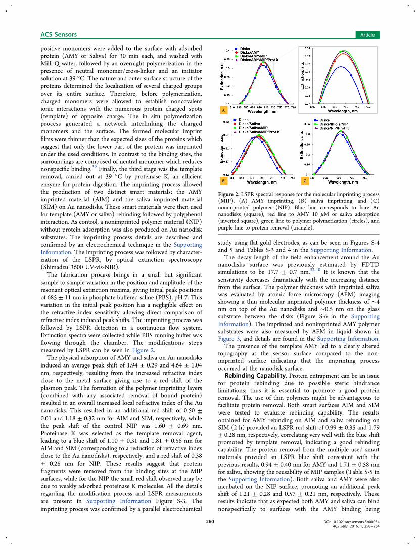
positive monomers were added to the surface with adsorbedprotein (AMY or Saliva) for 30 min each, and washed withMilli-Q water, followed by an overnight polymerization in thepresence of neutral monomer/cross-linker and an initiatorsolution at 39 °C. The nature and outer surface structure of theproteins determined the localization of several charged groupsover its entire surface. Therefore, before polymerization,charged monomers were allowed to establish noncovalentionic interactions with the numerous protein charged spots(template) of opposite charge. The in situ polymerizationprocess generated a network interlinking the chargedmonomers and the surface. The formed molecular imprintfilms were thinner than the expected sizes of the proteins whichsuggest that only the lower part of the protein was imprintedunder the used conditions. In contrast to the binding sites, thesurroundings are composed of neutral monomer which reducesnonspecific binding.39 Finally, the third stage was the templateremoval, carried out at 39 °C by proteinase K, an efficientenzyme for protein digestion. The imprinting process allowedthe production of two distinct smart materials: the AMYimprinted material (AIM) and the saliva imprinted material(SIM) on Au nanodisks. These smart materials were then usedfor template (AMY or saliva) rebinding followed by polyphenolinteraction. As control, a nonimprinted polymer material (NIP)without protein adsorption was also produced on Au nanodisksubstrates. The imprinting process details are described andconfirmed by an electrochemical technique in the SupportingInformation. The imprinting process was followed by character-ization of the LSPR, by optical extinction spectroscopy(Shimadzu 3600 UV-vis-NIR).The fabrication process brings in a small but significant
sample to sample variation in the position and amplitude of theresonant optical extinction maxima, giving initial peak positionsof 685 ± 11 nm in phosphate buffered saline (PBS), pH 7. Thisvariation in the initial peak position has a negligible effect onthe refractive index sensitivity allowing direct comparison ofrefractive index induced peak shifts. The imprinting process wasfollowed by LSPR detection in a continuous flow system.Extinction spectra were collected while PBS running buffer wasflowing through the chamber. The modifications stepsmeasured by LSPR can be seen in Figure 2.The physical adsorption of AMY and saliva on Au nanodisks
induced an average peak shift of 1.94 ± 0.29 and 4.64 ± 1.04nm, respectively, resulting from the increased refractive indexclose to the metal surface giving rise to a red shift of theplasmon peak. The formation of the polymer imprinting layers(combined with any associated removal of bound protein)resulted in an overall increased local refractive index of the Aunanodisks. This resulted in an additional red shift of 0.50 ±0.01 and 1.18 ± 0.32 nm for AIM and SIM, respectively, whilethe peak shift of the control NIP was 1.60 ± 0.69 nm.Proteinase K was selected as the template removal agent,leading to a blue shift of 1.10 ± 0.31 and 1.81 ± 0.58 nm forAIM and SIM (corresponding to a reduction of refractive indexclose to the Au nanodisks), respectively, and a red shift of 0.38± 0.25 nm for NIP. These results suggest that proteinfragments were removed from the binding sites at the MIPsurfaces, while for the NIP the small red shift observed may bedue to weakly adsorbed proteinase K molecules. All the detailsregarding the modification process and LSPR measurementsare present in Supporting Information Figure S-3. Theimprinting process was confirmed by a parallel electrochemical
study using flat gold electrodes, as can be seen in Figures S-4and 5 and Tables S-3 and 4 in the Supporting Information.The decay length of the field enhancement around the Au
nanodisks surface was previously estimated by FDTDsimulations to be 17.7 ± 0.7 nm.32,40 It is known that thesensitivity decreases dramatically with the increasing distancefrom the surface. The polymer thickness with imprinted salivawas evaluated by atomic force microscopy (AFM) imagingshowing a thin molecular imprinted polymer thickness of ∼4nm on top of the Au nanodisks and ∼0.5 nm on the glasssubstrate between the disks (Figure S-6 in the SupportingInformation). The imprinted and nonimprinted AMY polymersubstrates were also measured by AFM in liquid shown inFigure 3, and details are found in the Supporting Information.The presence of the template AMY led to a clearly altered
topography at the sensor surface compared to the non-imprinted surface indicating that the imprinting processoccurred at the nanodisk surface.
Rebinding Capability. Protein entrapment can be an issuefor protein rebinding due to possible steric hindrancelimitations; thus it is essential to promote a good proteinremoval. The use of thin polymers might be advantageous tofacilitate protein removal. Both smart surfaces AIM and SIMwere tested to evaluate rebinding capability. The resultsobtained for AMY rebinding on AIM and saliva rebinding onSIM (2 h) provided an LSPR red shift of 0.99 ± 0.35 and 1.79± 0.28 nm, respectively, correlating very well with the blue shiftpromoted by template removal, indicating a good rebindingcapability. The protein removal from the multiple used smartmaterials provided an LSPR blue shift consistent with theprevious results, 0.94 ± 0.40 nm for AMY and 1.71 ± 0.58 nmfor saliva, showing the reusability of MIP samples (Table S-5 inthe Supporting Information). Both saliva and AMY were alsoincubated on the NIP surface, promoting an additional peakshift of 1.21 ± 0.28 and 0.57 ± 0.21 nm, respectively. Theseresults indicate that as expected both AMY and saliva can bindnonspecifically to surfaces with the AMY binding being
Figure 2. LSPR spectral response for the molecular imprinting process(MIP). (A) AMY imprinting, (B) saliva imprinting, and (C)nonimprinted polymer (NIP). Blue line corresponds to bare Aunanodisks (square), red line to AMY 10 μM or saliva adsorption(inverted square), green line to polymer polymerization (circles), andpurple line to protein removal (triangle).
ACS Sensors Article
DOI: 10.1021/acssensors.5b00054ACS Sens. 2016, 1, 258−264
260

comparable to that of the AMY MIP. The saliva imprintedsurface provides more robust and higher specific responses. Thecapture of target proteins from saliva as achieved through themolecular imprinting of Au disks, with the salivary proteinsbeing immobilized and available for use as a sensor element.FDTD Calculations. Finite-difference time-domain
(FDTD) calculations of the Au nanodisks optical responsewere performed by matching to the experimental extinctionspectra and determined sensitivities, plotted in Figure 4.Experimental and FDTD spectra present similar peak shapesand positions although relative intensity is slightly different.Additionally, both spectra with peaks resulting from a dipoleresonance are in good agreement, Figure 4A and B. FDTDsimulations matched, as a starting point, the Au nanodisksdimensions of 100 nm diameter by 22 nm height (20 nm Auwith 2 nm Ti adhesion layer) and the correspondingexperimental extinction peak. The Au nanodisk modificationled to an LSPR peak shift which is directly correlated withexperimental bulk refractive index changes. The sensitivity torefractive index changes in thin films was measured by variationof the films refractive index in the FDTD model and assuming amaximum thickness of 7/14 nm for the AMY and saliva films,respectively. The obtained calibration curves for 7 and 14 nmfilms are plotted in Figure 4C and D. Measurements by SPR onhomogeneous surfaces indicated a surface coverage of AMY
consistent with a side on configuration of AMY with maximumfootprint (AMY size is indicated to be 11.5 × 11.9 × 7.0 nm3)which points to an AMY thickness of 7 nm. Salivary proteinsare known to form thicker layers,41 and here we assumed thatthe layer thickness was 14 nm, roughly determined based onthe protein composition of saliva and their three-dimensionalsize. The final refractive index of the saliva layer usingcalibration from FDTD simulations, assuming a 14 nm layerthickness, was 1.38 which fits well with the refractive indicesmeasured on adsorbed saliva films.41
FDTD also provided additional information about the spatialconfinement of the plasmon induced near−field, whichindicates that highest values of the near-field enhancement ofthe incoming optical field are localized around the upper andlower rims of the Au nanodisk, as can be seen in Figure 4E. Theoutcome of FDTD calculations allows the quantification of therebinding of protein and the amount of interacting poly-phenols.
Binding Affinities. In this work, we carry out proof ofprinciple studies of the interaction of different polyphenolcompounds with the complex saliva MIP. Polyphenols fromdifferent classes, flavonoid and nonflavonoid, were tested.Three polyphenols were selected for the binding affinitystudies: PGG from the nonflavonoid class consisting of aglucose molecule esterified with five gallic acid and twodifferent structural catechins; (+)-catechin and procyanidindimer B3 [(+)-catechin-(4−8)-(+)-catechin].
Figure 3. AFM images in liquid of (a) Au nanodisks (control), (b)nonimprinted material, and (c) amylase imprinted on Au nanodiskssubstrate.
Figure 4. Experimental (A) and FDTD simulated (B) extinctionspectra of Au nanodisks substrates immersed in different refractiveindex solutions. Correlation peak position vs refractive index unitsthrough FDTD for 7 nm (C) and 14 nm (D) layer thickness. (E) Fieldenhancement (E2 scale 0−150) distribution image of the Aunanodisks. (F) Two-dimensional cross section field map.
ACS Sensors Article
DOI: 10.1021/acssensors.5b00054ACS Sens. 2016, 1, 258−264
261

Each polyphenol was injected in the flow system interactingwith the protein content on both AIM and SIM surfaces, whilecontrol NIP and AIM/SIM without protein rebinding were alsoevaluated. Generally, the imprinted materials provided betterresponse than nonimprinted materials, Figure S-7, SupportingInformation. Additionally, the obtained signals when usingsaliva are significantly higher than those for the tests with AMY.The imprinted material with no rebinding was used as a controlproviding the lower signal (Table S-6 and Figure S-7 in theSupporting Information). Binding characteristics were meas-ured by repeated injection of increasing concentrations of PGGranging 0.1−955 μM, catechin 160−56 500 μM, and B3 100−57 000 μM, all shown in Figure 5.
When PGG, catechin or B3 were injected in the system, theyinteracted with surface bound protein and gave red shifts of theplasmon peak that remained after rinsing indicating a stronginteraction. The obtained shifts indicate a linear dependencewith log Polyphenol concentration (LSPR λmax shift vs logconcentration). Though, for all polyphenols, the linear responsewas only achieved for a smaller region within the testedconcentration range.The signal from PGG binding to the LSPR sensor surface
(Figure S-7, Supporting Information) showed a consistentlyhigher binding at MIP surfaces compared to NIP. Thedifference was much larger for saliva surfaces compared toAMY surfaces, which correlated to an overall lower proteinbinding of the AMY. The variation in the measurements for theNIP surfaces was substantially larger than MIP surfaces thatmay result from weaker or more variable protein-NIP surfaceinteractions. Binding of polyphenols directly to the polymersurface in the absence of protein, showed a low but significantlevel. When protein is attached to the surface (in particular forthe saliva surfaces) it likely reduces this background bindingboth competing for PGG adsorption and blocking the surface.We correlate the PGG binding to the amount of protein inorder to estimate the binding characteristics. The quantificationfor the AMY assumed a 7 nm layer while that of the saliva useda 14 nm layer. It should be noted that the ratio of the amountof polyphenol to protein is unaffected by the assumedthickness. In Figure 5A, the ratio of polyphenol mass/proteinmass is plotted versus polyphenol concentration (log scale).This mass ratio can be indirectly corrected with the peak shiftthrough the refractive index increment which is higher by afactor of 3 for PGG (0.532 cm2/g experimentally measuredvalue) in comparison with the protein (0.180 cm2/g literaturevalue42). PGG binding to saliva is higher than binding to AMY(∼2 times higher). We cannot rule out a contribution of PGGbinding directly to the MIP surface; however, the PGG bindingto AMY corresponds to around 20−25 PGGs/protein for thehighest concentration which is comparable to that previouslyobserved for PGG binding to AMY covalently attached tosurfaces.32 The stronger PGG attachment to the complexprotein mixture of saliva (we observe more than 36 proteins atthe MIP layer by mass spectrometry including AMY; see TableS-7 in the Supporting Information) indicates that otherproteins within the saliva are significantly stronger binders forPGG than AMY, suggesting that sensors based on specificsingle proteins (even those which are the most abundant) riskwrongly evaluating the global response. The binding profiles forPGG showed a linear regime (in the log scale) at higherconcentrations but with significant deviations toward higherbinding at low concentrations suggesting that there are rangesof binding strength sites at the proteins (which has beenobserved before for PGG binding to AMY). The overallamount of PGG molecules binding reaches a very high level athigher concentrations at the saliva. While we cannot calculatethe number of PGG molecules binding to each protein in salivawe estimate the binding to an AMY sized protein would onaverage be ∼40 which indicates that protein unfolding revealsadditional binding sites or that PGG cluster growth occurs atalready bound PGG molecules.We applied our sensing approach to study the differences
between polyphenol interaction with proteins. The combina-tion of ultrathin MIP layer with LSPR allowed us to evaluatethe binding of two small polyphenols (the monomeric basicunit of condensed tannins, (+)-catechin MW 290.3, and a
Figure 5. Ratio of polyphenols mass/protein mass for (A) PGG, (B)B3, and (C) catechin. AMY (triangles) and saliva (circles).
ACS Sensors Article
DOI: 10.1021/acssensors.5b00054ACS Sens. 2016, 1, 258−264
262

catechin dimer, the procyanin-B3 MW 2 × 290.3) and the PGG(MW 940.7), with both AMY and saliva.The LSPR signals for B3 (Figure S-7B, Supporting
Information) and catechin (Figure S-7C, Supporting Informa-tion) were significantly lower compared to that for the PGG asmight be expected from their lower molecular weight; however,significantly larger concentrations of the polyphenols wererequired to induce binding, indicating a concomitant weakerinteraction (see Figure 5). Binding to the polymer surface inthe absence of the protein was also significantly lower than forPGG. Interestingly, the two smaller polyphenols studiedshowed different profiles of binding to AMY versus saliva. B3binding to AMY was stronger (∼factor of 2) than to thecomplex saliva protein mixture and higher binding tendencywith the increase of B3 concentration. While for catechin thebinding to AMY is higher than to the saliva complex but withsimilar binding tendency with increased catechin concentration,also suggesting a stronger interaction. This fact could indicatethat AMY has a limited number of higher binding sites whilethe complex of saliva proteins has some proteins withsubstantial numbers of low binding strength sites. The signallevels observed for the catechin binding with its low molecularweight to AMY were similar to those for the maximal bindingobserved at nonprotein covered surface (albeit at higherconcentrations of catechin) and with significant errorsindicating that the catechin binding to AMY is likely close tothe detection limit for our system.Overall, we observed a significant variation in the binding
strength of polyphenols to the complex mixture of salivaproteins compared to binding to one specific protein (AMY).Amylase is shown to bind PGG weakly relative to saliva but tobind B3 and catechin stronger. This demonstrates that forstudying global effects of different small molecule binding onproteins, a complex saliva protein mixture has the potential togive a better response than sensors based on single or a fewproteins. For polyphenol binding to the complex saliva mixture,we observe a strong variation in binding of specific polyphenols.The effect of a complex food matrix on the protein andpolyphenol interaction was also tested showing someinterference, although the signal was affected in a verysystematic way indicating that for the analysis of complexfood matrices a careful choice of an appropriated matrix forcalibration needs to be made (Figure S-8, SupportingInformation). In the field of food science, this provides amethod and proof of principle to study quantitatively both theimportance of different polyphenols in astringency but also tounderstand and correct for the astringency response fromspecific wines and base ingredients during production. Inparticular, these approaches may be used to study thebioavailability of polyphenols from food and beverages afterpassing through the oral environment. A similar concept forstudying global binding response of drug molecules to thecomplex protein environment of tumors or intracellularenvironment could be applied to estimate retention or localbioavailability of drug molecules in therapeutic situations on thesite of action.The presented results indicate that all three polyphenols
showed large affinity with proteins and may interact withmultiple binding sites. The binding affinity provides informa-tion about barriers to polyphenol transport through interactionwith complex protein matrices before its absorption by theorganism and consequently about their bioavailability. Accord-ing to these results it seems that PGG will be the less available
polyphenol after passing through the mouth while catechin willbe the most available. Catechin is a monomer with lowmolecular size which may favor its absorption by the intestinelumen.
■ CONCLUSIONSHere we have developed a plasmonic biosensor to study globalinteractions between small molecules and complex proteinmatrices. We have made use of smart MIP layers which areultrathin, combining with the high localization of the opticalfields around the plasmonic sensor and imprint complexprotein matrices to allow the capture and study of saliva and itsinteraction with polyphenols. The sensor would allow the studyof bioavailability of small molecules in food or drug deliveryapplications associated with a broad range of disordersincluding neurodegenerative diseases, cardiovascular disease,and cancer.
■ ASSOCIATED CONTENT*S Supporting InformationThe Supporting Information is available free of charge on theACS Publications website at DOI: 10.1021/acssen-sors.5b00054.
Materials and methods including saliva characterization,Au nanodisks fabrication, molecular imprinted synthesis,and evaluation, interaction measurements by LSPR,electrochemical measurements, as well as description ofdata analysis; bare Au disks MIP and nonimprinted layercharacterized by atomic force microscopy; mass spec-troscopy of both pure saliva and Au disks/MIP saliva/rebinding saliva (PDF)
■ AUTHOR INFORMATIONCorresponding Author*E-mail: [email protected] ContributionsThe manuscript was written through contributions of allauthors. All authors have given approval to the final version ofthe manuscript.NotesThe authors declare no competing financial interest.
■ ACKNOWLEDGMENTSThe authors acknowledge FCT, Fundacao para a Ciencia eTecnologia, for the financial support (SFRH/BD/72479/2010), the FSE, Fundo Social Europeu for the cofinancialsupport. Vladimir Bochenkov acknowledges the support fromRFBR Grant #15-03-99582.
■ REFERENCES(1) Ehrnhoefer, D. E.; Bieschke, J.; Boeddrich, A.; Herbst, M.;Masino, L.; Lurz, R.; Engemann, S.; Pastore, A.; Wanker, E. E. EGCGredirects amyloidogenic polypeptides into unstructured, off-pathwayoligomers. Nat. Struct. Mol. Biol. 2008, 15, 558−566.(2) Ono, K.; Yoshiike, Y.; Takashima, A.; Hasegawa, K.; Naiki, H.;Yamada, M. Potent anti-amyloidogenic and fibril-destabilizing effectsof polyphenols in vitro: implications for the prevention andtherapeutics of Alzheimer’s disease. J. Neurochem. 2003, 87, 172−181.(3) Porat, Y.; Abramowitz, A.; Gazit, E. Inhibition of amyloid fibrilformation by polyphenols: Structural similarity and aromaticinteractions as a common inhibition mechanism. Chem. Biol. DrugDes. 2006, 67, 27−37.
ACS Sensors Article
DOI: 10.1021/acssensors.5b00054ACS Sens. 2016, 1, 258−264
263

(4) Corder, R.; Mullen, W.; Khan, N. Q.; Marks, S. C.; Wood, E. G.;Carrier, M. J.; Crozier, A. Red wine procyanidins and vascular health.Nature 2006, 444, 566−566.(5) Corder, R.; Douthwaite, J. A.; Lees, D. M.; Khan, N. Q.; Viseudos Santos, A. C.; Wood, E. G.; Carrier, M. J. Endothelin-1 synthesisreduced by red wine - Red wines confer extra benefit when it comes topreventing coronary heart disease. Nature 2001, 414, 863−864.(6) Fito, M.; Cladellas, M.; de la Torre, R.; Marti, J.; Munoz, D.;Schroder, H.; Alcantara, M.; Pujadas-Bastardes, M.; Marrugat, J.;Lopez-Sabater, M. C.; Bruguera, J.; Covas, M. I. Anti-inflammatoryeffect of virgin olive oil in stable coronary disease patients: arandomized, crossover, controlled trial. Eur. J. Clin. Nutr. 2008, 62,570−574.(7) Gescher, A.; Pastorino, U.; Plummer, S. M.; Manson, M. M.Suppression of tumour development by substances derived from thediet - mechanisms and clinical implications. Br. J. Clin. Pharmacol.1998, 45, 1−12.(8) Middleton, E.; Kandaswami, C.; Theoharides, T. C. The effects ofplant flavonoids on mammalian cells: Implications for inflammation,heart disease, and cancer. Pharmacol. Rev. 2000, 52, 673−751.(9) Aggarwal, B. B.; Shishodia, S. Molecular targets of dietary agentsfor prevention and therapy of cancer. Biochem. Pharmacol. 2006, 71,1397−1421.(10) Daglia, M. Polyphenols as antimicrobial agents. Curr. Opin.Biotechnol. 2012, 23, 174−181.(11) Ferrazzano, G. F.; Amato, I.; Ingenito, A.; Zarrelli, A.; Pinto, G.;Pollio, A. Plant Polyphenols and Their Anti-Cariogenic Properties: AReview. Molecules 2011, 16, 1486−1507.(12) Link, A.; Balaguer, F.; Goel, A. Cancer chemoprevention bydietary polyphenols: Promising role for epigenetics. Biochem.Pharmacol. 2010, 80, 1771−1792.(13) Izzotti, A.; Cartiglia, C.; Steele, V. E.; De Flora, S. MicroRNAsas targets for dietary and pharmacological inhibitors of mutagenesisand carcinogenesis. Mutat. Res., Rev. Mutat. Res. 2012, 751, 287−303.(14) Li, Y. Y.; Zhang, T. Targeting cancer stem cells by curcumin andclinical applications. Cancer Lett. 2014, 346, 197−205.(15) Santangelo, C.; Vari, R.; Scazzocchio, B.; Di Benedetto, R.;Filesi, C.; Masella, R. Polyphenols, intracellular signalling andinflammation. Ann. Ist. Super. Sanita 2007, 43, 394−405.(16) Albini, A.; Tosetti, F.; Li, V. W.; Noonan, D. M.; Li, W. W.Cancer prevention by targeting angiogenesis. Nat. Rev. Clin. Oncol.2012, 9, 498−509.(17) Potapovich, A. I.; Lulli, D.; Fidanza, P.; Kostyuk, V. A.; De Luca,C.; Pastore, S.; Korkina, L. G. Plant polyphenols differentiallymodulate inflammatory responses of human keratinocytes byinterfering with activation of transcription factors NF kappa B andAhR and EGFR-ERK pathway. Toxicol. Appl. Pharmacol. 2011, 255,138−149.(18) Sinclair, D. A.; Guarente, L. Small-Molecule Allosteric Activatorsof Sirtuins. In Annual Review of Pharmacology and Toxicology, Vol 54;Insel, P. A., Ed.; Annual Reviews: Palo Alto, CA, 2014; Vol. 54, pp363−380.(19) Surh, Y. J. Cancer chemoprevention with dietary phytochem-icals. Nat. Rev. Cancer 2003, 3, 768−780.(20) Ramos, S. Cancer chemoprevention and chemotherapy: Dietarypolyphenols and signalling pathways. Mol. Nutr. Food Res. 2008, 52,507−526.(21) Seeram, N. P.; Zhang, Y. J.; Nair, M. G. Inhibition ofproliferation of human cancer cells and cyclooxygenase enzymes byanthocyanidins and catechins. Nutr. Cancer 2003, 46, 101−106.(22) Xagorari, A.; Roussos, C.; Papapetropoulos, A. Inhibition ofLPS-stimulated pathways in macrophages by the flavonoid luteolin. Br.J. Pharmacol. 2002, 136, 1058−1064.(23) Im, H.; Bantz, K. C.; Lee, S. H.; Johnson, T. W.; Haynes, C. L.;Oh, S.-H. Self-Assembled Plasmonic Nanoring Cavity Arrays for SERSand LSPR Biosensing. Adv. Mater. 2013, 25, 2678−2685.(24) Truong, P. L.; Kim, B. W.; Sim, S. J. Rational aspect ratio andsuitable antibody coverage of gold nanorod for ultra-sensitivedetection of a cancer biomarker. Lab Chip 2012, 12, 1102−1109.
(25) Alivisatos, A. P.; Johnsson, K. P.; Peng, X. G.; Wilson, T. E.;Loweth, C. J.; Bruchez, M. P.; Schultz, P. G. Organization of’nanocrystal molecules’ using DNA. Nature 1996, 382, 609−611.(26) Wu, B.; Chen, L. C.; Huang, Y. J.; Zhang, Y. M.; Kang, Y. J.;Kim, D. H. Multiplexed Biomolecular Detection Based on SingleNanoparticles Immobilized on Pneumatically Controlled MicrofluidicChip. Plasmonics 2014, 9, 801−807.(27) Balamurugan, S.; Mayer, K. M.; Lee, S.; Soper, S. A.; Hafner, J.H.; Spivak, D. A. Nanostructure shape effects on response ofplasmonic aptamer sensors. J. Mol. Recognit. 2013, 26, 402−407.(28) Bhagawati, M.; You, C. J.; Piehler, J. Quantitative Real-TimeImaging of Protein-Protein Interactions by LSPR Detection withMicropatterned Gold Nanoparticles. Anal. Chem. 2013, 85, 9564−9571.(29) Cohavi, O.; Reichmann, D.; Abramovich, R.; Tesler, A. B.;Bellapadrona, G.; Kokh, D. B.; Wade, R. C.; Vaskevich, A.; Rubinstein,I.; Schreiber, G. A Quantitative, Real-Time Assessment of Binding ofPeptides and Proteins to Gold Surfaces. Chem. - Eur. J. 2011, 17,1327−1336.(30) Chen, S.; Svedendahl, M.; Van Duyne, R. P.; Kall, M. Plasmon-Enhanced Colorimetric ELISA with Single Molecule Sensitivity. NanoLett. 2011, 11, 1826−1830.(31) Zhang, J.; Wang, L.; Pan, D.; Song, S.; Boey, F. Y. C.; Zhang, H.;Fan, C. Visual cocaine detection with gold nanoparticles and rationallyengineered aptamer structures. Small 2008, 4, 1196−1200.(32) Guerreiro, J. R. L.; Frederiksen, M.; Bochenkov, V. E.; DeFreitas, V.; Ferreira Sales, M. G.; Sutherland, D. S. MultifunctionalBiosensor Based on Localized Surface Plasmon Resonance forMonitoring Small Molecule-Protein Interaction. ACS Nano 2014, 8,7958−7967.(33) Chianella, I.; Guerreiro, A.; Moczko, E.; Caygill, J. S.; Piletska, E.V.; De Vargas Sansalvador, I. M.; Whitcombe, M. J.; Piletsky, S. A.Direct Replacement of Antibodies with Molecularly ImprintedPolymer Nanoparticles in ELISA-Development of a Novel Assay forVancomycin. Anal. Chem. 2013, 85, 8462−8468.(34) Abbas, A.; Tian, L. M.; Morrissey, J. J.; Kharasch, E. D.;Singamaneni, S. Hot Spot-Localized Artificial Antibodies for Label-Free Plasmonic Biosensing. Adv. Funct. Mater. 2013, 23, 1789−1797.(35) Fredriksson, H.; Alaverdyan, Y.; Dmitriev, A.; Langhammer, C.;Sutherland, D. S.; Zaech, M.; Kasemo, B. Hole-mask colloidallithography. Adv. Mater. 2007, 19, 4297−4302.(36) Whitcombe, M. J.; Chianella, I.; Larcombe, L.; Piletsky, S. A.;Noble, J.; Porter, R.; Horgan, A. The rational development ofmolecularly imprinted polymer-based sensors for protein detection.Chem. Soc. Rev. 2011, 40, 1547−1571.(37) Li, S.; Cao, S.; Whitcombe, M. J.; Piletsky, S. A. Size matters:Challenges in imprinting macromolecules. Prog. Polym. Sci. 2014, 39,145−163.(38) Hinrichsen, E. L.; Feder, J.; Jossang, T. Geometry of randomsequential adsorption. J. Stat. Phys. 1986, 44, 793−827.(39) Moreira, F. T. C.; Sharma, S.; Dutra, R. A. F.; Noronha, J. P. C.;Cass, A. E. G.; Sales, M. G. F. Smart plastic antibody material (SPAM)tailored on disposable screen printed electrodes for proteinrecognition: Application to myoglobin detection. Biosens. Bioelectron.2013, 45, 237−244.(40) Mazzotta, F.; Johnson, T. W.; Dahlin, A. B.; Shaver, J.; Oh, S.-H.; Hook, F. Influence of the evanescent field decay length on thesensitivity of plasmonic nanodisks and nanoholes. ACS Photonics 2015,2, 256−262.(41) Cardenas, M.; Arnebrant, T.; Rennie, A.; Fragneto, G.; Thomas,R. K.; Lindh, L. Human saliva forms a complex film structure onalumina surfaces. Biomacromolecules 2007, 8, 65−69.(42) Hook, F.; Voros, J.; Rodahl, M.; Kurrat, R.; Boni, P.; Ramsden, J.J.; Textor, M.; Spencer, N. D.; Tengvall, P.; Gold, J.; Kasemo, B. Acomparative study of protein adsorption on titanium oxide surfacesusing in situ ellipsometry, optical waveguide lightmode spectroscopy,and quartz crystal microbalance/dissipation. Colloids Surf., B 2002, 24,155−170.
ACS Sensors Article
DOI: 10.1021/acssensors.5b00054ACS Sens. 2016, 1, 258−264
264