Embed Size (px)
Citation preview

C H A P T E R
1
Molecular Biology for
Computer Scientists
Lawrence Hunter
“Computers are to biology what mathematics is to physics.”
— Harold Morowitz
One of the major challenges for computer scientists who wish to work in thedomain of molecular biology is becoming conversant with the daunting intri-cacies of existing biological knowledge and its extensive technical vocabu-lary. Questions about the origin, function, and structure of living systemshave been pursued by nearly all cultures throughout history, and the work ofthe last two generations has been particularly fruitful. The knowledge of liv-ing systems resulting from this research is far too detailed and complex forany one human to comprehend. An entire scientific career can be based in thestudy of a single biomolecule. Nevertheless, in the following pages, I attemptto provide enough background for a computer scientist to understand muchof the biology discussed in this book. This chapter provides the briefest ofoverviews; I can only begin to convey the depth, variety, complexity andstunning beauty of the universe of living things.
Much of what follows is not about molecular biology per se. In order to

explain what the molecules are doing, it is often necessary to use conceptsinvolving, for example, cells, embryological development, or evolution. Bi-ology is frustratingly holistic. Events at one level can effect and be affectedby events at very different levels of scale or time. Digesting a survey of thebasic background material is a prerequisite for understanding the significanceof the molecular biology that is described elsewhere in the book. In life, as incognition, context is very important.
Do keep one rule in the back of your mind as you read this: for every gen-eralization I make about biology, there may well be thousands of exceptions.There are a lot of living things in the world, and precious few generalizationshold true for all of them. I will try to cover the principles; try to keep the ex-istence of exceptions in mind as you read. Another thing to remember is thatan important part of understanding biology is learning its language. Biolo-gists, like many scientists, use technical terms in order to be precise aboutreference. Getting a grasp on this terminology makes a great deal of the bio-logical literature accessible to the non-specialist. The notes contain informa-tion about terminology and other basic matters. With that, let’s begin at thebeginning.
1. What Is Life?
No simple definition of what it is to be a living thing captures our intuitionsabout what is alive and what is not. The central feature of life is its ability toreproduce itself. Reproductive ability alone is not enough; computer pro-grams can create endless copies of themselves—that does not make themalive. Crystals influence the matter around them to create structures similarto themselves but they’re not alive, either. Most living things take in materi-als from their environment and capture forms of energy they can use to trans-form those materials into components of themselves or their offspring. Virus-es, however, do not do that; they are nearly pure genetic material, wrapped ina protective coating. The cell that a virus infects does all the synthetic workinvolved in creating new viruses. Are viruses a form of life? Many peoplewould say so.
Another approach to defining “life” is to recognize its fundamental inter-relatedness. All living things are related to each other. Any pair of organisms,no matter how different, have a common ancestor sometime in the distantpast. Organisms came to differ from each other, and to reach modern levelsof complexity through evolution. Evolution has three components: inheri-tance, the passing of characteristics from parents to offspring; variation, theprocesses that make offspring other than exact copies of their parents; andselection, the process that differentially favors the reproduction of some or-ganisms, and hence their characteristics, over others. These three factorsdefine an evolutionary process. Perhaps the best definition of life is that it is
2 ARTIFICIAL INTELLIGENCE & MOLECULAR BIOLOGY
the result of the evolutionary process taking place on Earth. Evolution is thekey not only to defining what counts as life but also to understanding howliving systems function.
Evolution is a cumulative process. Inheritance is the determinant of al-most all of the structure and function of organisms; the amount of variationfrom one generation to the next is quite small. Some aspects of organisms,such as the molecules that carry energy or genetic information, have changedvery little since that original common ancestor several billion of years ago.Inheritance alone, however, is not sufficient for evolution to occur; perfectinheritance would lead to populations of entirely identical organisms, all ex-actly like the first one.
In order to evolve, there must be a source of variation in the inheritance.In biology, there are several sources of variation. Mutation, or randomchanges in inherited material, is only one source of change; sexual recombi-nation and various other kinds of genetic rearrangements also lead to varia-tions; even viruses can get into the act, leaving a permanent trace in thegenes of their hosts. All of these sources of variation modify the messagethat is passed from parent to offspring; in effect, exploring a very large spaceof possible characteristics. It is an evolutionary truism that almost all varia-tions are neutral or deleterious. As computer programmers well know, smallchanges in a complex system often lead to far-reaching and destructive con-sequences (And computer programmers make those small changes by design,and with the hope of improving the code!). However, given enough time, thesearch of that space has produced many viable organisms.
Living things have managed to adapt to a breathtaking array of chal-lenges, and continue to thrive. Selection is the process by which it is deter-mined which variants will persist, and therefore also which parts of the spaceof possible variations will be explored. Natural selection is based on the re-productive fitness of each individual. Reproductive fitness is a measure ofhow many surviving offspring an organism can produce; the better adaptedan organism is to its environment, the more successful offspring it will cre-ate. Because of competition for limited resources, only organisms with highfitness will survive; organisms less well adapted to their environment thancompeting organisms will simply die out.
I have likened evolution to a search through a very large space of possibleorganism characteristics. That space can be defined quite precisely. All of anorganism’s inherited characteristics are contained in a single messenger mol-ecule: deoxyribonucleic acid, or DNA. The characteristics are represented ina simple, linear, four-element code. The translation of this code into all theinherited characteristics of an organism (e.g. its body plan, or the wiring ofits nervous system) is complex. The particular genetic encoding for an organ-ism is called its genotype. The resulting physical characteristics of an organ-ism is called its phenotype. In the search space metaphor, every point in the
HUNTER 3

space is a genotype. Evolutionary variation (such as mutation, sexual recom-bination and genetic rearrangements) identifies the legal moves in this space.Selection is an evaluation function that determines how many other points apoint can generate, and how long each point persists. The difference betweengenotype and phenotype is important because allowable (i.e. small) steps ingenotype space can have large consequences in phenotype space. It is alsoworth noting that search happens in genotype space, but selection occurs onphenotypes. Although it is hard to characterize the size of phenotype space,an organism with a large amount of genetic material (like, e.g., that of theflower Lily) has about 1011 elements taken from a four letter alphabet, mean-ing that there are roughly 1070,000,000,000 possible genotypes of that size orless. A vast space indeed! Moves (reproductive events) occur asynchronous-ly, both with each other and with the selection process. There are many non-deterministic elements; for example, in which of many possible moves istaken, or in the application of the selection function. Imagine this searchprocess running for billions of iterations, examining trillions of points in thisspace in parallel at each iteration. Perhaps it is not such a surprise that evolu-tion is responsible for the wondrous abilities of living things, and for theirtremendous diversity.*
1.1 The Unity and the Diversity of Living Things
Life is extraordinarily varied. The differences between a tiny archebacteriumliving in a superheated sulphur vent at the bottom of the ocean and a two-tonpolar bear roaming the arctic circle span orders of magnitude in many dimen-sions. Many organisms consist of a single cell; a Sperm Whale has more than1015 cells. Although very acidic, very alkaline or very salty environments aregenerally deadly, living things can be found in all of them. Hot or cold, wet ordry, oxygen-rich or anaerobic, nearly every niche on the planet has been in-vaded by life. The diversity of approaches to gathering nutrients, detectingdanger, moving around, finding mates (or other forms of reproduction), rais-ing offspring and dozens of other activities of living creatures is truly awe-some. Although our understanding of the molecular level of life is less de-tailed, it appears that this diversity is echoed there. For example, proteins withvery similar shapes and identical functions can have radically different chemi-cal compositions. And organisms that look quite similar to each other mayhave very different genetic blueprints. All of the genetic material in an organ-ism is called its genome. Genetic material is discrete and hence has a particularsize, although the size of the genome is not directly related to the complexityof the organism. The size of genomes varies from about 5,000 elements in avery simple organism (e.g. the viruses SV40 or φx) to more than 1011 elements
4 ARTIFICIAL INTELLIGENCE & MOLECULAR BIOLOGY
*Evolution has also become an inspiration to a group of researchers interested in de-signing computer algorithms, e.g. Langton (1989).

in some higher plants; people have about 3x10⁹ elements in their genome.Despite this incredible diversity, nearly all of the same basic mechanisms
are present in all organisms. All living things are made of cells*: membrane-enclosed sacks of chemicals carrying out finely tuned sequences of reactions.The thousand or so substances that make up the basic reactions going on in-side the cell (the core metabolic pathways) are remarkably similar across allliving things. Every species has some variations, but the same basic materialsare found from bacteria to human. The genetic material that codes for all ofthese substances is written in more or less the same molecular language inevery organism. The developmental pathways for nearly all multicellular or-ganisms unfold in very similar ways. It is this underlying unity that offers thehope of developing predictive models of biological activity. It is the processof evolution that is responsible both for the diversity of living things and fortheir underlying similarities. The unity arises through inheritance from com-mon ancestors; the diversity from the power of variation and selection tosearch a vast space of possible living forms.
1.2 Prokaryotes & Eukaryotes, Yeasts & People
Non-biologists often fail to appreciate the tremendous number of differentkinds of organisms in the world. Although no one really knows, estimates ofthe number of currently extant species range from 5 million to 50 million(May, 1988).† There are at least 300,000 different kinds of beetles alone, andprobably 50,000 species of tropical trees. Familiar kinds of plants and ani-mals make up a relatively small proportion of the kinds of living things, per-haps only 20%. Vertebrates (animals with backbones: fish, reptiles, amphib-ians, birds, mammals) make up only about 3% of the species in the world.
Since Aristotle, scholars have tried to group these myriad species intomeaningful classes. This pursuit remains active, and the classifications are, tosome degree, still controversial. Traditionally, these classifications have beenbased on the morphology of organisms. Literally, morphology means shape,but it is generally taken to include internal structure as well. Morhpology isonly part of phenotype, however; other parts include physiology, or the func-tioning of living structures, and development. Structure, development andfunction all influence each other, so the dividing lines are not entirely clear.
In recent years, these traditional taxonomies have been shaken by infor-mation gained from analyzing genes directly, as well as by the discovery ofan entirely new class of organisms that live in hot, sulphurous environmentsin the deep sea.
HUNTER 5
*A virus is arguably alive, and is not a cell, but it depends on infecting a cell in orderto reproduce.
†May also notes that it is possible that half the extant species on the planet may be-come extinct in the next 50 to 100 years.
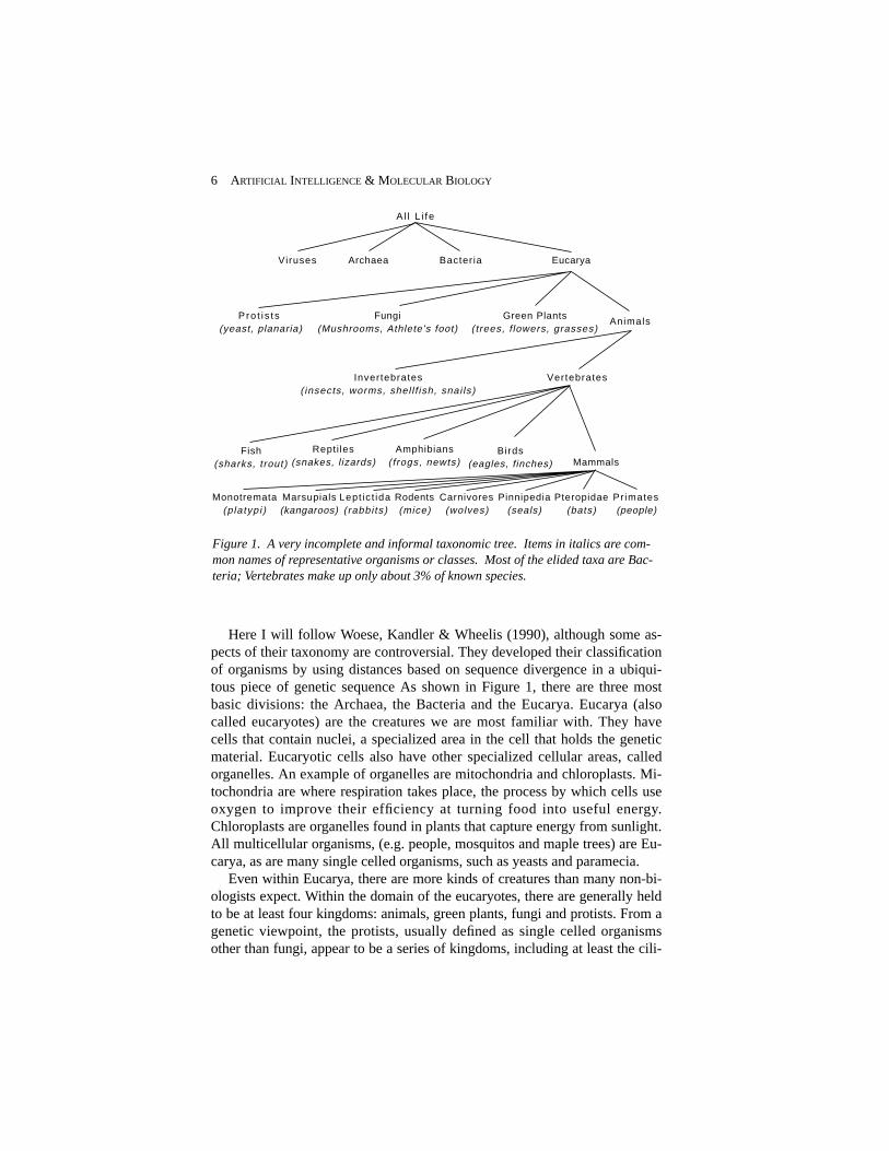
Here I will follow Woese, Kandler & Wheelis (1990), although some as-pects of their taxonomy are controversial. They developed their classificationof organisms by using distances based on sequence divergence in a ubiqui-tous piece of genetic sequence As shown in Figure 1, there are three mostbasic divisions: the Archaea, the Bacteria and the Eucarya. Eucarya (alsocalled eucaryotes) are the creatures we are most familiar with. They havecells that contain nuclei, a specialized area in the cell that holds the geneticmaterial. Eucaryotic cells also have other specialized cellular areas, calledorganelles. An example of organelles are mitochondria and chloroplasts. Mi-tochondria are where respiration takes place, the process by which cells useoxygen to improve their efficiency at turning food into useful energy.Chloroplasts are organelles found in plants that capture energy from sunlight.All multicellular organisms, (e.g. people, mosquitos and maple trees) are Eu-carya, as are many single celled organisms, such as yeasts and paramecia.
Even within Eucarya, there are more kinds of creatures than many non-bi-ologists expect. Within the domain of the eucaryotes, there are generally heldto be at least four kingdoms: animals, green plants, fungi and protists. From agenetic viewpoint, the protists, usually defined as single celled organismsother than fungi, appear to be a series of kingdoms, including at least the cili-
6 ARTIFICIAL INTELLIGENCE & MOLECULAR BIOLOGY
Al l L i fe
Archaea Bacteria Eucarya
AnimalsGreen Plants
(trees, f lowers, grasses)Fungi
(Mushrooms, Athlete's foot)Pro t i s ts
(yeast, planaria)
VertebratesInvertebrates( insects, worms, shel l f ish, snai ls)
Fish(sharks, trout)
Repti les(snakes, l izards)
Amphibians(frogs, newts)
Birds(eagles, finches) Mammals
Monotremata(platypi )
Marsupials(kangaroos)
Lept ic t ida(rabbi ts)
Rodents(mice)
Carnivores(wolves)
Pinnipedia(seals)
Pteropidae(bats)
Pr imates(people)
Viruses
Figure 1. A very incomplete and informal taxonomic tree. Items in italics are com-mon names of representative organisms or classes. Most of the elided taxa are Bac-teria; Vertebrates make up only about 3% of known species.

ates (cells with many external hairs, or cillia), the flagellates (cells with a sin-gle, long external fiber) and the microsporidia. The taxonomic tree continuesdown about a dozen levels, ending with particular species at the leaves. Allof these many eucaryotic life forms have a great deal in common with humanbeings, which is the reason we can learn so much about ourselves by study-ing them.
Bacteria (sometimes also called eubacteria, or prokaryotes) are ubiquitoussingle-celled organisms. And ubiquitous is the word; there are millions ofthem everywhere — on this page, in the air you are breathing, and in yourgut, for example. The membranes that enclose these cells are typically madeof a different kind of material than the ones that surround eucarya, and theyhave no nuclei or other organelles (they do have ribosomes, which are some-times considered organelles; see below). Almost all bacteria do is to makemore bacteria; it appears that when food is abundant, the survival of thefittest in bacteria means the survival of those that can divide the fastest (Al-berts, et al., 1989). Bacteria include not only the disease causing “germs,”but many kinds of algae, and a wide variety of symbiotic organisms, includ-ing soil bacteria that fix nitrogen for plants and Escherichia coli, a bacteriumthat lives in human intestines and is required for normal digestion. E. coli isubiquitous in laboratories because it is easy to grow and very well studied.
Archaea are a recently discovered class of organism so completely unlikeboth bacteria and eucarya, both genetically and morphologically, that theyhave upset a decades old dichotomy. Archaea live in superheated sulphurvents in the deep sea, or in hot acid springs, briney bogs and other seeminglyinhospitable places. They are sometimes called archebacteria even thoughthey bear little resemblence to bacteria. Their cell membranes are unlike ei-ther Bacteria or Eucarya. Although they have no nuclei or organelles, at a ge-netic level, they are a bit more like Eucarya than like Bacteria. These organ-isms are a relatively recent discovery, and any biological theories have yet toinclude Archaea, or consider them simply another kind of procaryote. Ar-chaea will probably have a significant effect on theories about the early his-tory of life, and their unusual biochemistry has already turned out to be sci-entifically and commercially important (e.g. see the discussion of PCR in thelast section of this chapter).
Viruses form another important category of living forms. They are obliga-tory parasites meaning that they rely on the biochemical machinery of theirhost cell to survive and reproduce. Viruses consist of just a small amount ofgenetic material surrounded by a protein coat. A small virus, such as φX,which infects bacteria, can have as few as 5000 elements in its genetic mater-ial. (Viruses that infect bactieria are called bacteriophages, or just phages.)Their simplicity and their role in human disease make viruses an active areaof study. They also play a crucial role in the technology of molecular biolo-gy, as is described in the last section in this chapter.
HUNTER 7

1.3 Evolutionary Time and Relatedness
There are so many different kinds of life, and they live in so many differentways. It is amazing that their underlying functioning is so similar. The reasonthat there is unity within all of that diversity is that all organisms appear tohave evolved from a common ancestor. This fundamental claim underpinsnearly all biological theorizing, and there is substantial evidence for it.
All evolutionary theories hold that the diversity of life arose by inheritedvariation through an unbroken line of descent. This common tree of descentis the basis for the taxonomy described above, and pervades the character ofall biological explanation. There is a great deal of argument over the detailedfunctioning of evolution (e.g. whether it happens continuously or in bursts),but practically every biologist agrees with that basic idea.
There are a variety of ways to estimate how long ago two organisms di-verged; that is, the last time they had a common ancestor. The more relatedtwo species are, the more recently they diverged. To the degree that pheno-typic similarity indicates genotypic similarity, organisms can be classified onthe basis of their structure, which is the traditional method. Growing knowl-edge of the DNA sequences of many genes in many organisms makes possi-ble estimates of the time of genetic divergence directly, by comparing theirgenetic sequences. If the rate of change can be quantified, and standards set,these differences can be translated into a “molecular clock;” Li & Graur,(1991) is a good introduction to this method. The underlying and somewhatcontroversial assumption is that in some parts of the genome, the rate of mu-tation is fairly constant. There are various methods for trying to find theseareas, estimate the rate of change, and hence calibrate the clock. The tech-nique has mostly confirmed estimates made with other methods, and is wide-ly considered to be potentially reliable, if not quite yet so. Most of the dates Iwill use below were derived from traditional (archaeological) dating.
In order to get a rough idea of the degrees of relatedness among creatures,it is helpful to know the basic timeline of life on Earth. The oldest knownfossils, stromalites found in Australia, indicate that life began at least 3.8 bil-lion years ago. Geological evidence indicates that a major meteor impactabout 4 billion years ago vaporized all of the oceans, effectively destroyingany life that may have existed before that. In effect, life on earth began al-most as soon as it could have. Early life forms probably resembled modernbacteria in some important ways. They were simple, single celled organisms,without nuclei or other organelles. Life remained like that for nearly 2 billionyears. Then, about halfway through the history of life, a radical change oc-curred: Eucarya came into being. There is evidence that eucarya began assymbiotic collections of simpler cells which were eventually assimilated andbecame organelles (see, e.g. Margolis (1981)). The advantages of these spe-cialized cellular organelles made early eucarya very successful. Single-celled
8 ARTIFICIAL INTELLIGENCE & MOLECULAR BIOLOGY

Eucarya become very complex, for example, developing mechanisms formoving around, detecting prey, paralyzing it and engulfing it.
The next major change in the history of life was the invention of sex. Evo-lution, as you recall, is a mechanism based on the inheritance of variation.Where do these variations come from? Before the advent of sex, variationsarose solely through individual, random changes in genetic material. A muta-tion might arise, changing one element in the genome, or a longer piece of agenome might be duplicated or moved. If the changed organism had an ad-vantage, the change would propagate itself through the population. Most mu-tations are neutral or deleterious, and evolutionary change by mutation is avery slow, random search of a vast space. The ability of two successful or-ganisms to combine bits of their genomes into an offspring produced variantswith a much higher probability of success. Those moves in the search spaceare more likely to produce an advantageous variation than random ones. Al-though you wouldn’t necessarily recognize it as sex when looking under amicroscope, even some Bacteria exchange genetic material. How and whensexual recombination first evolved is not clear, but it is quite ancient. Somehave argued that sexual reproduction was a necessary precursor to the devel-opment of multicellular organisms with specialized cells (Buss, 1987). Theadvent of sex dramatically changed the course of evolution. The new mecha-nism for the generation of variation focused nature’s search through thespace of possible genomes, leading to an increase in the proportion of advan-tageous variations, and an increase in the rate of evolutionary change.
This is probably a good place to correct a common misperception, namelythat some organisms are more "primitive" than others. Every existing organ-ism has, tautologically, made it into the modern era. Simple modern organ-isms are not primitive. The environment of the modern world is completelyunlike that of earth when life began, and even the simplest existing creatureshave evolved to survive in the present. It is possible to use groups of verydistantly related creatures (e.g. people and bacteria) to make inferences aboutancient organisms; whatever people and bacteria have in common are char-acteristics that were most likely shared by their last common ancestor, manyeons ago. Aspects of bacteria which are not shared with people may haveevolved as recently as any human characteristic not shared with bacteria.This applies to the relation between people and apes, too: apes are not anymore like ancestral primates than we are. It is what we have in common withother organisms that tells us what our ancestors were like; the differences be-tween us and other organisms are much less informative.
Whether or not it occurred as a result of the advent of sexual recombina-tion, the origin of multicellular organisms led to a tremendous explosion inthe kinds of organisms and in their complexity. This event occurred onlyabout a billion years ago, about three quarters of the way through the historyof life.
HUNTER 9

Of course, nearly all of the organisms people can see are multicellular (al-though the blue-green algae in ponds and swimming pools are a kind of bac-teria). Multicellular organisms gain their main evolutionary advantagethrough cellular specialization. Creatures with specialized cells have the abil-ity to occupy environmental niches that single-celled organisms cannot takeadvantage of. In multicellular organisms, cells quite distant from each othercan exchange matter, energy or information for their mutual benefit. For ex-ample, cells in the roots of a higher plant exist in a quite different environ-ment than the cells in the leaves, and each supplies the other with matter orenergy not available in the local environment.
An important difference between multicellular organisms and a colony ofunicellular organisms (e.g. coral) is that multicellular organisms have sepa-rated germ line (reproductive) cells from somatic (all the other) cells. Spermand eggs are germ cells; all the other kinds of cells in the body are somatic.Both kinds of cells divide and make new cells, but only germ cells make neworganisms. Somatic cells are usually specialized for a particular task; theyare skin cells, or nerve cells, or blood cells. Although these cells divide,when they divide, they create more of the same kind of cell. The division ofsomatic cells and single celled organisms is a four stage process that endswith mitosis, resulting in the production of two identical daughter cells. Theprocess as a whole is referred to as the cell cycle.
Only changes in germ cells are inherited from an organism to its off-spring. A variation that arises in a somatic cell will affect all of the cell’s de-scendents, but it will not affect any of the organism’s descendents. Germcells divide in a process called meiosis; part of this process is the productionof sperm and egg cells, each of which have only half the usual genetic mater-ial. The advent of this distinction involved a complex and intricate balancebetween somatic cells becoming an evolutionary deadends and the improvedcompetitive ability of a symbiotic collection of closely related cells.
Multicellular organisms all begin their lives from a single cell, a fertilizedegg. From that single cell, all of the specialized cells arise through a processcalled cellular differentiation. The process of development from fertilizedegg to full adult is extremely complex. It involves not only cellular differen-tiation, but the migration and arrangement of cells with respect to each other,orchestrated changes in which genes are used and which are not at any givenmoment, and even the programmed death of certain groups of cells that actas a kind of scaffolding during development. The transition from single-celled organism to multicellular creature required many dramatic innova-tions. It was a fundamental shift of the level of selection: away from the indi-vidual cell and to a collection of cells as a whole. The reproductive successof a single cell line within a multicellular individual may not correlate withthe success of the individual.* Embryology and development are complexand important topics, but are touched on only briefly in this chapter.
10 ARTIFICIAL INTELLIGENCE & MOLECULAR BIOLOGY

Most of the discussion so far has focused on organisms that seem verysimple and only distantly related to people. On a biochemical level, however,people are much like other eucaryotes, especially multicellular ones. Geneticand biochemical distance doesn’t always correlate very well with morpho-logical differences. For example, two rather similar looking species of frogsmay be much more genetically distant from each other than are, say, peopleand cows (Cherty, Case & Wilson, 1978). A great deal of human biochem-istry was already set by the time multicellular organisms appeared on theEarth. We can learn a lot about human biology by understanding how yeastswork.
We’ve now covered, very briefly, the diversity of living things, and someof the key events in the evolution of life up to the origin of multicellular or-ganisms. In the next section, we’ll take a closer look at how these complexorganisms work, and cover the parts of eucaryotic cells in a bit more detail.
2. Living Parts: Tissues, Cells, Compartments and Organelles
The main advantage multicellular organisms possess over their single-celledcompetitors is cell specialization. Not every cell in a larger organism has tobe able to extract nutrients, protect itself, sense the environment, move itselfaround, reproduce itself and so on. These complex tasks can be divided up,so that many different classes of cells can work together, accomplishing featsthat single cells cannot. Groups of cells specialized for a particular functionare tissues, and their cells are said to have differentiated. Differentiated cells(except reproductive cells) cannot reproduce an entire organism.
In people (and most other multicellular animals) there are fourteen majortissue types. There are many texts with illustrations and descriptions of thevarious cell types and tissue, e.g. Kessel and Kardon (1979) which is full ofbeautiful electron micrographs. Some of these tissue types are familiar:bones, muscles, cardiovascular tissue, nerves, and connective tissue (like ten-dons and ligaments). Other tissues are the constituents of the digestive, respi-ratory, urinary and reproductive systems. Skin and blood are both distinctivetissue types, made of highly specialized cells. Lymphatic tissue, such as thespleen and the lymph nodes make up the immune system. Endocrine tissuecomprises a network of hormone-producing glands (for example, the adrenalgland, source of adrenaline) that exert global control over various aspects ofthe body as a whole. Finally, epithelium, the most basic tissue type, lines allof the body’s cavities, secreting materials such as mucus, and, in the in-
HUNTER 11
*Cancer is an example where a single cell line within a multicellular organism repro-duces to the detriment of the whole.
testines, absorbing water and nutrients.There are more than 200 different specialized cell types in a typical verte-
brate. Some are large, some small; for example, a single nerve cell connectsyour foot to your spinal cord, and a drop of blood has more than 10,000 cellsin it. Some divide rapidly, others do not divide at all; bone marrow cells di-vide every few hours, and adult nerve cells can live 100 years without divid-ing. Once differentiated, a cell cannot change from one type to another. Yetdespite all of this variation, all of the cells in a multicellular organism haveexactly the same genetic code. The differences between them come from dif-ferences in gene expression, that is, whether or not a the product a genecodes for is produced, and how much is produced. Control of gene expres-sion is an elaborate dance with many participants. Thousands of biologicalsubstances bind to DNA, or bind to other biomolecules that bind to DNA.Genes code for products that turn on and off other genes, which in turn regu-late other genes, and so on. One of the key research areas in biology is devel-opment: how the intricate, densely interrelated genetic regulatory process ismanaged, and how cells "know" what to differentiate into, and when andwhere they do it. A prelude to these more complex topics is a discussion ofwhat cells are made of, and what they do.
2.1 The Composition of Cells
Despite their differences, most cells have a great deal in common with eachother. Every cell, whether a Archaea at the bottom of the ocean or a cell in ahair follicle on the top of your head has certain basic qualities: they containcytoplasm and genetic material, are enclosed in a membrane and have thebasic mechanisms for translating genetic messages into the main type of bio-logical molecule, the protein. All eucaryotic cells share additional compo-nents. Each of these basic parts of a cell is described briefly below:
Membranes are the boundaries between the cell and the outside world.Although there is no one moment that one can say life came into being, theorigin of the first cell membrane is a reasonable starting point. At that mo-ment, self-reproducing systems of molecules were individuated, and cellscame into being. All present day cells have a phospholipid cell membrane.Phospholipids are lipids (oils or fats) with a phosphate group attached. Theend with the phosphate group is hydrophillic (attracted to water) and the lipidend is hydrophobic (repelled by water). Cell membranes consist of two lay-ers of these molecules, with the hydrophobic ends facing in, and the hy-drophillic ends facing out. This keeps water and other materials from gettingthrough the membrane, except through special pores or channels.
A lot of the action in cells happens at the membrane. For single celled or-ganisms, the membrane contains molecules that sense the environment, andin some cells it can surround and engulf food, or attach and detach parts of it-self in order to move. In Bacteria and Archaea, the membrane plays a crucial
12 ARTIFICIAL INTELLIGENCE & MOLECULAR BIOLOGY

role in energy production by maintaining a large acidity difference betweenthe inside and the outside of the cell. In multicellular organisms, the mem-branes contain all sorts of signal transduction mechanisms, adhesion mole-cules, and other machinery for working together with other cells.
Proteins are the molecules that accomplish most of the functions of theliving cell. The number of different structures and functions that proteinstake on in a single organism is staggering. They make possible all of thechemical reactions in the cell by acting as enzymes that promote specificchemical reactions, which would otherwise occur only so slowly as to beotherwise negligible. The action of promoting chemical reactions is calledcatalysis, and enzymes are sometimes refered to as catalysts, which is a moregeneral term. Proteins also provide structural support, and are the keys tohow the immune system distinguishes self from invaders. They provide themechanism for acquiring and transforming energy, as well as translating itinto physical work in the muscles. They underlie sensors and the transmis-sion of information as well.
All proteins are constructed from linear sequences of smaller moleculescalled amino acids. There are twenty naturally occurring amino acids. Longproteins may contain as many as 4500 amino acids, so the space of possibleproteins is very large: 204500 or 105850. Proteins also fold up to form partic-ular three dimensional shapes, which give them their specific chemical func-tionality. Although it is easily demonstrable that the linear amino acid se-quence completely specifies the three dimensional structure of most proteins,the details of that mapping is one of the most important open questions of bi-ology. In addition a protein's three dimensional structure is not fixed; manyproteins move and flex in constrained ways, and that can have a significantrole in their biochemical function. Also, some proteins bind to other groupsof atoms that are required for them to function. These other structures arecalled prosthetic groups. An example of a prosthetic group is heme, whichbinds oxygen in the protein hemoglobin. I will discuss proteins in more de-tail again below.
Genetic material codes for all the other constituents of the the cell. Thisinformation is generally stored in long strands of DNA. In Bacteria, the DNAis generally circular. In Eucaryotes, it is linear. During cell division Eucary-otic DNA is grouped into X shaped structures called chromosomes. Someviruses (like the AIDS virus) store their genetic material in RNA. This genet-ic material contains the blueprint for all the proteins the cell can produce. I’llhave much more to say about DNA below.
Nuclei are the defining feature of Eucaryotic cells. The nucleus containsthe genetic material of the cell in the form of chromatin. Chromatin containslong stretches of DNA in a variety of conformations,* surrounded by nuclearproteins. The nucleus is separated from the rest of the cell by a nuclear mem-brane. Nuclei show up quite clearly under the light microscope; they are per-
HUNTER 13

haps the most visible feature of most cells.Cytoplasm is the name for the gel-like collection of substances inside the
cell. All cells have cytoplasm. The cytoplasm contains a wide variety of dif-ferent substances and structures. In Bacteria and Archaea, the cytoplasm con-tains all of the materials in the cell. In Eucarya, the genetic material is segre-gated into the cell nucleus.
Ribosomes are large molecular complexes, composed of several proteinsand RNA molecules. The function of ribosomes is to assemble proteins. Allcells, including Bacteria and Archaea have ribosomes. The process of trans-lating genetic information into proteins is described in detail below. Ribo-somes are where that process occurs, and are a key part of the mechanism foraccomplishing that most basic of tasks.
Mitochondria and Chroloplasts are cellular organelles involved in theproduction the energy that powers the cell. Mitochondria are found in all eu-caryotic cells, and their job is respiration: using oxygen to efficiently turnfood into energy the cell can use. Some bacteria and archaea get their energyby a process called glycolysis, from glyco- (sugar) and -lysis (cleavage or de-struction). This process creates two energy-carrying molecules for everymolecule of sugar consumed. As oxygen became more abundant†, some or-ganisms found a method for using it (called oxidative phosphorylation) tomake an order of magnitude increase in their ability to extract energy fromfood, getting 36 energy-carrying molecules for every sugar.
These originally free living organisms were engulfed by early eucaryotes.This symbiosis gradually became obligatory as eucaryotes came to dependon their mitochondria for energy, and the mitochondria came to depend onthe surrounding cell for many vital functions and materials. Mitochondriastill have their own genetic material however, and, in sexually reproducingorganisms, are inherited only via the cytoplasm of the egg cell. As a conse-quence, all mitochondria are maternally inherited.
Like the mitochondria, chloroplasts appear to have originated as free-liv-ing bacteria that eventually became obligatory symbionts, and then parts ofeucaryotic plant cells. Their task is to convert sunlight into energy-carryingmolecules.
Other Parts of Cells. There are other organelles found in eucaryotic
14 ARTIFICIAL INTELLIGENCE & MOLECULAR BIOLOGY
*Conformation means shape, connoting one of several possible shapes. DNA confor-mations include the traditional double helix, a supercoiled state where certain parts ofthe molecule are deeply hidden, a reverse coiled state called Z-DNA, and several oth-ers.
†There was very little oxygen in the early atmosphere. Oxygen is a waste product ofglycolysis, and it eventually became a significant component of the atmosphere. Al-though many modern organisms depend on oxygen to live, it is a very corrosive sub-stance, and living systems had to evolve quite complex biochemical processes fordealing with it.

cells. The endoplasmic reticulum (there are two kinds, rough and smooth) isinvolved in the production of the cell membrane itself, as well as in the pro-duction of materials that will eventually be exported from the cell. The Golgiapparatus are elongated sacs that are involved in the packaging of materialsthat will be exported from the cell, as well as segregating materials in the cellinto the correct intracellular compartment. Lysosomes contain substances thatare used to digest proteins; they are kept separate to prevent damage to othercellular components. Some cells have other structures, such as vacuoles oflipids for storage (like the ones often found around the abdomen of middle-aged men).
Now that you have a sense of the different components of the cell, we canproceed to examine the activities of these components. Life is a dynamicalsystem, far from equilibrium. Biology is not only the study of living things,but living actions.
3. Life as a Biochemical Process
Beginning with the highest levels of taxonomy, we have taken a quicktour of the varieties of organisms, and have briefly seen some of their impor-tant parts. So far, this account has been entirely descriptive. Because of thetremendous diversity of living systems, descriptive accounts are a crucial un-derpinning to any more explanatory theories. In order to understand how bio-logical systems work, one has to know what they are.
Knowledge of cells and tissues makes possible the functional accounts ofphysiology. For example, knowing that the cells in the bicep and in the heartare both kinds of muscle helps explain how the blood circulates. However, atthis level of description, the work that individual cells are able to do remainsmysterious. The revolution in biology over the last three decades resultedfrom the understanding cells in terms of their chemistry. These insightsbegan with descriptions of the molecules involved in living processes, andnow increasingly provides an understanding of the molecular structures andfunctions that are the fundamental objects and actions of living material.
More and more of the functions of life (e.g. cell division, immune reac-tion, neural transmission) are coming to be understood as the interactions ofcomplicated, self-regulating networks of chemical reactions. The substancesthat carry out and regulate these activities are generally referred to as bio-molecules. Biomolecules include proteins, carbohydrates, lipids—all calledmacromolecules because they are relatively large—and a variety of smallmolecules. The genetic material of the cell specifies how to create proteins,as well as when and how much to create. These proteins, in turn, control theflow of energy and materials through the cell, including the creation andtransformation of carbohydrates, lipids and other molecules, ultimately ac-complishing all of the functions that the cell carries out. The genetic material
HUNTER 15

itself is also now known to be a particular macromolecule: DNA.In even the simplest cell, there are more than a thousand kinds of biomol-
ecules interacting with each other; in human beings there are likely to bemore than 100,000 different kinds of proteins specified in the genome (it isunlikely that all of them are present in any particular cell). Both the amountof each molecule and its concentration in various compartments of the celldetermines what influence it will have. These concentrations vary over time,on scales of seconds to decades. Interactions among biomolecules are highlynon-linear, as are the interactions between biomolecules and other moleculesfrom outside the cell. All of these interactions take place in parallel amonglarge numbers of instances of each particular type. Despite this dauntingcomplexity, insights into the structure and function of these molecules, andinto their interactions are emerging very rapidly.
One of the reasons for that progress is the conception of life as a kind ofinformation processing. The processes that transform matter and energy inliving systems do so under the direction of a set of symbolically encoded in-structions. The “machine” language that describes the objects and processesof living systems contains four letters, and the text that describes a personhas about as many characters as three years’ worth of the New York Times(about 3x10⁹). In the next section, we will delve more deeply into the thechemistry of living systems.
4. The Molecular Building Blocks of Life
Living systems process matter, energy and information. The basic principleof life, reproduction, is the transformation of materials found in the environ-ment of an organism into another organism. Raw materials from the local en-vironment are broken down, and then reassembled following the instructionsin the genome. The offspring will contain instructions similar to the parent.The matter, energy and information processing abilities of living systems arevery general; one of the hallmarks of life is its adaptability to changing cir-cumstances. Some aspects of living systems have, however, stayed the sameover the years. Despite nearly 4 billion years of evolution, the basic molecu-lar objects for carrying matter, energy and information have changed very lit-tle. The basic units of matter are proteins, which subserve all of the structuraland many of the functional roles in the cell; the basic unit of energy is aphosphate bond in the molecule adenosine triphosphate (ATP); and the unitsof information are four nucleotides, which are assembled together into DNAand RNA.
The chemical composition of living things is fairly constant across the en-tire range of life forms. About 70% of any cell is water. About 4% are smallmolecules like sugars and inorganic ions*. One of these small molecules isATP, the energy carrier. Proteins make up between 15% and 20% of the cell;
16 ARTIFICIAL INTELLIGENCE & MOLECULAR BIOLOGY
DNA and RNA range from 2% to 7% of the weight. The cell membranes,lipids and other, similar molecules make up the remaining 4% to 7% (Al-berts, et al., 1989).
4.1 Energy
Living things obey all the laws of chemistry and physics, including the sec-ond law of thermodynamics, which states that the amount of entropy (disor-der) in the universe is always increasing. The consumption of energy is theonly way to create order in the face of entropy. Life doesn’t violate the sec-ond law; living things capture energy in a variety of forms, use it to create in-ternal order, and then transfer energy back to the environment as heat. An in-crease in organization within a cell is coupled with a greater increase indisorder outside the cell.
Living things must capture energy, either from sunlight through photosyn-thesis or from nutrients by respiration. The variety of chemicals that can beoxidized by various species to obtain energy through respiration is immense,ranging from simple sugars to complex oils and even sulfur compounds fromdeep sea vents (in the case of Archaea).
In many cases, the energy is first available to the cell as an electrochemi-cal gradient across the cell membrane. The cell can tap into electrochemicalgradient by coupling the energy that results from moving electrons across themembrane to other processes. There are many constraints on the flow of en-ergy through a living system. Most of the chemical reactions that organismsneed to survive require an input of a minimum amount of energy to takeplace at a reasonable rates; efficient use of energy dictates that this must bedelivered in a quanta exceeding the minimum requirement only slightly.
The energy provided for biochemical reactions has to be useable by manydifferent processes. It must be possible to provide energy where it is needed,and to store it until it is consumed. The uses of energy throughout livingsystems are very diverse. It is needed to synthesize and transport biomole-cules, to create mechanical action through the muscle proteins actin andmyosin, and to create and maintain electrical gradients, such as the ones thatneurons use to communicate and compute.
Storing and transporting energy in complex biochemical systems runs the
HUNTER 17
*An inorganic ion is a charged atom, or a charged small group of atoms, not involv-ing carbon. These substances, like iron and zinc, play small but vital role. For exam-ple, changing the balance of calcium and sodium ions across a cell membrane is thebasic method for exciting of neurons.
The individual building blocks of the larger molecules, i.e. amino acids and nucleicacids, are also considered small molecules when not part of a larger structure. Someof these molecules play roles in the cell other than as components of large molecules.For example, the nucleic acid adenine is at the core of the energy carrying moleculeadenosine triphosphate (ATP).

risk of disrupting chemical bonds other than the target ones, so the unit of en-ergy has to be small enough not to do harm, but large enough to be useful.The most common carrier of energy for storage and transport is the outer-most phosphate bond in the molecule adenosine triphosphate, or ATP. Thismolecule plays a central role in every living system: it is the carrier of ener-gy. Energy is taken out of ATP by the process of hydrolysis, which removesthe outermost phosphate group, producing the molecule adenosine diphos-phate (ADP). This process generates about 12 kcal per mole* of ATP, a quan-tity appropriate for performing many cellular tasks. The energy “charge” of acell is expressed in the ratio of ATP/ADP and the electrochemical differencebetween the inside and the outside of the cell (which is called the transmem-brane potential). If ATP is depleted, the movement of ions caused by thetransmembrane potential will result in the synthesis of additional ATP. If thetransmembrane potential has been reduced (for example, after a neuronfires), ATP will be consumed to pump ions back across the gradient and re-store the potential.
ATP is involved in most cellular processes, so it is sometimes called acurrency metabolite. ATP can also be converted to other high energy phos-phate compounds such as creatine phosphate, or other nucleotide triphos-phates. In turn, these molecules provide the higher levels of energy necessaryto transcribe genes and replicate chromosomes. Energy can also be stored indifferent chemical forms. Carbohydrates like glycogen provide a moderatedensity, moderately accessible form of energy storage. Fats have very highenergy storage density, but the energy stored in them takes longer to retrieve.
4.2 Proteins
Proteins are the primary components of living things, and they play manyroles. Proteins provide structural support and the infrastructure that holds acreature together; they are enzymes that make the chemical reactions neces-sary for life possible; they are the switches that control whether genes areturned on or off; they are the sensors that see and taste and smell, and the ef-fectors that make muscles move; they are the detectors that distinguish selffrom nonself and create an immune response. Finding the proteins that makeup a creature and understanding their function is the foundation of explana-tion in molecular biology.
Despite their radical differences in function, all proteins are made of thesame basic constituents: the amino acids. Each amino acid shares a basicstructure, consisting of a central carbon atom (C), an amino group (NH3) at
18 ARTIFICIAL INTELLIGENCE & MOLECULAR BIOLOGY
*kcal is an abbreviation for kilocalorie, the amount of energy necessary to raise a literof water one degree centigrade at standard temperature and pressure. It is equivalentto 1 dieter's calorie. A mole is an amount of a substance, measured in terms of thenumber of molecules, rather than by its mass. One mole is 6 x 10²³ molecules.

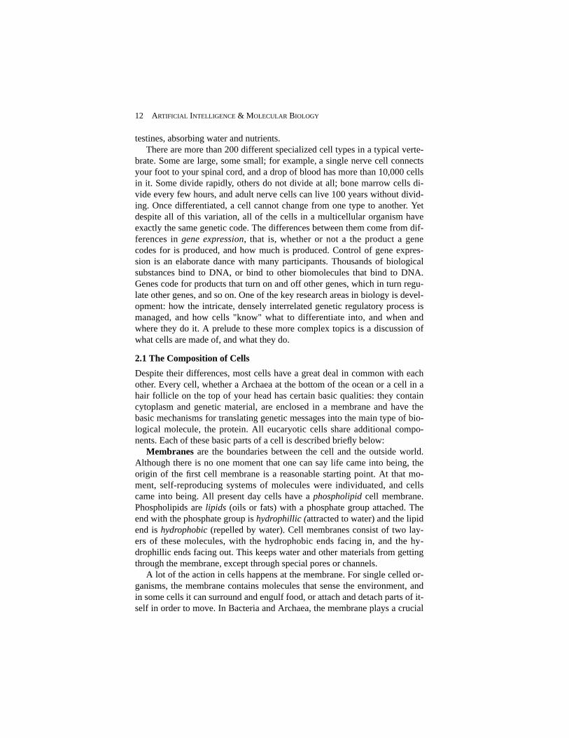
one end, a carboxyl group (COOH) at the other, and a variable sidechain (R),as shown in Figure 2. These chemical groups determine how the moleculefunctions, as Mavrovouniotis’s chapter in this volume explains. For example,under biological conditions the amino end of the molecule is positivelycharged, and the carboxyl end is negatively charged. Chains of amino acidsare assembled by a reaction that occurs between the nitrogen atom at theamino end of one amino acid and the carbon atom at the carboxyl end of an-other, bonding the two amino acids and releasing a molecule of water. Thelinkage is called a peptide bond, and long chains of amino acids can bestrung together into polymers*, called polypeptides, in this manner. All pro-teins are polypeptides, although the term polypeptide generally refers tochains that are shorter than whole proteins.
When a peptide bond is formed, the amino acid is changed (losing twohydrogen atoms and an oxygen atom), so the portion of the original moleculeintegrated into the polypeptide is often called a residue. The sequence ofamino acid residues that make up a protein is called the protein’s primarystructure. The primary structure is directly coded for in the genetic material:The individual elements of a DNA molecule form triples whichunambiguously specify an amino acid. A genetic sequence maps directly intoa sequence of amino acids. This process is discussed in greater detail below.
It is interesting to note that only a small proportion of the very many pos-sible polypeptide chains are naturally occurring proteins. Computationally,this is unsurprising. Many proteins contain more than 100 amino acids (some
HUNTER 19
*Polymers are long strings of similar elements; -mer means “element,” as inmonomer, dimer, etc. Homopolymer is a term that refers to polymers made up of allthe same element; heteropolymers are made of several different units. Proteins andDNA are both heteropolymers. Glycogen, a substance used for the medium-termstorage of excess energy, is an example of a homopolymer.
Carboxyl group: COOH
Amino Group: NH3
Central Carbon (C)
Sidechain (variable region)
Figure 2: The basic chemical structure of an amino acid. Carbon atoms are black,Oxygen is dark grey, Nitrogen light grey, and hydrogen white.
have more than 4000). The number of possible polypeptide chains of length100 is 20100 or more than 10130. Even if we take the high estimates of thenumber of species (5x10⁷) and assume that they all have as many differentproteins as there are in the most complex organism (<107) and that no twoorganisms share a single protein, the ratio of actual proteins to possiblepolypeptides is much less than 1:10100—a very small proportion, indeed.
The twenty naturally occuring amino acids all have the common elementsshown in Figure 2. The varying parts are called sidechains; the two carbonsand the nitrogen in the core are sometimes called the backbone. Peptidebonds link together the backbones of a sequence of amino acids. That linkcan be characterized as having two degrees of rotational freedom, the phi (φ)and psi (ψ) angles (although from the point of view of physics this is a dras-tic simplification, in most biological contexts it is valid). The conformationof a protein backbone (i.e. its shape when folded) can be adequately de-scribed as a series of φ/ψ angles, although it is also possible to represent theshape using the Cartesian coordinates of the central backbone atom (thealpha carbon, written Cα), or using various other representational schemes(see, e.g., Hunter or Zhang & Waltz in this volume).
The dimensions along which amino acids vary are quite important for anumber of reasons. One of the major unsolved problems in molecular biolo-gy is to be able to predict the structure and function of a protein from itsamino acid sequence. It was demonstrated more than two decades ago thatthe amino acid sequence of a protein determines ultimate conformation and,therefore, its biological activity and function. Exactly how the properties ofthe amino acids in the primary structure of a protein interact to determine theprotein’s ultimate conformation remains unknown. The chemical propertiesof the individual amino acids, however, are known with great precision.These properties form the basis for many representations of amino acids, e.g.in programs attempting to predict structure from sequence. Here is a briefsummary of some of them.
Glycine is the simplest amino acid; its sidechain is a single hydrogenatom. It is nonpolar, and does not ionize easily. The polarity of a moleculerefers to the degree that its electrons are distributed asymmetrically. A non-polar molecule has a relatively even distribution of charge. Ionization is theprocess that causes a molecule to gain or lose an electron, and hence becomecharged overall. The distribution of charge has a strong effect on the behav-ior of a molecule (e.g. like charges repel). Another important characteristicof glycine is that as a result of having no heavy (i.e. non-hydrogen) atoms inits sidechain, it is very flexible. That flexibility can give rise to unusual kinksin the folded protein.
Alanine is also small and simple; its sidechain is just a methyl group (con-sisting of a carbon and three hydrogen atoms). Alanine is one of the most
20 ARTIFICIAL INTELLIGENCE & MOLECULAR BIOLOGY

commonly appearing amino acids. Glycine and alanine’s sidechains arealiphatic, which means that they are straight chains (no loops) containingonly carbon and hydrogen atoms. There are three other aliphatic amino acids:valine, leucine and isoleucine. The longer aliphatic sidechains are hydropho-bic. Hydrophobicity is one of the key factors that determines how the chainof amino acids will fold up into an active protein. Hydrophobic residues tendto come together to form compact core that exclude water. Because the envi-ronment inside cells is aqueous (primarily water), these hydrophobicresidues will tend to be on the inside of a protein, rather than on its surface.
In contrast to alanine and glycine, the sidechains of amino acids pheny-lalanine, tyrosine and tryptophan are quite large. Size matters in protein fold-ing because atoms resist being too close to one another, so it is hard to packmany large sidechains closely. These sidechains are also aromatic, meaningthat they form closed rings of carbon atoms with alternating double bonds(like the simple molecule benzene). These rings are large and inflexible.Phenylalanine and tryptophan are also hydrophobic. Tyrosine has a hydroxylgroup (an OH at the end of the ring), and is therefore more reactive than theother sidechains mentioned so far, and less hydrophobic. These large aminoacids appear less often than would be expected ifproteins were composedrandomly. Serine and threonine also contain hydroxyl groups, but do nothave rings.
Another feature of importance in amino acids is whether they ionize toform charged groups. Residues that ionize are characterized by their pK,which indicates at what pH (level of acidity) half of the molecules of thatamino acid will have ionized. Arginine and lysine have high pK’s (that is,they ionize in basic environments) and histidine, gluatmic acid and asparticacid have low pK’s (they ionize in acidic ones). Since like charges repel andopposites attract, charge is an important feature in predicting protein confor-mation. Most of the charged residues in a protein will be found at its surface,although some will form bonds with each other on the inside of the molecule(called salt-bridges) which can provide strong constraints on the ultimatefolded form.
Cysteine and methionine have hydrophobic sidechains that contain a sul-phur atom, and each plays an important role in protein structure. The sul-phurs make the amino acids' sidechains very reactive. Cysteines can formdisulphide bonds with each other; disulphide bonds often hold distant partsof a polypeptide chain near each other, constraining the folded conformationlike salt bridges. For that reason, cysteines have a special role in determiningthe three dimensional structure of proteins. The chapter by Holbrook, Muskaland Kim in this volume discusses the prediction of this and other foldingconstraints. Methionine is also important because all eucaryotic proteins,when originally synthesized in the ribosome, start with a methionine. It is akind of “start” signal in the genetic code. This methionine is generally re-
HUNTER 21
moved before the protein is released into the cell, however.Histidine is a relatively rare amino acid, but often appears in the active
site of an enzyme. The active site is the small portion of an enzyme that ef-fects the target reaction, and it is the key to understanding the chemistry in-volved. The rest of the enzyme provides the necessary scaffolding to bringthe active site to bear in the right place, and to keep it away from bonds thatit might do harm to. Other regions of enzymes can also act as a switch, turn-ing the active site on and off in a process called allosteric control. Becausehistidine’s pK is near the typical pH of a cell, it is possible for small, localchanges in the chemical environment to flip it back and forth between beingcharged and not charged. This ability to flip between states makes it usefulfor catalyzing chemical reactions. Other charged residues also sometimesplay a similar role in catalysis.
With this background, it is now possible to understand the basics of theprotein folding problem which is the target of many of the AI methods ap-plied in this volume. The genetic code specifies only the amino acid se-quence of a protein. As a new protein comes off the ribosome, it folds up intothe shape that gives it its biochemical function, sometimes called its activeconformation (the same protein unfolded into some other shape is said to bedenatured, which is what happens, e.g. to the white of an egg when you cookit). In the cell, this process takes a few seconds, which is a very long time fora chemical reaction. The complex structure of the ribosome may play a rolein protein folding, and a few proteins need helper molecules, termed chaper-ones to fold properly. However, these few seconds are a very short time com-pared to how long it takes people to figure out how a protein will fold. In rawterms, the folding problem involves finding the mapping from primary se-quence (a sequence of from dozens to several thousand symbols, drawn froma 20 letter alphabet) to the real-numbered locations of the thousands of con-stituent atoms in three space.
Although all of the above features of amino acids play some role in proteinfolding, there are few absolute rules. The conformation a protein finally as-sumes will minimize the total “free” energy of the molecule. Going against thetendencies described above (e.g. packing several large sidechains near eachother) increases the local free energy, but may reduce the energy elsewhere inthe molecule. Each one of the tendencies described can be traded off againstsome other contribution to the total free energy of the folded protein. Givenany conformation of atoms, it is possible in principle to compute its free ener-gy. Ideally, one could examine all the possible conformations of a protein, cal-culate the free energy by applying quantum mechanical rules, and select theminimum energy conformation as a prediction of the folded structure. Unfortu-nately, there are very many possible conformations to test, and each energycalculation itself is prohibitively complex. A wide variety of approaches havebeen taken to making this problem tractable, and, given a few hours of super-
22 ARTIFICIAL INTELLIGENCE & MOLECULAR BIOLOGY
computer time, it is currently possible to evaluate several thousand possibleconformations. These techniques are well surveyed in Karplus & Petsko(1990). An alternative to the pure physical simulations are the various AI ap-proaches which a significant portion of this volume is dedicated to describing.
The position of the atoms in a folded protein is called its tertiary struc-ture. The primary structure is the amino acid sequence. Secondary structurerefers to local arrangements of a few to a few dozen amino acid residues thattake on particular conformations that are seen repeatedly in many differentproteins. These shapes are stabilized by hydrogen bonds (a hydrogen bond isa relatively weak bond that also plays a role in holding the two strands of theDNA molecule together). There are two main kinds of secondary structure:corkscrew-shaped conformations where the amino acids are packed tightlytogether, called α-helices, and long flat sheets made up of two or more adja-cent strands of the molecule, extended so that the amino acids are stretchedout as far from each other as they can be. Each extended chain is called a β-strand, and two or more β-strands held together by hydrogen bonds arecalled a β-sheet. β-sheets can be composed of strands running in the same di-rection (called a parallel β-sheet) or running in the opposite direction (an-tiparallel). Other kinds of secondary structure include structures that areeven more tightly packed than α-helices called 3-10 helices, and a variety ofsmall structures that link other structures, called β-turns. Some local combi-nations of secondary structures have been observed in a variety of differentproteins. For example, two α-helices linked by a turn with an approximately60° angle have been observed in a variety of proteins that bind to DNA. Thispattern is called the helix-turn-helix motif, and is an example of what isknown as super-secondary structure. Finally, some proteins only becomefunctional when assembled with other molecules. Some proteins bind tocopies of themselves; for example, some DNA-binding proteins only func-tion as dimers (linked pairs). Other proteins require prostehtic groups such asheme or chlorophyl. Additions necessary to make the folded protein activeare termed the protein’s quaternary structure.
4.3 Nucleic Acids
If proteins are the workhorses of the biochemical world, nucleic acids aretheir drivers; they control the action. All of the genetic information in anyliving creature is stored in deoxyribonucleic acid (DNA) and ribonucleic acid(RNA), which are polymers of four simple nucleic acid units, called nu-cleotides. There are four nucleotides found in DNA. Each nucleotide consistsof three parts: one of two base molecules (a purine or a pyrimidine), plus asugar (ribose in RNA and deoxyribose DNA), and one or more phosphategroups. The purine nucleotides are adenine (A) and guanine (G), and thepyrimidines are cytosine (C) and thymine (T). Nucleotides are sometimescalled bases, and, since DNA consists of two complementary strands bonded
HUNTER 23

together, these units are often called base-pairs. The length of a DNA se-quences is often measured in thousands of bases, abbreviated kb. Nucleotidesare generally abbreviated by their first letter, and appended into sequences,written, e.g., CCTATAG. The nucleotides are linked to each other in thepolymer by phosphodiester bonds. This bond is directional, a strand of DNAhas a head (called the 5’ end) and a tail (the 3’ end).
One well known fact about DNA is that it forms a double helix; that is,two helical (spiral-shaped) strands of the polypeptide, running in opposite di-rections, held together by hydrogen bonds. Adenines bond exclusively withthe thymines (A-T) and guanines bond exclusively with cytosines (G-C). Al-though the sequence in one strand of DNA is completely unrestricted, be-cause of these bonding rules the sequence in the complementary strand iscompletely determined. It is this feature that makes it possible to make highfidelity copies of the information stored in the DNA. It is also exploitedwhen DNA is transcribed into complementary strands of RNA, which directthe synthesis of protein. The only difference is that in RNA, uracil (U) takesthe place of thymine; that is, it bonds to adenine.
DNA molecules take a variety of conformations (shapes) in living sys-tems. In most biological circumstances, the DNA forms a classic doublehelix, called B-DNA; in certain circumstances, however, it can become su-percoiled or even reverse the direction of its twist (this form is called Z-DNA). These alternative forms may play a role in turning particular genes onand off (see below). There is some evidence that the geometry of the B-DNAform (e.g for example, differing twist angles between adjacent base pairs)may also be exploited by cell mechanisms. The fact that the conformation ofthe DNA can have a biological effect over and above the sequence it encodeshighlights an important lesson for computer scientists: there is more infor-mation available to a cell than appears in the sequence databases. This les-son also applies to protein sequences, as we will see in the discussion ofpost-translational modification.
Now that we have covered the basic structure and function of proteins andnucleic acids, we can begin to put together a picture of the molecular pro-cessing that goes on in every cell.
5. Genetic Expression: From Blueprint to Finished Product
5.1 Genes, the Genome and the Genetic Code
The genetic information of an organism can be stored in one or more distinctDNA molecules; each is called a chromosome. In some sexually reproducingorganisms, called diploids, each chromosome contains two similar DNAmolecules physically bound together, one from each parent. Sexually repro-ducing organisms with single DNA molecules in their chromosomes are
24 ARTIFICIAL INTELLIGENCE & MOLECULAR BIOLOGY

called haploid. Human beings are diploid with 23 pairs of linear chromo-somes. In Bacteria, it is common for the ends of the DNA molecule to bindtogether, forming a circular chromosome. All of the genetic information ofan organism, taken together as a whole, is refered to as its genome.
The primary role of nucleic acids is to carry the encoding of the primarystructure of proteins. Each non-overlapping triplet of nucleotides, called acodon, corresponds to a particular amino acid (see table 1). Four nucleotidescan form 43 = 64 possible triplets, which is more than the 20 needed to codefor each amino acid (pairs would provide only 16 codons). Three of thesecodons are used to designate the end of a protein sequence, and are calledstop codons. The others all code for a particular amino acid. That means thatmost amino acids are encoded by more than one codon. For example, alanineis represented in DNA by the codons GCT, GCC, GCA and GCG. Noticethat the first two nucleotides of these codons are all identical, and that thethird is redundant. Although this is not true for all of the amino acids, mostcodon synonyms differ only in the last nucleotide. This phenomenon iscalled the degeneracy of the code. Whether it is an artifact of the evolution,or serves a purpose such as allowing general changes in the global composi-tion of DNA (e.g. increasing the proportion of purines) without changing thecoded amino acids is still unknown.
There are some small variations in the translation of codons into aminoacids from organism to organism. Since the code is so central to the function-ing of the cell, it is very strongly conserved over evolution. However, thereare a few systems that use a slightly different code. An important example isfound in mitochondria. Mitochondria have their own DNA, and probablyrepresent previously free living organisms that were enveloped by eucary-otes. Mitochondrial DNA is translated using a slightly different code, whichis more degenerate (has less information in the third nucleotide) than thestandard code. Other organisms that diverged very early in evolution, such asthe ciliates, also use different codes.
The basic process of synthesizing proteins maps from a sequence ofcodons to a sequence of amino acids. However, there are a variety of impor-tant complications. Since codons come in triples, there are three possibleplaces to start parsing a segment of DNA. For example, the chain...AATGCGATAAG... could be read ...AAT-GCG-ATA... or ...ATG-CGA-TAA... or ...TGC-GAT-AAG.... This problem is similar to decoding an asyn-chronous serial bit stream into bytes. Each of these parsings is called a read-ing frame. A parsing with a long enough string of codons with no interveningstop codons is called an open reading frame, or ORF; and could be translatedinto a protein. Organisms sometimes code different proteins with overlap-ping reading frames, so that if the reading process shifts by one character, acompletely different, but still functional protein results! More often, frameshifts, which can be introduced by insertions and deletions in the DNA se-
HUNTER 25

quence or transcriptional “stuttering,” produce nonsense.Not only are there three possible reading frames in a DNA sequence, it is
possible to read off either strand of the double helix. Recall that the secondstrand is the complement of the first, so that our example above (AATGC-GATAAG) can also be read inverted and in the opposite direction, e.g. CT-TATCGCATT. This is sometimes called reading from the antisense or comple-mentary strand. An antisense message can also be parsed three ways, making atotal of 6 possible reading frames for every DNA sequence. There are knownexamples of DNA sequences that code for proteins in both directions with sev-eral overlapping reading frames: quite a feat of compact encoding.
And there’s more. DNA sequences coding for a single protein in most eu-caryotes have noncoding sequences, called introns, inserted into them.These introns are spliced out before the sequence is mapped into aminoacids. Different eucaryotes have a variety of different systems for recogniz-ing and removing these introns. Most bacteria don’t have introns. It is notknown whether introns evolved only after the origin of eucaryotes, orwhether selective pressure has caused bacteria to lose theirs. The segmentsof DNA that actually end up coding for a protein are called exons. You cankeep these straight by remembering that introns are insertions, and thatexons are expressed.
DNA contains a large amount of information in addition to the coding se-quences of proteins. Every cell in the body has the same DNA, but each celltype has to generate a different set of proteins, and even within a single celltype, its needs change throughout its life. An increasing number of DNA sig-nals that appear to play a role in the control of expression are being charac-terized. There are a variety of signals identifying where proteins begin andend, where splices should occur, and an exquisitely detailed set of mecha-nisms for controlling which proteins should be synthesized and in whatquantities. Large scale features of a DNA molecule, such as a region rich inCs and Gs can play a biologically important role, too.
Finally, some exceptions to the rules I mentioned above should be noted.DNA is sometimes found in single strands, particularly in some viruses.Viruses also play other tricks with nucleic acids, such as transcribing RNAinto DNA, going against the normal flow of information in the cell. Evennon-standard base-pairings sometimes play an important role, such as in thestructure of transfer RNA (see below).
5.2 RNA: Transcription, Translation, Splicing & RNA Structure
The process of mapping from DNA sequence to folded protein in eucaryotesinvolves many steps (see Figure 3). The first step is the transcription of aportion of DNA into an RNA molecule, called a messenger RNA (mRNA).This process begins with the binding of a molecule called RNA polymerase
26 ARTIFICIAL INTELLIGENCE & MOLECULAR BIOLOGY

to a location on the DNA molecule. Exactly where that polymerase binds de-termines which strand of the DNA will be read and in which direction. Partsof the DNA near the beginning of a protein coding region contain signalswhich can be recognized by the polymerase; these regions are called promot-ers. (Promoters and other control signals are discussed further below.) Thepolymerase catalyzes a reaction which causes the DNA to be used as a tem-plate to create a complementary strand of RNA, called the primary tran-script. This transcript contains introns as well as exons. At the end of thetranscript, 250 or more extra adenosines, called a poly-A tail, are often addedto the RNA. The role of these nucleotides is not known, but the distinctivesignature is sometimes used to detect the presence of mRNAs.
The next step is the splicing the exons together. This operation takestakes place in a ribosome-like assembly called a spliceosome. The RNA re-maining after the introns have been spliced out is called a mature mRNA. Itis then transported out of the nucleus to the cytoplasm, where it then bindsto a ribosome.
A ribosome is a very complex combination of RNA and protein, and itsoperation has yet to be completely understood. It is at the ribosome that themRNA is used as a blueprint for the production of a protein; this process iscalled translation. The reading frame that the translation will use is deter-mined by the ribosome. The translation process depends on the presence ofmolecules which make the mapping from codons in the mRNA to aminoacids; these molecules are called transfer-RNA or tRNAs. tRNAs have ananti-codon (that binds to its corresponding codon) near one end and the cor-responding amino acid on the other end. The anti-codon end of the tRNAsbind to the mRNA, bringing the amino acids corresponding the mRNA se-quence into physical proximity, where they form peptide bonds with eachother. How the tRNAs find only the correct amino acid was a mystery untilquite recently. This process depends on the three dimensional structure of theRNA molecule, which is discussed in Steeg’s chapter of this volume. As theprotein comes off the ribosome, it folds up into its native conformation. Thisprocess may involve help from the ribosome itself or from chaperone mole-cules, as was described above.
Once the protein has folded, other transformations can occur. Variouskinds of chemical groups can be bound to different places on the proteins, in-cluding sugars, phosphate, actyl or methyl groups. These additions canchange the hyrogen bonding proclivity or shape of the protein, and may benecessary to make the protein active, or may keep it from having an effectbefore it is needed. The general term for these transformations is post-trans-lational modifications. Once this process is complete, the protein is thentransported to the part of the cell where it will accomplish its function. Thetransport process may be merely passive diffusion through the cytoplasm, orthere may be an active transport mechanism that moves the protein across
HUNTER 27

28 ARTIFICIAL INTELLIGENCE & MOLECULAR BIOLOGY
α-he l i x
β-s t rand
β-sheet
Hydrogen bonds
Disulphide bond
Coil
{
DNA
RNA polymerase
Promoter region
Pr imary t ranscr ip t(RNA)in t ronexon
Transcription (takes place in nucleus)
Intron splicing (takes place at spliceosomes)
mRNA
Unfolded protein
Translation (takes place at r ibosomes)
Stop codon
Splice junction consensus sites
Folded protein
Folding and post-translat ional modif icat ion
met
ATG
tRNA
Glycosylat ion si te
Figure 3. A schematic drawing of the entire process of protein synthesis. An RNAPolymerase binds to a promoter region of DNA, and begins the transcription process,which continues until a stop codon is reached. The product is an RNA moleculecalled the primary transcript, which contains regions that code for proteins (exons)and regions which do not (introns). The introns are spliced out at splicosomes, andthe joined exons are transported to a ribosome. There, transfer RNAs match aminoacids to the appropriate codons in the RNA; the amino acids form peptide bonds andbecome an unfolded protein. The protein then folds into local formations like helicesand sheets, and forms internal bonds across longer distances. Post-translationalprocessing can add additional substance; e.g., glycosylation adds sugar molecules tothe protein.

membranes or into the appropriate cellular compartment.
5.3 Genetic RegulationEvery cell has the same DNA. Yet the DNA in some cells codes for the pro-teins needed to function as, say, a muscle, and other code for the proteins tomake the lens of the eye. The difference lies in the regulation of the geneticmachinery. At any particular time, a particular cell is producing only a smallfraction of the proteins coded for in its DNA. And the amount of each pro-tein produced must be precisely regulated in order for the cell to functionproperly. The cell will change the proteins it synthesizes in response to theenvironment or other cues. The mechanisms that regulate this process consti-tute a finely tuned, highly parallel system with extensive multifactoral feed-back and elaborate control structure. It is also not yet well understood.
Genes are generally said to be on or off (or expressed/not expressed), al-though the amount of protein produced is also important. The productionprocess is controlled by a complex collection of proteins in the nucleus ofeucaryotic cells that influence which genes are expressed. Perhaps the mostimportant of these proteins are the histones, which are tightly bound to theDNA in the chromosomes of eucaryotes. Histones are some of the most con-served proteins in all of life. There are almost no differences in the sequenceof plant and mammalian histones, despite more than a billion years of diver-gence in their evolution. Other proteins swarm around the DNA, someinfluencing the production of a single gene (either encouraging or inhibitingit), while others can influence the production of large numbers of genes atonce. An important group of these proteins are called topoisomerases; theyrearrange and untangle the DNA in various ways, and are the next mostprevalent proteins in the chromosome.
Many regulatory proteins recognize and bind to very specific sequences inthe DNA. The sequences that these proteins recognize tend to border the pro-tein coding regions of genes, and are known generally as control regions. Se-quences that occur just upstream (towards the 5' end) of the coding regionthat encourage the production of the protein are called promoters. Similar re-gions either downstream of the coding region or relatively far upstream arecalled enhancers. Sequences that tend to prevent the production of a proteinare called repressors. Karp’s chapter in this volume discusses how this com-plex set of interactions can be modeled in knowledge-based systems.
Cells need to turn entire suites of genes on and off in response to manydifferent events, ranging from normal development to trying to repair dam-age to the cell. The control mechanisms are responsive to the level of a prod-uct already in the cell (for homeostatic control) as well as to a tremendousvariety of extracellular signals. Perhaps the most amazing activities in generegulation occur during development; not only are genes turned on and offwith precise timing, but the control can extend to producing alternative splic-
HUNTER 29
ings of the nascent primary transcripts (as is the case in the transition fromfetal to normal hemoglobin).
5.4 Catalysis & Metabolic Pathways
The translation of genes into proteins, crucial as it is, is only a small portionof the biochemical activity in a cell. Proteins do most of the work of manag-ing the flow of energy, synthesizing, degrading and transporting materials,sending and receiving signals, exerting forces on the world, and providingstructural support. Systems of interacting proteins form the basis for nearlyevery process of living things, from moving around and digesting food tothinking and reproducing. Somewhat surprisingly, a large proportion of thechemical processes that underlie all of these activities are shared across avery wide range of organisms. These shared processes are collectively re-ferred to as intermediary metabolism. These include the catabolic processesfor breaking down proteins, fats and carbohydrates (such as those found infood) and the anabolic processes for building new materials. Similar collec-tions of reactions that are more specialized to particular organisms are calledsecondary metabolism. The substances that these reactions produce and con-sume are called metabolites.
The biochemical processes in intermediary metabolism are almost all cat-alyzed reactions. That is, these reactions would barely take place at all atnormal temperatures and pressures; they require special compounds that fa-cilitate the reaction — these compounds are called catalysts or enzymes. (It isonly partially in jest that many biochemistry courses open with the professorsaying that the reactions that take place in living systems are ones you weretaught were impossible in organic chemistry.) Catalysts are usually namedafter the reaction they facilitate, usually with the added suffix -ase. For ex-ample, alcohol dehydrogenase is the enzyme that turns ethyl alcohol into ac-etaldehyde by removing two hydrogen atoms. Common classes of enzymesinclude dehydrogenases, synthetases, proteases (for breaking down proteins),decarboxylases (removing carbon atoms), transferases (moving a chemicalgroup from one place to another), kinases, phosphatases (adding or removingphosphate groups, respectively) and so on. The materials transformed by cat-alysts are called substrates. Unlike the substrates, catalysts themselves arenot changed by the reactions they participate in. A final point to note aboutenzymatic reactions is that in many cases the reactions can proceed in eitherdirection. That is, and enzyme that transforms substance A into substance Bcan often also facilitate the transformation of B into A. The direction of thetransformation depends on the concentrations of the substrates and on the en-ergetics of the reaction (see Mavrovouniotis’ chapter in this volume for fur-ther discussion of this topic).
Even the basic transformations of intermediary metabolism can involve
30 ARTIFICIAL INTELLIGENCE & MOLECULAR BIOLOGY

dozens or hundreds of catalyzed reactions. These combinations of reactions,which accomplish tasks like turning foods into useable energy or compoundsare called metabolic pathways. Because of the many steps in these pathwaysand the widespread presence of direct and indirect feedback loops, they canexhibit many counterintuitive behaviors. Also, all of these chemical reactionsare going on in parallel. Mavrovouniotis’s chapter in this volume describesan efficient system for making inferences about these complex systems.
In addition to the feedback loops among the substrates in the pathways,the presence or absence of substrates can affect the behavior of the enzymesthemselves, through what is called allosteric regulation. These interactionsoccur when a substance binds to an enzyme someplace other than its usualactive site (the atoms in the molecule that have the enzymatic effect). Bind-ing at this other site changes the shape of the enzyme, thereby changing itsactivity. Another method of controlling enzymes is called competitive inhibi-tion. In this form of regulation, substance other than the usual substrate ofthe enzyme binds to the active site of the enzyme, preventing it from havingan effect on its substrate.
These are the basic mechanisms underlying eucaryotic cells (and much ofthis applies to bacterial and archaeal ones as well). Of course, each particularactivity of a living system, from the capture of energy to immune response,has its own complex network of biochemical reactions that provides themechanism underlying the function. Some of these mechanisms, such as thesecondary messenger system involving cyclic adenosine monophosphate(cAMP) are widely shared by many different systems. Others are exquisitelyspecialized for a particular task in a single species: my favorite example ofthis is the evidence that perfect pitch in humans (being able to identify musi-cal notes absolutely, rather than relative to each other) is mediated by a sin-gle protein. The functioning of these biochemical networks is being unrav-elled at an ever increasing rate, and the need for sophisticated methods toanalyze relevant data and build suitable models is growing rapidly.
5.5 Genetic Mechanisms of Evolution
In the beginning of this chapter, I discussed the central role that evolutionplays in understanding living systems. The mechanisms of evolution at themolecular level are increasingly well understood. The similarities and differ-ences among molecules that are closely related provide important informa-tion about the structure and function of those molecules. Molecules (or theirsequences) which are related to one another are said to be homologous. Al-though genes or proteins that have similar sequences are often assumed to behomologous, there are well known counterexamples due to convergent evo-lution. In these cases, aspects of very distantly related organisms come to re-semble one another through very different evolutionary pathways. Unlessthere is evidence to the contrary, it is usually safe to assume that macromole-
HUNTER 31

cular sequences that are similar to each other are homologous.The sources of variation at the molecular level are very important to un-
derstanding how molecules come to differ from each other (or diverge). Per-haps the best known mechanism of molecular evolution is the point muta-tion, or the change of a single nucleotide in a genetic sequence. The changecan be to insert a new nucleotide, to delete an existing one, or to change onenucleotide into another. Other mechanisms include large scale chromosomalrearrangements and inversions. An important kind of rearrangement is thegene duplication; in which additional copies of a gene are inserted into thegenome. These copies can then diverge, so that, for example, the originalfunctionality may be preserved at the same time as related new genes evolve.These duplication events can lead to the presence of pseudogenes, which arequite similar to actual genes, but are not expressed. These pseudogenes pre-sent challenges for gene recognition algorithms, such as the one proposed inSearls chapter in this volume. Sexual reproduction adds another dimension tothe exchange of genetic material. DNA from the two parents of a sexually re-producing organism undergoes a process called crossover, which forms akind of mosaic that is passed on to the offspring.
Most mutations have relatively little effect. Mutations in the middle ofintrons generally have no effect at all (although mutations at the ends of anintron can affect the splicing process). Mutations in the third position ofmost codons have little effect at the protein level because of the redundancyof the genetic code. Even mutations that cause changes in the sequence of aprotein are often neutral, as demonstrated by Sauer, et al (1989). Their ex-perimental method involved saturation mutagenesis which explores are rel-atively large proportion of the space of possible mutations in parallel. Neu-tral mutations are the basis of genetic drift, which is the phenomena thataccounts for the differences between the DNA that codes for functionallyidentical proteins in different organisms. This drift is also the basis for themolecular clock, described above. Of course, some point mutations arelethal, and others lead to diseases such as cystic fibrosis. Very rarely, a mu-tation will be advantageous; it will then rapidly get fixed in the population,as the organisms with the conferred advantage out reproduce the ones with-out it. Diploid sexually reproducing organisms have two copies of eachgene (one from each parent), resulting in an added layer of complexity inthe effect of mutations. Sometimes the extra copy can compensate (or par-tially compensate) for a mutation.
Molecular evolution also involves issues of selection and inheritance. In-heritance requires that the genes from the parent be passed to the offspring.DNA itself is replicated by splitting the double helix into two complimentarystrands and then extending a primer by attaching complementary nucleotides.This process is modelled in detail Brutlag, et al’s chapter in this volume. Themolecular mechanisms underlying the whole complex process of cell divi-
32 ARTIFICIAL INTELLIGENCE & MOLECULAR BIOLOGY

sion (i.e. the cell cycle) are strikingly conserved in eucaryotes, and knowl-edge about this process is growing rapidly (see, e.g., Hartwell (1991) for areview). Selection also occurs on factors that are only apparent on the molec-ular level, such as the efficiency of certain reaction pathways (see, e.g.Hochachka & Somero [1984]).
6. Sources of Biological Knowledge
The information in this chapter has been presented textbook style, with littlediscussion of how the knowledge arose, or where errors might have crept in.The purpose of this section is to describe some of the basic experimentalmethods of molecular biology. These methods are important not only in un-derstanding the source of possible errors in the data, but also because compu-tational methods for managing laboratory activities and analyzing raw dataare another area where AI can play a role (see the chapters by Edwards, et aland Glasgow, et al, in this volume). I will also describe some of the manyonline information resources relevant to computational molecular biologythat are available.
6.1 Model Organisms: Germs, Worms, Weeds, Bugs & Rodents
The investigation of the workings of even a single organism is so complex asto take many dedicated scientists many careers worth of time. Trying tostudy all organisms in great depth is simply beyond the abilities of modernbiology. Furthermore, the techniques of biological experimentation are oftencomplex, time consuming and difficult. Some of the most valuable methodsin biological research are invasive, or require organisms to be sacrificed, orrequire many generations of observation, or observations on large popula-tions. Much of this work is impractical or unethical to carry out on humans.For these reasons, biologists have selected a variety of model organisms forexperimentation. These creatures have qualities that make possible con-trolled laboratory experiments at reasonable cost and difficulty with resultsthat can often be extrapolated to people or other organisms of interest.
Of course, research involving humans can be done ethically, and in someareas of biomedical research, such as final drug testing, it is obligatory. Otherresearch methods involve kinds of human cells can be grown successfully inthe laboratory. Not many human cell types thrive outside of the body. Somekinds of human cancer cells do grow well in the laboratory, and these cellsare an important vehicle for research.
Sometimes the selection of a new model organism can lead to great ad-vances in a field. For example, the use of a particular kind of squid madepossible the understanding of the functioning of neurons because it containeda motor neuron that is more than 10 times the size of most neural cells, andhence easy to find and use in experiments. There are experimentally useful
HUNTER 33

correlates of nearly every aspect of human biology found in some organismor another, but the following six organisms form the main collection of mod-els used in molecular biology:
E. coli The ubiquitous intestinal bacterium Escherichia coli is a work-horse in biological laboratories. Because it is a relatively simple organismwith fast reproduction time and is safe and easy to work with, E. coli hasbeen the focus of a great deal of research in genetics and molecular biologyof the cell. Although it is a Bacterium, many of the basic biochemical mech-anisms of E. coli are shared by humans. For example, the first understandingof how genes can be turned on and off came from the study of a virus that in-fects these bacteria (Ptashne, 1987). E. coli is a common target for geneticengineering, where genes from other organisms are inserted into the bacterialgenome then produced in quantity. E. coli is now the basis of the internation-al biotechnology industry, churning out buckets full of human insulin, theheart attack drug TPA, and a wide variety of other substances.
Saccharomyces Saccharomyces cervesiae is better known as brewer’syeast, and it is another safe, easy to grow, short generation time organism.Other yeasts, such as Schizosaccharomyces pombe, are also used extensively.Surprisingly, yeasts are very much like people in many ways. Unlike the bac-terium E. coli, yeasts are eucaryotes, with a cell nucleus, mitochondria, a eu-caryotic cell membrane, and many of the other cellular components andprocesses found in most other eucaryotes, including people. Because theseyeasts are so easy to grow and manipulate, and because they are so biochemi-cally similar to people, many insights about the molecular processes involvedin metabolism, biosynthesis, cell division, and other crucial areas of biologyhave come from the investigation of Saccharomyces (Saccharomyces is agenus name, which, when used alone, refers to all species that are within thatgenus). Yeasts play another important role in molecular biology. One of thecrucial steps in sequencing large amounts of DNA is to be able to preparemany copies of moderate sized pieces of DNA. An widely used method fordoing this is the yeast artificial chromosome (or YAC), which is discussedbelow.
Arabidopsis The most important application of increased biological under-standing is generally thought to be in medicine, and increased understandingof human biology has indeed led to dramatic improvements in health care.However, in terms of effect on human life, agriculture is just as significant. Agreat deal of research into genetics and biochemistry has been motivated bythe desire to better understand various aspects of plant biology. An importantmodel organism for plants is Arabidopsis thaliana, a common weed. Ara-bidopsis makes a good model because it undergoes the same processes ofgrowth, development, flowering and reproduction as most higher plants, butit’s genome has 30 times less DNA than corn, and very little repetitive DNA.It also produces lots of seeds, and takes only about six weeks to grow to matu-
34 ARTIFICIAL INTELLIGENCE & MOLECULAR BIOLOGY

rity. There are several other model organisms used to investigate botanicalquestions, including tomatoes, tobacco, carrots and corn.
C. elegans One of the most exciting model organisms to emerge recentlyhas been the nematode worm Caenorhabditis elegans. This tiny creature,thousands of which can be found in a spadeful of dirt, has already been usedto generate tremendous insight about cellular development and physiology.The adult organism has exactly 959 cells, and every normal worm consists ofexactly the same collection of cells in the same places doing the same thing.It is one of the simplest creatures with a nervous system (which involvesabout a third of its cells). Not only is the complete anatomy of the organismknown, but a complete cell fate map has been generated, tracing the develop-mental lineage of each of each cell throughout the lifespan of the organism.This map allows researchers to relate behaviors to particular cells, to tracethe effects of genetic mutations very specifically, and perhaps to gain insightinto the mechanisms of aging as well as development. A large, highly inte-grated picture and text database of information about the cell fates, geneticmaps and sequences, mutation effects and other relevant information aboutC. elegans is currently under construction at the University of Arizona.
D. melanogaster Drosophila melanogaster, a common fruit fly, has longbeen a staple of classical genetics research. These flies have short generationtimes, and many different genetically determined morphological characteris-tics (e.g. eye color) that can readily be determined by visual inspection.Drosophila were used for decades in exploring patterns of inheritance; nowthat molecular methods can be applied, they have proven invaluable for a va-riety of studies of genetic expression and control. An important class of ge-netic elements that regulate many other genes, in effect, specifying complexgenetic programs, were first discovered in Drosophila; these areas are calledhomeoboxes. Molecular genetics in Drosophila is also providing great in-sights into how complex body plans are generated.
M. musculus Mus musculus is the basic laboratory mouse. Mice are mam-mals, and, as far as biochemistry is concerned, are practically identical topeople. Many questions about physiology, reproduction, functioning of theimmune and nervous systems and other areas of interest can only beaddressed by examining creatures that are very similar to humans; mice near-ly always fit the bill. The similarities between mice and people mean alsothat the mouse is a very complicated creature; it has a relatively large, com-plex genome, and mouse development and physiology is not as regular orconsistent as that of C. elegans or Drosophila. Although our depth of under-standing of the mouse will lag behind understanding of simpler organisms,the comparison of mouse genome to human is likely to be a key step, both inunderstanding their vast commonalities, and in seeing the aspects of ourgenes that make us uniquely human.
HUNTER 35

7. Experimental Methods
Molecular biologists have developed a tremendous variety of tools to addressquestions of biological function. This chapter can only touch briefly on a fewof the most widely used methods, but the terminology and a sense of thekinds of efforts required to produce the data used by computer scientists canbe important for understanding the strengths and limitations of varioussources of data.
Imaging. The first understanding of the cellular nature of life came short-ly after the invention of the light microscope, and microscopy remaines cen-tral to research in biology The tools for creating images have expandedtremendously. Not only are there computer controled light microscopes witha wide variety of imaging modalities, but there are now many other methodsof generating images of the very small. The electon microscope offers ex-tremely high resolution, although it requires exposing the imaged sample tohigh vacuum and other harsh treatments. New technologies including theAtomic Force Microscope (AFM) and the Scanning Tunnelling Microscope(STM) offer the potential to create images of individual molecules. Biolo-gists use these tools extensively.
Gel Electrophoresis. A charged molecule, when placed in an electricfield, will be accelerated; positively charged molecules will move towardnegative electrodes and vice versa. By placing a mixture of molecules of in-terest in a medium and subjecting them to an electric charge, the moleculeswill migrate through the medium and separate from each other. How fast themolecules will move depends on their charge and their size—bigger mole-cules see more resistance from the medium. The procedure, called elec-trophoresis involves putting a spot of the mixture to be analyzed at the top ofa polyacrylamide or agarose gel, and applying an electric field for a period oftime. Then the gel is stained so that the molecules become visible; the stainsappear as stripes along the gel, and are called bands. The location of thebands on the gel are proportional to the charge and size of the molecules inthe mixture (see Figure 4 for an example). The intensity of the stain is an in-dication of the amount of a particular molecule in the mixture. If themolecules are all the same charge, or have charge proportional to their size(as, for example, DNA does) then electrophoresis separates them purely bysize.
Often, several mixtures are run simultaneously on a single gel. This al-lows for easy calibration to standards, or comparison of the contents of dif-ferent mixtures, showing, for example, the absence of a particular molecularcomponent in one. The adjacent, parallel runs are sometimes called lanes. Avariation on this technique allows the sorting of molecules by a chemicalproperty called the isoelectric point, which is related to its pK. A combina-tion of the two methods, called 2D electrophoresis is capable of very fine
36 ARTIFICIAL INTELLIGENCE & MOLECULAR BIOLOGY

distinctions, for example, mapping each protein in a cell to a unique spot intwo-space, the size of the spot indicating the amount of the protein. Althoughthere are still some difficulties in calibration and repeatability, this method ispotentially a very powerful tool for monitoring the activities of large bio-chemical systems. In addition, if a desired molecule can be separated fromthe mixture this way, individual spots or bands can be removed from the gelfor further processing, in a procedure called blotting.
Cloning. A group of cells with identical genomes are said to be clones ofone another. Unless there are mutations, a single cell that reproduces asexu-ally will produce identical offspring; these clones are sometimes called a cellline, and certain standardized cell lines, for example the HeLa cell line, playan important role in biological research.
This concept has been generalize to cloning individual genes. In this case,a piece of DNA containing a gene of interest is inserted into the genome of atarget cell line, and the cells are screened so that all of the resulting cellshave an identical copy of the desired genetic sequence. The DNA in thesecells is said to be recombinant, and the cell will produce the protein codedfor by the inserted gene.
Cloning a gene requires some sophisticated technology. In order for acloned gene to be expressed, it must contain the appropriate transcription sig-nals for the target cell line. One way biologists ensure that this will happen isto put the new gene into a bacteriophage (a virus that infects bacteria), or aplasmid (a circular piece of DNA found outside of the chromosome of bacte-ria that replicates independently of the bacteria’s chromosomal DNA). These
HUNTER 37
Figure 4. This is an example of a gel electrophoresis run.. Each column was loadedwith a different mixture. The mixtures are then separated vertically by their chargeand size. The gel is then stained, producing dark bands where a molecule of a givensize or charge is present in a mixture. In this gel, the columns marked with a - are acontrol group. The band marked with an arrow is filled only in the + columns.
-- + - + - + - + - + - + - +
devices for inserting foreign DNA into cells are called vectors.In order to cut and paste desired DNA fragments into vectors, biologists
use restriction enzymes, which cut DNA at precisely specified points. Theseenzymes are produced naturally by bacteria as a way of attacking foreignDNA. For example, the commonly used enzyme EcoRI (from E. coli) cutsDNA between the G and the A in the sequence GAATTC; these target se-quences are called restriction sites. Everywhere a restriction site occurs in aDNA molecule treated with EcoRI, the DNA will be broken. Restriction en-zymes play many roles in biology in addition to making gene cloningpossible; a few others will be described below.
Both the insertion of the desired gene into the vector and the uptake of thevector by the target cells are effective only a fraction of the time. Fortunately,cells and vectors are small and it is relatively easy to grow a lot of them. Theprocess is applied to a population of target cells, and then the resulting popu-lation is screened to identify the cells where the gene was successfully insert-ed. This can be difficult, so many vectors are designed to facilitate screening.One popular vector, pBR322, contains a naturally occurring transcriptionstart signal and some antibiotic resistance genes, designed with convenientlyplaced restriction sites. If this vector is taken up by the target cells, it willconfer resistance to certain antibiotics to them. By applying the anitbiotic tothe whole colony, the researcher can kill all the cells that did not get thecloned gene. More sophisticated manipulations involving multiple antibioticresistances and carefully placed restriction sites can also be used to ensurethat the gene was correctly taken up by the vector.
There are many variations on these techniques for inserting foreign genes.It is now possible to use simple bacteria to produce large amounts of almostany isolated protein, including, for example, human insulin. Although it is amore complex process, it is also possible to insert foreign genes into plantsand animals, even people. A variety of efforts are underway to use these tech-niques to engineer organisms for agriculture, medicine and other applications.Not all of these applications are benign. One of the most successful early ef-forts was to increase the resistance of tobacco plants to pesticides, and thereare clear military applications. On the other hand, these methods also promisenew approaches to producing important rare biological compounds inexpen-sively (e.g. for novel cancer treatments or cleaning up toxic waste) and im-proving the nutritional value or hardiness of agricultural products. The entirefield of genetic engineering is controversial, and there are a variety of controlson what experiments can be done and how they can be done.
Hybridization and Immunological Staining. Biological compoundscan show remarkable specificity, for example, binding very selectively onlyto one particular compound. This ability plays an important role in the labo-ratory, where researchers can identify the presence or absence of a particularmolecule (or even a region of a molecule) in vanishingly small amounts.
38 ARTIFICIAL INTELLIGENCE & MOLECULAR BIOLOGY
Antibodies are the molecules that the immune system uses to identify andfight off invaders. Antibodies are extremely specific, recognizing and bind-ing to only one kind of molecule. Dyes can be attached to the antibody,forming a very specific system for identifying the presence (and possiblyquantifying the amount) of a target molecule that is present in a system.
There is a conceptually related method for identifying very specificallythe presence of a particular nucleotide sequence in a macromolecule. Thecomplement to a single-stranded DNA sequence will bind quite specificallyto that sequence. One technique measures how similar two related DNA se-quences are by testing how strongly the single-stranded versions of the mole-cules stick to each other, or hybridize. The more easily they come apart, themore differences there are between their sequences. It is also possible to at-tach a dye or other marker to a specific piece of DNA (called a probe) andthen hybridize it to a longer strand of DNA. The location along the strandthat is complementary to the probe will then be marked. There are manyvariations on hybridization and immunological staining that are customizedto the needs of a particular experiment.
Gene Mapping and Sequencing. The Human Genome Project is the ef-fort to produce a map and then the sequence of the human genome. The pur-pose of a genetic map is to identify the location and size of all of the genes ofan organism on its chromosomes. This information is important for a varietyof reasons. First, because crossover is an important component of inheritancein sexually reproducing organisms, genes that are near each other on thechromosome will tend to be inherited together. In fact, this forms the basisfor linkage analysis, which is a technique that looks at the relationships be-tween genes (or phenotypes) in large numbers of matings (in this context,often called crosses) to identify which genes tend to be inherited together,and are therefore likely to be near each other. Second, it is possible to clonegenes of known locations, opening up a wide range of possible experimentalmanipulations. Finally, it is currently possible to determine the sequence ofmoderate size pieces of DNA, so if an important gene has been mapped, it ispossible to find the sequence of that area, and discover the protein that is re-sponsible for the genetic characteristic. This is especially important for un-derstanding the basis of inherited diseases.
The existence of several different kinds of restriction enzymes makes pos-sible a molecular method of creating genetic maps. The application of eachrestriction enzyme (the process is called a digest) creates a different collec-tion of restriction fragments (the cut up pieces of DNA). By using gel elec-trophoresis, it is possible to determine the size of these fragments. Usingmultiple enzymes, together and separately, results in sets of fragments whichcan be (partially) ordered with respect to each other, resulting in a geneticmap. AI techniques for reasoning about partial orders have been effectivelyapplied to the problem of assembling the fragments into a map (Letovsky &
HUNTER 39

Berlyn, 1992). These physical maps divide a large piece of DNA (like achromosome) into parts, and and there is an associated method for obtainingany desired part.
Restriction fragment mapping becomes problematic when applied to largestretches of DNA, because the enzymes can produce many pieces of aboutthe same size, making the map ambiguous. The use of different enzymes canhelp address this problem to a limited degree, but a variety of other tech-niques are now also used.
Being able to divide the genome into moderate sized chunks is a prerequi-site to determining its sequence. Although there are several clever methodsfor determining the sequence of DNA molecule, all of them are limited to aresolution of well under a thousand basepairs at a time. In order to take thissequencing ability and determine the sequence of large pieces of DNA, manydifferent overlapping chunks must be sequenced, and then these sequencesmust be assembled. In order to accomplish this task, it is necessary to breakthe DNA in an entire genome down into a set of more manageable sizedpieces. The ordering of these pieces must be known (so they can be reassem-bled into a complete sequence), taken together the pieces must cover the en-tire genome, and the same set of pieces must be accessible to many differentlaboratories. This process is usually accomplished in several stages. The firststage generates relatively large pieces called contigs. Contigs are maintainedin cloned cell lines so that they can be reproduced and distributed. Often,these pieces of DNA are made into Yeast artificial chromosomes, or YACs,which can hold up to about a million basepairs of sequence each, requiringon the order of 10,000 clones to adequately cover the entire human genome.Each of these is then broken down into sets of smaller pieces, often in theform of cosmids. A cosmid is a particular kind of bacteriophage (a virus thatinfects bacteria) that is capable of accepting inserts of 30,000 or so basepairs.The difficulties in generating and maintaining collections of clones that largehave led to alternative technologies for large scale sequencing.
One alternative involves a new technology based on the polymerase chainreaction, or PCR. This mechanism was revolutionary becauase it made itpossible to rapidly produce huge amounts of a specific region of DNA, sim-ply by knowing a little bit of the sequence around the desired region. PCRexponentially amplifies (makes copies of) a segment of a DNA molecule,given a unique pair of sequences that bracket the desired piece. First, shortsequences of DNA (called oligonucleotides, or oligos) complementary to
40 ARTIFICIAL INTELLIGENCE & MOLECULAR BIOLOGY
*There are many interesting uses of this technology. For example, it gives law en-forcement the ability to generate enough DNA for identification from vanishing smallsamples of tissue. A more amusing application is the rumored use of PCR to spy onwhat academic competitors are doing in their research. Almost any correspondencefrom a competitor’s lab will contain traces of DNA which can be amplified by PCR toidentify the specific clones the lab is working with.

each of the bracketing sequences are synthesized. Creating short pieces ofDNA with a specific sequence is routine technology, now often performed bylaboratory robots. These pieces are called primers. The primers, the targetDNA and the enzyme DNA polymerase are then combined. The mixture isheated, so that the hydrogen bonds in the DNA break and the molecule splitsinto two single strands. When the mixture cools sufficiently, the primersbond to the regions around the area of interest, and the DNA polymerasereplicates the DNA downstream of the primers. By using a heat resistantpolymerase from an Archaea species that lives at high temperatures, it is pos-sible to rapidly cycle this process, doubling the amount of desired segment ofDNA each time. This technology makes possible the exponentialamplification of entire DNA molecules or any specific region of DNA forwhich bracketing primers can be generated.*
In order to use PCR for genome mapping and sequencing, a collection ofunique (short) sequences spread throughout the genome must be identifiedfor use as primers. The sequences must be unique in the genome so that thesource of amplified DNA is unambiguous, and they have to be relativelyshort so that they are easy to synthesize. The sites in the genome that corre-spond to these sequences are called sequence tagged sites or STSs. The moreSTSs that are known, the finer grained the map of the genome they provide.Finding short, unique sequences even in 3x10⁹ bp of DNA is not thatdifficult; a simple calculation shows that most sequences of length 16 or socan reasonably be expected to be unique in a genome of that size. An earlygoal of the Human Genome Project is to generate a list of STSs spaced at ap-proximately 100kbp intervals over the entire human genome. If it is possibleto find STSs that adequately cover the genome, it will not be necessary tobuild and maintain libraries of 10,000 YACs and ten times as many cosmids.Any region of DNA of interest can be identified by two STSs that bracket it.Instead of having to maintain large clone collections, these STSs can bestored in a database, and any researcher who needs a particular section ofDNA can synthesize the appropriate primers and use PCR to produce manycopies of just of that section.
Another issue that has been raised about the project to sequence thegenome is the need to know the sequences of all of the introns and other non-coding regions of DNA. One way to address this issue is to target only cod-ing regions for sequencing. The ability to find the sequences that a particularcell is using to produce proteins at a particular point in time is also useful ina variety of other areas as well. This information can be gleaned by gatheringthe mRNAs present in the cytoplasm of the cell, and sequencing them. In-stead of sequencing the mRNAs directly, biologists use an enzyme called re-verse transcriptase to make DNA molecules complementary to the mRNAs(called cDNAs) and then sequence that DNA. Using PCR and other technolo-gy, it is possible to capture at least portions of most of the mRNAs a cell is
HUNTER 41

producing. By sequencing these cDNAs, researchers can focus their attentionon just the parts of the genome that code for expressed proteins.
Large scale attempts to sequence at least part of all of the cDNAs that canbe produced from brain tissue have resulted in partial sequences for morethan 2500 new proteins in a very short period of time (Adams, et al, 1992).These sequences called ESTs, for expressed sequence tags can be used asPCR primers for future, more detailed experiments. This work has createdcontroversy because of the ensuing attempt by the National Institutes ofHealth to patent the EST sequences.
Crystallography and NMR. Until the relationship between protein se-quence and structure is more fully understood, the sequences produced bygenome projects will provide only part of the biochemical story. Additionalinformation about protein structure is necessary to understand how the pro-teins function. This structural information is at the present primarily gatheredby X-ray crystallography. In order to determine the structure of a protein inthis manner, a very large, pure crystal of the protein must be grown (thisprocess can take years, and may never succeed for certain proteins). Then theX-ray diffraction pattern of the crystal is measured, and this information canbe used indirectly to determine the positions of the atoms in the molecule.Glasgow, et al’s chapter in this volume describes this process in more detail.Because of the difficulties in crystallography, relatively few structures areknown, but the number of new structures is growing exponentially, with adoubling time of a bit over two years.
A promising alternative to crystallography for determining protein struc-ture is multi-dimensional nuclear magnetic resonance, or NMR. Althoughthis process does not require the crystallization of the protein, there are tech-nical difficulties in analyzing the data associated with large molecules likeproteins. Edwards, et al’s chapter in this volume describes some of the chal-lenges. Both crystallography and NMR techniques result in static proteinstructures, which are to some degree misleading. Proteins are flexible, andthe patterns of their movement are likely to play an important role in theirfunction. Although NMR has the potential to provide information about thisfacet of protein activity, there is very little data available currently.
7.1 Computational Biology
In the last five years, biologists have come to understand that sharing theresults of experiments now takes more than simple journal publication. In the1980s, many journals were overwhelmed with papers reporting novel se-quences and other biological data. Paper publications of sequences are hardto analyze, prone to typographical errors, and take up valuable journal space.
42 ARTIFICIAL INTELLIGENCE & MOLECULAR BIOLOGY
*Researchers without internet access can contact NCBI by writing to NCBI/NationalLibrary of Medicine/Bethesda, MD 20894 USA or calling +1 (301) 496-2475.
Databases were established, journals began to require deposition into thedatabases before publication, and various tools began to appear for managingand analyzing the databases.
When Doolittle, et al (1983) used the nascent genetic sequence databaseto prove that a cancer causing gene was a close relative of a normal growthfactor, molecular biology labs all over the world began installing computersor linking up to networks to do database searches. Since then, a bewilderingvariety of computational resources for biology have arisen. These databasesand other resources are a valuable service not only to the biological commu-nity, but also to the computer scientist in search of domain information.
There is a database of databases, listing these resources which is main-tained at Los Alamos National Laboratory. It is called LiMB(Lawton, Burks& Martinez, 1989), and contains descriptions, contacts and access methodsfor more than 100 molecular biology related databases. It is a very valuabletool for tracking down information. Another general source for databases andinformation about them is the National Center for Biotechnology Informa-tion (NCBI), which is part of the National Library of Medicine. Many data-bases are available via anonymous ftp from the NCBI server,ncbi.nlm.nih.gov.*
A few of the databases that may be of particular interest to computer sci-entists are described here. There are several databases that maintain geneticsequences, and they are increasingly coordinated. They are Genbank (Moore,Benton & Burks, 1990), the European Molecular Biology Laboratory nu-cleotide sequence database (EMBL) (Hamm & Cameron, 1986), and theDNA Database, Japan (DDBJ) (Miyazawa, 1990). NCBI will also provide asequence database beginning in 1992. The main protein sequence database isthe Protein Identification Resource (PIR) (George, Barker & Hunt, 1986).NCBI also provides a non-redundant combination of protein sequences fromvarious sources (including translations of genetic sequences) in its NRDB.
Several databases contain information about three dimensional structures ofmolecules. The Protein Data Bank (PDB) maintained by Brookhaven NationalLaboratory, contains protein structure data, primarily from crystallographicdata. BioMagRes (BMR) is a database of NMR derived data about proteins, in-cluding three dimensional coordinates, that is maintained at the University ofWisconsin, Madison (Ulrich, Markley & Kyogoku, 1989). CARBBANK, con-tains structural information for complex carbohydrates (Doubet, Bock, Smith,Albersheim & Darvill, 1989). Chemical Abstracts Service (CAS) Online Reg-istry File is a commercial database that contains more than 10 million chemicalsubstances, many with three dimensional coordinates and other useful informa-tion. The Cambridge Structural Database contains small molecule structures,and is available to researchers at moderate charge.
Genetic map databases (GDB), as well as a database of inherited humandiseases and characteristics (OMIM) are maintained at the Welch Medical
HUNTER 43

Library at Johns Hopkins University. To get access to these databases, sendemail to [email protected]. Other genetic map databases are available formany of the model organisms listed above; consult LiMB for more informa-tion about them.
There is a database of information about compounds involved in interme-diary metabolism called CompoundKB, developed by Peter Karp that isavailable from NCBI. This database is available in KEE knowledge baseform as well as several others, and there is associated LISP code whichmakes it attractive for artificial intelligence researchers; see Karp’s andMavrovouniotis’s chapters in this volume for possible applications of theknowledge base.
Finally, one of the most important computer-based assets for a computerscientist interested in molecular biology information is the bulletin boardsystem called bionet. This bboard is available through usenet as well as byelectronic mail. The discussion groups include computational biology, infor-mation theory and software, as well as more than 40 other areas. Bionet is anexcellent source for information and contacts with computationally sophisti-cated biologists.
8. Conclusion
AI researchers have often had unusual relationships with their collabora-tors. “Experts” are somehow “knowledge engineered” so that what theyknow can be put into programs. Biology has a long history of collaborativeresearch, and it does not match this AI model. Computer scientists and biolo-gists often have differing expectations about collaboration, education, con-ferences and many other seemingly mundane aspects of research. In order towork with biologists, AI researchers must understand a good deal about thedomain and find ways to bridge the gap between these rather different scien-tific cultures.
This brief survey of biology is intended to help the computer scientist getoriented and understand some of the commonly used terms in the domain.Many more detailed, but still accessible books are listed in the references. Ifind this material fascinating. Not only is it interesting as a domain for AI re-search, but it provides a rich set of metaphors for thinking about intelligence:genetic algorithms, neural networks and Darwinian automata are but a few ofthe computational approaches to behavior based on biological ideas. Therewill, no doubt, be many more.
Acknowledgements
This chapter was written at the instigation of Harold Morowitz, who un-derstood long before I did that such an introduction could indeed be accom-
44 ARTIFICIAL INTELLIGENCE & MOLECULAR BIOLOGY

plished in less than 1000 pages. He also taught the biochemistry course that Ifinally took, two years after finishing my Ph.D. David J. States deserves muchof the credit as well. In the three years we have been working together, hegreatly extended my understanding of not only what biologists know, but howthey think. He has read several drafts of this chapter and made helpful sugges-tions. David Landsman, Mark Boguski, Kalí Tal and Jill Shirmer have alsoread the chapter and made suggestions. Angel Lee graciously supplied the gelused in Figure 4. Of course, all remaining mistakes are my responsibility.
ReferencesAdams, M. D., Dubnick, M., Kerlavage, A. R., Moreno, R., Kelley, J. M., Utterback, T. R.,
Nagle, J. W., Fields, C. & Venter, J. C. (1992). Sequence Identification of 2,375 BrainGenes. Nature, 355(6361), 632-4.
Alberts, B., Bray, D., Lewis, J., Raff, M., Roberts, K. & Watson, J. (1989). The Molecular Biol-ogy of the Cell (2nd. ed. ed.). New York, NY: Garland Publishing.
Buss, L. (1987). The Evolution of Individuality. Princeton, NJ: Princeton University Press.
Cherty, L. M., Case, S. M. & Wilson, A. C. (1978). Frog Perspectives on the Morphological dif-ference between Humans and Chimpanzees. Science, 200, 209-211.
Doolittle, R. F., Hunkapiller, M. W., Hood, L. E., Devare, S. G., Robbins, K. C., Aaronson, S.A., & Antoniades, H. N. (1983). Simian Sarcoma Onc Gene, v-sis, Is Derived from the Gene(or Genes) Encoding Platelet Derived Growth Factor. Science, 221, 275-277.
Doubet, S., Bock, K., Smith, D., Albersheim, P. & Darvill, A. (1989). The Complex Carbohy-drate Structure Database. Trends in Biochemical Sciences, 14, 475.
George, D., Barker, W. & Hunt, L. (1986). The Protein Identification Resource. Nucleic AcidsResearch, 14, 11-15.
Hamm, G. & Cameron, G. (1986). The EMBL Data Library. Nucleic Acids Research, 14, 5-9.
Hartwell, L. (1991). Twenty-five Years of Cell Cycle Genetics. Genetics, 129(4), 975-980.
Hochachka, P. W. & Somero, G. N. (1984). Biochemical Adaptation. Princeton, NJ: PrincetonUniversity Press.
Karplus, M. & Petsko, G. A. (1990). Molecular Dynamics Simulations in Biology. Nature,347(October), 631-639.
Kessel, R. G. & Kardon, R. H. (1979). Tissues and Organs: A Text-Atlas of Scanning electronMicroscopy. San Francisco, CA: W.H. Freeman and Company.
Langton, C., eds. (1989). Artificial Life (VI.). Redwood City, CA: Addison Wesley.
Lawton, J., Burks, C. & Martinez, F. (1989). Overview of the LiMB Database. Nucleic AcidsResearch, 17, 5885-5899.
Letovsky, S. & Berlyn, M. (1992). CPROP: A Rule-based Program for Constructing a GeneticMap. Genomics, 12, 435-446.
Li, W.-H. & Graur, D. (1991). Fundamentals of Molecular Evolution. Sunderland, MA: SinauerAssociates, Inc.
Margolis, L. (1981). Symbiosis and Cell Evolution. San Francisco: Freeman.
May, R. M. (1988). How Many Species Are There on Earth? Science, 241(September 16), 1441-1450.
HUNTER 45
Miyazawa, S. (1990). DNA Data Bank of Japan: Present Status and Future Plans. Computersand DNA, 7, 47-61.
Moore, J., Benton, D. & Burks, C. (1990). The GenBank Nucleic Acid Data Bank. Focus, 11(4),69-72.
Ptashne, M. (1987). A Genetic Switch: Gene Control and the Phage Lambda. Palo Alto, CA:Blackwell Scientific Publications.
Sauer, R. T. (1989). Genetic Analysis of Protein Stability and Function. Annual Review of Ge-netics, 23, 289-310.
Ulrich, E., Markley, J. & Kyogoku, Y. (1989). Creation of Nuclear Magnetic Resonance DataRepository and Literature Base. Protein Sequence and Data Analysis, 2, 23-37.
Woese, C. R., Kandler, O. & Wheelis, M. L. (1990). Towards a Natural System of Organisms:Proposal for the Domains Archaia, Bacteria, and Eucarya. (June), 4576-4579.
46 ARTIFICIAL INTELLIGENCE & MOLECULAR BIOLOGY