Embed Size (px)
Citation preview

Bioinformatics (Globex, Summer2015)
Molecular Biology
A Primer
1
2
• What is Life? Three kingdoms The Cell theory
• Central Dogma Genetic code Transcription Translation • Molecular biology tools
Clone PCR Sequencing Microarray Yeast 2 hybrid

Organisms: three kindoms of life -- eukaryotes, eubacteria, and archea – Observation: a lot of living things
– Why does Mother nature have this biodiversity?
– Answers
• Simple classification based on morphological features
• Theory: evolution – mutations, natural selection, …
– Tree of life • NCBI Taxonomy
http://www.ncbi.nlm.nih.gov/entrez/query.fcgi?db=Taxonomy
Model organisms: E. coli, Drosophila, C. elegans, Yeast, Arabidopsis, Mouse, …
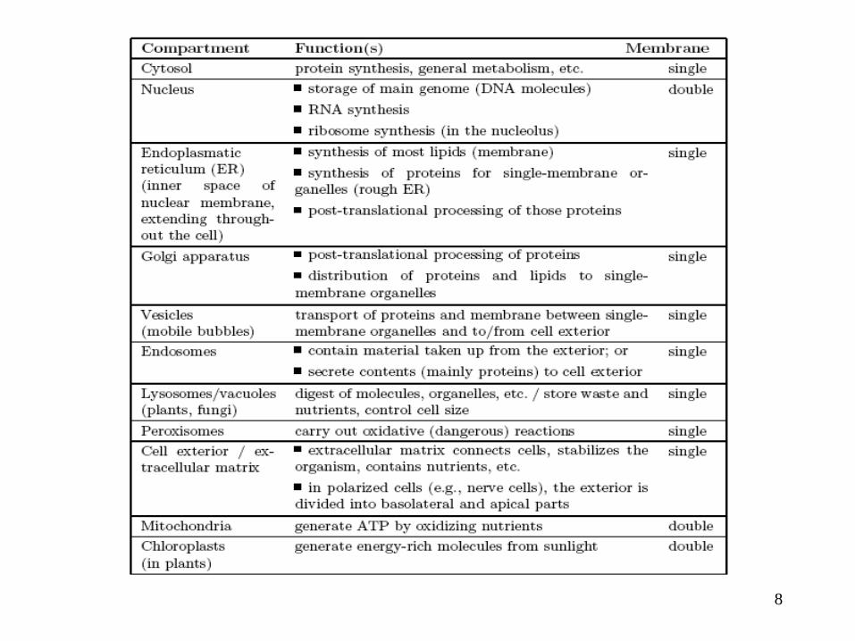
Cell: the basic unit of life
– Every living thing is made of cells.
– Every cell comes from a pre-existing cell.
3

Tree of Life
4
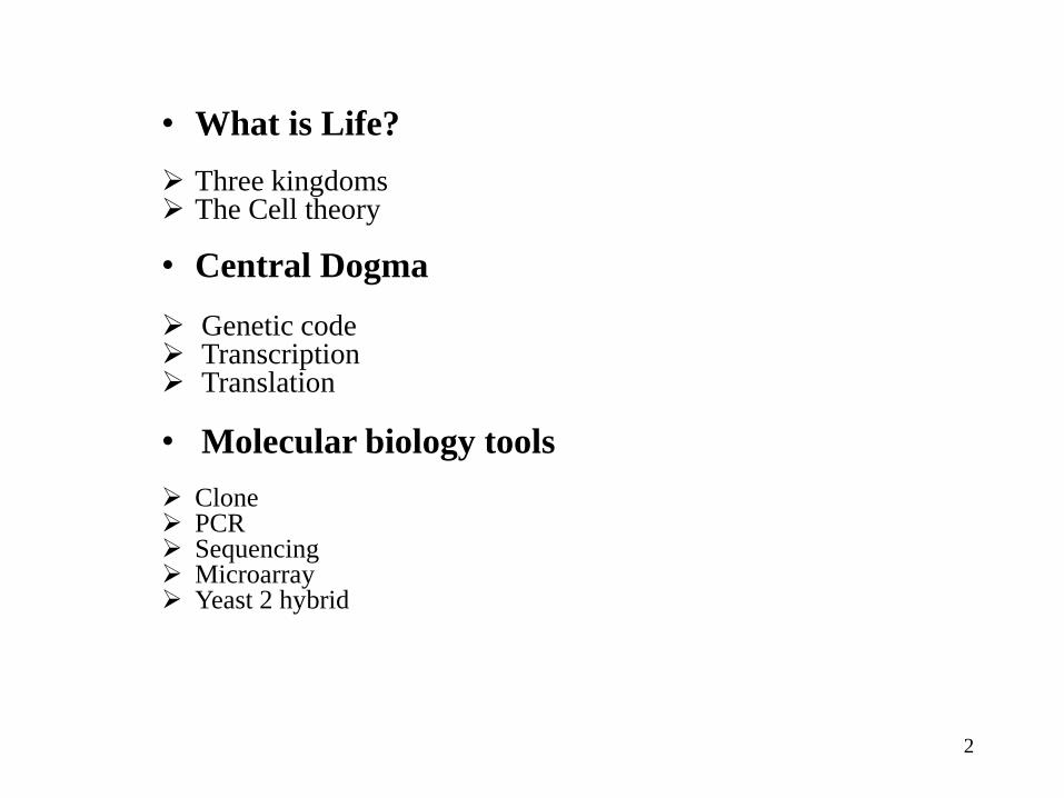
10-3 10-6 10-9
5

6

7
8

9

10
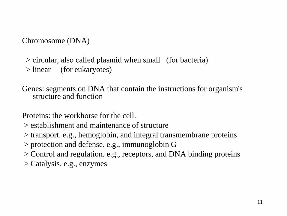
Chromosome (DNA)
> circular, also called plasmid when small (for bacteria)
> linear (for eukaryotes)
Genes: segments on DNA that contain the instructions for organism's structure and function
Proteins: the workhorse for the cell.
> establishment and maintenance of structure
> transport. e.g., hemoglobin, and integral transmembrane proteins
> protection and defense. e.g., immunoglobin G
> Control and regulation. e.g., receptors, and DNA binding proteins
> Catalysis. e.g., enzymes
11

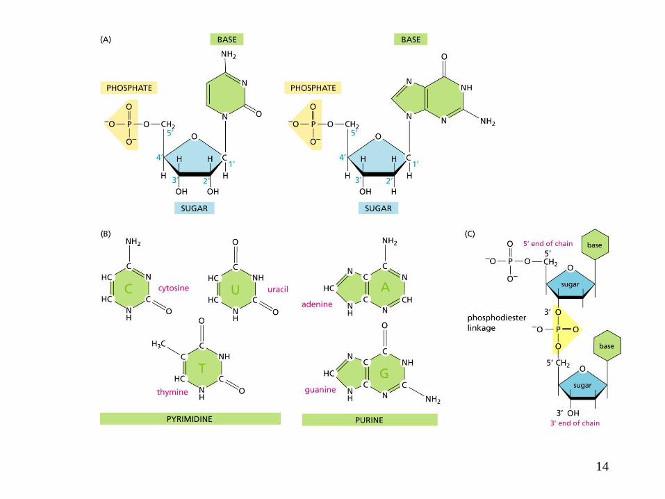
Small molecules:
> sugar: carbohydrate
> fatty acids
> nucleotides: A, C, G, T (Purines: A and G; Pyrimidines: C and T)
12

1
5
• Purines:A and G
• Pyrimidines: C and T
• Oligonucleotide: a DNA of a
few tens of nucleotides
• ATP, ADP, AMP
3
Structure of the bases (Thymine is not shown here)
13
14

15

DNA (double helix, hydrogen bond, complementary bases A-T, G-C)
5' end phosphate group
3' end is free
1' position is attached with the base
double strand DNA sequences form a helix via hydrogen bonds between complementary bases
hydrogen bond:
- weak: about 3~5 kJ/mol (A covalent C-C bond has 380 kJ/mol), will break when heated
- saturation:
- specific:
16
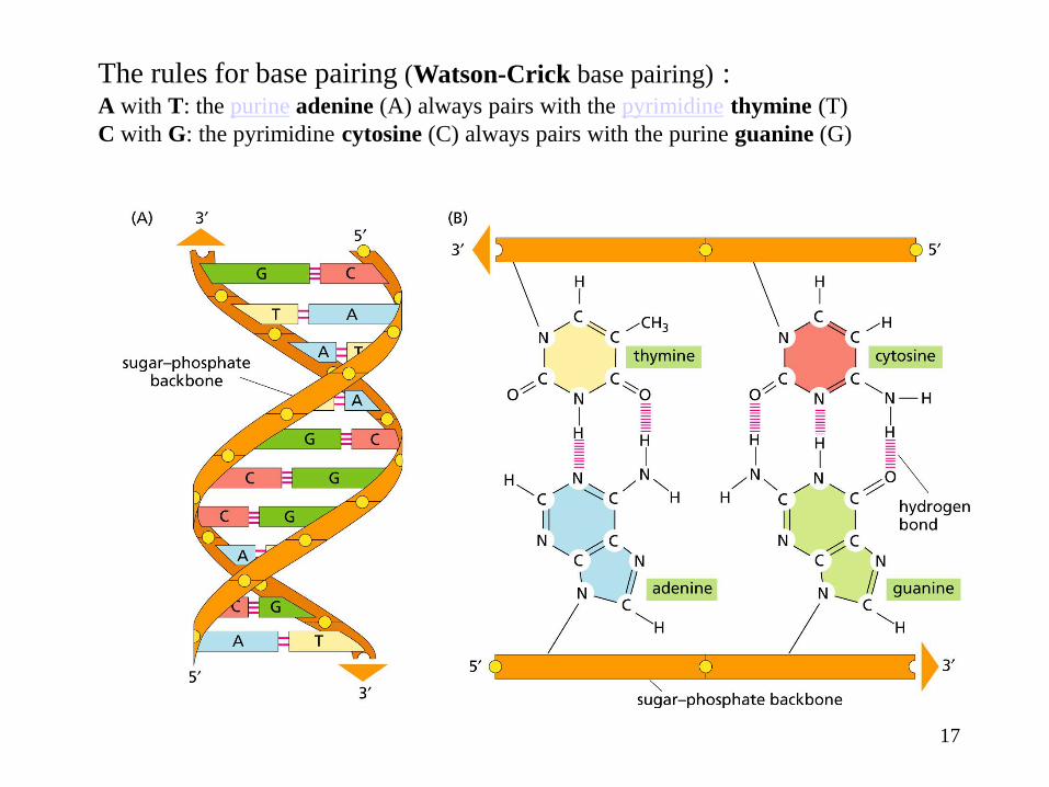
The rules for base pairing (Watson-Crick base pairing) : A with T: the purine adenine (A) always pairs with the pyrimidine thymine (T)
C with G: the pyrimidine cytosine (C) always pairs with the purine guanine (G)
17

DNA replication
18

19

20

21

22

23

Peptide bond
24

C- terminal
N-terminal
Polypeptide
25

26

• Helix complete turn
every 3.6 AAs
• Hydrogen bond
between (-C=O) of one
AA and (-N-H) of its 4th
neighboring AA
27

Hydrogen bond b/w carbonyl oxygen atom on one
chain and NH group on the adjacent chain
28

29

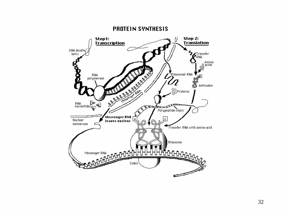
Information Expression
1-D information array 3-D biochemical structure
Central Dogma: DNA RNA Protein
30

Genetic Code: codons
31
32
33

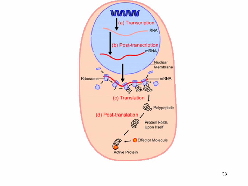
Transcription
34

Translation http://www.youtube.com/watch?v=B6O6uRb1D38
35

36

Gene Structure
5’ regulatory domains
Transcriptional control
Exons
Introns
DNA
3’ regulatory domains
Post-transcriptional processing: hnRNA to mRNA
Protein
Translation: mRNA to protein
37

How complex can a 4 letter code
really be? atcgggctatcgatagctatagcgcgatatatcgcgcgtatatgcgcgcatattag tagctagtgctgattcatctggactgtcgtaatatatacgcgcccggctatcgcgct atgcgcgatatcgcgcggcgctatataaatattaaaaaataaaatatatatatatgc tgcgcgatagcgctataggcgcgctatccatatataggcgctcgcccgggcgcga tgcatcggctacggctagctgtagctagtcggcgattagcggcttatatgcggcga gcgatgagagtcgcggctataggcttaggctatagcgctagtatatagcggctagc cgcgtagacgcgatagcgtagctagcggcgcgcgtatatagcgcttaagagcca aaatgcgtctagcgctataatatgcgctatagctatatgcggctattatatagcgca gcgctagctagcgtatcaggcgaggagatcgatgctactgatcgatgctagagca gcgtcatgctagtagtgccatatatatgctgagcgcgcgtagctcgatattacgcta cctagatgctagcgagctatgatcgtagca…………………………………….
38

• Alternative splicing
– Exception to the “One gene one protein” rule.
• Codon usage
– http://www.kazusa.or.jp/codon/
39
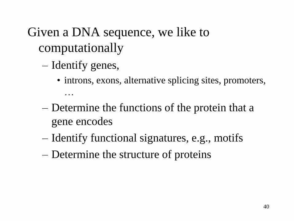
Given a DNA sequence, we like to
computationally
– Identify genes,
• introns, exons, alternative splicing sites, promoters,
…
– Determine the functions of the protein that a
gene encodes
– Identify functional signatures, e.g., motifs
– Determine the structure of proteins
40

41
Molecular biology tools
Clone PCR Sequencing Microarray Yeast 2 hybrid

DNA Cloning
Courtesy of Color Atlas of Biochemistry
42

Restriction endonucleases
43
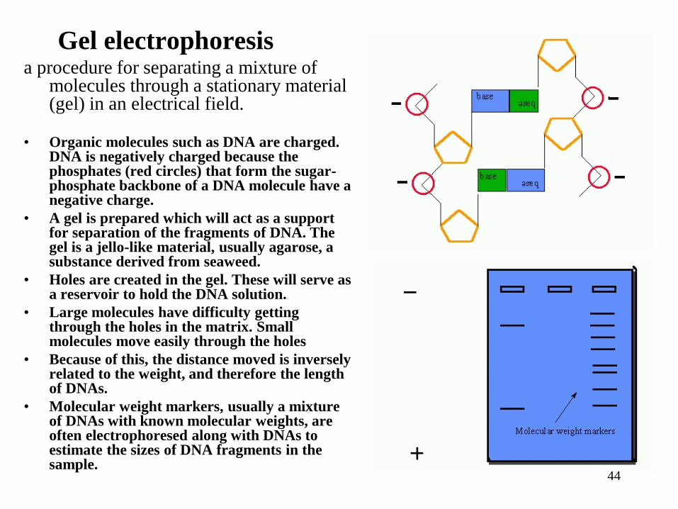
Gel electrophoresis a procedure for separating a mixture of
molecules through a stationary material (gel) in an electrical field.
• Organic molecules such as DNA are charged. DNA is negatively charged because the phosphates (red circles) that form the sugar-phosphate backbone of a DNA molecule have a negative charge.
• A gel is prepared which will act as a support for separation of the fragments of DNA. The gel is a jello-like material, usually agarose, a substance derived from seaweed.
• Holes are created in the gel. These will serve as a reservoir to hold the DNA solution.
• Large molecules have difficulty getting through the holes in the matrix. Small molecules move easily through the holes
• Because of this, the distance moved is inversely related to the weight, and therefore the length of DNAs.
• Molecular weight markers, usually a mixture of DNAs with known molecular weights, are often electrophoresed along with DNAs to estimate the sizes of DNA fragments in the sample.
44

Gel electrophoresis
apparatus - An
agarose gel is placed
in this buffer-filled
box and electrical
field is applied via the
power supply to the
rear. The negative
terminal is at the far
end (black wire), so
DNA migrates toward
the camera.
45

The bases (complementary to the template) are coupled to the primer on the 3' side (the
polymerase adds dNTP's from 5' to 3', reading the template from 3' to 5' side, bases are added
complementary to the template)
46
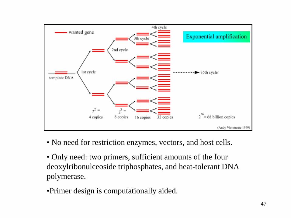
• No need for restriction enzymes, vectors, and host cells.
• Only need: two primers, sufficient amounts of the four
deoxylribonulceoside triphosphates, and heat-tolerant DNA
polymerase.
•Primer design is computationally aided.
47
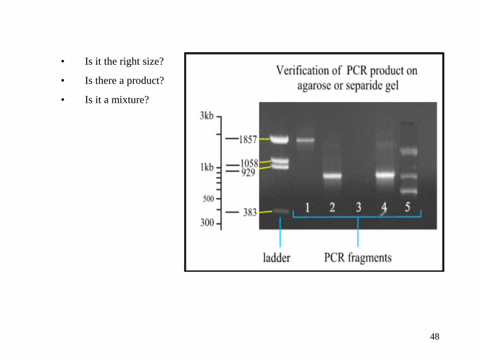
• Is it the right size?
• Is there a product?
• Is it a mixture?
48

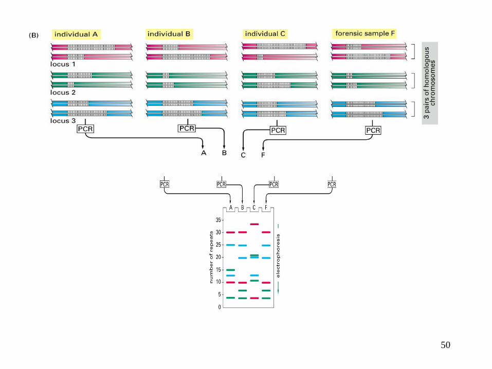
How PCR is used in forensic science
49
50

Evolution of Sequencing
Technology
• ABI Platform
• 1988
– 16 samples per/day ~250 bp/sample
• 1992
– 48 samples 3 times/day ~450 bp/sample
• 1996-8
– 96 samples 4 times/day ~600 bp/sample
• 2006
– 96 samples 4 times/day ~700-800 bp/sample
• 454
– 300,000 – 400,000 samples
2 times per day 100
bp/sample
• Solexa
– 2,000,000 – 3,000,000
samples once every 3 days
35-50 bp/sample
– 40,000,000 samples once
every 3 days 50 bp/sample
51

Sequencing DNA (Sanger’s method)
Courtesy of Color Atlas of Biochemistry 52

53

Chromatograph of Sequence data
54

• Four nucleotides are added stepwise to the template hybridized to a primer.
• Incorporation of a deoxynucleotide, determined by complementing with the template,
will release PPi and light, which can be detected by a CCD (charge-coupled device)
camera
• Unincorporated deoxynucleotides and the produced ATP are degraded between each
cycle by the nucleotide-degrading enzyme.
Sequencing by synthesis
55
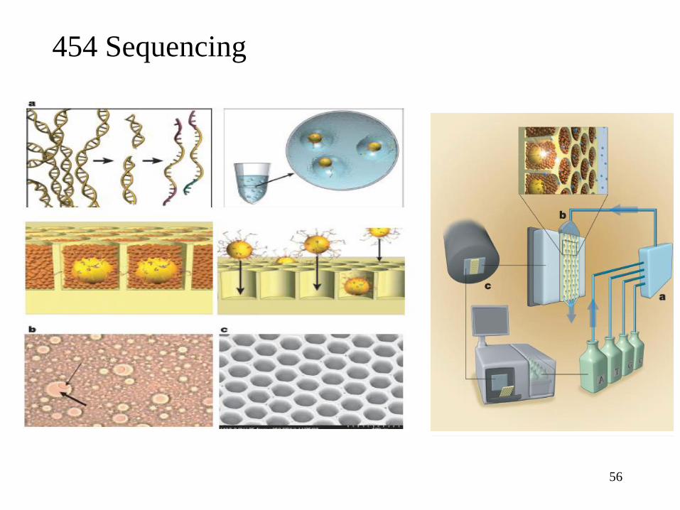
454 Sequencing
56

SBS Sequencing. cDNA or genomic template (nebulized DNA) is prepared by the addition
of 5’ and 3’ adapters. This is diluted and applied to the surface of a chip pre-coated with a
dense lawn of primers (A); “bridge-PCR” with unlabelled nucleotides is used to create a
PCR colony covalently linked to the surface. This creates millions of dense clusters of
dsDNA template (B). Sequencing is performed by addition of four dye-labeled reversible
nucleotide terminators along with the same primers and a custom polymerase. Laser
excitation and image capture is used to determine the first nucleotide for all clusters in
parallel (C). The 3’ blocked terminus is removed along with the fluorophore (dye), and the
second bases are determined the same way. The process continues for 25-35 nucleotides.
(Figure Modified from www.solexa.com).
SBS Sequencing
57

58
59
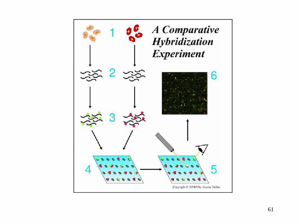
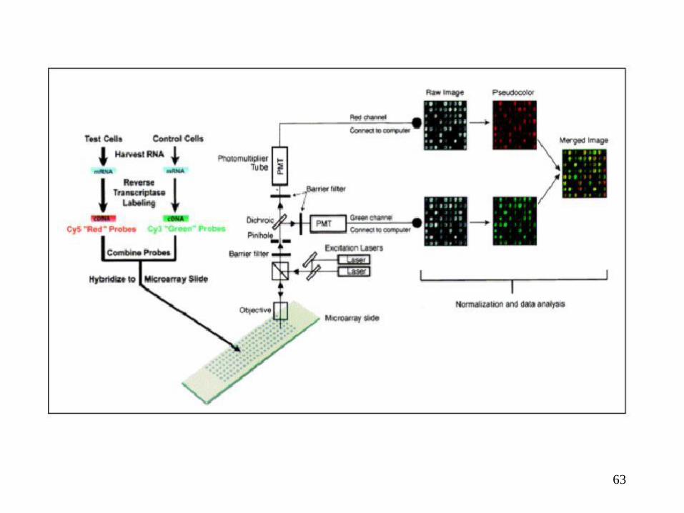
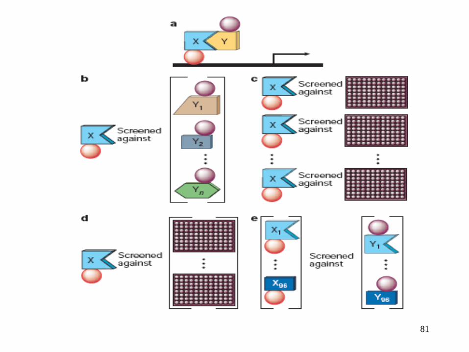
DNA Microarray, 2d gel, MSMS, yeast
2-hybrid.
60
Gene expression
– How many copies of a gene (its product) is present in the cell?
– For experimental reasons, gene expressions are measured by numbers of mRNAs, not directly by proteins. (See Proteomics)
– Various cell types are due to different genes expressed.
– The difference between diseased (e.g., cancerous) and non-diseased
– Diseased cells are often resulted from the abnormal levels of expression of key genes.
61

62
• Microarray
– Oligonucleotide (Affymetrix) array
• Oligo (~ 25 bases long)
• High density (1cm2 contain 100k oligos)
– cDNA array
• cDNA (RT-PCR), much longer (> 1000 bases)
• Varied density of cDNA on each spot, hybridization depends
on length
• Less possibility for false positives
– Image processing
– Background subtraction
– Normalization
63

64

65
Applications • Inferring transcription regulatory
networks
• Understanding correlation between
genotype and phenotype
• predicting genotype <=> phenotype
• Phenotypes:
– drug/therapy response
– drug-drug interactions for expression
– drug mechanism
– interacting pathways of metabolism
66
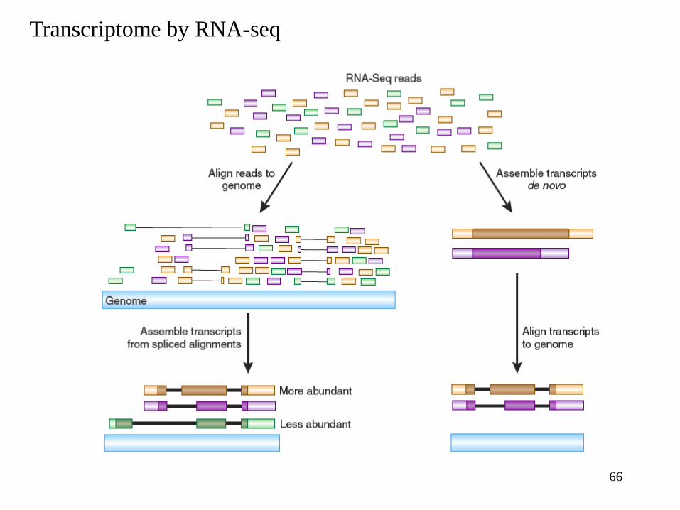
Transcriptome by RNA-seq

67
What is proteomics?
• Like genomics is the study of all genes in a genome, proteomics is the study of all proteins of a cell at a given time.
• Three aspects – Biological process (why is this being done? e.g. movement of cell)
– Molecular function (what kind of molecule is this? e.g., ATPase)
– Cellular component (where is this located? e.g., ribosome)
Why is it difficult?
• Moving target – Cell-to-cell variations
– Cell behavior changes with time
• Lack of high throughput technology – Protein chips? Protein sequences do not have hybridization that
DNA sequences have.
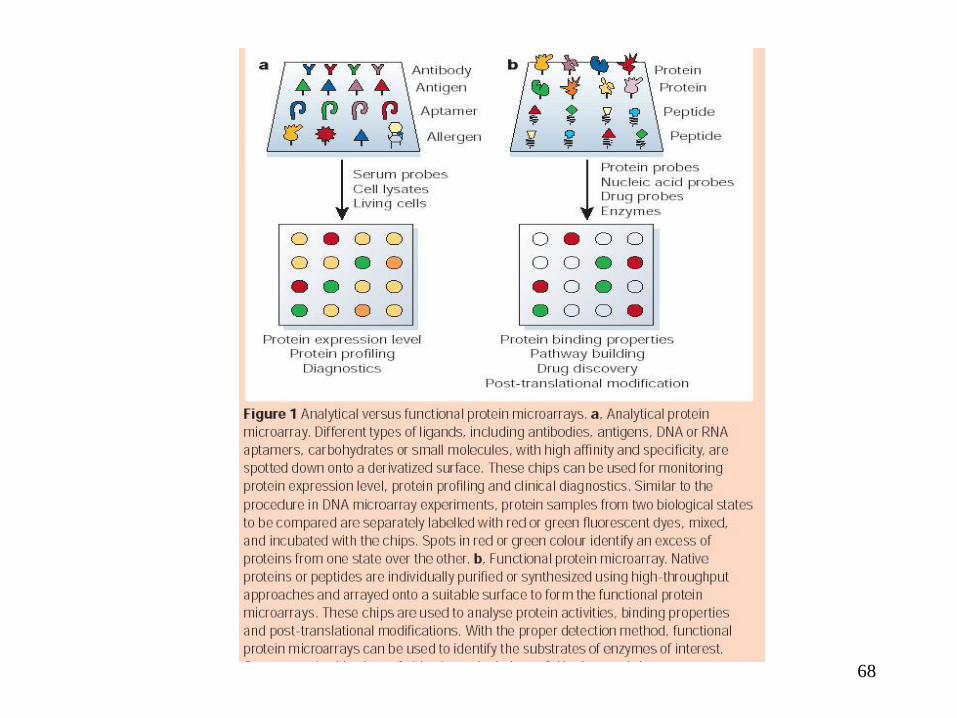
68

69
2D gel electrophoresis
– Isoelectric points (first dimension)
– Molecular weights (second dimension)
Both pI and MW are functions of amino acid sequence of a protein.
Some proteins do not resolve well by 2D gels.
Issues:
• Detection of spots (image processing)
• Quantification of each spot
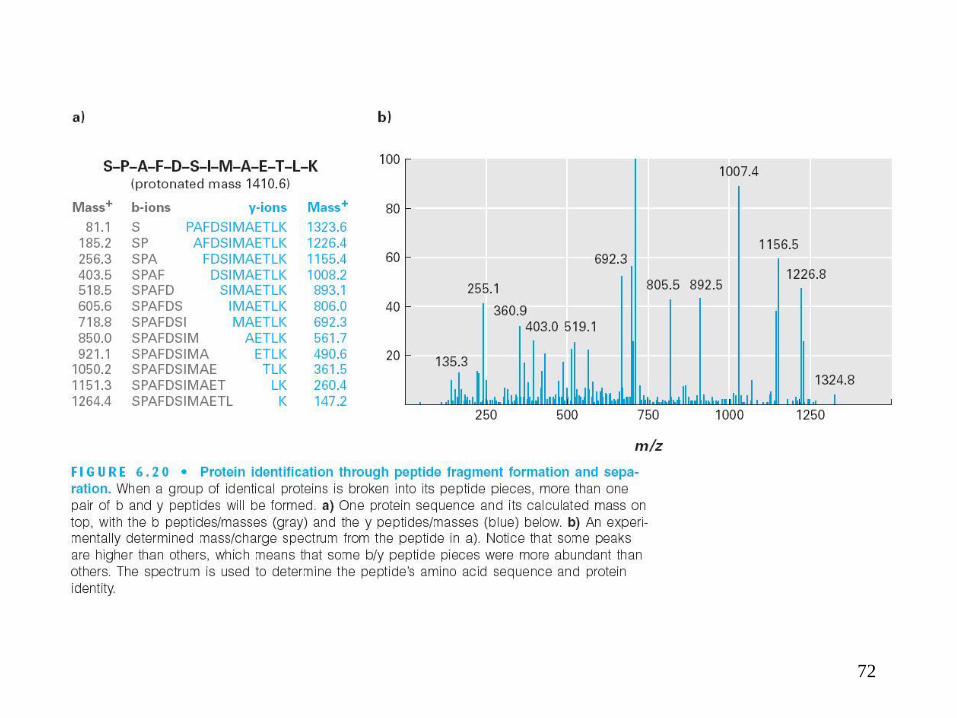
• Identification of each spot (Mass Spectrometry)

70

71
72

73

74
75
76

77
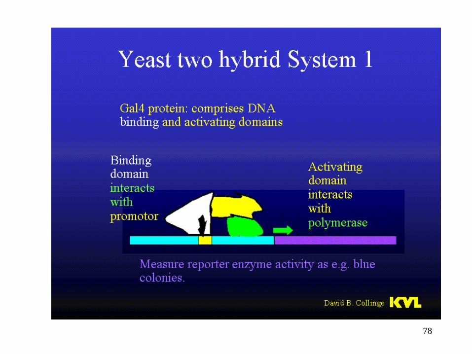
78

79
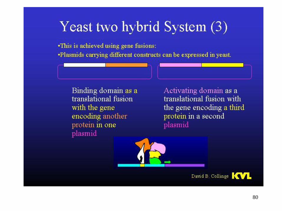
80
81