Embed Size (px)
Citation preview

Annu. Rev. Biophys. Biomol. Struct. 1995. 24:85-116 Copyright © 1995 by Annual Reviews Inc. All rights reserved
MOLECULAR AND STRUCTURAL BASIS OF TARGET RECOGNITION BY CALMODULIN Anna Crivici and Mitsuhiko Ikura
Division of Molecular and Structural Biology, Ontario Cancer Institute and
Department of Medical Biophysics, University of Toronto, Toronto, Ontario, Canada M4X lK9
KEY WORDS: calcium binding, conformational change, molecular model, regulation, signal transduction
CONTENTS PERSPECTIVES AND OVERVIEW . . . . . . ............. ............. ................ ......... 86 CALMODULIN-DEPENDENT ENZYMES AND PROTEINS . . . . . . ........ ........ 87
Protein Kinases and Phosphatases . . . . .. ..... . . . . . . . . . . . . . . . . . . . . . . . . . . . .. . . . . . . ..... . . . . . 87 Proteins Involved in Second-Messenger Generation . . . . . . . . . . . . ...... .......... ..... 89 Proteins Involved in Regulation of Cytoskeletal Elements ....... .... ............ ... 89
STRUCTURAL ASPECTS OF CALMODULIN-TARGET INTERACTIONS . . 89 Calmodulin-Binding Domains on Target Proteins . . . . . . . . . . .. ................ ......... 89 Structure of Ca2 + -Calmodulin .............................................................. 90 Conformational Changes upon ctl+ Binding . . . . . . . . . . . . . . . . . . . . . . . . . . . .. . . . . . ...... . . 94 Models of the Calmodulin-Target Complex . . . . . . . . . . . . . . . . . . . . .. . . . . . . . . . . . . . . . . . . . . . . . 94 Structure of the Calmodulin-Target Peptide Complex . . . . . .... . . . . . . . . . . . . . . . . . . . . . 96 Calcium-Independent Calmodulin-Target Interactions.. .... . ....... ................. 103
POSTTRANSLATIONAL MODIFICATIONS OF CALMODULIN AND TARGET PROTEINS .......................... ..................................... .............. 104
Phosphorylation of Target Proteins . . . . . . . . . . . . .. ............ ................ ............. 104 Posttranslational Modification of Calmodulin.......................................... 107
SUMMARY AND FUTURE DIRECTIONS . . . . . . . . ...... ............ ................ ..... 108
ABSTRACT Calmodulin (CaM) acts as an intracellular calcium sensor that translates the Ca2 + signal into a variety of cellular processes. Ca2 + -CaM recogni-
85 1056-8700/95/0610-0085$05.00
Ann
u. R
ev. B
ioph
ys. B
iom
ol. S
truc
t. 19
95.2
4:85
-116
. Dow
nloa
ded
from
ww
w.a
nnua
lrev
iew
s.or
gby
Roy
al M
elbo
urne
Ins
titut
e of
Tec
hnol
ogy
(RM
IT)
on 0
9/30
/13.
For
per
sona
l use
onl
y.
86 CRIVICI & IKURA
tionuf a short polypeptide segment in target proteins induces conformational changes in both CaM and the target, enabling the target protein to become functionally active. The solution and crystal structures of Ca2+ -CaM bound to peptides derived from three CaM-dependent enzymes reveal structural features that are common in target recognition by Ca2+ -CaM. Phosphorylation of the target proteins at sites in or near the CaM-binding region modulates binding of CaM, thereby providing an additional mechanism of functional regulation. The structural aspects of target recognition by Ca2 + -CaM are discussed using mainly the three-dimensional structural information obtained with nuclear magnetic resonance spectroscopy and X-ray diffraction methods.
PERSPECTIVES AND OVERVIEW Calmodulin (CaM) is a small (148 residues), ubiquitous, highly conserved protein that is an integral modulator of many calcium-dependent processes in virtually every eukaryotic cell type. It functions as a cytosolic calcium receptor and binds four calcium ions with high affinity (Kd 10-5 to 10-6 M) in response to a variety of extracellular signals (reviewed in 39, 75). Saturation of the calcium-binding sites induces a conformational change that enables Ca2 + -CaM to recognize and bind target proteins with high affinity (Kd 10-7 to 10-11 M) (75). Interaction of CaM with its targets further induces a conformational change in Ca2+ -CaM and the target to yield an activated target protein complex. A number of natural peptides also bind CaM with high affinity, including several hormones, neurotransmitters, and venoms (91-93). Because these are either exogenous or secreted peptides, the physiological relevance of these interactions is unclear.
Many Ca2+ -CaM-dependent proteins are involved in cell signaling through phosphorylation or dephosphorylation of intracellular proteins or participate in signal transduction by modulating levels of intracellular second messengers such as Ca2+, cAMP, cGMP, and nitric oxide (e.g. 21,48,49,88, 141). Another important area of CaM regulation involves the regulation of cytoskeletal elements (e.g. 2, 127, 139). New cellular targets of CaM are continuously being identified, but their functions and the role of their interactions with CaM often remain unknown. Biochemical characterization of various CaM -binding proteins have defined a role for CaM as an integrator of several different signal-transduction pathways as well as a mediator of Ca2 + -dependent cellular events.
The functional role of the interaction of CaM with target proteins and the molecular and structural features of the mode of recognition and regulation by CaM are intriguing. Although no single consensus
Ann
u. R
ev. B
ioph
ys. B
iom
ol. S
truc
t. 19
95.2
4:85
-116
. Dow
nloa
ded
from
ww
w.a
nnua
lrev
iew
s.or
gby
Roy
al M
elbo
urne
Ins
titut
e of
Tec
hnol
ogy
(RM
IT)
on 0
9/30
/13.
For
per
sona
l use
onl
y.

TARGET RECOGNITION BY CALMODULIN 87
sequence for CaM recognition exists, the complexes formed are highly specific and of high affinity. Activation of the majority of CaM-dependent proteins requires Ca2+ (see section on calcium-independent calmodulin-target interactions for exceptions). The Ca2+ -saturated form of CaM is conformationally distinct from the Ca2 + -free form, which adds to the complexity of the mode of target recognition because the formation of the ternary complex requires adjustments in the structure of CaM as well as a conformational change in the target that elicits activation.
Because most of the target proteins are large and multimeric, CaMtarget complexes have been difficult to study at the molecular level. Consequently, much of the structural information available has been obtained from the study of CaM complexed with peptide venoms (72, 91, 93), model peptides (24, 32, 112), and peptide fragments of the CaMbinding domains of several target proteins (e.g. 14, 63, 100, 101, 134, 157). Functional studies (e.g. 42, 69, 72, 79, 121, 128, 146) using structurally modified CaM have also contributed to the elucidation of the different modes of recognition and complex formation found in different types of targets.
Several comprehensive reviews describing structural (24, 113, 153, 163) and functional (103, 124) aspects of CaM are available, including collections edited by Cohen & Klee (26), Means & Conn (102), and Carafoli & Klee (18). This review includes a description of the current model of CaM-target interaction and focuses on the structural and conformational features required for recognition and activation by CaM and their relevance to the biological activities of this protein and its targets. We briefly discuss the results of investigations into the regulation of CaM-target interactions by structural modifications; this examination defines a role for CaM in the integration of different intracellular signal-transduction pathways.
CALMODULIN-DEPENDENT ENZYMES AND PROTEINS Protein Kinases and Phosphatases CaM-dependent protein kinases and phosphatases, which include both dedicated and multifunctional enzymes, seem to share a common mode of CaM recognition and regulation. The best-studied members of this group include skeletal- and smooth-muscle myosin light chain kinases (MLCKs) (14, 89, 124), multifunctional CaM-dependent protein kinase II (CaMKII) (27, 29, 50, 120), CaM-dependent protein phosphatase-2B
Ann
u. R
ev. B
ioph
ys. B
iom
ol. S
truc
t. 19
95.2
4:85
-116
. Dow
nloa
ded
from
ww
w.a
nnua
lrev
iew
s.or
gby
Roy
al M
elbo
urne
Ins
titut
e of
Tec
hnol
ogy
(RM
IT)
on 0
9/30
/13.
For
per
sona
l use
onl
y.
88 CRIVICI & IKURA
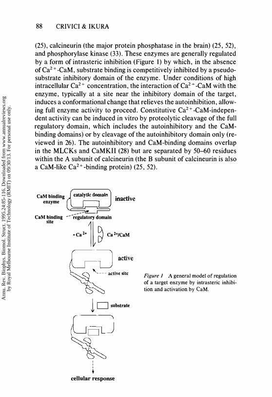
(25), calcineurin (the major protein phosphatase in the brain) (25, 52), and phosphorylase kinase (33). These enzymes are generally regulated by a form of intrasteric inhibition (Figure 1) by which, in the absence of Ca2 + -CaM, substrate binding is competitively inhibited by a pseudosubstrate inhibitory domain of the enzyme. Under conditions of high intracellular Ca2 + concentration, the interaction of Ca2 + -CaM with the enzyme, typically at a site near the inhibitory domain of the target, induces a conformational change that relieves the auto inhibition , allowing full enzyme activity to proceed. Constitutive Ca2+ -CaM-independent activity can be induced in vitro by proteolytic cleavage of the full regulatory domain, which includes the autoinhibitory and the CaMbinding domains) or by cleavage of the autoinhibitory domain only (reviewed in 26). The auto inhibitory and CaM-binding domains overlap in the MLCKs and CaMKII (28) but are separated by 50-60 residues within the A subunit of calcineurin (the B subunit of calcineurin is also a CaM-like Ca2+ -binding protein) (25, 52).
CaM binding enzyme
CaM binding site ---r;�iatory domain
-�'l � �U/�
1 D substrate
I I I +
cellular response
Figure 1 A general model of regulation of a target enzyme by intrasteric inhibition and activation by CaM.
Ann
u. R
ev. B
ioph
ys. B
iom
ol. S
truc
t. 19
95.2
4:85
-116
. Dow
nloa
ded
from
ww
w.a
nnua
lrev
iew
s.or
gby
Roy
al M
elbo
urne
Ins
titut
e of
Tec
hnol
ogy
(RM
IT)
on 0
9/30
/13.
For
per
sona
l use
onl
y.

TARGET RECOGNITION BY CALMODULIN 89
Proteins Involved in Second-Messenger Generation CaM-dependent cyclic nucleotide phosphodiesterase (PDE) (21, 111), type I adenylyl cyclase (83, 160), plasma membrane Ca2+ -ATPase (35, 159), and nitric oxide synthase (88, 160) are all CaM-dependent enzymes involved in signal transduction through generation or regulation of intracellular second messengers. In addition, inositol trisphosphate receptors bind CaM, but the role of CaM binding is presently unclear (41).
The molecular mechanism of CaM regulation has not been defined for all of these proteins but may follow a general mode of regulation analogous to those of the protein kinases and phosphatases. For example, interaction of CaM with the CaM domain of PDE relieves inhibition caused by an autoinhibitory domain (22). The CaM-binding domain of Ca2+ -ATPase likely interacts with two discontinuous sites on the energy-transduction domain (which couples Ca2 + transport to the ATPase activity) in the latent state, restricting the mobility of the cytoplasmic domain. Interaction with Ca2+ -CaM induces a conformational change that dissociates the CaM-binding and transduction domains, allowing Ca2+ transport and ATPase activity to proceed (35). Interaction between Ca2 + -CaM and these proteins enables cross-talk between the Ca2+ -CaM pathway and other signal-transduction pathways.
Proteins InvoLved in Regulation of CytoskeletaL ELements
The molecular basis of CaM regulation and the functional relevance of CaM interaction with cytoskeletal proteins and their regulatory proteins is not clearly defined. CaM-binding proteins thought to participate in various cytoskeleton-mediated events such as motility, cell growth and development, and morphogenesis include spectrin (149,150), f3-adducin (139), caldesmon (59, 94, 169), brush border myosin-I (BBMI) (30, 154), and the myristoylated alanine-rich C kinase substrate (MARCKS) (2, 12) and a related protein, F52 (12, 13). Details on the interaction of CaM with BBMI and MARCKS are described in later sections (CalciumIndependent Calmodulin-Target Interactions and Phosphorylation of Target Proteins, respectively).
STRUCTURAL ASPECTS OF CALMODULINTARGET INTERACTIONS Calmodulin-Binding Domains on Target Proteins
The identification of calmodulin-target proteins among Ca2 + -dependent proteins has largely been based on binding affinity and a functional
Ann
u. R
ev. B
ioph
ys. B
iom
ol. S
truc
t. 19
95.2
4:85
-116
. Dow
nloa
ded
from
ww
w.a
nnua
lrev
iew
s.or
gby
Roy
al M
elbo
urne
Ins
titut
e of
Tec
hnol
ogy
(RM
IT)
on 0
9/30
/13.
For
per
sona
l use
onl
y.

90 CRIVICI & IKURA
requirement for CaM (26, 113). CaM dependency is characterized by activation constants in the nanomolar range (75) and by a sensitivity to inhibition by CaM-binding drugs (26, 70) and high-affinity peptide targets of CaM (6, 34).
The CaM-binding domains of the targets have generally been mapped using limited proteolysis or by means of deletion, truncation, and sitedirected mutagenesis. Extensive mapping is also carried out using synthetic peptides and proteolytic fragments that incorporate full or partial sequences of the protein segment of interest. Positive identification of a sequence as a CaM-binding domain requires that the synthetic peptide retain a high affinity for CaM, with a Kd value comparable to those of the intact target protein (26) and with an ability to inhibit CaM binding to the native target protein as well as the native protein's CaM-dependent activity (26). In some cases, the precise boundaries of the binding domain have not been positively determined because of the complexity in the regulatory mechanisms of these target proteins.
Examination of the CaM-binding sequences of various target proteins reveals that the CaM-binding domain is limited to a short region of 14-26 residues that has a propensity to form a basic amphiphilic ahelix (reviewed in 113). DeGrado and coworkers (34) have developed a computer algorithm that can be used to examine cDNA libraries or protein sequences for potential CaM-binding domains. This technique involves a quantitative evaluation of a sequence on the basis of its electronic and hydrophobic properties and secondary-structure propensity to identify putative basic amphiphilic a-helical motifs (reviewed in 113). This approach has been used to correctly predict several CaMbinding domains (e.g. 4, 15,20, 160) but has also identified regions that do not have high CaM affinity when reproduced within a synthetic peptide (33, 73, 94). However, residues in the primary sequence of known CaM-binding domains do not always exhibit a propensity to form an amphipathic a-helix (Table 1). In phosphorylase kinase, one segment of the CaM-binding domain, reproduced in the synthetic peptide PhK13, is predicted to form an extended (3-turn-(3-sheet structure (33).
Structure of Ca2+ -Calmodulin The crystal structure of Ca2+ -CaM, first solved by Babu et al in 1985 (9), subsequently solved by Kretsinger et al (81), and further refined by several groups (8, 23, 130), has provided a starting point for the development of a model of the Ca2 + -CaM -target protein complex and for describing the conformational changes that must occur upon Ca2+ binding to CaM and upon target complexation. The crystal form of
Ann
u. R
ev. B
ioph
ys. B
iom
ol. S
truc
t. 19
95.2
4:85
-116
. Dow
nloa
ded
from
ww
w.a
nnua
lrev
iew
s.or
gby
Roy
al M
elbo
urne
Ins
titut
e of
Tec
hnol
ogy
(RM
IT)
on 0
9/30
/13.
For
per
sona
l use
onl
y.

TARGET RECOGNITION BY CALMODULIN 91
N-terminal domain
C-terminal domain
Figure 2 X-ray crystal structure of Ca2+ -CaM determined at 2 .2-A. resolution. Residues 78-81 of the interconnecting helix (residues 65-93) show some deviations from ideal helix geometry (8, 23). The structure was reproduced from atomic coordinates deposited by Babu et al (8) in the Brookhaven Protein Data Bank (structure code 3CLN).
Ca2+ -CaM (Figure 2) is a dumbbell-shaped molecule, approximately 65 A long, with N- and C-terminal globular domains or lobes (25 x 20 x 20 A) separated by an interconnecting helix of approximately eight turns. Each globular domain contains two Ca2+ -binding sites of the EF-hand (helix-loop-helix) type related by a two-fold axis of symmetry. The globular lobes are cup shaped and have a concave hydrophobic surface in the center of each and negatively charged residues at the rims. The extreme ends of the interconnecting helix form part of the helix-loop-helix Ca2+ sites, and the central six residues (76-82) are solvent exposed. NMR studies (10, 67) indicate that, in solution, residues 78-81 adopt a nonhelical conformation with considerable flexibility. In the refined crystal structure, residues 79-81 show some deviations from ideal helix geometry and contain some of the highest temperature factors in the molecule (8, 23).
Ann
u. R
ev. B
ioph
ys. B
iom
ol. S
truc
t. 19
95.2
4:85
-116
. Dow
nloa
ded
from
ww
w.a
nnua
lrev
iew
s.or
gby
Roy
al M
elbo
urne
Ins
titut
e of
Tec
hnol
ogy
(RM
IT)
on 0
9/30
/13.
For
per
sona
l use
onl
y.

\0 N
n :;c <: B R<>
� C :;c >--
Table 1 Primary sequences of some known and putative calmodulin binding domains of protein and peptide calmodulin targets
Targeta Sequenceb Reference
skMLCK (MI3)C K R R W K K NF l A V 5 A A N R F K K I 5 SSG A L 14
smMLCK (smMLCKp) A R R K W Q K T G H A V R A I G R L 5 5 88 CaMKII A R R K L K G A I L T T M L A T R N F 5 III Caldesmon G V R N I K 5 M W E K G N V F 5 5 169 Calspermin A R R K L K A A V K A V V ASS R L G 5 120
PFK (Mil) F M NN W E V ! K L L A H I R P PAP K 5 G 5 Y T V 15 Calcineurin ARK E V I R N K I R A I G K M AR V F 5 V L R 74
PhK (PhK5) L R R L ID A Y A F R I Y G H W V K K G Q Q Q N R G 33
(PhKI3) R G K F K V I C L TV L AS V R I Y Y Q Y R R V K P G 33 Ca2+ -ATPase (C28W) L R R G Q I L W F R G L N R I Q T Q I K V V N A F 5 5 5 159 59-kOa POE R R K H L Q R P I F R L R C L V K Q L E K 160 6O-kDa POE T E K M H Q R L K G IL R C L V K Q L E K III
NOS (NO-30) K R R A I G F K K L A E A V K F 5 A K L M G Q 160
Type I AC (AC-28) K P A K R M K £ K T V C Y L L V Q L M HeR K M F K A 160
Bordetella pertussis AC ! D L L W K I A RAG A R 5 A V G TE A 118
Neuromodulin K A H K A A T K I Q A 5 F R G H IT R K K L K G E K K 20
Spectrin K T ASP W K 5 A R L M V H T V A T F N 5 IKE 85
MARCKS KKK K K R £ 5 F K K 5 F K L 5 G F 5 £ K K 5 K K 45
Ann
u. R
ev. B
ioph
ys. B
iom
ol. S
truc
t. 19
95.2
4:85
-116
. Dow
nloa
ded
from
ww
w.a
nnua
lrev
iew
s.or
gby
Roy
al M
elbo
urne
Ins
titut
e of
Tec
hnol
ogy
(RM
IT)
on 0
9/30
/13.
For
per
sona
l use
onl
y.

F52 or MacMARCKS KKK K K E 5 F K K P F K L 5 G L 5 E K R N R K 13 /3-Adducin K Q Q K E K T R � L N T P N T Y L R V N V � D E V Q R N M G 5 139 HSP90a K D Q V A N 5 A F Q"E R L R K H G LE V I 106 HIV-l gp160 r H R L R D L L L I V K R ! VEL L G R R 147
BBMHC[ Q Q l A T l 1 Q K T Y R G W R CRT H r Q L M 104 Dilute MHC R A A C I R I Q K T I R G W L l R K R Y l C M Q 104 Mastoparan I N L K A L A A L A K K I h 93 Melittin G G A V L K V l T T G L PAL I 5 W I K R K R Q Q 93
Glucagon H 5 Q G T F T T 5 D Y 5 K Y L D 5 R R A Q D F V Q W L M N T 92 Secretin H 5 D G T F T 5 E h 5 R l R D 5 A R l Q R l h Q G l V 92 VIP H 5 D A V F T D N Y T R l R K Q M A V K K Y L N 5 I l N 92
GIP Y A D G T F I 5 D Y 5 A I M N K IRQ Q D F V N W L L A Q Q Q K 5 92 Model peptide CBP2 K L � K K L L K L L K K L L K h G 34
a Abbreviations: AC, adenylyl cyclase; BBMHCI, brush-border myosin heavy chain-I; CaMKII, calmodulin kinase II; CBP2, calmodulin binding peptide-2; GIP, gastrin inhibitory peptide; HIV-I gpl60, hUman immunodeficiency virus envelope glycoprotein 160; HSP, heat-shock protein; MARCKS, myristoylated alanine-rich C kinase substrate; MHC, myosin heavy chain; NOS. nitric oxide synthase; PDE, phosphodiesterase; PFK. phosphofructokinase; PhK, phosphorylase kinase; sk-, smMLCK, skeletal muscle- and smooth muscle-myosin light chain kinase; VIP, vasoa"tive intestinal peptide.
b Alignment of the CaM domains was made by visual inspection based on alignment of the putatively conserved major (bold and underlined) and minor (bold) hydrophobic anchors that intereact with the hydrophobic patches of the C- and N-terminal domains of CaM (63), an d on the alignment of the conserved basic residue (bQld and italicized) analogous to that residue of MLCK that is required for activation by CaM (100, 101). Precise ooundanes of the CaM-binding domain are not known for all targets.
c Names in parentheses are those used in the literature for the synthetic peptides containing the sequences listed.
� >-� � � tI1 n o a z :::J (5 z OJ -< n >r-' � o ti c:::: r-' Z
�
Ann
u. R
ev. B
ioph
ys. B
iom
ol. S
truc
t. 19
95.2
4:85
-116
. Dow
nloa
ded
from
ww
w.a
nnua
lrev
iew
s.or
gby
Roy
al M
elbo
urne
Ins
titut
e of
Tec
hnol
ogy
(RM
IT)
on 0
9/30
/13.
For
per
sona
l use
onl
y.

94 CRIVICI & IKURA
Studies of Ca2 + -CaM by small-angle X-ray scattering and fluorescence techniques have also indicated the presence of an equilibrium between several conformational substates in solution. The results of these measurements reflect an average conformation that is more compact than the elongated crystal structure, in which the distance between the globular domains varies over a range of values (55, 142).
Conformational Changes upon Ca2+ Binding
Solution techniques, such as NMR (e.g. 10, 36, 62-64, 66, 68, 134, 143, 166, 172), fluorescence (e.g. 19, 91-93, 112, 115, 146) and CD spectroscopy (e.g. 54, 76, 97), and small-angle X-ray scattering (e.g. 15, 30, 31, 54, 55, 71, 73, 76, 78), have also been carried out to elucidate the Ca2 + -induced conformational changes in CaM that occur in the first step in the transduction of the Ca2+ signal by CaM. Early NMR studies (5, 38, 65, 66, 77) indicated a two-step conformational change in CaM upon binding Ca2+ and clearly demonstrated that the C- and N-terminal domains contain high- and low-affinity sites for Ca2+, respectively. Both the affinity and cooperativity of Ca2+ binding is affected by target-complex formation. Kd values for Ca2+ can increase significantly in the presence of target peptides (75). Changes in the cooperativity of Ca2+ binding suggest that some conformational communication occurs between the globular domains of CaM when this molecule is complexed to a target protein or peptide (64, 86, 167). This type of communication has also been observed in studies of CaM containing mutations in the Ca2+ -binding sites that affect the protein's ability to bind Ca2+ . These mutations influence both binding and activation of target proteins (42, 97, 98, 108, 148).
In addition to measuring changes in Ca2 + -binding properties, solution experiments using small-angle X-ray scattering (55, 142) and fluorescence anisotropy measurements of photo-cross-linked CaM (146) indicate that CaM is more compact in the Ca2+ -free state. Upon saturation with Ca2+, the molecule becomes more elongated, and CD measurements indicate that the helical content also increases (56, 79, 95). The elongation of CaM upon binding Ca2 + may be necessary to expose the hydrophobic surfaces on the globular domains to enable target binding (16, 58, 70, 84, 153, 156, 161).
Models of the Calmodulin-Target Complex The dumbbell-shaped conformation of Ca2 + -bound CaM, whether fully elongated as found in the crystal structure or more compact as indicated by the solution studies, has been difficult to reconcile with the high affinities of either CaM-protein or CaM-peptide complexes. Sev-
Ann
u. R
ev. B
ioph
ys. B
iom
ol. S
truc
t. 19
95.2
4:85
-116
. Dow
nloa
ded
from
ww
w.a
nnua
lrev
iew
s.or
gby
Roy
al M
elbo
urne
Ins
titut
e of
Tec
hnol
ogy
(RM
IT)
on 0
9/30
/13.
For
per
sona
l use
onl
y.

TARGET RECOGNITION BY CALMODULIN 95
eral important observations have contributed to the structural models of CaM-target complexes based on the crystal structure of Ca2+ -CaM and on structural and functional studies of CaM-target peptide complexes in solution. Large conformational changes both in CaM and in peptide targets have been evidenced by small-angle X-ray and neutron scattering studies of CaM in complexes with several natural and synthetic peptides (reviewed in 157) and by IH NMR studies that indicate major perturbations in the chemical-shift patterns of the complex (76). Although many of the peptide targets are unstructured in solution (e.g. 76, 169, 170), they adopt a helical conformation within the complex (e.g. 63,76, 100, 101, 134, 169, 170). KIevit et al (76) first made this observation from CD measurements that showed a significant increase in the helical content of the CaM-M13 complex that was greater than that expected from the sum of contributions from the individual components. NMR studies reveal that both melittin (143) and a peptide derived from smMLCK (134) are helical when bound to CaM. DeGrado and coworkers (115) carried out fluorescence studies of CaM complexed with model tryptophan-containing peptides and showed that the emission maxima of these complexes were consistent with a helical conformation of the peptide. Cross-linking experiments of CaM have suggested that both of the globular N- and C-terminal domains of CaM interact with the target (112, 114) and can be in close proximity to each other (72,96, 123). Proteolytic cleavage of Ca2+ -CaM yields independent globular domains that can bind target peptides and proteins with reduced affinity but that cannot activate all target enzymes (69, 125).
The most difficult problem in developing models of CaM-target peptide complexes involves the integrity of the central helical linker found in the crystal structure of Ca2+ -CaM. The refined crystal structure includes some deviations from ideal geometry in the central portion of this helix (9). Mutations and deletions in this region are tolerated in many CaM-target complexes with respect to both binding and activation (122, 129). As previously noted, NMR experiments indicate that residues 78-81 adopt a nonhelical structure with a high degree of flexibility (10, 67).
Persechini & Kretsinger (124), and subsequently O'Neil & DeGrado (113), proposed that each globular domain of CaM could interact with one of two hydrophobic patches approximately 1800 apart in the helical form of the skMLCK peptide, M13. The main concept of this model, which uses the central helix as a flexible tether, proved to be correct (see below), but the detailed mode of interaction between the peptide and the globular domains of CaM could not be accurately predicted.
Strynadka & James (153) proposed a model for the complex of CaM
Ann
u. R
ev. B
ioph
ys. B
iom
ol. S
truc
t. 19
95.2
4:85
-116
. Dow
nloa
ded
from
ww
w.a
nnua
lrev
iew
s.or
gby
Roy
al M
elbo
urne
Ins
titut
e of
Tec
hnol
ogy
(RM
IT)
on 0
9/30
/13.
For
per
sona
l use
onl
y.

96 CRIVICI & IKURA
and mastoparan, a peptide venom similar in sequence to that of helix A of troponin C (151, 152). Although, unlike Kretsinger's model, no attempt was made to alter the conformation of the central helix, the relative orientation of the mastoparan helix and the C-terminal domain of CaM in this model is very similar to that found in NMR- and xray-derived structures of the CaM-M13 complex (see below) (63, 100).
Structure of the Calmodulin-Target Peptide Complex
In 1992, the three-dimensional structure of CaM complexed with M13 was solved by using multidimensional NMR techniques (Figure 3) (63). The conformations of the globular domains are nearly identical to those of the crystal structure of Ca2+ -CaM, but the interconnecting central helix (residues 65-93) is disrupted into two smaller helices (residues 65-73 and 83-93), which form part of the helix-loop-helix Ca2+ -binding sites. The flexible region in the central helix of Ca2 + -CaM (residues 78-81) is expanded in the complex to include residues 74-82, which do not appear to form any important contacts with the target peptide or with the globular domains. The overall shape of the molecule is more compact than the crystal structure of Ca2 + -CaM. The globular domains lie close to one another and, along with the flexible linker, form a hydrophobic channel that passes through the molecule at an approximate 45° angle relative to its long axis. The channel is occupied by the target peptide that adopts a helical conformation. Residues 3-21 of the 26-residue M13 peptide make up the helix and form contacts with the globular domains of CaM. The remaining residues of M13 are disordered and lie outside of the channel.
The target peptide M13 contains two important hydrophobic residues, Trp4 and Phe17, that form numerous contacts with the C- and Nterminal domains of CaM, respectively (Figure 4). These two residues establish hydrophobic interactions between the target and CaM and may represent anchors that determine the relative orientation of the target within the channel formed by the globular domains (Figure 5). Other hydrophobic residues of the peptide, Phe8, Ile9, AlalO, Valli, Ala13, and Ala14, also contribute to the hydrophobic contacts with the globular domains of CaM (Figure 4). In addition to the hydrophobic interactions, several possible electrostatic interactions can be deduced from the NMR-derived structure (63).
Almost at the same time that the NMR solution structure of CaMM13 was solved, the crystal structure of the analogous complex with a peptide from smMLCK (smMLCKp) was determined by Meador et al (100). The overall features of the complex are very similar to those of the solution structure of Ikura et al (63). The orientation of the helical
Ann
u. R
ev. B
ioph
ys. B
iom
ol. S
truc
t. 19
95.2
4:85
-116
. Dow
nloa
ded
from
ww
w.a
nnua
lrev
iew
s.or
gby
Roy
al M
elbo
urne
Ins
titut
e of
Tec
hnol
ogy
(RM
IT)
on 0
9/30
/13.
For
per
sona
l use
onl
y.

a
TARGET RECOGNITION BY CALMODULIN 97
C-terminal domain
interconnecting loop
(residues 74-82)
N-terminal domain
Figure 3 (a) NMR solution structure of Ca2+ -CaM (light ribbon) complexed with the skMLCK peptide, M 13 (dark ribbon). The major hydrophobic anchors of the peptide, Trp4 and Phe17, interact with the C-terminal and N-terminal domains of CaM, respectively. (b) Structure is rotated to illustrate the hydrophobic channel formed by the terminal domains of CaM that enclose the target peptide. Side chains have been omitted for clarity. The structures were reproduced from atomic coordinates deposited by Ikura et al (63) in the Brookhaven Protein Data Bank (structure code 2BBM).
Ann
u. R
ev. B
ioph
ys. B
iom
ol. S
truc
t. 19
95.2
4:85
-116
. Dow
nloa
ded
from
ww
w.a
nnua
lrev
iew
s.or
gby
Roy
al M
elbo
urne
Ins
titut
e of
Tec
hnol
ogy
(RM
IT)
on 0
9/30
/13.
For
per
sona
l use
onl
y.

98 CRIVICI & IKURA
a
F89 VI21 185 F92 MI24 A88
F89 F92
A88 V91 F92 1100 1125
b
F92 FI41 V 108
I(E�4) I Lll2 185 I (E87) I
! �i � !1(E�4)lil(E813)1
W4\ ! i8\! V'��2 /N\15 i :.:\! /\ ', ': : Vll i\': :Kl�\ Kl9 ,';- S22 -
K5 N7 19 l/f I Ai4 ,! \ \ S�l: ) ; \ ! / \/�.: 1 i j I 1: ... : : I I , / ' . I I I R16 I I r I ' ,) �,/" AIO ,Al3 : , \ Fl7 ' ' ,', .;' K6., I I I I \: I .. I I 120 I "-
F68 M72
L105
I �I I , ... I I : �: FI9 127 IM36 I i�
127 L32 M51 , AI5 FI6 L32 M36 L18 FI9 V35 FI9 127 M36 F68 L39 M72
M51 152 M51
152 V55 F68 V55 163 M71 M71
EI14 1"11441 ' IMI4s l �88 I I V91 I�: I � F92 rEs4l � II ;,1. __ � I
(' G9 I I.· ·· .,..,' ;G16 I � .... ·-S20 ' ; \: " R13 : \' " !: 'V12 '\: , SI9:,
I 18 HID: / l 11� ) f i : \. ' . , " " , , : ... : �!' : i: R17 ,: :, : ) • I \All I A14: I '\ LIS >, I I K7 I • I '\: I I .. , \1
Figure 4 (a) Schematic summary of the intermolecular nuclear Overhauser effect (NOE) interactions «5-A distances) between residue pairs in M 1 3 (residues indicated in the schematic helix) and Ca2 + -CaM (residues written above and below the helix) observed in the NMR solution structure (63), Potential electrostatic interactions between MI3 and glutamic acid residues of CaM are inferred from the three-dimensional structure, (b) Schematic summary of the interactions «4-A distances) between the residues of the peptide smMLCKp and Ca2+ -CaM as observed in the crystal structure of the complex reported by Meador et al ( 100), (c) Schematic summary of the interactions «4-A distances) between the residues of the peptide fragment of CaMKII and Ca2+ -CaM as observed in the crystal structure of the complex reported by Meador et al ( 10 1 ) . Major hydrophobic anchors of the peptide targets: (a) Trp4 and Phel7 , (b) Trp5 and Leul8 , and (c) LeulO and Leul9.
Ann
u. R
ev. B
ioph
ys. B
iom
ol. S
truc
t. 19
95.2
4:85
-116
. Dow
nloa
ded
from
ww
w.a
nnua
lrev
iew
s.or
gby
Roy
al M
elbo
urne
Ins
titut
e of
Tec
hnol
ogy
(RM
IT)
on 0
9/30
/13.
For
per
sona
l use
onl
y.

TARGET RECOGNITION BY CALMODULIN 99
LIOS MI24 [M144 [ FI41 � 1112X MI4S MI44 A88 M l-l-l ' MI4S F92
fEl20l ' IW7l : IM:091
I �L��l l Ll12l�:� A6, 'LI0 I ,/,.!, ,; Tl7, :' : . \: : i \: A�; 11\4: :' : \ : A20 T21i
I I I: : ,,! I : : : :!\ \ I . R\7 �9 KIlt': r, �16, ) : \\ \: NS : , ' \. , !:' 'M18: I' / .:: / I : : L1S:: � \ Li9 : '"
..•• F4 .• ' R8 , ( , G12, 'V, \ ,. , ' '\
,/ II I I � I � I I \ E7 @TI :[!:ill : � : AIS � :
IM36 1�
AIO � � L18 L39 Ell � Ell FI2 FI9 FI9 Q41 EI4 EI4 M72 F68
C A15 M72
Figure 4 (continued)
R22-
peptide within the complex is nearly identical to that of the CaM-M13 complex, with analogous residues of smMLCKp making similar contacts with the globular domains of CaM. A major difference between the structures lies in the conformation of the loop that connects the globular domains of CaM. In the crystal structure, residues 73-77 are found within an extended but well-defined loop that separates shorter helices on either side that make up a part of the helix-loop-helix Ca2+binding sites. Note that residues 74-82 are nonhelical and flexible in the solution NMR structure of the CaM-M13 complex (10, 63, 67). Several hydrophobic interactions between CaM and smMLCKp appear in the crystal structure (Figure 4).
The crystal structure of Ca2 + -CaM complexed with a 25-residue peptide fragment of CaMKII, recently reported by Meador et al (10 1), represents the third CaM-target complex solved to date. The overall topology of the structure is similar to that of the MLCK peptide complexes: As observed in the MLCK peptide complexes, the terminal domains of CaM are wrapped around the helical target peptide, enclosing the peptide in a hydrophobic channel within a globular core, and the interconnecting linker region of CaM is partially extended to form a loop joining the terminal domains. In addition, the hydrophobic patches of the terminal domains of CaM are anchored at two hydrophobic residues of the target peptide. However, a significant difference is that the major hydrophobic anchors within the CaM-binding domain of CaMKII, LeulO, and Leu19 (Figure 4) are approximately one turn of a helix closer together than those analogous residues in the MLCK peptides M13 and smMLCKp. The relative positions of the minor hy-
Ann
u. R
ev. B
ioph
ys. B
iom
ol. S
truc
t. 19
95.2
4:85
-116
. Dow
nloa
ded
from
ww
w.a
nnua
lrev
iew
s.or
gby
Roy
al M
elbo
urne
Ins
titut
e of
Tec
hnol
ogy
(RM
IT)
on 0
9/30
/13.
For
per
sona
l use
onl
y.

100 CRIVICI & IKURA
Figure 5 Space-filling models of the C-terminal (left, residues 84-148) and N-terminal (right, residues 1-72) domains of CaM extracted from the average NMR solution structure of Ca2+ -CaM complexed with M13 (63). The structures in the lower part of the diagram are rotated to show the surface of CaM that forms the hydrophobic channel and interacts directly with M13. The dark circles represent atoms of the hydrophobic residues of CaM that are adjacent to those hydrophobic residues of the peptide target, as determined from the NOEs observed in the NMR studies (63). The highlighted atoms are those of the hydrophobic residues of CaM listed in Figure 4a.
drophobic anchors (Table 1) are unchanged with respect to those in the MLCK peptides.
The arrangement of hydrophobic residues in CaM-binding domains may be an important determinant in the mode of CaM recognition of its targets. The positions of the major (residues 4 and 17) and minor (residues 8 and 11) hydrophobic anchors of M13 are conserved in many of the known CaM-binding domains of target proteins (Table 1). Recently, synthetic peptide analogues of melittin and a smMLCK peptide,
Ann
u. R
ev. B
ioph
ys. B
iom
ol. S
truc
t. 19
95.2
4:85
-116
. Dow
nloa
ded
from
ww
w.a
nnua
lrev
iew
s.or
gby
Roy
al M
elbo
urne
Ins
titut
e of
Tec
hnol
ogy
(RM
IT)
on 0
9/30
/13.
For
per
sona
l use
onl
y.

TARGET RECOGNITION BY CALMODULIN 101
which contain hydrophobic anchoring sites separated by 12 residues, were prepared using all D-amino acids (36). Both peptides formed complexes with Ca2 + -CaM that spectroscopically resembled those prepared with the native forms of the target peptides. Truncated peptides that are lacking in one of the terminal hydrophobic residues analogous to Trp4 or Phe17 of M13 bind to CaM with reduced affinity (73) because they may only form contacts with one of the globular domains. Failure to bind both domains simultaneously may result in an elongated rather than a compact complex structure, as has been observed in small-angle X-ray scattering studies of peptide fragments of the CaM-binding domain of Ca2+ -ATPase (73).
In addition to hydrophobic interactions between the target peptides and CaM, the crystal structures of Ca2+ -CaM complexed with smMLCKp and the CaMKII peptide reveal important electrostatic interactions that involve salt bridges between the basic residues of the peptide and glutamic acid residues in the N- and C-terminal domains of CaM (100,101). Arg17 of smMLCKp appears to be particularly important because it establishes hydrogen bonds and electrostatic interactions with Glu84 and Arg74 and van der Waals contacts with Met71, Met72 , and Met76. These residues are in the interconnecting loop region of CaM and may therefore be critical for establishing and maintaining the bend required to bring the globular domains close together (10 1).
Two major features of CaM may therefore be important in the interaction of Ca2+ -CaM with target peptides of varying primary sequence. In many of the known cases, the target peptide adopts a helical conformation upon complex formation (e.g. 63, 76, 100, 101, 169, 170), but the length of the helical region bound by the terminal domains of CaM may vary. Helices of varying length may be accommodated by the flexibility of the interconnecting linker region of CaM, which acts as a hinge (43), joining the globular domains as they fold around the target peptide. The length of the target peptide helix and the relative position of the hydrophobic anchors on the target peptide is reflected in the distance separating the terminal lobes of CaM in the globular structure of the complex. Although many of the known primary sequences of CaM-binding domains (Table 1) contain residues that may act as the major hydrophobic anchors that are separated by 12 residues (as found in the MLCK peptide complexes), other target domains, such as that of caldesmon (169), may more closely resemble the CaM-binding domain of CaMKII, in which the hydrophobic anchors are separated by eight residues. The terminal domains of Ca2+ -CaM may recognize and bind to different target amphipathic a-helices by making adjustments in the conformations of the flexible, hydrophobic methionine side
Ann
u. R
ev. B
ioph
ys. B
iom
ol. S
truc
t. 19
95.2
4:85
-116
. Dow
nloa
ded
from
ww
w.a
nnua
lrev
iew
s.or
gby
Roy
al M
elbo
urne
Ins
titut
e of
Tec
hnol
ogy
(RM
IT)
on 0
9/30
/13.
For
per
sona
l use
onl
y.

102 CRIVICI & IKURA
chains (113, 161, 168, 169) and acidic glutamic acid side chains that interact with the basic residues of the target peptide (100, 101). CaM is unique in that it contains numerous methionine residues (nine in total: residues 36, 51, 71, 72, 76, 109, 124, 144, and 145), which is unusual for a protein of this size.
The structures of CaM complexed with the three different peptides may be representative of the mode of interaction of CaM with its targets and can be used to explain much of the experimental data obtained for CaM-target complexes. The results of cross-linking studies between the globular domains of CaM, and between CaM and a bound peptide, can be explained by the close proximity of the cross-linked residues in the structures. Mutations and deletions in the flexible linker of CaM can be accommodated without destroying the activity of CaM because these residues form few contacts with the target domain and may only be important in allowing the globular domains to move close together upon complexation (71,80, 90,121). The NMR structure, which shows that only residues 3-21 of M13 are in contact with CaM, supports binding studies indicating that only 17 residues of M13 are required to bind CaM (14,89). The inaccessibility of the CaM-binding domain to proteolysis while CaM is bound (14) can be explained by the manner in which the target domain is enclosed within the hydrophobic channel.
The interactions between the light and heavy chains of the regulatory domain of scallop adductor (165) and of chicken skeletal muscle myosin (131) represent an analogous mode of target recognition. The essential and regulatory light chains, which share a sequence homology with CaM, form hydrophobic interactions with helical segments of the socalled IQ-motif of the heavy chain, particularly at two hydrophobic residues that are located 12 residues apart in the primary sequence. The regulatory light chain-binding region of the heavy chain has a kink in the middle of it.
The mode of interaction (described above) in the a-helical target model may not apply to all CaM-binding proteins and peptides. We have preliminary evidence that the complex formed between Ca2 + -CaM and a peptide fragment of the MARCKS-related protein, F52, which binds CaM in a Ca2+ -dependent manner in a 1: 1 complex, does not involve a helical conformation of the peptide target (T Porumb, A Crivici, PJ Blackshear & M Ikura, in preparation). The a-helical target model also does not readily accommodate the interaction between Ca2+ -CaM and target proteins believed to contain a sequentially discontinuous CaM-binding domain, such as phosphorylase kinase (33), Ca2+ -ATPase (35), and caldesmon (94). Because Ca2+ is required for the formation of the hydrophobic patches necessary to bind to the hy-
Ann
u. R
ev. B
ioph
ys. B
iom
ol. S
truc
t. 19
95.2
4:85
-116
. Dow
nloa
ded
from
ww
w.a
nnua
lrev
iew
s.or
gby
Roy
al M
elbo
urne
Ins
titut
e of
Tec
hnol
ogy
(RM
IT)
on 0
9/30
/13.
For
per
sona
l use
onl
y.

TARGET RECOGNITION BY CALMODULIN 103
drophobic face of the target peptides, the model described above does not account for the target recognition of CaM in a Ca2 + -independent manner.
Calcium-Independent Calmodulin-Target Interactions
Several proteins have been identified that bind CaM in the Ca2+ -free state, but all are believed to be sensitive to Ca2+ concentration. These proteins can be divided into three groups. The first case is represented by neuromodulin, which binds one molecule of CaM with higher affinity in the absence of Ca2 + than in the presence of Ca2 + under conditions of low ionic strength (3, 4, 6). The functional significance of this observation is not clear, but the finding suggests a role for neuromodulin as a plasma membrane-associated CaM trap that releases CaM into the cytosol in response to increases in Ca2+ concentration (87).
The second group of proteins includes those that contain CaM as an integral subunit that does not dissociate even at low Ca2+ concentration. Phosphorylase kinase (PhK) contains CaM as an integral subunit of the multimeric enzyme complex, which has an (af3y5)4 subunit composition (33). CaM, the 5 subunit, remains tightly associated with the complex under conditions of low Ca2 + concentration, but activation of the catalytic subunit is Ca2 + dependent (33). CaM may be bound to the y subunit in its Ca2+ -free conformation, thereby forming numerous intersubunit contacts (158). Knowledge of the conformation of CaM in the PhK complex may be important to our understanding of the mode of target recognition because two discrete segments of the y subunit are thought to bind to CaM simultaneously (158). These discontinuous segments have been reproduced in the peptides PhK5 and PhK13 (158). Solution X-ray scattering studies of synthetic CaM-domain pep tides complexed with CaM showed that the complex between CaM and PhK13 , or between CaM and both PhK5 and PhK13 , forms an extended complex whose conformational parameters are similar to those of Ca2 + -
CaM (33, 158). The CaM-dependent adenylyl cyclase of Bordetella pertussis may
also contain two distinct CaM-binding segments that form a single CaMbinding domain (83). This extracellular bacterial enzyme, which may be involved in the pathogenesis of Whooping cough (reviewed in 26), binds CaM in vitro in the presence and absence of Ca2+ (48). Interestingly, Ca2+-stimulated CaM-dependent activation is not reversible by Ca2+ chelators (48, 83). Like PhK, a PDE isolated from bovine lung contains CaM as an integral subunit (145). The mechanism of Ca2 + -CaM regulation of these latter two proteins is currently unclear because of the limited structural information available.
Ann
u. R
ev. B
ioph
ys. B
iom
ol. S
truc
t. 19
95.2
4:85
-116
. Dow
nloa
ded
from
ww
w.a
nnua
lrev
iew
s.or
gby
Roy
al M
elbo
urne
Ins
titut
e of
Tec
hnol
ogy
(RM
IT)
on 0
9/30
/13.
For
per
sona
l use
onl
y.

104 CRIVICI & IKURA
Brush border myosin-I (BBMI) represents a third example of a Ca2 + independent CaM-target association. This protein functions by coupling ATP hydrolysis to actin binding to enable the movement of the plasma membrane along the cytoskeleton in a CaM-dependent manner (127). This myosin-I-type protein consists of a single myosin heavy chain and three or four closely associated CaM molecules with low Ca2 + affinity, referred to as the light chains. The activating or inhibitory effect of Ca2+ on this protein is the subject of debate. Several groups have reported that Ca2+ induces the dissociation of CaM (30,31), suggesting an inhibitory role for Ca2+, whereas others have shown that Ca2+ is required for both ATPase activity and motility (107). More recent studies suggest that BBMI activity is sensitive to subtle changes in Ca2+ concentration within its normal physiological ranges. The four bound CaM molecules can be categorized into three groups that display unique Ca2+ - and temperature-dependent affinities for the myosin heavy chain subunit (154). Ca2+ -induced dissociation of one CaM stimulates the activity of this protein through a putative conformational change in the ATPase- and actin-binding sites. Dissociation of the first CaM is also believed to influence the binding of two of the three remaining molecules so that Ca2 + - or temperature-dependent dissociation of this second group results in a complete loss of activity (154).
POSTTRANSLATIONAL MODIFICATIONS OF CALMODULIN AND TARGET PROTEINS Phosphorylation of Target Proteins
Studies of the phosphorylation-dephosphorylation cycle of CaM-dependent enzymes have only recently led to the identification of the sites of phosphorylation. Many CaM-dependent enzymes are frequently sensitive to phosphorylation by serine/threonine kinases (e.g. 17,25, 45, 53, 61,99, 105) and are phosphorylated in vitro by one or more of the three major multifunctional serine/threonine kinases: cyclic AMP-dependent protein kinase (cAPK), CaMKII, and protein kinase C (PKC) (e.g. 7, 45, 51, 60, 61, 155). As with the general mode of regulation by CaM, CaM-binding proteins seem to share a common mechanism of regulation by phosphorylation, with some variations and exceptions. In many cases, phosphorylation of a target residue in or near the CaM-binding domain reduces CaM affinity and inhibits CaMinduced activation of the enzyme activity. Conversely, phosphorylation is inhibited when CaM is bound to the regulatory domain.
Ann
u. R
ev. B
ioph
ys. B
iom
ol. S
truc
t. 19
95.2
4:85
-116
. Dow
nloa
ded
from
ww
w.a
nnua
lrev
iew
s.or
gby
Roy
al M
elbo
urne
Ins
titut
e of
Tec
hnol
ogy
(RM
IT)
on 0
9/30
/13.
For
per
sona
l use
onl
y.

TARGET RECOGNITION BY CALMODULIN 105
This basic mode of regulation has been found in MLCK phosphorylation by cAPK, CaMKII, and PKC, which all modify the same serine residue at the C-terminal end of the CaM-binding domain (1, 60, 61, 110). Calcineurin A is phosphorylated by CaMKII and PKC at a serine residue at the C-terminal end of the CaM-binding domain (17,53). This phosphorylation is inhibited by bound CaM, although CaM can still bind to the phosphorylated domain (17, 51, 53). Different isozymes of CaM-dependent PDE are targets of different kinases. Phosphorylation of the 60-kDa isozyme by cAPK is inhibited by CaM, but CaM can still bind to phospho-PDE (144). CaMKII phosphorylates the 63-kDa isozyme at two major sites, which reduces the enzyme affinity for CaM (111, 144). The phosphorylation sites of these isoforms of PDE have not been determined, and the CaM-binding domains do not contain the necessary kinase consensus sequences.
CaMKII exhibits an interesting variation on this theme. Phosphorylation occurs at a primary site through inter subunit autophosphorylation. This site, Thr286, is located in the autoinhibitory domain, which is Nterminal to the boundary of the overlapping CaM-binding domain (28). Phosphorylation of this threonine residue has the opposite effect on both CaM-binding and enzyme activity compared with the examples mentioned above. Autophosphorylation is activated by CaM, which relieves the intrasteric inhibition on the catalytic site. Dissociation of the inhibitory domain exposes the target threonine, and phosphorylation inhibits the reassociation of this segment with the catalytic domain, thus producing a CaM-independent active form of the enzyme. A secondary effect of this modification is a 1000-fold increase in the CaMbinding affinity ( l 05); hence this form of CaMKII has one of the highest affinities of all known CaM-binding proteins. If CaM is dissociated by the addition of Ca2+ chelators, autophosphorylation of a secondary site can occur, which reduces the enzyme affinity for CaM. Not surprisingly, this secondary site, Thr306, is located within the CaM-binding domain. A recent study also showed that CaMKII is a substrate of PKC in vitro and that phosphorylation occurs at Thr286 in the auto inhibitory domain (162).
Several CaM-binding cytoskeletal proteins are also substrates of serine/threonine protein kinases, and in all known cases phosphorylation occurs within the CaM-binding domain. Phosphorylation of {3-adducin by cAPK, CaMKII, and PKC on a serine or threonine residue at the C-terminal end of the CaM-binding domain inhibits CaM binding (139), but the functional significance of this modification is not known. MARCKS and the related protein F52 contain multiple serine residues
Ann
u. R
ev. B
ioph
ys. B
iom
ol. S
truc
t. 19
95.2
4:85
-116
. Dow
nloa
ded
from
ww
w.a
nnua
lrev
iew
s.or
gby
Roy
al M
elbo
urne
Ins
titut
e of
Tec
hnol
ogy
(RM
IT)
on 0
9/30
/13.
For
per
sona
l use
onl
y.

106 CRIVICI & IKURA
within the CaM-binding domain that are phosphorylated by PKC (2), a process that reduces CaM-binding affinity and is inhibited by bound CaM (46). Another important effect of phosphorylation involves the translocation of the plasma membrane-anchored MARCKS to the cytosol, where it interacts with actin (2, 12). The phosphoprotein can bind to actin but cannot cross-link it. Dephosphorylation in the absence of actin causes MARCKS to revert to the membrane-bound form and, in the presence of actin, allows MARCKS to participate in actin-filament cross-linking. CaM binding to MARCKS, like phosphorylation, inhibits actin cross-linking but, unlike phosphorylation, does not result in translocation of the plasma membrane-associated form of the protein .
. In addition to cross-linking actin, MARCKS may function as a CaM
store by sequestering CaM at the plasma membrane in the absence of PKC activity (12). Release of Ca2+ to the cytosol in response to some extracellular signal may be r.lediated by the activity of PKC (12). A similar function has been proposed for neuromodulin (119), which is anchored to the plasma membrane through a palmitic acid residue (44) and binds CaM in a Ca2+ -independent manner. Phosphorylation of a serine residue in neuromodulin near the N-terminal end of the CaMbinding domain reduces the affinity for CaM (4, 11). Thus, mobilization of neuromodulin-associated CaM may be directly mediated through an increase in Ca2 + concentration, which reduces CaM affinity for neuromodulin, or through the kinase activity of PKC.
Regulation of the majority of the CaM-dependent protein functions described above can likely be explained on the basis of the destabilizing effects on the interaction with CaM, a largely hydrophobic protein, caused by the introduction of a negatively charged phosphate group into or near the CaM-binding domain on the target. Steric effects cannot be discounted in the putative destabilization of the CaM-phosphoprotein complex.
Secondary conformational effects induced by phosphorylation may also be important. In the case of CaMKII, the observed increase in CaM affinity for the Thr286-phosphorylated form of the protein may arise from a direct or indirect interaction between the phosphorylated inhibitory domain and the CaM-target domain complex, inducing some conformational change in CaM, its binding domain, or both components, with the result that additional interactions between the proteins increase the affinity of CaMKII for CaM. An alternative explanation may be that CaM does not optimally bind to its target domain in the dephosphoprotein because of some direct or indirect influence of the inhibitory domain on the conformational accessibility of the CaM domain. Phosphorylation may release this constraint and allow the CaM
Ann
u. R
ev. B
ioph
ys. B
iom
ol. S
truc
t. 19
95.2
4:85
-116
. Dow
nloa
ded
from
ww
w.a
nnua
lrev
iew
s.or
gby
Roy
al M
elbo
urne
Ins
titut
e of
Tec
hnol
ogy
(RM
IT)
on 0
9/30
/13.
For
per
sona
l use
onl
y.

TARGET RECOGNITION BY CALMODULIN 107
domain to bind CaM with higher affinity. The effect of mutating the target threonine residue to an aspartic acid in the native protein mimics the autophosphorylation by inducing Ca2+ -CaM-independent activity (37). These results suggest that the introduction of a negative charge is sufficient to generate this activity; however, how this change in the electronic properties of this domain enhances CaM-binding affinity is unknown. Further structural studies are required to identify the structural basis of this enhanced binding.
Posttranslational Modification of Calmodulin
Less attention has been paid to endogenous posttranslational modifications of CaM itself and the implications of the molecular and structural effects of these changes on CaM-target recognition. Studies have shown that posttranslational modifications of CaM have both a physiological basis and effects on the activation of various CaM targets. The effects on target protein activation, however, appear to be quite specific. Much, but not all, of the calmodulin isolated from higher organisms contains an E-trimethyllysine residue at position 115 (26, 102), but unmethylated calmodulin can activate most known target proteins, except for NAD kinase, which is significantly more sensitive to activation by unmethylated CaM (116,117,132,133,140). The structural basis for this biological effect is not known (171).
A much rarer modification, glycation of one or more lysine side chains of CaM, reduces CaM's ability to bind Ca2 + and to activate several target enzymes (79) but does not appear to be associated with a major structural rearrangement, as determined by CD spectroscopy and small-angle X-ray scattering (79). Although glyco-CaM is not normally found, it has been isolated from diabetic individuals and may be involved in the histopathological development of this disease (78).
Phosphorylation of CaM at the serine and threonine residues of the central linker region by multifunctional casein kinase II results in a reduction in the activation of smMLCK but does not prevent CaM from binding to this target (135). The same modifications to CaM reduced the binding affinity of CaM for PDE without changing the activation kinetics (135). Tyr99 phosphorylation generates a form of CaM that more effectively activates PDE and has a greater affinity for this target than the wild-type CaM (164). The conformational effects of these forms of CaM phosphorylation are not known.
Phospho-CaM has been isolated from several tissues (109, 126) and has been shown to be a substrate of the insulin receptor tyrosine kinase in vitro (47, 136-138) and of several serine/threonine kinases in vitro (57,82, 109, 126) and in intact cells (40, 109, 126). Because the effects
Ann
u. R
ev. B
ioph
ys. B
iom
ol. S
truc
t. 19
95.2
4:85
-116
. Dow
nloa
ded
from
ww
w.a
nnua
lrev
iew
s.or
gby
Roy
al M
elbo
urne
Ins
titut
e of
Tec
hnol
ogy
(RM
IT)
on 0
9/30
/13.
For
per
sona
l use
onl
y.
108 CRIVICI & IKURA
of phosphorylation appear to be target specific, this type of modification may represent not only a form of regulation that modulates the ability of CaM to recognize and activate target proteins, but also a mechanism by which CaM differentially regulates targets within a single cell. The effects of these structural modifications on CaM may provide some insights into an alternate regulatory pathway that can influence target protein response to Ca2+ -dependent signals.
SUMMARY AND FUTURE DIRECTIONS These and other observations have raised some questions concerning the universality of a single mode of interaction between CaM and its targets. Current investigations focusing on the primary, secondary, and tertiary structural features required for CaM recognition are addressing the following questions: (a) Is a basic amphiphilic a-helix the only structural motif recognized by CaM? Although many of the known peptide fragments of CaM-binding domains apparently bind Ca2+-CaM as an a-helix, several examples suggest other modes of interaction in which CaM recognizes and binds to nonhelical or discontinuous segments of the primary sequence. Unknown are the conformations of these CaMbinding regions in the intact , native proteins and the relevant conformational changes that occur within these targets that enable CaM recognition or that result from CaM binding. (b) How does CaM accommodate such a diverse selection of target sequences with such high affinity? The interconnecting linker region of CaM acts as a hinge that allows the terminal lobes of CaM to wrap around a target to varying degrees, depending on the length and three-dimensional structure of the target domain. The hydrophobic patches in the terminal domains of CaM contain many flexible residues, particularly methionine residues, that can optimize the contact between the CaM domains and the hydrophobic faces of a variety of targets.
Finally, recent studies of posttranslational modifications of CaM and its targets have prompted investigations into the role and mechanism of regulation of CaM and its targets by secondary regulatory pathways, as well as of the effects of structural modifications of both components on complex formation.
ACKNOWLEDGMENTS We thank Tim Harvey for his expert assistance in producing the figures, and all members of our laboratory for comments on the manuscript. Research in our laboratory is supported by the Medical Research Coun-
Ann
u. R
ev. B
ioph
ys. B
iom
ol. S
truc
t. 19
95.2
4:85
-116
. Dow
nloa
ded
from
ww
w.a
nnua
lrev
iew
s.or
gby
Roy
al M
elbo
urne
Ins
titut
e of
Tec
hnol
ogy
(RM
IT)
on 0
9/30
/13.
For
per
sona
l use
onl
y.

TARGET RECOGNITION BY CALMODULIN 109
cil of Canada (MRC). AC and MI are the recipients of an MRC Postdoctoral Fellowship and an MRC Scholarship, respectively.
Any Annual Review chapter, as well as any article cited in an Annual Review chapter,
may be purchased from the Annual Reviews Preprints and Reprints service.
1 -800.347-8007; 415-259-50 17; email:[email protected]
Literature Cited
1 . Adelstein RS, Conti MA, Hathaway DR, Klee CB. 1978. Phosphorylation of smooth muscle myosin light chain kinase by the catalytic subunit of adenosine 3 ' : 5 ' -monophosphate-dependent protein kinase. 1. Bioi. Chem . 253:8347
2. Aderem A. 1992. Signal transduction and the actin cytoskeleton: the roles of MARCKS and profilin. Trends Biochem. Sci. 17 :438
3. Alexander A, Cimler BM, Meier KE, Storm DR. 1987. Regulation of calmodulin binding to P-57, a neurospecific calmodulin binding protein. 1. Bioi. Chem. 262:6108
4 . Alexander KA, Wakim BT, Doyle GS, Walsh KA, Storm DR. 1988. Identification and characterization of the calmodulin-binding domain of neuromodulin, a neurospecific calmodulin-binding protein. 1. Bioi. Chem. 263:7544
5. Andersson A, Drakenberg T, Thulin E, Forsen S. 1983. A l 13Cd and lH NMR study of the interaction of calmodulin with 0600, trifluoperazine and some other hydrophobic drugs. Eur. 1. Biochem. 134:459
6. Andreasen TJ, Keller CH, LaPorte DC, Edelman AM, Storm DR. 198 1 . Preparation of azidocalmodulin: a photoaffinity label for calmodulinbinding proteins. Proc. Natl. Acad. Sci. USA 78:2782
7. Ape! ED, Byford MF, Au 0, Walsh KA, Storm DR. 1990. Identification of the protein kinase C phosphorylation site in neuromodulin. Biochemistry 29:2330
8. Babu YS, Bugg CE, Cook WJ. 1988. �tructure of calmodulin refined at 2.2 A resolution. 1. Mol. Bioi. 204 : 19 1
9 . Babu YS, Sack IS , Greenhough TJ, Bugg CE, Means AR, Cook WI. 1985. Three dimensional structure of calmodulin. Nature 3 15:37
10 . Barbato G, Ikura M, Kay LE, Pastor RW, Bax A. 1992. Backbone dvnam-
ics of calmodulin studied by 15N relaxation using inverse detected twodimensional NMR spectroscopy: the central helix is flexible. Biochemistry 3 1 :5269
1 1 . Baudier 1, Deloulme IC, Dorsselaer AV, Black 0, Matthes HWD. 199 1 . Purification and characterization of a brain-specific protein kinase C substrate, neurogranin. 1. Bioi. Chem. 266:229
12 . Blackshear PI. 1993. The MARCKS family of cellular protein kinase C substrates. 1. Bioi. Chem. 268: 1501
13. Blackshear PI, Verghese GM, Johnson ID, Haupt OM, Stumpo OJ. 1992. Characteristics of the F52 protein, a MARCKS homologue. 1. Bioi. Chem. 267: 13540
14. Blumenthal OK, Krebs EG. 1987. Preparation and properties of the calmodulin binding domain of skeletal muscle myosin light-chain kinase. Methods Enzymol. 139: 1 15
15 . Buschmeier B , Meyer HE, Mayr GW. 1987. Characterization of the calmodulin-binding sites of muscle phosphofructokinase and comparison with known calmodulin binding domains. 1. Bioi. Chem. 262:9454
16. Cachia PJ, Gariepy J, Hodges RS. 1985. Structural studies on calmodulin and troponin C . In Calmodulin Antagonists and Cellular Physiology, ed. H Hidaka, OJ Hartshorne, pp. 63-88. New York: Academic
17 . Calalb MB, Kincaid RL, Soderling TR. 1990. Phosphorylation of calcineurin: effect on calmodulin binding. Biochem. Biophys. Res. Commun. 172:55 1
18. Carafoli E, Klee C, eds. 1992. New developments in the calmodulin field. Cell Calcium 13 :353
19. Chapman ER, Alexander K, Vorherr T, Carafoli E, Storm DR. 1992. Fluorescence energy transfer analysis of calmodulin-peptide complexes. Biochemistry 3 1 : 1 2819
Ann
u. R
ev. B
ioph
ys. B
iom
ol. S
truc
t. 19
95.2
4:85
-116
. Dow
nloa
ded
from
ww
w.a
nnua
lrev
iew
s.or
gby
Roy
al M
elbo
urne
Ins
titut
e of
Tec
hnol
ogy
(RM
IT)
on 0
9/30
/13.
For
per
sona
l use
onl
y.

110 CRIVICI & IKURA
20. Chapman ER, Au D, Alexander KA, Nicolson TA, Storm DR. 199 1 . Characterization of the calmodulin binding domain of neuromodulin. J. BioI. Chem. 266:207
2 1 . Charbonneau H. 1990. Structurefunction relationships among cyclic nucleotide phosphodiesterases. In Cyclic Nucleotide Phosphodiesterases: Structure, Regulation and Drug Action, ed. J Beavo, MD Houslay, pp. 267-96. New York: Wiley & Sons
22. Charbonneau H, Kumar S, Novack JP, Blumenthal DK, Griffith PR, et al. 1992. Evidence for domain organization within the 61-kDa calmodulindependent cyclic nucleotide phosphodiesterase from bovine brain. Biochemistry 30:793 1
23. Chattopadhyaya R, Meador WE, Means AR, Quiocho FA. 1992. Calmodulin structure refined at 1 .7 A resolution. J. Mol. Bioi. 228: 1 177
24. Clore GM, Bax A, Ikura M, Gronenbom AM. 1994. Structure of calmodulin-target peptide complexes. Curro Opin. Struct. Bioi. 3 :838
25. Cohen P. 1989. The structure and regulation of protein phosphatases. Annu. Rev. Biochem . 58:453
26. Cohen P, Klee CB, eds. 1988. Calmodulin. Amsterdam: Elsevier
27. Colbran RJ, Schworer CM, Hashimoto Y, Fong YL, Rich DP, et al. 1989. Calcium/calmodulin-dependent protein kinase II. Biochem. J. 258: 3 1 3
28. Colbran RJ, Smith M, Schworer CM, Fong YL, Soderling TR. 1989. Regulatory domain of calcium/calmodulindependent protein kinase II. J. BioI. Chem. 264:4800
29. Colbran RJ, Soderling TR. 1990. Calcium/calmodulin-dependent protein kinase II . Curro Topics Cell. Reg. 3 1 : 1 8 1
30. Collins K , Sellers JR, Matsudaira P. 1990. Calmodulin dissociation regulates brush border myosin I ( l 1O-kDcalmodulin) mechanochemical activity in vitro. J. Cell. Bioi. 1 10: 1 1 37
3 1 . Coluccio LM, Bretscher A. 1987. Calcium-regulated cooperative binding of the microvillar I IOK-calmodulin complex to F-actin: formation of decorated filaments. J. Cell. Bioi. 105:325
32. Cox JA, Comte M, Fitton JE, DeGrado WF. 1985. The interaction of calmodulin with amphiphilic peptides. J. Biol. Chem. 260:2527
33. Dasgupta M, Honeycutt T, Blumenthal DK. 1989. The )I-subunit of skeletal muscle phosphorylase kinase contains two noncontiguous domains that act in concert to bind calmodulin. 1. Bioi. Chem. 264 : 1 7 1 56
34. DeGrado WF, Prendergast F, Wolfe HR, Cox JA. 1985. The design, synthesis, and characterization of tightbinding inhibitors of calmodulin. J. Cell. Biochem. 29:83
35. Falchetto R, Vorherr T, Carafoli E. 1992. The calmodulin-binding site of the plasma membrane Ca2+ pump interacts with the transduction domain of the enzyme. Protein Sci. I: 1613
36 . Fisher PJ, Prendergast FG, Ehrhardt MR, Urbauer JL, Wand JL, et al. 1994. Calmodulin interacts with amphiphilic peptides composed of all Damino acids. Nature 368:65 1
37. Fong Y-L, Taylor WL, Means AR, Soderling TR. 1989. Studies of the regulatory mechanism of Ca2 + Icalmodulin-dependent protein kinase II . J. Bioi. Chem. 264: 16759
38. Forsen S, Andersson A, Drakenberg T, Teleman 0, Thulin E , Vogel HL 1983. 25Mg, 43Ca and 1 l 3Cd NMR studies of regulatory calcium binding proteins. In Calcium-Binding Proteins, ed. B de Bernard, GL Sottocasa, G Sandri, E Carafoli, AN Taylor, et ai, pp. 12 1-3 1 . Amsterdam: Elsevier
39. Forsen S, Linse S, Drakenberg T, Kordel J, Akke M, et al. 199 1 . Ca2+ binding in proteins of the calmodulin superfamily: cooperativity, electrostatic contributions and molecular mechanisms. In Protein Conformations, ed. OJ Chadwick, K Widdows, pp. 222-36. Chichester: Wiley & Sons
40. Fukami Y, Nakamura T, Nakayama A, Kanehisa T. 1986. Phosphorylation of tyrosine residues of calmodulin in Rous sarcoma virus-transformed cells. Proc. Natl. Acad. Sci. USA 83 :4190
4 1 . Furuichi T, Kohda K, Miyawaki A, Mikoshiba K. 1994. Intracellular channels . Curro Opin. Neurobiol. 4: 294
42. Gao ZH, Krebs J , VanBerkum MFA, Tang WJ, Maune JF, et a!. 1993. Activation of four enzymes by two series of calmodulin mutants with point mutations in individual Cal + binding sites. J. Biol. Chem. 268:20096
43 . Gerstein M, Lesk AM, Chothia C.
Ann
u. R
ev. B
ioph
ys. B
iom
ol. S
truc
t. 19
95.2
4:85
-116
. Dow
nloa
ded
from
ww
w.a
nnua
lrev
iew
s.or
gby
Roy
al M
elbo
urne
Ins
titut
e of
Tec
hnol
ogy
(RM
IT)
on 0
9/30
/13.
For
per
sona
l use
onl
y.

TARGET RECOGNITION BY CALMODULIN 1 1 1
1994. Structural mechanisms for domain movement in proteins. Biochemistry 33:6739
44. Gorgels TGMF, Van Lookeren Champagne M, Oestreicher AB , Gribnau AAM, Gispen WHo 1989. B-50/GAP43 is localized at the cytoplasmic side of the plasma membrane in developing and adult rat pyramidal tract. Neuroscience 9:3861
45. Graff JM, Rajan RR, Randall RR, Nairn AC, Blackshear PJ. 199 1 . Protein kinase C substrate and inhibitor characteristics of peptides derived from the myristoylated alanine-rich C kinase substrate (MARCKS) protein phosphorylation site domain. J. Bioi. Chern. 266: 14390
46. Graff JM, Young TM, Johnson JD, Blackshear Pl. 1989. Phosphorylation-regulated calmodulin binding to a prominent cellular substrate for protein kinase C. J. Bioi. Chern. 264: 2 18 1 8
47. Graves CB, Gale RD, Laurino JP, McDonald JM. 1986. The insulin receptor and calmodulin: calmodulin enhances insulin-mediated receptor kinase activity and insulin stimulates phosphorylation of calmodulin. J. Bioi. Chern. 261 : 10429
48. Greenlee DV, Andreas TJ, Storm DR. 1982. Calcium-independent stimulation of Bordetella pertussis adenylate cyclase by calmodulin. Biochemistry 21 :2759
49. Guerini D, Krebs J, Carafoli E. 1987. Stimulation of the erythrocyte Ca2 + -ATPase and of bovine brain cyclic nucleotide phosphodiesterase. Eur. J. Biochem. 170:35
50. Hanson PI , Schulman H. 1992. Neuronal Ca2 + Icalmodulin-dependent protein kinases. Annu. Rev. Biochern. 61 :559
5 1 . Hashimoto Y, King MM, Soderling TR. 1 988. Regulatory interactions of calmodulin-binding proteins: phosphorylation of calcineurin by autophosphorylated Ca2 + Icalmodulin-dependent protein kinase II. Proc. Natl. Acad. Sci. USA 85:7001
52. Hashimoto Y, Perrino BA, Soderling TR. 1990. Identification of an autoinhibitory domain in calcineurin. J. Bioi. Chern. 265: 1924
53. Hashimoto Y, Soderling TR. 1989. Regulation of calcineurin by phosphorylation. J. Bioi. Chern. 264: 16524
54. Heidorn DB, Seeger PA, Rokop SE,
Blumenthal OK, "Means AR, et al. 1989. Changes in the structure of calmodulin induced by a peptide based on the calmodulin-binding domain of myosin light-chain kinase. Biochemistry 28:6757
55. Heidorn DB, Trewhella I. 1988. Comparison of the crystal and solution structures of calmodulin and troponin C. Biochemistry 27:909
56. Hennessey JP, Parthasarathy M, Johnson WC. 198 1 . Conformational transitions of calmodulin as studied by vacuum-UV CD. Biopolymers 26: 561
57. Heppel LA, Newton DL, Klee CB, Pinna LA. 1988. The phosphorylation of calmodulin and calmodulin fragments by kinase fractions from bovine brain. Biochim. Biophys. Acta 972:69
58. Herzberg 0, Moult J, James MNG. 1986. A model for the Ca2+ -induced conformational transition of troponin C. J. Bioi. Chern. 261 : 2638
59. Ikebe M. 1990. Phosphorylation of smooth muscle caldesmon by calmodulin dependent protein kinase II. J. Bioi. Chern. 265 : 17607
60. Ikebe M, Inagaki M, Kanamura K, Hidaka H. 1985. Phosphorylation of smooth muscle myosin light chain kinase by Ca2+ -activated, phospholipid-dependent protein kinase. J. Bioi. Chern. 260:4547
6 1 . Ikebe M, Reardon S. 1990. Phosphorylation of smooth muscle light chain kinase by smooth muscle Ca2+ Icalmodulin-dependent multifunctional protein kinase. J. Bioi. Chern. 265: 8975
62. Ikura M. 1986. Proton nuclear magnetic resonance studies on the kinetics of tryptic fragments of calmodulin upon calcium binding. Biochim. Biophys. Acta 872: 195
63. Ikura M, Clore GM, Gronenbom AM, Zhu G, Klee CB, Bax A. 1992. Solution structure of a calmodulintarget peptide complex by multidimensional NMR. Science 256:632
64. Ikura M, Hasegawa N, Hikichi K, Aimoto S, Yazawa M, Vagi K. 1989. 1 1 3Cd-NMR evidence for cooperative interaction between the amino- and carboxyl-terminal domains of calmodulin. Biochem. Biophys. Res. Commun. 1 6 1 : 1233
65. Ikura M, Hiraoki T, Hikichi K, Mikuni T, Yazawa M , Yagi K. 1983. Nuclear magnetic resonance studies on
Ann
u. R
ev. B
ioph
ys. B
iom
ol. S
truc
t. 19
95.2
4:85
-116
. Dow
nloa
ded
from
ww
w.a
nnua
lrev
iew
s.or
gby
Roy
al M
elbo
urne
Ins
titut
e of
Tec
hnol
ogy
(RM
IT)
on 0
9/30
/13.
For
per
sona
l use
onl
y.
112 CRIVICI & IKURA
calmodulin: calcium-induced conformational change. Biochemistry 22: 2573
66. Ikura M, Hiraoki T, Minowa 0, Yamaguchi H, Yazawa M, Yagi K. 1984. Nuclear magnetic resonance studies on calmodulin: calcium-dependet spectral change of proteolytic fragments. Biochemistry 23:3 124
67. Ikura M, Kay LE, Krinks M, Bax A. 199 1 . Triple-resonance multidimensional NMR study of calmodulin complexed with the binding domain of skeletal muscle myosin light-chain kinase: indication of a conformational change in the central helix. Biochemistry 30:5498
68. Ikura M, Minowa 0, Hikichi K. 1985 . Hydrogen bonding in the carboxylterminal half-fragment 78-148 of calmodulin as studied by two-dimensional nuclear magnetic resonance. Biochemistry 24:4264
69. Izumi Y, Wakita M, Yoshino H, Matsushima N. 1992. Structure of the proteolytic fragment F34 of calmodulin in the absence and presence of mastoparan as revealed by solution X-ray scattering. Biochemistry 3 1 : 12266
70. Jarrett HW. 1984. The synthesis and reaction of a specific affinity label for the hydrophobic drug-binding domains of calmodulin. 1. Bioi. Chem. 259: 10 136
7 1 . Kataoka M, Head JF, Persechini A, Kretsinger RH, Engelman DM. 199 1 . Small angle X-ray scattering studies of calmodulin mutants with deletions in the linker region of the central helix indicate that the linker region retains a predominantly �-helical conformation. Biochemistry 30: 1 188
72. Kataoka M, Head IF, Seaton BA, Engelman DM. 1989. Melittin binding causes a large calcium dependent conformational change in calmodulin. Proc. Natl. Acad. Sci. USA 86: 6944
73. Kataoka M, Head IF, Vorherr T, Krebs J, Carafoli E . 199 1 . Smallangle X-ray scattering study of calmodulin bound to two peptides corresponding to parts of the calmodulinbinding domain of the plasma membrane Ca2+ pump. Biochemistry 30: 6247
74. Kincaid RL, Nightingale MS, Martin BM. 1988. Characterization of a cDNA clone encoding the calmodulin binding domain of mouse calcineurin. Proc. Natl. Acad. Sci. USA 85:8983
75. Klee CB. 1988. Interaction of calmodulin with Ca2+ and target proteins. See Ref. 26, pp. 36-56
76. Kievit RE, Blumenthal DK, Wemmer DE, Krebs EG. 1985. Interaction of calmodulin with a calmodulin-binding peptide from myosin light chain kinase: major spectral changes in both occur as a result of complex formation. Biochemistry 24:8152
77. Kievit RE, Vanaman TC. 1984. Azidotyrosylcalmodulin derivatives: specific probes for protein-binding domains. 1. Bioi. Chem. 259: 15414
78 . Kowluru A, Kowluru RA, B itensky MW. 1987. Nonenzymatic glycosidation of erythrocyte cytoskeletal proteins in diabetes mellitus. Front. Diabetes 8: 190
79. Kowluru RA, Heidorn DB, Edmonson SP, Bitensky MW, Kowluru A, et al. 1989. Glycation of calmodulin: chemistry and structural and functional consequences. Biochemistry 28:2220
80. Kretsinger RH. 1992. The linker of calmodulin-to helix or not to helix. Cell Calcium 1 3 :363
8 1 . Kretsinger RH, Rudnick SE, Weisman LJ. 1986. Crystal structure of calmodulin. 1. lnorg. Biochem. 28: 289
82. Kubo M, Strott CA. 1988. Phosphorylation of calmodulin on threonine residue(s) by cytosol prepared from the adrenal cortex . Biochem. Biophys. Res. Commun. 156: 1 333
83. Ladant D. 1988. Interaction of Bordetella pertussis adenylate cyclase with calmodulin. 1. Bioi. Chem. 263 :2612
84. Laporte PC, Wierman BM, Storm DR. 1980. Calcium-induced exposure of a hydrophobic surface on calmodulin. Biochemistry 19:3814
85. Leto TL, Pleasic S, Forget BO, Benz EJ, Harchesi VT. 1989. Characterization of the calmodulin-binding site of nonerythroid a-spectrin. 1. Bioi. Chem. 264:5826
86. Linse S, Drakenberg T, Forsen S . 1986. Mastoparan binding induces a structural change affecting both the N-terminal and C-terminal domains of calmodulin: a 1 l3Cd-NMR study. FEBS Lett. 199:28
87. Liu Y, Storm DR. 1990. Regulation of free calmodulin levels by neuromodulin: neuron growth and regeneration. Trends Pharmacol. Sci. 1 1 : 107
88. Lowenstein CI, Snyder SH. 1992. Nitric oxide, a novel biologic messenger. Cell 70:705
Ann
u. R
ev. B
ioph
ys. B
iom
ol. S
truc
t. 19
95.2
4:85
-116
. Dow
nloa
ded
from
ww
w.a
nnua
lrev
iew
s.or
gby
Roy
al M
elbo
urne
Ins
titut
e of
Tec
hnol
ogy
(RM
IT)
on 0
9/30
/13.
For
per
sona
l use
onl
y.
TARGET RECOGNITION BY CALMODULIN 1 1 3
89. Lukas TJ, Burgess WH, Prendergrast FO, Lau W, Watterson OM. 1986. Calmodulin binding domains: characterization of a phosphorylation and calmodulin binding site from myosin light chain kinase. Biochemistry 25: 1458
90. Mackall J, Klee CB . 199 1 . Calciuminduced sensitization of the central helix of calmodulin to proteolysis. Biochemistry 30:7242
9 1 . Malencik DA, Anderson SR. 1982. Binding of simple peptides, hormones, and neurotransmitters by calmodulin. Biochemistry 2 1 :348 1
92. Ma1encik DA, Anderson SR. 1983. Binding of hormones and neuropeptides by calmodulin. Biochemistry 22 : 1995
93 . Malencik DA, Anderson SR. 1983 . High affinity binding of the mastoparans by calmodulin. Biochem. Biophys. Res. Commun . 1 1 4:50
94. Marston SB, Fraser IDC, Huber PAJ, Pritchard K, Gusev NB, Torok K. 1994. Location of two contact sites between human smooth muscle caldesmon and Ca2+ -calmodulin. J. Bioi. Chem. 269:8 134
95. Martin SR, Bayley PM. 1986. The effects of Ca2+ and Cd2+ on the secondary and tertiary structure of bovine testis calmodulin. Biochem. J. 238:485
96. Matsushima N , Izumi Y , Matsuo T , Yoshino H , Ueki T, Miyake Y . 1989. Binding of both Ca2 + and mastoparan to calmodulin induces a large change in the tertiary structure. J. Biochem. 105:883
97. Maune JF, Beckingham K, Martin SR, Bayley PM. 1992. Circular dichroism studies on calcium binding to two series of Ca2 + binding site mutants of Drosophila melanogaster calmodulin. Biochemistry 31 :7779
98. Maune JF, Klee CB , Beckingham K. 1992. Ca2+ binding and conformational change in two series of point mutations to the individual Caz + binding sites of calmodulin. J . Bioi. Chern. 267:5286
99. McIlroy BK, Walters JD, Blackshear PJ, Johnson JD. 199 1 . Phosphorylation-dependent binding of a synthetic MARCKS peptide to calmodulin. J. Bioi. Chem. 266:4959
100. Meador WE, Means AR, Quiocho FA. 1992. Target enzyme recognition by calmodulin: 2.4 A structure of a calmodulin-peptide complex. Science 257: 125 1
101 . Meador WE, Means AR, Quiocho FA. 1993. Modulation of calmodulin plasticity in molecular recognition on the basis of X-ray structures. Nature 262 : 1 7 1 8
102. Means AR, Conn PM, eds. 1987. Cellular regulator. Part A. Calcium- and calmodulin-binding proteins. Methods Enzymol. 139: 1 -9 1 7
103. Means AR, VanBerkum MFA, Bagchi I, Lu KP, Rasmussen CD. 1 99 1 . Regulatory functions of calmodulin. Pharmacol. Ther. 50:255
104. Mercer JA, Seperack PK, Strobel MC, Copeland NG, Jenkins NA. 199 1 . Novel myosin heavy chain encoded by murine dilute coat colour locus. Nature 349:709
105. Meyer T, Hanson PI, Stryer L, Schulman H . 1992. Calmodulin trapping by calcium/calmodulin-dependent protein kinase. Science 256: 1 199
106. Minami Y , Kawasaki H , Suzuki K, Yahara 1. 1993. The calmodulin-binding domain of the mouse 90-kDa heat shock protein. J. Bioi. Chem. 268: 9604
107. Mooseker MS, Con zelman KA, Coleman TR, Heuser JE, Sheetz MP. 1989. Characterization of intestinal microvillar membrane disks: detergent-resistant membrane sheets enriched in associated brush border myosin I (I 10K-calmodulin). J. Cel/. BioI. 109: 1 153
108. MukheJjea P, Beckingham K . 1 993. Calcium binding mutants of calmodulin adopt abnormal conformation in complex with model target peptides. Biochem. Mol. Bioi. Int. 29:555
109. Nakajo S, Hayashi K, Daimatsu T, Tanaka M, Nakaya K, Nakamura Y. 1986. Phosphorylation of rat brain calmodulin in vivo and in vitro. Biochem. Int. 1 3 :687
1 10. Nishikawa M, Shirakawa S, Adelstein RS. 1982. J. Bioi. Chem . 260: 8978
I I I . Novack JP, Charbonneau H , Bentley JK, Walsh KA, Beavo JA. 199 1 . Sequence comparison of the 63-, 6 1-, and 59-kDa calmodulin-dependent cyclic nucleotide phosphodiesterases. Biochemistry 30:7940
1 1 2. O'Neil KT, OeGrado WF. 1989. The interaction of calmodulin with fluorescent and photoreactive model peptides: evidence for a short interdomain separation. Proteins 6:284
1 13 . O'Neil KT, OeGrado WF. 1990. How calmodulin binds its targets: sequence independent recognition of
Ann
u. R
ev. B
ioph
ys. B
iom
ol. S
truc
t. 19
95.2
4:85
-116
. Dow
nloa
ded
from
ww
w.a
nnua
lrev
iew
s.or
gby
Roy
al M
elbo
urne
Ins
titut
e of
Tec
hnol
ogy
(RM
IT)
on 0
9/30
/13.
For
per
sona
l use
onl
y.

114 CRIVICI & IKURA
amphiphilic a-helices. Trends Biochem. Sci. 1 5 :59
1 14. O'Neil KT, Erickson-Viitanen S, DeGrado WF. 1989. Photo labeling of calmodulin with basic, amphiphilic ahelical peptides containing p-benzoylphenylalanine. J. Bioi. Chem. 264: 14571
1 15 . O'Neil KT, Wolfe HR, Erickson-Viitanen S, DeGrado WF. 1987. Fluorescence properties of calmodulinbinding peptides reflect a-helical periodicity. Science 236: 1454
1 16. Oh S-H, Roberts OM. 1990. Analysis of the state of posttranslational calmodulin methylation in developing pea plants. Plant Physiol. 93:880
1 17. Oh S-H, Steiner H-Y, Dougall OK, Roberts OM. 1992. Modulation of calmodulin levels, calmodulin methylation and calmodulin binding proteins during carrot cell growth and embryogenesis. Arch. Biochem. Biophys. 297:28
l lS. Oldenburg OJ, Gross MK, Wong CS, Storm DR. 1992. High-affinity calmodulin binding is required for the rapid entry of Bordetella pertussis adenylyl cyclase into neuroblastoma cells. Biochemistry 3 1 :8884
1 19. Paudel HK, Zwiers H, Wang JR. 1993. Phosphorylase kinase phosphorylates the calmodulin-binding regulatory proteins of neuronal tissuespecific proteins B-50 (GAP-43) and neurogranin. J. Bioi. Chern. 268: 6207
120. Payne ME, Fong YL, Ono T, Colbran RJ, Kemp BE, et al. 1988. Calcium! calmodulin-dependent protein kinase II. J. BioI. Chern. 263:7 190
12 1 . Persechini A, Blumenthal OK, Jarrett HW, Klee CB, Hardy DO, Kretsinger RH. 1989. The effects of deletions in the central helix of calmodulin on enzyme activation and peptide binding. J. Bioi. Chern. 264:8052
122. Persechini A, Hardy DO, Blumenthal OK, Jarrett HW, Kretsinger RH. 1988. The effects of genetically engi-
• neered amino acid deletions in the calmodulin long helix. Biophys. J. 53: 252a
123. Persechini A, Kretsinger RH. 1988. The central helix of calmodulin functions as a flexible tether. J. Bioi. Chern. 263 : 1 2175
124. Persechini A, Kretsinger RH. 1988. Toward a model of the calmodulinmyosin light-chain kinase complex: implications for calmodulin function. J. Cardiovasc. Pharrn. 12(Suppl. 5): SI
125 . Persechini A, McMillan A, Leakey P. 1994. Activation of myosin light chain kinase and nitric oxide synthase activities by calmodulin fragments. J. Bioi. Chem. 269: 16148
126. Plancke YO, Lazarides E. 1983. Evidence for a phosphorylated form of calmodulin in chicken brain and muscle. Mol. Cell. BioI. 3: 1412
127 . Pollard TO, Doberstein SK, Zot HG. 199 1 . Myosin-I . Annu. Rev. Physiol. 53:653
128. Putkey JA, Draetta GF, Slaughter GR, Klee CB, Cohen P, et al. 1986. Genetically engineered calmodulins differentially activate target enzymes. J. Bioi. Chem. 261 :9896
129. Putkey JA, Ono T, VanBerkum MFA, Means AR. 1988. Functional significance of the central helix in calmodulin. J. Bioi. Chem. 263: 1242
1 30. Rao ST, Wu S, Satyshur KA, Ling KY, Kung C, Sundaralingam M . 1993. Structure o f Para11!,ecium tetraurelia calmodulin at 1 .8 A resolution. Protein Sci. 2:436
1 3 1 . Rayment I, Rypniewski WR, Schmidt-Base RS, Tomchick DR, Benning MM, et al. 1994. Three-dimensional structure of myosin subfragment- I : a molecular motor. Science 261 :50
1 32. Roberts OM, Besl L, Oh SH, Masterson RV, Schell J, Stacey G. 1992. Expression of a calmodulin methylation mutant affects the growth and development of transgenic tobacco plants. Proc. Natl. Acad. Sci. USA 89:8394
1 33. Roberts OM, Rowe PM, Siegel FL, Lukas TJ, Watterson OM. 1986. Trimethyllysine and protein function: effect of methylation and mutagenesis of lysine-1 I5 of calmodulin on NAD kinase activation. J. Bioi. Chem. 261 : 1491
1 34. Roth SM, Schneider OM, Strobel LA, VanBerkum MFA, Means AR, Wand AJ. 199 1 . Structure of the smooth muscle myosin light-chain kinase calmodulin-binding domain peptide bound to calmodulin. Biochemistry 30: 10078
135 . Sacks DB, Davis HW, Williams JP, Sheehan EL, Garcia JGN, McDonald JM. 1992. Phosphorylation by casein kinase II alters the biological activity of calmodulin. Biochem. J. 283:21
136 . Sacks DB, Fujita-Yamaguchi Y , Gale RD, McDonald JM. 1989. Tyrosinespecific phosphorylation of calmodulin by the insulin receptor kinase puri-
Ann
u. R
ev. B
ioph
ys. B
iom
ol. S
truc
t. 19
95.2
4:85
-116
. Dow
nloa
ded
from
ww
w.a
nnua
lrev
iew
s.or
gby
Roy
al M
elbo
urne
Ins
titut
e of
Tec
hnol
ogy
(RM
IT)
on 0
9/30
/13.
For
per
sona
l use
onl
y.

TARGET RECOGNITION BY CALMODULIN 115
fied from human placenta. Biochem. J. 263:803
137. Sacks DB, McDonald JM. 1988. Insulin-stimulated phosphorylation of calmodulin by rat liver insulin receptor preparations . J. Bioi. Chem. 263:2377
138. Sacks DB, McDonald JM. 1989. Calmodulin as substrate for insulin-receptor kinase. Phosphorylation by receptors from rat skeletal muscle. Diabetes 38:84
139. Scaramuzzino DA, Morrow JS. 1993. Calmodulin-binding domain of recombinant erythrocyte-adducin. Proc. Natl. Acad. Sci. USA 90:3398
140. Schaefer WH, Lukas TJ, Blair lA, Schultz JE, Watterson OM. 1987. Amino acid sequence of a novel calmodulin from Paramecium tetraurelia that contains dimethyllysine in the first domain. 1. BioI. Chem . 262: 1025
14 J . Schmidt HHHW, Pollock JS, Nakane M, Forstermann U, Murad F. 1992. Ca2+ /calmodulin-regulated nitric oxide synthases. Cell Calcium 13:427
142. Seaton BA, Head JF, Engelman OM, Richards F. 1985. Calcium induced increase in the radius of gyration and maximum dimension of calmodulin measured by small angle scattering. Biochemistry 24:6740
143 . Seeholzer SH, Cohn M, Putkey JA, Means AR, Crespi HL. 1986. NMR studies of a complex of deuterated calmodulin with melittin. Proc. Natl. Acad. Sci. USA 83:3634
144. Sharma RK. 1991 . Phosphorylation and characterization of bovine heart calmodulin-dependent phosphodiesterase. Biochemistry 30:5963
145. Sharma RK, Wang J-H. 1986. Purification and characterization of bovine lung calmodulin-dependent cyclic nucleotide phosphodiesterase, an enzyme containing calmodulin as a subunit. J. Bioi. Chem . 261 : 14160
146. Small EW, Anderson SR. 1988. Fluorescence anisotropy decay demonstrates calcium-dependent shape changes in photo-cross-linked calmodulin. Biochemistry 27:419
147. Srinivas SK, Srinivas RV, Anantharamaiah OM , Compans RW, Segrest JP. 1993. Cytosolic domain of the human immunodeficiency virus envelope glycoproteins binds to calmodulin and inhibits calmodulin-dependent proteins. 1. Bioi. Chem. 268:22895
148. Starovasnik MA, Su D-R, Beckingham K, Kievit RE. 1992. A series of point mutations reveal interactions between the calcium-binding sites of calmodulin. Protein Sci. 1 : 245
149. Steiner JP, Walke HT Jr, Bennett V . 1989. Calcium/calmodulin inhibits direct binding of spectrin to synaptosomal membranes. 1. Bioi. Chem. 264:2783
150. Stromqvist M, Berglund A, Shanbhag VP, Backman LJ. 1988. Influence of calmodulin on the human red cell membrane skeleton. Biochemistry 27: 1 104
1 5 1 . Strynadka NCJ, James MNG. 1988. Two trifluoroperazine-binding sites on calmodulin predicted from comparative modelling with troponin C. Proteins 3 : 1
152. Strynadka NCJ, James MNG. 1989. Crystal structures of the helix-Ioophelix calcium-binding proteins. Annu. Rev. Biochem. 58:95 1
153. Strynadka NCJ , James MNG. 1990. Model for the interaction of amphiphilic helices with troponin C and calmodulin. 1990. Proteins 7:234
154. Swanljun�-Collins H, Collins JH. 1991 . Ca + stimulates the Mg2+ _ ATPase activity of brush border myosin I with three or four calmodulin light chains but inhibits with less than two bound. J. Bioi. Chem. 266: 13 i2
I S S . Tan JL, Ravid S, Spudich JA. 1992. Control of non-muscle myosins by phosphorylation. Annu. Rev. Biochem. 61 :721
156. Tanaka T, Hidaki H. 1980. Hydrophobic regions function in calmodulin-enzyme interactions. J. Bioi. Chem. 255: 1 1078
157. Trewhella J. 1992. The solution structures of calmodulin and its complexes with synthetic peptides based on target enzyme binding domains. Cell Calcium 13 :377
158. Trewhella J , Blumenthal OK, Rokop SE, Seeger PA. 1990. Small-angle scattering studies show distinct conformations of calmodulin in its complexes with two peptides based on the regulatory domain of the catalytic subunit of phosphorylase kinase. Biochemistry 29:93 1 6
159. Vorherr T, James P , Krebs J , Enyedi A, McCormick DJ, et al. 1990. Interaction of calmodulin with the calmodulin binding domain of the plasma membrane Ca2+ pump. Biochemistry 29:355
160. Vorherr T, Knopfel L, Hofmann F, Mollner S, Pfeuffer T, Carafoli E. 1993. The calmodulin binding domain of nitric oxide synthase and adenylyl cyclase. Biochemistry 32:6081
Ann
u. R
ev. B
ioph
ys. B
iom
ol. S
truc
t. 19
95.2
4:85
-116
. Dow
nloa
ded
from
ww
w.a
nnua
lrev
iew
s.or
gby
Roy
al M
elbo
urne
Ins
titut
e of
Tec
hnol
ogy
(RM
IT)
on 0
9/30
/13.
For
per
sona
l use
onl
y.

1 16 CRIVICI & IKURA
1 6 1 . Walsh M, Stevens FC , Oikawa K, Kay CM. 1978. Chemical modification studies on the Ca2 + -dependent protein modulator: the role of methionine residues in the activation of cyclic nucleotide phosphodiesterase. Biochemistry 17 :3924
162. Waxham MN, Aronowski 1. 1993. Ca2+ (calmodulin-dependent protein kinase II is phosphorylated by protein kinase C in vitro. Biochemistry 32:2923 ,
163 . Weinstein H, Mehler EL. 1994. Ca2 + -binding and structrual dynamics in the functions of calmodulin. Annu. Rev. Physiol. 56:213
1 64. Williams J , Jo H , McDonald JM. 199 1 . Phosphotyrosyl calmodulin has increased affinity for cyclic nucleotide phosphodiesterase. FASEB 1. 5 : 448a
1 65 . Xie X, Harrison DH, Schlichting I, Sweet RM, Kalabokis VN, et at. 1994. Structure of the regulatqry domain of scallop myosin at 2.8 A resolution. Nature 368: 306
166. Yazawa, M, Ikura M, Hikichi K, Luan Y , Vagi K. 1987. Communication between two globular domains of calmodulin in the presence of mas toparan or caldesmon fragment: Ca2 +
binding and 'H NMR. J. Bioi. Chern. 262 : 1 095 1
167. Yazawa M, Ikura M, Hikichi K, Ying L, Vagi K. 1987. Communication between two globular domains of calmodulin in the presence of mas toparan or caldesmon fragment: CaH binding and 'H NMR. J. Bioi. Chern. 262: 1095 1
168. Zhang M, Li M , Wang JH, Vogel HJ. 1994. The effect of Met---+Leu mutations on calmodulin's ability to activate cyclic nucleotide phosphodiesterase. J. Bioi. Chern. 269: 1 5546
169. Zhang M, Vogel HJ . 1994. The calmodulin binding domain of caldesmon binds to calmodulin in an a-helical conformation. Biochemistry 33: 1 163
170. Zhang M, Vogel HI. 1994. Characterization of the calmodulin-binding domain of rat cerebellar nitric oxide synthase. J. Bioi. Chern. 269:98 1
1 7 1 . Zhang M , Vogel HJ. 1994. Characterization of trimethyllysine- 1 I5 in calmodulin by nitrogen- 14 and carbon-13 NMR spectroscopy. 1. Bioi. Chern. 269:5099
172. Zhang M, Vogel HJ. 1994. Two-dimensional NMR studies of selenomethionyl-calmodulin. 1. Mol. Bioi. 239:545
Ann
u. R
ev. B
ioph
ys. B
iom
ol. S
truc
t. 19
95.2
4:85
-116
. Dow
nloa
ded
from
ww
w.a
nnua
lrev
iew
s.or
gby
Roy
al M
elbo
urne
Ins
titut
e of
Tec
hnol
ogy
(RM
IT)
on 0
9/30
/13.
For
per
sona
l use
onl
y.