Embed Size (px)
Citation preview
Protein Science (1997), 6:794-807. Cambridge University Press. Printed in the USA. Copyright 0 1997 The Protein Society
Intermolecular tuning of calmodulin by target peptides and proteins: Differential effects on Ca2+ binding and implications for kinase activation
OLVE B. PEERSEN, TRAVIS S. MADSEN, AND JOSEPH J. FALKE Department of Chemistry and Biochemistry, University of Colorado, Boulder, Colorado 80309-0215
(RECEIVED October 22, 1996; ACCEPTED January 22, 1997)
Abstract
Ca2+-activated calmodulin (CaM) regulates many target enzymes by docking to an amphiphilic target helix of variable sequence. This study compares the equilibrium Ca2+ binding and Ca2+ dissociation kinetics of CaM complexed to target peptides derived from five different CaM-regulated proteins: phosphorylase kinase, CaM-dependent protein kinase 11, skeletal and smooth myosin light chain kinases, and the plasma membrane Ca2+-ATPase. The results reveal that different target peptides can tune the Ca2+ binding affinities and kinetics of the two CaM domains over a wide range of Ca2+ concentrations and time scales. The five peptides increase the Ca2+ affinity of the N-terminal regulatory domain from 14- to 350-fold and slow its Ca2+ dissociation kinetics from 60- to 140-fold. Smaller effects are observed for the C-terminal domain, where peptides increase the apparent Ca2+ affinity 8- to 100-fold and slow dissociation kinetics 13- to 32-fold. In full-length skeletal myosin light chain kinase the inter-molecular tuning provided by the isolated target peptide is further modulated by other tuning interactions, resulting in a CaM-protein complex that has a 10-fold lower Ca2+ affinity than the analogous CaM-peptide complex. Unlike the CaM-peptide complexes, Ca2+ dissociation from the protein complex follows monoexponential kinetics in which all four Ca2+ ions dissociate at a rate comparable to the slow rate observed in the peptide complex. The two Ca2+ ions bound to the CaM N-terminal domain are substantially occluded in the CaM-protein complex. Overall, the results indicate that the cellular activation of myosin light chain kinase is likely to be triggered by the binding of free CaZ2+-CaM or Cq2+-CaM after a Ca2+ signal has begun and that inactivation of the complex is initiated by a single rate-limiting event, which is proposed to be either the direct dissociation of Ca2+ ions from the bound C-terminal domain or the dissociation of Ca2+ loaded C-terminal domain from skMLCK. The observed target-induced variations in Ca2+ affinities and dissociation rates could serve to tune CaM activation and inactivation for different cellular pathways, and also must counterbalance the variable energetic costs of driving the activating conformational change in different target enzymes.
Keywords: calcium signaling; calmodulin; kinase regulation; protein-protein interactions
The ubiquitous protein calmodulin (CaM) plays a crucial role in Ca2+ signaling (Beckingham, 1995; Braun & Schulman, 1995; Clapham, 1995; Wolenski, 1995; Berridge, 1996), where it regu- lates a wide range of cellular processes by serving as an intra- cellular receptor for Ca2+ ions (Wheeler et al., 1995; Calakos & Scheller, 1996; Dawson & Dawson, 1996; Ghosh & Greenberg, 1996; Restrepo et al., 1996). CaM binds four Ca2+ ions and then undergoes a conformational change that greatly increases its af- finity for various target proteins (Chazin, 1995a; Finn et al., 1995; Kuboniwa et al., 1995; Zhang et al., 1995; Ikura, 1996), leading to complex formation and activation or inactivation of numerous bio- chemical pathways (Crivici & Ikura, 1995; James et al., 1995). The
Reprint requests to: Joseph J. Falke, Department of Chemistry and Bio- chemistry, University of Colorado, Boulder, Colorado 80309-0215; e-mail [email protected].
targets of such activation include cytoplasmic enzymes as diverse as protein kinases and phosphatases, adenylate cyclase, cytoskel- eta1 proteins, ion channels, and Ca2+ pumps. There is increasing evidence that CaM is also involved in nuclear functions, including transcriptional control through interactions with specific DNA bind- ing proteins (Bachs et al., 1994; Corneliussen et al., 1994; Baudier et al., 1995).
CaM is prototypical of a large family of ion binding proteins that utilize a helix-loop-helix motif known as the EF-hand to bind Ca2+ and Mg2+ (Kawasaki & Kretsinger, 1995; Kretsinger, 1996). A key functional requirement of the EF-hand motif is that the Ca2+ binding characteristics of a given site must be tailored, or “tuned,” to provide the optimal CaZ+ activation parameters required by a specific biochemical pathway (Chazin, 1995a,b; Falke et al., 1994; Linse & Forsen, 1995). In particular, the Ca2+ affinity, selectivity, and kinetics must be such that a given EF-hand protein is activated
794

Intermolecular tuning of Ca" binding to calmodulin 795
or inactivated at the appropriate Ca2+ concentration threshold and with the appropriate kinetics. Two broad classes of tuning mech- anisms are used to achieve this goal; intra-molecular tuning and inter-molecular tuning. Intra-molecular tuning arises from the unique primary and tertiary structural features of different EF-hand pro- teins, which exhibit over 105-fold ranges of Ca2+ binding affinities and kinetics. Such intra-molecular tuning is the result of a carefully orchestrated set of structural and electrostatic interactions in the EF-hand, particularly in the Ca2+ coordinating EF-loop itself (Falke et al., 1994; Linse & ForsCn, 1995; Drake et al., 1996; Drake & Falke, 1996). While intra-molecular tuning is sufficient to tailor a given protein to a single biochemical role, multi-functional pro- teins such as CaM use additional inter-molecular tuning inter- actions to satisfy the diverse activation requirements of different signaling pathways.
Inter-molecular tuning relies on interactions between different proteins to modulate ion binding at an EF-hand site, providing an additional mechanism whereby ion binding characteristics can be optimized. CaM is one of the best characterized examples of inter- molecular EF-hand tuning because its Ca2+ affinity increases sub- stantially in the presence of the activatable target enzymes to which it binds. Olwin and Storm (1985) showed that the macroscopic Ca2+ affinity of CaM increases from - 14 p M in the absence of a target protein to 2.1, 1.1, and 0.6 p M in the presence of skeletal muscle troponin I, skeletal muscle myosin light chain kinase, and phosphodiesterase, respectively. In many target enzymes, CaM bind- ing to its target helix results in the removal of an adjacent sequence from the enzyme active site, thereby triggering enzyme activation through relief of auto-inhibition (reviewed by James et al., 1995). Studies of CaM-dependent enzymes have revealed a consensus CaM-binding motif consisting of a -20 residue long basic and amphiphilic a-helical sequence that exhibits structural similarity, but remarkably little sequence identity, among different target pro- teins (Cox et al., 1985; Erickson-Viitanen & DeGrado, 1987; O'Neil & DeGrado, 1990a; Strynadka &James, 1990; Afshar et al., 1994). Synthetic peptides corresponding to the CaM binding sequences of specific proteins serve as potent antagonists in Ca2+/CaM-dependent activation assays and bind CaM with nanomolar affinities that are comparable to or tighter than those of the full-length proteins (Blumenthal et al., 1985; Lukas et al., 1986; Dasgupta et al., 1989; Anagli et al., 1995). Peptides can also increase the Ca2+ affinity and slow Ca2+ dissociation rates as compared to CaM in the absence of a target sequence (Yazawa et al., 1992; Stemmer & Klee, 1994; Bayley et al., 1996; Johnson et al., 1996; Persechini et al., 1996).
CaM has a dumbbell shaped structure in which two domains, termed the N and C domains, are linked by a flexible helix (Taylor et al., 1991; Finn et al., 1995; Kuboniwa et al., 1995; Zhang et al., 1995). Each domain binds two Ca2+ ions into a pair of EF-hand type sites. In the presence of a target peptide, the middle portion of the inter-domain helix melts and the two domains wrap around and engulf the target helix (Klevit et al., 1985; Ikura et al., 1992; Meador et al., 1992, 1993; Meador et al., 1995; Crivici & Ikura, 1995). The N domain of CaM interacts with the C-terminal end of the target peptide and vice versa. Due to variations in target se- quences, the CaM-target interfaces are unique for each target helix, and different target helices can alter the conformation of a given CaM domain, as well as the relative orientation between the two domains (Meador et al., 1993; Crivici & Ikura, 1995). Moreover, different natural target sequences exhibit varying free energies of binding to Ca2+-loaded CaM, and thus differ in their abilities to
drive the equilibrium toward the Ca2+-loaded state (Crivici & Ikura, 1995). Together, these factors provide multiple sources of structural and thermodynamic variability that could be used to provide inter-molecular tuning of Ca2+ binding.
Previous studies have characterized the effects of individual target peptides on Ca2+ binding to CaM (Vorherr et al., 1990; Yazawa et al., 1992; Bayley et al., 1996) or have compared the effects of multiple target sequences on Ca2+ dissociation kinetics (Johnson et al., 1996; Persechini et al., 1996). The present work extends these studies by systematically comparing both the equi- librium and kinetic aspects of Ca2+ binding to nine CaM-peptide complexes and one CaM-protein complex. This comparative anal- ysis utilizes target peptides derived from five CaM-regulated en- zymes: CaM-dependent protein kinase IIa (MKII), phosphorylase kinase (PhKS), smooth and skeletal myosin light chain kinases (smMLCK and skMLCK), and the plasma membrane Ca2+ pump (CaATPase). Direct detection of Ca2+ binding by flow dialysis experiments reveals that each of the target peptides substantially increases the Ca2+ affinity of CaM, but the range of these affinity enhancements is significantly broader than previously determined using intact target proteins. Stopped-flow kinetic studies indicate that the affinity enhancements are largely accounted for by slowed Ca2+ dissociation from the Ca2+-CaM-peptide complexes, while the calculated Ca2+ association rates remain relatively constant. Although all five peptides yield similar ratios of Ca2+ affinities and dissociation rates for the two domains, experiments using shorter peptides confirm that it is possible to tune Ca2+ binding to the two domains independently of each other, and the magnitude of tuning increases with the length of the peptide. Finally, a direct compar- ison of Ca2+ binding and kinetic data obtained using the full- length skMLCK protein and its corresponding CaM-binding peptide reveal the presence of important additional tuning interactions in the intact protein. Thus, the overall Ca2+ concentration threshold of CaM-dependent activation is controlled both by the docking of CaM to the target helix and other features specific to a given target protein. The implications of these findings for different mecha- nisms of Ca2'/CaM-dependent kinase activation are discussed.
Results
Selection of target peptides
The goal of this inter-molecular tuning study was to directly com- pare the effects of different target peptides and proteins on the equilibrium and kinetics of Ca2+ binding to calmodulin using a representative set of well-defined target sequences. The use of identical laboratory conditions is crucial to determining the range of tuning effects because differences in ionic strength, temperature, and pH can have profound effects on Ca2+ bipding parameters. A total of nine peptides corresponding to full or partial CaM binding motifs from five different enzymes were examined, as summarized in Table 1. The five full-length peptides bind CaM with high afiinities in the presence of Ca2+, having dissociation constants in the low nanomolar range as listed in Table 1.
Three peptides (MKII, smMLCK, and skMLCK) correspond to the full CaM binding motifs of regulatory kinases where structures of the CaM-peptide complexes have been determined by solution NMR (Ikura et al., 1992) and X-ray crystallography methods (Mea- dor et al., 1992, 1993). The fourth full-length target peptide is from the plasma membrane Ca2+ATPase, and the fifth peptide, PhK5, corresponds to one of two CaM binding sequences found in the

796 O.B. Peersen et al.
Table 1. Sequences of fill-length and partial CaM target peptides
Peptide Sequence" Affinity
PhK5 . L R R L I D A Y A F R I Y G H W V K K G Q Q Q N R . <20 nMb CaATPase R G Q I L W F R G L N R I Q T Q I K V V N A F S -1 nM' smMLCK R R K W Q K T G H A V R A I G R L S S 1-2 nMd skMLCK K R R W K K N F I A V S A A N R F K K I S S S 5 1 nMe MKII F N A R R K L K G A I L T T M L A T R N F S < I nMf
Sk-NI 1 - K R R W K K N F I A V . 0.4 / . L M ~ sk-C10 * A N R F K K I S S S . ND
CaATP-C 17 R G L N R I Q T Q I K V V N A F S ND CaATP-N 18 R G Q I L W F R G L N R I Q T Q I K ND
aUnderline indicates regions of smMLCK, skMLCK, and MKII peptides established to be an ordered a-helix in the known structures of the CaM-peptide complexes. Bold indicates residues making the primary hydrophobic interactions with CaM in these complexes, putative anchor residues for other peptides are indicated in italics, . denotes peptide termini that were blocked by acetylation or amidation, and ND = affinity not determined.
. .
bDasauuta et al. (1989). 'Vorherr et al. (1990). Envedi et al. (1989). dLukas et al. (1986) Malencik et al. (1982). eFitzsimons et al. (1992), kfumenthal et al.'(1985), Findlay et al. (1995). 'Hanley et al. (1987). gDetermined by tryptophan fluorescence (see Methods).
catalytic y subunit of phosphorylase kinase (PhK), a multi-subunit enzyme that tightly binds CaM as its 8-subunit. Four shorter pep- tides, representing the N- or C-terminal portions of the skMLCK and CaATPase helices, were generated to examine the effects of partial peptides on Ca2+ binding to CaM (Table 1). In each case, these shorter peptides contain only one of the two hydrophobic anchors proposed to dominate the binding interactions with the corresponding hydrophobic patches found on each domain of CaM.
Peptides were added in 3- to 30-fold molar excess to ensure that CaM was fully saturated with peptide during the Ca2+ binding experiments. Control experiments confirmed that, as expected given the nanomolar affinity of each full-length peptide for CaM, vary- ing concentrations of peptide ranging from 3-30 p M yielded the same Ca2+ affinity, within error (data not shown). For technical reasons (see Methods), the equilibrium binding experiments in- cluded 0.5 mM Mg2+, which significantly reduced the scatter in the flow-dialysis data, presumably by blocking non-specific diva- lent ion binding sites on the dialysis membrane. At this concen- tration, Mg2+ interacts quite weakly with CaM and the CaM- target complexes, and is largely a spectator ion. Parallel CaZ+ binding experiments carried out in the absence and presence of Mg2+ for some of the CaM-peptide complexes showed that the inclusion of Mg2+ resulted in less than a twofold change in the observed Ca2+ dissociation constants, consistent with the nearly Mg2+-independent binding of Ca2+ previously noted for the CaM- skMLCK protein complex (Johnson et al., 1981). Addition of Mg2+ at concentrations as high as 10 mM was unable to promote for- mation of CaM-peptide complexes, as judged by peptide trypto- phan fluorescence (data not shown).
Measurements of Ca2+ binding to CaM-peptide complexes
Equilibrium Ca2+ binding to CaM and its target peptide complexes was measured using flow dialysis, a technique that enables rapid determination of a complete binding curve for high-affinity ligand interactions (Colowick & Womack, 1969; Porumb, 1994). Figure 1 directly compares the Ca2+ binding profiles of CaM alone and in the presence of the five full-length peptides. The Ca2+ concentra- tions yielding half-saturation ([Ca2+],,2), summarized in Table 2, reveal that target peptides increase the macroscopic Ca2+ affinity
of CaM by as much as two orders of magnitude, as previously shown in similar peptide studies using a variety of experimental conditions (Yazawa et al., 1992; Stemmer & Klee, 1994; Bayley et al., 1996). Here, the controlled conditions enable direct com- parison of intermolecular tuning by the five different peptides. The MKII and skMLCK peptides generate the greatest enhancement of macroscopic Ca2+ affinity (240- and 220-fold, respectively), while the PhKS peptide provides the smallest enhancement (13-fold). Additionally, the steepness of the Ca2+ binding profile varies slightly between different CaM-peptide pairs, suggesting that selected tar- gets can differentially adjust either the cooperativity of Ca2+ bind- ing or the relative CaZC affinities of the two CaM domains. Together,
4
3
2
-0- Calmodulbn -D- + PhKS + + CaATPase 1
-. +smMLCK .. -,--- +skMLCK + + MKII n
i o 9 1 0-8 I 0" 1 0 6 10-5 I o - ~ [ Ca2 + 1 Frw (M)
Fig. 1. Ca2+ binding curves for calmodulin in the presence of synthetic peptides corresponding to CaM target sequences of five different enzymes. Shown are data for peptides derived from phosphorylase kinase (PhKS), erythrocyte Ca2+ATPase (CaATPase), smooth and skeletal myosin light chain kinases (smMLCK and skMLCK), and the CaM-dependent multi-
tained by flow dialysis in 100 mM KCI, 0.5 mM MgCI2, 20 mM MOPS, functional kinase IIa from bovine brain (MKII). Binding data were ob-
pH 7.4 at 25 "C. Each curve represents the calculated best fit to a pair of independent domains that each bind two Ca2+ ions in a cooperative man- ner, yielding the parameters listed in Table 2. Data from three or four different determinations were combined to give the range of points shown for each CaM-peptide complex.

Intermolecular tuning of Ca2+ binding to calmodulin 797
Table 2. Effects of target peptides on the equilibrium and kinetics of Ca" binding to calmodulin
Equilibrium Caz+ binding to the two CaM domainsb CaZ+ dissociation'
[C~II /Z" KDI KDZ keg. k O J P
( m ) Peptide (CLM 1 (PM ) HI HZ ( S - 7 (Sf')
None 7.5 2 . 0 + 0 . 1 1 3 . 0 f 0 . 6 1 . 4 k 0 . 2 1.7 kO.3 1 1 . 8 k 0 . 1 2 8 5 0 k 7 0 PhK5 0.60 0.24 f 0.08 0.9 f 0.2 1.1 f 0.3 1.7 f 0.4 0.90 f 0.01 13.2 + 0.2 CaATPase 0.12 0.09 f 0.01 0.2 f 0.1 2.0 f 0.3 1.4 f 0.2 0.69 f 0.03 12.1 f 0.5 smMLCK 0.17 0.10 f 0.06 0.2 f 0.1 1.5 f 0.7 1.7 + 0.7 0.70 k 0.02 12.5 f 0.6 skMLCK 0.034 0.02 k 0.01 0.08 k 0.06 2.3 f 1.8 0.9 f 0.6 0.36 f 0.01 6.4 f 0.6 MKII 0.031 0.03 f 0.01 0.04 f 0.01 0.9 f 0.3 2.0 f 0.8 0.49 k 0.03 6.0 f 0.4
sk-N 1 1 0 . 2 8 f 0 . 0 1 6 . 0 f 0 . 2 1.7 t 0 . 1 1.8 kO.l 0.73 kO.01 450 f 10
sk-N11 + sk-C10 0 . 3 2 k 0 . 0 1 4 . 7 k 0 . 2 1 . 3 f 0 . 1 2 . 0 f 0 . 1 ND ND CaATP-N 1 8 0.12 f 0.01 3.9 f 0.2 1.6 f 0.3 1.7 k 0.1 0.63 k 0.01 260 k 10 CaATP-c 17 0 . 6 6 f 0 . 1 3 2 . 4 f 0 . 2 1 . 2 f 0 . 1 1.8 k 0 . 3 1 . 7 2 k 0 . 0 4 1 5 . 2 k 0 . 4
sk-CI0 3.4 + 0.8 4.0 f 1.0 1.4 f 0.4 1.2 k 0.4 6.8 k 0.1 650 f 50
aMacroscopic Ca" affinity determined by the midpoint of the Ca2+ saturation curve determined at 25 "C in 100 mM KC1.0.5 mM MgCIz, 20 mM MOPS, pH 7.4.
bDissociation constants ( K D I , KDZ) and Hill coefficients (HI, Hz) obtained by fitting the data as two independent binding events, each of which cooperatively binds a pair of Ca2+ ions. Domain assignments are discussed in the text (KDI, HI, kORA represent the C-terminal domain; KDZ, Hz, kom the N-terminal domain).
'Ca2+ dissociation rates determined by fitting the stopped-flow Quin-2 fluorescence signal to a double exponential decay curve. The indicated values are averages of rates determined over a range of peptide concentrations (10-1 5 pM, see Results). Data were obtained at 25°C in 100 mM KC1, 20 mM MOPS, pH 7.4.
these results provide direct, model-independent evidence for inter- molecular tuning of Ca2+ binding to calmodulin.
Quantitative analysis of the Ca2+ binding curves was carried out by fitting the data to a model of two independent domains, each of which bind a pair of Ca2+ ions in a single cooperative event (Porumb, 1994). This approach is justified by observations that the Ca2+ binding profile of free CaM can be reconstructed from those of its two isolated domains, by evidence for domain-independence in the binding and dissociation of Ca2+ in CaM-target complexes, and by the observation that individual Ca2+ site mutations perturb mainly the two sites in the same domain (Linse et al., 1991; Starovas- nik et al., 1992; Finn et al., 1995; Bayley et al., 1996; Johnson et al., 1996). Table 2 presents the resulting equilibrium dissociation constants and Hill coefficients of the high-affinity ( K D l , H I ) and low affinity (KD2, Hz) CaM domains. In the absence of target peptide, the two domains bind Ca2+ with dissociation constants of 2 and 13 p M and Hill coefficients of 1.4 and 1.7, respectively. These data agree with previously published values from other lab- oratories, which have assigned the high-affinity Ca2+ binding com- ponent as the C-terminal domain and the low-affinity component as the N-terminal domain (Haiech et al., 1981; Linse et al., 1991). Upon addition of peptides, the Ca2+ dissociation constants of the high- and low-affinity components decrease up to 100-fold and 320-fold, respectively. With the exception of PhK5, peptide bind- ing decreases the ratio of the two affinities (KDzIKDI) from 6.5 to about 2, indicating that the Ca2+ affinities of the two CaM do- mains become more equal upon association with a target peptide. Such equalization of the two binding events accounts, in part, for the increased steepness observed for Ca2+ binding to the four highest affinity complexes (Fig. 1) . Enhanced Ca2+ binding co- operativity within one or both CaM domains may also contribute to the greater steepness, but the precision of the measured Hill coefficients is insufficient to ascertain the importance of such effects.
Ca2+ dissociation kinetics
The rates of Ca2+ dissociation from CaM were determined at 25 "C by stopped-flow fluorescence measurements using Quin-2, a fluorescent chelator that exhibits rapid, high-affinity Ca2+ binding, resulting in a fivefold reduction in its fluorescence (Martin et al., 1985). Solutions were made in the same buffer used for the flow- dialysis experiments but without the 0.5 mM MgC12 used to in- crease binding curve precision because Mg2+ binds to Quin-2 and would interfere with the kinetic assay. Because Mg2+ is largely a spectator ion in the equilibrium binding experiments (see above), its omission does not obviate direct comparisons of the equilibrium and kinetic data, such as calculation of Ca2+ association rates.
In the absence of a target sequence, free CaM exhibited bi- exponential Ca2+ dissociation kinetics yielding time constants of 2850 s- ' and 11.8 s-' (Fig. 2), where each dissociation event can be attributed to the essentially simultaneous dissociation of two Ca2+ ions from one domain of CaM (Suko et al., 1986; Martin et al., 1992; Johnson et al., 1996; Persechini et al., 1996). In the presence of the target peptides these events are slowed dramati- cally, with the highest Ca2+ affinity CaM-peptide complexes yield- ing the slowest Ca2+ dissociation rates. The best-fit apparent rate constants for the slow (kofA) and fast (k,@) dissociation events are listed in Table 2. The rate of the slow dissociation event decreases as much as 33-fold, from 12 S - I in the absence of peptide to 0.36 s-' in the presence of the skMLCK peptide. Even more drastic changes are observed for the fast dissociation event, which slows at least 140-fold, decreasing from 2850 to 6 s". As was observed for the Ca2+ affinities of the two domains, all five target peptides bring the relative Ca2+ dissociation rates of the two domains closer together. In the absence of a target peptide the ratio ko,pk0flA is -70, but this value decreases to -15 in the presence of target peptide (Table 3).

798 O.B. Peersen et al.
h
Y >
cu
030 I ~ ~.
0.25
0.20
0 15
0.10
0 05
O r n C 00.0 0020 0030
O . o 0 1 8 I ~ I # , r I # , r , 0.0 0.2 0.4 0.6 0.8 1.0 1 2
Time (s)
Fig. 2. Stopped-flow measurements of Ca2+ dissociation from CaM- peptide complexes. Shown are Quin-2 fluorescence data for CaM and Ca"PhK5 and CaM-smMLCK complexes. Data were obtained at 25 "C by mixing solutions of 5 p M CaM and 10 p M peptide in 60 pM CaCI2 with 150 p M Quin-2, both in 100 mM KCI, 20 mM MOPS, pH 7.4. Inset shows first 35 ms of the dissociation curves and Table 2 lists the koflvalues determined by fitting to double exponential decay curves after masking the first 1.0 ms to account for the dead time of the Applied Photophysics model 17MV stopped flow apparatus.
Domain assignments of equilibrium and kinetic parameters
It is important to assign the pairs of equilibrium dissociation con- stants (KD,, KD2) and dissociation rate constants (koffA, k,@) to the N and C domains of CaM. For free CaM, it is well established that the N domain provides the low affinity, rapidly dissociating com- ponent, while the C domain is responsible for the high affinity, slowly dissociating component (Martin et al., 1985, 1992; Linse et al., 1991; Johnson et al., 1996). We have confirmed this assign- ment in our Drosophila CaM by stopped-flow absorbance mea- surements at 276 nm, where the Ca2+-dependent absorbance of Tyr-138 provides a specific probe of Ca2+ binding to EF-hand site IV in the C domain (Maune et al., 1992). Tyr absorbance was found to change exponentially at a rate of 12 s-' when Ca2+- saturated CaM was mixed with EDTA (data not shown), which is in excellent agreement with the slow Quin-2 detected rate of
1 1.8 s ~ and shows that the slowly dissociating component can be attributed to the C domain. However, in the presence of target peptide this method of domain assignment is compromised by the background absorbance of aromatic side chains on the peptides.
Evidence provided by several other approaches argues that the same domain assignments made in the absence of peptide con- tinue to hold in the Ca2+ binding reactions of all five CaM-target peptide complexes. First, the fast dissociating component (/corn)
observed for the various CaM-peptide complexes is generally com- parable to the slow C component (kOffA) observed in CaM alone. Because peptide binding greatly increases the Ca2+ affinities of both domains, it is unlikely that the koff value for one domain would be relatively unaffected while that of the other domain undergoes a 3000-fold decrease (from a k,@ of 2850 s-' to a kOffA of 0.26 s-'). Second, if one assumes that the low- and high-affinity equilibrium constants, KD2 and KDI, also continue to represent binding to the N and C domains in the CaM-peptide complexes, then the apparent on-rate constants of the two domains can be calculated as konN = korntKD2 and konc = koffA/KD1. As shown in Table 3, these calculations show that the konN of the N domain remains generally constant at -6 X lo7 M" s-' for free CaM and its peptide complexes, while the konc of the C domain is constant at -1 X 10' M" s" . These relationships indicate that the N and C domains of CaM retain their respective identities as the (i) low-affinity, rapidly dissociating, and (ii) high-affinity, slowly dissociating components of the Ca2+ binding reactions in the CaM- peptide complexes. Furthermore, these data show that the affinity increases observed for both domains arise almost purely from de- creases in the Ca2+ dissociation rates. Finally, the same domain assignments have also been obtained in equilibrium and kinetic studies of CaM complexed with MLCK and several other target sequences. In their recent kinetic studies, Johnson et al. (1996) used disulfide locked CaM domains and Ca2+ transients to show that the N domain both binds and releases Ca2+ faster than does the C domain in four CaM-peptide complexes, and Persechini et al. (1996) used N and C domain fragments of CaM to assign the slower Ca2+ dissociation events to the C domain in MLCK and nitric oxide synthase target peptide complexes. Combined with the results presented here, these data suggest that most, if not all, CaM-peptide complexes retain a conserved order of Ca2+ affinity and release rates from the two CaM domains.
Table 3. Calculated peptide-induced effects on ea2' binding to the two CaM domains
Peptide
None PhK5 CaATPase smMLCK skMLCK MKII
Ca affinitya enhancement
I X
12x 62 X
44 x 220x 240 X
Km/Km
6.5 f 0.4 3.8 f 1.5 1.9 f 1.2 2.0 f 1.6 4.0 f 3.4 1.3 f 0.6
(X lo6 M" s" kFn (calc)
IC 2 7 2 * 6 2 6 5 f 6 14.7 f 0.1 15 f 2 17.5 k 0.1 60 f 20 18.0 f 0.3 60 f 20 15.9 f 0.9 80 f 40 11.8 f 0.6 150 f 30
5.9 f 0.3 3.8 f 0.9 7.7 _t 0.6
7 f 3 18 f 5 16 f 4
Talculated as the ratio [ of CaM]/( [Ca]l,2 of CaM-peptide] based on values found in Table 2. bAverage ratio determined over a range of peptide concentrations (10-15 pM) approaching saturation of the observed k,f
values (see Results). 'For the N-terminal domain, k,", = kOm/KD2; for the C-terminal domain, k:" = koflA/KDI (see Results and Table 2). Note
that the indicated apparent rates include small systematic errors due to (1) the presence of Mg2+ in the KD determinations and (2) the peptide concentration dependence of the kof measurements (see Results).

Intermolecular tuning of Ca2+ binding to calmodulin
Effects of partial peptides on e a 2 + binding and dissociation
To further investigate the inter-molecular tuning of the two CaM domains, four partial peptides were designed to interact more strongly with a specific domain of CaM. First, the 23-residue skMLCK peptide was split into an N-terminal 1 I-residue peptide, sk-N11, and a C-terminal 10-residue peptide, sk-C10. The Ser-Ala residues in the middle of the full-length peptide were omitted to minimize steric clash between the two partial peptides, making it theoretically possible for both peptides to bind CaM simulta- neously. The termini were blocked to prevent adverse interactions between a charged peptide terminus and hydrophobic groups on the CaM interface. Based on the structure of the CaM-skMLCK peptide complex (Ikura et al., 1992), sk-N1 1 is predicted to inter- act only with the C domain of CaM, while sk-C10 peptide would interact only with the N domain. Recent NMR studies have con- firmed that a shorter N-terminal peptide selectively binds the C domain of CaM and shifts its Ca2+ binding parameters (Findlay et al., 1995; Bayley et al., 1996). Second, a pair of longer partial peptides were made based on the CaATPase peptide sequence. Designated CaATP-N18 and CaATP-C17, these two peptides are N- and C-terminal fragments that contain all the residues up to, but not including, the putative second hydrophobic anchor sequence, Le., the Trp-Phe residues in the N-terminal half and the two valines in the C-terminal half of the peptide (Table 1). Previous NMR (Vorherret al., 1990) and equilibrium Ca2+ binding studies (Yazawa et al., 1992) have shown that a CaATP-N18 peptide with a Leu-Arg N-terminal extension binds selectively to the C-terminal domain of CaM and elicits a biphasic Ca2+ binding curve. Here, we seek to directly compare the effects of this N-terminal peptide with a similarly constructed C-terminal peptide starting immediately after the N-terminal hydrophobic anchor residues.
Figure 3 summarizes the CaM Ca2+ binding curves obtained in the presence of these peptides, and the corresponding equilibrium and kinetic constants are listed in Table 2. Data from the N-terminal peptides, sk-N11 and CaATP-N18, show that these peptides can greatly enhance the Ca2+ affinity of one domain over the other, resulting in a clearly biphasic binding curve. Qualitatively, the data are in good agreement with earlier Ca2+ binding data from similar peptides (Yazawa et al., 1992; Bayley et al., 1996), and both the observed Ca2+ dissociation rates and binding constants confirm that these N-terminal peptides mainly affect the higher affinity, slower dissociating C domain. The sk-N11 peptide enhances the Ca2+ affinity of the C domain sevenfold and decreases its Ca2+ dissociation rate l6-fold and CaATF"N18 increases the Ca2+ af- finity of the C domain 17-fold and decreases its Ca2+ dissociation rate 19-fold. The effects on the N domain are only two- to threefold for both N-terminal peptides.
Domain specific tuning is also observed for the two C-terminal peptides, sk-C10 and CaATP-C17, which selectively perturb the CaZf binding parameters of the N domain to a greater degree than those of the C domain. CaATP-C17 decreases the fast Ca2+ dis- sociation rate from the N domain 56-fold while having only a sevenfold effect on the C domain, and the respective effects on equilibrium Ca2+ binding are five- and threefold. Smaller effects are seen for sk-ClO, which selectively increases the Ca2+ affinity of the N domain only threefold even at peptide concentrations as high as 500 p M (data not shown). Finally, an equimolar mixture of the sk-N1 1 and sk-C10 peptides have approximately the same effects on Ca2+ binding to the two CaM domains as do the indi- vidual peptides (Fig. 3A). These data indicate that the two peptides
799
are not able to act in concert to increase the Ca2+ affinity of the complex to the level seen for the full-length skMLCK peptide and argue against the existence of strong inter-domain tuning effects whereby the binding of Ca2+ and/or peptide to one CaM domain significantly alters the binding equilibria of the other.
Three conclusions can be drawn from the partial peptide results. First, N-terminal peptides as short as 11 residues are capable of selectively tuning the Ca2+ affinity and kinetics of the C domain, with only minor effects on the N domain, as recently observed by Bayley et al. (1996). Increasing the peptide length further enhances the Ca2+ binding properties of both CaM domains, but the Ca2+ binding curve remains clearly biphasic, indicating that the prefer- ential tuning of the C domain is retained. Second, C-terminal pep- tides appear to interact selectively with the N domain, but they do not elicit biphasic binding curves even if their length is sufficient to demonstrate clear effects on the Ca2+ affinities of both domains. The lack of a biphasic curve is not unexpected because there is an
4-1 -0- + sk-NI1 - 0. - + sk-C10
4 - + skMLCK 0"- + s k - N i l 8 sk-C10
- + CaATPase
Fig. 3. A: Ca2+ binding curves for calmodulin in the presence of the two half-length peptides sk-N11 and sk-C10. Heavy lines show data obtained for sk-N11 (diamonds) and sk-C10 (open circles) at 17 pM, and filled circles delineate a mixture of both peptides present at 15 p M each. Essen- tially identical results were obtained with higher peptide concentrations (up to 500 p M each). The Ca2+ binding curves of CaM alone (- - - - -) and in the presence of the full-length skMLCK peptide (-) are shown for comparison. Other conditions were as in the Figure 1 legend. B: Anal- ogous data obtained using the CaATP-N18 (diamonds) and CaATP-C17 (open circles) partial peptides present at 30 pM each. Data for CaM alone
are shown for comparison. Best-fit Ca2+ dissociation constants are listed in Table 2.
(- . - - -) and in the presence of the full-length CaATPase peptide (-)

800 O.B. Peersen et al.
inherent sixfold difference in the Ca2+ affinities of the two do- mains that must be overcome by C-terminal peptides, but only enhanced by N-terminal peptides, in order to see a biphasic Ca2+ binding curve. Third, both hydrophobic anchor residues are re- quired to elicit the full enhancement of Ca2+ affinity seen with the full-length peptides, and these residues must be presented on the same peptide.
Comparison of intermolecular tuning by a full-length protein and its isolated target peptide
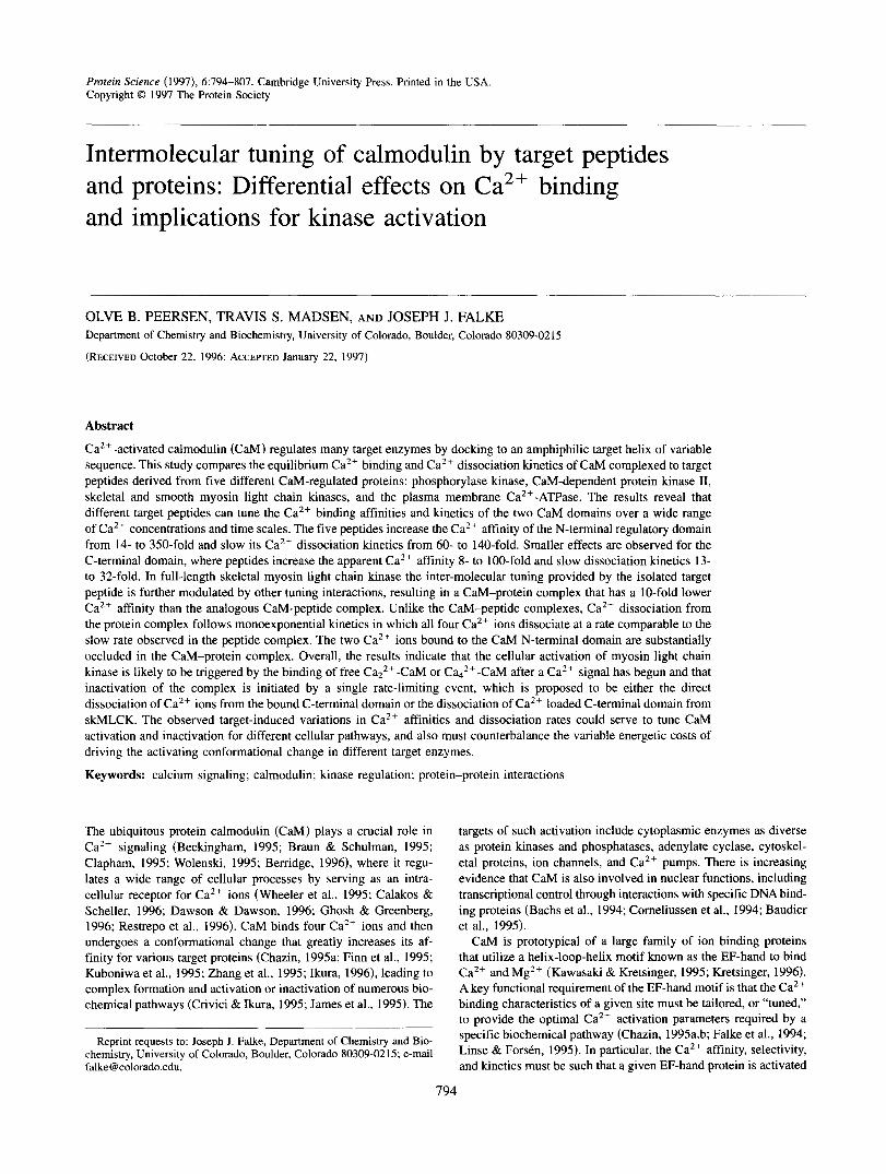
Figure 4 compares equilibrium Ca2+ binding curves obtained from CaM in the presence of the full-length skMLCK protein (gener- ously provided by Dr. Peter Kennelly, Virginia Tech, Blacksburg, VA). The temperature of these experiments was lowered to 20°C to maintain maximal stability of the Ca2+ activated CaM-skMLCK complex during the experiment (Kennelly et al., 1990). It is readily apparent that the intact protein increases the Ca2+ affinity of CaM to a significantly smaller extent than does the isolated target pep- tide, which enhances the macroscopic Ca2+ affinity 220-fold (Table 2). The protein increases the macroscopic Ca2+ affinity only 13-fold to a value of 560 nM, in excellent agreement with the value of 700 nM measured by Crouch et al. (1981). The new flow-dialysis data for the CaM-protein complex further enable resolution of the Ca2+ affinities of the two CaM domains, yielding values of 380 and 800 nM (Table 5). These affinities result in only partial Ca2+ occupancy of the CaM-skMLCK complex at resting cellular Ca2+ levels (-100 nM), as required for effective Ca2+- CaM mediated activation in vivo. In contrast, the much higher affinities observed for the skMLCK peptide complex, 20 and 80 nM, would predict significant Ca2+ occupancy and activation even in the absence of a Ca2+ signal.
At 20 "C the Ca2+ dissociation timecourse of free CaM showed -2 Ca2+ dissociating from the N domain at a rate of 730 s- ' and -2 Ca2+ dissociating from the C domain at a rate of 8.4 s - ' (Table 5). When skMLCK was added, the Ca2+ dissociation time course became a single exponential process exhibiting a rate of 0.52 s" and a stoichiometry of 3.4 Ca2+ per CaM (Fig. 5,
I + skMLCK 9 Peptide /
1 o - ~ 10-8 10" 10-6 1 o - ~ 1
[Ca2+l Frae (M)
Fig. 4. Comparison of Ca2+ binding to CaM in the presence of the sk- MLCK peptide and the complete skMLCK protein. Experiments were carried out using 1 p M CaM and 4 pM skMLCK protein or peptide at 20°C in 100 mM KCI, 0.5 mM MgCl2, 20 mM MOPS, pH 7.4. Best-fit Ca2+ binding constants are listed in Table 5 .
4
N B 0 0
+ skMLCK Protein
I ' I ' I ' I ' I ' I 0.0 1 .o 2.0 3.0 4.0 5.0
Time (sec)
Fig. 5. Kinetics of Ca2+ release from the CaM-skMLCK protein complex. The fitted curve corresponds to a single exponential dissociation curve having a rate of 0.52 s" and an observed amplitude 3.4 Ca2+ per CaM, as summarized in Table 5. The inset shows an expanded view of the first 85 ms, which were sampled with a dwell time of 25 ps; note the absence of a significant rapid component in the protein complex. The experiment was carried out at 20°C by stopped-flow mixing a solution of 150 pM Quin-2 with an equal volume of I pM CaM and 2 p M skMLCK in 100 pM CaC12, where both solutions were buffered in 20 mM MOPS, 100 mM KCI, 1 mM DTT, pH 7.4. At the post-mixing concentrations of Quin-2 and CaC12, the Ca2+ occupancy of the CaM-skMLCK complex is expected to be - 17%. resulting in -0.7 Ca2+ ions per CaM being retained by the complex. Thus, the observed amplitude of the kinetic trace corre- sponds to the maximum expected stoichiometry for the dissociation of four bound Ca2+ ions (see Results).
Table 5). This rate was independent of skMLCK concentration when skMLCK was present at 1.5- to 2.5-fold excess over CaM. Although the observed amplitude of this dissociation event is 3.4 Ca2+ per CaM, it most likely represents the complete release of all four Ca2+ ions from the CaM-skMLCK complex for two reasons. First, at the end of the stopped flow experiment the free Ca2+ concentration will be about 0.2 p M based on the post-mixing concentrations of Quin-2 (75 pM) and Ca2+ (50 pM) and a KO of -0.1 p M for the interaction between Ca2+ and Quin-2. This final free Ca2+ concentration is effectively being buffered by the Quin-2 and is sufficient to maintain partial Ca2+ saturation of the 1 p M CaM-skMLCK complex. Based on the KO values reported in Table 5, the complex will be 17% saturated and have a stoichiom- etry of -0.7 Ca2+ ions per complex, thereby fully accounting for the "missing" Ca2+ ions in the dissociation time course. Second, the single exponential behavior of the dissociation time course argues against the existence of another dissociation event-attempts to add a second exponential component to the curve fit are not statistically justified based on the resulting x 2 values and param- eter errors. It follows that the complete dissociation of all four Ca2+ ions occurs in response to a single rate-limiting event that triggers skMLCK inactivation.
An alternative interpretation that three Ca2+ ions dissociate at the observed rate while a fourth ion dissociates too rapidly to ob- serve at a rate of > 1000 s- ' (Persechini et al., 1996) is ruled out by several factors. First, this interpretation does not account for the Ca2+ still bound to the protein complex at the end of the dissociation time course, as defined by the final free Ca2+ concentration due to buff- ering by the Ca2+ chelator (see above). Second, the binding of CaM to skMLCK is expected to slow, rather than increase, the rates of Ca2+ dissociation from CaM because the Ca2+ affinity of the CaM-

Intermolecular tuning of e a Z f binding to calmodulin
skMLCK complex is 13-fold higher than that of free CaM. Third, the biphasic time course observed for free CaM clearly becomes monophasic for the CaM-skMLCK complex.
Discussion
CaM tuning by isolated target peptides
Overall, the results indicate that isolated peptides representing five different natural CaM binding sequences can increase the macro- scopic Ca2+ affinity and slow Ca2+ dissociation by over two or- ders of magnitude relative to in the absence of peptide. These tuning effects are extremely peptide specific, with different CaM target sequences eliciting a 20-fold range of Ca2+ affinity enhance- ments and a threefold range of Ca2+ dissociation rates. De- convolution of the data into two independent equilibrium constants and dissociation rates, each representing a cooperative 2Ca2+ pro- cess occurring at one of the two CaM domains, reveals that the two domains are differentially tuned by target peptides. Inter-molecular tuning of both Ca2+ affinity and dissociation kinetics is three- to fivefold stronger for the N domain than for the C domain, thereby making the Ca2+ binding parameters of the two domains more similar than in the absence of peptide. This domain specific tuning may have important functional implications, because it is thought that the N domain plays a central role in activation within cells while the C domain may be largely Ca2+ occupied even in the absence of a Ca2+ signal (Bayley et al., 1996; Johnson et al., 1996).
It is not yet clear what structural and electrostatic features of isolated target peptides are responsible for their different effects on Ca2+ affinity. The range of peptide-induced Ca2+ affinities, which corresponds to a free energy range of -6 kcal mol" per four Ca2+ ions (Table 41, is unlikely to stem from the varying helix-formation propensities of aqueous target peptides for two reasons. First, the Ca2+ binding experiments were conducted at peptide concentra- tions sufficient to saturate the peptide binding equilibrium (see
80 1
Methods). Under such conditions, assuming that CaM selectively binds a helical conformer, the peptide association reaction is dom- inated by the binding of pre-formed target helices, which are ex- pected to account for -20% of the total solution population. Second, the free energy of the helix-coil transition for a typical 20-25 residue peptide is on the order of 1 kcal mol" (O'Neil & De- Grado, 1990b; Scholtz & Baldwin, 1992; Chakrabartty et al., 1994), which is small compared to the 6 kcal mol-' range of Ca2+ bind- ing energy triggered by the different target peptides (Table 4).
Instead, the results suggest that two distinct types of inter- molecular contacts may dominate the tuning of Ca2+ binding to CaM-target complexes: domain-specific contacts and dual-domain contacts. Domain-specific tuning stems from direct interactions between a given CaM domain and the region of the target peptide that is docked to that domain. Such tuning is illustrated by partial peptides from two different target sequences. N-terminal partial peptides (sk-N11 and CaATP-N18) selectively tighten Ca2+ bind- ing to the high-affinity C domain of CaM, and C-terminal peptides (sk-C10 and CaATF"C17) have dominant effects on Ca2+ binding to the lower affinity N domain. It is clear that partial peptides from both ends of the target sequence retain the same domain specificity of tuning that they exhibit in the full-length peptide. However, the dual-domain contacts provided by the full-length skMLCK and CaATPase peptides are able to enhance the Ca2+ affinity of both CaM domains by a significantly larger factor than do their corre- sponding partial peptides; for the skMLCK peptide there is an additional ICfold affinity enhancement beyond that attributable to the domain-specific interaction of sk-N1 1. This extra enhancement is not observed when the sk-N11 and sk-C10 peptides are added to CaM simultaneously, demonstrating that the full effect requires a covalent linkage between the N- and C-terminal regions of the skMLCK target sequence. Such covalent continuity is likely to increase the stability of the Ca42+-CaM-target complex, thereby favoring the fully Ca2+-occupied form of CaM. The Ca2+ binding curves obtained in the presence of the CaATP-N18 and CaATP- C17 peptides further support this model. The increased lengths of
Table 4. Estimated free energies of e a 2 + binding to CaM-target complexes
AGla AG2a 2AAG1 + 2AAGzb Target peptide (kcal mol" per CaZ+) (kcal mol" per Ca2+) (kcal mol-' per 4 Ca2+)
None PhK5 CaATPase smMLCK skMLCK MKII
-7.8 -9.0 -9.6 -9.5 - 10 - 10
-6.7 - 8.2 -9.1 -9.1 -9.7 - 10
- -5.7 -8.6 -8.5
-12 - 12
skMLCK Protein (20 "C) -8.8 -8.3 -5.3
aCalculated at AG = -RT InKO based on the KDI and KDZ values determined by fitting the equilibrium binding data to the two cooperative event model (Table 2). Using the domain assignments described in Results, AG, and AG? correspond to the average free energy of binding a single Ca2+ ion to the C and N domain, respectively.
bThe net effect of target peptide binding to CaM on the binding free energy of four Ca2+, relative to the binding free energy of four CaZ+ in the absence of target peptide. For each domain, the effect of target peptide on the binding of a single Ca2+ (AAG,) is given by
AAG, = AGpM.agct - AGpM
where n specifies the domain and AGF""Wet and AGpM are the appropriate values from elsewhere in this table.

802
these peptides result in CaZ+ affinities which are closer, but not equal, to those seen in the full-length CaATPase peptide complex. The CaATPase partial peptides reiterate the seemingly strict re- quirement for hydrophobic anchor residues at both ends of the peptide in order to elicit the full enhancement of Ca2+ binding, as suggested by the deep hydrophobic pockets formed on each do- main of CaM upon Ca2' binding (reviewed by Ikura, 1996).
Ca2+ affinity tuning: Target peptides vs. target proteins
The target sequences that generate the highest Ca2+ affinities, MKII and skMLCK, actually shift the macroscopic KO for Ca2+ binding to -30 nM, which is below the resting level of free Ca2+ in a typical eukaryotic cell, estimated to be -100 nM (Clapham, 1995). Even the KO of the regulatory N domain is observed to shift below the resting Ca2+ level (Table 2), which could lead to con- stitutive CaM-dependent activation in vivo. It follows that intact target proteins must have additional tuning interactions that weaken the macroscopic Ca2+ affinity of their CaM complexes to ensure that the affinity of the N domain, in particular, is above the resting intracellular Ca2+ level. The native skMLCK protein, for example, has been shown to generate a macroscopic Ca2+ affinity of -700 nM under conditions comparable to those of the present study (Crouch et al., 1981) and similar results have been observed for the smooth muscle isoform (Fitzsimons et al., 1992). The onset of Ca2+-CaM-dependent activation of skMLCK has also been observed to require significantly higher Ca2+ concentrations than those needed to saturate direct Ca2+ binding to CaM in the pres- ence of the skMLCK peptide (Yagi et al., 1989), further suggesting that the interactions between CaM and its targets are weaker for full-length proteins than for their individual target helices. Using our experimental conditions, we have confirmed that the Ca2+
O.B. Peersen et al.
affinity of the CaM-skMLCK protein complex is at least an order of magnitude weaker than that of the corresponding CaM-skMLCK peptide complex. For the protein complex the KD values of the N and C domains are 800 nM and 380 nM, respectively, as compared to 80 nM and 20 nM for two domains in the peptide complex.
The lower Ca2+ affinity observed for the CaM-skMLCK pro- tein complex has important implications for the activation of its CaM-regulated pathway in a cellular environment. One proposed mechanism for activation, based on the higher Ca2+ affinity ob- served for the C domain of a CaM-peptide complex, begins with CaM pre-bound to the target protein via its C-terminal domain at resting levels of intracellular Ca2+. Enzyme activation then only requires the binding of Ca2+ to the lower affinity N domain during a Ca2+ signal (Bayley et al., 1996; Johnson et al., 1996). Such a mechanism is quite plausible and is likely to describe the activa- tion of a number of CaM target proteins. However, the present results for the CaM-skMLCK protein complex do not predict sig- nificant Ca2+ occupancy (< 10%) of the C domain at resting levels of intracellular Ca2+. Rather, the higher Ca2+ threshold required for saturation of the CaM-protein complex as compared to the CaM-peptide complex suggests that, within cells, skMLCK acti- vation is initiated some time after the Ca2+ signal has begun, either by the binding of CaZ2+-CaM via its Ca2+ loaded C domain or by the essentially simultaneous binding of both domains of Cad2+- CaM to the target enzyme. Clearly, both the Ca2+ threshold and kinetics will be quite different for mechanisms beginning with pre-bound versus freely diffusing CaM. Because different cell types vary greatly in the fraction of CaM that is pre-bound, and because certain target complexes are known to use CaM as a bound subunit (Klee & Vanaman, 1982; Means, 1988; Luby-Phelps et a1.,'1995), it appears likely that both extremes of activation mechanisms are physiologically important, providing a wide range of thresholds and timecourses for Ca2+/CaM activation.
Table 5. Ca" binding to calmodulin-skMLCK peptide and protein complexes
CaM-skMLCK CaM-skMLCK CaM peptide proteina
Equilibrium Ca2+ bindingb KDI (PM) 2.0 k 0.1 0.02 t 0.01
HI 1.4 f 0.2 2.3 t 1.8 H2 1.7 i 0.3 0.9 i 0.6
0.38 k 0.05 KDZ (PM) 13.0 t 0.6 0.08 t 0.06 0.80 t 0.06
1.6 k 0.2 1.6 f 0.5
Ca2+ dissociation' koffA (s ~ ) 11.8 k 0.1 8.4 t 0.2a 0.36 t 0.01 0.52 k 0.01 nA (Ca2+/CaM) 2.1 t O . l a ND* 3.4 i 0.1 kom (s") 2850 k 70 730 f 60" 6.4 f 0.6 nB (Ca2+/CaM) 2.4 f 0.2" ND*
-
Calculated Ca2+ kOne N domain (X io6 M" S" ) 2 6 5 i 6 C domain (X lo6 M" S" 1 5.9 i 0.3
80 t 40 0.65 k 0.04 18 f 5 1.4 + 0.1
'Data obtained at 20°C to maximize CaM-skMLCK complex stability (see Results). bTwo cooperative site binding model (see Methods and Table 2). Data obtained in 20 mM MOPS, 100 mM
'Same conditions as above except MgClz was omitted. n indicates observed dissociation stoichiometry. dNot determined since the high CaZf affinities of the CaM-peptide complexes interfere with stoichiometry
eApparent k,, value as described in Table 3.
KC1, 0.5 mM MgClZ, pH 7.4 at 25 "C.
determination (see Results and Methods).
Intermolecular tuning of Ca2+ binding to calmodulin 803
Ca2+ release tuning: Target peptides vs. target proteins
The kinetics of Ca2+ release from the CaM-skMLCK protein complex differ substantially from those of all the CaM-peptide complexes in that Ca2+ dissociation from the N domain is slowed to the same time scale as C domain dissociation, yielding mono- exponential kinetics in which all four Ca2+ ions dissociate at a single observed rate of 0.52 s". Caz+ dissociation from the CaM N domain is 10-fold slower in the protein complex than in the peptide complex, while dissociation from the C domain is essen- tially unchanged. The observation that Ca2+ dissociation from the N domain is substantially slower in the protein complex than in the peptide complex, despite having a lower Ca2+ affinity, indicates that the N domain Ca2+ sites are somehow occluded in the com- plex with the full-length protein. Such occlusion may stem from steric or electrostatic barriers blocking a dissociation pathway to the solvent or could represent the inhibition of a structural re- arrangement required for CaZf release from the coordination site. Functionally, the occlusion serves to extend the lifetime of the activated protein complex.
The monophasic Ca2+ dissociation curve observed for the re- lease of all four Ca2+ ions from the activated CaM-skMLCK pro- tein complex implies that there is a single rate-limiting event resulting
Calcium Influx (e)
Resting State
* I
in complete enzyme inactivation. The specific nature of this trigger event is not known, but several lines of evidence suggest that it may be driven by the dissociation of the CaM C domain from the N-terminal end of the target sequence. A similar model has been pro- posed by Bayley et al. (1996), who have argued that dissociation of a C domain-Ca2+-target complex is an ordered two-step process initiated by the target peptide dissociating from Ca2+-loaded Cah4 and followed by the rapid loss of Ca2+ from the resulting Ca2+-C domain complex. As predicted by their model, the observed Ca2+ dissociation rate of -0.52 s-l from the CaM-skMLCK protein com- plex (Table 5) correlates well with the - 1 s-' rate of skMLCK in- activation when triggered by addition of Ca2+ chelators (Persechini et al. 1996; Stull et al., 1986) and the -2 s-l rate determined for CaM-skMLCK complex dissociation (Johnson et al., 1981).
Significantly, the observed Ca2+ dissociation rate of the pro- tein complex (0.52 s-') is similar to the rate of Ca2+ dissociation from the C domain in both the CaM-skMLCK peptide complex (0.36 s- I ) and the CaM-sk-N1 1 partial peptide complex (0.73 s - I ) ,
supporting the hypothesis that a dissociation event involving the C domain is the rate-limiting step in the breakdown of the C a " protein complex. Dissociation of two Ca2+ ions from the bound C domain, or dissociation of the fully Ca2+-occupied C domain, would trigger dissociation of the CaM-protein complex and the
Activated State
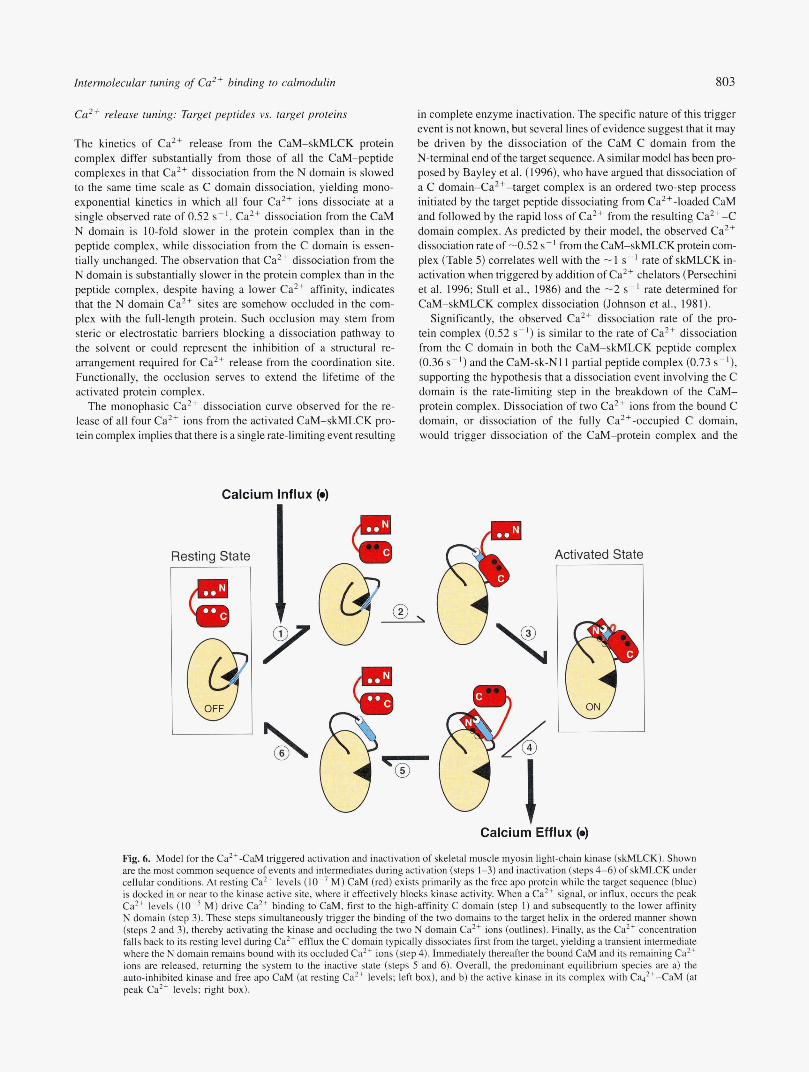
Fig. 6. Model for the Ca2+CaM triggered activation and inactivation of skeletal muscle myosin light-chain kinase (skhKCK). Shown are the most common sequence of events and intermediates during activation (steps 1-3) and inactivation (steps 4-6) of skMLCK under cellular conditions. At resting Ca2+ levels (10" M) CaM (red) exists primarily as the free apo protein while the target sequence (blue) is docked in or near to the kinase active site, where it effectively blocks kinase activity. When a CaZ+ signal, or influx, occurs the peak Ca2+ levels M) drive Ca2+ binding to CaM, first to the high-affinity C domain (step 1) and subsequently to the lower affinity N domain (step 3). These steps simultaneously trigger the binding of the two domains to the target helix in the ordered manner shown (steps 2 and 3). thereby activating the kinase and occluding the two N domain Ca2+ ions (outlines). Finally, as the Ca2+ concentration falls back to its resting level during Ca2+ efflux the C domain typically dissociates first from the target, yielding a transient intermediate where the N domain remains bound with its occluded Ca2+ ions (step 4). Immediately thereafter the bound CaM and its remaining Ca2+ ions are released, returning the system to the inactive state (steps 5 and 6). Overall, the predominant equilibrium species are a) the auto-inhibited kinase and free apo CaM (at resting Ca2+ levels; left box), and b) the active kinase in its complex with Cq2+-CaM (at peak Ca2+ levels; right box).

O.B. Peersen et al.
rapid release of the remaining Ca2+ from the N and C domains of CaM at rates comparable to those observed in free CaM. Such a model is attractive because it implies that the rate of target enzyme inactivation is dominated by interactions between the CaM C do- main and the N-terminal half of the target helix, with little addi- tional tuning provided by the rest of the protein, and as such can be predicted by studying the behavior of CaM-peptide complexes. It should be pointed out, however, that the kinetic data presented here do in no way exclude the CaM N domain from playing a role in triggering the deactivation of the CaM-skMLCK protein com- plex, especially considering the striking occlusion of Ca2+ disso- ciation from the N domain sites, which may lie at a critical docking interface. Figure 6 presents a schematic model summarizing the predominant molecular events during the activation and inactiva- tion of skMLCK in vivo.
Origins of the different tuning observed for target peptides and proteins
One molecular explanation for the different tuning effects observed when CaM is complexed with isolated peptides and full-length pro- teins can be derived from the mechanism of CaM-dependent en- zyme regulation. Many CaM regulated kinases, including skMLCK, smMLCK, and MKII, are activated by the removal of an auto- inhibitory sequence from the active site of the enzyme, an event that can be triggered either by the binding of CaM to a nearby sequence or by phosphorylation of key residues (Zurini et al., 1984; Edelman et al., 1985; Pearson et al., 1988; Olson et al., 1990; Gallagher et al., 1993; Brickey et al., 1994; Krueger et al., 1995; Goldberg et al., 1996). For these proteins, the smaller Ca2+ affinity enhancement ob- served for the full-size enzyme relative to the isolated target peptide is likely to stem, at least in part, from the energetic cost of removing autoinhibition. The full-length Ca2+ATPase molecule provides an analogous energetic cost to CaM binding that is absent in the cor- responding target peptide, because CaM binding is 40-fold tighter for the isolated peptide than to the intact protein (Enyedi et al., 1989). Such considerations imply that target peptides from enzymes ex- hibiting the strongest autoinhibition will yield the highest Ca'+ af- finity CaM-peptide complexes.
Superimposed on the need to counterbalance the energetic cost of auto-inhibition is the independent requirement that each CaM- target enzyme complex must exhibit Ca2+ concentration threshold and kinetic parameters that are optimized for its specific physio- logical role. As a result, the interactions between CaM and the target sequence must be carefully adjusted to account not only for the unique features of the target molecule, but also for the require- ments of the pathway that the target molecule regulates. It should be emphasized that while the binding of CaM to its target sequence provides an important component of the overall tuning, the bound CaM appears to interact with additional surfaces of the target molecule, thereby increasing the number of contacts that could contribute to intermolecular tuning. An example of such additional tuning effects is the 10-fold reduction in the observed rate of Ca2+ dissociation from the N domain in the CaM-skMLCK protein complex as compared to the analogous peptide complex. Such kinetic tuning may serve a critical physiological role by optimizing the rate of kinase inactivation for skeletal muscle function.
In closing, the present study further defines the nature of the intermolecular tuning generated by the binding of CaM to isolated target helices. The data show that target peptides can tune the Ca2+ binding characteristics of CaM over a much broader range than
previously observed for intact target proteins. Although the iso- lated target sequence clearly provides a major component of inter- molecular tuning, additional interactions occur in the full-length CaM-protein complex that bring the threshold for Ca2+ activation into the physiologically appropriate range and further tune the kinetics of Ca2+ dissociation. It is clear that a molecular under- standing of the interaction between CaM and its targets will require further dissection of the target sequences to identify the key struc- tural and electrostatic motifs that regulate Ca2+ binding to and release from this important mediator of Caz+ signaling.
Methods
Culmodulin and peptides
The Ca2+ binding experiments were carried out using Drosophila calmodulin that was overexpressed with the pJFM39 vector (Maune et al., 1992) in E. coli strain AR68 (A. Shatzman, SmithKline Beecham Inc., King of Prussia, PA) using temperature induction of the phage A PI promoter. Drosophila CaM differs from vertebrate CaM at only three positions (Tyr 99 + Phe, Gln 143 + Thr, Ala 147 + Ser) (Smith et al., 1987) and activates target enzymes with an efficiency comparable to that of the vertebrate form (Gao et al., 1993). The protein was purified using phenyl-sepharose chromatography followed by a (3-25 gel filtration column to re- move EGTA (Putkey et al., 1985). exchanged into 100 mM KCI, 20 mM MOPS, 5 mM CaCI2, pH 7.4 and concentrated to - 150 p M using an Amicon YM 10 membrane prior to being snap frozen in for storage at -70 "C. The final protein preparation ex- hibited its characteristic Ca*+-dependent mobility shift on SDS- polyacrylamide gels (Klee et al., 1979) and was >99% pure as judged by Coomassie and silver staining. Liquid chromatography electrospray mass-spectrometry (LCIESI-MS) showed that in the presence of Ca" the protein eluted from a Poros C4 reverse phase column in two peaks; a less hydrophobic species with a molecular weight of 16,679 Da and a more hydrophobic 16,838 Da species that is consistent with the binding of four Ca2+ ions.
Peptides corresponding to the CaM binding domains of C a " regulated enzymes were synthesized and purified to >90% homo- geneity by Macromolecular Resources (Colorado State University, Fort Collins, CO) or by Dr. Spencer Anthony-Cahill (Somatogen Inc., Boulder, CO), and the expected molecular masses were con- firmed by mass spectrometry. Stock solutions of 0.9-1.5 mM pep- tide were prepared in the buffer used for flow dialysis experiments (see below), where peptide concentrations were determined by absorbance based on Trp = 5600 M" cm") or Phe (€258 = 200 M" cm") residues. The affinity of the sk-N11 peptide for Ca2+ loaded CaM was determined by observing peptide Trp flu- orescence during direct titration with CaM, resulting in a 0.4 p M K D . In vivo total concentrations of CaM and its target proteins have generally been determined to be in the 1 p M range, although the concentration of freely diffusible CaM is lower (Klee & Vana- man, 1982; Luby-Phelps et al., 1995), and experimental CaM and target concentrations similar to these were used. A CaM con- centration of -0.9 p M was used in experiments involving the skMLCK, smMLCK, Ca2+ATPase, and MKII peptides, where the Ca2+ affinities of the complexes were 0.2 p M or less. Exper- iments involving PhK5 and the four partial peptides were done at 1-7 p M CaM to more accurately detect the weaker Ca2+ binding of these complexes, and free CaM experiments were conducted at 10-25 p M protein. At high stock peptide concentrations the

Intermolecular tuning of e a 2 + binding to calmodulin 805
Ca2+ATPase peptide exhibited solubility problems, as previously noted by Vorherr et al. (1990). It did, however, become soluble in the standard flow dialysis buffer upon addition of formic acid to 1 % . To compensate for the increased acidity, the MOPS concen- tration of the flow dialysis buffer was increased from 20 to 50 mM and the pH was adjusted to 7.6 prior to adding the peptide stock solution, resulting in a final pH of 7.4.
Determination of e a 2 + binding curves and dissociation constants
Ca2+ binding measurements were carried out by flow dialysis (Porumb, 1994; Colowick & Womack, 1969) at 25 "C using a home-built, temperature-controlled three-cell flow dialyzer ma- chined from Teflon. The flow dialyzer and all plasticware were decalcified by washing in 33% nitric acid, followed by a 20 mM EDTA bath and extensive rinsing with distilled water. The flow dialysis buffer, containing 100 mM KCI, 0.5 mM MgC12, 20 mM MOPS at pH 7.4, was decalcified by passage over a Chelex-100 column (BioRad) prior to adding MgCI2. The presence of this second divalent ion significantly shortened the response time of the flow-dialysis apparatus and reduced scatter in the data points, presumably because Mg2+ acts to block non-specific divalent ion binding sites on the dialysis membrane. The enhanced precision resulting from the inclusion of Mg2+ was most striking for the highest Ca2+ affinity complexes (i.e., those experiments extending to very low free Ca2+ concentrations), where reliable binding curves could not be obtained in the absence of Mg2+.
Freshly decalcified CaM was prepared for each Ca2+ binding experiment using a small Chelex-100 column whose effectiveness was ascertained by atomic absorption spectroscopy. The upper sample chamber of the dialyzer held a 1-1.7 mL sample and the lower pumped chamber had a total volume of 70 pL, which fol- lowed a serpentine path in contact with the dialysis membrane. Specta-Por 7 metal-free membranes with a 1000 MWCO were used after extensive washing. The lower chamber was pumped at a rate of -300 pL/min using a Pharmacia P-3 peristaltic pump while collecting five 0.9-min fractions for each Ca2+ addition to the upper sample chamber. All Ca2+ additions were made using a 100 p L Hamilton syringe attached to a 50-step repeating injector and fitted with narrow plastic protein-gel loading tips. Ca2+ solu- tions were made using 99.99% pure CaCI2 (Aldrich) and all buffer and protein solution volumes were determined by weight after correcting for buffer density.
Ca2+ binding curves were generated from 45Ca scintillation counting data and were corrected for the loss of both radioactive 45Ca and non-radioactive 40Ca during the experiment as previously described (Maune et al., 1992; Yazawa et al., 1992; Stemmer & Klee, 1994). Typically, only 5-10% of the 45Ca initially added to the sample cell diffused across the membrane during the experi- ment. Finally, the 2a confidence interval reported by the scintil- lation counter was propagated by standard methods to produce errors in both the free Ca2+ concentration and the Ca2+/CaM stoichiometry at each point in the titration curve. Ca2+ binding constants were determined by fitting the data to the following equation describing the binding of four ions as two cooperative binding events (Porumb, 1994), where X is the free Ca2+ concen- tration, the factor c absorbs errors in the stoichiometry determina- tion, and the KD and H terms are the macroscopic dissociation constants and Hill coefficients of the two binding events.
The data were fit using KaleidaGraph 3.0 software for the Mac- intosh (Synergy Software, Reading, PA), which employs a Leven- berg-Marquardt algorithm to determine the non-linear least squares best fit. For each point in the titration curve, the root mean square of the relative errors in both the [Ca2+lFree and Ca2+/CaM dimen- sions (RMS error = [(Ax/x)' + (AY/Y)*]"~) was designated as a data weighting factor during curve fitting. Data points determined to higher precision were thus given more emphasis during the fit- ting process. Accounting for error in both dimensions significantly improved the quality of the curve fits by balancing the dominant error in the [Ca2+IFree early in the titration with the dominant error in the Ca2+/CaM stoichiometry found late in the titration.
Stopped flow dissociation kinetics
The time courses of Ca2+ dissociation from CaM and its peptide complexes were determined by fluorescence using the chelator Quin-2 (Molecular Probes, Eugene, OR) in an Applied Photophys- ics model 17MV stopped flow apparatus with a minimum dwell time of 20 ps and a dual time-base signal acquisition mode that allows one to accurately digitize both fast and slow events in a single experiment. The dead time of the stopped-flow apparatus was determined using the reduction of 2,6-dichlorophenolindophenol by ascorbic acid as described by Tonomura et al. (1978), revealing a dead time of 1.0 ms and a linear response up to an apparent reaction rate of -750 s-'. Consequently, the first 1.5 ms of the observed dissociation time courses were omitted when curve fit- ting the data and rates in excess of 750 s" are designated as lower limits.
For the CaM-peptide complex studies, solutions containing 4-6 p M CaM, a range of 0-15 p M peptide, and 60 pM CaCI2 in Mg2+-free flow dialysis buffer were stopped-flow mixed with a solution of 200 p M Quin-2 in the same buffer. Quin-2 fluores- cence was observed through a 490 nm bandpass filter (Aex = 332 nm). A total of eight transients were acquired at each peptide concentration. The stopped flow apparatus was used in absorbance mode to observe changes in Tyr 138 absorbance at 276 nm as CaM solutions were mixed with 5 mM EDTA.
Although the major effect on the Ca2+ dissociation rate was established at equimolar concentrations of CaM and peptide, the observed dissociation rate decreased further when increasing con- centrations of peptide were added. As recently pointed out by Bayley et al. (1996), this effect can be attributed to the rebinding of Ca2' to CaM rather than Quin-2 after an initial dissociation event; such back-flow of Ca2' reduces the observed dissociation rate and the effect becomes greater as the CaM-target equilibrium is pushed further toward formation of the high Ca2+ affinity CaM- target complex by addition of excess peptide. Consequently, the Ca2+ dissociation rates for all five peptides were determined over a range of peptide concentrations, and the reported rates (Table 2) are averages representing the asymptotic values achieved at satu- rating peptide concentrations (10-15 p M peptide and 3-5 pM CaM).
The Ca2+ dissociation rate of the CaM-skMLCK protein com- plex was determined at 20 "C using 150 pM Quin-2 in one syringe and 100 p M CaCI2 with 1 p M CaM and 0-2.5 p M skMLCK in the other. Dithiothreitol was added to 1 mM. A skMLCK titration was used to confirm that the skMLCK to CaM ratio, based on

O.B. Peersen et al.
Bradford and BCA protein concentration assays, was sufficient to saturate the interaction between the two proteins. Separate exper- iments using a series of CaC12 solutions with concentrations rang- ing from 73 to 112 p M showed that the Quin-2 fluorescence response was linear over this range with a slope of 15.0 mV/pM Caz+ added, and this slope was used to convert the amplitudes of the observed dissociation rates into Ca'+ stoichiometries. Note that the high Ca2+ affinities of the CaM-peptide complexes inter- fere with the amplitude-based stoichiometry determination due to significant residual binding of Ca2+ at the end of the kinetic time- course (see Results). Consequently, only data from free CaM and the CaM-skMLCK protein complex were analyzed in terms of the number of Ca2+ ions dissociating per CaM molecule.
Calculated association constants
It should be noted that the calculated k,, (= k,fl/K,) values listed in Table 3 are best considered as apparent constants for two rea- sons. First, the flow dialysis binding experiments required the presence of 0.5 mM Mg2+ to get reliable Ca2+ KO values for the high affinity CaM-target complexes, but the k, values were de- termined in the absence of Mg2+ as it would interfere with Ca2+ binding to Quin-2. Although Mg2+ is largely a spectator ion in this system, the 0.5 mM concentration of Mg2+ may increase the Ca2+ KD values up to a maximum of twofold due to competitive binding to EF-hand sites (see below). Second, the observed values for the CaM-target complexes are themselves apparent rate constants that are slightly dependent on the target peptide concentration (Bayley et al., 1996; see Methods). Overall, these two effects will have minimal impact on the comparisons among the different CaM- peptide complexes because the binding experiments were all car- ried out under identical conditions and the reported koflrates represent the plateau values observed at saturating amounts of peptide.
Comparison of our k,, values with those obtained by other lab- oratories in the absence of Mg2+ can be made by considering the effect of competing Mg2+ ions on the observed Ca'+ KO value, which is described by the relationship
Tsai et al. (1987) investigated the binding of Mg2+ to CaM by NMR methods and determined that Mg2+ binds the CaM N and C domains with dissociation constants of -0.5 mM and 3.3 mM, respectively, in the absence monovalent ions. Applying this cor- rection to our binding data determined in the presence of 0.5 mM Mg2+ indicates that the added Mg2+ results in a small twofold increase in the N domain KO and a negligible - 1.15-fold effect on the C domain K,,.
Acknowledgments
The authors wish to thank Spencer Anthony-Cahill of Somatogen, Inc. for peptide synthesis and Eric Nalefski and Andrea Hazard for helpful discus- sions. We are especially indebted to Peter Kennelly for providing us with the skMLCK protein sample, Katherine Beckingham for the clone of Dm- sophila calmodulin, and A. Shatzman of SmithKline Beecham Pharmaceu- ticals for E. coli strain AR68. This research was funded through grants from the National Institutes of Health (Postdoctoral Fellowship GM-17045 to OBP, and Grant R01-GM-48203 to JJF). Support for this work was pro- vided by NIH Grant GM48203At.
References
Afshar M, Caves LSD, Guimard L, Hubbard RE, Calas B, Grassy G, Haiech J . 1994. Investigating the high affhity and low sequence specificity of cal- modulin binding to its targets. J Mol Biol 244554-571.
Anagli J, Hofmann F, Quadroni M, Vorherr T, Carafoli E. 1995. The calmodulin- binding domain of the inducible (macrophage) nitric oxide synthase. Eur J Biochern 233:701-708.
Bachs 0, Agell N, Carafoli E. 1994. Calmodulin and calmodulin-binding pro- teins in the nucleus. Cell Calcium 16:289-296.
Baudier J, Bergeret E, Bertacchi N, Weintraub H, Gagnon J, Garin J. 1995. Interactions of myogenic bHLH transcription factors with calcium-binding calmodulin and S lO0a (aa) proteins. Biochemistry 34:7834-7846.
Bayley PM. Findlay WA, Martin SR. 1996. Target recognition by calmodulin: Dissecting the kinetics and affinity of interactions using short peptide se- quences. Protein Sci 5: 121 5-1 228.
Beckingham K. 1995. Calcium regulation of Drosophila development. Adv Sec Mess Phosphoprotein Res 30:359-394.
Bemdge MJ. 1996. Calcium signalling and cell proliferation. Bioessays 17491- 500.
Blumenthal DK. Takio K, Edelman AM, Charbonneau H, Titani K, Walsh KA, Krebs EG. 1985. Identification of the calmodulin-binding domain of skeletal muscle myosin light chain kinase. Proc Natl Acad Sci USA 82:3 187-3 19 I .
Braun AP, Schulman H. 1995. The multifunctional calcium calmodulin-dependent protein-kinase-From form to function. Annu Rev Physiol 57417-445.
Brickey DA, Bann JG. Fong YL, Pemno L, Brennan RG. Soderling TR. 1994. Mutational analysis of the autoinhibitory domain of calmodulin kinase 11. J Biol Chem 26929047-29054.
Calakos N, Scheller RH. 1996. Synaptic vesicle biogenesis, docking, and
Chakrabartty A, Kortemme T. Baldwin RL. 1994. Helix propensities of the fusion-A molecular description. Physiol Rev 76: 1-29.
ammo acids measured in alanine-based peptides without helix-stabilizing side-chain interactions. Protein Sci 3:843-852.
Chazin WJ. 1995a. Releasing the calcium trigger. Naf Struct Biol 2:707-710. Chazin WJ. 1995b. Signal transduction versus buffering activity in Ca2+ bind-
Clapham DE. 1995. Calcium signaling. Cell 80:259-268. Colowick SP, Womack E 1969. Binding of diffusible molecules by macromol-
ecules: Rapid measurement by rate of dialysis. J Biol Chem 244:774-777. Comeliussen B, Holm M, Waltersson Y, Onions J , Hallberg B, Thomell A,
Grundstrom T. 1994. Calcium/calmodulin inhibition of basic-helix-loop- helix transcription factor domains. Nature 368:760-764.
Cox JA, Comte M, Fitton JE, DeGrado WE 1985. The interaction of calmodulin with amphiphilic peptides. J Biol Chem 26U2527-2534.
Crivici A, Ikura M. 1995. Molecular and structural basis of target recognition by calmodulin. Annu Rev Biophys Biornol Struct 24.85-1 16.
Crouch TH, Holroyde MJ. Collins JH, Solaro RJ, Potter JD. 1981. Interaction of calmodulin with skeletal muscle myosin light chains kinase. Biochemistry 20:63 18-6325.
Dasgupta M, Honeycutt T, Blumenthal DK. 1989. The y-subunit of skeletal muscle phosphorylase kinase contains two noncontiguous domains that act in concert to bind calmodulin. JBiol Chem 264:17156-17163.
Dawson VL, Dawson TM. 1996. Nitric-oxide actions in neurochemistry. Neuro- chem In / 2997-1 10.
Drake SK, Falke JJ. 1996. Kinetic tuning of the EF-hand Ca2+ binding motif The gateway residue independently adjusts (i) barrier height and (ii) equi- librium. Biochemisrp 35:1753-1760.
Drake SK, Lee K, Falke JJ. 1996. Tuning the ion affinity and selectivity of the EF-hand Ca2' binding motif Substitutions at the gateway position. Bio- chemistry 35:6697-6705.
Edelman AM, Takio K, Blumenthal DK, Hansen RS, Walsh KA, Titani K, Krebs EG. 1985. Characterization of the calmodulin-binding and catalytic domains in skeletal muscle myosin light chain kinase. JBiol Chem 260:l 1275-1 1285.
Enyedi A, Vorherr T, James P, McCormick DJ, Filoteo AG, Carafoli E, Pennis- ton IT. 1989. The calmodulin binding domain of the plasma membrane Ca2+ pump Interacts both with calmodulin and with another part of the pump. J Biol Chem 264: 123 13-1 232 I .
Erickson-Viitanen S, DeGrado WF. 1987. Recognition and characterization of calmodulin-binding sequences in peptides and proteins. Methods Enzymol 139455-478.
Falke JJ, Drake SK, Hazard AL, Peersen OB. 1994. Molecular tuning of ion
Findlay WA, Gradwell MJ, Bayley PM. 1995. Role of the N-terminal region of binding to Ca2+ signaling proteins. Q Rev Biophys 27219-290.
the skeletal muscle myosin light chain kinase target sequence in its inter- action with calmodulin. Protein Sci 42375-2382.
Finn BE, Evenas J, Drakenberg T, Waltho JP, Thulin E, Forstn S. 1995. Calcium-
B i d 2:777-783. induced structural changes and domain autonomy in calmodulin. Nat Strucr
ing proteins. Nat Struct Biol /:239-245.

Intermolecular tuning of Ca2+ binding to calmodulin 807
Fitzsimons DP, Hening BP, Stull JT, Gallagher PJ. 1992. Identification of basic residues involved in activation and calmodulin binding of rabbit smooth muscle myosin light chain kinase. J Biol Chem 26723903-23909.
Gallagher PJ, Herring BP, Trafny A, Sowadski J, Stull JT. 1993. A molecular mechanism for autoinhibition of myosin light-chain kinases. J Biol Chem 26826578-26582.
Gao ZH, Krebs J, VanBerkum MFA, Tang W-J, Maune JF, Means AR, Stull JT, Beckingham K. 1993. Activation of four enzymes by two series of calmod- ulin mutants with point mutations in individual Ca2+ binding sites. J Biol Chem 26820096-20104.
Ghosh A, Greenberg ME. 1996. Calcium signaling in neurons: Molecular mech- anisms and cellular consequences. Science 268:239-247.
Goldberg J, Naim AC, Kuriyan J. 1996. Structural basis for the autoinhibition of calcium/calmodulin-dependent protein kinase I. Cell 84375-887.
Haiech J , Klee CB, Demaille JG. 1981. Effects of cations on affinity of cal- modulin for calcium: Ordered binding of calcium ions allows the specific
Ikura M, Clore GM, Gronenbom AM, Zhu G, Klee CB, Bax A. 1992. Solution activation of calmodulin-stimulated enzymes. Biochemistry 20:3890-3897.
structure of a calmodulin-target peptide complex by multidimensional NMR. Science 256632-638.
Ikura M. 1996. Calcium binding and conformational response in EF-hand pro- teins. Trends Biochem Sci 21:14-17.
James P, Vorherr T, Carafoli E. 1995. Calmodulin-binding domains: Just two faced or multi-faceted? Trends Biochem Sci 20:38-41.
Johnson JD, Snyder C, Walsh M, Flynn M. 1996. Effects of myosin light chain kinase and peptides on Ca2+ Exchange with the N- and C-terminal CaZ+ binding sites of calmodulin. J Biol Chem 271:761-767.
Johnson JD, Holroyde MJ, Crouch TH, Solaro RJ, Potter JD. 1981. Fluores- cence studies of the interaction of calmodulin with myosin light chain kinase. J Biol Chem 256:12194-12198.
Kawasaki H, Kretsinger RH. 1995. Calcium-binding proteins 1: EF-hands. Pro- tein Profile 1:383-517.
Kennelly PJ, Starovasnik MA, Edelman AM, Krebs EG. 1990. Modulation of the stability of rabbit skeletal muscle myosin light chain kinase through the calmodulin-binding domain. J Biol Chem 265:1742-1749.
Klee CB, Crouch TH, Krinks MH. 1979. Calcineurin: A calcium- and calmodulin- binding protein of the nervous system. Proc Natl Acad Sci USA 76:6270- 6273.
Klee CB, Vanaman TC. 1982. Calmodulin. Adv Protein Chem 35:213-321. Klevit RE, Blumenthal DK, Wemmer DE, Krebs EG. 1985. Biochemistry
Kretsinger RH. 1996. EF-hands reach out. Nat Struct B i d 3: 12-15. Krueger JK, Padre RC, Stull JT. 1995. Intrasteric regulation of myosin light-
chain kinase. J Biol Chem 270:16848-16853. Kuboniwa H, Tjandra N, Grzesiek S, Ren H, Klee CB. Bax A. 1995. Solution
structure of calcium-free calmodulin. Nat Struct Biol 2:768-776. Lime S, Helmersson A, Forsen S. 1991. Calcium binding to calmodulin and its
globular domains. J Biol Chem 266:8050-8054. Linse S, ForsCn S. 1995. Determinants that govern high-affinity calcium bind-
ing. Adv Sec Mess Phosphoprotein Res 30:89-15 I . Luby-Phelps K, Hori M, Phelps JM, Won D. 1995. Ca2+-regulated dynamic
compartmentalization of calmodulin in living smooth muscle cells. J Biol Chem 270:21532-21538.
Lukas TJ, Burgess WH. Prendergast FG, Lau W, Watterson DM. 1986. Calmod- ulin binding domains: Characterization of a phosphorylation and calmodulin binding site from myosin light chain kinase. Biochemistry 25:1458-1464.
Martin SR, Teleman AA, Bayley PM, Drakenberg T, ForsCn S. 1985. Kinetics of calcium dissociation from calmodulin and its tryptic fragments. A stopped-
J Biochem 151543-550. flow fluorescence study using Quin-2 reveals a two-domain structure. Eur
Martin SR, Maune IF, Beckingham K, Bayley PM. 1992. Stopped-flow studies
melanogaster calmoddin. Eur J Biochem 205: 1107-1 114. of calcium dissociation from calcium-binding-site mutants of Drosophila
Maune JF, Klee CB, Beckingham K. 1992. Ca2+ binding and conformational change in two series of point mutations to the individual Caz+-binding sites of calmodulin. J Biol Chem 2675286-5295.
Meador WE, Means AR, Quiocho FA. 1992. Target enzyme recognition by calmodulin: 2.4 .& structure of a calmodulin-peptide complex. Science 2571251-1255.
Meador WE, Means AR, Quiocho FA. 1993. Modulation of calmodulin plas-
262:1718-1721. ticity in molecular recognition on the basis of X-ray structures. Science
Meador WE, George SE, Means AR, Quiocho FA. 1995. X-ray analysis reveals
Nat Struct Biol 2:943-945. conformational adaptation of the linker in functional calmodulin mutants.
24:8152-8157.
Means AR. 1988. Molecular mechanisms of action of calmodulin. Recent Prog Horn Res 44:223-262.
O’Neil KT, DeGrado WE 1990a. How calmodulin binds its targets: Sequence independent recognition of amphiphilic a-helices. Trends Biochem Sci 1559- 64.
O’Neil KT, DeGrado W E 1990b. A thermodynamic scale for the helix-form-
651. ing tendencies of the commonly occurring amino acids. Science 250:646-
Olson N, Pearson RB, Needleman DS, Hunvitz MY, Kemp BE, Means AR. 1990. Regulation and structural motifs of chicken gizzard myosin light chain kinase. Proc Natl Acad Sci USA 872284-2288.
Olwin BB, Storm DR. 1985. Calcium binding to complexes of calmodulin and
Pearson RB, Wettenhall REH, Means AR, Hartshorne DJ, Kemp BE. 1988. calmodulin binding proteins. Biochemistry 24:8081-8086.
Autoregulation of enzymes by pseudosubstrate prototypes: Myosin light chain kinase. Science 241:970-973.
Persechini A, White HD, Gansz KJ. 1996. Different mechanisms for Ca2+ dissociation from complexes of calmodulin with nitric oxide synthase or myosin light chain kinase. J Biol Chem 271:62-67.
Porumb T. 1994. Determination of calcium-binding constants by flow dialysis. Anal Biochem 220227-237.
Putkey JA, Slaughter GR, Means AR. 1985. Bacterial expression and charac- terization of proteins derived from the chicken calmodulin cDNA and a calmodulin processed gene. J Biol Chem 2604704-4712.
Restrepo D, Teeter JH, Schild D. 1996. Znd-Messenger signaling in olfactory transduction. J Neurobiol 30:37-48.
Scholtz JM, Baldwin RL. 1992. The mechanism of a-helix formation by pep- tides. Annu Rev Biophys Biomol Struct 21:95-118.
Smith VL, Doyle KE, Maune JF, Munjaal RP, Beckingham K. 1987. Structure and sequence of the Drosophila melanogaster calmodulin gene. J Mol Biol 196:471-485.
Starovasnik MA, Su DR, Beckingham K, Klevit RE. 1992. A series of point mutations reveal interactions between the calcium-binding sites of calmod- din. Protein Sci 1:245-253.
Stemmer PM, Klee CB. 1994. Dual calcium ion regulation of calcineurin by calmodulin and calcineurin B. Biochemistry 33:6859-6866.
Strynadka NCJ, James MNG. 1990. Model for the interaction of amphiphilic helices with troponin C and calmodulin. Proteins 7234-248.
Stull JT, Nunnally MH. Michnoff CH. 1986. Calmodulin-dependent protein kinases. The Enzymes, Vol XVll. New York: Academic Press. pp 113-166.
Suko J, Wyskovsky W, Pidlich J , Hauptner R, Plank B, Hellmann G. 1986. Calcium release from calmodulin and its C-terminal and N-terminal halves in the presence of the calmodulin antagonists phenoxybenzamine and me- littin measured by stopped-flow fluorescence with Quin 2 and intrinsic
Taylor DA, Sack JS, Maune JF, Beckingham K. Quiocho FA. 1991. Structure of tyrosine. Eur J Biochem 159425-434.
a recombinant calmodulin from Drosophila rnelanogaster refined at 2.2-8, resolution. J B i d Chem 266:21375-21380.
Tonomura B, Nakatani H, Ohnishi M, Yamaguchi-Ito J, Hiromi K. 1978. Test reactions for a stopped-flow apparatus. Reduction of 2,6-dichlorophenolin- dophenol and potassium ferricyanide by L-ascorbic acid. Anal Biochem 84.370-383.
Tsai M-D, Drakenberg T. Thulin E, Forsen S. 1987. Is the binding of magne- sium(I1) to calmodulin significant? An investigation by magnesium-25 nu-
Vorherr T. James P, Krebs J, Enyedi A, McCormick DJ, Penniston JT. Carafoli clear magnetic resonance. Biochemistry 26:3635-3643.
E. 1990. Interaction of calmodulin with the calmodulin binding domain of the plasma membrane Ca2+ pump. Biochemistry 29:355-365.
Wheeler DB, Randall A, Sather WA, Tsien RW. 1995. Neuronal calcium chan- nels encoded by the alpha(lA) subunit and their contribution to excitatory synaptic transmission in the CNS. Prog Brain Res 1055-78.
Wolenski JS. 1995. Regulation of calmodulin-binding myosins. Trends Cell Biol 5:310-316.
Yagi K, Yazawa M, Ikura M, Hikichi K. 1989. Interaction between calmodulin
Med B i d , Vol 255. New York: Plenum Press. pp 147-154. and target proteins. In: Hidaka, H., ed. Calcium Protein Signaling Adv Exp
Yazawa M, Vorherr T, James P, Carafoli E, Yagi K. 1992. Binding of calcium by calmodulin: Influence of the calmodulin binding domain of the plasma membrane calcium pump. Biochemistry 3/:3171-3176.
Zhang M, Tanaka T, Ikura M. 1995. Calcium-induced conformational transition revealed by the solution structure of apo calmodulin. Nar Struct B i d 2:758- 767.
Zurini M, Krebs J, Penniston JT, Carafoli E. 1984. Controlled proteolysis of the purified CaZ+-ATPase of the erythrocyte membrane; A correlation between the structure and the function of the enzyme. J Biol Chem 259:618-627.