Embed Size (px)
Citation preview
MODELLING STENOSIS DEVELOPMENT IN
THE CAROTID ARTERY AT THE EARLY
STAGES OF STENOSIS DEVELOPMENT
A.C.Stamou1, J.M.Buick2
1 Faculty of Engineering and Computing, Coventry University
2 School of Engineering, Anglesea Building, Portsmouth
Objectives of the talk
Presentation outline
Description of the problem
Methodology
Lattice Boltzmann method
Stenosis growth model
Results
Conclusion
Presentation outline
In the present work, the position of the development of a stenosis in the carotid artery has been simulated based on regions of stagnation in blood flow.
The interest is focused on:
the blood flow properties, computed here using the Lattice Boltzmann Method,
prediction of the initial growth of the stenosis, based on regions of blood stagnation at the artery wall structure,
variation of the haemodynamic properties with the growth of the stenosis.
4
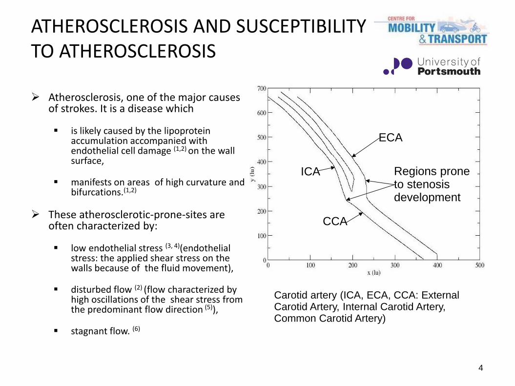
Carotid artery (ICA, ECA, CCA: External Carotid Artery, Internal Carotid Artery, Common Carotid Artery)
ICA
CCA
ECA
Regions prone to stenosis development
ATHEROSCLEROSIS AND SUSCEPTIBILITY TO ATHEROSCLEROSIS
Atherosclerosis, one of the major causes of strokes. It is a disease which
is likely caused by the lipoprotein accumulation accompanied with endothelial cell damage (1,2) on the wall surface,
manifests on areas of high curvature and bifurcations.(1,2)
These atherosclerotic-prone-sites are often characterized by:
low endothelial stress (3, 4)(endothelial stress: the applied shear stress on the walls because of the fluid movement),
disturbed flow (2) (flow characterized by high oscillations of the shear stress from the predominant flow direction (5)),
stagnant flow. (6)
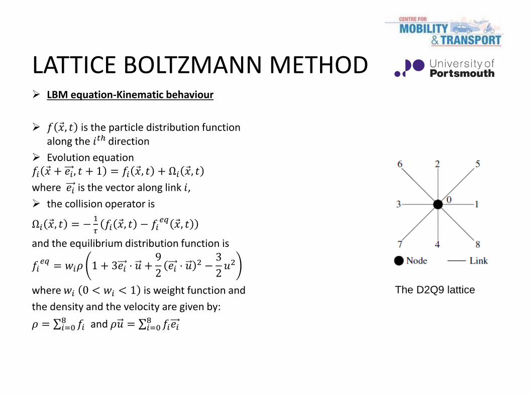
LATTICE BOLTZMANN METHOD LBM equation-Kinematic behaviour
𝑓 𝑥 , 𝑡 is the particle distribution function along the 𝑖𝑡ℎ direction
Evolution equation 𝑓𝑖 𝑥 + 𝑒𝑖 , 𝑡 + 1 = 𝑓𝑖 𝑥 , 𝑡 + Ω𝑖 𝑥 , 𝑡
where 𝑒𝑖 is the vector along link 𝑖,
the collision operator is
Ω𝑖 𝑥 , 𝑡 = −1
𝜏𝑓𝑖 𝑥 , 𝑡 − 𝑓𝑖
𝑒𝑞 𝑥 , 𝑡
and the equilibrium distribution function is
𝑓𝑖𝑒𝑞 = 𝑤𝑖𝜌 1 + 3𝑒𝑖 ⋅ 𝑢 +
9
2𝑒𝑖 ⋅ 𝑢 2 −
3
2𝑢2
where 𝑤𝑖 0 < 𝑤𝑖 < 1 is weight function and
the density and the velocity are given by:
𝜌 = 𝑓𝑖8𝑖=0 and 𝜌𝑢 = 𝑓𝑖𝑒𝑖
8𝑖=0
The D2Q9 lattice

STENOSIS GROWTH MODELLING (CRITERIA)
The velocity magnitude along the wall is considered of significant effect on the simulation of the growth of the stenosis.
However, the maximum stagnation of the flow is considered in our work as the criterion for the growth of the stenosis, with stagnation index:
SI = N/T
where T is the period of the cardiac pulse and N is the number of time-steps in a period when 𝑢1, the local velocity magnitude at the 1 grid length from the wall, is less than 1% of the average 𝑢1 over the whole of the wall during the previous period.
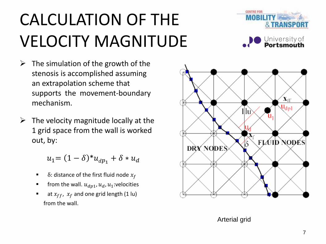
7
Arterial grid
CALCULATION OF THE VELOCITY MAGNITUDE The simulation of the growth of the
stenosis is accomplished assuming an extrapolation scheme that supports the movement-boundary mechanism.
The velocity magnitude locally at the 1 grid space from the wall is worked out, by:
𝑢1= 1 − 𝛿 *𝑢𝑑𝑝1+ 𝛿 ∗ 𝑢𝑑
δ: distance of the first fluid node 𝑥𝑓
from the wall. 𝑢𝑑𝑝1, 𝑢𝑑 , 𝑢1:velocities
at 𝑥𝑓𝑓, 𝑥𝑓 and one grid length (1 lu)
from the wall.
8
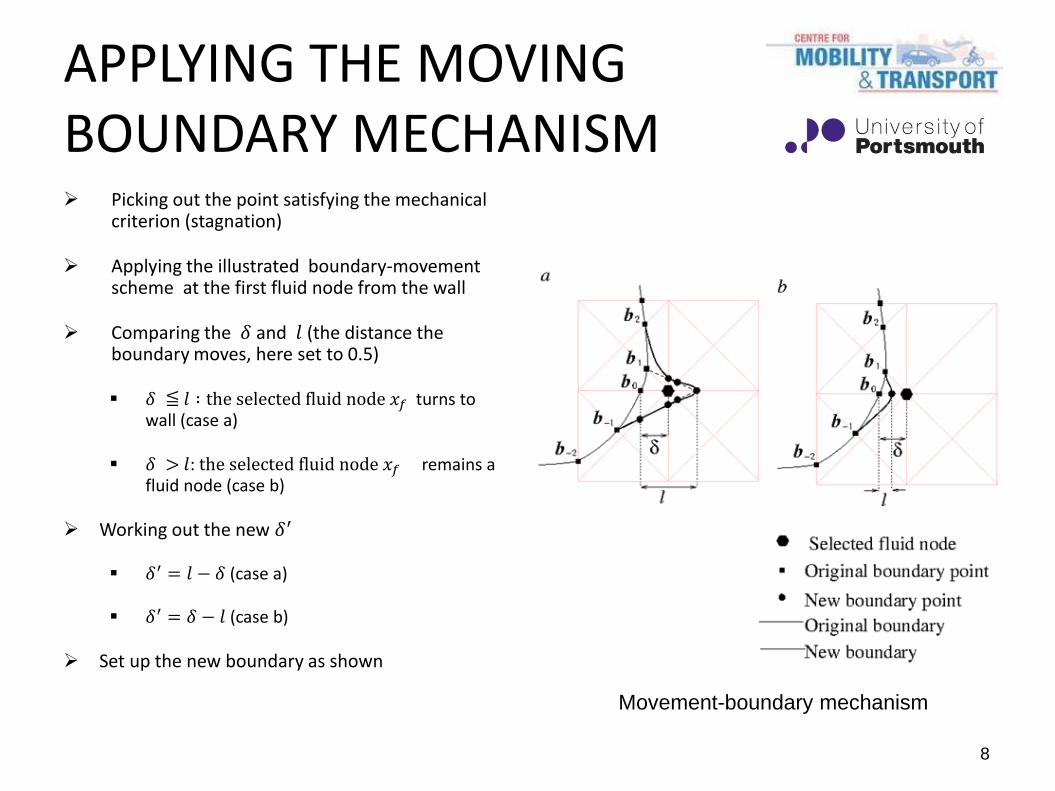
APPLYING THE MOVING BOUNDARY MECHANISM Picking out the point satisfying the mechanical
criterion (stagnation)
Applying the illustrated boundary-movement scheme at the first fluid node from the wall
Comparing the 𝛿 and 𝑙 (the distance the boundary moves, here set to 0.5)
𝛿 ≦ 𝑙 ∶ the selected fluid node 𝑥𝑓 turns to wall (case a)
𝛿 > 𝑙: the selected fluid node 𝑥𝑓 remains a fluid node (case b)
Working out the new 𝛿′
𝛿′ = 𝑙 − 𝛿 (case a)
𝛿′ = 𝛿 − 𝑙 (case b)
Set up the new boundary as shown
Movement-boundary mechanism
RESULTS ANALYSIS
The presentation of the results is structured as follows:
Stenosis development in layers
Effect of stenosis growth on the blood flow along
the wall
Flow profiles

10
STENOSIS DEVELOPMENT
The stenosis is formed in layers built up on the outer walls of the ECA and ICA.
The stenosis layers are built up consecutively on either the same arterial wall or on the opposite wall.
The growth occurs on the ECA below the bifurcation.
In the ICA the stenosis occurs from slightly below the bifurcation and extends significantly in to the ICA.
Stenosis growth in layers with Ts, the number of grid sites, originally in the blood flow region, which have been enclosed by the growth of the stenosis.

11
CHANGE IN THE WALL VELOCITY WITH STENOSIS GROWTH
B) A)
The growth of the stenosis influences mainly the flow at the stenosed regions.
A) Velocity magnitude along the ECA
B) Velocity variation with the growth of the stenosis as a function of TS
12
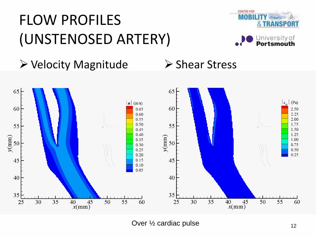
FLOW PROFILES (UNSTENOSED ARTERY)
Velocity Magnitude Shear Stress
Over ½ cardiac pulse
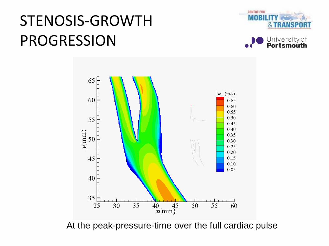
At the peak-pressure-time over the full cardiac pulse
STENOSIS-GROWTH PROGRESSION

14
FLOW PROFILES (FULLY STENOSED ARTERY)
Velocity Magnitude Shear Stress
Over ½ cardiac pulse

Conclusions
The position of the stenosis has been simulated here based on regions of flow stagnation.
The stenosis is formed on the outer wall of both the ICA and the ECA in the region where vortex motion develops in the healthy artery.
The growth of the stenosis influences mainly the flow close to the stenosed wall.
An increased velocity was observed in both the ICA and the ECA with the growth of the stenosis.
Future research
Simulation of the progression of the stenosis in a number of other arteries which are similarly susceptible to this disease, in particular the human aorta
Comparison of haemodynamics between stenosed and stented arteries
Simulation of the non-Newtonian blood considering alternative models to the Carreau-Yasuda
Development of a 3D model to enable the secondary flows in the artery to be assessed and their effect on stenosis development
References
1. C.G. Caro. Atherioscler Thromb Vasc Biol. 29: 18-161, 2009.
2. X. He, D.N. Ku. Journal of Biomechanical Engineering. 118: 74-82, 1996.
3. Y.S. Chatzizisis, M. Yoman, A.U. Coskun, R. Beigel, B.V. Stone, C. Meynard et al. Circulation. 117: 993-1002, 2008.
4. J. Knight, U. Olgac, S.C. Saur, D. Poulikakos, W. Marshall, P.C. Cattin et al. Atherosclerosis. 211: 445-450, 2010.
5. D.N. Ku, D.P. Giddens, C.K. Zarins and S.Glagov. Atherioscler Thromb Vasc Biol. 5: 293-302, 1985.
6. C.V. Soulis, O. P. Lampri, D.K. Fytanidis and G.D. Giannoglou. Biomedical Engineering, 2011 10th International Workshop on 5-7 Oct. 2011 1-4.