Embed Size (px)
Citation preview
Surg Today (2009) 39:144–147DOI 10.1007/s00595-008-3811-x
Reprint requests to: T. Hirokawa (address 3)Received: February 25, 2008 / Accepted: May 14, 2008
Middle-Colic Artery Aneurysm Associated with Segmental Arterial Mediolysis, Successfully Managed by Transcatheter Arterial Embolization: Report of a Case
TAKAHISA HIROKAWA1,3, HIROZUMI SAWAI
1, KOJI YAMADA1, TAKEHIRO WAKASUGI
1, HIROMITSU TAKEYAMA1,
HIROYUKI OGINO2, MASAKATSU TSURUSAKI
3, and YASUAKI ARAI3
Departments of 1 Gastroenterological Surgery and 2 Radiology, Nagoya City University Graduate School of Medical Sciences, Nagoya, Japan3 Department of Diagnostic Radiology, National Cancer Center Hospital, 5-1-1 Tsukiji, Chuo-ku, Tokyo 104-0045, Japan
AbstractAn aneurysm of the middle-colic artery, associated with segmental arterial mediolysis (SAM), is a rare condi-tion. This report describes a case of a middle-colic artery aneurysm that was associated with SAM. A 57-year-old man was admitted to our hospital because of severe abdominal pain. A rupture of a middle-colic artery aneurysm was diagnosed by computed tomography, and angiography showed that it may have been associated with SAM. The ruptured aneurysm was successfully treated with transcatheter arterial embolization. Trans-catheter arterial embolization might be one of the best treatments for such a complicated aneurysm occurring in a visceral artery.
Key words Middle-colic artery aneurysm · Segmental arterial mediolysis · Transcatheter arterial embolization
Introduction
The most frequent site of a visceral artery aneurysm is the splenic artery, but it is a rare occurrence. Therefore, an aneurysm of the middle-colic artery is even more uncommon.1 An aneurysm that may be caused by seg-mental arterial mediolysis (SAM) is also a rare condi-tion. This report documents a case of a SAM-associated, ruptured, middle-colic artery aneurysm that was suc-cessfully managed by transcatheter arterial emboliza-tion (TAE).
Case Report
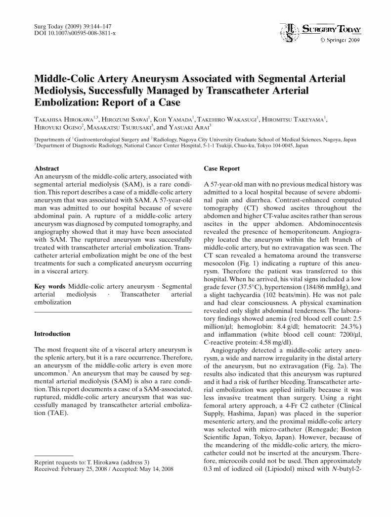
A 57-year-old man with no previous medical history was admitted to a local hospital because of severe abdomi-nal pain and diarrhea. Contrast-enhanced computed tomography (CT) showed ascites throughout the abdomen and higher CT-value ascites rather than serous ascites in the upper abdomen. Abdominocentesis revealed the presence of hemoperitoneum. Angiogra-phy located the aneurysm within the left branch of middle-colic artery, but no extravagation was seen. The CT scan revealed a hematoma around the transverse mesocolon (Fig. 1) indicating a rupture of this aneu-rysm. Therefore the patient was transferred to this hospital. When he arrived, his vital signs included a low grade fever (37.5°C), hypertension (184/86 mmHg), and a slight tachycardia (102 beats/min). He was not pale and had clear consciousness. A physical examination revealed only slight abdominal tenderness. The labora-tory fi ndings showed anemia (red blood cell count: 2.5 million/μl; hemoglobin: 8.4 g/dl; hematocrit: 24.3%) and infl ammation (white blood cell count: 7200/μl, C-reactive protein: 4.58 mg/dl).
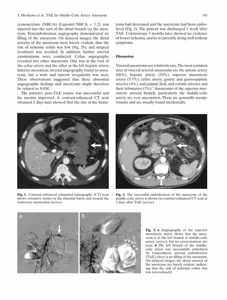
Angiography detected a middle-colic artery aneu-rysm, a wide and narrow irregularity in the distal artery of the aneurysm, but no extravagation (Fig. 2a). The results also indicated that this aneurysm was ruptured and it had a risk of further bleeding. Transcatheter arte-rial embolization was applied initially because it was less invasive treatment than surgery. Using a right femoral artery approach, a 4-Fr C2 catheter (Clinical Supply, Hashima, Japan) was placed in the superior mesenteric artery, and the proximal middle-colic artery was selected with micro-catheter (Renegade; Boston Scientifi c Japan, Tokyo, Japan). However, because of the meandering of the middle-colic artery, the micro-catheter could not be inserted at the aneurysm. There-fore, microcoils could not be used. Then approximately 0.3 ml of iodized oil (Lipiodol) mixed with N-butyl-2-
T. Hirokawa et al.: TAE for Middle-Colic Artery Aneurysm 145
cyanoacrylate (NBCA) (Lipiodol : NBCA = 3 : 2) was injected into the root of the distal branch via the aneu-rysm. Postembolization angiography demonstrated no fi lling of the aneurysm. On delayed images, the distal arteries of the aneurysm were barely evident, thus the risk of ischemic colitis was low (Fig. 2b), and surgical treatment was avoided. In addition, further arterial examinations were conducted. Celiac angiography revealed two other aneurysms. One was at the root of the celiac artery and the other at the left hepatic artery. Inferior mesenteric arterial angiography found no aneu-rysm, but a wide and narrow irregularity was seen. These observations suggested that these abnormal angiographic fi ndings and aneurysms might therefore be related to SAM.
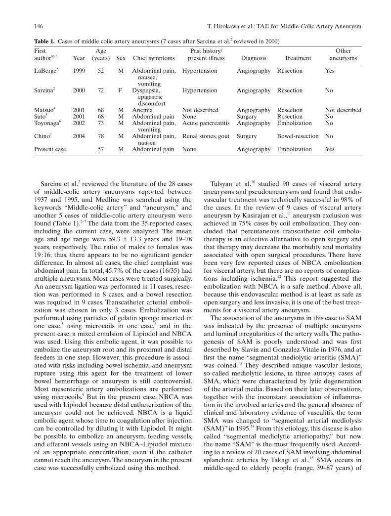
The patient’s post-TAE course was uneventful and his anemia improved. A contrast-enhanced CT scan obtained 2 days later showed that the size of the hema-
toma had decreased and the aneurysm had been embo-lized (Fig. 3). The patient was discharged 1 week after TAE. Colonoscopy 3 months later showed no evidence of bowel ischemia, and he is currently doing well without symptoms.
Discussion
Visceral aneurysms are relatively rare. The most common sites of visceral arterial aneurysms are the splenic artery (60%), hepatic artery (20%), superior mesenteric artery (5.5%), celiac artery, gastric and gastroepiploic arteries (4%), and jejunal, ileal, and colonic arteries and their tributaries (3%).1 Aneurysms of the superior mes-enteric arterial branch, particularly the middle-colic artery, are very uncommon. These are generally asymp-tomatic and are usually found incidentally.
Fig. 1. Contrast-enhanced computed tomography (CT) scan shows extensive ascites in the omental bursa and around the transverse mesocolon (arrow)
Fig. 2. a Angiography of the superior mesenteric artery shows that the aneu-rysm is in the left branch of middle-colic artery (arrow), but no extravasations are seen. b The left branch of the middle-colic artery was successfully embolized by transcatheter arterial embolization (TAE); there is no fi lling of the aneurysm. On delayed images, the distal arteries of the aneurysm are barely evident, indicat-ing that the risk of ischemic colitis was low (arrowheads)
Fig. 3. The successful embolization of the aneurysm of the middle-colic artery is shown on contrast-enhanced CT scan at 2 days after TAE (arrow)
146 T. Hirokawa et al.: TAE for Middle-Colic Artery Aneurysm
Sarcina et al.2 reviewed the literature of the 28 cases of middle-colic artery aneurysms reported between 1937 and 1995, and Medline was searched using the keywords “Middle-colic artery” and “aneurysm,” and another 5 cases of middle-colic artery aneurysm were found (Table 1).2–7 The data from the 35 reported cases, including the current case, were analyzed. The mean age and age range were 59.3 ± 13.3 years and 19–78 years, respectively. The ratio of males to females was 19 : 16; thus, there appears to be no signifi cant gender difference. In almost all cases, the chief complaint was abdominal pain. In total, 45.7% of the cases (16/35) had multiple aneurysms. Most cases were treated surgically. An aneurysm ligation was performed in 11 cases, resec-tion was performed in 8 cases, and a bowel resection was required in 9 cases. Transcatheter arterial emboli-zation was chosen in only 3 cases. Embolization was performed using particles of gelatin sponge inserted in one case,8 using microcoils in one case,6 and in the present case, a mixed emulsion of Lipiodol and NBCA was used. Using this embolic agent, it was possible to embolize the aneurysm root and its proximal and distal feeders in one step. However, this procedure is associ-ated with risks including bowel ischemia, and aneurysm rupture using this agent for the treatment of lower bowel hemorrhage or aneurysm is still controversial. Most mesenteric artery embolizations are performed using microcoils.9 But in the present case, NBCA was used with Lipiodol because distal catheterization of the aneurysm could not be achieved. NBCA is a liquid embolic agent whose time to coagulation after injection can be controlled by diluting it with Lipiodol. It might be possible to embolize an aneurysm, feeding vessels, and efferent vessels using an NBCA–Lipiodol mixture of an appropriate concentration, even if the catheter cannot reach the aneurysm. The aneurysm in the present case was successfully embolized using this method.
Tulsyan et al.10 studied 90 cases of visceral artery aneurysms and pseudoaneurysms and found that endo-vascular treatment was technically successful in 98% of the cases. In the review of 9 cases of visceral artery aneurysm by Kasirajan et al.,11 aneurysm exclusion was achieved in 75% cases by coil embolization. They con-cluded that percutaneous transcatheter coil embolo-therapy is an effective alternative to open surgery and that therapy may decrease the morbidity and mortality associated with open surgical procedures. There have been very few reported cases of NBCA embolization for visceral artery, but there are no reports of complica-tions including ischemia.12 This report suggested the embolization with NBCA is a safe method. Above all, because this endovascular method is at least as safe as open surgery and less invasive, it is one of the best treat-ments for a visceral artery aneurysm.
The association of the aneurysms in this case to SAM was indicated by the presence of multiple aneurysms and luminal irregularities of the artery walls. The patho-genesis of SAM is poorly understood and was fi rst described by Slavin and Gonzalez-Vitale in 1976, and at fi rst the name “segmental mediolytic arteritis (SMA)” was coined.13 They described unique vascular lesions, so-called mediolytic lesions, in three autopsy cases of SMA, which were characterized by lytic degeneration of the arterial media. Based on their later observations, together with the inconstant association of infl amma-tion in the involved arteries and the general absence of clinical and laboratory evidence of vasculitis, the term SMA was changed to “segmental arterial mediolysis (SAM)” in 1995.14 From this etiology, this disease is also called “segmental mediolytic arteriopathy,” but now the name “SAM” is the most frequently used. Accord-ing to a review of 20 cases of SAM involving abdominal splanchnic arteries by Takagi et al.,15 SMA occurs in middle-aged to elderly people (range, 39–87 years) of
Table 1. Cases of middle colic artery aneurysms (7 cases after Sarcina et al.2 reviewed in 2000)
First authorRef. Year
Age (years) Sex Chief symptoms
Past history/present illness Diagnosis Treatment
Other aneurysms
LaBerge3 1999 52 M Abdominal pain, nausea, vomiting
Hypertension Angiography Resection Yes
Sarcina2 2000 72 F Dyspepsia, epigastric discomfort
Hypertension Angiography Resection No
Matsuo4 2001 68 M Anemia Not described Angiography Resection Not describedSato5 2001 68 M Abdominal pain None Surgery Resection NoToyonaga6 2002 73 M Abdominal pain,
vomitingAcute pancreatitis Angiography Embolization No
Chino7 2004 78 M Abdominal pain, nausea
Renal stones, gout Surgery Bowel-resection No
Present case 57 M Abdominal pain None Angiography Embolization Yes
T. Hirokawa et al.: TAE for Middle-Colic Artery Aneurysm 147
both sexes, usually involves more than one visceral artery, and most frequently, branches of the celiac axis. The current case was compatible with these cases. Gen-erally the visceral aneurysms are related to an infection represented by subacute bacterial endocarditis, polyar-teritis nodosa, and others. In the present case, there had been no previous infectious disease. Following the TAE, the patient is doing well and there have been no signs of infl ammation, and thus no need for any steroid or immunosuppressive drug therapies. One limitation to this study is that no pathological examination was carried out, but the clinical and radiographical fi ndings strongly suggested SAM. In conclusion, this was a rare case of a ruptured middle-colic artery aneurysm associ-ated with SMA, which is the fi rst case to be treated by Lipiodol and NBCA embolization.
References
1. Moore WS. Vascular surgery: a comprehensive review. 3rd ed. Philadelphia, PA: Saunders; 1991.
2. Sarcina A, Bellosta R, Magnaldi S, Luzzani L. Aneurysm of the middle colic artery — case report and literature review. Eur J Vasc Endovasc Surg 2000;20:198–200.
3. LaBerge K. SCVIR Annual Meeting fi lm panel session: case 3. JVIR 1999;10:509–13.
4. Matsuo S, Yamaguchi S, Miyamoto S, Ishii T, Tsuneoka N, Obata S, et al. Ruptured aneurysm of the visceral artery: report of two cases. Surg Today 2001. 31:660–4.
5. Sato T, Itoh M, Ohta N, Funaki H, Saito Z, Takayanagi N. Spontaneous ruptured middle colic artery aneurysm with concur-rent renal cell carcinoma. Hepato-Gastroenterology 2001;48:678–80.
6. Toyonaga T, Nagaoka S, Ouchida K, Nagata M, Shirota T, Ogawa T, et al. Case of a bleeding pseudoaneurysm of the middle colic artery complicating acute pancreatitis. Hepato-Gastroenterology 2002;49:1141–3.
7. Chino O, Kijima H, Shivuya M, Yamamoto S, Kashiwagi H, Kondo Y, et al. A case report: spontaneous rupture of dissecting aneurysm of the middle colic artery. Tokai J Exp Clin Med 2004;29:155–8.
8. Naito A, Toyota N, Ito K. Embolization of a ruptured middle colic artery aneurysm. Cardiovasc Intervent Radiol 1995;18:56–8.
9. d’Othee BJ, Surapaneni P, Rabkin D, Nasser I, Clouse M. Microcoil embolization for acute lower gastrointestinal bleeding. Cardiovasc Intervent Radiol 2006;29:49–58.
10. Tulsyan N, Kashyap VS, Greenberg RK, Sarac TP, Clair DG, Pierce G, et al. The endovascular management of visceral artery aneurysms and pseudoaneurysms. J Vasc Surg 2006;45:276–83.
11. Kasirajan K, Greenberg RK, Clair D, Ouriel K. Endovascular management of visceral artery aneurysm. J Endovasc Ther 2001;8:150–5.
12. Kish JW, Katz MD, Marx MV, Harrell DS, Hanks SE. N-butyl cyanoacrylate embolization for control of acute arterial hemor-rhage. J Vasc Interv Radiol 2004;15:689–95.
13. Slavin RE, Gonzalez-Vitale JC. Segmental mediolytic arteritis. A clinical pathologic study. Lab Interv 1976;35:23–9.
14. Slavin RE, Saeki K, Bhagavan B, Maas AE. Segmental arterial mediolysis: a precursor to fi bromuscular dysplasia?. Mod Pathol 1995;8:287–94.
15. Takagi C, Ashizawa N, Eishi K, Ashizawa K, Hayashi T, Tanaka K, et al. Segmental mediolytic arteriopathy involving celiac to splenic and left renal arteries. Intern Med 2003;42:818–23.