Embed Size (px)
Citation preview

PEER-REVIEWED ARTICLE bioresources.com
Zhang et al. (2016). “Polysaccharides extraction,” BioResources 11(2), 5100-5112. 5100
Microwave-assisted Extraction of Polysaccharides from Bamboo (Phyllostachys acuta) Leaves and their Antioxidant Activity
Xiudong Zhang,a Mingfei Li,b Linxin Zhong,a Xinwen Peng a,*and Runcang Sun,b
Polysaccharides were isolated from Phyllostachys acuta leaves by microwave-assisted extraction under various temperatures and time. The obtained polysaccharides were characterized by acid hydrolysis, the Folin-Ciocalteu method, and Fourier transform infrared spectroscopy (FTIR). The major monosaccharides presented in the extracts were arabinose (258.0 mg/g to 414.5 mg/g), galactose (167.0 mg/g to 289.2 mg/g), and glucose (157.4 mg/g to 246.7 mg/g) along with some mannose, fructose, and xylose. The total phenol yield of the bamboo leaves was 0.31 mg/g to 0.73 mg/g. The FTIR spectra revealed that the polysaccharides mostly consisted of β-glycosidic linkages. For the cytotoxicity, the presence of polysaccharides considerably elevated the multiplication of HepG2 cells and showed no growth inhibition for the samples. For the antioxidant activities, the polysaccharides exhibited excellent abilities both in the diphenyl picrylhydrazyl radical potential (DPPH) assay and ferric reducing antioxidant potential (FRAP) assay. The results suggest that bamboo leaf polysaccharides have great potential to be applied in the food, healthcare, and pharmaceutical fields.
Keywords: Polysaccharides; Microwave-assisted extraction; Bamboo leaf; Phyllostachys acuta;
Antioxidant
Contact information: a: State Key Laboratory of Pulp and Paper Engineering, South China University of
Technology, Guangzhou 510640, China; b: Beijing Key Laboratory of Lignocellulosic Chemistry, Beijing
Forestry University, Beijing 100083, China; *Corresponding author: [email protected]
INTRODUCTION
Bamboo, a perennial green plant belonging to the Gramineae family and
Bambusoideae subfamily, is one of the most abundant biomass resources in China. For a
long time, bamboo leaves had been considered as residues from traditional applications,
and most of them were burned or discarded, which brought in serious environmental
pollution and resource waste. It is therefore imperative to find an adequate disposal method
for the waste bamboo leaves or means to convert them into useful products.
Many residues, originating from the process of utilization, are important
agricultural resources because of their enriched content of bioactive compounds such as
proteins, phenolic compounds, phytic acid, and polysaccharides (Zhu and Wu 2009). As a
traditional Chinese medicine, bamboo leaves were used as an antipyretic to stop bleeding,
and for detoxification and detumescence (Chen et al. 1999). Moreover, the antioxidant
materials in bamboo have also been considered as a novel natural food antioxidant (Liu et
al. 2014).
Previous research showed that bamboo leaves could be used as a raw material for
obtaining co-products in medical and food industries, such as polysaccharides and
flavonoids (Kweon et al. 2001) and C-glycosides (Zhang et al. 2008). Furthermore,
PEER-REVIEWED ARTICLE bioresources.com
Zhang et al. (2016). “Polysaccharides extraction,” BioResources 11(2), 5100-5112. 5101
bamboo leaf extracts have been reported to have great potential for anticancer purposes
(Seki and Maeda 2010), protection of immune systems (Seki et al. 2010), and inhibiting
diseases (Jiao et al. 2011).
The most common method used for polysaccharides isolation was through hot
water extraction (HWE) (Shi et al. 2011). The weaknesses of HWE, including high energy
consumption and low yields, had inhibited this method from being widely applied. To
optimize the extraction technology, many novel techniques had been employed, such as
ultrasonic-assisted extraction (UAE), enzyme-assisted extraction (EAE), supercritical fluid
extraction (SFE), and pressurized liquid extraction (PLE) (Kadam et al. 2013; Matsunaga
et al. 2013; Ye et al. 2015). These novel extraction technologies had been successfully
used in food and pharmaceutical applications for extraction of bioactive compounds
(Easmin et al. 2015).
Among all the novel extraction technologies, microwave-assisted extraction (MAE)
has been considered as one of the predominant methods to extract polysaccharides from
plant structures (leaves, seeds, flowers, and roots) because of its simpler operation, shorter
extraction time, flexibility, lower energy consumption, and higher efficacy. Moreover,
MAE has also been reported to have the potential to minimize the disassembly and possible
degradation of the extracted polysaccharides (Marshall et al. 2010). Rodriguez-Jasso et al.
(2011) reported that high yields of fucoidan could be isolated in a very short time with the
MAE method.
Vazquez-Delfin et al. (2014) elaborated an extraction system based on microwave
irradiation, and the results showed lower yields but higher anhydrogalactose content than
conventional techniques. Karabegovic et al. (2013) optimized the MAE of polysaccharides
from cherry laurel leaves with a response surface method (RSM). The previous research
suggested that MAE was a promising method for extracting bio-active components,
including polysaccharides, flavonoids, and polyphenols.
Phyllostachys acuta leaf is a forestry residue with a variety of bio-active
components. Feedstocks used to isolate bio-components were generally confined to
medicinal plants. To our best knowledge, there has been a lack of reports about ordinary
bamboo leaf polysaccharide extraction. Thus, the Phyllostachys acuta leaf was used as
feedstock to isolate crude polysaccharides for bio-antioxidants. The results revealed that
the crude polysaccharides had potent antioxidant activities, no cytotoxicity, and great
potential to be used in food and pharmaceutical areas.
EXPERIMENTAL
Raw Materials Bamboo leaves were harvested from Yunnan province of China, in 2013, from
three-year-old bamboo (Phyllostachys acuta). The raw material was first dried at room
temperature for 15 days and then ground into a fine powder to obtain a fraction under 20-
mesh. The powder was stored in a desiccator at room temperature.
Chemicals and Reagents 2, 2’-Diphenyl-1-picryl-hydrazyl (DPPH), 1, 3, 5’-tri (2’-pyridyl) -2, 4, 6’-triazine
(TPTZ), and ascorbic acid were purchased from Sinopharm Chemical Company (Beijing,
China). All chemicals were of analytical grade and were used without any further
purification.

PEER-REVIEWED ARTICLE bioresources.com
Zhang et al. (2016). “Polysaccharides extraction,” BioResources 11(2), 5100-5112. 5102
Extraction Process The polysaccharide extraction was based on a previously reported method with
some modifications (Liu et al. 2015). The extraction process was carried out in a 500-mL
beaker, followed by heating in a microwave oven (MicroSYNTH, Milestone, USA). Then,
20 g (oven-dried weight) of bamboo leaf flour was added to 300 mL of ultra-pure water in
the beaker. After that, the beaker was subjected to microwave heating for predetermined
temperatures (60, 70, 80, and 90 °C) and time intervals (20, 40, and 60 min).
Once the reaction was complete, the supernatant and sediments were separated by
vacuum filtration. The residues were washed with ultra-pure water three times and then
discarded. The solutions were condensed to approximately 100 mL using a rotary
evaporator at 60 ºC under vacuum. Anhydrous ethanol (300 mL) was slowly added to the
resulting solution, which was then precipitated overnight at 4 °C for 24 h. Subsequently,
the precipitated polysaccharide fractions were centrifuged and freeze-dried to obtain crude
polysaccharides.
Except for polysaccharides, a small amount of monosaccharide, protein, as well as
flavones could also be precipitated. The main components were polysaccharides, so the
extracted solid was considered as crude polysaccharides. The extraction yield was utilized
to optimize the extraction conditions.
FTIR Analysis
The functional groups of the extracted crude polysaccharides were identified on a
FTIR spectrophotometer (NicoletiN10, Thermo, USA). The crude polysaccharides were
ground into powder and pressed into 1 mm disks for FTIR measurement in the frequency
range of 4,000 to 400 cm-1.
Monosaccharide Component Analysis The total neutral monosaccharide and uronic acids of the samples were determined
by high-performance anion exchange chromatography (HPAEC). Each sample (0.03 g)
was first hydrolyzed with 0.03 mL of 72% sulfuric acid at 30 °C for 1 h. During this
procedure, a stirring rod was used to stir the sample every 5 to 10 min. Subsequently, the
hydrolysates were diluted with 8.4 mL of ultra-pure water, and then the tubes were sealed
and incubated in a bake oven at 105 °C for 1 h. After that, the sample solutions were diluted
with ultra-pure water and filtered via a 0.22-μm filter membrane before injecting into the
HPAEC system (Dionex ISC 300, USA) with an amperometric detector, a CarbopacTMP-
20 column (4 mm × 250 mm, Dionex), and a guard PA-20 column (3 mm × 30 mm, Dionex).
The peaks were identified by comparing the retention time to the standards.
Total Phenol Analysis Total phenol analysis was conducted according to the method reported by Jerez et
al.(2007). Briefly, 0.5 mL of the extracted crude polysaccharide solution (0.1 g/L) was
added to 2.5 mL of Folin-Ciocalteu reagent (2.0 M). The mixture was shaken continuously
and incubated for 30 min. Then, 2.5 mL of Na2CO3 solution (7.5 g/mL) was added to the
previous mixture. After maintaining for 1.5 h at 37 °C, the mixture was measured at 765
nm against a blank (ultrapure water), and the total phenol content was calculated according
to the standard curve of gallic acid solutions (0 to 0.06 g/L).
PEER-REVIEWED ARTICLE bioresources.com
Zhang et al. (2016). “Polysaccharides extraction,” BioResources 11(2), 5100-5112. 5103
Cytotoxicity Analysis Cytotoxicity was evaluated using the MTS (3-(4,5-dimethylthiazol-2-yl)-5(3-
carboxymethoxyphenyl)-2-(4-sulfopheny)-2H-tetrazolium, inner salt) assay. In detail,
HepG2 cells were cultivated in trypsin-EDTA medium and 5% CO2 atmosphere at 37 ºC
in advance. Then, a density of 1 × 10⁵ HepG2 cells were seeded into 96-well plates and
treated with polysaccharide (extracted at 70 °C and 40 min) solutions (0, 0.01, 0.05, 0.1,
0.25, 0.5, and 1 mg/mL) for 0, 4, or 12 h. At predetermined time intervals,1.0% MTS
solution was added to the medium, and then the mixtures were subjected to incubation for
4 h at 37 ºC. After that, the absorbance of the resulting solutions at 490 nm was measured
by a spectra-photometric plate reader (Multiscan MK3, Thermo Fisher Scientific, USA).
DPPH Radical Scavenging Activity The DPPH radical scavenging activity of the extracted polysaccharides was carried
out according to previous methods with minor modifications (Yin et al. 2010). In detail,
the DPPH powder was dissolved in methanol to obtain 25 mg/L solutions. Then, 3 mL of
DPPH fluid was added to 0.5 mL of polysaccharide liquor (1, 2, 4, 6, 8, and 10 mg/mL,
respectively). The mixtures were shaken vigorously and maintained in the darkness for 30
min. After that, the absorbance was measured at 517 nm using ascorbic acid as a positive
control. All tests were carried out in parallel. The scavenging activity on DPPH radicals
was calculated by the following equation,
DPPH radical scavenging activity (%) =DPPH
sampleDPPH
A
AA -×100 (1)
where ADPPH is the absorbance of the DPPH radical solution with methanol and Asample is
the absorbance of the DPPH radical solution with polysaccharide fluid.
Ferric Reducing Activity The FRAP assay was carried out according to a previously described method with
some modifications (Tian et al. 2009). The FRAP solution consisted of 250 mL of acetate
buffer (300 mM, pH 3.6), 25 mL of TPTZ solution (10 mM in 40 mM HCl), and 25 mL of
FeCl3•6H2O solution (20 mM). After preheating to 37 ºC, 3 mL of FRAP reagent was
mixed with 0.12 mL sample solutions (0.5, 1, 2, 3, 4, and 5 mg/mL, respectively) and
incubated for 30 min. Subsequently, the absorbance was measured at 593 nm, and the final
result was calculated as the concentration of FeSO4•7H2O.
RESULTS AND DISCUSSION
Extraction of Polysaccharides from Bamboo Leaves The effects of time and temperature on crude polysaccharides yield were
investigated with a heating power of 800 W, and the mass ratio of raw material to water
was fixed at 1:15. As shown in Figure 1, the yield of crude polysaccharides was positively
correlated with increasing temperature (from 60 to 90 °C) and time (from 20 to 60 min).
At 70 °C and 40 min, the yield reached 1993 mg/L. After that, increasing temperature and
time only slightly increased the yield. Harsher extraction conditions, in terms of time and
temperature, may have led to an efficient opening of the cell wall network and hence to a
higher yield. In detail, when the temperature was set to be 60 °C, increasing reaction time
from 20 to 60 min brought in only 126 mg/L polysaccharides of augmented yield. On the

PEER-REVIEWED ARTICLE bioresources.com
Zhang et al. (2016). “Polysaccharides extraction,” BioResources 11(2), 5100-5112. 5104
other hand, from 70 to 90 °C, the solid product yield could be significantly enhanced by
increasing reaction time from 20 to 40 min. With further prolonging of the reaction time to
60 min, the solid yield was almost the same as that of 40 min. The results suggested that
both reaction time and temperature played an essential role in the extraction processes.
Furthermore, 60 °C was not strong enough for sufficient polysaccharides dissolution. At
higher reaction temperatures, small particles of polysaccharides could be isolated from the
cell wall efficiently within 20 min; increasing reaction time contributed to polysaccharides
dissolution, as well as degradation. From the beginning to 40 min, the dissolution reaction
was dominant, and after that, the degradation rate was enhanced and comparable with
dissolution rate, resulting in the slight increase of production yield at longer reaction time.
Compared with the hot water extraction reported by Cui et al. (2014), the extraction time
in this study was effectively reduced to less than 60 min.
60-20 60-40 60-60 70-20 70-40 70-60 80-20 80-40 80-60 90-20 90-40 90-60
500
1000
1500
2000
2500
Po
lys
ac
ch
ari
de
s y
ield
(m
g/L
)
Temperature-Time (°C-min)
Fig. 1. Yield of crude polysaccharides extracted under various conditions
FTIR Analysis FTIR spectra demonstrated that no considerable differences were observed between
the fractions under both mild and severe conditions, suggesting that the tested range of
temperature (60 to 90 °C) and time (20 to 60 min) had no enormous effects on the structure
of the extracted crude polysaccharides. The samples gave signals for -OH stretching
vibration at 3280 cm-1 and for C-H stretching at approximately 2930 cm-1 (asymmetrical
stretching)and 2862 cm-1 (symmetrical stretching) (Buranov and Mazza 2012). The peaks
at 400 to 2000 cm-1 are the characteristic absorptions of polysaccharides (Luo et al. 2012;
Peng et al. 2013).The strong absorption signals at 1613 cm-1 can be assigned to the
stretching vibration of the CHO and C=O bonds (Mao et al. 2013). The bands at
approximately 1413 cm-1 can be attributed to C-H bending vibration of -CH2-. The
absorption peaks at 1328 cm-1 are related to C-O stretching vibration in the C-OH groups
or C-C stretching vibration in the carbohydrate structure (Sandula et al. 1999). There are
intense peaks at around 1100 cm-1, which can be assigned to the uronic acids that had not
been broken by the extraction process (Ying et al. 2011). As shown in Figure 2, S6 showed
a lower absorption peak than other extracted solids at this region. It was assumed that the
uronic acids were instable at the extraction conditions. When prolonging the reaction time
from 40 min to 60 min, the uronic acids degraded much faster than its formation, resulting
in lower uronic acids content and weak absorption signals at around 1100 cm-1. In addition,

PEER-REVIEWED ARTICLE bioresources.com
Zhang et al. (2016). “Polysaccharides extraction,” BioResources 11(2), 5100-5112. 5105
the absorption signals at approximately 900 cm-1 can be attributed to the β-glycosidic
linkages between sugar units (Sun et al. 2003). In conclusion, the FTIR spectra revealed
the general characteristic absorption peaks of polysaccharides, and no novel absorbance
signals were found.
4000 3500 3000 2500 2000 1500 1000
Tra
ns
mit
tan
ce
(%
)
795.5
5
1118.5
8
1326.3
11413.0
5
1616.7
3
2076.1
32137.9
6
3297.8
3
S4
S6
S11
S8
S2
2929.9
0
S5
Wave number (cm-1
)
Fig. 2. FTIR spectra of crude polysaccharides extracted under various conditions (S2, S4, S5, S6, S8, and S11 represent samples extracted at 60 °C and 40 min, 70 °C and 20 min, 70 °C and 40 min, 70 °C and 60 min, 80 °C and 40 min, and 90 °C and 40 min, respectively)
Chemical Components The HPAEC analysis (Table 1) showed that the component sugars of the extracted
crude polysaccharides were mannose, fructose, xylose, glucose, galactose, and arabinose.
Arabinose, galactose, and glucose were the predominant monosaccharides (relative mass
258.0 mg/g extracted solids to 414.5 mg/g extracted solid, 167.0 mg/g extracted solids to
289.2 mg/g extracted solids, and 157.4 mg/g extracted solids to 246.7 mg/g extracted solids,
respectively), along with low levels of mannose (relative mass 6.9 mg/g extracted solids to
14.6 mg/g extracted solids), fucose (relative mass 7.8 mg/g extracted solids to 13.0 mg/g
extracted solids), and xylose (relative mass 40.9 mg/g extracted solids to 109.0 mg/g
extracted solids). The differences in monosaccharide contents were associated with
extraction conditions, which was seen varied at different temperatures and time. In addition
to monosaccharides, the extraction conditions also had an apparent effect on the release of
total phenol in the final products. At 40 min, there was a higher total phenol content than
that with other conditions, except for that at 90 °C (the highest phenol yielded at 20 min).
The results could be assumed that the dissolved phenol particles were unstable in the
reaction conditions. When the reaction temperature was sever (at 90 °C, for example), it
would degraded sharply, resulting in low yield. As relatively severe reaction conditions
were favorable for the phenol dissolution, the highest final phenol release reached as high
as 0.73 mg/g from bamboo leaves at 70 °C, 40 min.

PEER-REVIEWED ARTICLE bioresources.com
Zhang et al. (2016). “Polysaccharides extraction,” BioResources 11(2), 5100-5112. 5106
Table 1.The Monosaccharide Compositions and Phenol Contents of the Crude Polysaccharides Extracted at Different Time and Temperatures*
T (°C)
t (min)
Man (mg/g)
Fuc (mg/g)
Xyl (mg/g)
Glu (mg/g)
Gal (mg/g)
Ara (mg/g)
Phenol** (mg/g)
Others*** (mg/g)
60
20 12.8 9.0 55.3 182.8 282.6 293.3 0.31 1.74
40 9.7 9.8 42.5 182.8 175.0 412.9 0.34 2.17
60 12.3 8.0 69.8 199.8 273.8 294.3 0.35 2.12
70
20 10.2 10.0 58.4 173.8 289.2 318.4 0.48 2.28
40 9.9 10.2 43.4 182.0 176.5 414.6 0.73 3.54
60 12.5 11.7 76.2 223.7 259.8 277.8 0.42 3.66
80
20 11.8 13.0 62.9 246.7 243.7 266.3 0.31 1.89
40 8.0 9.1 40.9 157.4 203.3 416.5 0.58 3.96
60 14.6 8.9 109.0 237.4 231.0 258.0 0.44 4.64
90
20 11.8 7.8 65.2 244.4 238.4 260.6 0.50 3.97
40 6.9 9.1 56.3 167.0 167.0 413.8 0.39 5.03
60 10.7 8.5 73.9 171.9 244.4 275.3 0.43 5.69
*T: temperature; t: time; Man: mannose; Fuc: fucose; Xyl: xylose; Glu: glucose; Gal: galactose; Ara: arabinose. All the monosaccharide content was calculated as mass fraction of extracted solid materials. ** The phenol content was calculated as mass fraction of raw bamboo leaves. *** Others were represented as other particles in the crude polysaccharides like protein, fat, and so on. Its content was calculated as mass fraction of raw bamboo leaves.
Cytotoxicity Analysis
In order to be used in food and pharmaceutical industry applications, the extracted
crude polysaccharides must exhibit minimal toxicity. To evaluate the effects of crude
polysaccharides on cell proliferation, the cytotoxicity was evaluated against HepG2 cells,
taking the samples extracted at 70 °C and 40 min as arepresentative. Cells were exposed to
increasing doses of extracted crude polysaccharides solutions with concentrations of 0.01
mg/mL to 1 mg/mL for 0, 12, or 24 h, and the cell viability was determined by the MTS
assay. As shown in Figure3, cell viabilities of HepG2 were markedly increased after
exposure to extracted crude bamboo leaf polysaccharides for 12 h in a dose-dependent
manner. The proliferation of HepG2 cells was increased in correspondence to the
increasing extracted crude polysaccharides concentrations (from 0.01 to 0.5 mg/mL).
When further increasing the dose to 1 mg/mL, the cell proliferation rate was decreased and
resulted in a comparable OD 490 value of the blank sample. The inhibition ratio of HepG2
cells was considerably decreased after exposure to the crude polysaccharides in a dose- and
time- dependent manner. The inhibition ratio was -26.77% after 24 h of exposure to 0.5
mg/mL crude polysaccharides, while a -3.02% inhibition ratio was observed after 24 h of
exposure to 1.0 mg/mL crude polysaccharides. The results revealed that the extracted crude
polysaccharides had no obvious cytotoxicity against HepG2 cells. In addition, in the
presence of the bamboo leaf extracts, the cell multiplication rate was markedly accelerated.

PEER-REVIEWED ARTICLE bioresources.com
Zhang et al. (2016). “Polysaccharides extraction,” BioResources 11(2), 5100-5112. 5107
0 0.01 0.05 0.1 0.25 0.5 10.0
0.2
0.4
0.6
0.8
1.0
1.2
1.4
O.D
49
0
Concentration (mg/mL)
0 h
4 h
12 h
Fig. 3. Cytotoxicity profile of crude polysaccharides extracted at 70 ºC and 40 min against HepG2 cells
Scavenging Activity of DPPH Radical Analysis DPPH is a stable free radical that has a maximum absorption at 517 nm in methanol.
When encountering a proton-donating substance (antioxidants), DPPH radicals are
scavenged and the absorbance at 517 nm is reduced. Based on this principle, the antioxidant
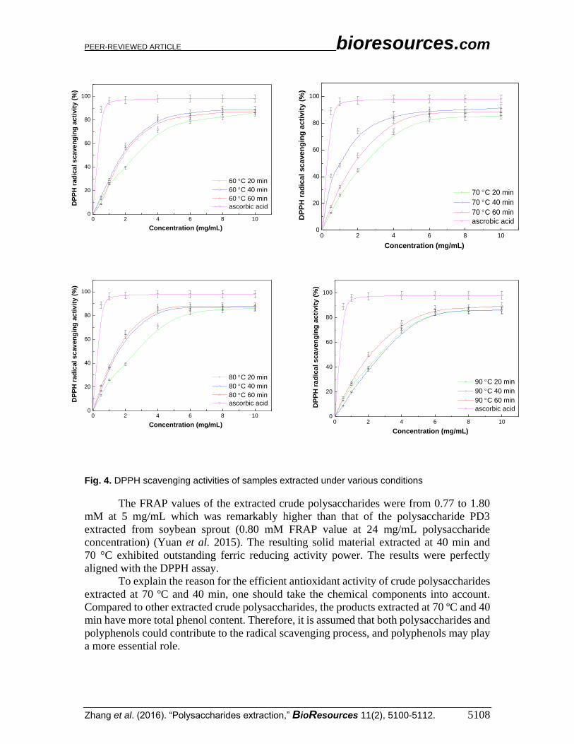
activity of a substance can be measured. Figure 4 shows that the scavenging activities of
all the extracted crude polysaccharides were clearly observed under all tested
concentrations and correlated well with increased concentrations up to 10 mg/mL. At 10
mg/mL, the antioxidant effects of all the samples were higher than 85% with less than 3.5
mg/L DPPH residue. The resulting solid material from the extracts at 40 min and 70 °C
had the highest antioxidant activity, with an IC50 of 1.15 mg/mL. The IC50 of ascorbic acid
was 0.30 mg/mL, indicating that the DPPH scavenging activity of ascorbic acid was much
higher than that of extracted bamboo leaf crude polysaccharides. However, the antioxidant
activity was remarkably higher than that of other polysaccharides, such as CMCP from
Cucurbita moschata (IC50 = 7.8 mg/mL, tested with 20 mg/mL DPPH solution) (Wu et al.
2014). These results indicated that at appropriate extraction conditions, the extracted crude
polysaccharides could have noticeable scavenging effects on free radicals. The mechanism
of the extracted crude polysaccharides radical scavenging can be explained as follows:
hemiacetal hydroxyl in reducing sugars, phenolic hydroxyl groups, and other components
acted as hydrogen donators to react with DPPH radicals to convert them to more stable
products.
Ferric Reducing Activity Power The FRAP assay is commonly used for the routine analysis of single antioxidant
and total antioxidant activity of plant extracts (Pulido et al. 2000; Schlesier et al. 2002).
When colorless Fe (III)-TPTZ form reacts with electron-donating antioxidants, a colored
Fe (II)-TPTZ form is generated. Antioxidants with higher efficiency generate more Fe (II)-
TPTZ, and a deeper color is observed. Figure 5 shows that the reducing power of all
samples was notable at all tested concentrations and positively correlated with increased
concentrations up to 5.0 mg/mL.
PEER-REVIEWED ARTICLE bioresources.com
Zhang et al. (2016). “Polysaccharides extraction,” BioResources 11(2), 5100-5112. 5108
0 2 4 6 8 100
20
40
60
80
100
DP
PH
ra
dic
al
sc
av
en
gin
g a
cti
vit
y (
%)
Concentration (mg/mL)
60 C 20 min
60 C 40 min
60 C 60 min
ascorbic acid
0 2 4 6 8 10
0
20
40
60
80
100
DP
PH
rad
ica
l sc
ave
ng
ing
ac
tiv
ity
(%
)
Concentration (mg/mL)
70 C 20 min
70 C 40 min
70 C 60 min
ascrobic acid
0 2 4 6 8 100
20
40
60
80
100
DP
PH
ra
dic
al
sc
av
en
gin
g a
cti
vit
y (
%)
Concentration (mg/mL)
80 C 20 min
80 C 40 min
80 C 60 min
ascorbic acid
0 2 4 6 8 100
20
40
60
80
100
DP
PH
ra
dic
al
scav
en
gin
g a
cti
vit
y (
%)
Concentration (mg/mL)
90 C 20 min
90 C 40 min
90 C 60 min
ascorbic acid
Fig. 4. DPPH scavenging activities of samples extracted under various conditions
The FRAP values of the extracted crude polysaccharides were from 0.77 to 1.80
mM at 5 mg/mL which was remarkably higher than that of the polysaccharide PD3
extracted from soybean sprout (0.80 mM FRAP value at 24 mg/mL polysaccharide
concentration) (Yuan et al. 2015). The resulting solid material extracted at 40 min and
70 °C exhibited outstanding ferric reducing activity power. The results were perfectly
aligned with the DPPH assay.
To explain the reason for the efficient antioxidant activity of crude polysaccharides
extracted at 70 ºC and 40 min, one should take the chemical components into account.
Compared to other extracted crude polysaccharides, the products extracted at 70 ºC and 40
min have more total phenol content. Therefore, it is assumed that both polysaccharides and
polyphenols could contribute to the radical scavenging process, and polyphenols may play
a more essential role.

PEER-REVIEWED ARTICLE bioresources.com
Zhang et al. (2016). “Polysaccharides extraction,” BioResources 11(2), 5100-5112. 5109
0 1 2 3 4 50.0
0.4
0.8
1.2
1.6
2.0
Fe(I
I) (
mM
)
Concentration (mg/mL)
60 C 20 min
60 C 40 min
60 C 60 min
0 1 2 3 4 50.0
0.4
0.8
1.2
1.6
2.0
Fe
(II)
(m
M)
Concentration (mg/mL)
70 C 20 min
70 C 40 min
70 C 60 min
0 1 2 3 4 50.0
0.4
0.8
1.2
1.6
2.0
Fe(I
I) (
mM
)
Concentration (mg/mL)
80 C 20 min
80 C 40 min
80 C 60 min
0 1 2 3 4 50.0
0.4
0.8
1.2
1.6
2.0
Fe(I
I) (
mM
)
Concentration (mg/mL)
90 C 20 min
90 C 40 min
90 C 60 min
Fig. 5. Ferric reducing power of polysaccharides extracted under various conditions
CONCLUSIONS
1. Over 2000 mg/L of bamboo leaf crude polysaccharides were isolated with the MAE
method, and all the extracted solids exhibited excellent antioxidant activities in both
DPPH and FRAP assays.
2. Cytotoxicity experiments against HepG2 cells showed that the extracted crude
polysaccharides had no inhibitory effects on cell proliferation.
3. There is great potential for the crude polysaccharides extracted from bamboo leaves to
be applied as functional food ingredients and pharmaceuticals.
ACKNOWLEDGMENTS
The project is supported by the National Natural Science Foundation of China
(21404043, 31430092 and 21336002), Pearl River S&T Nova Program of Guangzhou
(2014J2200063), Science and Technology Project of Guangdong Province
PEER-REVIEWED ARTICLE bioresources.com
Zhang et al. (2016). “Polysaccharides extraction,” BioResources 11(2), 5100-5112. 5110
(2015A010105005), Research Fund for the Doctoral Program of Higher Education
(201301721200240), and Fundamental Research Funds for the Central Universities.
REFERENCES CITED
Buranov, A. U., and Mazza, G. (2012). “Fractionation of flax shives with pressurized
aqueous ethanol,” Ind. Crop. Prod. 35(1), 77-87.DOI: 10.1016/j.indcrop.2011.06.014
Chen, C. Y., Ding, Y. Q., Elmahadi, E. A., Zhou, J. Y., Li, Y., and Xu, H. B. (1999).
“Study on the isolation, purification and physicochemical properties of
polysaccharides from Indocalamus tesselatus,” Biomed. Chromatogr. 13(1), 11-14.
DOI: 10.1002/(sici)1099-0801(199902)13:1
Cui, G. T., Zhang, W. X., Wang, Q. J., Zhang, A. M., Mu, H. B., Bai, H. J., and Duan, J.
Y. (2014). “Extraction optimization, characterization and immunity activity of
polysaccharides from Fructus jujubae,” Carbohydr. Polym. 111, 245-255. DOI:
10.1016/j.carbpol.2014.04.041
Easmin, M. S., Sarker, M. Z. I., Ferdosh, S., Shamsudin, S. H., Bin Yunus, K., Uddin, M.
S., Sarker, M. M. R., Akanda, M. J. H., Hossain, M. S., and Khalil, H. P. S. A. (2015).
“Bioactive compounds and advanced processing technology: Phaleria macrocarpa
(sheff.) Boerl, A review,” J. Chem. Technol. Biotechnol. 90(6), 981-991. DOI:
10.1002/jctb.4603
Jerez, M., Selga, A., Sineiro, J., Torres, J. L., and Nunez, M. J. (2007). “A comparison
between bark extracts from Pinus pinaster and Pinus radiata: Antioxidant activity
and procyanidin composition,” Food Chem. 100(2), 439-444. DOI:
10.1016/j.foodchem.2005.09.064
Jiao, J. J., Lue, G. S., Liu, X. J., Zhu, H., and Zhang, Y. (2011). “Reduction of blood lead
levels in lead-exposed mice by dietary supplements and natural antioxidants,” J. Sci.
Food Agric. 91(3), 485-491. DOI: 10.1002/jsfa.4210
Kadam, S. U., Tiwari, B. K., and O'Donnell, C. P. (2013). “Application of novel
extraction technologies for bioactives from marine algae,” J. Agric. Food Chem.
61(20), 4667-4675. DOI: 10.1021/jf400819p
Karabegovic, I. T., Stojicevic, S. S., Velickovic, D. T., Nikolic, N. C., and Lazic, M. L.
(2013). “Optimization of microwave-assisted extraction and characterization of
phenolic compounds in cherry laurel (Prunus laurocerasus) leaves,” Sep. Purif.
Technol. 120, 429-436. DOI: 10.1016/j.seppur.2013.10.021
Kweon, M. H., Hwang, H. J., and Sung, H. C. (2001). “Identification and antioxidant
activity of novel chlorogenic acid derivatives from bamboo (Phyllostachys edulis),” J.
Agric. Food Chem. 49(10), 4646-4655. DOI: 10.1021/jf010514x
Liu, L. Y., Xia, B. N., Jin, C., Zhang, Y., and Zhang, Y. (2014). “Chemical acylation of
water-soluble antioxidant of bamboo leaves (AOB-w) and functional evaluation of
oil-soluble AOB (cAOB-o),” J. Food Sci. 79(10), C1886-C1894.DOI: 10.1111/1750-
3841.12578
Liu, J., Wen, X. Y., Zhang, X. Q., Pu, H. M., Kan, J., and Jin, C. H. (2015). “Extraction,
characterization and in vitro antioxidant activity of polysaccharides from black
soybean,” Int. J. Biol. Macromol. 72, 1182-1190. DOI:
10.1016/j.ijbiomac.2014.08.058
Luo, Q., Peng, H., Zhou, M. Y., Lin, D., Ruan, R., Wan, Y. Q., Zhang, J. S., and Liu, Y. H.
(2012). “Alkali extraction and physicochemical characterization of hemicelluloses

PEER-REVIEWED ARTICLE bioresources.com
Zhang et al. (2016). “Polysaccharides extraction,” BioResources 11(2), 5100-5112. 5111
from young bamboo (Phyllostachys pubescens Mazel),” BioResources 7(4), 5817-
5828. DOI: 10.15376/biores.7.4.5817-5828
Mao, J. W., Yin, J., Ge, Q., Jiang, Z. L., and Gong, J. Y. (2013). “In vitro antioxidant
activities of polysaccharides extracted from moso bamboo-leaf,” Int. J. Biol.
Macromol. 55, 1-5. DOI: 10.1016/j.ijbiomac.2012.12.027
Marshall, L. F., Peter, H. C., and Arland, T. H. (2010). “Extraction and characterization of
sugar beet polysaccharides,” in: Green Polymer Chemistry: Biocatalysis and Bio-
materials, Cheng, H. (ed.), American Chemical Society, Washington, DC, pp. 71-86.
Matsunaga, Y., Wahyudiono, Machmudah, S., Askin, R., Quitain, A. T., Sasaki, M., and
Goto, M. (2013). “Hydrothermal extraction and micronization of polysaccharides
from Ganoderma lucidum in a one-step process,” BioResources 8(1), 461-471. DOI:
10.15376/biores.8.1.461-471
Peng, H., Zhou, M. Y., Yu, Z. P., Zhang, J. S., Ruan, R. G., Wan, Y. Q, and Liu, Y. H.
(2013). “Fractionation and characterization of hemicelluloses from young bamboo
(Phyllostachys pubescens Mazel) leaves,” Carbohydr. Polym. 95(1), 262-271. DOI:
10.1016/j.carbpol.2013.03.007
Pulido, R., Bravo, L., and Saura-Calixto, F. (2000). “Antioxidant activity of dietary
polyphenols as determined by a modified ferric reducing/antioxidant power assay,” J
Agric. Food Chem. 48(8), 3396-3402. DOI: 10.1021/jf9913458
Rodriguez-Jasso, R. M., Mussatto, S. I., Pastrana, L., Aguilar, C. N., and Teixeira, J. A.
(2011). “Microwave-assisted extraction of sulfated polysaccharides (fucoidan) from
brown seaweed,” Carbohydr. Polym. 86(3), 1137-1144. DOI:
10.1016/j.carbpol.2011.06.006
Sandula, J., Kogan, G., Kacurakova, M., and Machova, E. (1999). “Microbial (1-3)-beta-
D-glucans, their preparation, physico-chemical characterization and
immunomodulatory activity,” Carbohydr. Polym. 38(3), 247-253. DOI:
10.1016/s0144-8617(98)00099-x
Schlesier, K., Harwat, M., Bohm, V., and Bitsch, R. (2002). “Assessment of antioxidant
activity by using different in vitro methods,” Free Radical Res. 36(2), 177-187. DOI:
10.1080/10715760290006411
Seki, T., Kida, K., and Maeda, H. (2010). “Immunostimulation-mediated anti-tumor
activity of bamboo (Sasa senanensis) leaf extracts obtained under vigorous
condition,” J. Evid. Complem. Altern. Med. 7(4), 447-457. DOI:
10.1093/ecam/nen026
Seki, T., and Maeda, H. (2010). “Cancer preventive effect of kumaizasa bamboo leaf
extracts administered prior to carcinogenesis or cancer inoculation,”Anticancer Res.
30(1), 111-118.
Shi, Z. J., Xiao, L. P., Deng, J., Xu, F., and Sun, R. C. (2011). “Isolation and character-
ization of soluble polysaccharides of Dendrocalamus brandishii: A high yielding
bamboo,” BioResources 6(4), 5151-5166. DOI: 10.15376/biores.6.4.5151-5166
Sun, X. F., Sun, R. C., Tomkinson, J., and Baird, M. S. (2003). “Preparation of sugarcane
bagasse hemicellulosic succinates using NBS as a catalyst,” Carbohydr. Polym. 53(4),
483-495. DOI: 10.1016/s0144-8617(03)00150-4
Tian, F., Li, B., Ji, B., Yang, J., Zhang, G., Chen, Y., and Luo, Y. (2009). “Antioxidant and
antimicrobial activities of consecutive extracts from Galla chinensis: The polarity
affects the bioactivities,” Food Chem. 113(1), 173-179. DOI:
10.1016/j.foodchem.2008.07.062
Vazquez-Delfin, E., Robledo, D., and Freile-Pelegrin, Y. (2014). “Microwave-assisted

PEER-REVIEWED ARTICLE bioresources.com
Zhang et al. (2016). “Polysaccharides extraction,” BioResources 11(2), 5100-5112. 5112
extraction of the carrageenan from Hypnea musciformis (Cystocloniaceae,
Rhodophyta),” J. Appl. Phycol. 26(2), 901-907. DOI: 10.1007/s10811-013-0090-8
Wu, H., Zhu, J. X., Diao, W. C., and Wang, C. R. (2014). “Ultrasound-assisted enzymatic
extraction and antioxidant activity of polysaccharides from pumpkin (Cucurbita
moschata),” Carbohydr. Polym. 113, 314-324. DOI: 10.1016/j.carbpol.2014.07.025
Ye, Y., Li, Y., and Fang, F. (2015). “Cost-effective isolation of bioactive compounds from
a discarded bioresource-defatted seeds of Camellia oleifera,” BioResources 10(1),
1060-1072. DOI: 10.15376/biores.110.1.1060-1072
Yin, J. Y., Nie, S. P., Zhou, C., Wan, Y., and Xie, M. Y. (2010). “Chemical characteristics
and antioxidant activities of polysaccharide purified from the seeds of Plantago
asiatica L,” J. Sci. Food Agric. 90(2), 210-217. DOI: 10.1002/jsfa.3793
Ying, Z., Han, X. X., and Li, J. R. (2011). “Ultrasound-assisted extraction of
polysaccharides from mulberry leaves,” Food Chem. 127(3), 1273-1279. DOI:
10.1016/j.foodchem.2011.01.083
Yuan, M., Jia, X. J., Yang, Y., Ding, C. B., Du, L., Yuan, S., Zhang, Z. W., and Chen, Y.
G. (2015). “Effect of light on structural properties and antioxidant activities of
polysaccharides from soybean sprouts,” Process Biochem. 50(7), 1152-1157. DOI:
10.1016/j.procbio.2015.03.024
Zhang, Y., Jiao, J. J., Liu, C. M., Wu, X. Q., and Zhang, Y. (2008). “Isolation and
purification of four flavone C-glycosides from antioxidant of bamboo leaves by
macroporous resin column chromatography and preparative high-performance liquid
chromatography,” Food Chem. 107(3), 1326-1336. DOI:
10.1016/j.foodchem.2007.09.037
Zhu, J. F., and Wu, M. C. (2009). “Characterization and free radical scavenging activity
of rapeseed meal polysaccharides WPS-1 and APS-2,” J. Agric. Food Chem. 57(3),
812-819. DOI: 10.1021/jf802687t
Article submitted: October 22, 2015; Peer review completed: January 14, 2016; Revised
version received and accepted: March 18, 2016; Published: April 25, 2016.
DOI: 10.15376/biores.11.2.5100-5112