Embed Size (px)
Citation preview

MicroRNA-133 controls cardiac hypertrophyAlessandra Care1,11, Daniele Catalucci2,3,11, Federica Felicetti1, Desiree Bonci1, Antonio Addario1,Paolo Gallo3,4, Marie-Louise Bang2,3, Patrizia Segnalini1, Yusu Gu2, Nancy D Dalton2, Leonardo Elia2,Michael V G Latronico3,4, Morten Høydal5, Camillo Autore6, Matteo A Russo7, Gerald W Dorn II8,Øyvind Ellingsen5, Pilar Ruiz-Lozano9, Kirk L Peterson2, Carlo M Croce10, Cesare Peschle1,11 &Gianluigi Condorelli2,3,11
Growing evidence indicates that microRNAs (miRNAs or miRs)
are involved in basic cell functions and oncogenesis. Here
we report that miR-133 has a critical role in determining
cardiomyocyte hypertrophy. We observed decreased expression
of both miR-133 and miR-1, which belong to the same
transcriptional unit, in mouse and human models of cardiac
hypertrophy. In vitro overexpression of miR-133 or
miR-1 inhibited cardiac hypertrophy. In contrast, suppression
of miR-133 by ‘decoy’ sequences induced hypertrophy,
which was more pronounced than that after stimulation
with conventional inducers of hypertrophy. In vivo inhibition of
miR-133 by a single infusion of an antagomir caused marked
and sustained cardiac hypertrophy. We identified specific
targets of miR-133: RhoA, a GDP-GTP exchange protein
regulating cardiac hypertrophy; Cdc42, a signal transduction
kinase implicated in hypertrophy; and Nelf-A/WHSC2,
a nuclear factor involved in cardiogenesis. Our data show
that miR-133, and possibly miR-1, are key regulators of
cardiac hypertrophy, suggesting their therapeutic application
in heart disease.
MicroRNAs (miRNAs) are small conserved RNA molecules of B22nucleotides1 which negatively modulate gene expression in animalsand plants, primarily through base paring to the 3¢ untranslated region(UTR) of target mRNAs; this leads to mRNA cleavage and/ortranslation repression1. MiRNAs are involved in a variety of basicbiological processes, for example, cell proliferation and apoptosis2–4
and stress responses5. In fact, bioinformatic analysis predicts that eachmiRNA may regulate hundreds of targets, suggesting that miRNAsmay play a role in almost every biological pathway6. Furthermore,miRNAs are implicated in cancer, where they can act as tumorsuppressors or oncogenes7.
Our studies focused on the possible functional role of miRNAs incardiac hypertrophy. Cardiac myocytes respond to stress by under-
going hypertrophy, which is mediated by extracellular stimuli, includ-ing cytokines or pressure overload that activate diverse signal trans-duction pathways8. These in turn induce a reprogramming of cardiacgene expression and the activation of ‘fetal’ cardiac genes8. A fewmiRNAs, particularly miR-133 and miR-1, which are included in thesame bicistronic unit, are specifically expressed in skeletal muscle andcardiac myocytes (refs. 9,10). Notably, miR-133 and miR-1 play keyroles in skeletal myoblast proliferation and differentiation, respec-tively9. Here we investigated the expression and functional role ofmiR-133 in cardiac myocyte hypertrophy. Some expression and func-tional studies also examined miR-1.
We initially explored the expression profile of miR-133 and miR-1 indifferent tissues (Supplementary Fig. 1 online). Microarray andnorthern blot analyses revealed that both miR-133 and miR-1 areexpressed only in the heart and skeletal muscle of human embryos andadults. We found a similar expression pattern in mice. Furthermore,microarray analysis revealed increased miR-133 expression in develop-ing mouse embryos from embryonic day (E) 12 through at least E18.In situ hybridization analysis confirmed that miR-133 is selectivelyexpressed in embryonic heart and skeletal muscle, whereas it isvirtually absent from other tissues.
We then assessed miR-133 and miR-1 expression levels in threemurine models of cardiac hypertrophy: transverse aortic arch–constricted (TAC) mice, transgenic (Tg) mice with selective cardiacoverexpression of a constitutively active mutant of the Akt kinase11
(hemodynamic data in Supplementary Table 1 online), and exercisedrats. In the first model, multiple signal transduction pathways areinduced simultaneously by pressure overload, leading to cardiacmyocyte hypertrophy12. In the second, hypertrophy is mediated bythe downstream effects of Akt on mRNA translation and geneexpression11. In the third, exercised rats are analyzed as a model ofadaptive cardiac hypertrophy, in which the signaling cascade, activatedby insulin growth factor (IGF)-1 and/or insulin and includingphosphatidyl-inositol 3-kinase (PI-3K) and Akt, plays a key role in
Received 26 January; accepted 27 March; published online 22 April 2007; doi:10.1038/nm1582
1Department of Hematology, Oncology and Molecular Medicine, Istituto Superiore Sanita, 00161 Rome, Italy. 2Department of Medicine, Division of Cardiology,University of California San Diego, La Jolla, California 92093, USA. 3Istituto di Ricovero e Cura a Carattere Scientifico Multimedica, 20099 Milan, Italy. 4San RaffaeleBiomedical Science Park, 00128 Rome, Italy. 5The Norwegian University of Science and Technology (NTNU), 7491 Trondheim, Norway. 6II Faculty of Medicine,University ‘La Sapienza’, 00161 Rome, Italy. 7Istituto di Ricovero e Cura a Carattere Scientifico San Raffaele Pisana, 00163 Rome, Italy. 8Division of Cardiology,University of Cincinnati, Cincinnati, Ohio 45267, USA. 9The Burnham Institute for Medical Research, La Jolla, California 92037, USA. 10Comprehensive CancerCenter, Ohio State University, Columbus, Ohio 43210, USA. 11These authors contributed equally to this work. Correspondence should be addressed to C.P.([email protected]) or G.C. ([email protected]).
NATURE MEDICINE VOLUME 13 [ NUMBER 5 [ MAY 2007 613
L E T T ERS©
2007
Nat
ure
Pub
lishi
ng G
roup
ht
tp://
ww
w.n
atur
e.co
m/n
atur
emed
icin
e
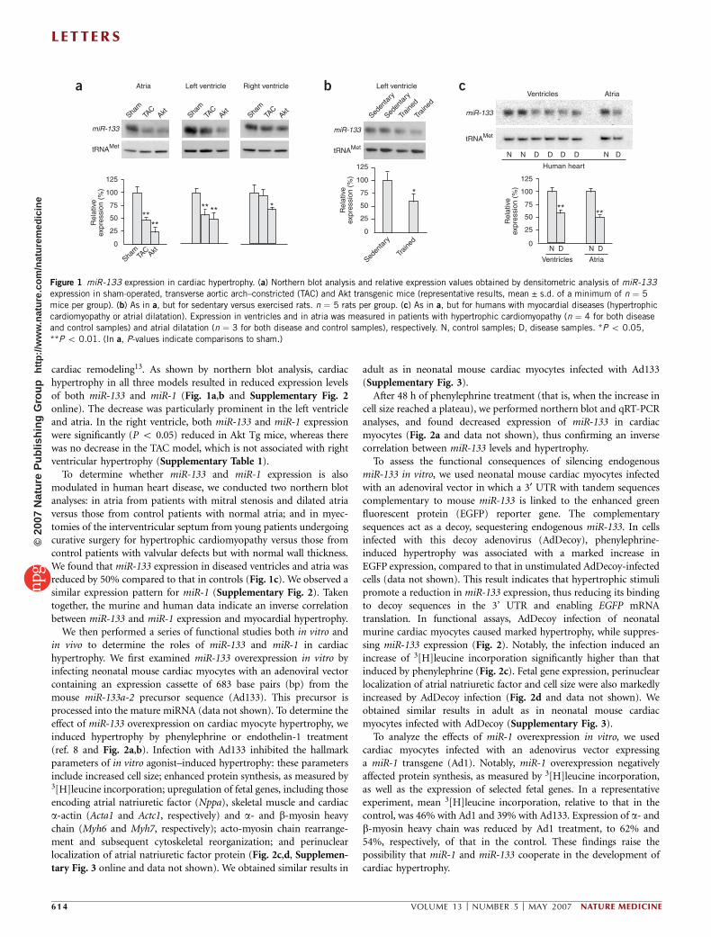
cardiac remodeling13. As shown by northern blot analysis, cardiachypertrophy in all three models resulted in reduced expression levelsof both miR-133 and miR-1 (Fig. 1a,b and Supplementary Fig. 2online). The decrease was particularly prominent in the left ventricleand atria. In the right ventricle, both miR-133 and miR-1 expressionwere significantly (P o 0.05) reduced in Akt Tg mice, whereas therewas no decrease in the TAC model, which is not associated with rightventricular hypertrophy (Supplementary Table 1).
To determine whether miR-133 and miR-1 expression is alsomodulated in human heart disease, we conducted two northern blotanalyses: in atria from patients with mitral stenosis and dilated atriaversus those from control patients with normal atria; and in myec-tomies of the interventricular septum from young patients undergoingcurative surgery for hypertrophic cardiomyopathy versus those fromcontrol patients with valvular defects but with normal wall thickness.We found that miR-133 expression in diseased ventricles and atria wasreduced by 50% compared to that in controls (Fig. 1c). We observed asimilar expression pattern for miR-1 (Supplementary Fig. 2). Takentogether, the murine and human data indicate an inverse correlationbetween miR-133 and miR-1 expression and myocardial hypertrophy.
We then performed a series of functional studies both in vitro andin vivo to determine the roles of miR-133 and miR-1 in cardiachypertrophy. We first examined miR-133 overexpression in vitro byinfecting neonatal mouse cardiac myocytes with an adenoviral vectorcontaining an expression cassette of 683 base pairs (bp) from themouse miR-133a-2 precursor sequence (Ad133). This precursor isprocessed into the mature miRNA (data not shown). To determine theeffect of miR-133 overexpression on cardiac myocyte hypertrophy, weinduced hypertrophy by phenylephrine or endothelin-1 treatment(ref. 8 and Fig. 2a,b). Infection with Ad133 inhibited the hallmarkparameters of in vitro agonist–induced hypertrophy: these parametersinclude increased cell size; enhanced protein synthesis, as measured by3[H]leucine incorporation; upregulation of fetal genes, including thoseencoding atrial natriuretic factor (Nppa), skeletal muscle and cardiaca-actin (Acta1 and Actc1, respectively) and a- and b-myosin heavychain (Myh6 and Myh7, respectively); acto-myosin chain rearrange-ment and subsequent cytoskeletal reorganization; and perinuclearlocalization of atrial natriuretic factor protein (Fig. 2c,d, Supplemen-tary Fig. 3 online and data not shown). We obtained similar results in
adult as in neonatal mouse cardiac myocytes infected with Ad133(Supplementary Fig. 3).
After 48 h of phenylephrine treatment (that is, when the increase incell size reached a plateau), we performed northern blot and qRT-PCRanalyses, and found decreased expression of miR-133 in cardiacmyocytes (Fig. 2a and data not shown), thus confirming an inversecorrelation between miR-133 levels and hypertrophy.
To assess the functional consequences of silencing endogenousmiR-133 in vitro, we used neonatal mouse cardiac myocytes infectedwith an adenoviral vector in which a 3¢ UTR with tandem sequencescomplementary to mouse miR-133 is linked to the enhanced greenfluorescent protein (EGFP) reporter gene. The complementarysequences act as a decoy, sequestering endogenous miR-133. In cellsinfected with this decoy adenovirus (AdDecoy), phenylephrine-induced hypertrophy was associated with a marked increase inEGFP expression, compared to that in unstimulated AdDecoy-infectedcells (data not shown). This result indicates that hypertrophic stimulipromote a reduction in miR-133 expression, thus reducing its bindingto decoy sequences in the 3’ UTR and enabling EGFP mRNAtranslation. In functional assays, AdDecoy infection of neonatalmurine cardiac myocytes caused marked hypertrophy, while suppres-sing miR-133 expression (Fig. 2). Notably, the infection induced anincrease of 3[H]leucine incorporation significantly higher than thatinduced by phenylephrine (Fig. 2c). Fetal gene expression, perinuclearlocalization of atrial natriuretic factor and cell size were also markedlyincreased by AdDecoy infection (Fig. 2d and data not shown). Weobtained similar results in adult as in neonatal mouse cardiacmyocytes infected with AdDecoy (Supplementary Fig. 3).
To analyze the effects of miR-1 overexpression in vitro, we usedcardiac myocytes infected with an adenovirus vector expressinga miR-1 transgene (Ad1). Notably, miR-1 overexpression negativelyaffected protein synthesis, as measured by 3[H]leucine incorporation,as well as the expression of selected fetal genes. In a representativeexperiment, mean 3[H]leucine incorporation, relative to that in thecontrol, was 46% with Ad1 and 39% with Ad133. Expression of a- andb-myosin heavy chain was reduced by Ad1 treatment, to 62% and54%, respectively, of that in the control. These findings raise thepossibility that miR-1 and miR-133 cooperate in the development ofcardiac hypertrophy.
a cb
****
*
*****
****
N DN D
Ventricles Atria
125
100
75
50
25
0
Rel
ativ
eex
pres
sion
(%
)
Rel
ativ
eex
pres
sion
(%
)
NNN DDDDD
Human heart
AtriaVentricles
tRNAMet
miR-133
miR-133
125
100
75
50
25
0
Sham
TAC Akt
Rel
ativ
eex
pres
sion
(%
)
125
100
75
50
25
0
Seden
tary
Traine
d
tRNAMet
miR-133
tRNAMet
Traine
d
Traine
d
Seden
tary
Seden
tary
Left ventricleLeft ventricle Right ventricle
AktTAC
Sham
AktTAC
Sham
AktTAC
Sham
Atria
Figure 1 miR-133 expression in cardiac hypertrophy. (a) Northern blot analysis and relative expression values obtained by densitometric analysis of miR-133
expression in sham-operated, transverse aortic arch–constricted (TAC) and Akt transgenic mice (representative results, mean ± s.d. of a minimum of n ¼ 5
mice per group). (b) As in a, but for sedentary versus exercised rats. n ¼ 5 rats per group. (c) As in a, but for humans with myocardial diseases (hypertrophic
cardiomyopathy or atrial dilatation). Expression in ventricles and in atria was measured in patients with hypertrophic cardiomyopathy (n ¼ 4 for both disease
and control samples) and atrial dilatation (n ¼ 3 for both disease and control samples), respectively. N, control samples; D, disease samples. *P o 0.05,
**P o 0.01. (In a, P-values indicate comparisons to sham.)
L E T TERS
614 VOLUME 13 [ NUMBER 5 [ MAY 2007 NATURE MEDICINE
©20
07 N
atur
e P
ublis
hing
Gro
up
http
://w
ww
.nat
ure.
com
/nat
urem
edic
ine

To investigate the role of miR-133 in cardiac remodeling in vivo,we implanted mice subcutaneously with osmotic minipumpsfor a continuous delivery of a chemically modified (ref. 14) antisenseRNA oligonucleotide (termed an antagomir) targeted to miR-133(antagomir-133). Antagomir oligonucleotides can efficiently and sta-bly knockdown specific miRNAs in living cells14. At 1 month afterminipump implantation, echocardiographic analysis showed a markedincrease in key hypertrophic parameters, such as diastolic left ven-tricular posterior wall and diastolic interventricular septum thickness,left ventricular mass index and the ratio of left ventricle weight tobody weight, in antagomir-treated compared to saline-treated mice(Fig. 3a and Supplementary Table 1). We confirmed this effect by
histological analysis (Fig. 3a). Furthermore, we noted that cardiachypertrophy in antagomir-treated mice was associated with a reinduc-tion of fetal gene expression (Fig. 3b). Northern blot analysis revealedthat the miR-133 level in antagomir-treated mice was 70% lower thanthat in controls (Fig. 3c).
We confirmed these results in short-term experiments using trans-coronary gene delivery15. In a miR-133 gain-of-function model, wetested the effect of Ad133 infection on cardiac hypertrophy in Akt Tgmice (Supplementary Fig. 4 online). At 14 d after Ad133 infection,overexpression of miR-133 resulted in a significant reduction in thesize of left ventricular cardiac myocytes and a significant decrease inthe expression of fetal genes, except for that encoding skeletal muscle
NppaMyh7Myh6Actc1Acta1
NppaMyh7Myh6Actc1Acta1
0
1
2
Fol
d in
crea
se
Fol
d in
crea
se
**
*
*
*
*
*
*
* ***
**
**
*
**
Control AdAdDecoy
Control Ad
Control Ad + PEAd133
Ad133 + PE
0
1
2*
AdDec
oy
AdDec
oy
Ad133
Ad133
Ad133
Ad133
+ P
E
Ad133
+ E
T1
Ad133
+ P
E
Contro
l Ad
Contro
l Ad
Contro
l Ad
Contro
l Ad
Contro
l Ad
+ PE
Contro
l Ad
+ PE
Contro
l Ad
+ ET1
Gapdh
miR-133
0
0.5
1.0
1.5
2.0
[3H
]leuc
ine
inco
rpor
atio
n(f
old
incr
ease
)
[3H
]leuc
ine
inco
rpor
atio
n(f
old
incr
ease
)
Gapdh
miR-133
miR-133
tRNAMet
PE/48
h
PE/6 h
C
0
0.3
0.6
0.9
1.2
1.5
a c
d
b
Figure 2 Infection of neonatal cardiac myocytes with Ad133 and AdDecoy. (a) Hypertrophy, evaluated
as [3H]leucine incorporation in neonatal cardiac myocytes infected with Ad133 or control adenovirus
(control Ad; multiplicity of infection, 100), and with or without 100 mM phenylephrine (PE) or 100 nM
endothelin-1 (ET1). *P o 0.01 versus control Ad; 1P o 0.01 versus control Ad + PE or control Ad +
ET1. Also shown are northern blot analyses of miR-133 in cardiac myocytes after PE stimulation, and
after infection with Ad133 or control Ad. Northern blots are representative of at least three experiments.
(b) Dot blot analysis performed with total RNA extracted from neonatal cardiac myocytes. The expression
level of fetal cardiac genes (Acta1, encoding skeletal a-actin; Actc1, encoding cardiac a-actin; Myh6,
encoding myosin heavy chain-a; Myh7, encoding myosin heavy chain-b; Nppa, encoding atrial natriuretic factor), normalized for Gapdh expression, is
evaluated as fold induction over that in control Ad cells. *P o 0.01 versus control Ad; DP o 0.05 and 1P o 0.01 versus control Ad + PE. (c) [3H]leucine
incorporation in cardiac myocytes. Northern blot analysis of miR-133 is also shown. (d) Dot blot analysis of expression of fetal cardiac genes (see legend topanel b) in AdDecoy-infected cardiac myocytes, evaluated as fold induction over that in control Ad-infected controls. *P o 0.05 and **P o 0.01 versus
control Ad; 1P o 0.01 versus control + PE. In each panel, data represent mean ± s.d., with a minimum of 3 experiments.
a
b cAntagomir Saline
AntagomirSaline
Antagomir
Saline
0
20
40
60
80
100
120
*
*
**
**
******
Rel
ativ
e R
NA
exp
ress
ion
(%)
tRNAMet
miR-133
Antag
omir
Antag
omir
Saline
Saline
Saline
NppaMyh7Myh6Actc1Acta10
1
2
3
Fol
d in
crea
se
150
100
50
0
LVW
/BW
(%
)
0
20
40
60
80
100
120
LVM
(m
g)
1.2
1.0
0.8
0.6
0.4
0.2
0
LVP
Wd
(mm
)
Aftertreatment
Basal0
0.10.20.30.40.50.60.70.80.9
IVS
d (m
m)
AntagomirSaline
Figure 3 In vivo effects of antagomir-133 administration on cardiac hypertrophy. (a) Histological evaluation of hearts from saline- versus antagomir-treated
mice. Echocardiographic parameters of cardiac hypertrophy were evaluated (IVSd, diastolic interventricular septum thickness (mm); LVPWd, diastolic left
ventricular posterior wall thickness (mm); LVM, left ventricular mass (mg); LVW/BW, ratio of left ventricle weight to body weight). (b) Dot blot analysis of
fetal cardiac genes (see legend to Fig. 2b for abbreviations). (c) Northern blot and densitometric analysis of miR-133. n ¼ 8 per group (in all panels).
*P o 0.05 versus saline control.
L E T T ERS
NATURE MEDICINE VOLUME 13 [ NUMBER 5 [ MAY 2007 615
©20
07 N
atur
e P
ublis
hing
Gro
up
http
://w
ww
.nat
ure.
com
/nat
urem
edic
ine

actin. In a miR-133 loss-of-function model, we tested the effect ofAdDecoy infection on cardiac hypertrophy in wild-type mice. At 14 dafter gene transfer, we observed a significant increase in the size of leftventricular cardiac myocytes and upregulation of cardiac hypertrophymarkers, as compared to that in controls infected with the EGFP virus(Supplementary Fig. 4). Notably, in the control group, only B40% ofleft ventricle cardiac myocytes were infected (on the basis of EGFPexpression, data not shown), and the miR-133 level was only mildlyreduced (mean ± s.e.m., 0.69 ± 0.04% of control values). In view ofthese findings, the effects seen on hypertrophy may underestimate theconsequences of complete suppression of miR-133. Notably, miR-1expression was also reduced (0.81 ± 0.07% of control values),suggesting a link between the two miRNAs.
Using a bioinformatic approach, we then searched for candidatemiR-133 target genes that have been reported to be involved in cardiachypertrophy. This analysis led to the identification of Rhoa, Cdc42 andWhsc2 (human gene: NELFA), whose mRNA 3¢ UTR regions comprise‘seed’ sequences and flanking nucleotides matching miR-133 (Fig. 4,and Supplementary Figs. 5 and 6 online). RhoA and Cdc42 aremembers of the Rho subfamily (RhoA, Rac1 and Cdc42) of smallGTP-binding proteins and are involved in cardiac hypertrophy16,17.NELF-A/Whsc2, a negative regulator of RNA polymerase II, is linked
to Wolf-Hirschhorn syndrome, which is char-acterized by cardiac dysgenesis as well asseveral other abnormalities18.
Several lines of evidence indicate that Rhoa,Cdc42 and NELFA/Whsc2 are regulated bymiR-133 in cardiac hypertrophy. In TAC-treated and Akt Tg mice, the expressionlevel of miR-133 was inversely related to theamount of these proteins (SupplementaryFigs. 5 and 6; see also Fig. 1). Furthermore,in both neonatal and adult cardiac myocytes,miR-133 suppression with AdDecoy infection,or overexpression following Ad133 infection,caused an increase or a decrease in the level ofthese three proteins, respectively; mRNAlevels, however, were not affected (Fig. 4and Supplementary Fig. 6). These data sug-gest that Rhoa, Cdc42 and Whsc2 mRNAs aretargeted by miR-133 in cardiac myocytes. Totest this possibility, we performed luciferasereporter assays in HeLa cells, which do notexpress miR-133. We identified at leasttwo ‘high score’ seed sequences in the 3¢UTR of each of the three candidate targetgenes. Cotransfection of miR-133 with theluciferase reporter gene linked to thewild-type 3’ UTR of Rhoa, Cdc42 or Whsc2resulted in a significant (P o 0.01) decreasein luciferase activity (Fig. 4 and Supplemen-tary Fig. 6). In contrast, cotransfection ofa control miRNA (not complementary to the3¢ UTRs of these three genes) with the wild-type 3¢ UTR constructs did not result in adecrease in luciferase activity; similarly,cotransfection of miR-133 with constructscontaining mutated or deleted 3¢ UTRsequences also did not result in a decreasein luciferase activity (Fig. 4 and Supplemen-tary Fig. 6).
To test the effects of Whsc2 overexpression on cardiac hypertrophy,we infected cardiac myocytes in vitro with an adenovirus vectorexpressing a Whsc2 transgene (AdWhsc2). Although Whsc2 over-expression resulted in reduced cardiomyocyte protein synthesis (datanot shown), reactivation of fetal gene expression clearly showedinduction of the hypertrophic gene program (SupplementaryFig. 6). Whsc2 overexpression also upregulated RhoA protein expres-sion (Supplementary Fig. 6). In addition, we tested the effect ofAdWhsc2 infection on cardiac hypertrophy in wild-type mice. At 14 dafter infection, overexpression of Whsc2 resulted in a notable increasein fetal gene expression (Supplementary Fig. 6).
Taken together, our studies indicate a key role for miR-133 in theregulation of cardiac hypertrophy. First, miR-133 expression wasinversely related to cardiac hypertrophy in three different murinemodels. Second, functional studies performed on neonatal and adultcardiac myocytes in vitro showed that overexpression of miR-133inhibited the increase in cardiac myocyte size and other hallmarks ofhypertrophy; suppression of endogenous miR-133 using a decoysequence induced a marked cardiac myocyte hypertrophy in theabsence of any hypertrophic stimulus. Third, a single infusionin vivo of an antagomir oligonucleotide suppressing miR-133 induceda marked and sustained cardiac hypertrophy. Last, myocardial
***
**
Site 2 - 3′ UTRSite 1 - 3′ UTR
Luci
fera
se a
ctiv
ity (
%)
Contro
l Ad
Ad133
AdDec
oy
Contro
l Ad
Ad133
AdDec
oy
Contro
l Ad
Ad133
AdDec
oy150
100
50
0
Control miRNAmiR-133
MutWtMutWt
++
++
++
++–
––
––
––
–
125
100
75
50
25
0
125
100
75
50
25
0Rel
ativ
e ex
pres
sion
(%
)
Gapdh
Rhoa
Tubulin
RhoA
a b
**
**
0
50
100
150
200
250
300125
100
75
50
25
0
Contro
l Ad
Ad133
AdDec
oy
Contro
l Ad
Ad133
AdDec
oy
Contro
l Ad
Ad133
AdDec
oy
Rel
ativ
e ex
pres
sion
(%
)
Actin
Cdc42
Gapdh
Cdc42
c
****
MutMut WtWt
miR-133Control miRNA +
++
++
++
+–
––
––
––
–
Site 2 - 3′ UTRSite 1 - 3′ UTR
0
50
100
150Lu
cife
rase
act
ivity
(%
)d
Figure 4 Analysis of the miR-133 target genes Rhoa and Cdc42. (a) Top, representative northern (left)
and western (right) blot analyses for RhoA after infection of cardiac myocytes with control adenovirus
(control Ad), Ad133 or AdDecoy. Bottom, relative densitometric analyses (mean ± s.d. of a minimum
of n ¼ 5 mice per group). (b) Luciferase reporter assays (mean ± s.d., minimum of 4 experiments
per group) performed by cotransfection of miR-133 oligonucleotide with a luciferase reporter gene
linked to the Rhoa 3¢ UTR, containing either wild-type (Wt) or mutated or deleted (Mut) miR-133
complementary sites; a control nontargeting oligonucleotide (control miRNA) was also included.
Mutated 3’ UTR sequences are listed in the Supplementary Methods. (c) As in a, but measuring
expression of Cdc42. (d) As in b, but using the Cdc42 3¢ UTR. *P o 0.05 and **P o 0.01 versus
control Ad (a and c) or control miRNA (b and d).
L E T TERS
616 VOLUME 13 [ NUMBER 5 [ MAY 2007 NATURE MEDICINE
©20
07 N
atur
e P
ublis
hing
Gro
up
http
://w
ww
.nat
ure.
com
/nat
urem
edic
ine

miR-133 expression was downregulated in the hearts of patients withhypertrophic cardiomyopathy or atrial dilatation.
We identified three targets of miR-133—Rhoa, Cdc42 andNELFA/Whsc2—relevant to cardiac hypertrophy development. RhoAand Cdc42 are associated with cytoskeletal and myofibrillar rearrange-ments during hypertrophy16,17, whereas NELF-A/Whsc2 is not knownto be involved in hypertrophy18. In gene transfer studies, we showedthat Whsc2 overexpression upregulates myocardial fetal gene expres-sion but not protein synthesis (Supplementary Fig. 6), suggesting thatNELF-A/Whsc2 plays a role in only some aspects of the hypertrophygene program.
Recent studies have suggested that miRNAs may function accordingto a ‘combinatorial circuitry model’, whereby a single miRNA targetsmultiple mRNAs and several coexpressed miRNAs may target a singlemRNA (refs. 19,20). In line with this model, we identified multipletargets of miR-133 implicated in heart hypertrophy. Although othermiRNAs may also be involved13, our results indicate that miR-133functions as a critical gene for establishing and sustaining thehypertrophy gene program.
The expression of miR-133 and miR-1 in the same bicistronic unitraises the possibility that they may cooperate in cardiac hypertrophy.In skeletal myoblast culture, miR-133 and miR-1 promote differentia-tion and proliferation, respectively9. In cardiac hypertrophy, we foundthat miR-133 and miR-1 were downregulated in an identical pattern,suggesting that these two miRNAs may functionally interact toregulate the translation of two complementary sets of target mRNAsinvolved in hypertrophy development. This model is in line within vitro functional studies on miR-1 reported by others21 and byus in this study. The regulation of miR-133 and miR-1 transcription isalso of interest. In mouse skeletal muscle, MyoD and myogeninmodulate the transcription of miR-1 and miR-133 according to aregulatory loop22, as suggested for miR-223 and its target, the NFI-Atranscription factor23. However, MyoD and myogenin are notexpressed in the heart (ref. 24 and data not shown), whereas it hasbeen suggested that serum response factor (SRF) regulates miR-1expression in transgenic mice25 and Drosophila26.
Our studies may have important clinical implications. Notably,in vivo miR-133 levels are downmodulated not only in murine modelsof cardiac hypertrophy, but also in human disease states associatedwith myocardial hypertrophy. More importantly, the effects ofantagomir-133 on hypertrophy imply that modulation of miR-133expression by oligonucleotide administration may have futuretherapeutic application in the clinical setting.
METHODSHuman tissues and animal experiments. Human embryos and fetuses were
obtained by legal abortions at 5–10 weeks after fertilization, according to
institutional guidelines (Istituto Superiore di Sanita); written, informed con-
sent was obtained in advance from the mothers. The age of the embryo was
carefully established by morphologic staging according to standard multiple
criteria. Different organs were dissected under an inverted microscope and
stored under liquid nitrogen. Samples from human hearts were obtained
according to institutional regulations (University ‘‘La Sapienza’’); written,
informed consent was obtained in advance from the patients. Experiments
on mice were performed according to institutional guidelines of the University
of California, San Diego, Animal Subjects Committee. Tissues were harvested,
frozen, and stored at –80 1C for RNA and protein extraction.
Pressure overload cardiac hypertrophy. We used 10- to 12-week old C57BL/6
female mice (Harlan). The pressure overload model was obtained through
transverse aortic arch constriction (TAC) under anesthesia, as described27.
Akt transgenic mice. Transgenic mice with cardiac-specific overexpression of
constitutively active Akt have previously been described11.
Endurance training. We analyzed a model of adaptive hypertrophy in Sprague
Dawley rats. The apparatus and method have been previously described and
validated28. Briefly, rats ran uphill on a treadmill for 1.5 h, alternating between
8 min at an exercise intensity corresponding to 85–90% of maximal oxygen
uptake (VO2max) and 2 min recovery at 50–60% VO2max. Rats performed this
exercise 2 d per week over 8 weeks; controls were age-matched rats that
remained sedentary.
Isolation, culture and treatment of mouse cardiac myocytes. We isolated and
cultured neonatal and adult cardiac myocytes using standard techniques29. We
performed adenoviral infection in serum-free medium; 5 h after infection, the
medium was replaced and cells further incubated for 48 h (Supplementary
Methods online).
miR-133 silencing by antagomir treatment. Chemically modified antisense
oligonucleotides (antagomir) have been used to inhibit miR-133 expression14.
The antagomir sequence complementary to miR-133 is 5¢-P-ACAGCUGGUUG
AAGGGGACAA-3¢. The 3¢ end of the oligonucleotides was conjugated to
cholesterol; all the bases were 2¢-OMe modified. Antagomir oligonucleotides
were deprotected, desalted and purified by high-performance liquid chromato-
graphy (HPLC; Dharmacon). C57BL/6female mice (8 weeks old) received
antagomirs at doses of 80 mg/kg body weight through Alzet osmotic mini-
pumps (model 1003D, Alza). Minipumps were prepared and placed in a petri
dish filled with sterile 0.9% saline at 37 1C, for at least 4 h before implantation,
in order to prime the pumps for continuous delivery of the drug. Controls
received a saline minipump.
Transcoronary gene delivery. Details are in Supplementary Methods.
Luciferase assays. We performed luciferase reporter experiments in the
HeLa cell line. 3¢ UTR segments of Rhoa, Cdc42 and Whsc2 predicted to
interact specifically with miR-133 were subcloned by standard procedures
into the pGL3 promoter vector (Promega) immediately downstream of
the stop codon of the luciferase gene. Mutagenesis was performed as
described by the manufacturer (Invitrogen). We made short constructs
(80–100 bases), encompassing wild-type or mutated (six point mutations)
seed sequences, in order to separately analyze the functional role of each
seed (Supplementary Methods). The seed sequences are indicated in
Supplementary Figures 5 and 6. Using Lipofectamine 2000 (Invitrogen), cells
were transfected with 0.8 mg of pGL3-3¢ UTR plasmid, 20 pmol of either a
stability-enhanced 2¢-O-methyl nontargeting RNA control or miR-133a
oligonucleotides (Dharmacon), and an emerald GFP–expressing plasmid
(to evaluate the percentage of transfected cells). At 48 h after transfection,
cells were lysed and luciferase activity was measured (FemtomasterFB
12, Zylux).
Plasmids and vectors. See Supplementary Methods.
Accession numbers. GenBank: Rhoa, BC068115; Cdc42, NM_009861; Whsc2,
NM_011914.
Note: Supplementary information is available on the Nature Medicine website.
ACKNOWLEDGMENTSWe thank M. Blasi, M. Fontana and V. Michetti for editorial assistance, andG. Loreto for graphics. This work was supported by grants from the US NationalInstitutes of Health (HL078797-01A1 to G.C., HLO65484 to P.R.-L. and1R01HL63168 to C.P.), the Marie Curie Outgoing Fellowship 6th EuropeanFramework Programme (D.C.), EUGeneHeart (LSHM-CT-2005-018833 to G.C.),the Italian Ministry of Scientific Research (G.C. and C.P.), the Italian Ministry ofHealth (C.P. and G.C.) and the Italy-USA miR Oncology Program, IstitutoSuperiore di Sanita, Rome (C.P.).
AUTHOR CONTRIBUTIONSD.C., A.C., D.B., F.F., A.A., M.V.G.L., P.S., M.-L.B. and L.E. conducted the in vitroand in vivo experiments. D.C. and P.R-L. performed the assessment of miRNAsin mouse cardiac development. P.G., Y.G., N.D.D., G.W.D., Ø.E. and K.L.P.performed the in vivo models of cardiac hypertrophy. C.A. and M.A.R. collectedhuman samples. C.M.C. conducted the miRNA microarray analysis. A.C., D.C.,C.P. and G.C. planned the experiments. D.C., A.C., M.-L.B., P.R.-L., C.P. and G.C.
L E T T ERS
NATURE MEDICINE VOLUME 13 [ NUMBER 5 [ MAY 2007 617
©20
07 N
atur
e P
ublis
hing
Gro
up
http
://w
ww
.nat
ure.
com
/nat
urem
edic
ine

wrote the manuscript. C.P. and G.C. were responsible for research coordinationand strategy.
COMPETING INTERESTS STATEMENTThe authors declare no competing financial interests.
Published online at http://www.nature.com/naturemedicine
Reprints and permissions information is available online at http://npg.nature.com/
reprintsandpermissions
1. Bartel, D.P. MicroRNAs: genomics, biogenesis, mechanism, and function. Cell 116,281–297 (2004).
2. Xu, P., Guo, M. & Hay, B.A. MicroRNAs and the regulation of cell death. Trends Genet.20, 617–624 (2004).
3. Cheng, A.M., Byrom, M.W., Shelton, J. & Ford, L.P. Antisense inhibition of humanmiRNAs and indications for an involvement of miRNA in cell growth and apoptosis.Nucleic Acids Res. 33, 1290–1297 (2005).
4. Felli, N. et al. MicroRNAs 221 and 222 inhibit normal erythropoiesis and erythro-leukemic cell growth via kit receptor down-modulation. Proc. Natl. Acad. Sci. USA 102,18081–18086 (2005).
5. Dresios, J. et al. Cold stress-induced protein Rbm3 binds 60S ribosomal subunits,alters microRNA levels, and enhances global protein synthesis. Proc. Natl. Acad. Sci.USA 102, 1865–1870 (2005).
6. Krek, A. et al. Combinatorial microRNA target predictions. Nat. Genet. 37, 495–500(2005).
7. Esquela-Kerscher, A. & Slack, F.J. Oncomirs-microRNAs with a role in cancer. Nat. Rev.Cancer 6, 259–269 (2006).
8. McKinsey, T.A. & Olson, E.N. Toward transcriptional therapies for the failing heart:chemical screens to modulate genes. J. Clin. Invest. 115, 538–546 (2005).
9. Chen, J.F. et al. The role of microRNA-1 and microRNA-133 in skeletal muscleproliferation and differentiation. Nat. Genet. 38, 228–233 (2006).
10. van Rooij, E. et al. A signature pattern of stress-responsive microRNAs that can evokecardiac hypertrophy and heart failure. Proc. Natl. Acad. Sci. USA 103, 18255–18260(2006).
11. Condorelli, G. et al. Akt induces enhanced myocardial contractility and cell size in vivoin transgenic mice. Proc. Natl. Acad. Sci. USA 99, 12333–12338 (2002).
12. Dorn, G.W., II, Robbins, J. & Sugden, P.H. Phenotyping hypertrophy: eschew obfusca-tion. Circ. Res. 92, 1171–1175 (2003).
13. Shiojima, I. & Walsh, K. Regulation of cardiac growth and coronary angiogenesis by theAkt/PKB signaling pathway. Genes Dev. 20, 3347–3365 (2006).
14. Krutzfeldt, J. et al. Silencing of microRNAs in vivo with antagomirs. Nature 438,685–689 (2005).
15. Iwatate, M. et al. In vivo high-efficiency transcoronary gene delivery and Cre-LoxP geneswitching in the adult mouse heart. Gene Ther. 10, 1814–1820 (2003).
16. Brown, J.H. et al. The Rac and Rho hall of fame: a decade of hypertrophic signalinghits. Circ. Res. 98, 730–742 (2006).
17. Nagai, T. et al. Cdc42 plays a critical role in assembly of sarcomere units in series ofcardiac myocytes. Biochem. Biophys. Res. Commun. 305, 806–810 (2003).
18. Bergemann, A.D., Cole, F. & Hirschhorn, K. The etiology of Wolf-Hirschhorn syndrome.Trends Genet. 21, 188–195 (2005).
19. Lin, H. et al. A microRNA polycistron as a potential human oncogene. Nature 435,828–833 (2005).
20. Stark, A. et al. Animal microRNAs confer robustness to gene expression and have asignificant impact on 3¢UTR evolution. Cell 123, 1133–1146 (2005).
21. Sayed, D., Hong, C., Chen, I.Y., Lypowy, J. & Abdellatif, M. MicroRNAs play anessential role in the development of cardiac hypertrophy. Circ. Res. 100, 416–424(2007).
22. Rao, P.K. et al. Myogenic factors that regulate expression of muscle-specific micro-RNAs. Proc. Natl. Acad. Sci. USA 103, 8721–8726 (2006).
23. Fazi, F. et al. A minicircuitry comprised of microRNA-223 and transcription factorsNFI-A and C/EBPa regulates human granulopoiesis. Cell 123, 819–831 (2005).
24. Kattman, S.J. et al. Multipotent flk-1+ cardiovascular progenitor cells give rise to thecardiomyocyte, endothelial and vascular smooth muscle lineages. Dev. Cell 11,723–732 (2006).
25. Zhao, Y., Samal, E. & Srivastava, D. Serum response factor regulates a muscle-specificmicroRNA that targets Hand2 during cardiogenesis. Nature 436, 214–220 (2005).
26. Kwon, C., Han, Z., Olson, E.N. & Srivastava, D. MicroRNA1 influences cardiacdifferentiation in Drosophila and regulates Notch signaling. Proc. Natl. Acad. Sci.USA 102, 18986–18991 (2005).
27. Rockman, H.A. et al. Segregation of atrial-specific and inducible expression of an atrialnatriuretic factor transgene in an in vivo murine model of cardiac hypertrophy. Proc.Natl. Acad. Sci. USA 88, 8277–8281 (1991).
28. Wisløff, U. et al. Intensity-controlled treadmill running in rats: VO(2 max) and cardiachypertrophy. Am. J. Physiol. 280, H1301–H1310 (2001).
29. Ikeda, Y. et al. Simian immunodeficiency virus-based lentivirus vector for retinalgene transfer: a preclinical safety study in adult rats. Gene Ther. 10, 1161–1169(2003).
L E T TERS
618 VOLUME 13 [ NUMBER 5 [ MAY 2007 NATURE MEDICINE
©20
07 N
atur
e P
ublis
hing
Gro
up
http
://w
ww
.nat
ure.
com
/nat
urem
edic
ine