Embed Size (px)
Citation preview
C16:1n7-Palmitoleate and physiological cardiac hypertrophy
1
Adipose Tissue Lipolysis Promotes Exercise-Induced Cardiac Hypertrophy Involving the Lipokine C16:1n7-Palmitoleate Anna Foryst-Ludwig1,9; Michael C. Kreissl2; Verena Benz1; Sarah Brix1; Elia Smeir1; Zsofia Ban1; Elżbieta Januszewicz1; Janek Salatzki1; Jana Grune1; Anne-Kathrin Schwanstecher1; Annelie Blumrich1; Andreas Schirbel2; Robert Klopfleisch3; Michael Rothe4; Katharina Blume5; Martin Halle5,6; Bernd Wolfarth7; Erin E. Kershaw8; Ulrich Kintscher1,9
From the 1 Charité-Universitaetsmedizin Berlin, Institute of Pharmacology, Center for Cardiovascular Research, Berlin, Germany 2 University Hospital Wuerzburg, Department of Nuclear Medicine, Wuerzburg, Germany 3 Department of Veterinary Pathology, College of Veterinary Medicine, Freie Universität Berlin, Berlin, Germany 4 Lipidomix GmbH, Berlin, Germany 5 Department of Prevention, Rehabilitation, and Sports Medicine, Technische Universitaet Muenchen, Germany 6 DZHK (German Center for Cardiovascular Research), partner site Munich Heart Alliance, Munich, Germany 7 Department of Sports Medicine, Humboldt University/ Charité-Universitaetsmedizin Berlin, Germany 8 University of Pittsburgh, Division of Endocrinology and Metabolism, Pittsburgh, PA, USA 9 DZHK (German Center for Cardiovascular Research), partner site Berlin, Germany *Running title: C16:1n7-Palmitoleate and physiological cardiac hypertrophy To whom correspondence should be addressed: Ulrich Kintscher; Charité - Universitaetsmedizin Berlin; Institute of Pharmacology, Center for Cardiovascular Research, CCR; Hessische Str. 3-4, 10115 Berlin, Germany; Tel.: +49 30 450 525 276; Fax: +49 30 450 525 901; E-mail: [email protected]
Keywords: adipose tissue metabolism; adipose tissue lipolysis, adipose triglyceride lipase (ATGL) cardiac metabolism, cardiac hypertrophy, C16:1n7-Palmitoleate
http://www.jbc.org/cgi/doi/10.1074/jbc.M115.645341The latest version is at JBC Papers in Press. Published on August 10, 2015 as Manuscript M115.645341
Copyright 2015 by The American Society for Biochemistry and Molecular Biology, Inc.
by guest on February 4, 2018http://w
ww
.jbc.org/D
ownloaded from
C16:1n7-Palmitoleate and physiological cardiac hypertrophy
2
Background: Endurance training induces physiological cardiac hypertrophy and elevates adipose tissue lipolysis. Results: Adipose-specific adipose triglyceride lipase (ATGL)-knock out mice exhibit attenuated exercise-induced cardiac hypertrophy likely mediated by the lack of C16:1n7 palmitoleate actions on the heart. Conclusion: ATGL-mediated adipose lipolysis regulates physiological cardiac hypertrophy. Significance: Adipose-derived lipokines may serve as important molecular mediators of cardiac physiology and pathology. ABSTRACT
Endurance exercise training induces substantial adaptive cardiac modifications such as left ventricular hypertrophy (LVH). Simultaneously to the development of LVH, adipose tissue (AT) lipolysis becomes elevated upon endurance training to cope with enhanced energy demands. In the present study, we investigated the impact of adipose tissue lipolysis on the development of exercise-induced cardiac hypertrophy. Mice deficient for adipose triglyceride lipase (ATGL) in AT (atATGL-KO) were challenged with chronic treadmill running. Exercise-induced AT-lipolytic activity was significantly reduced in atATGL-KO mice accompanied by the absence of plasma fatty acid increase. These processes were directly associated with a prominent attenuation of myocardial FA uptake in atATGL-KO, and a significant reduction of the cardiac hypertrophic response to exercise. FA-serum profiling revealed palmitoleic acid (C16:1n7) as a new molecular comediator of exercise-induced cardiac hypertrophy by inducing non-proliferative cardiomyocyte growth. In parallel, serum FA-analysis and echocardiography was performed in 25 endurance athletes. In consonance, serum C16:1n7 palmitoleate level exhibited a significant positive correlation with diastolic interventricular septum thickness (IVSd) in those athletes. No correlation existed between linoleic acid (18:2n6) and IVSd. Collectively, our data provide the first evidence that adipose tissue lipolysis directly promotes the development of exercise-induced cardiac
hypertrophy involving the lipokine C16:1n7 palmitoleate as a molecular comediator. The identification of a lipokine involved in physiological cardiac growth may help to develop future lipid-based therapies for pathological LVH or heart failure. Intensive and prolonged physical exercise results in major cardiac adaptations leading to amplified cardiac output that meet increased peripheral oxygen and energy demands (1). Exercise-induced cardiac changes include enlarged left ventricular (LV) internal dimensions and augmented LV wall thickness in the presence of preserved cardiac function (1). These changes are accompanied by maintenance of fatty acid (FA) oxidation as the predominant cardiac energy source. In contrast, the development of pathological or maladaptive LV-hypertrophy during chronic hypertension or aortic valve disease results in augmented hypertrophic responses associated with LV-dysfunction, cardiac fibrosis, and prevailing glucose oxidation (2). An improved knowledge about the mechanisms underlying exercise-induced/ physiological cardiac hypertrophy is of clinical relevance since molecular effectors such as insulin-like growth factor 1 (IGF1) that induce beneficial cardiac growth are potential therapeutic targets for the treatment of maladaptive hypertrophy and heart failure (3). New mechanisms underlying the development of LVH may arise from shifting the focus from the heart to accompanied peripheral exercise-induced physiological responses. In order to provide the heart and skeletal muscle with a sufficient amount of energy during prolonged physical exercise, a switch in energy substrates occurs from an initial preference for glucose that later changing to fatty acids (FAs) (3). Increased catecholamine levels induce the hydrolysis of triacylglycerol (TG) in white adipose tissue (WAT) mediating the release of free fatty acids (FFAs) into the circulation that provide energy substrates for the heart and skeletal muscle (4). In addition to their role as biomolecules for energy production, FAs have been recently characterized as signaling molecules directly regulating cellular responses (5). Along this line, exercise-mediated stimulation of WAT lipolysis releases a broad variety of FAs into the
by guest on February 4, 2018http://w
ww
.jbc.org/D
ownloaded from
C16:1n7-Palmitoleate and physiological cardiac hypertrophy
3
bloodstream some of which may directly act as molecular effectors on other organs including the heart. In order to liberate FAs from WAT, TG-hydrolysis is catalyzed by two major adipose tissue lipases, hormone sensitive lipase (HSL) and adipose triglyceride lipase (ATGL) under hormonal control (6). In the present study, we aimed to identify new FA-based molecular effectors from WAT for exercise-induced, non-proliferative cardiac growth. For this, we studied adipose tissue specific ATGL-deficient mice (atATGL-KO) with attenuated WAT-lipolysis and reduced FA-release during chronic exercise. We then performed a serum FA-profiling using HPLC/ triplequad mass spectrometer technology to identify individual FAs involved in the development of physiological cardiac hypertrophy. Finally, to translate these results into the human situation we studied endurance athletes by echocardiography and performed serum FA-profiling to correlate individual FA-levels with the degree of training-induced LVH. EXPERIMENTAL PROCEDURES Animals All animals’ procedures were performed in accordance with the guidelines of the German Law on the Protection of Animals. Fat tissue specific ATGL-deficient mice (atATGL-KO) were generated by crossing B6.129-Pnpla2tm1Eek (ATGL-flox) mice (7) with B6.Cg-Tg(Fabp4-cre)1Rev/J mice. Highest expression of ATGL in mice has been determined in WAT/ brown AT, in heart, and testis (8). To exclude effects of ATGL deficiency in other tissues than adipose tissue we analyzed ATGL expression in WAT, heart, and macrophages (Fig. 1A). Mice were housed in a facility with a 12-h light/dark cycle (25oC), and fed at libitum with standard diet (9). 5 week-old female ATGLflox/flox Cre/+ (atATGL-KO) mice and control littermate ATGLflox/flox +/+ animals (wt) were randomized to a treadmill-trained group (run, n=10 atATGL-KO and n=10 wt mice) and control-sedentary animals (sed, n=10 atATGL-KO and n=10 wt mice). Run mice were adapted to treadmill training (Treadmill, TSE Sytems) by gradual increase of the training intensity (0.05-0.25 m/s, 7o slope, 15-90 min/day/mouse, for 3 weeks). After the adaptation phase mice were trained over 4 weeks using the following protocol: 0.25 m/s, 7o slope, 90 min/day/mouse, as described
previously (9). To avoid differences in nutritional status of the mice, all animals were trained in the fed-state. At the end of the training regime mice underwent echocardiographic analysis (10). Blood samples were collected before/after 45min training in fed mice for analysis of serum glucose and FFA (9). Body composition was determined by NMR (Bruker’s Minispec MQ10). The respiratory quotient (RQ) during exercise was determined using indirect calorimetric analysis combined with treadmill training (TSE Systems) (9). In an additional set of experiments atATGL-KO and wt mice were supplemented orally with C16:1n7 or C18:1 (300mg/kg/day) (11) during 4 weeks of the main training period. Afterwards the mice underwent echocardiographic analysis.
Small animal PET In order to assess glucose metabolism the animals were injected after an overnight rest with ~ 7 MBq of [18F]-2-fluorodesoxyglucose (18F-FDG) and imaged using a small animal PET (Inveon dedicated PET, Siemens) (9). In order to evaluate FA metabolism after a resting period and overnight starvation, ~ 2 MBq of [18F]-fluoro-4-thia-palmitate (18F-FTP) were injected (12). Data were reconstructed using 3D-OSEM, Image analysis was performed using the image analysis software AMIDE (13).
mRNA Analysis Total RNA was isolated using RNeasy Micro kit (Qiagen). RNA samples were reverse transcribed (Promega), and used in quantitative PCR reactions in the presence of a fluorescent dye (Sybrgreen, Life Sciences) (9). The expression analysis of bone marrow-derived cells BMdMφ was performed as described previously (14). Briefly, isolated primary bone marrow-derived cells from female atATGL-KO mice and their control littermates were differentiated in-vitro into macrophages using 10% L929-conditioned medium. After 7 days of differentiation, bone marrow-derived macrophages (BMdMφ) were harvested and total RNA was isolated, as described above.
Ex-vivo lipolysis assay in gonadal adipose tissue explants The ex-vivo lipolysis assay was described previously. Gonadal fat pads isolated from the mice directly after training (n = 3-4/group) were incubated in DMEM containing 2% FA-free BSA (basal lipolysis) and forskolin (10µM) (stimulated lipolysis) for 1 h at 37°C. FFA content was
by guest on February 4, 2018http://w
ww
.jbc.org/D
ownloaded from
C16:1n7-Palmitoleate and physiological cardiac hypertrophy
4
quantified using the HR-NEFA (Wako Diagnostics) (9).
FA profiling of serum samples and HL-1 cell fractions 100 µL serum were hydrolyzed under alkaline-methanolic conditions within 60 min at 80 °C. The samples were neutralized and diluted 1:10 with methanol containing internal standards (C15:0, C21:0 50 µg, C20:4-d8, C18:2-d4 5 µg, C20:5-d5, C22:6-d5 1 µg). HPLC-measurement was performed using an Agilent 1200 HPLC system with binary pump, autosampler and column thermostat equipped with a Phenomenex Kinetex-C18 column 2.6 µm, 2.1 x 150 mm column using a solvent system of aqueous formic acid (0.1%) and acetonitrile. The solvent gradient started at 30 % acetonitrile and was increased to 98 % within 10 min with a flow rate of 0.4 mL/min and 5 µL injection volume. The HPLC was coupled with an Agilent 6460 triplequad mass spectrometer with electrospray ionisation source operated in negative selected ion mode.
Measurement of ceramides in heart muscles About 30 mg tissue samples were homogenized with liquid nitrogen. Next, an internal standard consisting of 10 ng Ceramide 17:0 (Avanti Polar Lipids, AL, USA) in 2 mL Ethyl acetate / i-Propanol / Water (60:30:10) was added. The mixture was 15 sec vortexed, then 30 sec ultra-sonicated. This was repeated three times followed by centrifugation for 10 min at 5000 rpm. The clear supernatant was taken and evaporated to dryness under a stream of nitrogen at 50 °C. The residues were dissolved in 150 µL methanol and analysed using an Agilent 1290 HPLC system with binary pump, autosampler and column thermostat with a Kinetex C-18, 2.1 x 150 mm, 2.6 µm (Phenomenex, Aschaffenburg, Germany) column using a solvent system of aqueous formic acid (0.1%) and methanol. The elution gradient was started at 95 % methanol, which was increased within 10 minutes to 99 % and held there for 2 minutes. The flow rate was set at 0.4 mL/min. The injection volume was 1 µL. The HPLC was coupled with an Agilent 6490 Triplequad mass spectrometer (Agilent Technologies, Santa Clara, USA) with electrospray ionisation source. The source parameters were: Drying gas: 140 °C/14 L/min, Sheath gas: 380 °C/10 L/min, Capillary voltage: 5500 V, Nozzel voltage: 2000 V and Nebulizer pressure: 30 psi. Analysis was performed in Multiple Reaction Monitoring
(MRM) in positive mode. The results were calculated compared with Ceramide 17:0 as internal standard.
Cell culture experiments Mouse HL-1 cardiomyocytes, kindly provided by W.C. Claycomb (Louisiana State University, LA) were cultivated, as described previously (15). Human (GATA-4+/alpha-sarcomeric actin+) primary cardiomyocytes (primary HC) were purchased from Promocell, and cultivated accordingly to the manufacturer´s instructions, as described previously (16). After a 24 h starvation period (0.5% FBS), cells were stimulated with 100 nM endothelin 1 (Et-1), a mix of FFAs (FA-mix) dissolved in 10% FFAs-free BSA (C16:0, C16:1n7 and C14:0), (C16:0, C14:0), (C16:0, C18:1 and C18:2), or C16:1n7 alone for 6 h (mRNA expression) or 30min (protein phosphorylation). FFAs were used in equimolar serum concentrations estimated by FA-profiling, C16:1 low was 10x lower as serum concentration.
HL-1 cell fractionation For cell fractionation experiments the membrane fractionation kit (ab 139409, Abcam) were used according to the manufacturer`s protocol.
Western Immunoblotting For Western Blot analysis HL-1 and primary HC were lysed in RIPA buffer (50 mM Tris pH 7.5, 150 mM NaCl, 5 mM MgCl2, 1% Nonidet P-40, 2.5% glycerol, 1 mM EGTA, 50 mM NaF, 1 mM Na3VO4, 10 mM Na4P2O7, 100 µM phenylmethylsulfonyl fluoride and complete protease/ phosphatase inhibitor cocktail (Phos-stop and Complete Mini, Roche Diagnostics). Lysates were analyzed by immunoblotting using antibodies raised against pS473-Akt and total-Akt (4060 and 9272 from Cell Signaling Technologies, dilution 1:2000), and a secondary horseradish-conjugated antibodies (Jackson Immuno Research, dilution 1:10000). For detection, enhanced chemiluminiscent reagents (ECL kit, Thermo Scientific) were used. Cell fractions were analysed using the membrane fractionation WB cocktail (ab 140365, Abcam) providing the mix of Ab specific for different cellular fractions: anti-Sodium Potassium ATPase (plasma membrane marker); anti-GRP78 (endoplasmic reticulum marker), anti-ATP5a (mitochondrial membrane marker); anti-GAPDH (cytosolic marker) and anti-histone H3 (nuclear marker). WB analysis was performed according to the manufacturer`s protocol.
by guest on February 4, 2018http://w
ww
.jbc.org/D
ownloaded from
C16:1n7-Palmitoleate and physiological cardiac hypertrophy
5
Immunostaining and cell size
quantification Cells were fixed with formaldehyde (3.7%) in phosphate-buffered saline (PBS), permeabilized for 10min with 0.5% Triton X-100 in PBS and blocked for 1 hour with PBS containing 0.1% Triton X-100 and goat serum (10%). Cells were incubated with primary antibody for sarcomeric alpha-actinin (mouse monoclonal antibody, dilution 1:200, Sigma) for 1 hour in the same solution as blocking solution. Cells were washed three times with PBS, incubated with secondary AlexaFluor®488-conjugated antibody (goat anti-rabbit polyclonal IgG, dilution 1:400, Molecular Probes; green) and washed 3 times with PBS. Next, nuclei were stained for 5min with DAPI (dilution 1:1000, Thermo Scientific, blue), washed 3 times with PBS and mounted with mounting solution (Dako). Proteins were visualized with an inverted fluorescence phase-contrast microscope (BZ-9000E, All-in-one fluorescence microscope, Keyence) at a x20 and x60 magnification, and images were captured using a digital camera CFI60 (Nikon). Cell size was determined using BZ-II Analyzer software and Dynamic Cell Count Tool (Keyence).
Histology Cardiac tissue isolated from mice were formalin-fixed, paraffin-embedded and stained with hematoxylin and eosin (H&E). For evaluation of LV-cardiomyocyte size, HE-stained cross sections were analysed using Analysis Software (Olympus). For this, 50 randomly selected cells/ LV were quantified in at least 3 random fields from n=3 mice (x100 magnification). Picrosirius red staining was performed according to the manufacturer´s instruction (Morphisto, Germany) with small modification: the cardiac tissue was incubated two times in 6% acetic acid for 20 minutes prior staining
Athletes and Echocardiography 25 male, endurance athletes from the German cross-country skiing (n=9) / biathlon (n=16) national team underwent physical examinations, 2D-echocardiography and serum sampling. The cohort was aged between 18-28 years. All participants provided written consent, and ethical approval was obtained from the ethics committee (University Hospital Klinikum rechts der Isar, Munich, Germany). Serum samples were taken at
the early morning hour prior to breakfast and subjected to lipid profiling (100µL). Two-dimensional echocardiography at rest was performed by an experienced echocardiography specialist accordingly to standard procedures and measurements recommended previously (17). An IE 33 system with a 3.5 MHz transducer (Philips Healthcare, Hamburg, Germany) was used for all investigations. The examination included the documentation of standard parasternal and apical views and the use of continuous-, pulsed-wave- and coloured Doppler techniques. Left ventricular mass was calculated using the formula established by Devereux et al. (18).
Statistical analysis Comparison of mean values between groups was evaluated by 2-way ANOVA (Bonferroni posttest), 2-way ANOVA with repeated measures (Bonferroni posttest), 1-way ANOVA (Tukey's or Bonferroni Multiple Comparison Test) or unpaired t-tests, as appropriate. Exact value of n is provided for each type of experiments. Correlation analyses in the clinical study were performed using Pearson´s test. Statistical significance was assumed at p < 0.05. Vertical lines in the histograms indicate standard error of the mean (SEM). RESULTS
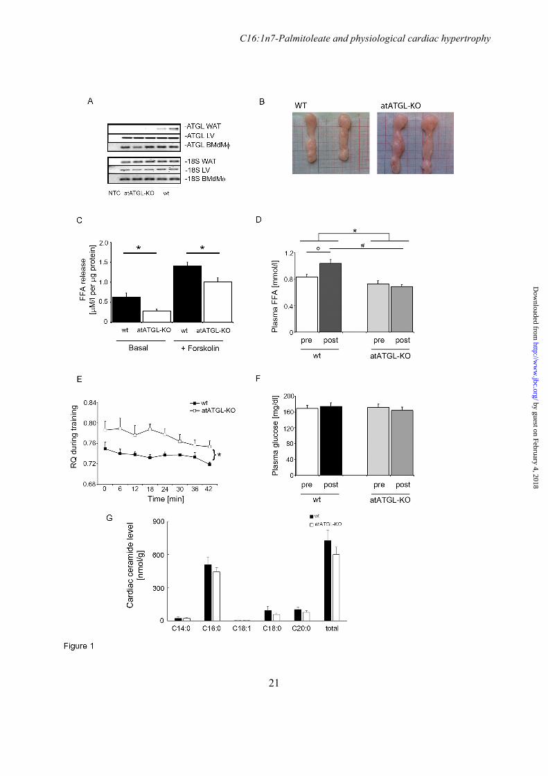
atATGL-KO mice showed complete deletion of ATGL expression in WAT but not in the heart or bone marrow-derived macrophages; an enlargement of WAT depots, increased fat pad weights, and increased overall fat mass (Fig. 1A/B and Table 1). Moreover, WAT lipolysis was significantly diminished in atATGL-KO mice, when compared to wt littermates (Figure 1C). In accordance with the importance of adipose ATGL for exercise-induced WAT lipolysis, circulating FFA level, measured before and after training did significantly increase after training in wt-mice but not in atATGL-KO mice (Figure 1D). Consistently, atATGL-KO mice exhibited a significantly higher peri-exercise RQ confirming attenuated systemic lipid oxidation in the absence of adipose ATGL (Figure 1E). Plasma glucose levels did not differ between the genotypes (Figure 1F). Previously, ATGL-deficiency in cardiomyocytes resulted in increased cardiac ceramide levels (19). However, deletion of ATGL in WAT did not affect cardiac ceramide levels (Fig. 1G). Taken together, atATGL-KO mice
by guest on February 4, 2018http://w
ww
.jbc.org/D
ownloaded from
C16:1n7-Palmitoleate and physiological cardiac hypertrophy
6
exhibited impaired WAT lipolysis associated with reduced exercise-induced plasma FFA appearance and systemic lipid oxidation.
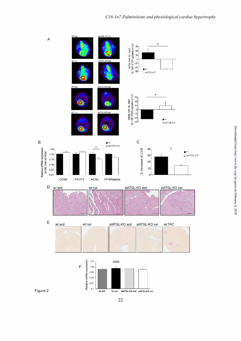
Subsequently small animal PET was performed in sedentary and trained mice to assess cardiac FA and glucose uptake. Trained mice were examined at the end of the 4-week training after an overnight starving/ resting period. As depicted in Figure 2A, trained wt-mice markedly increased cardiac FA-uptake when compared to sedentary controls. Accordingly, cardiac glucose uptake was reduced in exercising wt-mice. In consonance with the reduced FA-mobilization in trained atATGL-KO mice, exercise-mediated cardiac FA-uptake was completely abolished and even lower than sedentary controls, accompanied by an increase in cardiac glucose uptake (Figure 2A). Reduced cardiac FA-uptake in atATGL-KO mice did not result from diminished expression of cardiac FA-transporters (Figure 2B). In line with a reduction of cardiac FA-uptake and utilization, cardiac expression of key enzymes involved in downstream FA-metabolism such as long-chain acyl-CoA synthetase 1 (ACSL1) were reduced or tended to decrease (peroxisome proliferator-activated receptor alpha (PPARalpha)) in atATGL-KO mice (Figure 2B). In association with changes in cardiac metabolism, chronic exercise leads to the development of physiological cardiac hypertrophy. LV mass assessed by echocardiography was significantly increased in wt mice challenged with endurance training compared to sedentary mice (Table 2). This response was significantly attenuated in atATGL-KO mice (Figure 2C, Table 2). Histological analysis of myocyte size from LV-samples (HE-staining) revealed a moderate increase of cardiomyocyte size in trained wt-mice when compared to sedentary wt-mice (LV-myocyte cross sectional area: sedentary-wt: 258.6±6.9µm2 vs. trained-wt: 291.0±7.1µm2, p<0.01) (Fig. 2D). This regulation was not visible in atATGL-KO mice (LV-myocyte cross sectional area: sedentary-KO: 273.3±7.5µm2 vs. trained-KO: 277.1±6.8µm2, p=ns) (Fig. 2D). Together these data suggest that FA-mobilisation from adipose tissue, and peripheral FA availability may determine cardiac morphology during chronic exercise. During the development of pathological cardiac hypertrophy inflammatory processes such as macrophage invasion and fibrotic remodelling occur. In contrast, exercised-induced LV mass increase was neither associated with cardiac
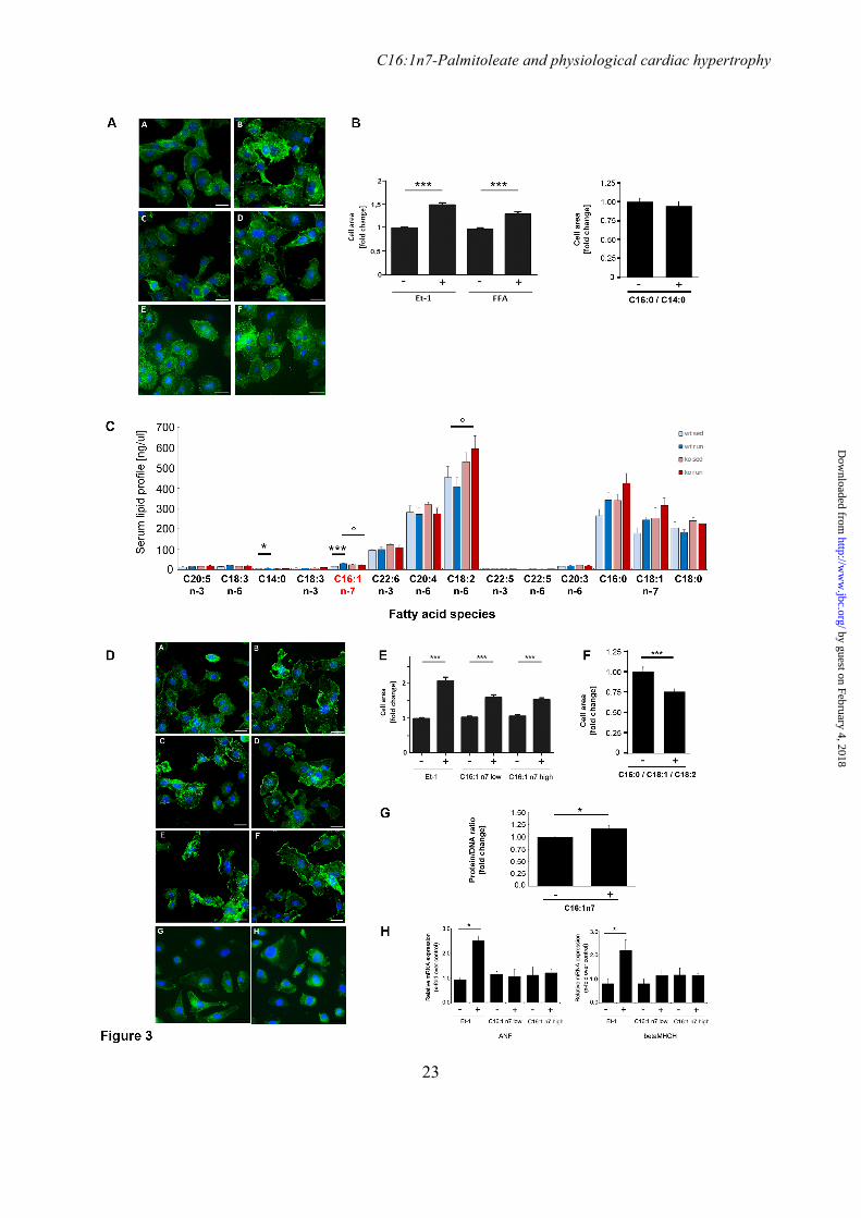
fibrosis (Fig. 2E) nor with macrophage invasion assessed by expression of the macrophage marker CD68 (Fig. 2F). And more importantly, deletion of ATGL in WAT did not affect these processes (Fig. 2E/F). Recently published data from Riquelme and colleagues indicated a prohypertrophic action of several FA species (5). The lack of an exercise-induced increase of circulating FA-levels in atATGL-KO mice associated with the attenuated cardiac hypertrophic response suggests also a potential role of FAs as prohypertrophic mediators in our model. To investigate the role of FAs in non-proliferative cardiomyocyte growth, we next stimulated HL-1 cells with the FA-mixture containing C14:0, C16:0 and C16:1n7, as previously described (5) and analyzed cellular hypertrophy (Figure 3A/B). The FA-mixture, effectively induced cardiomyocyte hypertrophy in exercise-relevant serum concentrations (Figure 3A/B). Lack of C16:1n7 in this mixture resulted in the absence of pro-hypertrophic effects (Figure 3A/B). To identify the putative FA-effector of hypertrophy development in our model we performed a comprehensive serum analysis of circulating FAs using HPLC/ MS measurements (Figure 3C). Among the prohypertrophic FAs only C16:1n7 serum levels followed a pattern corresponding to the observed cardiac phenotype (Figure 3C). C16:1n7 levels increased significantly in wt-mice after training but not in atATGL-KO mice (Figure 3C). In contrast, C14:0 and C16:0 also increased in atATGL-KO mice (Figure 3C). Despite, the overall low serum concentration of C16:1n7 compared to other FAs, C16:1n7 has recently been identified as an adipose-derived hormone regulating biochemical and physiological processes in other organs such as skeletal muscle and liver (20). Thus, we hypothesized that C16:1n7 might be a crucial molecular comediator of cardiac hypertrophy in our model. To prove this, we next treated HL-1 cells with C16:1n7 alone, or a FA-mixture reflecting the serum FA-composition (C16:0, C18:1, C18:2), and analyzed cellular hypertrophy by assessing the cell area (Fig. 3D-F) and cellular protein content (Fig.3G). C16:1n7 alone effectively induced non-proliferative cardiomyocyte growth in exercise-relevant serum concentrations whereas this effect was absent in HL-1 cells stimulated with the FA-mixture (Fig. 3D-F). These data suggest that C16:1n7 liberated from adipose tissue may support
by guest on February 4, 2018http://w
ww
.jbc.org/D
ownloaded from
C16:1n7-Palmitoleate and physiological cardiac hypertrophy
7
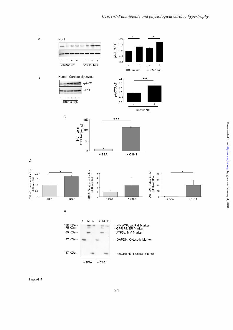
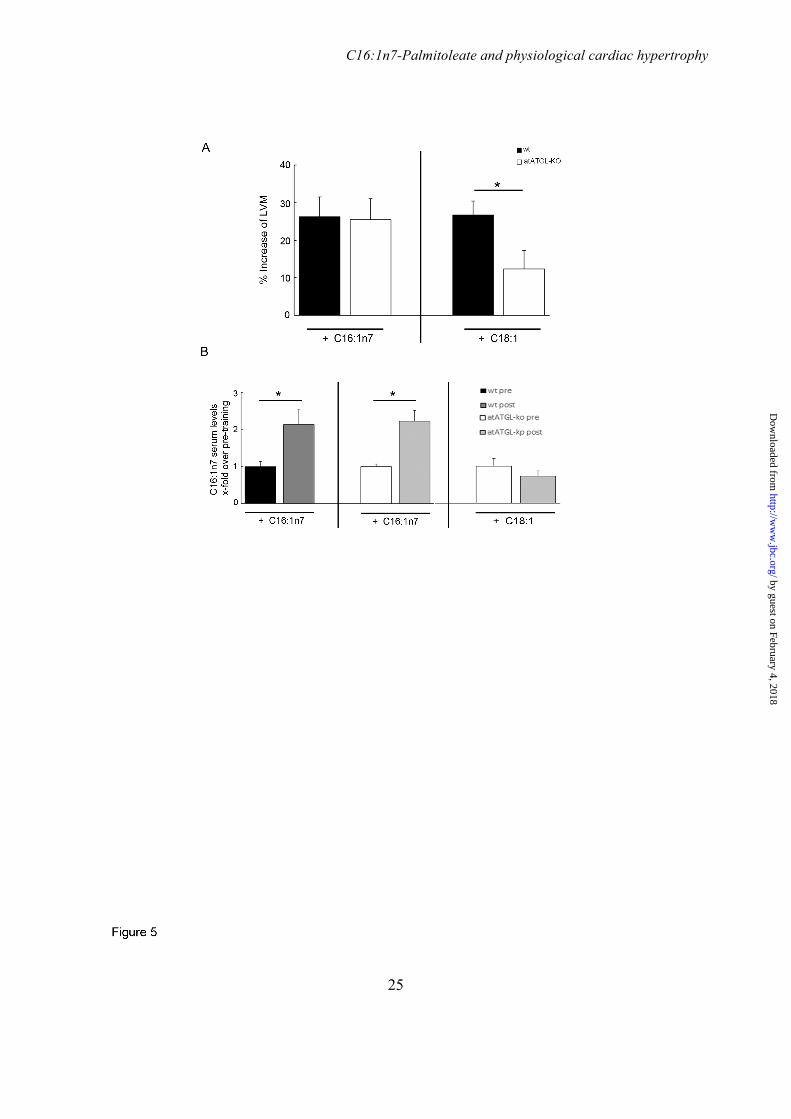
the development of training-induced LVH. Moreover, the mRNA expression of the atrial natriuretic factor (ANF) and beta-cardiac myosin heavy chain isogene (beta-MHCH), both markers of pathological hypertrophy, was not increased under C16:1n7-stimulation indicating the induction of a physiological response (Figure 3H). A hallmark of the development of physiological cardiac hypertrophy is the short-term activation of the cardiomyocytic serine/ threonine protein kinase AKT. Short-term treatment of HL-1 cells and primary human cardiomyocytes with C16:1n7 markedly stimulated AKT-phosphorylation supporting the induction of an adaptive, physiological hypertrophic response by C16:1n7 (Figure 4A/B). To further elicit the mechanism of C16:1n7´s action, we analysed cellular C16:1n7 appearance in HL-1 whole cells, and in subcellular fractions (membrane, cytosolic, nuclear) 6h after C16:1n7 stimulation by using cell fractionation followed by HPLC/ MS measurements (Figure 4C-E). Cells were washed multiple times after C16:1n7 stimulation to exclude contamination of cell pellets with the FA. As depicted in Fig. 4C, C16:1n7 stimulation led to a significant cellular accumulation of C16:1n7. Interestingly, C16:1n7 predominantly occurred in nuclear fractions, and modest induction of its occurrence was observed in cell membrane fractions (Figure 4D). Together these results suggest, that C16:1n7 likely acts via AKT-signaling and via an intracellular/ nuclear mechanism. To further support a role of C16:1n7 as a coregulator of training-induced cardiac hypertrophy in vivo, we assessed wt- and atATGL-KO mice who either received dietary enrichment of C16:1n7 or C18:1 during their 4 weeks training regimen. Only dietary supplementation with C16:1n7 rescued the impaired exercise-induced cardiac hypertrophy in atATGL-KO mice to levels comparable to wt mice, whereas C18:1 supplementation did not affect the hypertrophic response in atATGL-KO mice (Fig. 5A and Table 3). These data suggest that the lack of C16:1n7 contributes to the cardiac phenotype observed in atATGL-KO mice after exercise. Importantly, C16:1n7 supplementation led to a consistent increase of C16:1n7 plasma levels during the training period in wt- and atATGL-KO mice, which was not observed with C18:1 supplementation (Fig. 5B). We also analysed AKT-phosphorylation in LV samples from
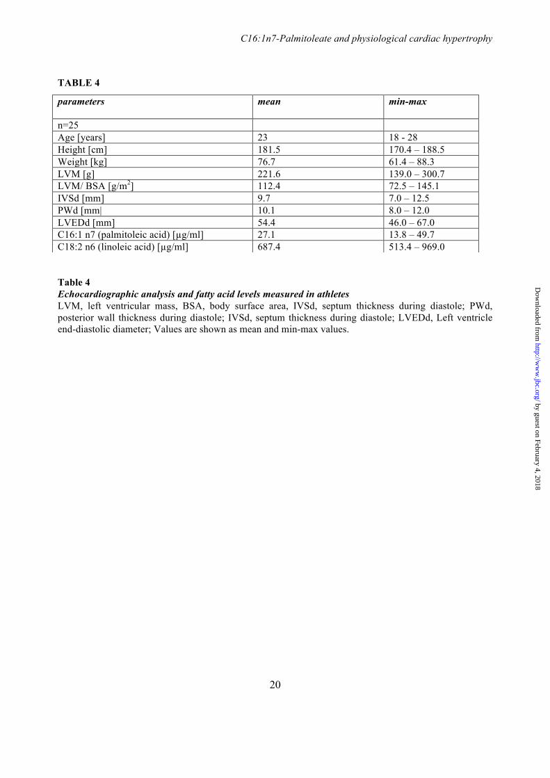
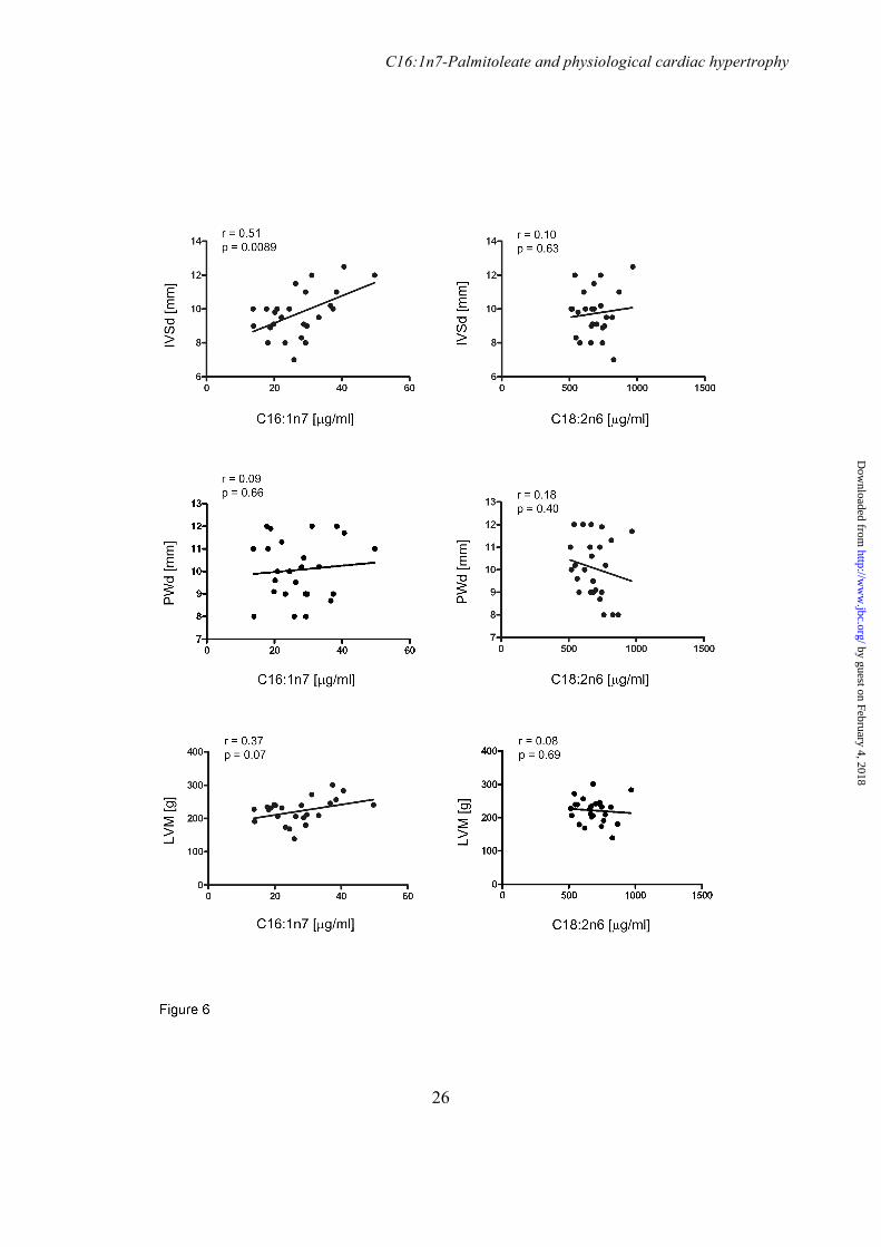
supplemented, trained mice. Cardiac AKT-phosphorylation in vivo was not regulated by FA-supplementation in trained mice (data not shown). For translation of our results to human physiology, we performed echocardiography and serum FA analysis (C16:1n7, C18:2n6) in 25 male, highly trained endurance athletes, and analyzed parameters of LVH including diastolic interventricular septum thickness (IVSd), diastolic posterior wall thickness (PWd), end-diastolic left ventricle diameter (LVEDd), and LVM. Mean IVSd-, PWd-, LVEDd-, and LVM-values (Table 4) indicated training-induced LVH in endurance athletes compared to previously studied control subjects (21). We observed a highly significant linear correlation between C16:1n7 serum level and IVSd (Figure 6, upper-left). C16:1n7 level did not correlate with PWd (Figure 6, middle-left). A positive correlation between C16:1n7 and LVM could be detected but did not reach statistical significance (Figure 6, lower-left). To exclude that this correlation is a result from an increase in serum FA-levels per se, we also studied C18:2n6 serum levels, a FA with highest serum concentrations in human serum samples. No correlation could be detected between C18:2n6 level and echocardiographic LVH parameters (Figure 6, right) suggesting a specific association between C16:1n7 and IVSd. DISCUSSION The present study demonstrates for the first time that adipose tissue (AT) lipolysis regulated by adipose ATGL, and associated with lipokine liberation (C16:1n7) promotes exercise-induced cardiac hypertrophy. The relevance of C16:1n7 as a pro-hypertrophic cofactor during training is further supported by a positive correlation between C16:1n7 serum level and echocardiographic LVH-parameters in endurance athletes. In consonance with the work of Ahmadian and colleagues (22), an impairment of ATGL-mediated lipolysis in WAT led to AT hypertrophy. Moreover, when subjected to chronic training, atATGL-KO mice exhibited an impaired AT lipolysis associated with the absence of exercise-induced plasma FFA appearance and reduced systemic lipid oxidation. Absence of adaptation of circulating FFAs to increased energy demands during exercise has been previously described in two other models of ATGL-deficiency (23,24).
by guest on February 4, 2018http://w
ww
.jbc.org/D
ownloaded from
C16:1n7-Palmitoleate and physiological cardiac hypertrophy
8
Schoiswohl and colleagues demonstrated in an acute exercise model that mice overexpressing ATGL specifically in the heart while lacking ATGL in all other tissues, exhibit significantly reduced appearance of plasma FFAs and glycerol after training (24). In contrast to our results, these authors also observed a significant decrease of plasma glucose in ATGL-deficient mice after training likely resulting from the relatively short and acute form of exercise. Likewise, in a separate study, the global ATGL-KO also resulted in a reduction of maximal running velocity and endurance capacity underscoring the importance of this enzyme for regular substrate metabolism during exercise (23). The effect of adipocyte-specific ATGL deletion on adaptive responses to chronic exercise training has not yet been previously examined. In the present study, chronically trained atATGLKO mice exhibited diminished development of adaptive LVH and reduced capability of cardiac FA uptake. Thus, this is the first report investigating the lack of ATGL in AT, and its impact on cardiac metabolism/ function. In contrast, previous studies mainly focussed on the role of myocardial ATGL. Kienesberger and colleagues demonstrated that cardiomyocyte-specific overexpression of ATGL results in reduced myocardial TG content and an improved systolic function (25). These data are in line with ATGL loss-of-function studies showing a marked increase of cardiac TG accumulation and impairment of LV-systolic function (26-28). In addition, Gao and colleagues demonstrated that ATGL-deficiency in neonatal rat cardiomyocytes leads to accumulation of ceramides, a process not observed in our model (19). More importantly, Haemmerle and colleagues also investigated transgenic mice with ATGL exclusively expressed in cardiac muscle (26). The authors show that plasma FA and TG concentrations were much lower in those mice accompanied by an increase in cardiac PPARalpha- and PPARdelta target gene expression, suggesting that plasma lipid concentrations do not correlate with the activity level of the myocardial PPAR pathway and the severity of the cardiac phenotype. This is in contrast to our data clearly indicating a close relationship between plasma lipid fuel availability, cardiac-uptake, -phenotype, and -gene expression. Accordingly, cardiac expression of the PPARalpha target gene ACSL1 (29) was downregulated in atATGL-KO mice. WAT-ATGL mediates the
release of systemic FA-ligands for PPARalpha activation in other organs (22). Thus, suppression of WAT-lipolysis in atATGL-KO mice results in reduced cardiac FA-availability which may attenuate cardiac PPARalpha activity and subsequent gene regulation. The discrepancy between observations by Haemmerle and colleagues and our data may result from distinct experimental settings. Cardiac ATGL overexpression may induce a distinct cardiac metabolic program, which differs from hearts with physiological ATGL levels, as in case of the mice used in our study. We identified C16:1n7, liberated by ATGL-mediated adipocyte lipolysis in response to training, as a molecular comediator of training-induced cardiac hypertrophy. In consonance with our data, Riquelme and colleagues recently identified similar FAs as prohypertrophic factors in a different model of physiological postprandial cardiac hypertrophy (5). The present study was not designed to prove that C16:1n7 alone is sufficient to directly induce cardiac hypertrophy. Rather our data supports the notion that FAs may act as coregulators of physiological responses in concert with other mediators in-vivo (3). C16:1n7 has been recently characterized as an adipose-derived lipid hormone regulating muscular insulin sensitivity and hepatic lipid metabolism (20). In this regard, C16:1n7 markedly potentiated AKT-signaling in skeletal muscle (20). Cardiac AKT-phosphorylation was not regulated by C16:1n7 during chronic administration in-vivo. However, we show that the C16:1n7-mediated stimulation of cardiac cellular hypertrophy is accompanied by an induction of AKT-phosphorylation in murine HL-1 cardiomyocytes and in primary human cardiomyocytes. AKT-phosphorylation in-vivo likely follows an intermittent pattern according to the training period (3). These characteristics make it difficult to analyse the AKT signal in-vivo during chronic FA-administration. The upstream signalling pathway of C16:1n7-induced AKT phosphorylation and subsequent cardiomyocyte hypertrophy remains to be determined. Potential candidates include the G-protein-coupled receptor GPR120 which signals through a Galpha-q-dependent pathway after C16:1n7 binding, or porcupine, a membrane-bound O-acyltransferase involved in the attachment of C16:1n7 to serine residues (30,31).
by guest on February 4, 2018http://w
ww
.jbc.org/D
ownloaded from
C16:1n7-Palmitoleate and physiological cardiac hypertrophy
9
Lipid modification of serine residues has been recently identified as a pivotal processes in Wnt signalling, a pathway upstream of AKT, and involved in cardiac remodelling (31,32). In addition, the marked occurrence of C16:1n7 in the cell nucleus 6h after stimulation points towards a nuclear action of the FA. Palmitoleic acid has been shown to induce PPARdelta transcriptional activity (33). Since, cardiomyocytic PPARdelta is an established regulator of cardiomyocyte morphology and function, this nuclear receptor might be a potential target of C16:1n7´s action in the cell nucleus (34). To prove the relevance of our results for human physiology we demonstrated that C16:1n7 serum level significantly correlate with echocardiographic LVH parameters in endurance athletes. Increased serum FFA levels have been previously documented during exercise (35). To exclude that LVH parameters in athletes correlate with increased FFA levels per se, we analysed C18:2n6 showing no correlation to LVH. Chiefly, liberation of FFAs during prolonged exercise occurs for the maintenance of enhanced skeletal muscle and cardiac energy demands. Compared to C18:2n6 serum concentrations, C16:1n7 level are very low making it unlikely that C16:1n7 serves as a major cardiac energy source. On the contrary, our data points towards an involvement of C16:1n7 in cardiac cellular growth, likely in concert with other mediators (3). We are aware
that the observed correlation does not allow any causative conclusions, however, these data, at least in part, support the potential action of C16:1n7 on LV-wall thickening. Recently published studies pointed out that molecular effectors involved in beneficial adaptive cardiac growth may be used as therapeutic targets for maladaptive cardiovascular disease (3). Along this line one may speculate about a potential therapeutic role of lipid-derived, e.g. C16:1n7-based, interventions. This is further supported by studies demonstrating a beneficial role of food-derived or supplemented other FAs such as omega-3 FAs in primary- and/or secondary prevention of cardiovascular disease (36). Future studies will be required to fully understand the therapeutic options of C16:1n7 itself or key hepatic enzymes involved in de-novo C16:1n7 synthesis, and to develop a lipid-based therapy for cardiovascular diseases. Collectively, our data provide the first evidence that adipose tissue lipolysis directly promotes the development of exercise-induced cardiac hypertrophy involving the lipokine C16:1n7 palmitoleate. The identification of a lipokine mediating physiological cardiac growth may help to develop future lipid-based therapies for pathological LVH or chronic heart failure.
ACKNOWLEDGEMENTS The authors thank Christiane Sprang, Beata Hoeft, and Manuela Sommerfeld (Charité-Universitaetsmedizin Berlin, Institute of Pharmacology, CCR, Berlin, Germany) for their excellent technical support; Xiang Li, Katja Hirsch (University Hospital Wuerzburg, Department of Nuclear Medicine, Wuerzburg, Germany) and Ina Israel for the help with PET-experiments; and Stefan Anker (Charité-Universitaetsmedizin Berlin, CCR, Germany) for using NMR device (Brucker’s Minispec MQ10). CONFLICT OF INTEREST MR is an employee of Lipidomix GmbH, Berlin, Germany. All other authors have no conflict of interest to disclose in connection with the submitted article. AUTHOR CONTRIBUTIONS AFL substantially contributed to conception and design of the study, acquisition of data, and data analysis and interpretation, drafted the article, and revised the article critically for important intellectual content. MCK, MR, and RK substantially contributed to conception of the study, acquisition of data, and data analysis and interpretation (MCK: PET analysis, MR: HPLC/MS analysis, RK: Histological analysis). VB (in-vivo experiments), SB (in-vivo/ in-vitro experiments), ES (in-vitro experiments), ZB (in-vitro experiments), EJ (in-vitro experiments), JS (in-vivo experiments), JG (in-vivo experiments), AKS (in-vitro experiments), AB (in-vitro experiments), AS (PET analysis), and KB (human studies) substantially
by guest on February 4, 2018http://w
ww
.jbc.org/D
ownloaded from
C16:1n7-Palmitoleate and physiological cardiac hypertrophy
10
contributed to acquisition of data, and data analysis and interpretation. MH and BW substantially contributed to conception of the human study, and revised the article critically for important intellectual content. EEK substantially contributed to conception and design of the study (in-vivo experiments with atATGL-KO mice), and data interpretation, and revised the article critically for important intellectual content. UK substantially contributed to acquisition of funding, conception and design of the study, data analysis and interpretation, drafted the article, and revised the article critically for important intellectual content. All authors finally approved the version to be published.
by guest on February 4, 2018http://w
ww
.jbc.org/D
ownloaded from
C16:1n7-Palmitoleate and physiological cardiac hypertrophy
11
REFERENCES 1. Rawlins, J., Bhan, A., and Sharma, S. (2009) Left ventricular hypertrophy in athletes. Eur
J Echocardiogr 10, 350-356 2. Heineke, J., and Molkentin, J. D. (2006) Regulation of cardiac hypertrophy by
intracellular signalling pathways. Nat Rev Mol Cell Biol 7, 589-600 3. Maillet, M., van Berlo, J. H., and Molkentin, J. D. (2013) Molecular basis of
physiological heart growth: fundamental concepts and new players. Nat Rev Mol Cell Biol 14, 38-48
4. Thompson, D., Karpe, F., Lafontan, M., and Frayn, K. (2012) Physical activity and exercise in the regulation of human adipose tissue physiology. Physiol Rev 92, 157-191
5. Riquelme, C. A., Magida, J. A., Harrison, B. C., Wall, C. E., Marr, T. G., Secor, S. M., and Leinwand, L. A. (2011) Fatty acids identified in the Burmese python promote beneficial cardiac growth. Science 334, 528-531
6. Young, S. G., and Zechner, R. (2013) Biochemistry and pathophysiology of intravascular and intracellular lipolysis. Genes Dev 27, 459-484
7. Sitnick, M. T., Basantani, M. K., Cai, L., Schoiswohl, G., Yazbeck, C. F., Distefano, G., Ritov, V., DeLany, J. P., Schreiber, R., Stolz, D. B., Gardner, N. P., Kienesberger, P. C., Pulinilkunnil, T., Zechner, R., Goodpaster, B. H., Coen, P., and Kershaw, E. E. (2013) Skeletal muscle triacylglycerol hydrolysis does not influence metabolic complications of obesity. Diabetes 62, 3350-3361
8. Villena, J. A., Roy, S., Sarkadi-Nagy, E., Kim, K. H., and Sul, H. S. (2004) Desnutrin, an adipocyte gene encoding a novel patatin domain-containing protein, is induced by fasting and glucocorticoids: ectopic expression of desnutrin increases triglyceride hydrolysis. J Biol Chem 279, 47066-47075
9. Foryst-Ludwig, A., Kreissl, M. C., Sprang, C., Thalke, B., Bohm, C., Benz, V., Gurgen, D., Dragun, D., Schubert, C., Mai, K., Stawowy, P., Spranger, J., Regitz-Zagrosek, V., Unger, T., and Kintscher, U. (2011) Sex differences in physiological cardiac hypertrophy are associated with exercise-mediated changes in energy substrate availability. Am J Physiol Heart Circ Physiol 301, H115-122
10. Fliegner, D., Schubert, C., Penkalla, A., Witt, H., Kararigas, G., Dworatzek, E., Staub, E., Martus, P., Ruiz Noppinger, P., Kintscher, U., Gustafsson, J. A., and Regitz-Zagrosek, V. (2010) Female sex and estrogen receptor-beta attenuate cardiac remodeling and apoptosis in pressure overload. Am J Physiol Regul Integr Comp Physiol 298, R1597-1606
11. Bolsoni-Lopes, A., Festuccia, W. T., Farias, T. S., Chimin, P., Torres-Leal, F. L., Derogis, P. B., de Andrade, P. B., Miyamoto, S., Lima, F. B., Curi, R., and Alonso-Vale, M. I. (2013) Palmitoleic acid (n-7) increases white adipocyte lipolysis and lipase content in a PPARalpha-dependent manner. Am J Physiol Endocrinol Metab 305, E1093-1102
12. DeGrado, T. R., Kitapci, M. T., Wang, S., Ying, J., and Lopaschuk, G. D. (2006) Validation of 18F-fluoro-4-thia-palmitate as a PET probe for myocardial fatty acid oxidation: effects of hypoxia and composition of exogenous fatty acids. J Nucl Med 47, 173-181
13. Loening, A. M., and Gambhir, S. S. (2003) AMIDE: a free software tool for multimodality medical image analysis. Mol Imaging 2, 131-137
14. Weischenfeldt, J., and Porse, B. (2008) Bone Marrow-Derived Macrophages (BMM): Isolation and Applications. CSH protocols 2008, pdb prot5080
15. Claycomb, W. C., Lanson, N. A., Jr., Stallworth, B. S., Egeland, D. B., Delcarpio, J. B., Bahinski, A., and Izzo, N. J., Jr. (1998) HL-1 cells: a cardiac muscle cell line that contracts and retains phenotypic characteristics of the adult cardiomyocyte. Proc Natl
by guest on February 4, 2018http://w
ww
.jbc.org/D
ownloaded from
C16:1n7-Palmitoleate and physiological cardiac hypertrophy
12
Acad Sci U S A 95, 2979-2984 16. Boon, R. A., Iekushi, K., Lechner, S., Seeger, T., Fischer, A., Heydt, S., Kaluza, D.,
Treguer, K., Carmona, G., Bonauer, A., Horrevoets, A. J., Didier, N., Girmatsion, Z., Biliczki, P., Ehrlich, J. R., Katus, H. A., Muller, O. J., Potente, M., Zeiher, A. M., Hermeking, H., and Dimmeler, S. MicroRNA-34a regulates cardiac ageing and function. Nature 495, 107-110
17. Lang, R. M., Bierig, M., Devereux, R. B., Flachskampf, F. A., Foster, E., Pellikka, P. A., Picard, M. H., Roman, M. J., Seward, J., Shanewise, J. S., Solomon, S. D., Spencer, K. T., Sutton, M. S., and Stewart, W. J. (2005) Recommendations for chamber quantification: a report from the American Society of Echocardiography's Guidelines and Standards Committee and the Chamber Quantification Writing Group, developed in conjunction with the European Association of Echocardiography, a branch of the European Society of Cardiology. Journal of the American Society of Echocardiography : official publication of the American Society of Echocardiography 18, 1440-1463
18. Devereux, R. B., Alonso, D. R., Lutas, E. M., Gottlieb, G. J., Campo, E., Sachs, I., and Reichek, N. (1986) Echocardiographic assessment of left ventricular hypertrophy: comparison to necropsy findings. The American journal of cardiology 57, 450-458
19. Gao, H., Feng, X. J., Li, Z. M., Li, M., Gao, S., He, Y. H., Wang, J. J., Zeng, S. Y., Liu, X. P., Huang, X. Y., Chen, S. R., and Liu, P. Q. (2015) Downregulation of adipose triglyceride lipase promotes cardiomyocyte hypertrophy by triggering the accumulation of ceramides. Archives of biochemistry and biophysics 565, 76-88
20. Cao, H., Gerhold, K., Mayers, J. R., Wiest, M. M., Watkins, S. M., and Hotamisligil, G. S. (2008) Identification of a lipokine, a lipid hormone linking adipose tissue to systemic metabolism. Cell 134, 933-944
21. Pluim, B. M., Zwinderman, A. H., van der Laarse, A., and van der Wall, E. E. (2000) The athlete's heart. A meta-analysis of cardiac structure and function. Circulation 101, 336-344
22. Ahmadian, M., Abbott, M. J., Tang, T., Hudak, C. S., Kim, Y., Bruss, M., Hellerstein, M. K., Lee, H. Y., Samuel, V. T., Shulman, G. I., Wang, Y., Duncan, R. E., Kang, C., and Sul, H. S. (2011) Desnutrin/ATGL is regulated by AMPK and is required for a brown adipose phenotype. Cell Metab 13, 739-748
23. Huijsman, E., van de Par, C., Economou, C., van der Poel, C., Lynch, G. S., Schoiswohl, G., Haemmerle, G., Zechner, R., and Watt, M. J. (2009) Adipose triacylglycerol lipase deletion alters whole body energy metabolism and impairs exercise performance in mice. Am J Physiol Endocrinol Metab 297, E505-513
24. Schoiswohl, G., Schweiger, M., Schreiber, R., Gorkiewicz, G., Preiss-Landl, K., Taschler, U., Zierler, K. A., Radner, F. P., Eichmann, T. O., Kienesberger, P. C., Eder, S., Lass, A., Haemmerle, G., Alsted, T. J., Kiens, B., Hoefler, G., Zechner, R., and Zimmermann, R. (2010) Adipose triglyceride lipase plays a key role in the supply of the working muscle with fatty acids. J Lipid Res 51, 490-499
25. Kienesberger, P. C., Pulinilkunnil, T., Sung, M. M., Nagendran, J., Haemmerle, G., Kershaw, E. E., Young, M. E., Light, P. E., Oudit, G. Y., Zechner, R., and Dyck, J. R. (2012) Myocardial ATGL overexpression decreases the reliance on fatty acid oxidation and protects against pressure overload-induced cardiac dysfunction. Mol Cell Biol 32, 740-750
26. Haemmerle, G., Moustafa, T., Woelkart, G., Buttner, S., Schmidt, A., van de Weijer, T., Hesselink, M., Jaeger, D., Kienesberger, P. C., Zierler, K., Schreiber, R., Eichmann, T., Kolb, D., Kotzbeck, P., Schweiger, M., Kumari, M., Eder, S., Schoiswohl, G.,
by guest on February 4, 2018http://w
ww
.jbc.org/D
ownloaded from
C16:1n7-Palmitoleate and physiological cardiac hypertrophy
13
Wongsiriroj, N., Pollak, N. M., Radner, F. P., Preiss-Landl, K., Kolbe, T., Rulicke, T., Pieske, B., Trauner, M., Lass, A., Zimmermann, R., Hoefler, G., Cinti, S., Kershaw, E. E., Schrauwen, P., Madeo, F., Mayer, B., and Zechner, R. (2011) ATGL-mediated fat catabolism regulates cardiac mitochondrial function via PPAR-alpha and PGC-1. Nat Med 17, 1076-1085
27. Zierler, K. A., Jaeger, D., Pollak, N. M., Eder, S., Rechberger, G. N., Radner, F. P., Woelkart, G., Kolb, D., Schmidt, A., Kumari, M., Preiss-Landl, K., Pieske, B., Mayer, B., Zimmermann, R., Lass, A., Zechner, R., and Haemmerle, G. (2013) Functional cardiac lipolysis in mice critically depends on comparative gene identification-58. J Biol Chem 288, 9892-9904
28. Kienesberger, P. C., Pulinilkunnil, T., Nagendran, J., Young, M. E., Bogner-Strauss, J. G., Hackl, H., Khadour, R., Heydari, E., Haemmerle, G., Zechner, R., Kershaw, E. E., and Dyck, J. R. (2013) Early structural and metabolic cardiac remodelling in response to inducible adipose triglyceride lipase ablation. Cardiovasc Res 99, 442-451
29. Schoonjans, K., Watanabe, M., Suzuki, H., Mahfoudi, A., Krey, G., Wahli, W., Grimaldi, P., Staels, B., Yamamoto, T., and Auwerx, J. (1995) Induction of the acyl-coenzyme A synthetase gene by fibrates and fatty acids is mediated by a peroxisome proliferator response element in the C promoter. J Biol Chem 270, 19269-19276
30. Glass, C. K., and Olefsky, J. M. Inflammation and lipid signaling in the etiology of insulin resistance. Cell Metab 15, 635-645
31. Takada, R., Satomi, Y., Kurata, T., Ueno, N., Norioka, S., Kondoh, H., Takao, T., and Takada, S. (2006) Monounsaturated fatty acid modification of Wnt protein: its role in Wnt secretion. Dev Cell 11, 791-801
32. Bergmann, M. W. WNT signaling in adult cardiac hypertrophy and remodeling: lessons learned from cardiac development. Circ Res 107, 1198-1208
33. Brown, J. D., Oligino, E., Rader, D. J., Saghatelian, A., and Plutzky, J. (2011) VLDL hydrolysis by hepatic lipase regulates PPARdelta transcriptional responses. PLoS One 6, e21209
34. Cheng, L., Ding, G., Qin, Q., Huang, Y., Lewis, W., He, N., Evans, R. M., Schneider, M. D., Brako, F. A., Xiao, Y., Chen, Y. E., and Yang, Q. (2004) Cardiomyocyte-restricted peroxisome proliferator-activated receptor-delta deletion perturbs myocardial fatty acid oxidation and leads to cardiomyopathy. Nat Med. 10, 1245-1250. Epub 2004 Oct 1210.
35. Mittendorfer, B., Horowitz, J. F., and Klein, S. (2002) Effect of gender on lipid kinetics during endurance exercise of moderate intensity in untrained subjects. Am J Physiol Endocrinol Metab 283, E58-65
36. Kromhout, D., and de Goede, J. (2014) Update on cardiometabolic health effects of omega-3 fatty acids. Curr Opin Lipidol 25, 85-90
37. Konhilas, J. P., Maass, A. H., Luckey, S. W., Stauffer, B. L., Olson, E. N., and Leinwand, L. A. (2004) Sex modifies exercise and cardiac adaptation in mice. Am J Physiol Heart Circ Physiol 287, H2768-2776
by guest on February 4, 2018http://w
ww
.jbc.org/D
ownloaded from
C16:1n7-Palmitoleate and physiological cardiac hypertrophy
14
FOOTNOTES The work was supported by Deutsche Forschungsgemeinschaft (DFG; FG 1054/2 (Ki712/5-2)). AFL was supported by the DFG (FG 1054/2, KFO 218/2); VB was supported by the DFG (KFO 218/2); SB was supported by the Deutsche Stiftung für Herzforschung; MH was supported by the Else Kröner-Fresenius Stiftung 2012_A82; EEK was supported by National Institutes of Health (NIH), Grant NIH R01DK090166 and Howard Hughes Medical Institute, Grant HHMI-ECA and UK was supported by the DFG (FG 1054/2, KFO 192/2, KFO 218/2) and the Else Kröner-Fresenius Stiftung (2014_A100).
by guest on February 4, 2018http://w
ww
.jbc.org/D
ownloaded from
C16:1n7-Palmitoleate and physiological cardiac hypertrophy
15
FIGURE LEGENDS Figure 1: Phenotypic characterization of atATGL-KO mice (A) RT-PCR analysis of ATGL expression, measured in WAT, the left ventricle of the heart (LV), and bone marrow derived macrophages (BMdMφ). B) Representative images of gonadal fat pads isolated from WT and atATGL-KO mice. Exercise-mediated metabolic changes: C) Ex-vivo lipolysis assay in murine gonadal adipose tissue explants collected from trained-mice immediately following their final running session (n = 3-4/group). FFA-release assays were performed using perigonadal white adipose tissue pads (n=3-4 / mouse), * p<0.05 vs. wt mice (unpaired ttest). D) Change in plasma FFAs and (F) glucose of trained mice before (pre) and directly after (post) their final running session; * p<0.05 vs. pre/ post difference in wt mice, ° p<0.05 vs. wt (pre), # p<0.05 vs. wt (post) (n=9; 2-way ANOVA with repeated measures, (Bonferroni posttest)). E) Respiratory quotient (RQ) from trained mice during their final running sessions using indirect calorimetry integrated into the treadmill system (CaloTreadmill; TSE System); * p<0.05 vs. wt (n=4; 2-way ANOVA with repeated measures (Bonferroni posttest)). G) Profile of selected cardiac ceramides. Results represent samples obtained from wt and atATGL-KO mice (n=3), analyzed using rapid resolution HPLC/ Tandem MS. Figure 2: Exercise-mediated changes in cardiac morphology/ metabolism A) Representative images of the myocardial FA-uptake (18F-FTP) (top 4 panels) and glucose- uptake (18F-FDG) (bottom 4 panels), measured using small animal PET. Quantification of 18F-FTP and 18F-FDG PET, showed as percent change of mean sedentary myocardial uptake, calculated as injection dose relative to LVM (%ID/ LVM), (n=5; * p<0.05 vs. wt, (unpaired ttest)). B) Analysis of mRNA expression in LV tissue collected from trained mice immediately following their final running session, as a marker of lipid uptake and mitochondrial fatty acid oxidation. qRT-PCR studies were carried out using total RNA isolated from LV tissue. Data are presented as x-fold over wt mice. CD36: Cluster of Differentiation 36; FATP1: Fatty acid transport protein 1; ACSL: Acyl-CoA synthetase-1; PPARalpha: peroxisome proliferator-activated receptor alpha; * p<0.05 vs. wt mice, (n=8; unpaired ttest). C) Cardiac adaptation expressed as percent change from mean sedentary LVM, as described previously (37); * p<0.05 vs. wt mice, (n=9; unpaired ttest). D) H&E –stained heart sections from wt and atATGL-KO mice. E) Picrosirius Red –stained heart sections from wt and atATGL-KO mice (fibrosis), positive control (right image): LV-samples from mice with transverse aortic constriction (TAC) resulting in pathological cardiac hypertrophy F) qRT-PCR analysis of CD68 mRNA expression levels measured in LVs of wt and atATGL-KO mice, as indicated. Figure 3: Hypertrophic response to C16:1n7 A) Hypertrophic response of murine cardiac HL-1 cells to FAs; representative immunofluorescence images of HL-1 cells, treated with 100 nM endothelin 1 (Et-1) (B), FA-mix: C14:0, C16:0 and C16:1n7 (D) or C14:0 and C16:0 (F),, or vehicle control (A, C, E) for 6h (left panel). B) Cell surface area calculated from the images of HL-1 cells, treated with the indicated compounds for 6h. Data are presented as x-fold over control; *** p<0.001 vs. control, 1-way ANOVA (Bonferroni posttest) or unpaired ttest. C) Profile of selected serum lipids. Results represent serum samples obtained from sedentary and trained mice (n=5), analyzed using rapid resolution HPLC/ Tandem MS; (* p<0.05 vs. wt sed; *** p<0.001 vs. wt sed; ° p<0.05 vs. wt run, (n=5; 2-way ANOVA (Bonferroni posttest)). D) Hypertrophic response of murine cardiac HL-1 cells to C16:1n7; representative immunofluorescence images of HL-1 cells, treated with 100 nM Et-1 (B), C16:1n7 low (D)/ high (F) concentration, C16:0; C18:1 and C18:2 (H), or vehicle controls (A, C, E, G) for 6h; E), F) cell surface area calculated from the images of HL-1 cells, treated with the indicated compounds for 6h. Data are presented as x-fold over control; (*** p<0.001 vs. control, 1-way ANOVA (Bonferroni posttest) (E) or unpaired ttest (F)). G) Ratio of Protein/DNA, n=8; * p< 0.05 vs control H) Analysis of ANF and beta-MHCH mRNA expression in cardiac HL-1 cells, treated with 100 nM Et-1, C16:1n7 low/ high concentration, or vehicle control for 6h. ANF: Atrial natriuretic factor; beta-MHCH: beta-cardiac myosin heavy chain isogene. * p<0.05 vs. control, (n=3; 1-way ANOVA (Tukey's or
by guest on February 4, 2018http://w
ww
.jbc.org/D
ownloaded from
C16:1n7-Palmitoleate and physiological cardiac hypertrophy
16
Bonferroni posttest)). Figure 4: Cellular actions of C16:1n7 A) WB analysis of P-AKT and AKT of HL-1 cells und (B) primary human cardiac myocytes stimulated with C16:1n7 for 30min, as indicated. Right panels: pAKT/AKT ratio calculated based on 3-4 independent experiments using ImageJ software, * p< 0.05 vs control, *** p< 0.01 vs. control, unpaired ttest. C) C16:1n7 levels measured in HL-1 cells stimulated with C16:1 or vehicle for 6h. Results represent cell pellet samples (n=3), analyzed using rapid resolution HPLC/ Tandem MS; *** p<0.001 vs. control, unpaired ttest; D) C16:1n7 levels measured in the different cellular fractions of HL-1 cells, stimulated with C16:1 or vehicle for 6h. Results represent fractionation samples: membrane-, cytosol-, and nuclear- fractions (n=3), analyzed using rapid resolution HPLC/ Tandem MS, * p<0.05 vs. control; unpaired ttest. E) WB analysis of different cellular fractions isolated from HL-1 cells treated with C16n7 or vehicle for 6h. MM-Marker: mitochondrial membrane marker; PM-Marker: plasma membrane marker; ER-Marker: endoplasmic reticulum marker. For details please see the method section. Figure 5: C16:1n7 supplementation in vivo A) Cardiac adaptation of mice supplemented with C16:1n7 or C18:1 during the exercise period. Cardiac adaptation is expressed as percent change from mean sedentary LVM, as described previously (37); (n=4-5; * p< 0.05 vs. wt mice, unpaired ttest). B) C16:1n7 serum levels in trained mice supplemented with C16:1n7 or C18:1 obtained before (pre) and (post) their final running sessions. Results represent serum samples (n=5), analyzed using rapid resolution HPLC/ Tandem MS, (* p< 0.05 vs. samples obtained before training (pre), unpaired ttest) Figure 6: LVH and serum FA-level in endurance athletes Echocardiographic analysis, including Interventricular septum thickness in diastole (IVSd), posterior wall thickness in diastole (PWd) and left ventricular mass (LVM) was performed in 25 endurance athletes. Echocardiography data was correlated to serum FA-levels of C16:1n7 (left panel) and C18:2n6 (right panel), measured in the serum of athletes. r = Pearson´s correlation coefficient.
by guest on February 4, 2018http://w
ww
.jbc.org/D
ownloaded from
C16:1n7-Palmitoleate and physiological cardiac hypertrophy
17
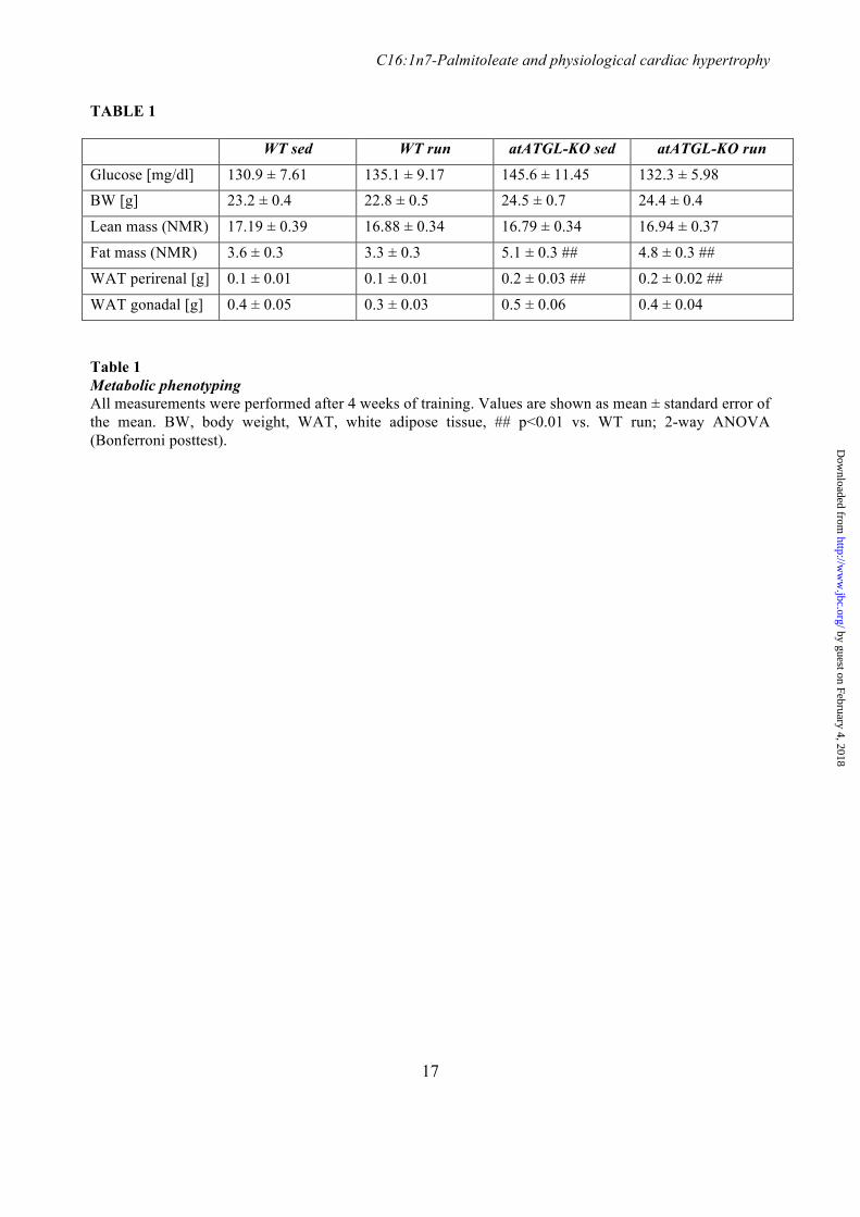
TABLE 1 WT sed WT run atATGL-KO sed atATGL-KO run
Glucose [mg/dl] 130.9 ± 7.61 135.1 ± 9.17 145.6 ± 11.45 132.3 ± 5.98
BW [g] 23.2 ± 0.4 22.8 ± 0.5 24.5 ± 0.7 24.4 ± 0.4
Lean mass (NMR) 17.19 ± 0.39 16.88 ± 0.34 16.79 ± 0.34 16.94 ± 0.37
Fat mass (NMR) 3.6 ± 0.3 3.3 ± 0.3 5.1 ± 0.3 ## 4.8 ± 0.3 ##
WAT perirenal [g] 0.1 ± 0.01 0.1 ± 0.01 0.2 ± 0.03 ## 0.2 ± 0.02 ##
WAT gonadal [g] 0.4 ± 0.05 0.3 ± 0.03 0.5 ± 0.06 0.4 ± 0.04
Table 1 Metabolic phenotyping All measurements were performed after 4 weeks of training. Values are shown as mean ± standard error of the mean. BW, body weight, WAT, white adipose tissue, ## p<0.01 vs. WT run; 2-way ANOVA (Bonferroni posttest).
by guest on February 4, 2018http://w
ww
.jbc.org/D
ownloaded from
C16:1n7-Palmitoleate and physiological cardiac hypertrophy
18
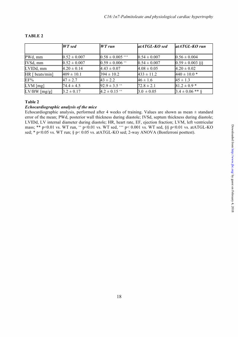
TABLE 2 WT sed
WT run atATGL-KO sed atATGL-KO run
PWd, mm 0.52 ± 0.007 0.58 ± 0.005 ‘‘‘ 0.54 ± 0.007 0.56 ± 0.004 IVSd, mm 0.52 ± 0.007 0.59 ± 0.006 ‘‘ 0.54 ± 0.007 0.59 ± 0.003 §§ LVIDd, mm 4.20 ± 0.14 4.43 ± 0.07 4.08 ± 0.05 4.20 ± 0.02 HR [ beats/min] 409 ± 10.1 394 ± 10.2 433 ± 11.2 440 ± 10.0 * EF% 47 ± 2.7 43 ± 2.2 46 ± 1.6 45 ± 1.3 LVM [mg] 74.4 ± 4.5 92.9 ± 3.5 ‘‘ 72.8 ± 2.1 81.2 ± 0.9 * LV/BW [mg/g] 3.2 ± 0.17 4.2 ± 0.15 ‘‘ 3.0 ± 0.05 3.4 ± 0.06 ** § Table 2 Echocardiographic analysis of the mice Echocardiographic analysis, performed after 4 weeks of training. Values are shown as mean ± standard error of the mean; PWd, posterior wall thickness during diastole; IVSd, septum thickness during diastole; LVIDd, LV internal diameter during diastole; HR, heart rate, EF, ejection fraction; LVM, left ventricular mass; ** p<0.01 vs. WT run, ‘‘ p<0.01 vs. WT sed, ‘‘‘ p< 0.001 vs. WT sed, §§ p<0.01 vs. atATGL-KO sed; * p<0.05 vs. WT run; § p< 0.05 vs. atATGL-KO sed; 2-way ANOVA (Bonferroni posttest).
by guest on February 4, 2018http://w
ww
.jbc.org/D
ownloaded from
C16:1n7-Palmitoleate and physiological cardiac hypertrophy
19
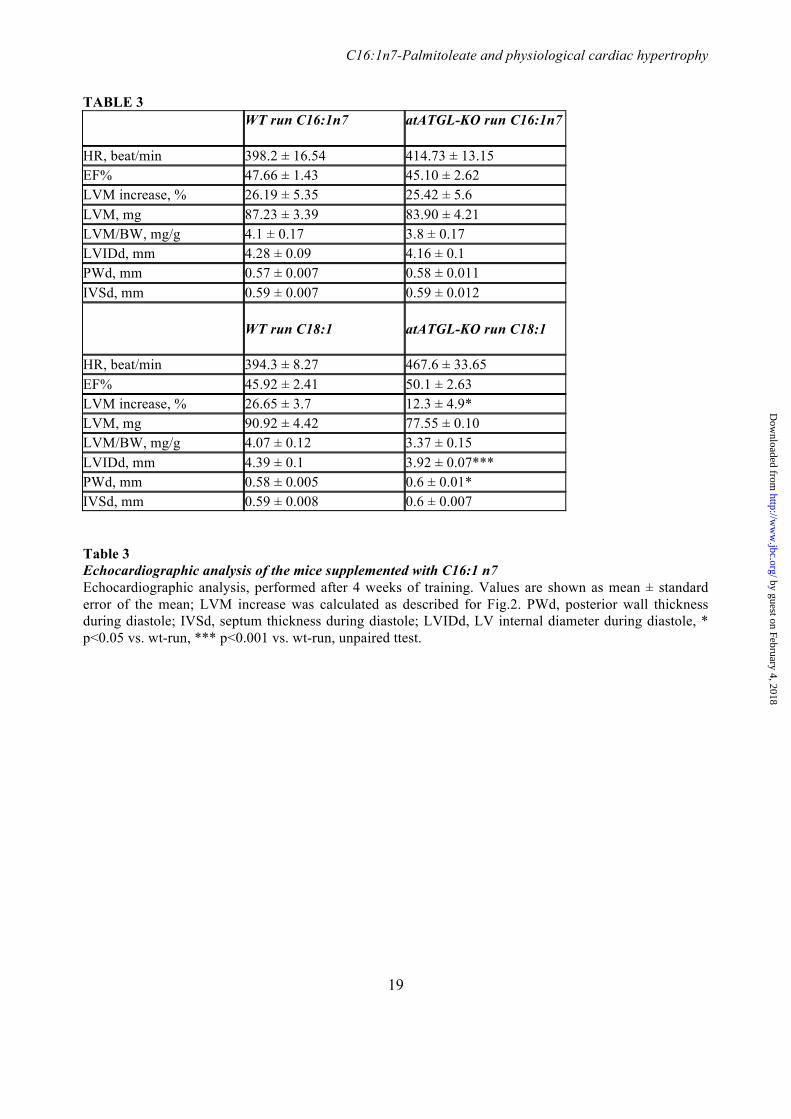
TABLE 3 WT run C16:1n7
atATGL-KO run C16:1n7
HR, beat/min 398.2 ± 16.54 414.73 ± 13.15 EF% 47.66 ± 1.43 45.10 ± 2.62 LVM increase, % 26.19 ± 5.35 25.42 ± 5.6 LVM, mg 87.23 ± 3.39 83.90 ± 4.21 LVM/BW, mg/g 4.1 ± 0.17 3.8 ± 0.17 LVIDd, mm 4.28 ± 0.09 4.16 ± 0.1 PWd, mm 0.57 ± 0.007 0.58 ± 0.011 IVSd, mm 0.59 ± 0.007 0.59 ± 0.012
WT run C18:1
atATGL-KO run C18:1
HR, beat/min 394.3 ± 8.27 467.6 ± 33.65 EF% 45.92 ± 2.41 50.1 ± 2.63 LVM increase, % 26.65 ± 3.7 12.3 ± 4.9* LVM, mg 90.92 ± 4.42 77.55 ± 0.10 LVM/BW, mg/g 4.07 ± 0.12 3.37 ± 0.15 LVIDd, mm 4.39 ± 0.1 3.92 ± 0.07*** PWd, mm 0.58 ± 0.005 0.6 ± 0.01* IVSd, mm 0.59 ± 0.008 0.6 ± 0.007 Table 3 Echocardiographic analysis of the mice supplemented with C16:1 n7 Echocardiographic analysis, performed after 4 weeks of training. Values are shown as mean ± standard error of the mean; LVM increase was calculated as described for Fig.2. PWd, posterior wall thickness during diastole; IVSd, septum thickness during diastole; LVIDd, LV internal diameter during diastole, * p<0.05 vs. wt-run, *** p<0.001 vs. wt-run, unpaired ttest.
by guest on February 4, 2018http://w
ww
.jbc.org/D
ownloaded from
C16:1n7-Palmitoleate and physiological cardiac hypertrophy
20
TABLE 4
Table 4 Echocardiographic analysis and fatty acid levels measured in athletes LVM, left ventricular mass, BSA, body surface area, IVSd, septum thickness during diastole; PWd, posterior wall thickness during diastole; IVSd, septum thickness during diastole; LVEDd, Left ventricle end-diastolic diameter; Values are shown as mean and min-max values.
parameters mean min-max
n=25 Age [years] 23 18 - 28 Height [cm] 181.5 170.4 – 188.5 Weight [kg] 76.7 61.4 – 88.3 LVM [g] 221.6 139.0 – 300.7 LVM/ BSA [g/m2] 112.4 72.5 – 145.1 IVSd [mm] 9.7 7.0 – 12.5 PWd [mm| 10.1 8.0 – 12.0 LVEDd [mm] 54.4 46.0 – 67.0 C16:1 n7 (palmitoleic acid) [µg/ml] 27.1 13.8 – 49.7 C18:2 n6 (linoleic acid) [µg/ml] 687.4 513.4 – 969.0
by guest on February 4, 2018http://w
ww
.jbc.org/D
ownloaded from
C16:1n7-Palmitoleate and physiological cardiac hypertrophy
21
by guest on February 4, 2018http://w
ww
.jbc.org/D
ownloaded from
C16:1n7-Palmitoleate and physiological cardiac hypertrophy
22
by guest on February 4, 2018http://w
ww
.jbc.org/D
ownloaded from
C16:1n7-Palmitoleate and physiological cardiac hypertrophy
23
by guest on February 4, 2018http://w
ww
.jbc.org/D
ownloaded from
C16:1n7-Palmitoleate and physiological cardiac hypertrophy
24
by guest on February 4, 2018http://w
ww
.jbc.org/D
ownloaded from
C16:1n7-Palmitoleate and physiological cardiac hypertrophy
25
by guest on February 4, 2018http://w
ww
.jbc.org/D
ownloaded from
C16:1n7-Palmitoleate and physiological cardiac hypertrophy
26
by guest on February 4, 2018http://w
ww
.jbc.org/D
ownloaded from
Blume, Martin Halle, Bernd Wolfarth, Erin E. Kershaw and Ulrich KintscherAnnelie Blumrich, Andreas Schirbel, Robert Klopfleisch, Michael Rothe, Katharina
Ban, Elzbieta Januszewicz, Janek Salatzki, Jana Grune, Anne-Kathrin Schwanstecher, Anna Foryst-Ludwig, Michael C. Kreissl, Verena Benz, Sarah Brix, Elia Smeir, Zsofia
Involving the Lipokine C16:1n7-PalmitoleateAdipose Tissue Lipolysis Promotes Exercise-Induced Cardiac Hypertrophy
published online August 10, 2015J. Biol. Chem.
10.1074/jbc.M115.645341Access the most updated version of this article at doi:
Alerts:
When a correction for this article is posted•
When this article is cited•
to choose from all of JBC's e-mail alertsClick here
by guest on February 4, 2018http://w
ww
.jbc.org/D
ownloaded from