Embed Size (px)
Citation preview

REVIEW
Microbiota and gut neuropeptides: a dual action of antimicrobialactivity and neuroimmune response
Julia Aresti Sanz1 & Sahar El Aidy1
Received: 2 October 2018 /Accepted: 10 March 2019 /Published online: 17 April 2019# The Author(s) 2019
AbstractThe gut microbiota is comprised of a vast variety of microbes that colonize the gastrointestinal tract and exert crucial roles for thehost health. These microorganisms, partially via their breakdown of dietary components, are able to modulate immune response,mood, and behavior, establishing a chemical dialogue in the microbiota–gut–brain interphase. Changes in the gut microbiotacomposition and functionality are associated with multiple diseases, in which altered levels of gut-associated neuropeptides arealso detected. Gut neuropeptides are strong neuroimmune modulators; they mediate the communication between the gut micro-biota and the host (including gut–brain axis) and have also recently been found to exert antimicrobial properties. This highlightsthe importance of understanding the interplay between gut neuropeptides and microbiota and their implications on host health.Here, we will discuss how gut neuropeptides help to maintain a balanced microbiota and we will point at the missing gaps thatneed to be further investigated in order to elucidate whether these molecules are related to neuropsychiatric disorders, which areoften associated with gut dysbiosis and altered gut neuropeptide levels.
Keywords Neuropeptides . Bacteria . Neuro-immune response . Gut-brain axis
Introduction
The gut microbiota comprises a complex community of met-abolically active microorganisms that have a strong influenceon a wide range of physiological processes, such as immuneand nervous system development, food metabolism, cellgrowth and differentiation, or mood and behavior (Dinanand Cryan 2017; El Aidy et al. 2013; El Aidy et al. 2016;Erny et al. 2015; Rowland et al. 2018; Taylor and Holscher2018). Though the composition of the gut microbiota isunique for each individual and therefore it is not possible todetermine what defines a Bhealthy microbiota,^ it seems clearthat alterations in the composition of the gut microbiota,known as dysbiosis, are associated with multiple diseases,
including neuropsychiatric disorders and inflammatory gas-trointestinal diseases (Koopman and El Aidy 2017;Pusceddu et al. 2018). Thus, it is of great significance to in-vestigate the mechanisms involved in the interplay betweenthe microbiota and the host.
One of the ways by which the gut bacteria establish theirintimate relationship with their host is through the productionof biologically active molecules. Bacteria produce these mol-ecules by breaking down dietary compounds that reach theintestine and/or the indigenously produced compounds thatare expelled from the intestine (Dodd et al. 2017; Strandwitz2018; Zhang and Davies 2016). Products of bacterial break-down range from immunomodulatory to antimicrobial andneuroactive compounds that not only have a local action onthe host but can also reach the blood stream to have an impacton distant parts of the body (Lyte and Cryan 2014).
Neuropeptides are now in the spotlight as one of the poten-tial mediators of the exchange of information between gutbacteria and other tissues and organs. Their role as modulatorsof neuronal and immune functions is well known and reveals astrikingly complex network through which neuropeptides ex-ert multiple functions. Although the term Bneuropeptide^ iscommonly used in the context of the central nervous system(CNS), the enteric nervous system (ENS) is another major
This article belongs to a Special Issue on Microbiome in Psychiatry &Psychopharmacology.
* Sahar El [email protected]
1 Department of Molecular Immunology and Microbiology,Groningen Biomolecular Sciences and Biotechnology Institute(GBB), University of Groningen, Nijenborgh 7, 9747AG Groningen, The Netherlands
Psychopharmacology (2019) 236:1597–1609https://doi.org/10.1007/s00213-019-05224-0

source of production of these peptides. Therefore, in this arti-cle, we will use the term Bgut neuropeptides^ to specificallyrefer to neuropeptides which are produced in the ENS, asopposed to other gut peptides which are also produced in theintestinal epithelium. This term also reinforces the idea thatgut neuropeptides are able to exert an extraintestinal action bysignaling to distant organs, such as the brain. Interestingly,several gut neuropeptides also have antimicrobial activity;for instance, neuropeptide Y (NPY) and substance P (SP),which have been shown to inhibit the growth of Escherichiacoli (Hansen et al. 2006). However, the antimicrobial activityof gut neuropeptides has not been studied in detail yet.Considering that gut antimicrobial neuropeptides are pro-duced not only in the CNS but also in the ENS and theirrespective receptors are widely expressed along the gastroin-testinal tract (GIT), it is important to unravel the function ofthese peptides in the context of gut microbiota homeostasis,which is key for a healthy state of the host. Finally, gut neu-ropeptides might also be key regulators of the so-called mi-crobiota–gut–brain axis; for example, through their receptorsexpressed on the vagus nerve, the main communication routebetween the gut and the CNS (Holzer and Farzi 2014). In thisreview article, we will describe the different types of antimi-crobial peptides (AMPs) that are present in the gut and theirmode of action. Thereafter, we will focus on gut neuropep-tides and will address their direct and indirect function inmaintaining homeostasis.
Antimicrobial peptides in the gastrointestinaltract
The GIT is a major entry point of microbes, and so, the organhas developed a complex defense mechanism to protect thehost from diseases, as part of the innate immune system. Oneof the components of this defense barrier is the AMPs. In theGIT, AMPs are produced not only by the intestinal epitheliumbut also by the gut microbiota in the lumen (Table 1). Aspresented in Table 1, human defense peptides have a ratherbroad spectrum of action, whereas their bacterial counterpartsdisplay a much narrower spectrum. This is due to a very spe-cific mode of action of bacterial AMPs where the antimicro-bial action takes place upon high-affinity binding to receptorsin the cell envelope (Martinez et al. 2016).
Antimicrobial peptides produced by the gutmicrobiota
Gut bacteria represent a major source of AMPs production inthe GIT. These bacteria synthesize the so-called bacteriocins.Similar to AMPs produced by human cells, bacteriocins aresmall, cationic peptides that can easily interact with bacterialmembranes. So far, 177 bacteriocins have been identified and
sequenced; 88% of them are produced by Gram-positive bac-teria, whereas the remaining 12% are produced by Gram-negative bacteria and Archaea. It is also remarkable that mostof the reported Gram-positive bacteriocins producers belongto the group of lactic acid bacteria, which carry out fermenta-tion of sugar into lactic acid (http://bactibase.hammamilab.org/statistics.php). However, it cannot be concluded thatlactic acid bacteria are the only bacteriocin producers;instead, given the interest that they pose for the foodindustry, it is likely that most of the research has beenfocused on this group of bacteria and accordingly morebacteriocins have been reported to be produced by lacticacid bacteria.
Bacteriocins are classified into many different subgroupsdue to the heterogenicity of this group of molecules.Bacteriocins that are produced by Gram-negative bacteriaare referred to as microcins, small peptides, or colicins, whichare larger proteins. Microcins are subsequently divided intoclass I (< 5 kDa, containing post-translational modifications)and class II (5–10 kDa, without post-translational modifica-tions) (Hassan et al. 2012). Despite their structural diversity,microcins share an interesting mechanism of action that hasbeen named the BTrojan Horse^ strategy. Some microcinssuch asMccJ25 andMccE492 mimic the structure of essentialbacterial molecules and take advantage of the natural recep-tors for these ligands, which allow them to enter and kill thetarget bacteria. On the other hand, MccC7 and MccC59 aresecreted as harmless molecules and further transformed intotoxic derivatives once they enter the susceptible bacteria(Duquesne et al. 2007). An interesting example of howmicrocins are relevant for the host was shown recently usinga mouse model of intestinal inflammation. In the study ofSassone-Corsi et al., probiotic E. coli Nissle 1917, which pro-duces microcins, was able to restrict the expansion of othercompeting Enterobacteriaceae during inflammation, includingpathogenic E. coli and Salmonella enterica. Also, therapeuticadministration of E. coli Nissle was sufficient to effectivelydisplace enteric pathogens from their niche due to their pro-duction of microcins (Sassone-Corsi et al. 2016). The secondsubgroup of Gram-negative bacteriocins, colicins, are largerpeptides produced by some strains of E. coli and other relatedEnterobacteriaceae. They are active against E. coli and closelyrelated bacteria such as Salmonella (Braun et al. 1994).Similarly to microcins, colicins are quite a diverse group thatincludes up to 30 types of proteins which slightly differ inlethal activity and mode of action (Smarda and Smajs 1998).The mechanism by which colicins kill bacteria has been ex-tensively studied and consists of three main steps. Initially,colicins bind to the outer membrane proteins of the target cellthrough the receptor binding domain. Then, the translocationdomain located in the N-terminus of the protein allows coli-cins to enter the target cell: depending on which protein com-plex they interact with, colicins are subdivided into Tol-
1598 Psychopharmacology (2019) 236:1597–1609

dependent or Ton-dependent colicins. These periplasmic pro-tein complexes allow colicins to translocate and reach theinner membrane where the killing activity takes place by dif-ferent mechanisms: pore formation, DNAse activity, or inhi-bition of protein synthesis (Cascales et al. 2007). Interestingly,a very recent study analyzed clinical isolates of E. coli fromthe intestinal mucosa of patients suffering from inflammatorybowel disease (IBD) and found a higher frequency ofbacteriocinogeny when compared to healthy subjects. Thestudy found that IBD E. coli strains had a higher prevalence
of virulence determinants when compared to healthy controls,suggesting that IBD E. coli strains closely resemble their path-ogenic counterparts. One of these determinants was group Bcolicins, which are actually encoded in plasmids containingadditional virulence genes. This association between IBD andgroup B colicins could therefore reflect additional virulencedeterminants present in the colicin plasmids highly prevalentin IBD (Micenková et al. 2018). The use of recombinant co-licins directed to specific pathogenic E. coli strains present inIBD has been proposed as a therapeutic method for IBDs
Table 1 Antimicrobial peptides in the gastrointestinal tract. Summary of host-derived and microbiota-derived antimicrobial peptides includingproducing cell types and antimicrobial spectrum
Source AMP family Cell type Antimicrobial activity References
Microbiota Lantibiotics Gram-positive bacteria S. pneumoniae, S. aureus, S. pneumoniae,C. difficile, E. faecium
(Dawson 2007)
Microcins Gram-negative bacteria E. coli, S. enterica, E. cloacae,K. pneumoniae, Citrobacter, Shigella
(Duquesneet al. 2007)
Host Defensins Paneth cells, monocytes, macrophages,T and B cells, dendritic cells
S. aureus, P. aeruginosa, E. coli,L. monocytogenes, S. typhimurium,C. difficile, C. albicans, B. fragilis,E. faecalis, S. pyogenes, E. faecium,S. cerevisiae, S. pneumonia, B. cepacia,HPV, HIV, influenza virus
(Sivieri et al.2017)
PhospholipaseA2
Paneth cells E. coli, S. typhimurium, L. monocytogenes,S. aureus, B. anthracis, P. aeruginosa,B. subtilis
(Timo J.Nevalainenet al. 2008)
Cathelicidins Enterocytes, macrophages, epithelial cells E. coli, K. pneumonia, P. aeruginosa,N. gonorrhoeae, Streptococcus sp.,H. pylori, Shigella sp., Salmonella sp.,C. albicans
(Sivieri et al.2017)
RegIII lectins Enterocytes, enteroendocrine cells L. monocytogenes, Y. pseudotuberculosis,C. rodentium, S. enteritidis, S. enterica,Enterococcus, C. butyricum, L. reuteri,E. coli, Bacteroides spp.
(Miki et al.2017)
Host neuropeptides NPY Enteric neurons, neutrophils, monocytes,macrophages, fibroblasts
E. coli, E. faecalis, Candida spp.,P. aeruginosa, S. mutans, L. acidophilus,L. major, M. catarrhalis, H. influenza,A. caviae, A. actinomycetemcomitans
(Augustyniaket al. 2012)
SP Enteric neurons, neutrophils, monocytes,macrophages, lymphocytes B, lymphocytesT, dendritic cells, natural killers, mast cells,fibroblasts
E. coli, C. albicans, P. aeruginosa,S. mutans, L. acidophilus, K. pneumoniae,E. faecalis, P. vulgaris, M. catarrhalis,H. influenza, S. aureus,A. actinomycetemcomitans
(Augustyniaket al. 2012)
α-MSH Enteric neurons, neutrophils, monocytes,macrophages, lymphocytes B, lymphocytesT, dendritic cells, natural killers, mast cells
Escherichia coli, Staphylococcus aureus,Candida albicans, Trypanosoma brucei
(Augustyniaket al. 2012)
CGRP Enteric neurons, macrophages, lymphocytes T,dendritic cells
E. coli,C. albicans, P. aeruginosa, S. aureus,E. faecalis, S. mutans, L. acidophilus,M. catarrhalis, H. influenzae
(Augustyniaket al. 2012)
AM Enteric neurons, neutrophils, macrophages,mast cells
B. fragilis, E. coli (Allaker et al.1999;Augustyniaket al. 2012)
VIP Enteric neurons, monocytes, macrophages,dendritic cells, T lymphocytes, Blymphocytes, mast cells
T. brucei (Augustyniaket al. 2012;Delgadoet al. 2009)
Psychopharmacology (2019) 236:1597–1609 1599

(Kotłowski 2016). Thus, genetically modified E. coli Nissle1917 could be used as a vehicle to introduce specific colicinsin the GIT that would reinforce the antimicrobial action thatthis strain exerts through the production of microcins(Kotłowski 2016). However, it is important to note that thishypothesis has not been yet tested in the laboratory, so in vitroand in vivo experiments are needed to confirm the potential ofcolicins as therapeutic agents in intestinal inflammation.
Bacteriocins produced by Gram-positive bacteria are divid-ed into lantibiotics and non-lantibiotics. Lantibiotics consti-tute the Gram-positive counterpart of microcins; they aresmall peptides, below 5 kDa, and contain unusual post-translationally modified residues such as lanthionine and 3-methly-lathionine (Perez et al. 2014). The first lantibiotic everdiscovered was Nisin A (Rogers and Whittier 1928), and a lotof studies are still being done on how to genetically modifythis peptide to create derivatives improving its antimicrobialproperties (Field et al. 2012; Li et al. 2018). Lantibiotics arequite unique molecules; they present unusual thioether-containing amino acid residues that form ring structures with-in the peptides (Jack and Jung 2000), which is probably linkedto their complex mechanism of action. The mode of action ofthese antimicrobials was for a long time thought to be restrict-ed to the pore formation mechanism of the prototypiclantibiotic nisin (Garcerá et al. 1993). However, upon struc-tural derivatization, new insights reveal a complex set ofmodes of action that are combined to eventually disrupt bac-terial growth. For instance, nisin takes the pore formationprocess one step further, increasing the specificity and stabilityof the pore through the interaction with lipid II. Lipid II is amolecule that is anchored to cell membranes and acts as aprecursor in the synthesis of peptidoglycan by translocatingacross the membrane the building blocks required for its syn-thesis (van Heijenoort 2007). By binding to lipid II, nisin isable to greatly improve the pore-forming efficiency (Breukinket al. 1999) and this mechanism has also been described inother lantibiotics, such as the two-peptide lantibiotic lacticin3147 (Wiedemann et al. 2006) and subtilin (Parisot et al.2008). Interestingly, the interaction with lipid II has anothereffect that also increases the potency of these molecules asantibiotics. As lipid II is a precursor in the synthesis of pepti-doglycan, the interaction with nisin sequesters lipid II from itsnormal function and results in the inhibition of cell membranebiosynthesis, which creates the dual effect of this lantibiotic(Wiedemann et al. 2001).
In a similar way to microcins, Gram-positive bacteriocinsalso have great therapeutic potential. For example, enterococ-cal bacteriocin 21 might constitute an effective approach todecolonize antibiotic-resistant enterococci from the gut. In arecent study, commensal Enterococcus faecalis deliveringbacteriocin 21 was used to specifically disrupt the growth ofmultidrug-resistant enterococci during infection in the mam-malian GIT. Thus, a particular niche from the gut microbiota
was inhibited without altering the rest of the microbial popu-lation, which poses a useful strategy to treat infections causedby antibiotic resistant strains that might otherwise be verychallenging to cure (Kommineni et al. 2015).
Antimicrobial peptides produced by the intestinalmucosa
The intestinal epithelium provides the first line of defenseagainst microorganisms that reach the gut lumen and can bepathogenic for the host. Intestinal epithelial cells are mainlyenterocytes, but they also comprise enteric neurons,enteroendocrine cells, tuft cells, and secretory cells, whichinclude goblet and Paneth cells. Goblet cells produce mucinand Paneth cells secrete AMPs, creating a physical and bio-chemical barrier that ensures the proper segregation betweenhost and microorganisms, helping to avoid infections (Allaireet al. 2018). It must be noted, however, that Paneth and gobletcells are not the only source of host-derived AMPs. Theseepithelial secretory cells are aided by classic immune cellslocated in the lamina propria, which include lymphocytes,macrophages, and dendritic cells for the production ofAMPs (Charles et al. 2001).
So far, five different AMPs and proteins have been found tobe produced by Paneth cells: defensins, cathelicidins, lysozymeC, phospholipase A2, and REGIIIα/β/γ. Lysozyme C, a gly-coside hydrolase that cleaves specific residues of peptidoglycancausing lysis of the bacterial membrane, was firstly found to beexpressed in Paneth cells (Mason and Taylor 1975).Phospholipase A2 was also found to be synthesized in Panethcells (Nevalainen et al. 1995) and described as a bactericidalagent against E. coli and Listeria monocytogenes. Furthermore,Paneth cells produce REGIIIα, a C-type lectin also known asHIP/PAP (hepatocarcinoma–intestine–pancreas/pancreatic-as-sociated protein), which is produced in enteroendocrine cellsas well (Lasserre et al. 1999). REGIIIα, as many other antimi-crobial peptides, is able to kill bacteria by formation of apermeabilizing transmembrane pore, although this mechanismis inhibited by lipopolysaccharides (LPS), which is why thispeptide is only effective against Gram positive-bacteria(Mukherjee et al. 2013). Additionally, REGIII peptides playan important role during the initial establishment of the gutmicrobiota in germ-free mice. Indeed, REGIIIγ and REGIIIβexpressions have been shown to peak during the process of gutbacterial colonization in both the small intestine and the colonof mouse, coinciding with induced expression of innate im-mune molecules phospholipase A2 (Pla2g2a) and resistin-likebeta (Retnlβ) in the colon (El Aidy et al. 2012). Later on, theexpression of REGIII peptides in the colon returned to basallevels detected in germ free animals. This suggests that REGIIIpeptides exert an antimicrobial function in the small intestine,while Pla2g2a and Retnlβ exert this antimicrobial function inthe colon (El Aidy et al. 2012).
1600 Psychopharmacology (2019) 236:1597–1609
The major AMP components in the intestinal mucosa aredefensins. So far, ten defensins have been identified and di-vided in two groups according to structural features: humanα-defensins (HDs) and human β-defensins (HBDs). Defensinsare small, cationic peptides with a rather broad spectrum ofantimicrobial activity in vitro and they exert their function bycreating micropores in the bacterial membranes that cause theleak of the cell’s content, loss of structure, and eventual celldeath (Cobo and Chadee 2013; Cunliffe 2003). They are syn-thesized as propeptides and are subsequently cleaved by pro-teases to form the mature, active forms of the peptides (Valore& Valore and Ganz 1992). HDs are produced by Paneth cellsand are therefore in charge of protection in the small intestine,whereas HBDs play the same role in the colon, where they aresecreted by epithelial cells. Additionally, HDs are produced byimmune cells including monocytes, macrophages, T and Bcells, which also produce HBDs together with dendritic cells(Dutta and Das 2016).
The other family of AMPs produced in the GIT by the hostis cathelicidins. Among cathelicidins, only LL-37 is expressedin humans. Cathelicidins and defensins share some commonfeatures; LL-37 is also synthesized as a propeptide, which islater cleaved by a protease to release the mature peptide withantimicrobial activity (Zaiou and Gallo 2002). Furthermore,the mode of action of LL-37 is also based on bacterial mem-brane disruption by pore formation (Xhindoli et al. 2016).However, in contrast to defensins, which are only expressedin the small intestine, LL-37 expression in this region of theintestinal tract is rather low, while it is highly abundant in thecolon (Schauber et al. 2003), where it is secreted bycolonocytes (Hase et al. 2002).
Antimicrobial gut neuropeptides
There is increasing evidence that gut neuropeptides are one ofthe axes of communication between the gut microbiota andthe host (El Aidy et al. 2016) and they might also be playing arole as antimicrobial agents. Gut neuropeptides are structural-ly similar to regular antimicrobial peptides; they are small (<10 kDa), cationic, and amphipathic molecules and also sharesimilarities with other AMPs in their mode of action. Eventhough their antimicrobial properties have not yet been studiedin detail, there are multiple indications that gut neuropeptidessuch as NPY, SP, α-melanocyte stimulating hormone (α-MSH), vasoactive intestinal peptide (VIP) calcitonin gene-related peptide (CGRP), and adrenomedullin (AM) might behaving an important role in the regulation of the gut microbi-ota composition.
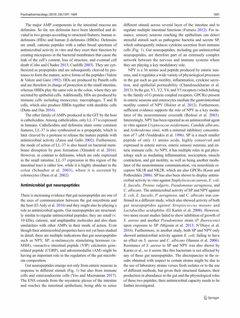
Gut neuropeptides emerge not only from enteric neurons inresponse to different stimuli (Fig. 1) but also from immunecells and enteroendocrine cells (Yoo and Mazmanian 2017).The ENS extends from the myenteric plexus of the intestineand reaches the intestinal epithelium, being able to sense
different stimuli across several layer of the intestine and toregulate multiple intestinal functions (Furness 2012). For in-stance, sensory neurons reaching the epithelium can detectstressful stimuli such as pathogenic bacteria and secrete SP,which subsequently induces cytokine secretion from immunecells (Fig. 1). Gut neuropeptides, including gut antimicrobialneuropeptides, are therefore part of an extremely complexnetwork between the nervous and immune systems wherethey are playing a key modulatory role.
NPY is a 36 amino acid peptide produced by enteric neu-rons, and it regulates a wide variety of physiological processesin the gut such as gut motility, inflammation, cytokine secre-tion, and epithelial permeability (Chandrasekharan et al.2013). In the gut, Y1, Y2, Y4, and Y5 receptors (which belongto the family of G protein coupled receptors, GPCRs) presentin enteric neurons and enterocytes mediate the gastrointestinalmotility control of NPY (Holzer et al. 2012). Furthermore,sufficient evidence supports the role of NPY as a key modu-lator of the neuroimmune crosstalk (Bedoui et al. 2003).Interestingly, NPY has been reported as an antimicrobial agentin vitro against Cryptococcus neoformans, Candida albicans,and Arthroderma simii, with a minimal inhibitory concentra-tion of 7 μM (Vouldoukis et al. 1996). SP is a much smallerpeptide of only 11 amino acids, highly conserved andexpressed in enteric nerves, enteric sensory neurons, and en-teric immune cells. As NPY, it has multiple roles in gut phys-iology such as mediating inflammation, nociception, musclecontraction, and gut motility, as well as being another modu-lator of the neuroimmune communication, via neurokinin re-ceptors NK1R and NK2R, which are also GPCRs (Koon andPothoulakis 2006). SP has also been shown to display antimi-crobial activity in vitro against Staphylococcus aureus, E. coli,E. faecalis, Proteus vulgaris, Pseudomonas aeruginosa, andC. albicans. The antimicrobial activity of SP and NPYagainstE. coli, E. faecalis, P. aeruginosa, and C. albicans was con-firmed in a different study, which also showed activity of bothgut neuropeptides against Streptococcus mutans andLactobacillus acidophilus (El Karim et al. 2008). However,two more recent studies failed to show inhibition of growth ofS. aureus and another Pseudomonas strain (P. fluorescens)upon exposure to SP (Mijouin et al. 2013; N’Diaye et al.2016). Furthermore, in another study, both SP and NPY onlyshowed antimicrobial activity against E. coli, failing to havean effect on S. aureus and C. albicans (Hansen et al. 2006).Resistance of S. aureus to SP and NPY was also shown byKarim et al., so it seems like this bacterium is not affected byany of these gut neuropeptides. The discrepancies in the re-sults obtained with respect to certain strains might be due tothe use of laboratory strains versus fresh isolates or to the useof different methods, but given their structural features, theirproduction in abundance in the gut and the physiological rolesof these two peptides, their antimicrobial capacity needs to befurther investigated.
Psychopharmacology (2019) 236:1597–1609 1601

Another important gut neuropeptide isα-MSH, a 13 aminoacid peptide that emerges from the post-translational process-ing of POMC (proopiomelanocortin). As other gut neuropep-tides, it is an important neuroimmune modulator being a po-tent antiinflammatory molecule, which allows it to regulateintestinal permeability (Váradi et al. 2017). α-MSH also ex-erts its function through another member of the GPCR familyof receptors; melanocortin receptors (MCRs) bind α-MSHtriggering different signaling pathways that regulate a varietyof functions. In the gut, MC1R and MC3R induction by α-MSH modulates its antiinflammatory response through cyclicadenosine 3′,5′-monophosphate (cAMP) signaling (Singh andMukhopadhyay 2014). α-MSH antimicrobial activity wasfirst shown against E. coli, C. albicans, and S. aureus(Cutuli et al. 2000), which was later extended toCryptococcus neoformans (Masman et al. 2006) andC. vaginitis (Catania et al. 2005).VIP is another gut neuropep-tide which has been shown to display promising antimicrobial
activity, at least against some pathogens (El Karim et al.2008). VIP is a 28 amino acid peptide that is also present inthe GIT, where it regulates different function through theVPAC2 receptors, which belong to GPCR family too. Apartfrom being an important immune regulator, VIP regulates va-sodilatation in the gut, as well as motility (Mario Delgado andGanea 2013). Regarding its antimicrobial activity, VIP wasshown to be effective in killing S. mutans, E. coli,P. aeruginosa, and C. albicans in vitro. Additionally, VIPwas also able to kill the pathogen Trypanosoma brucei inanother in vitro study (M. Delgado et al. 2009). However,not many follow-up studies on the antimicrobial propertiesof VIP are available. Instead, a few studies with syntheticanalogues of the peptide with improved stability have shownenhanced antimicrobial activity of VIP. Indeed, modified VIPpeptides were able to kill S. mutans, Micrococcus luteus, andpathogenic E. faecalis, while the native peptide was ineffec-tive against these bacteria against S. aureus and E. coli
Fig. 1 Direct and indirect effectsof gut neuropeptides in the GIT.Upon sensing of stressful stimuli,enteric neurons release gutneuropeptides that induce aresponse in innate and adaptiveimmune cells, which also secretethese peptides, resulting in astrong response to bacterialimbalance. Additionally, if gutneuropeptides cross the epithelialbarrier, they could exert a directantimicrobial activity in theintestinal lumen by differentkilling mechanisms
1602 Psychopharmacology (2019) 236:1597–1609

compared to the native form of the peptide (Campos-Salinaset al. 2014). Using also analogues of VIP, Xu et al. were ableto demonstrate antimicrobial activity against S. aureus andE. coli that was increased when compared to the naturalform of the peptide (Xu et al. 2017). Finally, CGRP is an-other gut neuropeptide that has been investigated as an an-timicrobial peptide. In its mature form, it is composed of 37amino acids and has two splicing variants; α-CGRP and β-CGRP. α-CGRP is predominantly expressed not only in theCNS but also in the peripheral nervous system, whereas β-CGRP is mostly found in the ENS. This peptide causesvasodilation in the GIT and has a protective role againstischemia (Ma 2016). Regarding its antimicrobial activity,CGRP is able to affect the growth of E. coli, C. albicansand P. aeruginosa (El Karim et al. 2008), therefore havingquite a restricted spectrum of action, as compared with theother gut neuropeptides discussed before. Within the CGRPfamily of peptides, it is also important to mention AM. This52 amino acid peptide is generated by post-translational en-zymatic processing of preproadrenomedullin, which also re-sults in the formation of its gene-related peptideproadrenomedullin N-terminal peptide (PAMP) (Bełtowskiand Jamroz 2004). AM shares structural and functional sim-ilarities with CGRP, such as the presence of an internalmolecular ring and a central helical region for receptor bind-ing, and both peptides exert their functions through the cal-citonin receptor-like receptor (CRLR) (Hay and Walker2017; Pérez-Castells et al. 2012). AM and PAMP are foundacross the entire GIT, being specially abundant in neuroen-docrine cells, and they regulate growth of the intestinal ep-ithelium, water, and ion transport in the colon, intestinalmotility, and vasodilatation (Martínez-Herrero and Martínez2016). Antimicrobial properties have also been described forboth peptides; antimicrobial activity of AM was initiallyshown against a range of bacteria known to be membersof the microbiota of different mucosal surfaces, includingintestinal mucosa. AM was able to inhibit the growth ofBacteroides fragilis and E. coli (Allaker et al. 1999), andthis was extended in a later study to PAMP, which wasactually shown to be even more potent than AM againstE. coli (Marutsuka et al. 2001).
As shown in Table 1, the antimicrobial activity of gut neu-ropeptides has mainly been tested against pathogenic bacteria.Although of high relevance, it is still necessary to study anti-microbial activity of gut neuropeptides on commensal bacte-ria. Not only inflammation can be caused by colonization ofpathogens, but also certain strains of commensal bacteria,such as E. coli, can trigger the production of cytokines andinduce an inflammatory state in the gut that can translate to thebrain. (Kittana et al. 2018). Therefore, the role of these pep-tides as antimicrobial agents against commensal strains shouldbe further explored in order to understand their potential im-pact in the host health.
Modes of action of antimicrobial gutneuropeptides
As mentioned before, the common structural features of hostdefense peptides and antimicrobial gut neuropeptides allowthe latter to exert their function through the same mechanismsof action on the target microorganisms. These include mem-brane disruption, interference with cell division, and metabo-lism or disruption of ATP synthesis among others.Furthermore, gut neuropeptides have an additional capacityof interacting with the neuro- and immune systems, whichultimately causes the release of other molecules with antimi-crobial activity. This combined action raises a great potentialfor gut neuropeptides to be used with therapeutic purposes inorder to treat diseases associated with microbiota alterations,especially in light of the increasing resistance to conventionalantibiotics that is putting at risk the use of these drugs.
Direct antimicrobial effects of gut neuropeptides
The mechanism of action of antimicrobial peptides has beenstudied in depth mostly in host defense peptides, but the struc-tural similarities of gut neuropeptides, which are also small,cationic molecules, allow them to exert their function in asimilar way. The predominant antimicrobial mode of actionof gut neuropeptides consists of binding and disruption of thebacterial cell wall. Indeed, membrane disruptive mechanismshave been shown to drive the action of NPY (Thomas et al.2005), α-MSH (Madhuri et al. 2009), and AM (Allaker et al.2006), all of which contain a rich combination of positivelycharge amino acids and hydrophobic residues in their C-terminal fragment. However, non-disruptive mechanisms alsoexist and will be discussed later on.
Membrane-disruptive peptides usually form α-helicalstructures with accumulation of positively charged residuestowards the C-terminal side of the peptide (Powers andHancock 2003). This allows the initial step to approach theirtarget, which occurs through electrostatic interactions of suchresidues with the anionic LPS that coat the outer bacterialmembrane. By displacement of Mg2+ and Ca2+ ions that areusually interacting with LPS, antimicrobial peptides createlocal disturbances in the membrane, allowing their transloca-tion and making the inner cytoplasmic membrane accessible(Hancock and Chapple 1999). The next step consists of thereorientation of the peptide to interact with the inner mem-brane. Different mechanistic models have been proposed forthe interaction of these peptides with inner bacterial mem-branes, and although, it is not yet clear which is correct, theyall lead to the disruption and depolarization of the membrane,which rapidly causes cell death (Sato and Feix 2006). It is easyto speculate that there might not be one unique model to ex-plain the mode of action of these peptides, but instead, they
Psychopharmacology (2019) 236:1597–1609 1603

might all be correct depending on the physicochemical prop-erties of each antimicrobial peptide.
Interestingly, another rather non-traditional mode of actionhas been described to take place in the trypanolytic activity ofgut neuropeptides AM and VIP. Trypanosomiasis is known tocause intestinal damage in humans, as an inflammatory state isinduced upon the invasion of this parasite with increased cy-tokine levels and intestinal permeability (Ben-Rashed et al.2003). Studying the antimicrobial activity of different gut neu-ropeptides on T. brucei, an unusual antimicrobial mechanismwas found in which the peptides are subjected to endocytosisthrough the flagellar pocket of this microorganism. Next, traf-ficking of the peptides through the endosomal network allowsthem to reach the lysosomes, where they disrupt endosome–lysosome vesicles. Disruption of these membranes causes therelease of the gut neuropeptides to the cytosol, together withglycolytic enzymes, which leads to a metabolic failure andfinally to cell death (Delgado et al. 2009).
Finally, AM has also been shown to have a distinct, unusu-al mechanism of action against S. aureus. Staphylococci divi-sion occurs though the inwards growth of the peripheral cellwall and formation of a transverse cross wall, known as sep-tum. Then, the newly synthesized peptidoglycan undergoeslocalized hydrolysis that results in complete cell separation(Giesbrecht et al. 1998). In S. aureus, AM has been shownto disrupt the formation of the septum (Allaker et al. 2006),which is known to lead to cell death in other organisms.
Even though these results are promising and presentgut neuropeptides as potential modulators of gut microbi-ota homeostasis by direct antimicrobial activity, somequestions need to be addressed before they can be imple-mented as therapeutic agents. The studies discussed aboveare in vitro studies, and it is not yet clear whether thesegut neuropeptides reach the gut microbiota in sufficientamounts. For them to be effective in direct killing of bac-teria, gut neuropeptides should be able to reach the gutlumen or areas in close proximity to the intestinal epithe-lium, where they may exert a very important role in gutmicrobiota homeostasis. Another concern for their poten-tial therapeutic implementation is that, as mentioned be-fore, most of the studies so far have tested antimicrobialactivity against pathogenic bacteria, which leaves com-mensal bacteria unexplored. It would therefore be crucialto unravel whether these peptides can keep infections un-der control without altering the composition of commen-sal bacteria residing in the GIT. In light of the growingresistance of pathogens to the currently used broad-spectrum antibiotics, exploring the therapeutic possibili-ties of gut neuropeptides is extremely significant; thesepeptides could be active against resistant strains and theirseemingly restricted spectrum of action would not influ-ence other untargeted bacteria, reducing the possibilitiesof developing resistance.
Indirect antimicrobial effects of gut neuropeptides
Besides their antimicrobial action on microbes, gut neuropep-tides have much more sophisticated roles and probably themore important antiinfective functions of these molecules relyon their neuroimmune modulatory properties. Such featureshave allowed the host to develop a complex and intrinsicnetwork of defense mechanisms to protect itself from micro-bial invasion. Gut neuropeptides respond to gut microbiotaalterations by mediating the so called neurogenic inflamma-tion (Houser and Tansey 2017). At the same time,neuroimmune modulation by gut neuropeptides translates tochanges in the ENS, which can eventually reach the brain andimpact mood and behavior. Thus, inflammation in the GITresults in changes in ENS, such as increased number of entericneurons, altered levels of gut neuropeptides and changes in themotor circuits of the intestine (Margolis and Gershon 2016).
Nociceptors, which are specialized peripheral sensory neu-rons, respond to damaging stimuli in the GITsuch as infectionby releasing gut neuropeptides. SP, CGRP, AM, and NPYarereleased from these neurons and mediate neurogenic inflam-mation as a defense mechanism for the host. This way, duringthe effector phase of inflammation, sensory neurons produceneuropeptides that promote proliferation and migration of im-mune cells, guiding them to the specific site of damage (Chiuet al. 2012). NPY, SP, and CGRP are able to stimulate therelease of proinflammatory cytokines, chemokines, and ara-chidonic acid from immune and non-immune cells, while VIP,α-MSH, and AM seem to have the opposite effect. Altogether,they mediate neurogenic inflammation (Fig. 1) (Mitchell andKing 2010; Souza-Moreira et al. 2011). A detailed descriptionof the action of neuropeptides on the immune system is pro-vided in the review articles by Souza-Moreira et al. andMitchell et al. Interestingly, one of the properties that makegut neuropeptides such potent mediators of the inflammatoryresponse is their often synergetic activity with other mole-cules, which allows them to orchestrate an intense and effi-cient response. For instance, SP and CGRP have been shownto act jointly on peripheral blood mononuclear cells (PBMC).This synergetic effect is in line with the additive effectsthat these peptides are known to have in other physiolog-ical processes involved in the inflammatory response; va-sodilatation, vascular permeability, and hyperalgesia(Cuesta et al. 2002). SP has been shown to enhance theresponse of macrophages and monocytes by being able tostrongly increase the release of cytokines and chemokinesfrom these cells in response to LPS (Sipka et al. 2010).Another example of this coordinated action with othermolecules is VIP, which is able to facilitate the bacteri-cidal activity of human cathelicidin LL-37; when used incombination, VIP and LL-37 can keep their antimicrobialactivity even at physiological concentrations of NaCl, atleast against E. coli and P. aeruginosa (Ohta et al. 2011).
1604 Psychopharmacology (2019) 236:1597–1609

The complex picture of gut neuropeptide modulation of theimmune response through their capacity to regulate innate andadaptive immune response via different receptors gives rise to agreat variety of modes of action. For example, activation ofneutrophil receptor Y5 by NPY potentiates a respiratory burstresulting in an increase of reactive oxygen species (ROS), oneof the critical functions of neutrophils. Conversely, when NPYbinds to Y1 and Y2 receptors, phagocytosis of E. coli isinhibited. Furthermore, the effect of this gut neuropeptide onneutrophils is also concentration-dependent; at low doses, NPYpromotes phagocytosis, whereas at higher doses phagocytosisis inhibited and the respiratory burst is potentiated to eliminatethe pathogen (Bedoui et al. 2008). On the other hand, a recentstudy using a SP agonist showed that stimulation of NK1R ofdendritic cells promotes the maturation of these cells togetherwith decreased secretion of IL-10 (Janelsins et al. 2013).Moreover, in line with its antiinflammatory effect describedbefore, α-MSH drives the maturation of macrophages by inter-action with MC1R receptors in these cells. Interestingly, thisprocess has been specifically shown to kill C. albicans in a ratvaginitismodel, again through cAMP signaling (Ji et al. 2013).CGRP is also able to regulate cells of the innate immune sys-tem. For instance, binding of the peptide to CRLR of macro-phages induces IL-6 and TNF-α (Fernandez et al. 2001).
Regarding the interaction of gut neuropeptides with adap-tive immune cells, SP has been reported to induce maturationof human memory CD4+ T cells into Th17 cells upon induc-tion of IL-1β production in monocytes, an effect that occursupon induction of NK1R in these cells (Cunin et al. 2011). α-MSH is also able to upregulate cytokine IL-10 in dendritic cellsvia MC1R, leading to the induction of regulatory T cells andeventual inhibition of effector T cells which points to an im-munosuppressive phenotype (Auriemma et al. 2012). Anotherexample is the ability of NPY to induce chemotaxis, adhesionto epithelial cells, and transepithelial migration of dendriticcells through Y1 receptor activation (Buttari et al. 2014).
In summary, gut neuropeptides allow the host to have astrong response to bacterial infections; damaging stimuli aresensed by sensory neurons, which in turn secrete these peptidesthat can directly act on microbes and at the same time induceinflammation through the interaction with immune cells in theintestinal mucosa. However, this complicated response has itsimplications on the GIT as it involves neuropeptide-mediatedgastrointestinal inflammation on the ENS. An inflammatorystate of the bowel induces severe changes in the ENS that resultin gastrointestinal dysfunction. It has been shown that inflam-mation causes multiple changes in the intrinsic circuitry of theENS, such as neuronal hyperexcitability, increased synapticfacilitation, and decreased inhibitory neuromuscular transmis-sion (Krauter et al. 2007; Linden et al. 2004; Strong et al.2010). This profound neuronal alteration ultimately leads tomajor, long-lasting disruption of the intestinal motor activity.Interestingly, bowel dysfunction is observed not only at the site
of inflammation but also in non-inflamed regions of the gut,which might be explained by the long lasting alteration ofenteric neuronal circuits. Altered motility and secretion canbe found at distant, non-inflamed regions of the GIT, as shownin an experimental model of colitis in guinea pigs (Hons et al.2009). In this study, non-cholinergic secretion was found to bedecreased in the ileum where no inflammation was observed,and this change was associated with a decrease in excitatorysynaptic transmission in secretomotor neurons. In contrast, il-eal cholinergic neurons were more excitable, while action po-tentials in primary afferent neurons were broader than in anormal, non-inflamed state (Hons et al. 2009). Although themechanism behind the distant effect of inflammation on non-affected regions of the gut is not yet understood, it seems clearthat altered enteric neuronal function caused by neuropeptide-mediated inflammation can have effects in other regions of theGIT and this might be explained by a neuronal-mediatedspread of the inflammation, resulting in profound and longlasting changes in the ENS across the whole bowel.
Conclusion
The gut microbiota is emerging lately as a key regulator ofhost health and disease. However, the mechanisms employedby these microbes to communicate with distant organs such asthe brain are only beginning to be understood and antimicro-bial gut neuropeptides are likely be involved in this process.Gut neuropeptides have the capacity to regulate gut microbi-ota homeostasis through both direct antimicrobial effects andneurogenic inflammation and therefore should be consideredas potential therapeutic targets for diseases where this functionis affected. Gut neuropeptides do seem to have a rather narrowspectrum as antimicrobial agents compared to defense pep-tides produced by Paneth cells. However, they should not beunderestimated as potential alternatives to classic antibiotics,given their potent effects as neuroimmune modulators.
An important reason to further investigate the role of gutneuropeptides on intestinal homeostasis, possibility via an ef-fect on gut microbiota composition, has to do with the fact thatseveral neuropsychiatric disorders, which are associated withdysbiosis in the gut and intestinal inflammation, have alsobeen described to be associated with altered levels of (gut)neuropeptides, though it is not yet clear whether disruptedlevels of these neuropeptides are also detected in the GIT.For example, Autism spectrum disorder (ASD), which is char-acterized by a wide range of symptoms, including immunedysregulation, gut microbiota dysbiosis, and gastrointestinaldysfunction (Vuong and Hsiao 2018), has been linked withincreased circulating levels of CGRP (Nelson et al. 2001).Major depressive disorder (MDD) seems to also be associatedwith increased levels of SP, which are restored after antide-pressant treatment (Bondy et al. 2003; Lieb et al. 2004),
Psychopharmacology (2019) 236:1597–1609 1605

together with altered levels of NPY (Morales-Medina et al.2010). Another example is Parkinson’s disease (PD), a neuro-degenerative disorder that presents gastrointestinal dysfunc-tion and dysbiosis as comorbidities (Poirier et al. 2016; Sunand Shen 2018). Although more clinical trials are required toobtain consistent results, SP, NPY, and CGRP levels also seemto be altered in PD patients (Svenningsson et al. 2017;Thornton and Vink 2008). Finally, it is worth mentioning thata direct influence of gut microbiota on eating disorders hasalso been shown in the last years. Increased levels of bacterialinducedα-MSH autoantibodies have been reported in patientswith anorexia nervosa, bulimia, and binge eating disorder. Thesame study found that induction of α-MSH autoantibodies inmice was able to influence food intake, anxiety, andmelanocortin signaling (Tennoune et al. 2014).
Taken together, there is sufficient data from both animaland human models that point to a key role of gut microbiotain neuropsychiatric disorders, whichmight bemediated in partby gut neuropeptides and their direct and indirect functions onthe gut microbiota itself and the brain. However, more insightsinto the mechanisms are needed before any solid conclusioncan be made. For example, there is no evidence regardingwhether the elevated levels of these neuropeptides have anorigin in the gut or how altered levels of circulating neuropep-tides correlate with the progressing of intestinal inflammationin these disorders. Given the strong association of intestinalmicrobial dysbiosis with many neuropsychiatric or neurode-generative disorders, it is worth addressing these questionswhich could potentially reveal gut neuropeptides as promisingtherapeutic agents in these pathological conditions.
Acknowledgments SEA is supported by Rosalind Franklin Fellowships,co-funded by the European Union and University of Groningen.
Author contributions Conceptualization, JAS, SEA; writing–originaldraft, JAS, SEA; writing–review and editing, JAS; funding acquisition,SEA.
Compliance with ethical standards
Conflicts of interest The authors declare that they have no conflict ofinterest.
Open Access This article is distributed under the terms of the CreativeCommons At t r ibut ion 4 .0 In te rna t ional License (h t tp : / /creativecommons.org/licenses/by/4.0/), which permits unrestricted use,distribution, and reproduction in any medium, provided you give appro-priate credit to the original author(s) and the source, provide a link to theCreative Commons license, and indicate if changes were made.
References
Allaire JM, Crowley SM, LawHT, Chang S.-Y., Ko H.-J., & Vallance BA(2018) The intestinal epithelium: central coordinator of mucosalimmunity https://doi.org/10.1016/j.it.2018.04.002
Allaker RP, Grosvenor PW, McAnerney DC, Sheehan BE, Srikanta BH,Pell K, Kapas S (2006) Mechanisms of adrenomedullin antimicro-bial action. Peptides 27(4):661–666. https://doi.org/10.1016/j.peptides.2005.09.003
Allaker RP, Zihni C, Kapas S (1999) An investigation into the antimicro-bial effects of adrenomedullin on members of the skin, oral, respi-ratory tract and gut microflora. FEMS Immunol Med Microbiol23(4):289–293. https://doi.org/10.1016/S0928-8244(98)00148-5
Augustyniak D, Nowak J, Lundy FT (2012) Direct and indirect antimi-crobial activities of neuropeptides and their therapeutic potential.Curr Protein Pept Sci 13(8):723–738. https://doi.org/10.2174/138920312804871139
Auriemma M, Brzoska T, Klenner L, Kupas V, Goerge T, Voskort M,Zhao Z, Sparwasser T, Luger TA, Loser K (2012) α-MSH-stimulated tolerogenic dendritic cells induce functional regulatoryT cells and ameliorate ongoing skin inflammation. J InvestigDermatol 132(7):1814–1824. https://doi.org/10.1038/jid.2012.59
Bedoui S, Kawamura N, Straub RH, Pabst R, Yamamura T, von HörstenS (2003) Relevance of neuropeptide Y for the neuroimmunecrosstalk. J Neuroimmunol 134(1–2):1–11. https://doi.org/10.1016/S0165-5728(02)00424-1
Bedoui S, Kromer A, Gebhardt T, Jacobs R, Raber K, Dimitrijevic M,Heine J, von Hörsten S (2008) Neuropeptide Y receptor-specificallymodulates human neutrophil function. J Neuroimmunol 195(1–2):88–95. https://doi.org/10.1016/j.jneuroim.2008.01.012
Bełtowski J, Jamroz A (2004) Adrenomedullin— what do we know 10years since its discovery? Pol J Pharmacol 56(1):5–27
Ben-Rashed M, Ingram GA, Pentreath VW (2003) Mast cells, histamineand the pathogenesis of intestinal damage in experimentalTrypanosoma brucei brucei infections. Ann Trop Med Parasitol97(8):803–809. https://doi.org/10.1179/000349803225002444
Bondy B, Baghai TC, Minov C, Schüle C, Schwarz MJ, Zwanzger P,et al. (2003) Substance P serum levels are increased in major de-pression: preliminary results. Retrieved from https://ac.els-cdn.com/S0006322302015445/1-s2.0-S0006322302015445-main.pdf?_tid=4024d1e2-6169-4127-98af-2c2722541ae1&acdnat=1550500946_803dd6d99b2963865c4add8dad0d2f19
Braun V, Pilsl H, Gross P (1994) Colicins: structures, modes of action,transfer through membranes, and evolution. ArchMicrobiol 161(3):199–206
Breukink E, Wiedemann I, van Kraaij C, Kuipers OP, Sahl H-G, deKruijff B (1999) Use of the cell wall precursor lipid II by a pore-forming peptide antibiotic. Science 286(5448):2361–2364. https://doi.org/10.1126/science.286.5448.2361
Buttari B, Profumo E, Domenici G, Tagliani A, Ippoliti F, Bonini S,Businaro R, Elenkov I, Riganò R (2014) Neuropeptide y inducespotent migration of human immature dendritic cells and promotes aTh2 polarization. FASEB J 28(7):3038–3049. https://doi.org/10.1096/fj.13-243485
Campos-Salinas J, Cavazzuti A, O’valle F, Forte-Lago I, Caro M,Beverley SM et al (2014) Therapeutic efficacy of stable analoguesof vasoactive intestinal peptide against pathogens. https://doi.org/10.1074/jbc.M114.560573
Cascales E, Buchanan SK, Duche D, Kleanthous C, Lloubes R, Postle K,Riley M, Slatin S, Cavard D (2007) Colicin biology. Microbiol MolBiol Rev 71(1):158–229. https://doi.org/10.1160/TH10-02-0101
Catania A, Grieco P, Randazzo A, Novellino E, Gatti S, Rossi C et al(2005) Three-dimensional structure of the alpha-MSH-derivedcandidacidal peptide [Ac-CKPV]2. J Pept Res 66(1):19–26.https://doi.org/10.1111/j.1399-3011.2005.00265.x
Chandrasekharan B, Nezami BG, Srinivasan S (2013) Emerging neuro-peptide targets in inflammation: NPY and VIP. Am J PhysiolGastrointest Liver Physiol 304(11):G949–G957. https://doi.org/10.1152/ajpgi.00493.2012
Charles A Janeway J, Travers P,Walport M, & ShlomchikMJ (2001) Themucosal immune system. In Immunobiology: the immune system in
1606 Psychopharmacology (2019) 236:1597–1609

health and disease (5th Edition). Garland Science. Retrieved fromhttps://www.ncbi.nlm.nih.gov/books/NBK27169/
Chiu IM, von HehnCA,Woolf CJ (2012) Neurogenic inflammation—theperipheral nervous system’s role in host defense and immunopathol-ogy. Nat Neurosci 15(8):1063–1067. https://doi.org/10.1038/nn.3144.Neurogenic
Cobo E, Chadee K (2013) Antimicrobial human β-defensins in the colonand their role in infectious and non-infectious diseases. Pathogens2(1):177–192. https://doi.org/10.3390/pathogens2010177
Cuesta MC, Quintero L, Pons H, Suarez-Roca H (2002) Substance P andcalcitonin gene-related peptide increase IL-1β, IL-6 and TNFα se-cretion from human peripheral blood mononuclear cells.Neurochem Int 40(4):301–306. https://doi.org/10.1016/S0197-0186(01)00094-8
Cunin P, Caillon A, Corvaisier M, Garo E, Scotet M, Blanchard S, DelnesteY, Jeannin P (2011) The tachykinins substance P and hemokinin-1favor the generation of human memory Th17 cells by inducing IL-1 ,IL-23, and TNF-like 1A expression by monocytes. J Immunol 186(7):4175–4182. https://doi.org/10.4049/jimmunol.1002535
Cunliffe RN (2003) α-Defensins in the gastrointestinal tract. MolImmunol 40(7):463–467. https://doi.org/10.1016/S0161-5890(03)00157-3
Cutuli M, Cristiani S, Lipton JM, Catania A (2000) Antimicrobial effectsof α-MSH peptides. J Leukoc Biol 67(2):233–239. https://doi.org/10.1002/jlb.67.2.233
Dawson DMJ (2007) Lantibiotics as antimicrobial agents. ExpertOpinion on Therapeutic Patents 17(4):365–369. https://doi.org/10.1517/13543776.17.4.365
Delgado M, Anderson P, Garcia-Salcedo JA, Caro M, Gonzalez-Rey E(2009) Neuropeptides kill African trypanosomes by targeting intra-cellular compartments and inducing autophagic-like cell death. CellDeath Differ 16(3):406–416. https://doi.org/10.1038/cdd.2008.161
DelgadoM,GaneaD (2013) Vasoactive intestinal peptide: a neuropeptidewith pleiotropic immune functions. Amino Acids 45(1):25–39.https://doi.org/10.1007/s00726-011-1184-8
Dinan TG, Cryan JF (2017) Microbes immunity and behavior: psychoneu-roimmunology meets the microbiome. Neuropsychopharmacology42(1):178–192. https://doi.org/10.1038/npp.2016.103
Dodd D, Spitzer MH, Van Treuren W, Merrill BD, Hryckowian AJ,Higginbottom SK et al (2017) A gut bacterial pathway metabolizesaromatic amino acids into nine circulating metabolites. Nature551(7682):648–652. https://doi.org/10.1038/nature24661
Duquesne S, Petit V, Peduzzi J, Rebuffat S (2007) Structural and func-tional diversity of microcins, gene-encoded antibacterial peptidesfrom enterobacteria. J Mol Microbiol Biotechnol 13(4):200–209.https://doi.org/10.1159/000104748
Dutta P, Das S (2016) Mammalian antimicrobial peptides: promisingtherapeutic targets against infection and chronic inflammation.Curr Top Med Chem 16(1):99–129. https://doi.org/10.2174/1568026615666150703121819
El Aidy S, Derrien M, Merrifield CA, Levenez F, Doré J, BoekschotenMV et al (2013) Gut bacteria-host metabolic interplay duringconventionalisation of the mouse germfree colon. ISME J 7(4):743–755. https://doi.org/10.1038/ismej.2012.142
El Aidy S, Stilling R, Dinan TG, Cryan JF (2016) Microbiome to brain:unravelling themultidirectional axes of communication. Adv ExpMedBiol 874:301–336. https://doi.org/10.1007/978-3-319-20215-0_15
El Aidy S, Van Baarlen P, Derrien M, Lindenbergh-Kortleve DJ,Hooiveld G, Levenez F et al (2012) Temporal and spatial interplayof microbiota and intestinal mucosa drive establishment of immunehomeostasis in conventionalized mice. Mucosal Immunol 5(5):567–579. https://doi.org/10.1038/mi.2012.32
El Karim IA, Linden GJ, Orr DF, Lundy FT (2008) Antimicrobial activityof neuropeptides against a range of micro-organisms from skin, oral,respiratory and gastrointestinal tract sites. J Neuroimmunol 200(1–2):11–16. https://doi.org/10.1016/j.jneuroim.2008.05.014
Erny D, de Angelis A, Jaitin D,Wieghofer P, Staszewski O, David E et al(2015) Host microbiota constantly control maturation and functionof microglia in the CNS. Nat Neurosci 18(7):965–977. https://doi.org/10.1038/nn.4030
Fernandez S, Knopf MA, Bjork SK, McGillis JP (2001) Bone marrow-derived macrophages express functional CGRP receptors and respondto CGRP by increasing transcription of c-fos and IL-6 mRNA. CellImmunol 209(2):140–148. https://doi.org/10.1006/cimm.2001.1795
Field D, BegleyM,O’Connor PM, DalyKM,Hugenholtz F, Cotter PD, HillC, Ross RP (2012) Bioengineered Nisin a derivatives with enhancedactivity against both gram positive and gram negative pathogens. PLoSOne 7(10). https://doi.org/10.1371/journal.pone.0046884
Furness JB (2012) The enteric nervous system and neurogastroenterology.Nature Reviews Gastroenterology & Hepatology, 9, 286. Retrievedfrom https://doi.org/10.1038/nrgastro.2012.32
Garcerá MJG, Elferink MGL, Driessen AJM, Konings WN (1993)In vitro pore-forming activity of the lantibiotic nisin: role ofprotonmotive force and lipid composition. Eur J Biochem 212(2):417–422. https://doi.org/10.1111/j.1432-1033.1993.tb17677.x
Giesbrecht P, Kersten T, Maidhof H, & Wecke J (1998) Staphylococcalcell wall: morphogenesis and fatal variations in the presence ofpenicillin. Microbiology and Molecular Biology Reviews
Hancock REW, Chapple DS (1999) Peptide antibiotics. AntimicrobAgents Chemother 43(6):1317–1323. https://doi.org/10.1016/S0140-6736(97)80051-7
Hansen CJ, Burnell KK, Brogden KA (2006) Antimicrobial activity ofsubstance P and neuropeptide Yagainst laboratory strains of bacteriaand oral microorganisms. J Neuroimmunol 177(1–2):215–218.https://doi.org/10.1016/J.JNEUROIM.2006.05.011
Hase K, Eckmann L, Leopard JD, Varki N, Kagnoff MF (2002) Celldifferentiation is a key determinant of cathelicidin LL-37/humancationic antimicrobial protein 18 expression by human colon epithe-lium. Society 70(2):953–963. https://doi.org/10.1128/IAI.70.2.953
Hassan M, Kjos M, Nes IF, Diep DB, Lotfipour F (2012) Natural antimi-crobial peptides from bacteria: characteristics and potential applica-tions to fight against antibiotic resistance. J Appl Microbiol 113(4):723–736. https://doi.org/10.1111/j.1365-2672.2012.05338.x
Hay DL, Walker CS (2017) CGRP and its receptors. Headache 57(4):625–636. https://doi.org/10.1111/head.13064
Holzer P, Farzi A (2014) Neuropeptides and the microbiota-gut-brainaxis. Adv Exp Med Biol 817:3–24. https://doi.org/10.1007/978-1-4939-0897-4_1
Holzer P, Reichmann F, Farzi A (2012) Neuropeptide Y, peptide YYandpancreatic polypeptide in the gut-brain axis. Neuropeptides 46(6):261–274. https://doi.org/10.1016/j.npep.2012.08.005
Hons IM, Burda JE, Grider JR, Mawe GM, Sharkey KA (2009)Alterations to enteric neural signaling underlie secretory abnormal-ities of the ileum in experimental colitis in the guinea pig. Am JPhysiol Gastrointest Liver Physiol 296(4):G717–G726. https://doi.org/10.1152/ajpgi.90472.2008
Houser MC, Tansey MG (2017) The gut-brain axis: is intestinal inflam-mation a silent driver of Parkinson’s disease pathogenesis? NpjParkinson’s Disease 3(1):3. https://doi.org/10.1038/s41531-016-0002-0
Jack RW, Jung G (2000) Lantibiotics and microcins: polypeptides withunusual chemical diversity. Curr Opin Chem Biol 4(3):310–317.https://doi.org/10.1016/S1367-5931(00)00094-6
Janelsins BM, Sumpter TL, Tkacheva OA, Rojas-Canales DM, Erdos G,Mathers AR, Shufesky WJ, Storkus WJ, Falo LD, Morelli AE,Larregina AT (2013) Neurokinin-1 receptor agonists bias therapeu-tic dendritic cells to induce type-1 immunity by licensing host den-dritic cells to produce IL-12. Blood 121(15):2923–2934. https://doi.org/10.1182/blood-2012-07-446054
Ji H.-X., Zou Y.-L., Duan J.-J., Jia Z.-R., Li X.-J., Wang Z., et al. (2013)The synthetic Melanocortin (CKPV) 2 exerts anti-fungal and anti-inflammatory effects against Candida albicans vaginitis via
Psychopharmacology (2019) 236:1597–1609 1607

inducing macrophage M 2 polarization. https://doi.org/10.1371/journal.pone.0056004
Kittana H, Gomes-Neto JC, Heck K, Geis AL, Segura Muñoz RR, CodyLA, Schmaltz RJ, Bindels LB, Sinha R, Hostetter JM, Benson AK,Ramer-Tait AE (2018) Commensal Escherichia coli strains can pro-mote intestinal inflammation via differential Interleukin-6 production.Front Immunol 9:2318. https://doi.org/10.3389/fimmu.2018.02318
Kommineni S, Bretl DJ, LamV, Chakraborty R, Hayward M, Simpson P,Cao Y, Bousounis P, Kristich CJ, Salzman NH (2015) Bacteriocinproduction augments niche competition by enterococci in the mam-malian gastrointestinal tract. Nature 526(7575):719–722. https://doi.org/10.1038/nature15524
Koon HW, Pothoulakis C (2006) Immunomodulatory properties of sub-stance P: the gastrointestinal system as a model. Ann N YAcad Sci1088(1):23–40. https://doi.org/10.1196/annals.1366.024
Koopman M, El Aidy S (2017) Depressed gut? The microbiota-diet-inflammation trialogue in depression. Curr Opin Psychiatry 30(5):369–377. https://doi.org/10.1097/YCO.0000000000000350
Kotłowski R (2016) Use of Escherichia coli Nissle 1917 producing re-combinant colicins for treatment of IBD patients. Med Hypotheses93:8–10. https://doi.org/10.1016/J.MEHY.2016.05.002
Krauter EM, Linden DR, Sharkey KA, Mawe GM (2007) Synaptic plas-ticity in myenteric neurons of the guinea-pig distal colon: presynap-tic mechanisms of inflammation-induced synaptic facilitation. JPhysiol 581(2):787–800. https://doi.org/10.1113/jphysiol.2007.128082
Lasserre C, Colnot C, Bréchot C, Poirier F (1999) HIP/PAP gene,encoding a C-type lectin overexpressed in primary liver cancer, isexpressed in nervous system as well as in intestine and pancreas ofthe postimplantation mouse embryo. Am J Pathol 154(5):1601–1610. https://doi.org/10.1016/S0002-9440(10)65413-2
Li Q, Montalban-Lopez M, Kuipers OP (2018) Increasing the antimicro-bial activity of Nisin-based lantibiotics against gram-negative path-ogens. Appl Environ Microbiol 84(12). https://doi.org/10.1128/AEM.00052-18
Lieb K, Walden J, Grunze H, Fiebich BL, Berger M, & Normann C(2004) Serum levels of substance P and response to antidepressantpharmacotherapy. Pharmacopsychiatry. Germany
Linden DR, Sharkey KA, Mawe GM (2004) Enhanced excitability ofmyenteric AH neurones in the inflamed guinea-pig distal colon. JPhysiol 547(2):589–601. https://doi.org/10.1113/jphysiol.2002.035147
LyteM, & Cryan JF (2014)Microbial endocrinology: the microbiota-gut-brain axis in health and disease. https://doi.org/10.1007/978-1-4939-0987-4
Ma H (2016) Calcitonin gene-related peptide (CGRP). In Handbook ofhormones (pp. 235–237). Elsevier Inc. https://doi.org/10.1016/B978-0-12-801028-0.00171-9
Madhuri ST, Venugopal SK, Ghosh D, Gadepalli R, Dhawan B,Mukhopadhyay K (2009) In vitro antimicrobial activity of alpha-melanocyte stimulating hormone against major human pathogenStaphylococcus aureus. Peptides 30(9):1627–1635. https://doi.org/10.1016/j.peptides.2009.06.020
Margolis KG, Gershon MD (2016) Enteric neuronal regulation of intes-tinal inflammation. Trends Neurosci 39(9):614–624. https://doi.org/10.1016/j.tins.2016.06.007
Martínez-Herrero S, Martínez A (2016) Adrenomedullin regulates intes-tinal physiology and pathophysiology. Domest Anim Endocrinol56:S66–S83. https://doi.org/10.1016/J.DOMANIEND.2016.02.004
Martinez B, Rodriguez A, & Suarez E (2016) Antimicrobial peptidesproduced by bacteria: the bacteriocins. In New weapons to controlbacterial growth. https://doi.org/10.1007/978-3-319-28368-5
MarutsukaK, NawaY, AsadaY, Hara S, Kitamura K, Eto T, Sumiyoshi A(2001) Adrenomedullin and proadrenomudullin N-terminal 20 pep-tide (PAMP) are present in human colonic epithelia and exert an
antimicrobial effect. Exp Physiol 86(5):543–545. https://doi.org/10.1113/eph8602250
Masman MF, Rodriguez AM, Svetaz L, Zacchino SA, Somlai C,Csizmadia IG et al (2006) Synthesis and conformational analysisof His-Phe-Arg-Trp-NH2 and analogues with antifungal properties.Bioorg Med Chem 14(22):7604–7614. https://doi.org/10.1016/j.bmc.2006.07.007
Mason DY, Taylor CR (1975, February) The distribution of muramidase(lysozyme) in human tissues. J Clin Pathol 28:124–132
Micenková L, Frankovičová L, Jaborníková I, Bosák J, Dítě P, Šmarda J,Vrba M, Ševčíková A, Kmeťová M, Šmajs D (2018) Escherichiacoli isolates from patients with inflammatory bowel disease: ExPECvirulence- and colicin-determinants are more frequent compared tohealthy controls. Int J Med Microbiol 308(5):498–504. https://doi.org/10.1016/J.IJMM.2018.04.008
Mijouin L, Hillion M, Ramdani Y, Jaouen T, Duclairoir-Poc C, Follet-Gueye, et al. (2013) Effects of a skin neuropeptide (substance P) oncutaneous microflora. PLoS One 8(11). https://doi.org/10.1371/journal.pone.0078773
Miki T, Okada N, Hardt WD (2017) Inflammatory bactericidal lectinRegIIIβ: friend or foe for the host? Gut Microbes 9(2):1–9. https://doi.org/10.1080/19490976.2017.1387344
MitchellMJ, KingMR (2010) Neuropeptides: keeping the balance betweenpathogen immunity and immune tolerance. Curr Opin Pharmacol10(4):473–481. https://doi.org/10.1016/j.coph.2010.03.003
Morales-Medina JC, Dumont Y, Quirion R (2010) A possible role ofneuropeptide Y in depression and stress. Brain Res 1314:194–205.https://doi.org/10.1016/J.BRAINRES.2009.09.077
Mukherjee S, Zheng H, Derebe MG, Callenberg KM, Partch CL, RollinsD, Propheter DC, Rizo J, Grabe M, Jiang QX, Hooper LV (2013)Antibacterial membrane attack by a pore-forming intestinal C-typelectin. Nature 505:103. Retrieved from. https://doi.org/10.1038/nature12729
N’Diaye A, Mijouin L, HillionM, Diaz S, Konto-Ghiorghi Y, Percoco G,et al. (2016) Effect of substance P in Staphylococcus aureus andStaphylococcus epidermidis virulence: implication for skin homeo-stasis. Frontiers in Microbiology, 7(APR), 1–15. https://doi.org/10.3389/fmicb.2016.00506
Nelson KB, Grether JK, Croen LA, Dambrosia JM, Dickens BF, JelliffeLL, Hansen RL, Phillips TM (2001) Neuropeptides andneurotrophins in neonatal blood of children with autism or mentalretardation. Ann Neurol 49(5):597–606
Nevalainen TJ, Graham GG, Scott KF (2008) Antibacterial actions ofsecreted phospholipases A2. Biochim Biophys Acta Mol Cell BiolLipids 1781(1–2):1–9. https://doi.org/10.1016/j.bbalip.2007.12.001
Nevalainen TJ, Grönroos JM, Kallajoki M (1995) Expression of group IIphospholipase A2 in the human gastrointestinal tract. Lab Invest72(2):201–208 Retrieved from http://europepmc.org/abstract/MED/7853853
Ohta K, Kajiya M, Zhu T, Nishi H, Mawardi H, Shin J et al (2011)Additive effects of Orexin B and vasoactive intestinal polypeptideon LL-37-mediated antimicrobial activities. J Neuroimmunol 233(1-2):37–45. https://doi.org/10.1021/nn300902w.Release
Parisot J, Carey S, Breukink E, Chan WC, Narbad A, Bonev B (2008)Molecular mechanism of target recognition by subtilin, a class Ilanthionine antibiotic. Antimicrob Agents Chemother 52(2):612–618. https://doi.org/10.1128/AAC.00836-07
Pérez-Castells J, Martín-Santamaría S, Nieto L, Ramos A, Martínez A,De Pascual-Teresa B, Jiménez-Barbero J (2012) Structure ofmicelle-bound adrenomedullin: a first step toward the analysis ofits interactions with receptors and small molecules. Biopolymers97(1):45–53. https://doi.org/10.1002/bip.21700
Perez RH, Zendo T, Sonomoto K (2014) Novel bacteriocins from lacticacid bacteria (LAB): various structures and applications. MicrobCell Factories 13(Suppl 1):S3. https://doi.org/10.1186/1475-2859-13-S1-S3
1608 Psychopharmacology (2019) 236:1597–1609

Poirier AA, Aubé B, Côté M, Morin N, Di Paolo T, Soulet D (2016)Gastrointestinal dysfunctions in Parkinson’s disease: symptomsand treatments. Parkinson’s Disease 2016. https://doi.org/10.1155/2016/6762528
Powers JPS, Hancock REW (2003) The relationship between peptidestructure and antibacterial activity. Peptides 24(11):1681–1691.https://doi.org/10.1016/j.peptides.2003.08.023
Pusceddu MM, Murray K, Gareau MG (2018) Targeting the microbiota,from irritable bowel syndrome to mood disorders: focus onprobiotics and prebiotics. Curr Pathobiology Rep 6(1):1–13.https://doi.org/10.1007/s40139-018-0160-3
Rogers LA, Whittier EO (1928) Limiting factors in the lactic fermenta-tion. J Bacteriol 16(4):211–229
Rowland I, Gibson G, Heinken A, Scott K, Swann J, Thiele I, Tuohy K(2018) Gut microbiota functions: metabolism of nutrients and otherfood components. Eur J Nutr 57(1):1–24. https://doi.org/10.1007/s00394-017-1445-8
Sassone-Corsi M, Nuccio SP, Liu H, Hernandez D, Vu CT, TakahashiAA, Edwards RA, Raffatellu M (2016) Microcins mediate compe-tition among Enterobacteriaceae in the inflamed gut. Nature540(7632):280–283. https://doi.org/10.1038/nature20557
Sato H, Feix JB (2006) Peptide-membrane interactions and mechanismsof membrane destruction by amphipathic α-helical antimicrobialpeptides. Biochim Biophys Acta Biomembr 1758(9):1245–1256.https://doi.org/10.1016/j.bbamem.2006.02.021
Schauber J, Svanholm C, Term??n, S., Iffland, K., Menzel, T.,Scheppach, et al. (2003) Expression of the cathelicidin LL-37 ismodulated by short chain fatty acids in colonocytes: relevance ofsignalling pathways. Gut 52(5):735–741. https://doi.org/10.1136/gut.52.5.735
Singh M, Mukhopadhyay K (2014) Alpha-melanocyte stimulating hor-mone: an emerging anti-inflammatory antimicrobial peptide.Biomed Res Int 2014:874610. https://doi.org/10.1155/2014/874610
Sipka A, Langner K, Seyfert HM, Schuberth HJ (2010) Substance P altersthe in vitro LPS responsiveness of bovine monocytes and blood-derived macrophages. Vet Immunol Immunopathol 136(3–4):219–226. https://doi.org/10.1016/j.vetimm.2010.03.011
Sivieri K, Bassan J, Peixoto G, Monti R (2017) Gut microbiota andantimicrobial peptides. Curr Opin Food Sci 13:56–62. https://doi.org/10.1016/j.cofs.2017.02.010
Smarda J, Smajs D (1998) Colicins - exocellular lethal proteins ofEscherichia coli. Folia Microbiol 43(6):563–582. https://doi.org/10.1007/BF02816372
Souza-Moreira L, Campos-Salinas J, Caro M, Gonzalez-Rey E (2011)Neuropeptides as pleiotropic modulators of the immune response.Neuroendocrinology 94(2):89–100. https://doi.org/10.1159/000328636
Strandwitz P (2018) Neurotransmitter modulation by the gut microbiota.Brain Res 1693:128–133. https://doi.org/10.1016/j.brainres.2018.03.015
Strong DS, Cornbrooks CF, Roberts JA, Hoffman JM, Sharkey KA,Mawe GM (2010) Purinergic neuromuscular transmission is selec-tively attenuated in ulcerated regions of inflamed guinea pig distalcolon. J Physiol 588(5):847–859. https://doi.org/10.1113/jphysiol.2009.185082
Sun M-F, Shen Y-Q (2018) Dysbiosis of gut microbiota and microbialmetabolites in Parkinson’s disease. Ageing Res Rev 45:53–61.https://doi.org/10.1016/J.ARR.2018.04.004
Svenningsson P, Pålhagen S, & Mathé AA (2017) Neuropeptide Y andcalcitonin gene-related peptide in cerebrospinal fluid in Parkinson’sdisease with comorbid depression versus patients with major depres-sive disorder. Frontiers in Psychiatry, 8(JUN), 1–5. https://doi.org/10.3389/fpsyt.2017.00102
Taylor AM, Holscher HD (2018) A review of dietary and microbialconnections to depression, anxiety, and stress. Nutr Neurosci:1–14.https://doi.org/10.1080/1028415X.2018.1493808
Tennoune N, Chan P, Breton J, Legrand R, Chabane YN, Akkermann K,Järv A, Ouelaa W, Takagi K, Ghouzali I, Francois M, Lucas N,Bole-Feysot C, Pestel-Caron M, do Rego JC, Vaudry D, Harro J,Dé E, Déchelotte P, Fetissov SO (2014) Bacterial ClpB heat-shockprotein, an antigen-mimetic of the anorexigenic peptide α-MSH, atthe origin of eating disorders. Transl Psychiatry 4(August):e458.https://doi.org/10.1038/tp.2014.98
Thomas L, Scheidt HA, Bettio A, Huster D, Beck-Sickinger AG, ArnoldK, Zschörnig O (2005) Membrane interaction of neuropeptide Ydetected by EPR and NMR spectroscopy. Biochim Biophys ActaBiomembr 1714(2):103–113. https://doi.org/10.1016/j.bbamem.2005.06.012
Thornton, E., &Vink, R. (2008). The role of the neuropeptide substance Pin the pathogenesis of Parkinson’s disease
Valore EV, & Ganz T (1992) Posttranslational processing of defensins inimmature humanmyeloid cells.Blood, 79(6), 1538–1544. Retrievedfrom http://www.ncbi.nlm.nih.gov/pubmed/1339298
van Heijenoort J (2007) Lipid intermediates in the biosynthesis of bacte-rial peptidoglycan. Microbiol Mol Biol Rev 71(4):620–635. https://doi.org/10.1128/MMBR.00016-07
Váradi J, Harazin A, Fenyvesi F, Réti-Nagy K, Gogolák P, Vámosi G,Bácskay I, Fehér P, Ujhelyi Z, Vasvári G, Róka E, Haines D, DeliMA, Vecsernyés M (2017) Alpha-melanocyte stimulating hormoneprotects against cytokine-induced barrier damage in Caco-2 intesti-nal epithelial monolayers. PLoS One 12(1):e0170537. https://doi.org/10.1371/journal.pone.0170537
Vouldoukis I, Shai Y, Nicolas P, Mor A (1996) Broad spectrum antibioticactivity of skin-PYY. FEBS Lett 380(3):237–240. https://doi.org/10.1016/0014-5793(96)00050-6
Vuong HE, Hsiao EY (2018) Emerging roles for the gut microbiome inautism spectrum disorder. Biol Psychiatry 81(5):411–423. https://doi.org/10.1016/j.biopsych.2016.08.024.Emerging
Wiedemann I, Böttiger T, Bonelli RR, Wiese A, Hagge SO, Gutsmann Tet al (2006) The mode of action of the lantibiotic lacticin 3147—acomplex mechanism involving specific interaction of two peptidesand the cell wall precursor lipid II. Mol Microbiol 61(2):285–296.https://doi.org/10.1111/j.1365-2958.2006.05223.x
Wiedemann I, Breukink E, van Kraaij C, Kuipers OP, Bierbaum G, deKruijff B, Sahl HG (2001) Specific binding of nisin to the peptido-glycan precursor lipid II combines pore formation and inhibition ofcell wall biosynthesis for potent antibiotic activity. J Biol Chem276(3):1772–1779. https://doi.org/10.1074/jbc.M006770200
Xhindoli D, Pacor S, Benincasa M, Scocchi M, Gennaro R, Tossi A(2016) The human cathelicidin LL-37—a pore-forming antibacterialpeptide and host-cell modulator. Biochim Biophys Acta Biomembr1858(3):546–566. https://doi.org/10.1016/j.bbamem.2015.11.003
Xu C, Guo Y, Qiao X, Shang X, Niu W, Jin M et al (2017) Design,recombinant fusion expression and biological evaluation of va-soactive intestinal peptide analogue as novel antimicrobialagent. Molecules 22(11):1963. https://doi.org/10.3390/molecules22111963
Yoo BB, & Mazmanian SK (2017) The enteric network: interactionsbetween the immune and nervous systems of the gut https://doi.org/10.1016/j.immuni.2017.05.011
Zaiou M, Gallo RL (2002) Cathelicidins, essential gene-encoded mam-malian antibiotics. J Mol Med 80(9):549–561. https://doi.org/10.1007/s00109-002-0350-6
Zhang LS, Davies SS (2016) Microbial metabolism of dietary compo-nents to bioactive metabolites: opportunities for new therapeuticinterventions. Genome Medicine. https://doi.org/10.1186/s13073-016-0296-x
Publisher’s note Springer Nature remains neutral with regard to juris-dictional claims in published maps and institutional affiliations.
Psychopharmacology (2019) 236:1597–1609 1609