Embed Size (px)
Citation preview

Cell Signaling Technology®:
The Story of a CST AntibodyPage 9
NEB UKExpressions MAY
201
4
NEBNext®
Library Preparation Reagents for
Next Generation Sequencing
• Microbiome Enrichment
• Automation
Clone with ConfidenceNEB® Cloning Reagents Page 3
Proteomics Grants for UK Scientists - win an academic grant from CST ™
Page 10

CONTENTS
Page 9
02 NEB CLONING REAGENTSWhen you think of cloning, think NEB! Learn more about our new, PCR Cloning Kit and Recombinant Shrimp Alkaline Phosphatase.
03 FIDELITY AT ITS FINESTQ5® DNA Polymerase sets the standard for both fidelity and performance. Discover robust amplification for a broad range of amplicons.
04 MICROBIOME DNA ANALYSISOur feature article addresses the challenges of analysing microbiome genome samples containing host DNA.
06 NEBNEXT MICROBIOME DNA ENRICHMENT KITEnrichment of microbial genome DNA from samples containing methylated host DNA.
07 AUTOMATED LIBRARY PREPARATION FOR ILLUMINA® ON THE HAMILTON® STAR™ LINEWe describe the automation of the NEBNext Ultra™ DNA Library Prep Kit for Illumina on a Hamilton Microlab® STARlet™ instrument.
09 SAME CST. NEW WEBSITE.The CST website has been reengineered to accelerate your research and discovery. Learn about new features and discover The Story of a CST Antibody.
10 POST TRANSLATIONAL MODIFICATION PROTEOMICSFind out how CST’s PTMScan® Proteomics Kits and Services can help you.
11 UK ACADEMIC PROJECT GRANTS FOR PTMSCAN DISCOVERYCST are awarding two grants to UK researchers for a PTMScan project, including a $2.5k travel stipend to present your findings at a relevant scientific meeting - read here about how to apply.
SAME CST. NEW WEBSITE. www.cellsignal.com
100% 100% recycled paper, vegetable based inks.
Read Me, Recycle Me
CELL SIGNALING TECHNOLOGY Visit the new www.cellsignal.com
© 2014 Cell Signaling Technology, Inc. Cell Signaling Technology® and PhosphoSitePlus® are trademarks of Cell Signaling Technology, Inc. FLY_0001WEBD_0070E
Same CST. New Website.Designed by scientists, for scientists, our new site has been reengineered to help accelerate your research and discovery.
Developed in close collaboration with CST scientists, our new website brings together the best of the old and the new: the in-depth scientific content and full breadth of resources CST is known for, plus new features that make it faster and easier to determine the right antibodies and companion products to meet your needs. If you value specificity, visit www.cellsignal.com first to find antibodies you can trust – and CST scientists who can be your partner at the bench.
New features include: Seamlessly integrated scientific resources for your research area or target of interest.:: Classic pathway diagrams:: PhosphoSitePlus® database integration:: Our extensive protocol library:: Tutorials and reference tables
Interactive product search portal to help you quickly find the right antibodies and companion products. :: Search for products through a wide range of parameters:: Validation and application data for every antibody:: Scientific and technical resource areas:: Validation images quick view by application
The Story of a CST Antibody In 2013, we reached out to research scientists globally and discovered that many did not think it was possible for any company to make, validate, and support all its antibodies in-house. This is in fact what CST does and we’re sharing our step-by-step process right on our new home page. Skeptical? See our story and judge for yourself. www.cellsignal.com
DNA phosphorylation is a key step in the cloning workflow. For your next cloning experiment, consider rSAP. It is a heat labile alkaline phosphatase purified from a recombinant source. rSAP contains no affinity tags or other modifications.
1 unit of rSAP was incubated with DNA under recommended reaction conditions for 30 mins, then heated at 65°C. Remaining phosphatase activity was measured by PNPP assay.
rSAP heat inactivation at 65°C
30
0 100
15
20
10
Rem
aini
ng A
ctiv
ity (
%)
Incubation at 65°C, minutes20
40
5
50
100
60
70
80
90
PRODUCT NEB # SIZE PRICE
NEB PCR Cloning Kit E1202S 20 rxns £329
Shrimp Alkaline Phosphatase (rSAP) M0371S/L 500/2500 units £51/£204
Ordering Information
The NEB PCR Cloning Kit allows quick and simple cloning of all your PCR amplicons, regardless of the polymerase used. This kit utilizes a novel mechanism for background colony suppression, and allows for direct cloning from your reaction with no purification step.
ADVANTAGES
• Easy cloning of all PCR products including blunt and TA ends
• Fast cloning, with low/no background and no blue/white selection required
• Save time–no purification steps required
• Provided primers allow for downstream colony PCR screening or sequencing
A 500 bp PCR product incubated with the linearized vector in a 3:1 ratio according to recommended protocol. 2 µl of reaction was transformed into provided NEB 10-beta Competent E. coli and 1/20th of the outgrowth was plated. The left plate serves as the control, with vector backbone only. The right plate contains PCR insert.
PCR cloning with no/low background
NEB PCR CLONING KIT
PRICE ADVANTAGEPRODUCT NEB # SIZE PRICE PRICE/RXN
Shrimp Alkaline Phosphatase (rSAP) M0371S 500u/500ml £51 10.2p
Antarctic Phosphatase (AnP) M0289S 1000u/200ml £54 27p
Dephosphorylating 1 mg of DNA is more cost-efficient with rSAP.
1 unit of rSAP de-phosphorylates 1 mg DNA. 5 units of AnP is required to de-phosphorylate 1-5 mg DNA.
Clone with ConfidenceExplore the wise choice at CloneWithNEB.com
NEW ENGLAND BIOLABS®, NEB®, Q5® AND NEBNEXT® are registered trademarks of New England Biolabs, Inc. ULTRA™ is a trademark of New England Biolabs, Inc. CELL SIGNALING TECHNOLOGY®, PTMSCAN®, UBISCAN® and XP® are registered trademarks of Cell Signaling Technology, Inc. CST™ is a trademark of Cell Signaling Technology, Inc. ACCUPRIME™ is a trademark and PLATINUM® is a registered trademark of Life Technologies, Inc. AGILENT® and BIOANALYZER® are registered trademarks and PFUULTRA™ is a trademark of Agilent Technologies, Inc. HAMILTON® and MICROLAB® are registered trade-marks, and STAR™ and STARLET™ are trademarks of the Hamilton Company. ILLUMINA® is a registered trademark of Illumina, Inc. PHUSION® is a registered trademark and property of Thermo Fisher Scientific. FSC® is a registered trademark of the Forest Stewardship Council.
Enjoy faster cloning with more flexible conditions
rSAPDiscover the advantages of Recombinant Shrimp Alkaline Phosphatase
TECHNICAL ADVANTAGES
• Inactivate rSAP completely, in 5 minutes at 65°C
• Significantly improved stability vs native enzyme
• No need for supplemental additives, such as zinc
• Add directly to restriction enzyme digests; active in all NEBuffers and no need to purify PCR products
NEB Cloning ReagentsFrom traditional to advanced molecular cloning techniques, NEB has the right solution for you. Our high-quality reagents are available for every step in the workflow. Educational tools and technical support are available to you each step of the way, ensuring that you can clone with confidence. When you think of cloning, think NEB!

03
HIG
H-FID
ELITY
PC
R
Clone with ConfidenceExplore the wise choice at CloneWithNEB.com
Q5 & Q5 HOT START HIGH-FIDELITY DNA POLYMERASES
Fidelity at its finest Q5 High-Fidelity DNA Polymerase sets a new standard for both fidelity and performance. With the highest-fidelity amplification available (>100 times higher than Taq and 2X higher than Thermo Scientific® Phusion®), Q5 DNA Polymerase results in ultra-low error rates. Q5 DNA Polymerase is composed of a novel polymerase that is fused to the processivity-enhancing Sso7d DNA binding domain, improving speed, fidelity and reliability of performance.
The Q5 buffer system is designed to provide superior performance with minimal optimization across a broad range of amplicons, regardless of GC content. For routine or complex amplicons up to ~65% GC content, the Q5 Reaction Buffer provides reliable and robust amplification. For amplicons with high GC content (>65% GC), addition of the Q5 High GC Enhancer ensures continued maximum performance. Q5 and Q5 Hot Start DNA Polymerases are available as standalone enzymes or in master mix format, for added convenience. Master mix formulations include dNTPs, Mg++ and all necessary buffer components to ensure robust performance, even on GC-rich templates..
In contrast to chemically-modified or antibody-based hot start polymerases, NEB’s Q5 Hot Start DNA Polymerase utilizes a unique synthetic aptamer. This structure binds to the polymerase through non-covalent interactions, blocking activity during the reaction setup. The polymerase is activated during normal cycling conditions, allowing reactions to be set up at room temperature. Q5 Hot Start does not require a separate high temperature activation step, shortening reaction times and increasing ease-of-use. Q5 Hot Start is an ideal choice for high specificity amplification and provides robust amplification of a wide variety of amplicons, regardless of GC content.
• Fidelity – the highest-fidelity amplification available (>100X higher than Taq and 2 X higher than Phusion)
• Robustness – high specificity and yield with minimal optimization
• Coverage – superior performance for a broad range of amplicons (from high AT to high GC)
• Speed – short extension times
• Amplicon length – robust amplifications up to 20 kb for simple templates (10 kb for complex)
Ordering Information
PRODUCT NEB # SIZE PRICE
Q5 High-Fidelity DNA Polymerase M0491S/L 100/500 units £68/£272
Q5 Hot Start High-Fidelity DNA Polymerase M0493S/L 100/500 units £78/£312
Q5 High-Fidelity 2X Master Mix M0492S/L 100/500 reactions £110/£440
Q5 Hot Start High-Fidelity 2X Master Mix M0494S/L 100/500 reactions £138/£552
Q5 High-Fidelity PCR Kit E0555S/L 50/200 reactions £58/£197
Q5 Site-Directed Mutagenesis Kit E0554S 10 reactions £153
Q5 Site-Directed Mutagenesis Kit (without competent cells) E0552S 10 reactions £110
Five Quality Features of Q5 DNA Polymerase
“Q5 works great. It was able to amplify a very difficult product, one I honestly didn’t think would work. I am extremely happy with the Q5 enzyme.”customer, vanderbilt university
Mandarin Ducks (Aix galericulata) are frequently featured in Chinese art and regarded as a symbol of fidelity.
Q5 provides superior performance across a wide range of genomic targets
PCR was performed with a variety of amplicons, with GC content ranging from high AT to high GC, using Q5 and several other commercially available polymerases. All polymerases were cycled according to manufacturers’ recommendations, including the use of GC Buffers and enhancers, when recommended. Yield and purity of reaction products were quantitated and represented, as shown in the figure key, by dot color and size. A large dark green dot represents the most successful performance. Q5 provides superior performance across the range of GC content.
Full product names
Q5™ High-Fidelity DNA Polymerase (NEB)Phusion® High-Fidelity DNA Polymerase (NEB)PfuUltra™ High-Fidelity DNA Polymerase (Agilent)KOD DNA Polymerase (EMD)
PfuUltra II Fusion HS DNA Polymerase (Agilent)AccuPrime™ Pfx DNA Polymerase (Invitrogen/Life)Platinum® Taq DNA Polymerase High Fidelity (Invitrogen/Life)
Visit www.Q5PCR.com to view the latest video tutorials on Q5 DNA Polymerase from NEB scientists.

04
MIC
RO
BIO
ME
DN
A A
NA
LYSI
S
functioning. Now, it is understood that an organism’s microbiome can influence many processes within the host organism. Discoveries including the role of the microbiome in conditions and disease states, such as obesity, diabetes mellitus and cardiovascular disease (reviewed in 7), have led to the potential for development of microbiome-based diagnostic and therapeutic tools. Additionally, the unique nature of an individual’s microbiome has enabled matching of skin-associated bacteria, on objects such as a keyboard, to specific individuals, leading to the potential for use in forensic applications (8). It should be noted that microbiome research is not limited to humans, and research into microbiomes of non-human organisms is also increasing rapidly in environmental and agricultural areas of research (9).
Although it is still not possible to isolate and culture the vast majority of microorganisms (estimated to be over 95%), analysis of total nucleic acid from microbiome samples has enabled significant advances in the field. Furthermore, advances in sequencing technologies have enabled significant progress in microbiome nucleic acid analysis.
Current Methods of AnalysisThe majority of microbiome DNA studies to date have employed 16S analysis (Figure 1). This analysis method takes advantage of the 16S rRNA gene that is specific to prokaryotes and some of the archaea and is not found in
eukaryotes. 16S rRNA genes from different species have significant homology, but the gene also includes hypervariable regions that are generally species-specific, and are determined by the microbial composition of the community. These characteristics enable the use of universal primer pairs to amplify 16S genes from many organisms in the same PCR reaction and then, through subsequent sequencing of the PCR products, the individual species represented can be identified.
While the 16S method is a fast and relatively inexpensive way to survey, at high throughput, the microbial organisms present within a sample, it provides very little information regarding function. Additionally, determining optimal PCR primers (for specific sample types and to distinguish between some species) can be challenging. In contrast, sequencing of the total DNA of a microbiome sample does not have these limitations and provides a more complex range of information. Through the identification of microbial sequences, genes, variants and polymorphisms, this method enables determination of information on microbiome species diversity and, also, putative functional information. Such sequencing-based studies have enabled the creation of many databases, including the Human Oral Microbiome Database (HOMD) [www.homd.org] (10). Approximately 700 prokaryotic species are present in the human oral cavity, and the stated goal of the HOMD databaseproject is to provide taxonomic and genomic
feature article
Fiona Stewart, Ph.D. and Erbay Yigit, Ph.D., New England Biolabs, Inc.
IntroductionA wealth of information about the composition of, and interactions between, the constituent microbes of a microbiome can provide insight into both the function and dysfunction of the host organism, as well as the host-microbiome unit as a whole. In particular, the relationships amongst and between resident microbes (bacteria, archea and fungi) and their hosts have recently become the topic of fervent research; the number of microbiome research publications has been steadily increasing since 2003 (2). Such research has demonstrated that the microbiome communities of individuals are unique, as are the microbiome communities of specific sites within an individual (reviewed in 3). In humans, the number of microorganisms present is estimated to exceed the number of human cells by 10-fold (4). Studies of the human microbiome (including the Human Microbiome Project (HMP) [www.hmpdacc.org] (5), and MetaHIT, the metagenomics of the intestinal tract [www.metahit.eu] (6)) may be the best known, and have led to the understanding that the human microbiome may be critical to health and disease.
Until relatively recently, the role of the microbiome was unknown, and an organism’s microbial load was considered to be potentially nothing more than cellular “hitchhikers”, having little impact on the organism’s
Addressing Challenges in Microbiome DNA AnalysisAmong the very many “-omes” now studied and discussed (1), microbiomes have received increasing attention in recent months, from both scientists and the general public. Used to describe the communities of microorganisms and their genes in a particular environment, including a body or part of a body, “microbiome” is becoming an increasingly common term in everyday language. One challenge in microbiome genome analysis is addressing the presence of host DNA in samples. As such, improved methods for solving this problem are needed.
Nucleic acid extraction/purification
16S rRNA sequencing Total microbiome DNA sequencingPCR amplify16S rRNA gene
Next-generation sequencing of total DNA from the microbiome sample
SequenceFilter host DNA sequences*
Group sequences into OTUsCompare OTU sequencesto databases
Compare microbial sequences to databases and reference genomes
Identification of:• Species• Relative abundance of
species within sample
Identification of:• Species• Relative abundance of
species within sample• Genes
• Variants• Polymorphisms• Functional
information
Microbiomesample
ATCGGTACCTATCGGTACCTACCATTAGGTACCATCGGTACCTACATTAGGTACCGACCATACGAC
ATCGGTACCTACATTATTAGGTACCGACCATACGACTTAGACCATACGACATCGGTACTTAGGTACCGACCATACGACATCGGTACCTACATTAGGTCTACATTAGGTATCGGTACCTACATTAGGTTAGGTACCGACCATACGACATCGGTACCTACATTAGGTTGGTACCGACCATACGACATCGGTACCTACATTAGGTACCGACCATACGACTACCGACCATATACCGACTTGAGACCTAGACATCGGTACCTACATTATTAGGTACCGACCATACGACTTAGACCATACGACATCGGTACTTAGGTACCGACCATACGACATCGGTACCTACATTAGGTCTACATTAGGTATCGGTACCTACATTAGGTTAGGTACCGACCATACGACATCGGTACCTACATTAGGTTGGTACCGACCATACGACATCGGTACCTACATTAGGTACCGACCATACGACTACCG
ATCGGTACCTACATTATTAGGTACCGACCATACGACTTAGACCATACGACATCGGTACTTAGGTACCGACCATACGACATCGGTACCTACATTAGGTCTACATTAGGTATCGGTACCTACATTAGGTTAGGTACCGACCATACGACATCGGTACCTACATTAGGTTGGTACCGACCATACGACATCGGTACCTACATTAGGTACCGACCATACGACTACCGACCATATACCGACTTGAGACCTAGACATCGGTACCTACATTATTAGGTACCGACCATACGACTTAGACCATACGACATCGGTACTTAGGTACCGACCATACGACATCGGTACCTACATTAGGTCTACATTAGGTATCGGTACCTACATTAGGTTAGGTACCGACCATACGACATCGGTACCTACATTAGGTTGGTACCGACCATACGACATCGGTACCTACATTAGGTACCGACCATACGACTACCG
OTU = Operational Taxonomic Unit, a group of very similar 16S sequences
While 16S analysis is fast and inexpensive, it provides little information regarding function. More detailed information can be obtained through microbiome sequencing, particularly once host DNA is removed.
* For many samples, host DNA constitutes a high percentage of sequence reads. Removal of host DNA, and enrichment of microbial DNA substantially increases the percentage of sequence reads from the microbial sequences of interest.
Figure 1. Microbiome DNA Analysis Methods

05
MIC
RO
BIO
ME D
NA
AN
ALY
SIS
information on these species. Comparison of microbiome sample sequences to databases, such as HOMD, further enables discovery, including genes, pathways and their relative frequencies in the sample.
Overcoming Difficulties with Microbiome SamplesMany microbiome samples are overwhelmed with host DNA, and the HMP has reported especially high levels of human DNA in soft tissue samples, such as mid-vagina and throat samples. Saliva samples also contain high lev-els of human DNA (11). In contrast, although human DNA is generally all but absent from fecal samples, some infections can substantially increase the level of human DNA in such sam-ples, likely due to widespread cell lysis during bacterial infection.
The presence of contaminating host genomic DNA in a microbiome sample complicates the genetic analysis of these samples. Since a single human cell contains approximately 1,000 times more DNA than a single bacterial cell (approxi-mately 6 billion bp versus 4-5 million bp), even a low level of human cell contamination within a microbiome sample can substantially complicate the sample processing and sequenc-ing. As a result, in the case of total microbiome DNA sequencing studies, only a small percent-age of sequencing reads from such samples per-tain to the microbes of interest, and therefore
a large percentage of sequencing reads (host) have to be discarded. Consequently, obtaining sufficient sequence coverage of the microbiome DNA can become cost-prohibitive or even tech-nically infeasible. Therefore, methods to enrich microbiome DNA are useful, and, in some cases, critical for sequencing of the microbiome. However, until now, options for such enrich-ment have been limited to selective cell lysis, with the disadvantages of a requirement for live cells, and low bacterial DNA recovery.
The NEBNext® SolutionThe NEBNext Microbiome DNA Enrichment Kit addresses this problem by providing a quick and effective way to remove contaminat-ing host DNA, thereby enriching for microbi-ome DNA. The kit exploits the different preva-lences of CpG methylation in the genomes of microbial and eukaryotic organisms. Eukaryotic DNA, including human DNA, is methylated at CpGs, while methylation at CpG sites in micro-bial species is rare.
The NEBNext Microbiome DNA Enrichment Kit uses a magnetic bead-based method to selectively bind and remove CpG-methylated host DNA. The kit contains the MBD2-Fc protein, which is composed of the methylated CpG-specific binding protein MBD2, fused to the Fc fragment of human IgG. The Fc frag-ment binds readily to Protein A, enabling effec-tive attachment to Protein A-bound magnetic beads. The MBD2 domain of this protein binds
specifically and tightly to CpG methylated DNA. Application of a magnetic field then pulls out the CpG-methylated (eukaryotic) DNA, leaving the non-CpG-methylated (microbial) DNA in the supernatant (see workflow figure on Page 5).
Microbiome Enrichment of Human SalivaHuman saliva samples can be especially chal-lenging, due to high levels of human genomic DNA and the poor-quality of the DNA itself. Despite these sample challenges, the data shown in Figure 2 demonstrates that substantial enrichment of microbiome DNA from saliva was achieved using the NEBNext Microbiome DNA Enrichment Kit.
An important consideration when assessing the validity of microbiome enrichment is that the enrichment should not be biased, and the diversity of microbiome organisms in the sample should remain intact after enrichment. As shown in Figure 3, measurement of the relative abundance of species represented in HOMD was equivalent between unenriched and enriched samples. Interestingly, Neisseria flavescens, highlighted with *, was a unique out-lier in this comparison and may have unusual methylation density, which enables binding to the MBD-Fc beads at a low level. It is notable that other Neisseria species (N. mucosa, N. sicca and N. elognata) are also represented, but do not exhibit this anomalous enrichment.
0
7
8
9
10
3
4
1
2
5
6
100 7 8 94 5 61 2 3Enriched Oral Microbes (%)
Inpu
t of O
ral M
icro
bes
(%)
*
0
0.05
0.10
0.15
0.20
0.20 0.05 0.10 0.15Enriched Oral Microbes (%)
Inpu
t of O
ral M
icro
bes
(%)
DNA was purified from pooled human saliva DNA (Innovative Research) and enriched using the NEBNext Microbiome DNA Enrich-ment Kit. Libraries were prepared from unenriched and enriched samples, followed by sequencing on the SOLiD4 platform. The graph shows a comparison between relative abundance of each bacterial species listed in HOMD[10] before and after enrichment with the NEBNext Microbiome DNA Enrichment Kit. Abundance is inferred from the number of reads mapping to each species as a percentage of all reads mapping to HOMD. High concordance continues even to very low abundance species (inset). We compared 501M 50 bp SOLiD4 reads in the enriched dataset to 537M 50 bp SOLiD4 reads in the unenriched dataset. Reads were mapped using Bowtie 0.12.7[13] with typical settings (2 mismatches in a 28 bp seed region, etc).
* Niesseria flavescens – This organism may have unusual methylation density, allowing it to bind the enriching beads at a low level. Other Niesseria species (N. mucosa, N. sicca and N. elognata) are represented, but do not exhibit this anomalous enrichment.
Figure 3. Microbiome Diversity is Retained After Enrichment with the NEBNext Microbiome DNA Enrichment Kit
DNA was purified from pooled human saliva DNA (Innovative Research) and enriched using the NEBNext Microbiome DNA Enrichment Kit. Libraries were prepared from unenriched and enriched samples and sequenced on the SOLiD 4 platform. The graph shows percentages of 500M-537M SOLiD4 50 bp reads that mapped to either the Human reference sequence (hg19) or to a microbe listed in the Human Oral Microbiome Database (HOMD)[10]. (Because the HOMD collection is not comprehensive, ~80% of reads in the enriched samples do not map to either database.) Reads were mapped using Bowtie 0.12.7[13] with typical settings (2 mismatches in a 28 bp seed region, etc.).
Figure 2. Salivary Microbiome DNA Enrichment
0
20
40
60
80
30
10
50
70
90
100
Map
ped
Rea
ds (%
)
Unenriched Enriched
Oral microbes (HOMD) Human (hg19)
Saliva Replicate 1
Saliva Replicate 2
Saliva Replicate 1
Saliva Replicate 2

06
MIC
RO
BIO
ME
DN
A E
NR
ICH
MEN
T
NEW
Advantages• Effective enrichment of microbial
genomic DNA from contaminating host DNA
• Fast, simple protocol
• Enables microbiome whole genome sequencing, even for samples with high levels of host DNA
• Compatible with downstream applications, including next generation sequencing on all platforms, qPCR and end point PCR
• Suitable for a wide range of sample types
• No requirement for live cells
ConclusionFrom forensic microbial “fingerprints” to dis-ease-causing pathogens, microbiomes comprise a vast and varied microcosm with a surprising degree of influence over the health and function of the host organism. The potential for signifi-cant and exciting discoveries to be achieved with microbiome analysis is enormous, but will require improved tools and methods to make this a reality. As a step towards this goal, the NEBNext Microbiome DNA Enrichment Kit now makes it possible to substantially enrich a variety of sample types for non-host, micro-bial DNA, while retaining microbial diversity, and thereby improving the quality and cost-effectiveness of downstream analyses and data generation.
References
1. Alphabetically ordered list of -omes and -omics (2013) Omics.org Retrieved on May 1, 2013, from www.omics.org.
2. Jones, S. (2013) Nature Biotechnology, 31, 277.
3. Morgan, X.C., et al. (2013) Trends in Genetics, 29, 51–58.
4. Backhed, F., et al. (2005) Science, 307, 1915–1920.
5. Peterson, J., et al. (2009) Genome Res. 19, 2317–2323.
6. Qin, J., et al. (2010) Nature, 464, 59–65.
7. Pflughoeft, K.J. and Versalovic J. (2012) Annu. Rev. of Pathol. 7, 99–122.
8. Fierer N., et al. (2010) Proc. Natl. Acad. Sci. USA, 107, 6477–6481.
9. Jansson, J.K. and Prosser, J. I. (2013) Nature, 494, 40–41.
10. Chen, T., et al. (2010) The Human Oral Microbiome Database Retrieved on May 1, 2013, from www.homd.org.
11. The Human Microbiome Project Consortium (2012) Nature, 486, 215–221.
12. Langmead, B., et al. (2009). Genome Biol. 10(3), R25.
Scientific Contribution
The scientific contributors to this article include: George R. Feehery, Erbay Yigit, Bradley W. Langhorst, Fiona J. Stewart, Eileen T. Dimalanta, Sriharsa Pradhan, James MacFarland, Christine Sumner and Theodore B. Davis.
Add NEBNext MBD2-Fc to Protein A Magnetic Beads.
Add clean, intact, genomic DNA mixture to beads.
Separate target microbial DNA from methylated host DNA bound to beads.
Methylated host DNA
Magnet
Incubate 10 minutes. Wash beads 2x with Bind/Wash Buffer.
Incubate 15 minutes to bind methylated host DNA to magnetic beads.
Microbial DNA remains in supernatant.
NEBNext MBD2-Fc NEBNext Protein A Magnetic Beads
The MBD2-Fc protein binds specifically to CpG methylated DNA. In the NEBNext Microbiome DNA Enrichment workflow, MBD2-Fc is attached to Protein A magnetic beads, enabling capture of methylated DNA, while the microbial DNA remains in the supernatant.
Microbiome DNA Enrichment Kit Workflow
NEBNext Microbiome DNA Enrichment KitMicrobiome DNA analysis can be challenging due to the high percentage of host DNA present in samples. The NEBNext Microbiome DNA Enrichment Kit facilitates enrichment of microbial genomic DNA from samples containing methylated host DNA (including human), by selective binding and removal of CpG-methylated host DNA. Importantly, microbial diversity remains intact after enrichment.
featured product
Ordering Information
PRODUCT NEB # SIZE PRICE
NEBNext Microbiome DNA Enrichment Kit E2612S 6 reactions £185
For information, visit www.neb.com/microbiome
Methylated Adaptor for Illumina® NGS Library Preparation PRODUCT NEB # SIZE PRICE
NEBNext Multiplex Oligos for Illumina
(Methylated Adaptor, Index Primers Set 1)E7535S/L 24/96 reactions £133/£485
For more details please visit www.neb.uk.com

07
AU
TO
MAT
ED LIB
RA
RY
PR
EPAR
ATIO
N FO
R N
GS
Automated Library Preparation using NEBNext® Ultra™ DNA Library Prep Kit for Illumina® (E7370) on the HAMILTON® STAR™ Line
Library preparation from low amounts of input DNA is a key requirement for many Next Generation Sequencing (NGS) applications such as sequencing precious clinical samples or ChIP-seq libraries. Reagents from New England Biolabs® provide an ideal solution for generation of indexed NGS libraries over a broad range of input amounts from 5 ng to 1μg of genomic DNA. Library preparation is a time-consuming process and a bottleneck for many laboratories. Automation can increase data output and consistency of results even for a small number of samples. Here, we describe the automa-tion of the NEBNext Ultra DNA Library Prep Kit for Illumina on a Hamilton Microlab® STARlet™ instrument.
A standardized NGS workstation, based on the Hamilton Microlab STARlet platform was developed. The STARlet provides deck space for the preparation of 8 - 48 libraries in one run with minimal hands-on time. The method is programmed in the Hamilton control software, Venus. All liquid classes are set for minimal chemical consumption; the pipetting parameters are optimized for maxi-mum recovery and purity of the libraries and the scripts are written to use the minimum number of filter tips.. Sample-dependent size selection in the library process is controlled by a simple excel working list. The application was validated using 5 ng – 1 μg E. coli and human DNA. Initial tests using ~1 ng of human ChIP DNA were made. Additional methods are currently under development.
The following is edited from an Application Note written by Jürgen Zimmermann1, Claudio Guetg2, Oliver Flieger2, Jonathon Blake1, Björn Textor3, Fiona Stewart4 and Vladimir Benes1. Please visit www.neb.uk.com to download the full transcript.
1 EMBL Heidelberg, GeneCore Facility; Meyerhofstr. 1; 69117 Heidelberg, Germany2 HAMILTON Bonaduz AG, Via Crusch, 8, 7402 Bonaduz, Switzerland3 New England Biolabs GmbH, Brüningstr. 50 Geb B852; Frankfurt, Germany4 New England Biolabs Inc, 240 County Road; Ipswich, MA01938-273, USA
The Hamilton Microlab Starlet (top) and deck layout required for automated NEBNext DNA library preparation (bottom). A user-friendly graphical user interface guides the operator through the setup process, displaying the status during the run.
Method DescriptionIn general the automated process follows the manual method (see flowchart), with several modifications. Input materials can be provided in microtiterplates (MTPs) or microcentrifuge tubes. The system prepares all mastermixes automatically on a cooled deck position. The stock chemicals can safely be removed at any time. End repair/dA-Tailing, Adaptor Ligation, size selection and preparation for PCR were performed consecutively. The PCR is performed off deck. After the PCR step is finished, the samples are returned to the deck for the final clean up step.
The NEBNext Ultra kits reduce the number of reaction steps by combination of end repair and dA-Tailing as well as the number of purifications steps, resulting in time gain and cost savings compared to other protocols.
In Plate Cross ContaminationAll liquid handling stations, like the Hamilton STARlet, are processing unsealed MTPs. To detect cross contamination 24 Covaris-sheared E. coli DNA samples and 24 water samples were arranged in a checkerboard pattern in a 96 well plate and processed with the automated method. The data reveal no cross contamination between the individual wells. Download the full Application Note transcript to see representative results.
08
AU
TO
MAT
ED L
IBR
AR
Y P
REP
AR
ATIO
N F
OR
NG
S
Reagents• NEBNext ULTRA DNA Library Prep Kit
for Illumina (NEB #E7370)
• NEBNext Multiplex Oligos for Illumina (Primer Set 1) (NEB #E7335)
• NEBNext Multiplex Oligos for Illumina (Primer Set 2) (NEB #E7500)
System Benefits• Optimized throughput from 8 to 48
libraries per run
• Wide range of input from 5 ng to 1 µg genomic DNA
• Automated master mix generation on deck
• Independent size selection per sample in a single run
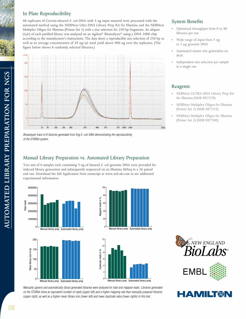
Bioanalyzer trace of 8 libraries generated from 5ng E. coli DNA demonstrating the reproducibility of the STARlet system.
In Plate Reproducibility48 replicates of Covaris-sheared E. coli DNA with 5 ng input material were processed with the automated method using the NEBNext Ultra DNA Library Prep Kit for Illumina and the NEBNext Multiplex Oligos for Illumina (Primer Set 1) with a size selection for 250 bp fragments. An aliquot (1μL) of each purified library was analysed on an Agilent® Bioanalyzer® using a DNA 1000 chip according to the manufacturer’s instructions. The data show a reproducible size selection of 250 bp as well as an average concentration of 29 ng/μl; total yield above 900 ng over the replicates. (The figure below shows 8 randomly selected libraries.)
Manual Library Preparation vs. Automated Library Preparation Two sets of 6 samples each containing 5 ng of sheared E. coli genomic DNA were provided for indexed library generation and subsequently sequenced on an Illumina MiSeq in a 36 paired end run. Download the full Application Note transcript at www.neb.uk.com to see additional experimental information.
0
1000000
2000000
3000000
4000000
5000000
Manual library prep Automated library prep
Tota
l rea
ds
0
20
40
60
80
100
Manual library prep Automated library prep
Map
ped
read
s in
%
0
50
100
150
200
Manual library prep Automated library prep
Mea
n lib
rary
siz
e in
bp
0
2
4
6
8
10
12
Manual library prep Automated library prep
Dupl
icat
e re
ads
in %
Manually (green) and automatically (blue) generated libraries were analyzed for total and mapped reads. Libraries generated on the STARlet show an equivalent number of reads (upper left) and a higher mapping rate than manually prepared libraries (upper right), as well as a higher mean library size (lower left) and lower duplicate rates (lower rights) in this test.
09
CELL SIG
NA
LING
TEC
HN
OLO
GY
CELL SIGNALING TECHNOLOGY Visit the new www.cellsignal.com
© 2014 Cell Signaling Technology, Inc. Cell Signaling Technology® and PhosphoSitePlus® are trademarks of Cell Signaling Technology, Inc. FLY_0001WEBD_0070E
Same CST. New Website.Designed by scientists, for scientists, our new site has been reengineered to help accelerate your research and discovery.
Developed in close collaboration with CST scientists, our new website brings together the best of the old and the new: the in-depth scientific content and full breadth of resources CST is known for, plus new features that make it faster and easier to determine the right antibodies and companion products to meet your needs. If you value specificity, visit www.cellsignal.com first to find antibodies you can trust – and CST scientists who can be your partner at the bench.
New features include: Seamlessly integrated scientific resources for your research area or target of interest.:: Classic pathway diagrams:: PhosphoSitePlus® database integration:: Our extensive protocol library:: Tutorials and reference tables
Interactive product search portal to help you quickly find the right antibodies and companion products. :: Search for products through a wide range of parameters:: Validation and application data for every antibody:: Scientific and technical resource areas:: Validation images quick view by application
The Story of a CST Antibody In 2013, we reached out to research scientists globally and discovered that many did not think it was possible for any company to make, validate, and support all its antibodies in-house. This is in fact what CST does and we’re sharing our step-by-step process right on our new home page. Skeptical? See our story and judge for yourself. www.cellsignal.com

CEL
L SI
GN
ALI
NG
TEC
HN
OLO
GY
10
What is PTMScan Proteomics?PTMScan® Proteomics Kits and Services employ proprietary methodologies for antibody-based peptide enrichment combined with tandem mass spectrometry for quantitative profiling of posttranslational modifications (PTMs), including phosphorylation,ubiquitination, methylation and acetylation.
How can it help you?:: Allows identification of novel or low abundance post translational modification events by enrichment of modified peptides with an antibody directed against phospho (Tyr, Ser, Thr)-peptides, acetylated or methylated peptides,or ubiquitin-tagged peptides
:: Enables identification of novel phosphorylation events in research involving disease state or drug treatment response
:: PTMScan® proteomics technology can be applied to many biological systems and species to support diverse research interests
:: Expert technical service is provided by CST™ proteomics scientists throughout your experiment
Post Translational Modification Proteomics
PTMScan® PRODUCTS AND SERVICES www.cellsignal.com
Available Products and Services:: AcetylScan® Proteomics Service
:: UbiScan® Proteomics Service
:: Ser/Thr PhosphoScan® Proteomics Service
:: Tyrosine PhosphoScan® Proteomics Service
:: MethylScan™ Proteomics Service
Ub Me
AcP
LC-MS/MS and Bioinformatic Analysis
Peptides
Cell or Tissue Samples
Ub
Me
Ac
P
Immunoprecipitation using motif antibody

11
CELL SIG
NA
LING
TEC
HN
OLO
GY
WIN A PROJECT GRANT
Application Dates: March 26, 2014 – July 1, 2014 (Two project grants will be awarded in 2014 to UK applicants.)
Use PTMScan® Discovery Services to characterize post-translational modifications with motif antibodies and mass spectrometry.
www.cellsignal.com/ptmgrantuk
Founded by research scientists in 1999, Cell Signaling Technology (CST) is a family-owned company active in applied systems biology research, particularly as it relates to cancer. Understanding the importance of using antibodies with high levels of specificity and consistency, CST scientists produce, validate, and support all of our antibodies in-house.
ABOUT CST:
TO ENTER:
PTMScan® Project Grants include: • PTMScan® Discovery project for up to 4 sample conditions
• Teleconference with CST PTMScan® scientists to discuss the details of the study design and the requirements for sample preparation
• Data package, including RAW data files and Excel® Result Tables and Summary PowerPoint® Report
• Post-project consultation with PTMScan® scientists to discuss results and address any study related questions
• A $2.5K travel stipend to present your PTMScan® results at a relevant scientific meeting
Ubiquitination Phosphorylation
Acetylation Methylation
www.cellsignal.com/ptmgrantuk
Visit www.cellsignal.com/ptmgrantuk for full elligibility details, application requirements and Terms & Conditions. If you have any questions please contact your NEB UK Account Manager or email [email protected].
Win a Proteomics Grant fromCell Signaling TechnologyUK Academic Project Grants for PTMScan® Discovery
Worthover
$19,000!

† Excludes Republic of Ireland, N Ireland and exports.
email: [email protected] tel: 0800 318486 fax: 0800 435682 web: www.neb.uk.com
FreeWith NEB
FreeNext Day Delivery
for All UK Mainland Orders†
No ice charges, No dry ice charges.
£0.00Our Delivery charge?