Embed Size (px)
Citation preview
Michael Cummings
David Reisman • University of South Carolina
Gene Expression and Gene Regulation
Part 1
Chapter 9
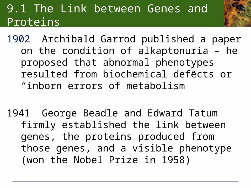
9.1 The Link between Genes and Proteins
1902 Archibald Garrod published a paper on the condition of alkaptonuria – he proposed that abnormal phenotypes resulted from biochemical defects or “inborn errors of metabolism”
1941 George Beadle and Edward Tatum firmly established the link between genes, the proteins produced from those genes, and a visible phenotype (won the Nobel Prize in 1958)
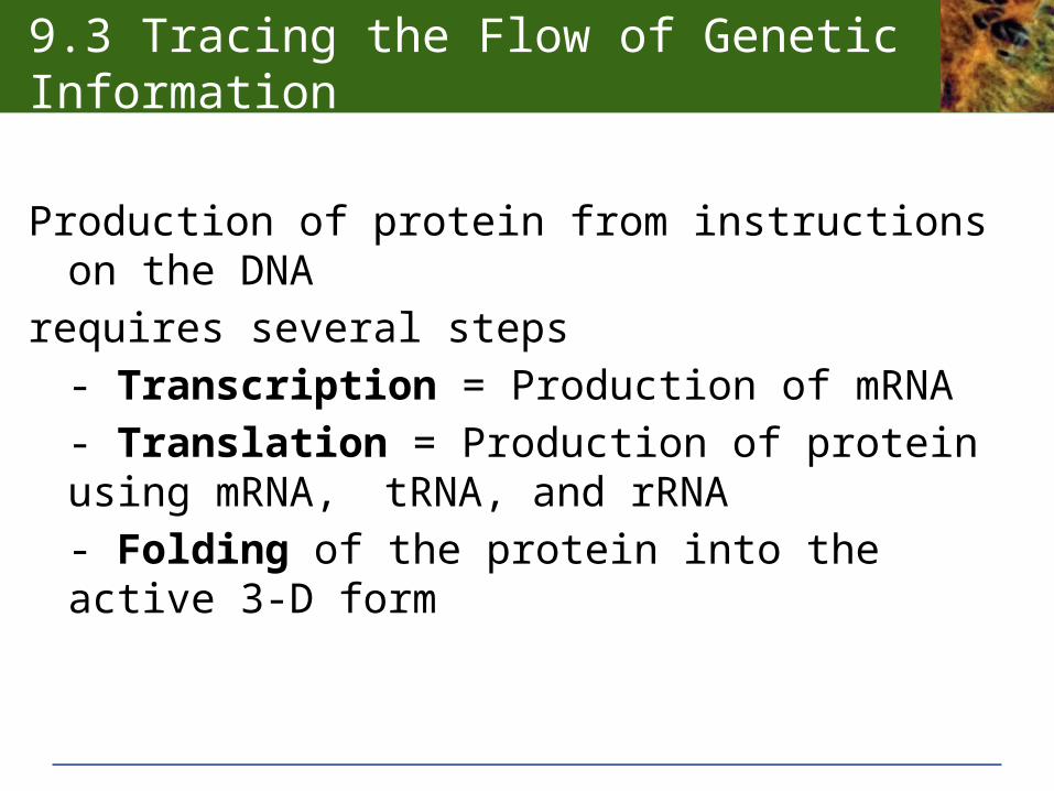
9.3 Tracing the Flow of Genetic Information
Production of protein from instructions on the DNA
requires several steps
- Transcription = Production of mRNA
- Translation = Production of protein using mRNA, tRNA, and rRNA
- Folding of the protein into the active 3-D form
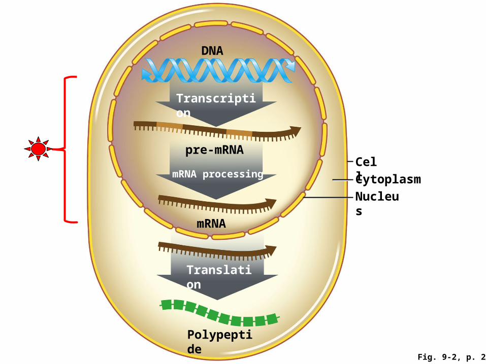
Fig. 9-2, p. 201
DNA
Transcription
pre-mRNACell
mRNA processing Cytoplasm
Nucleus
mRNA
Translation
Polypeptide
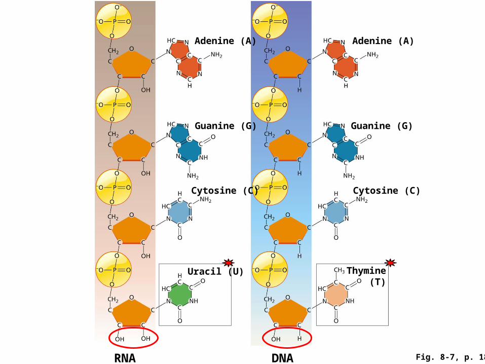
Fig. 8-7, p. 183
Adenine (A) Adenine (A)
Guanine (G) Guanine (G)
Cytosine (C) Cytosine (C)
Uracil (U) Thymine (T)
RNA DNA
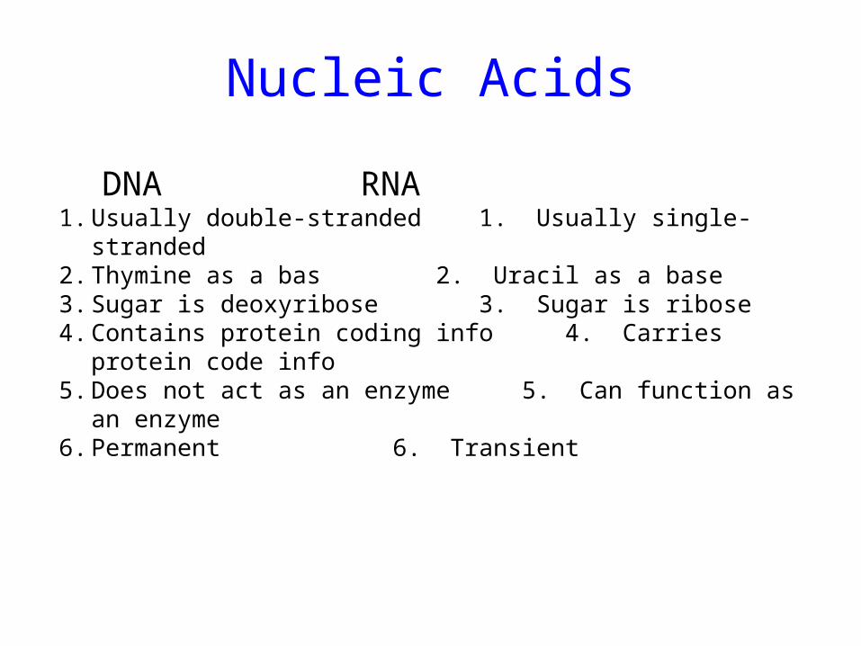
Table 10.2
Nucleic Acids
DNA RNA1. Usually double-stranded 1. Usually single-stranded2. Thymine as a bas 2. Uracil as a base3. Sugar is deoxyribose 3. Sugar is ribose4. Contains protein coding info 4. Carries protein code info5. Does not act as an enzyme 5. Can function as an
enzyme6. Permanent 6. Transient
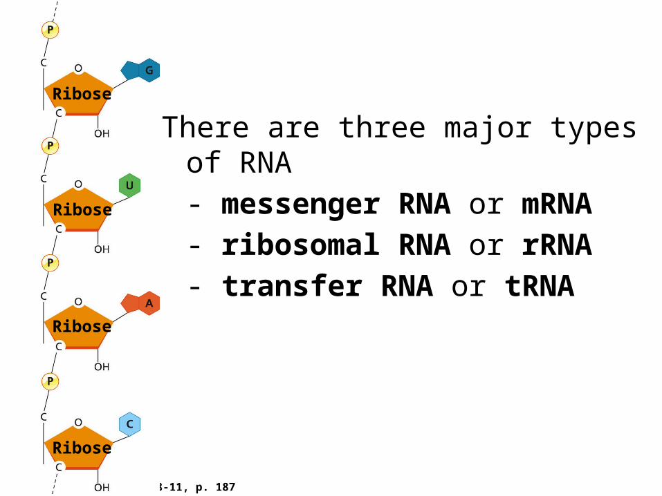
Fig. 8-11, p. 187
Ribose
Ribose
Ribose
Ribose
There are three major types of RNA
- messenger RNA or mRNA
- ribosomal RNA or rRNA
- transfer RNA or tRNA
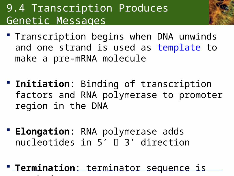
9.4 Transcription Produces Genetic Messages Transcription begins when DNA unwinds and one
strand is used as template to make a pre-mRNA molecule
Initiation: Binding of transcription factors and RNA polymerase to promoter region in the DNA
Elongation: RNA polymerase adds nucleotides in 5’ 3’ direction
Termination: terminator sequence is reached
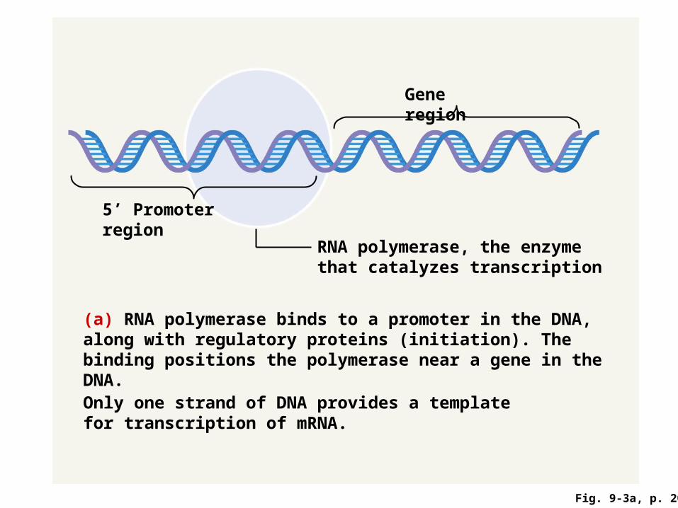
Fig. 9-3a, p. 200
Gene region
5’ Promoter region
RNA polymerase, the enzyme that catalyzes transcription
(a) RNA polymerase binds to a promoter in the DNA, along with regulatory proteins (initiation). The binding positions the polymerase near a gene in the DNA.
Only one strand of DNA provides a template for transcription of mRNA.
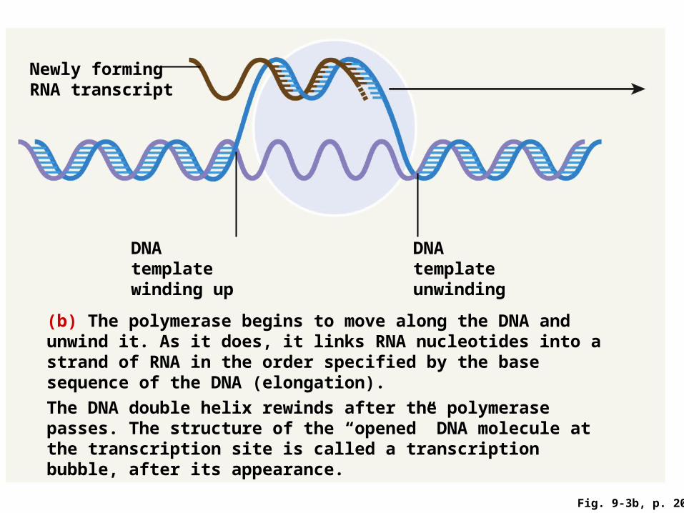
Fig. 9-3b, p. 200
Newly forming RNA transcript
DNA template winding up
DNA template unwinding
(b) The polymerase begins to move along the DNA and unwind it. As it does, it links RNA nucleotides into a strand of RNA in the order specified by the base sequence of the DNA (elongation).
The DNA double helix rewinds after the polymerase passes. The structure of the “opened” DNA molecule at the transcription site is called a transcription bubble, after its appearance.
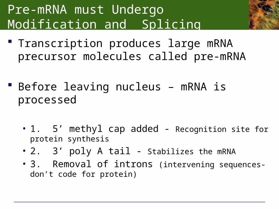
Pre-mRNA must Undergo Modification and Splicing Transcription produces large mRNA precursor
molecules called pre-mRNA
Before leaving nucleus – mRNA is processed
• 1. 5’ methyl cap added - Recognition site for protein synthesis
• 2. 3’ poly A tail - Stabilizes the mRNA
• 3. Removal of introns (intervening sequences- don’t code for protein)
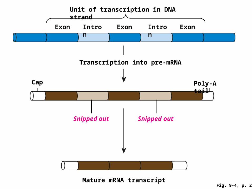
Fig. 9-4, p. 202
Unit of transcription in DNA strand
Exon Intron Exon Intron Exon
Transcription into pre-mRNA
Cap Poly-A tail
Snipped out Snipped out
Mature mRNA transcript
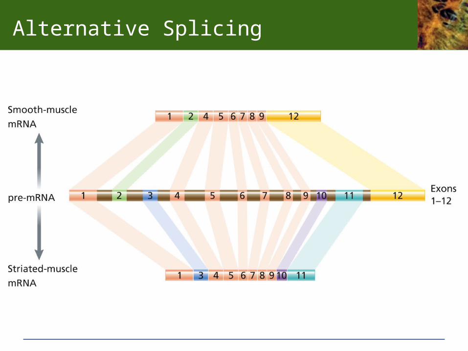
Alternative Splicing
Mutations in Splicing Sites and Genetic Disorders
Splicing defects cause several human genetic disorders
One hemoglobin disorder, b-thalassemia, is due to mutations at the exon/intron region that results in lower splicing efficiency and lower -b globin protein
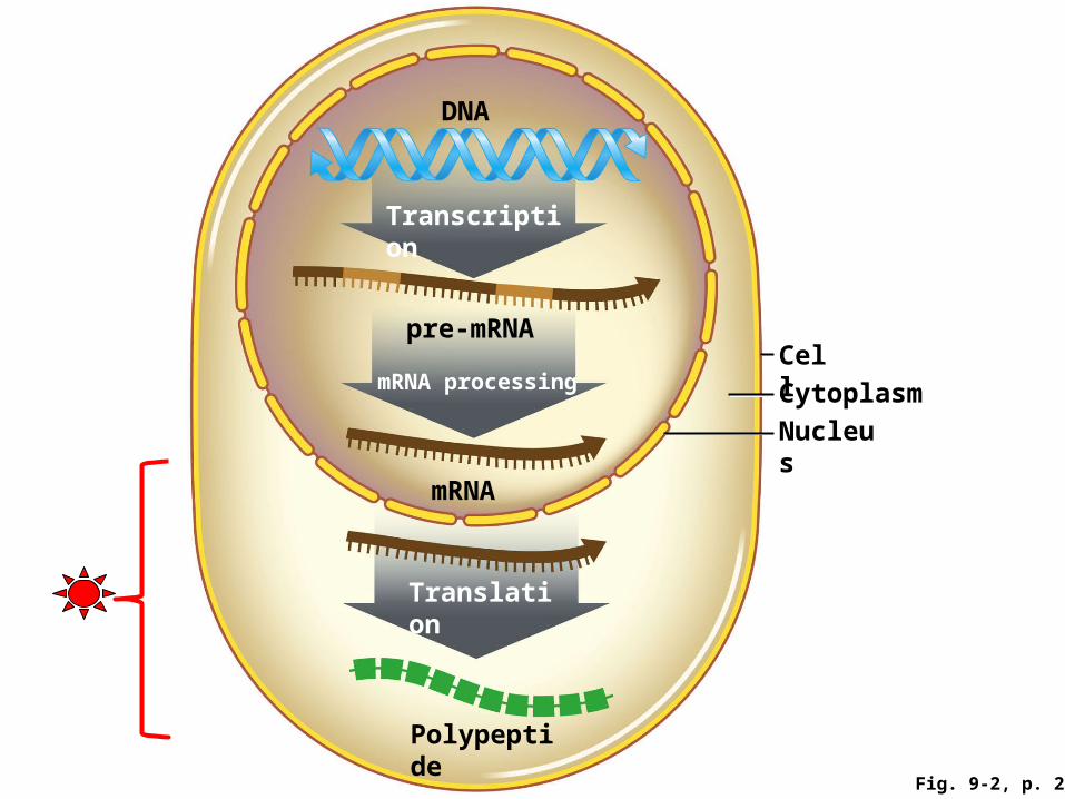
Fig. 9-2, p. 201
DNA
Transcription
pre-mRNACell
mRNA processing Cytoplasm
Nucleus
mRNA
Translation
Polypeptide
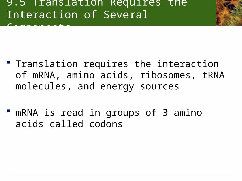
9.5 Translation Requires the Interaction of Several Components
Translation requires the interaction of mRNA, amino acids, ribosomes, tRNA molecules, and energy sources
mRNA is read in groups of 3 amino acids called codons
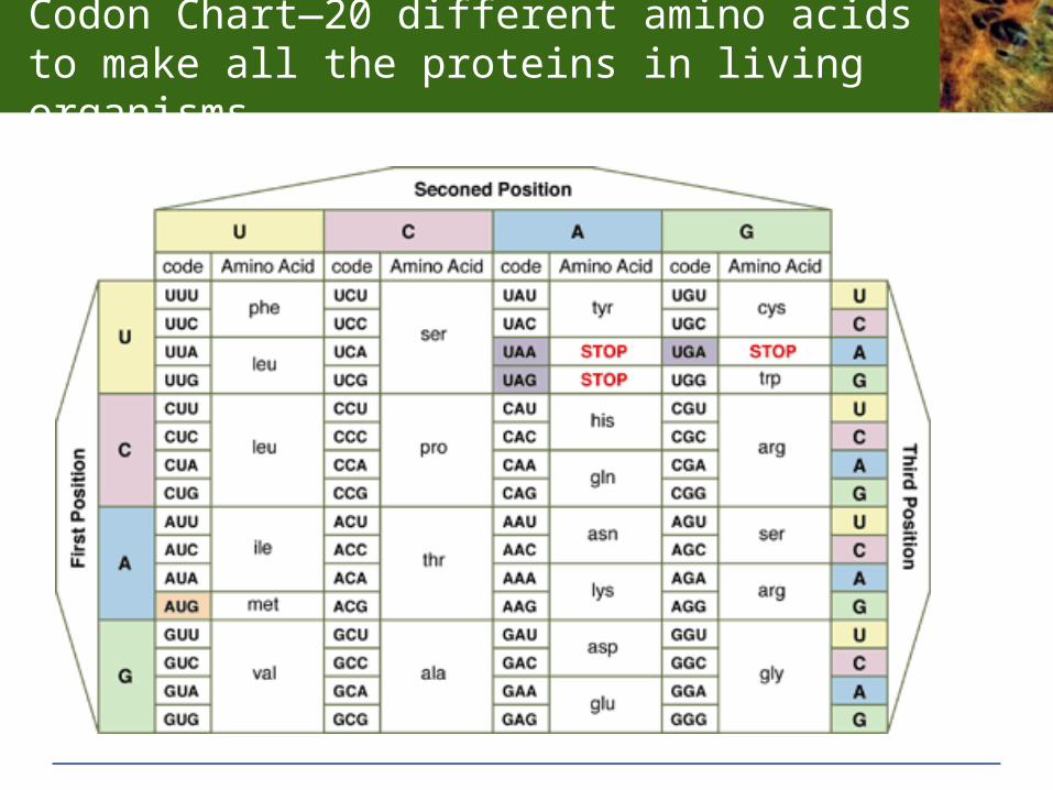
Codon Chart—20 different amino acids to make all the proteins in living organisms
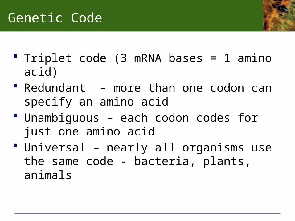
Genetic Code
Triplet code (3 mRNA bases = 1 amino acid) Redundant – more than one codon can specify an
amino acid Unambiguous – each codon codes for just one
amino acid Universal – nearly all organisms use the same
code - bacteria, plants, animals
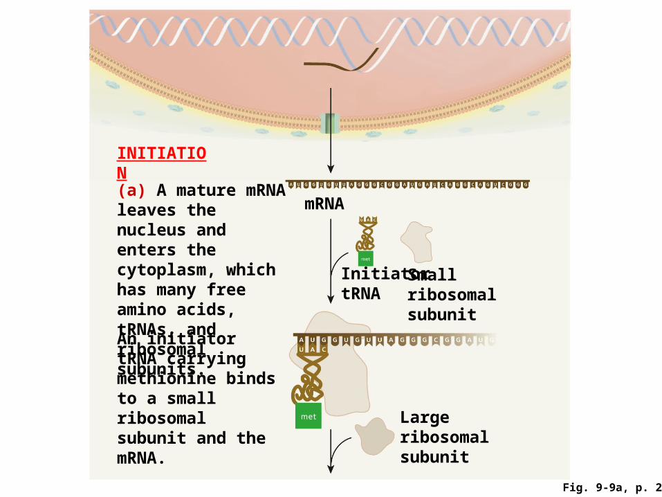
Fig. 9-9a, p. 206
INITIATION
(a) A mature mRNA leaves the nucleus and enters the cytoplasm, which has many free amino acids, tRNAs, and ribosomal subunits.
mRNA
Initiator tRNA
Small ribosomal subunit
An initiator tRNA carrying methionine binds to a small ribosomal subunit and the mRNA. Large
ribosomal subunit
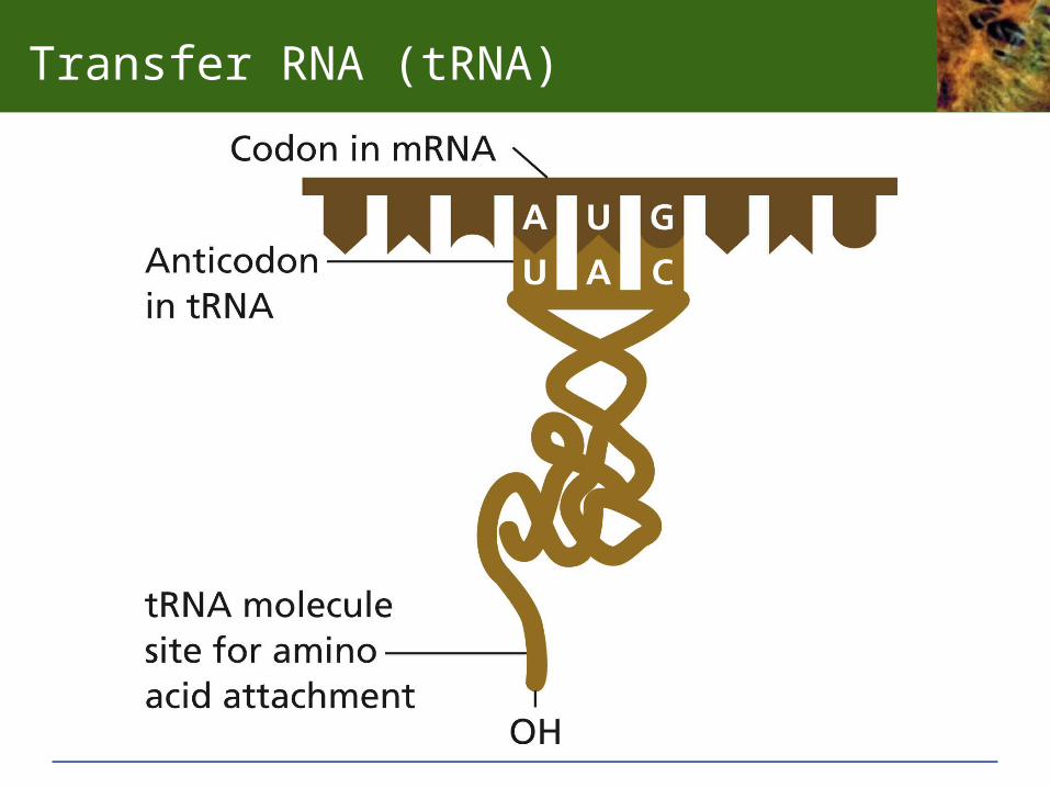
Transfer RNA (tRNA)
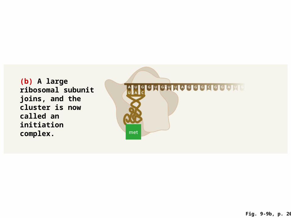
Fig. 9-9b, p. 206
(b) A large ribosomal subunit joins, and the cluster is now called an initiation complex.
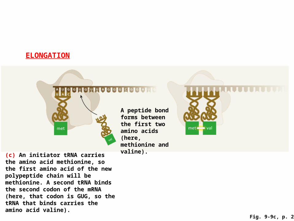
Fig. 9-9c, p. 207
ELONGATION
(c) An initiator tRNA carries the amino acid methionine, so the first amino acid of the new polypeptide chain will be methionine. A second tRNA binds the second codon of the mRNA (here, that codon is GUG, so the tRNA that binds carries the amino acid valine).
A peptide bond forms between the first two amino acids (here, methionine and valine).
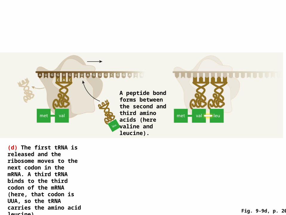
Fig. 9-9d, p. 207
(d) The first tRNA is released and the ribosome moves to the next codon in the mRNA. A third tRNA binds to the third codon of the mRNA (here, that codon is UUA, so the tRNA carries the amino acid leucine).
A peptide bond forms between the second and third amino acids (here valine and leucine).
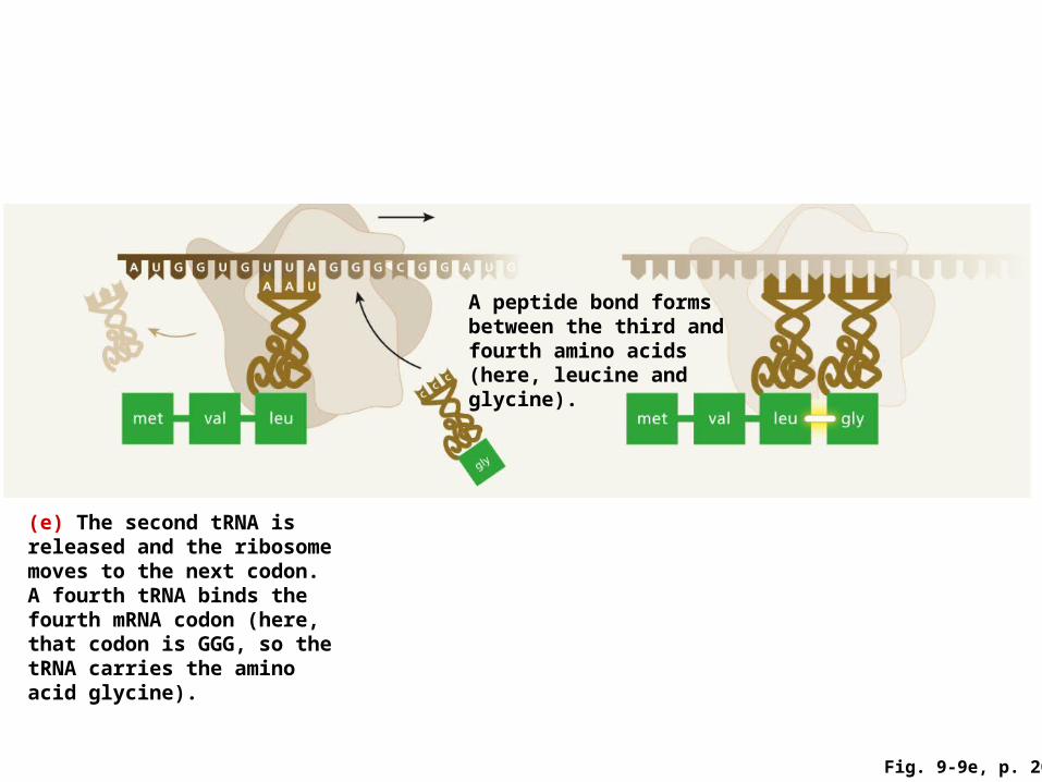
Fig. 9-9e, p. 207
(e) The second tRNA is released and the ribosome moves to the next codon. A fourth tRNA binds the fourth mRNA codon (here, that codon is GGG, so the tRNA carries the amino acid glycine).
A peptide bond forms between the third and fourth amino acids (here, leucine and glycine).
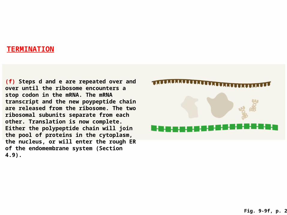
Fig. 9-9f, p. 207
TERMINATION
(f) Steps d and e are repeated over and over until the ribosome encounters a stop codon in the mRNA. The mRNA transcript and the new poypeptide chain are released from the ribosome. The two ribosomal subunits separate from each other. Translation is now complete. Either the polypeptide chain will join the pool of proteins in the cytoplasm, the nucleus, or will enter the rough ER of the endomembrane system (Section 4.9).
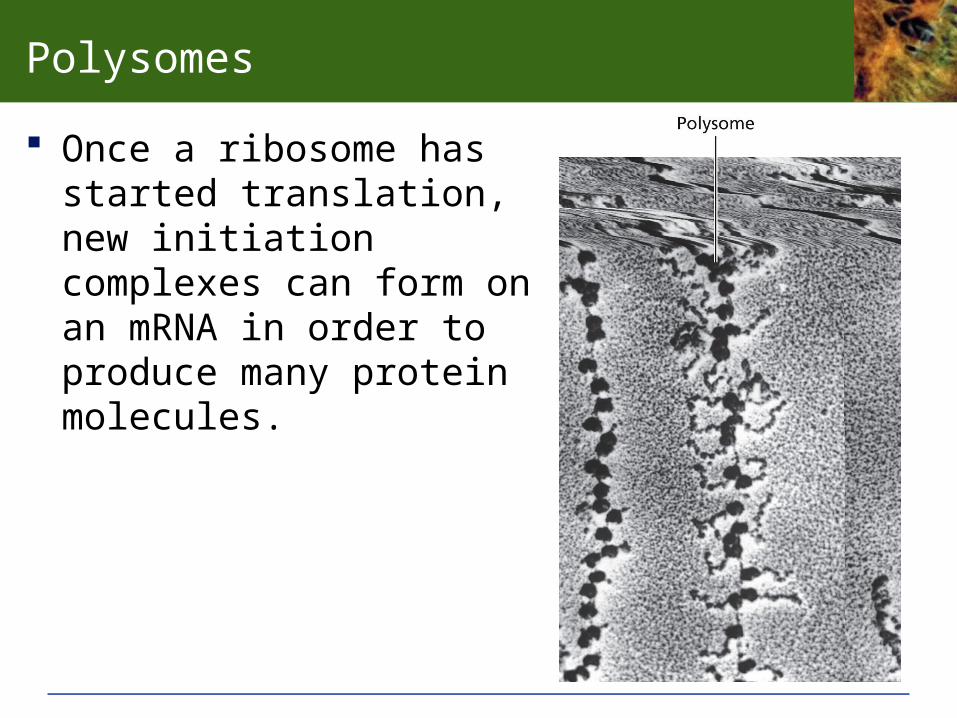
Polysomes
Once a ribosome has started translation, new initiation complexes can form on an mRNA in order to produce many protein molecules.
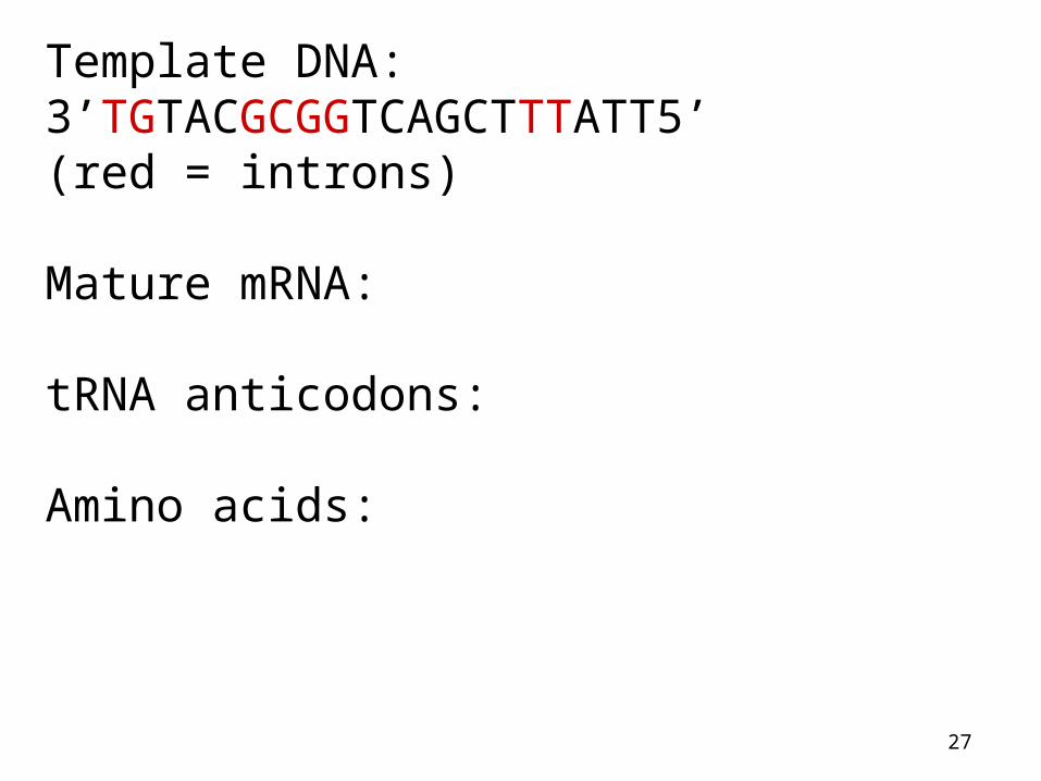
27
Template DNA: 3’TGTACGCGGTCAGCTTTATT5’(red = introns)
Mature mRNA:
tRNA anticodons:
Amino acids:
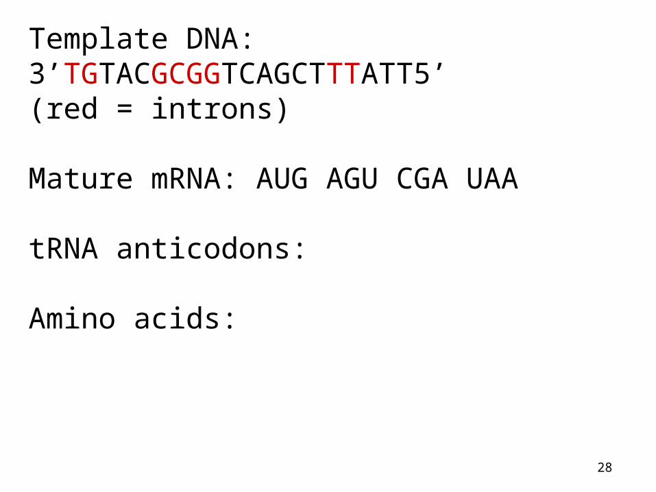
28
Template DNA: 3’TGTACGCGGTCAGCTTTATT5’(red = introns)
Mature mRNA: AUG AGU CGA UAA
tRNA anticodons:
Amino acids:
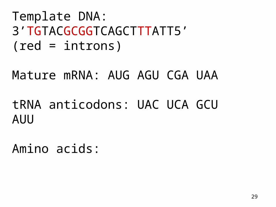
29
Template DNA: 3’TGTACGCGGTCAGCTTTATT5’(red = introns)
Mature mRNA: AUG AGU CGA UAA
tRNA anticodons: UAC UCA GCU AUU
Amino acids:
30
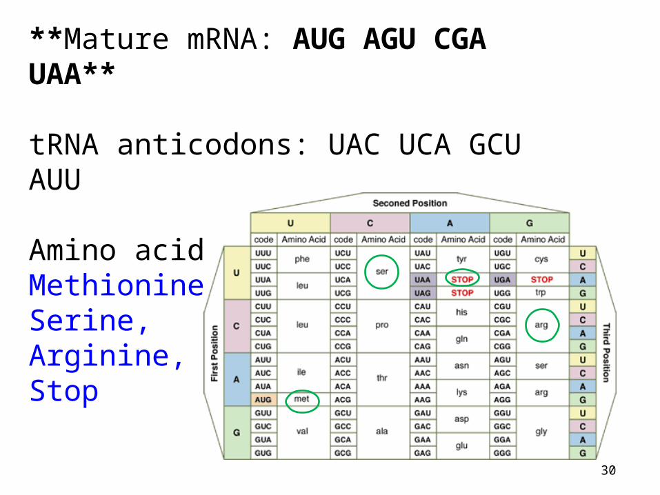
**Mature mRNA: AUG AGU CGA UAA**
tRNA anticodons: UAC UCA GCU AUU
Amino acids: Methionine,Serine,Arginine,Stop
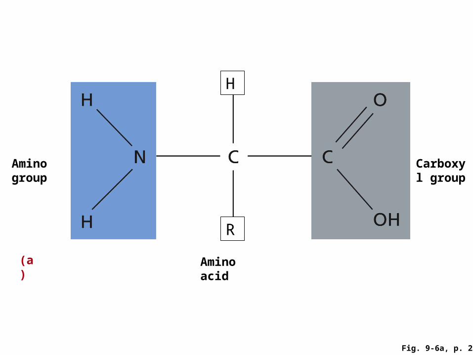
Fig. 9-6a, p. 203
Amino group
Carboxyl group
(a) Amino acid
H
R
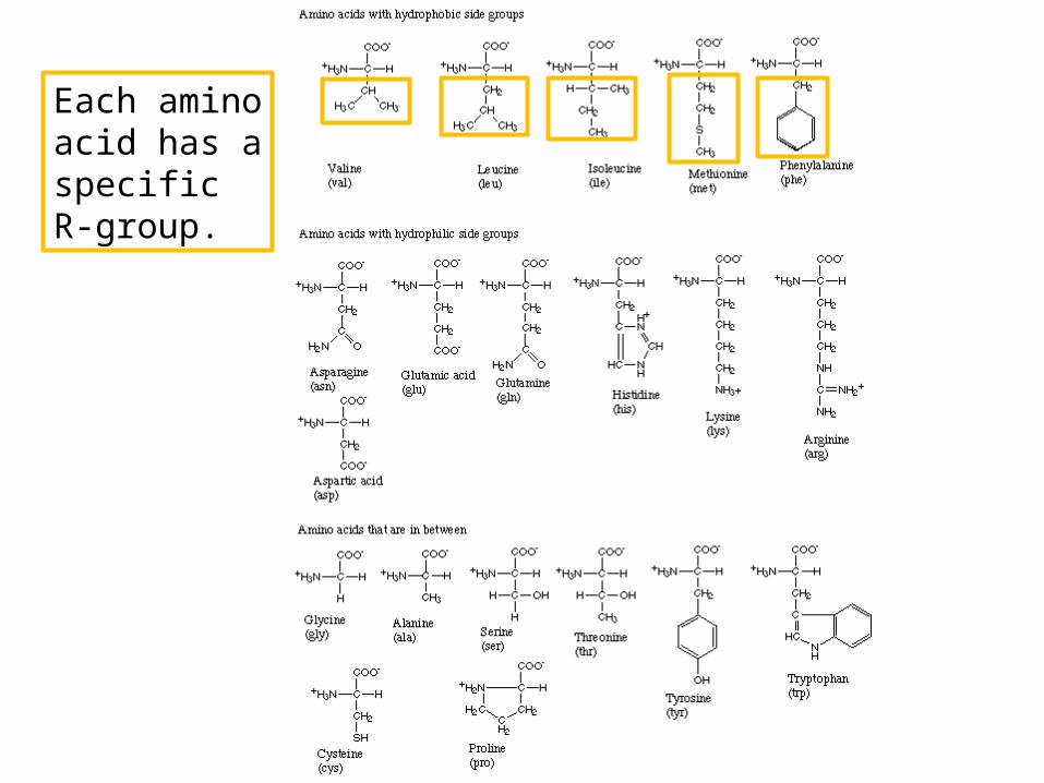
Each aminoacid has aspecificR-group.
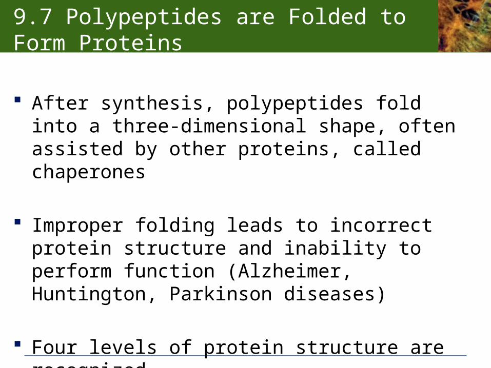
9.7 Polypeptides are Folded to Form Proteins
After synthesis, polypeptides fold into a three-dimensional shape, often assisted by other proteins, called chaperones
Improper folding leads to incorrect protein structure and inability to perform function (Alzheimer, Huntington, Parkinson diseases)
Four levels of protein structure are recognized

Four Levels of Protein Structure
Primary structure (1O) • The amino acid sequence in a polypeptide chain
Secondary structure (2O) • The pleated or helical structure in a protein molecule
resulting from the peptide bonds between amino acids
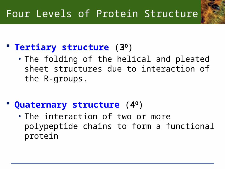
Four Levels of Protein Structure
Tertiary structure (3O) • The folding of the helical and pleated sheet
structures due to interaction of the R-groups.
Quaternary structure (4O) • The interaction of two or more polypeptide chains to
form a functional protein
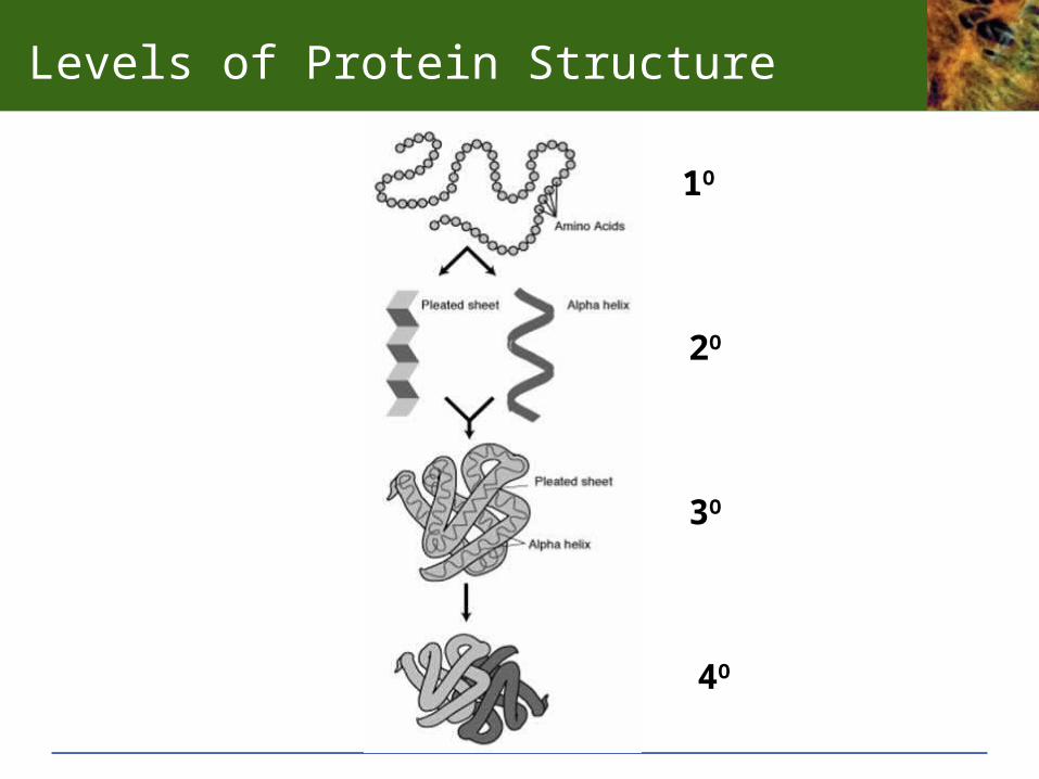
Levels of Protein Structure
1O
2O
3O
4O
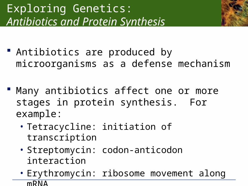
Exploring Genetics: Antibiotics and Protein Synthesis
Antibiotics are produced by microorganisms as a defense mechanism
Many antibiotics affect one or more stages in protein synthesis. For example:• Tetracycline: initiation of transcription• Streptomycin: codon-anticodon interaction• Erythromycin: ribosome movement along mRNA
Other topics in Chp 9 Part 2
Protein folding diseases Regulation of protein synthesis occurs at several
levels:• Timing of transcription• The rate of translation• The ways in which proteins are processed• The rate of protein break-down