Embed Size (px)
Citation preview
Copyright © 2005 Pearson Education, Inc. Publishing as Benjamin Cummings
Molecular Biology Of The Gene
Chapter 10
Copyright © 2005 Pearson Education, Inc. Publishing as Benjamin Cummings
Sabotage Inside Our Cells
• A saboteur
– Lies low waiting for the right moment to strike
Copyright © 2005 Pearson Education, Inc. Publishing as Benjamin Cummings
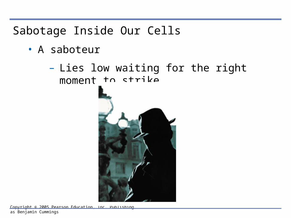
• Viruses are biological saboteurs
– Hijacking the genetic material of host cells in order to reproduce themselves
• Viruses provided some of the earliest evidence
– That genes are made of DNA
Copyright © 2005 Pearson Education, Inc. Publishing as Benjamin Cummings
THE STRUCTURE OF THE GENETIC MATERIAL
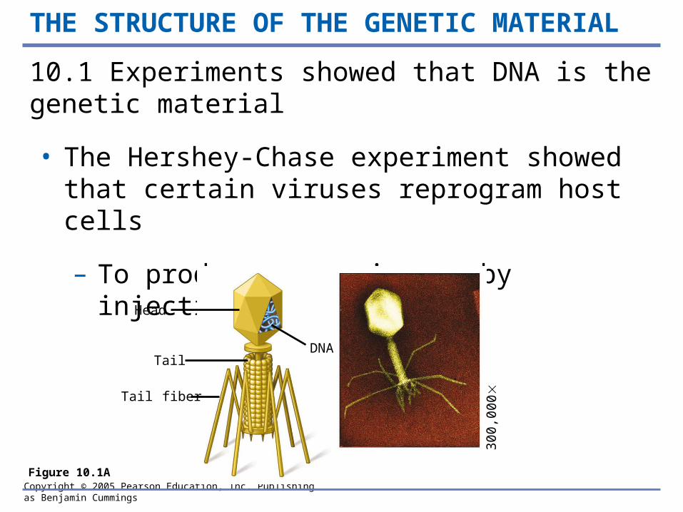
10.1 Experiments showed that DNA is the genetic material
• The Hershey-Chase experiment showed that certain viruses reprogram host cells
– To produce more viruses by injecting their DNA
Figure 10.1A
Head
Tail
Tail fiber
DNA
300
,00
0

Copyright © 2005 Pearson Education, Inc. Publishing as Benjamin Cummings
• The Hershey-Chase experiment
Phage
Bacterium
Radioactiveprotein
DNA
Phage DNA
Empty protein shell Radioactivity
in liquid
PelletCentrifuge
Batch 1Radioactiveprotein
Batch 2RadioactiveDNA
RadioactiveDNA
Centrifuge
Pellet
Radioactivityin pellet
Figure 10.1B
Mix radioactively labeled phages with bacteria. The phages infect the bacterial cells.
1 Agitate in a blender to separate phages outside the bacteria from the cells and their contents.
2 Centrifuge the mixture so bacteria form a pelletat the bottom of the testtube.
3 Measure the radioactivity in the pellet and the liquid.
4
Copyright © 2005 Pearson Education, Inc. Publishing as Benjamin Cummings
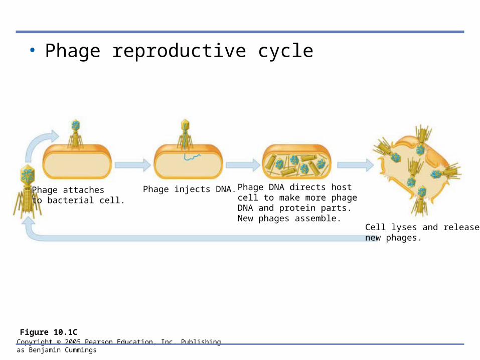
• Phage reproductive cycle
Figure 10.1C
Phage attachesto bacterial cell.
Phage injects DNA. Phage DNA directs hostcell to make more phageDNA and protein parts.New phages assemble.
Cell lyses and releases new phages.

Copyright © 2005 Pearson Education, Inc. Publishing as Benjamin Cummings
DNA polynucleotide
A
C
T
G
T
Sugar-phosphate backbone
Phosphate group
Nitrogenous base
SugarA
C
T
G
T
Phosphategroup
O
O–
OO P CH2
H3C C
C
C
CN
C
N
H
H
O
O
C
O
O
H
C H H
H
C
H
Nitrogenous base(A, G, C, or T)
Thymine (T)
Sugar(deoxyribose)
DNA nucleotide
DNA nucleotide
10.2 DNA and RNA are polymers of nucleotides
• DNA is a nucleic acid
– Made of long chains of nucleotide monomers
Figure 10.2A
Copyright © 2005 Pearson Education, Inc. Publishing as Benjamin Cummings
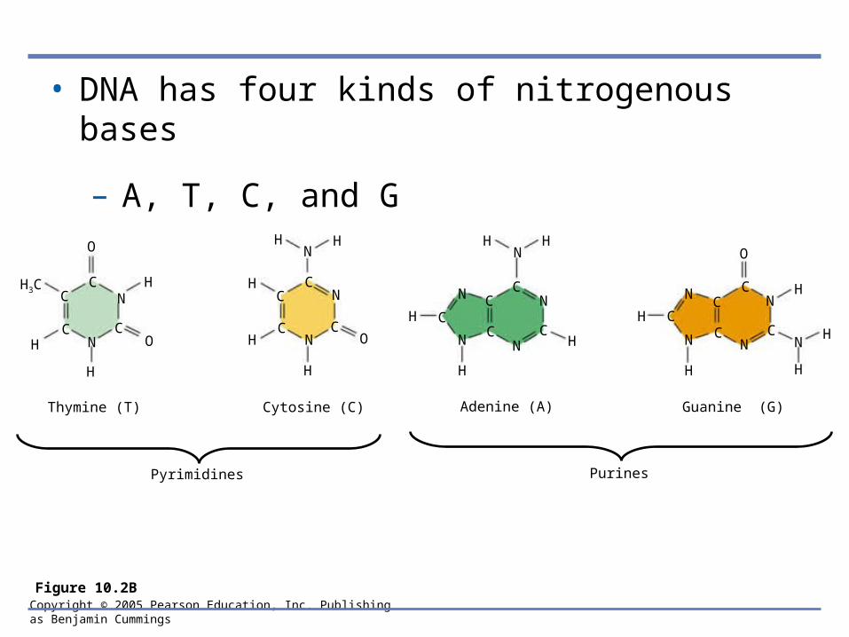
• DNA has four kinds of nitrogenous bases
– A, T, C, and G
CC
C
CC
C
O
N
C
H
H
ONH
H3C
H H
H
H
N
N
N
H
OC
H HN
H C
N
N N
N
C
CC
C
H
H
N
N
H
C
CN
C HN
CN
H C
O
H
H
Thymine (T) Cytosine (C) Adenine (A) Guanine (G)
PurinesPyrimidines
Figure 10.2B
Copyright © 2005 Pearson Education, Inc. Publishing as Benjamin Cummings
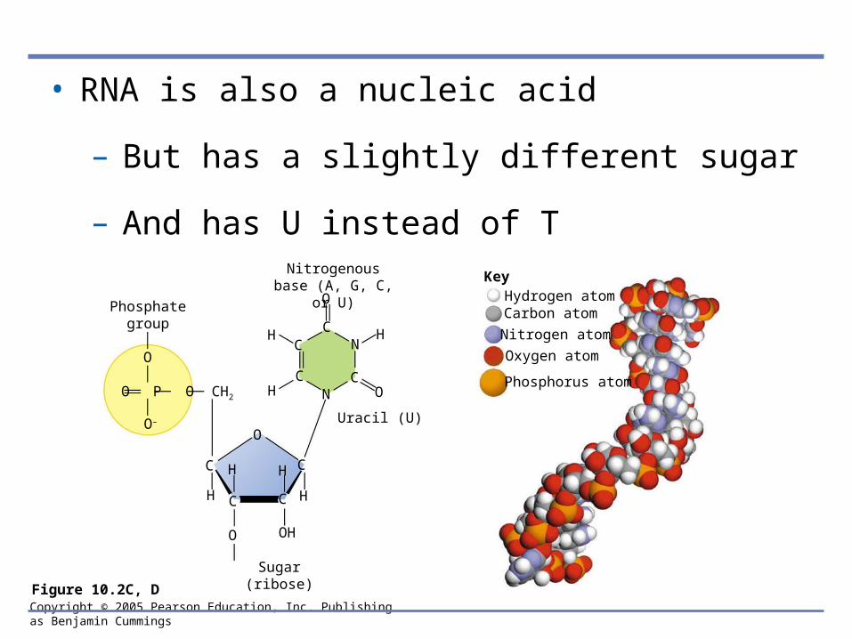
• RNA is also a nucleic acid
– But has a slightly different sugar
– And has U instead of TNitrogenous base
(A, G, C, or U)
Phosphategroup
O
O–
OO P CH2
HC
C
C
CN
C
N
H
H
O
O
C
O
O
H
C H H
OH
C
H
Uracil (U)
Sugar(ribose)
KeyHydrogen atomCarbon atom
Nitrogen atom
Oxygen atom
Phosphorus atom
Figure 10.2C, D

Copyright © 2005 Pearson Education, Inc. Publishing as Benjamin Cummings
10.3 DNA is a double-stranded helix
• James Watson and Francis Crick
– Worked out the three-dimensional structure of DNA, based on work by Rosalind Franklin
Figure 10.3A, B
Copyright © 2005 Pearson Education, Inc. Publishing as Benjamin Cummings
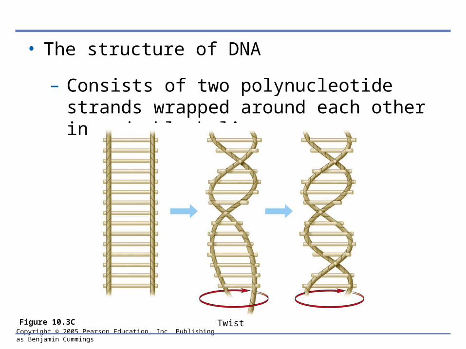
• The structure of DNA
– Consists of two polynucleotide strands wrapped around each other in a double helix
Figure 10.3C Twist
Copyright © 2005 Pearson Education, Inc. Publishing as Benjamin Cummings
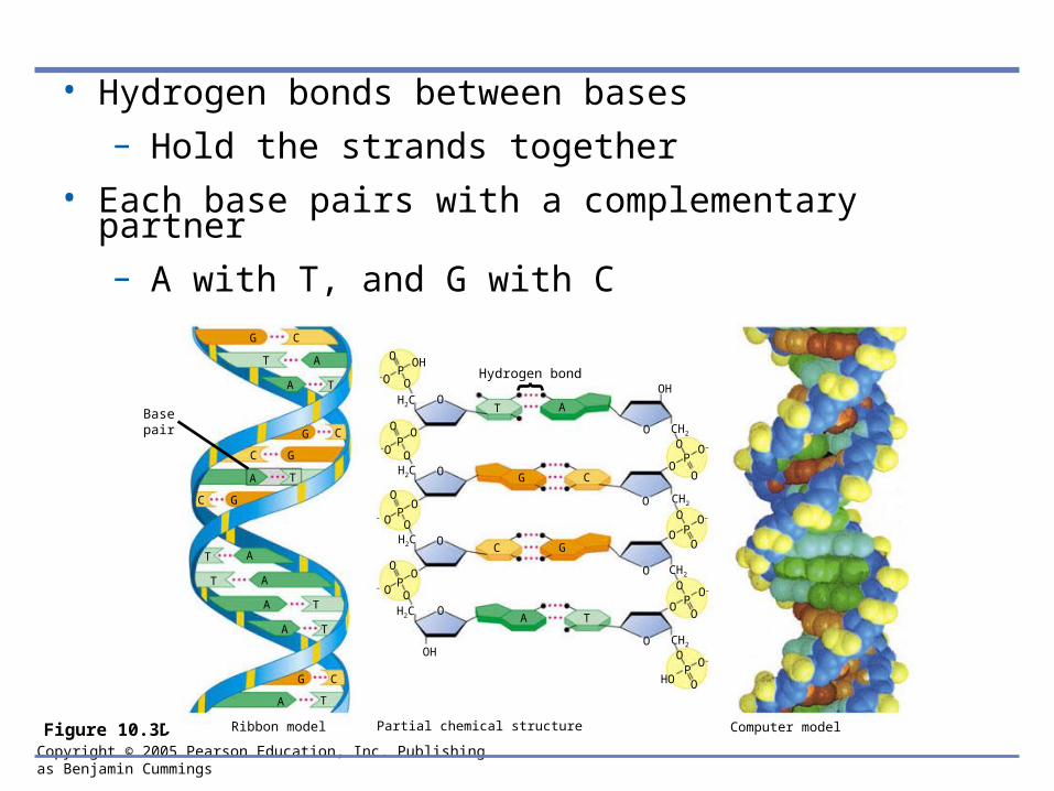
• Hydrogen bonds between bases
– Hold the strands together
• Each base pairs with a complementary partner
– A with T, and G with C
Figure 10.3D
G C
T A
A T
G
G
C
C
A T
GC
T A
T A
A T
A T
G C
A T
O
O
OH–O
P
OO
–OPO
OO
P– O
– O OP
OO
O
OH
H2C
H2C
H2C
H2C
O
O
O
O
O
O
O
O
PO–
O–
O–
O–
OH
HO
O
O
O
P
P
P
O
O
O
O
O
O
O
O
T A
G C
C G
A T
CH2
CH2
CH2
CH2
Hydrogen bond
Basepair
Ribbon model Partial chemical structure Computer model
Copyright © 2005 Pearson Education, Inc. Publishing as Benjamin Cummings
DNA REPLICATION
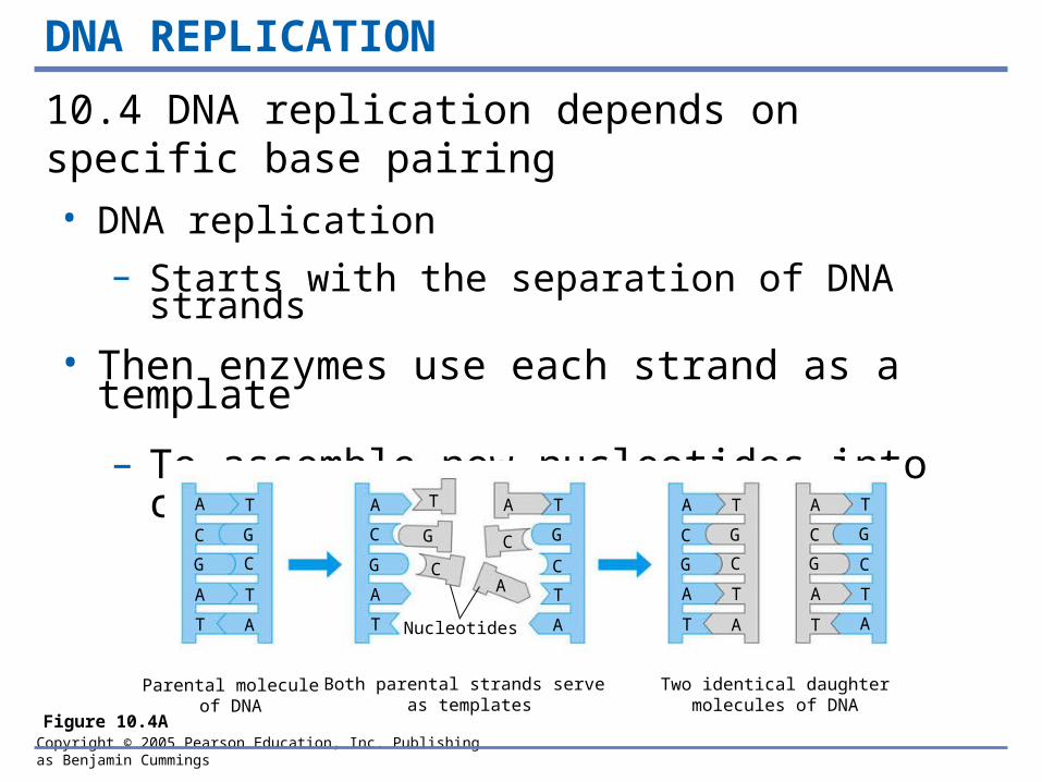
10.4 DNA replication depends on specific base pairing
• DNA replication
– Starts with the separation of DNA strands
• Then enzymes use each strand as a template
– To assemble new nucleotides into complementary strands
Figure 10.4A
A T
C G
G C
A T
T A
A T
C G
G C
A T
T A
A T
C G
G C
A T
T A
A T
C G
G C
A
T
A T
C G
AC
T
A
Parental moleculeof DNA
Both parental strands serve as templates
Two identical daughtermolecules of DNA
Nucleotides
Copyright © 2005 Pearson Education, Inc. Publishing as Benjamin Cummings
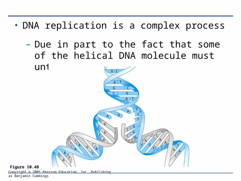
• DNA replication is a complex process
– Due in part to the fact that some of the helical DNA molecule must untwist
Figure 10.4B
G C
A T
G C
A T
C G
AGA
CG
C
GC
G
TA
G
C
TAT
AA
TT
A
CG
CG
CG
T
AG
C
T
A
T
A
AT
T
A
TC
T
Copyright © 2005 Pearson Education, Inc. Publishing as Benjamin Cummings
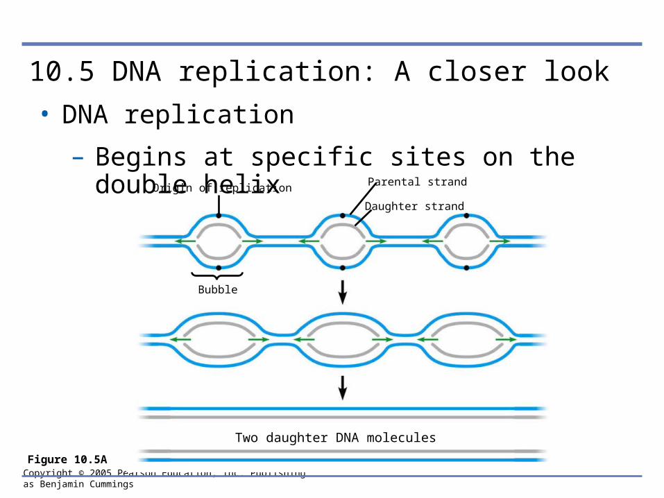
10.5 DNA replication: A closer look
• DNA replication
– Begins at specific sites on the double helix
Figure 10.5A
Origin of replication
Two daughter DNA molecules
Parental strand
Daughter strand
Bubble
Copyright © 2005 Pearson Education, Inc. Publishing as Benjamin Cummings
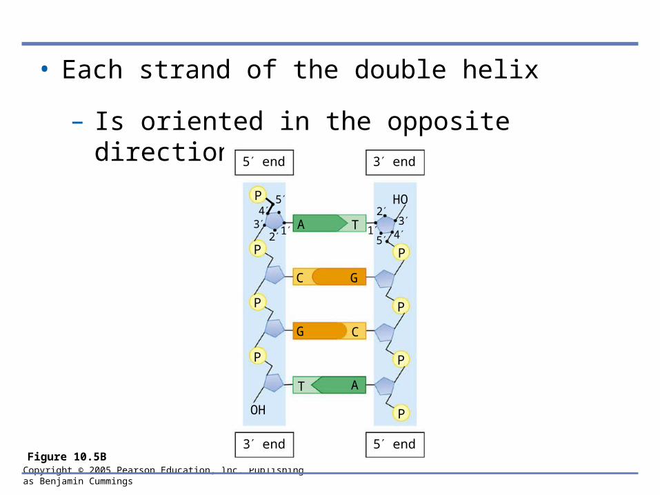
• Each strand of the double helix
– Is oriented in the opposite direction
Figure 10.5B
P
P
P
P
P
P
P
P
HO
OH
A
C
G
T
T
C
G
A
2 134
5
15 4
32
5 end 3 end
3 end 5 end
Copyright © 2005 Pearson Education, Inc. Publishing as Benjamin Cummings
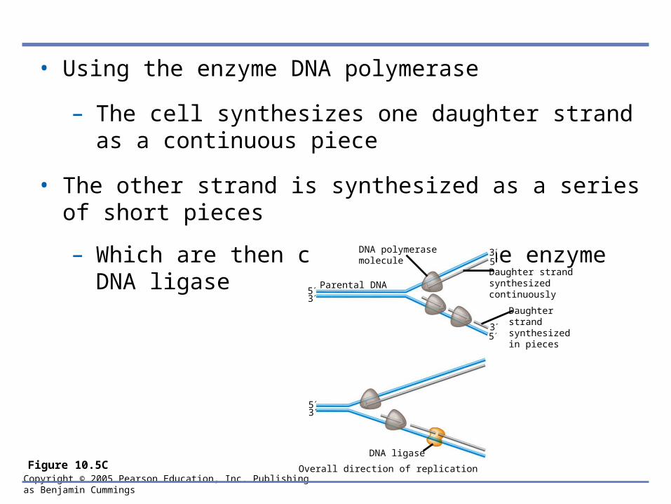
• Using the enzyme DNA polymerase
– The cell synthesizes one daughter strand as a continuous piece
• The other strand is synthesized as a series of short pieces
– Which are then connected by the enzyme DNA ligase
Figure 10.5C
3
53
53
5
53
Daughter strandsynthesizedcontinuously
Daughter strandsynthesizedin pieces
Parental DNA
DNA ligase
DNA polymerasemolecule
Overall direction of replication
Copyright © 2005 Pearson Education, Inc. Publishing as Benjamin Cummings
THE FLOW OF GENETIC INFORMATION FROM DNA TO RNA TO PROTEIN
• 10.6 The DNA genotype is expressed as proteins, which provide the molecular basis for phenotypic traits
• The information constituting an organism’s genotype
– Is carried in its sequence of its DNA bases
• A particular gene, a linear sequence of many nucleotides
– Specifies a polypeptide
Copyright © 2005 Pearson Education, Inc. Publishing as Benjamin Cummings
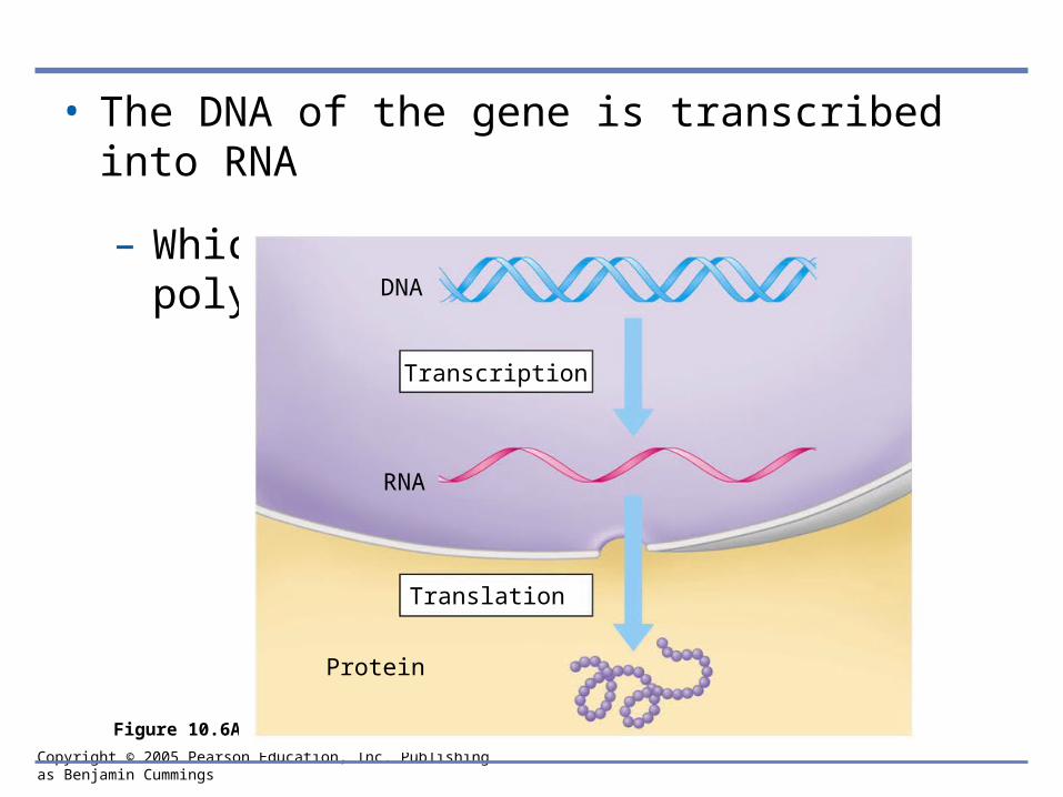
• The DNA of the gene is transcribed into RNA
– Which is translated into the polypeptide
Figure 10.6A
DNA
Transcription
RNA
Protein
Translation
Copyright © 2005 Pearson Education, Inc. Publishing as Benjamin Cummings
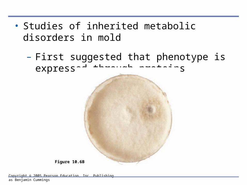
• Studies of inherited metabolic disorders in mold
– First suggested that phenotype is expressed through proteins
Figure 10.6B
Copyright © 2005 Pearson Education, Inc. Publishing as Benjamin Cummings
10.7 Genetic information written in codons is translated into amino acid sequences
• The “words” of the DNA “language”
– Are triplets of bases called codons
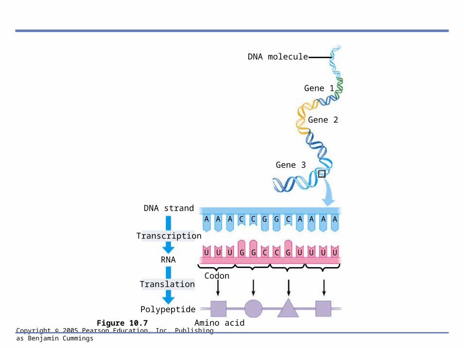
• The codons in a gene
– Specify the amino acid sequence of a polypeptide
Copyright © 2005 Pearson Education, Inc. Publishing as Benjamin Cummings
DNA strand
Transcription
Translation
Polypeptide
RNA
Amino acid
Codon
A A A C C G G C A A A A
U U U G G C C G U U U U
Gene 1
Gene 2
Gene 3
DNA molecule
Figure 10.7
Copyright © 2005 Pearson Education, Inc. Publishing as Benjamin Cummings
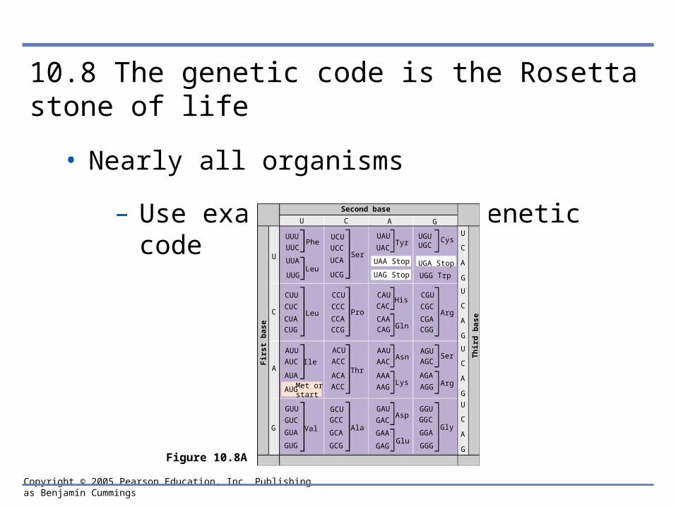
10.8 The genetic code is the Rosetta stone of life
• Nearly all organisms
– Use exactly the same genetic code
Figure 10.8A
UUC
UGUUGC
UGA Stop
Met or start
Phe
Leu
Leu
Ile
Val Ala
Thr
Pro
Ser
Asn
Lys
His
Gln
Asp
Glu
Ser
Arg
Arg
Gly
CysTyr
G
A
C
U
U C A G
Th
ird
bas
e
Second base
Fir
st b
ase
UUA
UUU
CUC
CUU
CUG
CUA
AUC
AUU
AUG
AUA
GUC
GUU
GUG
GUA
UCC
UCU
UCG
UCA
CCC
CCU
CCG
CCA
ACC
ACU
ACC
ACA
GCC
GCU
GCG
GCA
UAC
UAU
UAG Stop
UAA Stop
CAC
CAU
CAGCAA
AAC
AAU
AAG
AAA
GAC
GAU
GAG
GAA
UGG Trp
CGC
CGU
CGGCGA
AGCAGU
AGG
AGA
GGC
GGU
GGG
GGA
U
C
A
G
U
C
A
G
U
C
A
G
U
C
A
G
UUG
Copyright © 2005 Pearson Education, Inc. Publishing as Benjamin Cummings
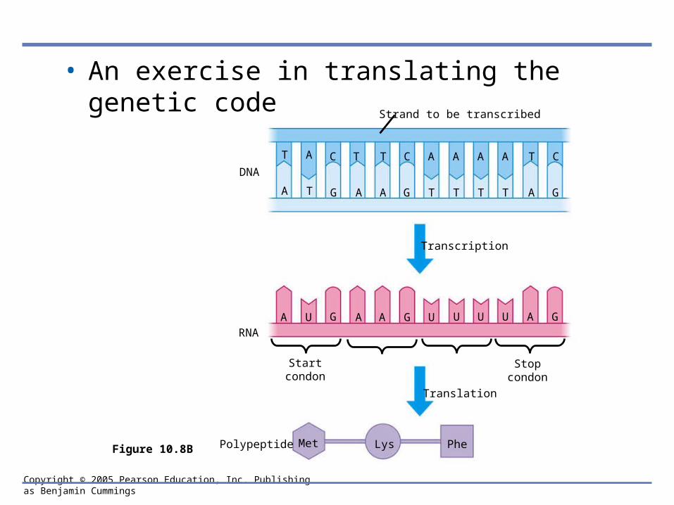
• An exercise in translating the genetic code
Figure 10.8B
T A C T T C A A A A T C
A T G A A G T T T T A G
A U G A A G U U U U A G
Transcription
Translation
RNA
DNA
Met Lys PhePolypeptide
Startcondon
Stopcondon
Strand to be transcribed

Copyright © 2005 Pearson Education, Inc. Publishing as Benjamin Cummings
10.9 Transcription produces genetic messages in the form of RNA
• A close-up view of transcription
RNApolymerase
RNA nucleotides
Direction of transcription
Template Strand of DNA
Newly made RNA
TC
AT C C A A T
T
GG
CC
AATTGGAT
G
U
C A U C C AA
U
Figure 10.9A

Copyright © 2005 Pearson Education, Inc. Publishing as Benjamin Cummings
• In the nucleus, the DNA helix unzips
– And RNA nucleotides line up along one strand of the DNA, following the base pairing rules
• As the single-stranded messenger RNA (mRNA) peels away from the gene
– The DNA strands rejoin
Copyright © 2005 Pearson Education, Inc. Publishing as Benjamin Cummings
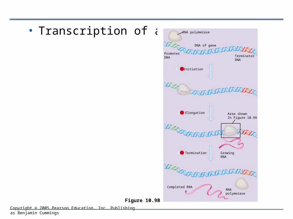
• Transcription of a gene RNA polymerase
DNA of gene
PromoterDNA Terminator
DNA
Area shownIn Figure 10.9A
GrowingRNA
Completed RNARNApolymerase
Figure 10.9B
1 Initiation
2 Elongation
3 Termination

Copyright © 2005 Pearson Education, Inc. Publishing as Benjamin Cummings
10.10 Eukaryotic RNA is processed before leaving the nucleus
• Noncoding segments called introns are spliced out
– And a cap and a tail are added to the endsExon Intron Exon Intron Exon
DNA
Cap TranscriptionAddition of cap and tail
RNAtranscript with capand tail
Introns removedTail
Exons spliced together
mRNA
Coding sequence Nucleus
Cytoplasm
Figure 10.10
Copyright © 2005 Pearson Education, Inc. Publishing as Benjamin Cummings
10.11 Transfer RNA molecules serve as interpreters during translation
• Translation
– Takes place in the cytoplasm
Copyright © 2005 Pearson Education, Inc. Publishing as Benjamin Cummings
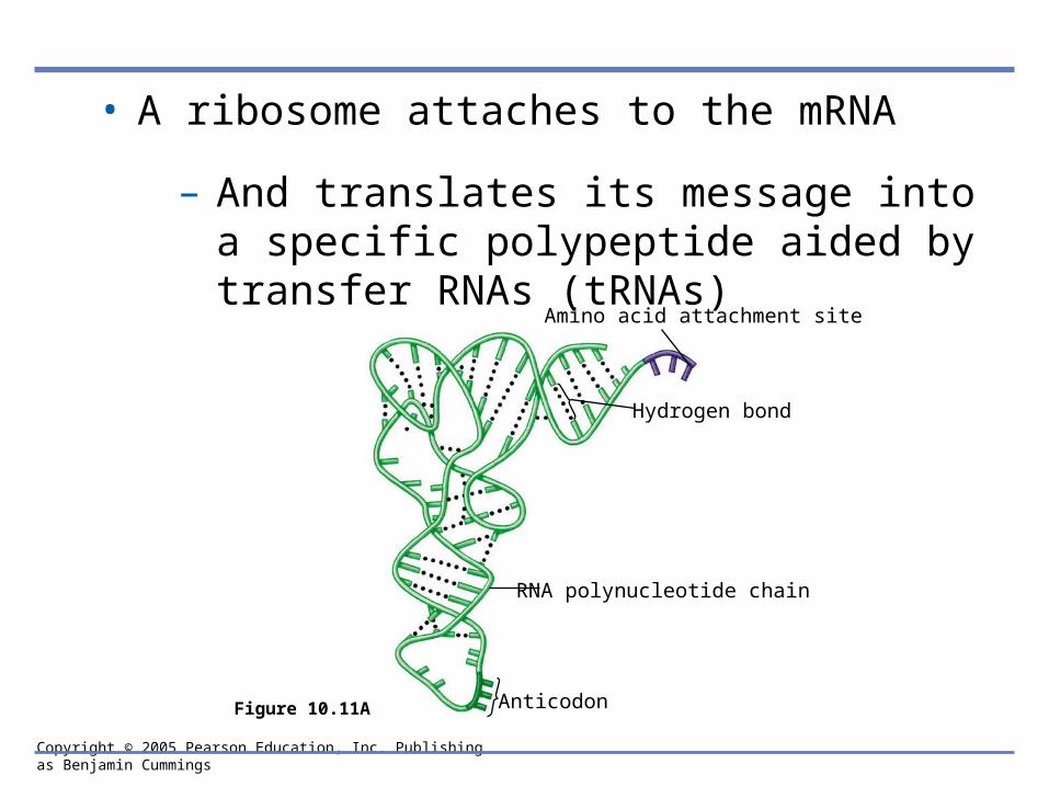
• A ribosome attaches to the mRNA
– And translates its message into a specific polypeptide aided by transfer RNAs (tRNAs)
Amino acid attachment site
Hydrogen bond
RNA polynucleotide chain
AnticodonFigure 10.11A
Copyright © 2005 Pearson Education, Inc. Publishing as Benjamin Cummings
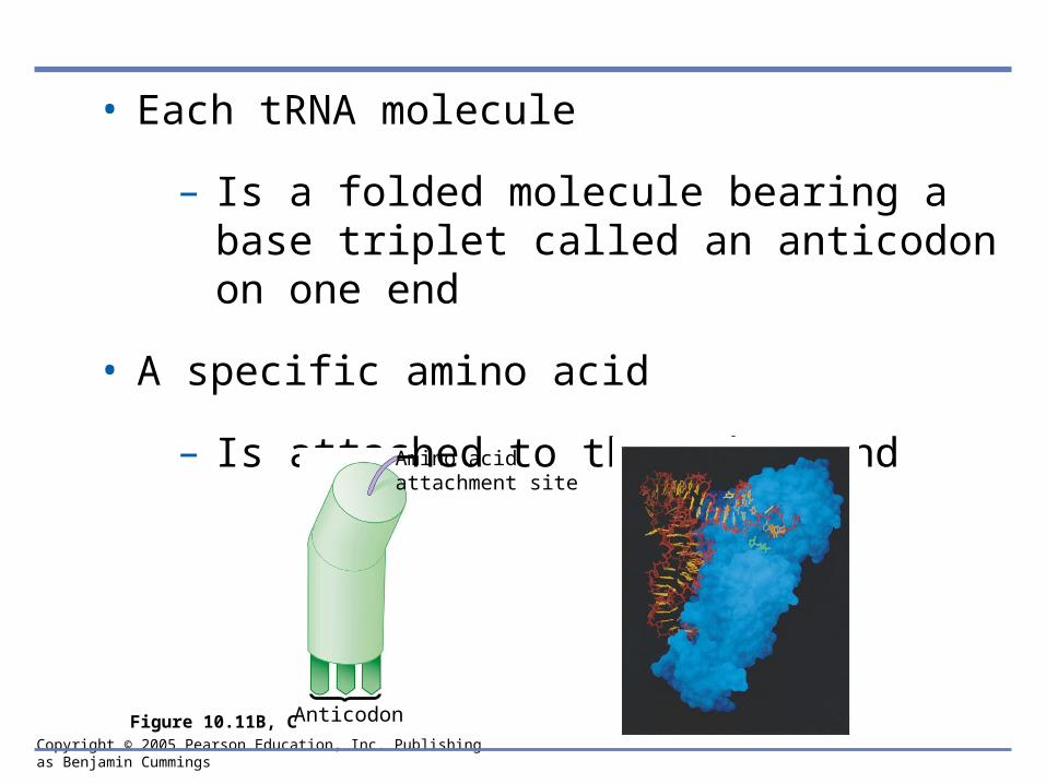
• Each tRNA molecule
– Is a folded molecule bearing a base triplet called an anticodon on one end
• A specific amino acid
– Is attached to the other endAmino acidattachment site
AnticodonFigure 10.11B, C
Copyright © 2005 Pearson Education, Inc. Publishing as Benjamin Cummings
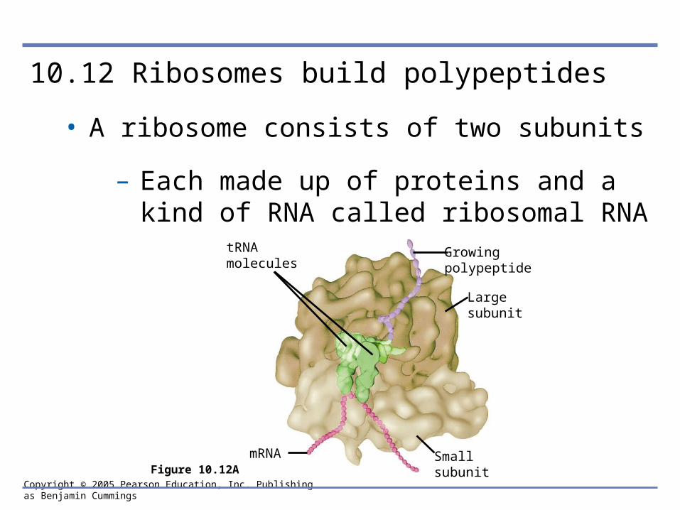
10.12 Ribosomes build polypeptides
• A ribosome consists of two subunits
– Each made up of proteins and a kind of RNA called ribosomal RNA
tRNAmolecules
mRNA Small subunit
Growingpolypeptide
Largesubunit
Figure 10.12A
Copyright © 2005 Pearson Education, Inc. Publishing as Benjamin Cummings
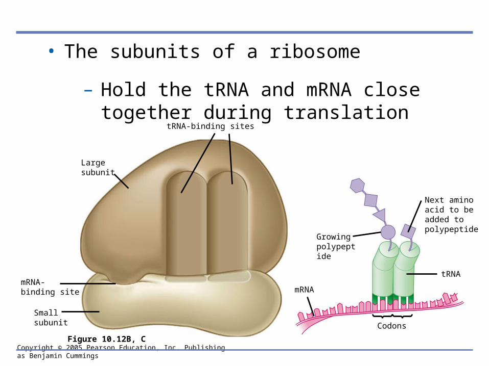
• The subunits of a ribosome
– Hold the tRNA and mRNA close together during translation
Largesubunit
mRNA-binding site
Smallsubunit
tRNA-binding sites
Growing polypeptide
Next amino acid to be added to polypeptide
mRNA
tRNA
Codons
Figure 10.12B, C
Copyright © 2005 Pearson Education, Inc. Publishing as Benjamin Cummings
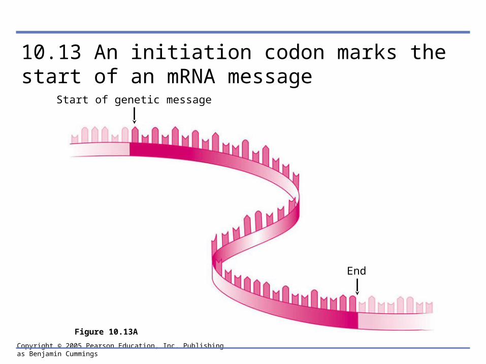
10.13 An initiation codon marks the start of an mRNA message
Start of genetic message
End
Figure 10.13A
Copyright © 2005 Pearson Education, Inc. Publishing as Benjamin Cummings
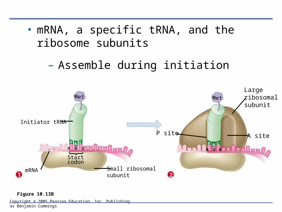
• mRNA, a specific tRNA, and the ribosome subunits
– Assemble during initiation
Met Met
Initiator tRNA
1 2mRNA Small ribosomal
subunit
Startcodon
Large ribosomalsubunit
A siteU A CAU C
A U G A U G
P site
Figure 10.13B
Copyright © 2005 Pearson Education, Inc. Publishing as Benjamin Cummings
10.14 Elongation adds amino acids to the polypeptide chain until a stop codon terminates translation
• Once initiation is complete
– Amino acids are added one by one to the first amino acid
Copyright © 2005 Pearson Education, Inc. Publishing as Benjamin Cummings
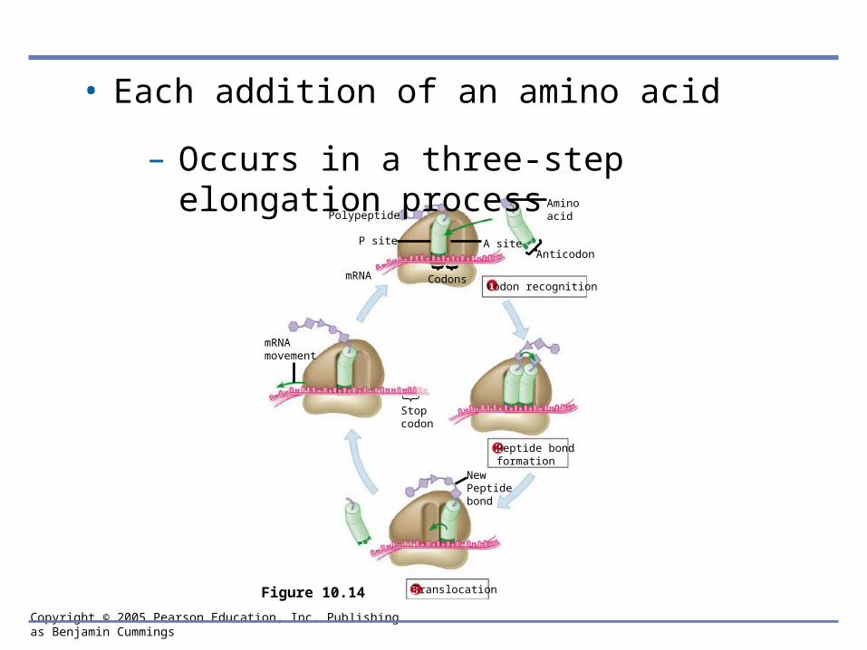
• Each addition of an amino acid
– Occurs in a three-step elongation process
Polypeptide
P site
mRNA Codons
mRNAmovement
Stopcodon
NewPeptidebond
Anticodon
Aminoacid
A site
Figure 10.14
1 Codon recognition
2 Peptide bondformation
3 Translocation

Copyright © 2005 Pearson Education, Inc. Publishing as Benjamin Cummings
• The mRNA moves a codon at a time
– And a tRNA with a complementary anticodon pairs with each codon, adding its amino acid to the peptide chain
Copyright © 2005 Pearson Education, Inc. Publishing as Benjamin Cummings
• Elongation continues
– Until a stop codon reaches the ribosome’s A site, terminating translation

Copyright © 2005 Pearson Education, Inc. Publishing as Benjamin Cummings
10.15 Review: The flow of genetic information in the cell is DNARNAprotein
• The sequence of codons in DNA, via the sequence of codons
– Spells out the primary structure of a polypeptide
Copyright © 2005 Pearson Education, Inc. Publishing as Benjamin Cummings
Polypeptide
TranscriptionDNA
mRNA
RNApolymerase
Amino acid Translation
tRNA
Enzyme
Anticodon
ATP
InitiatortRNA
Largeribosomalsubunit
Start Codon
Codons
mRNA
Stop codon
Smallribosomalsubunit
Growingpolypeptide
New peptidebond forming
mRNA
Figure 10.15
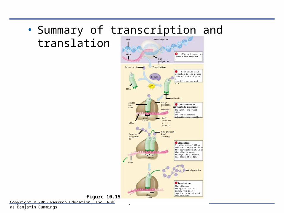
• Summary of transcription and translation
mRNA is transcribed from a DNA template.1
Each amino acidattaches to its propertRNA with the help of aspecific enzyme and ATP.
2
Initiation ofpolypeptide synthesis
The mRNA, the first tRNA,and the ribosomal subunits come together.
3
Elongation4A succession of tRNAsadd their amino acids to the polypeptide chain as the mRNA is moved through the ribosome, one codon at a time.
5
The ribosome recognizes a stop codon. The poly-peptide is terminated and released.
Termination

Copyright © 2005 Pearson Education, Inc. Publishing as Benjamin Cummings
10.16 Mutations can change the meaning of genes
• Mutations are changes in the DNA base sequence
– Caused by errors in DNA replication or recombination, or by mutagens
C T T C A T
Normal hemoglobin
Mutant hemoglobin DNA
G A A G U A
Sickle-cell hemoglobin
Normal hemoglobin DNA
Glu Val
mRNA mRNA
Figure 10.16A
Copyright © 2005 Pearson Education, Inc. Publishing as Benjamin Cummings
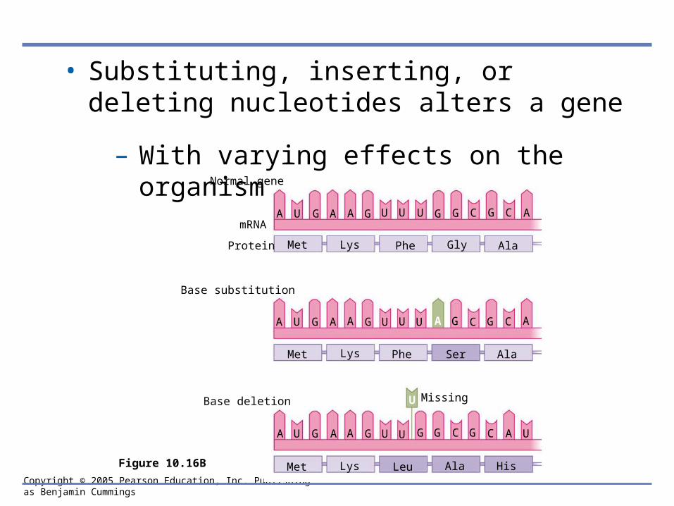
• Substituting, inserting, or deleting nucleotides alters a gene
– With varying effects on the organismNormal gene
mRNA
Base substitution
Base deletion Missing
Met Lys Phe Gly Ala
Met Lys Phe Ser Ala
Met Lys Leu Ala His
A U G A A G U U U G G C G C A
A U G A A G U U U A G C G C A
A U G A A G U U G G C G C A U
U
Protein
Figure 10.16B
Copyright © 2005 Pearson Education, Inc. Publishing as Benjamin Cummings
MICROBIAL GENETICS
10.17 Viral DNA may become part of the host chromosome
• Viruses
– Can be regarded as genes packaged in protein

Copyright © 2005 Pearson Education, Inc. Publishing as Benjamin Cummings
• When phage DNA enters a lytic cycle inside a bacterium
– It is replicated, transcribed, and translated
• The new viral DNA and protein molecules
– Then assemble into new phages, which burst from the host cell
Copyright © 2005 Pearson Education, Inc. Publishing as Benjamin Cummings
• In the lysogenic cycle
– Phage DNA inserts into the host chromosome and is passed on to generations of daughter cells
• Much later
– It may initiate phage production
Copyright © 2005 Pearson Education, Inc. Publishing as Benjamin Cummings
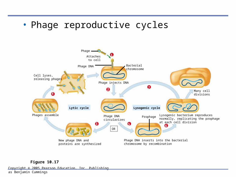
• Phage reproductive cycles
Lysogenic bacterium reproducesnormally, replicating the prophageat each cell division
Phage DNA inserts into the bacterialchromosome by recombination
New phage DNA andproteins are synthesized
Phages assemble
Cell lyses,releasing phages
Phage
Attachesto cell
Phage DNA
Phage injects DNA
Many celldivisions
Prophage
Lytic cycle Lysogenic cycle
OR
Bacterialchromosome
Phage DNAcircularizes
Figure 10.17
1
2
3
4
5 6
7
Copyright © 2005 Pearson Education, Inc. Publishing as Benjamin Cummings
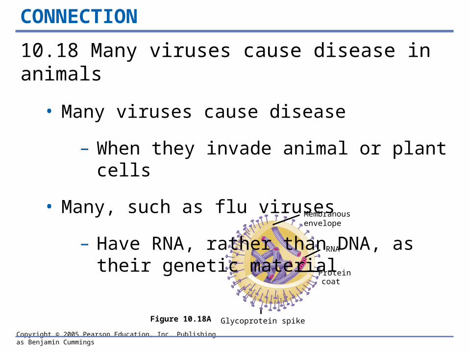
Membranousenvelope
RNA
Proteincoat
Glycoprotein spikeFigure 10.18A
CONNECTION
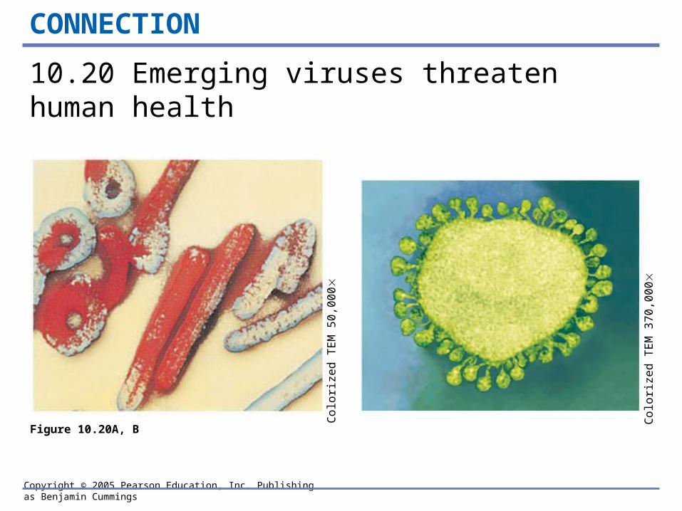
10.18 Many viruses cause disease in animals
• Many viruses cause disease
– When they invade animal or plant cells
• Many, such as flu viruses
– Have RNA, rather than DNA, as their genetic material

Copyright © 2005 Pearson Education, Inc. Publishing as Benjamin Cummings
• Some animal viruses
– Steal a bit of host cell membrane as a protective envelope
– Can remain latent in the host’s body for long periods Glycoprotein spike
EnvelopeProtein coat
Viral RNA(genome)
VIRUS
Plasma membraneof host cell
Viral RNA(genome)
Template
New viralgenome
Exit
mRNA
7
Newviral proteins
Figure 10.18B
Entry1
Uncoating2
RNA synthesisby viral enzyme3
Proteinsynthesis
4 RNA synthesis(other strand)
5
Assembly6
Copyright © 2005 Pearson Education, Inc. Publishing as Benjamin Cummings
CONNECTION
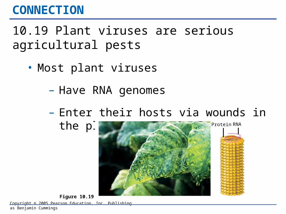
10.19 Plant viruses are serious agricultural pests
• Most plant viruses
– Have RNA genomes
– Enter their hosts via wounds in the plant’s outer layers
Protein RNA
Figure 10.19
Copyright © 2005 Pearson Education, Inc. Publishing as Benjamin Cummings
CONNECTION
10.20 Emerging viruses threaten human health
Co
loriz
ed
TE
M 5
0,0
00
Co
loriz
ed
TE
M 3
70
,00
0
Figure 10.20A, B
Copyright © 2005 Pearson Education, Inc. Publishing as Benjamin Cummings
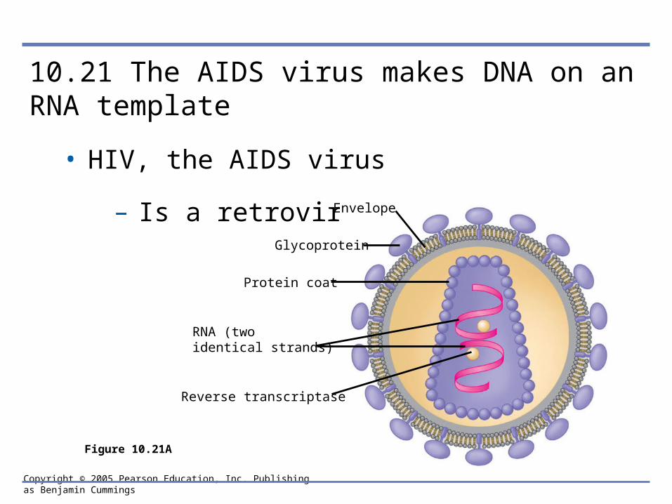
10.21 The AIDS virus makes DNA on an RNA template
• HIV, the AIDS virus
– Is a retrovirus Envelope
Glycoprotein
Protein coat
RNA (two identical strands)
Reverse transcriptase
Figure 10.21A

Copyright © 2005 Pearson Education, Inc. Publishing as Benjamin Cummings
• Inside a cell, HIV uses its RNA as a template for making DNA
– To insert into a host chromosome
Viral RNA
RNAstrand
Double-strandedDNA
Viral RNAand proteins
CYTOPLASM
NUCLEUSChromosomal DNA
Provirus DNA
RNA
Figure 10.21B
1
2
3
45
6

Copyright © 2005 Pearson Education, Inc. Publishing as Benjamin Cummings
10.22 Bacteria can transfer DNA in three ways
• Bacteria can transfer genes from cell to cell by one of three processes
– Transformation, transduction, or conjugation
DNA enterscell
Fragment of DNAfrom anotherbacterial cell
Bacterial chromosome
(DNA)
Phage
Fragment of DNA fromanotherbacterial cell(former phagehost)
Phage
Sex pili
Mating bridge
Donor cell(“male”)
Recipient cell(“female”)
Figure 10.22A–C
Copyright © 2005 Pearson Education, Inc. Publishing as Benjamin Cummings
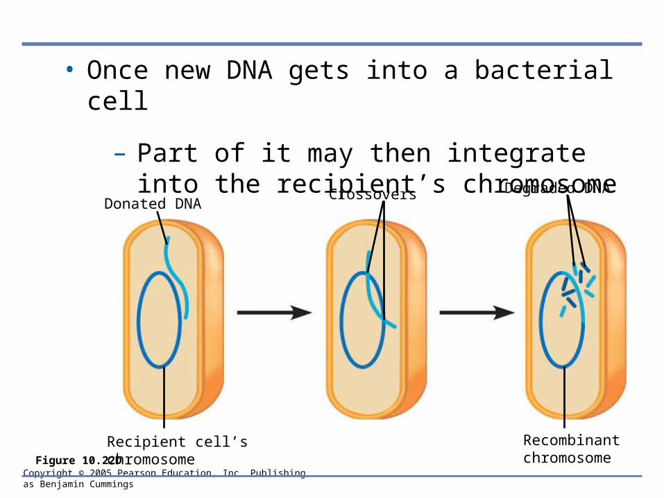
• Once new DNA gets into a bacterial cell
– Part of it may then integrate into the recipient’s chromosome
Recipient cell’schromosome
Recombinantchromosome
Donated DNACrossovers Degraded DNA
Figure 10.22D
Copyright © 2005 Pearson Education, Inc. Publishing as Benjamin Cummings
10.23 Bacterial plasmids can serve as carriers for gene transfer
• Plasmids
– Are small circular DNA molecules separate from the bacterial chromosome
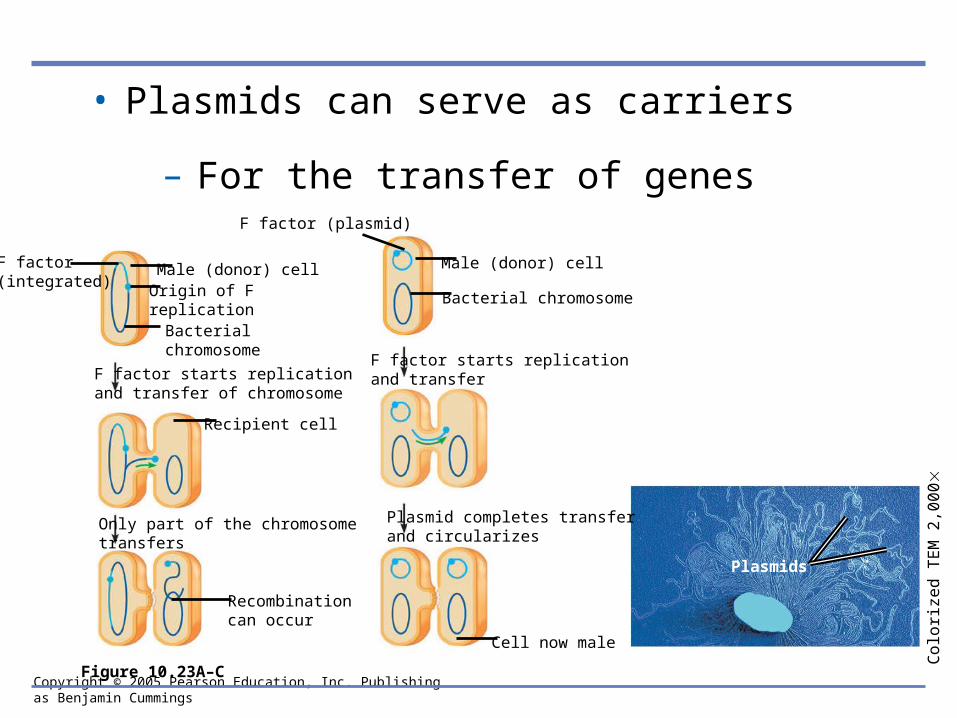
Copyright © 2005 Pearson Education, Inc. Publishing as Benjamin Cummings
• Plasmids can serve as carriers
– For the transfer of genes
Plasmids
Co
loriz
ed
TE
M 2
,00
0
Cell now male
Plasmid completes transferand circularizes
F factor starts replication and transfer
Male (donor) cell
Bacterial chromosome
F factor (plasmid)
Recombination can occur
Only part of the chromosome transfers
F factor starts replication and transfer of chromosome
Origin of F replicationBacterial chromosome
Male (donor) cellF factor (integrated)
Recipient cell
Figure 10.23A–C