Embed Size (px)
Citation preview


MGH PATHOLOGY | i
INTRODUCTION ..............................................................................................................................ii
TRAINING GRANT LABORATORIES ..............................................................................................iii
FACULTY
Molecular Pathology Unit J. Keith Joung .................................................................................................................................1 David M. Langenau ........................................................................................................................2Martin Aryee ..................................................................................................................................3 Atul K. Bhan ...................................................................................................................................4 A. John Iafrate ................................................................................................................................5 David N. Louis .................................................................................................................................6 Luca Pinello ....................................................................................................................................7Miguel N. Rivera .............................................................................................................................8 Dennis C. Sgroi ...............................................................................................................................9 Anat Stemmer-Rachamimov ........................................................................................................10Mario L. Suvà ................................................................................................................................11
Experimental Pathology Unit Bradley Bernstein .........................................................................................................................12 Frederic I. Preffer .........................................................................................................................13 James R. Stone ..............................................................................................................................14
Translational Oncology Laboratory Dora Dias-Santagata ....................................................................................................................15Long Phi Le ...................................................................................................................................16
Immunopathology Research Unit Robert B. Colvin ...........................................................................................................................17 Rex Neal Smith .............................................................................................................................18
Howard Hughes Medical Institute Jeannie T. Lee (Molecular Biology) .............................................................................................19
Pathology Imaging Guillermo J. Tearney (Wellman Center for Photomedicine) .....................................................20
Pathology Faculty Affiliated With Other Departments Matthew P. Frosch: MassGeneral Institute for Neurodegenerative Diseases .........................21 Gad A. Getz: Center for Cancer Research .................................................................................22 John M. Higgins: Center for Systems Biology ...........................................................................23Michael S. Lawrence: Center for Cancer Research ....................................................................24 Andrea I. McClatchey: Center for Cancer Research ..................................................................25 Eric S. Rosenberg: Infectious Diseases .......................................................................................26 Chin Lee Wu: Urology-Pathology Research Laboratory ...........................................................27Ömer H. Yilmaz: Koch Institute for Integrative Cancer Research at MIT ................................28 Lee Zou: Center for Cancer Research .........................................................................................29
TABLE OF CONTENTS

ii | MGH PATHOLOGY
Pathology plays a key role in academic medicine, as a natural bridge between the study of humandisease and experimental biological investigation. Major advances in molecular pathology and inpathology informatics are accelerating the pace of this translational research. In turn, the rapidityand frequency of interactions between the clinical and scientific areas makes this a very exciting timein the field of pathology.
Laboratory-based scientific research is a major component of MGH Pathology, and is complementedby productive clinical research activities. As a result, MGH Pathology provides an exciting stage for basicand translational research. The present brochure highlights the basic scientific research activities inMGH Pathology.
Basic research at MGH Pathology, which is organized under the Division of Research, is divided among a variety of laboratories, both within the Pathology Service and other MGH departments. Peer-review funded investigators in MGH Pathology are centered in the Molecular Pathology Unit, with additional departmental laboratories in the Howard Hughes Medical Institute, the Center for Integrated Diagnostics, the Experimental Pathology Unit, the Pathology Imaging Laboratory, and the Immunopathology Research Unit. In addition, many peer-review funded pathologists and members of the Harvard Medical School Department of Pathology have laboratories in other MGH departments, including the Cancer Center, the Infectious Disease Unit, Neurology, Urology and Wellman Photomedicine.
Basic research activities have been expanded greatly over the past ten years, including: creation of theDivision; recruitment of more than ten basic scientists at the Assistant Professor level (with nearly all as full members of the Center for Cancer Research, five as members of the Harvard-MIT Broad Institute and many as members of the Harvard Medical School Biological and Biomedical Sciences program); addition of five molecular diagnostic pathologists; acquisition of considerable additional Pathology research space that has been extensively renovated; expansion in the number of Harvard and MIT Ph.D.-candidate graduate students training in MGH Pathology laboratories; and provision of competitive pilot grants to junior clinical faculty. As a result, the group has seen an extraordinary increase in the amount of NIH funding over the past decade.
In coordination with the growth of molecular pathology research, molecular diagnostic activities have also expanded greatly, including: development of the MGH Center for Integrated Diagnostics (led by A. John Iafrate); organization of the MGH Pathology component of the Harvard-wide Molecular Genetic Pathology fellowship; extension and further development of the molecular pathology rotation for Pathology residents; and extensive expansion of the CLIA-approved molecular diagnostics laboratory, with implementation of a novel, high-throughput clinical mutation screening program through the Translational Research Laboratory.
We are currently implementing initiatives identified from our recent departmental strategic planning process. With support and resources from the department and the hospital, we are expanding computational biology and bioinformatics resources for pathology, expanding collaborations and interactions with the Center for Integrated Diagnostics, and building additional links between basic and clinical/translational researchers within MGH Pathology. We also plan to continue to recruit additional basic science principal investigators and to develop new research space. These efforts will ensure that MGH Pathology faculty remain at the forefronts of their fields, enabling them to continue advancing our understanding and diagnosis of human diseases.
J. Keith Joung, MD, PhDAssociate Chief of Pathology (Research) and Pathologist, Massachusetts General HospitalProfessor of Pathology, Harvard Medical School
INTRODUCTION

MGH PATHOLOGY | iii
INTRODUCTION
MGH Pathology directs an NIH Training Grant that provides post-doctoral fellowship support for residents wishing to pursue scientific training following their clinical years. MGH Pathology trainees have had considerable success garnering individual grants for research fellowships. Our trainees have been authors on well over 100 publications, in journals that include Cell, Science, Nature, Cancer Cell, Developmental Cell, Molecular Cell, Nature Genetics, Nature Biotechnology, Nature Methods, Current Biology and PNAS. Many trainees undertake post-doctoral fellowships in MGH Pathology laboratories. MGH Pathology trainees have also done fellowships with other investigators, including the following over the past 20 years:
MGH TRAINING GRANT LABORATORIES
Nancy Andrews, MD, PhDChildren’s Hospital
Spyros Artavanis-Tsakonas, PhDHarvard Medical School
David Bartel, PhDWhitehead Institute
Alan Beggs, PhDChildren’s Hospital
Brett Bouma, PhDMGH Wellman Center for Photomedicine
Connie Cepko, PhDHarvard Medical School
George Church, PhDHarvard Medical School
Michael Detmar, MDMGH Cutaneous Biology Research Center
Iain Drummond, PhDMGH Renal Unit
Benjamin Ebert, MD, PhDBrigham & Women’s Hospital
Kevin Eggan, PhDHarvard University
Stephen Elledge, PhDHarvard Medical School
James Fox, PhDMassachusetts Institute of Technology
Frank Gertler, PhDMassachusetts Institute of Technology
Daniel Haber, MD, PhDMGH Center for Cancer Research
Konrad Hochedlinger, PhDMGH Center for Regenerative Medicine
Bradley Hyman, MD, PhDMGH Neurology
Frank Haluska, MD, PhDMGH Hematology-Oncology
Donald Ingber, MD, PhDWyss Institute at Harvard
Ralph Isberg, PhDTufts University
Rudolf Jaenisch, MDMassachusetts Institute of Technology
Rakesh Jain, PhDMGH Radiation Oncology
Jeannie Lee, MD, PhDMGH Molecular Biology
Susan Lindquist, PhDMassachusetts Institute of Technology
Andrea McClatchey, PhDMGH Center for Cancer Research
Matthew Meyerson, MD, PhDBroad Institute
Carl Pabo, PhDMassachusetts Institute of Technology
Shiv Pillai, MD, PhDMGH Center for Cancer Research
Sridhar Ramaswamy, MDMGH Center for Cancer Research
David Sabatini, MD, PhDMassachusetts Institute of Technology
David Scadden, MDMGH Hematology-Oncology
Jeffrey Settleman, PhDMGH Center for Cancer Research
Phillip Sharp, PhDMassachusetts Institute of Technology
Carla Shatz, PhDHarvard Medical School
Melissa Suter, PhDMGH Wellman Center for Photomedicine
Jay Vacanti, MD, PhDMGH Pediatric Surgery
Amy Wagers, PhDHarvard University
Robert Weinberg, PhDMassachusetts Institute of Technology
Ramnik Xavier, MD, PhDMGH Gastrointestinal Unit
Gary Yellen, PhDHarvard Medical School
Lee Zou, PhDMGH Center for Cancer Research

1 | MGH PATHOLOGY
MOLECULAR PATHOLOGY UNIT
“Genome-editing nuclease technologies have important applications inbiological research and gene therapy…”
Selected PublicationsKleinstiver BP, Pattanayak V, Prew MS, Tsai SQ, Nguyen NT, Zheng Z, Joung JK. High-fidelity CRISPR-Cas9 nucleases with no detectable genome-wide off-target effects. Nature. 2016; 529(7587): 490-5.
Tsai SQ, Joung JK. Defining and improving the genome-wide specificities of CRISPR-Cas9 nucleases. Nat Rev Genet. 2016; 17(5): 300-12. Review
Kleinstiver BP, Prew MS, Tsai SQ, Nguyen NT, Topkar VV, Zheng Z, Joung JK. Broadening the targeting range of Staphylococcus aureus CRISPR-Cas9 by modifying PAM recognition. Nat Biotechnol. 2015; 33(12): 1293-1298.
Kleinstiver BP, Prew MS, Tsai SQ, Topkar VV, Nguyen NT, Zheng Z, Gonzales AP, Li Z, Peterson RT, Yeh JR, Aryee MJ, Joung JK. Engineered CRISPR-Cas9 nucleases with altered PAM specificities. Nature. 2015; 523(7561): 481-5.
Tsai SQ, Zheng Z, Nguyen NT, Liebers M, Topkar VV, Thapar V, Wyvekens N, Khayter C, Iafrate AJ, Le LP, Aryee MJ, Joung JK. GUIDE-seq enables genome-wide profiling of off-target cleavage by CRISPR-Cas nucleases. Nat Biotechnol. 2015; 33(2): 187-97.
The Joung laboratory is developing strategies to reprogram the genome and epigenome of living cells to better understand biology and treat disease. We have developed and optimized molecular tools for customized genome editing that enable scientists to alter the DNA sequence of a living cell—from fruit flies to humans—with great precision. These technologies are based on proteins engineered to recognize and cleave specific genomic sequences. We also use these targeting methodologies to enable activation, repression, or alteration of histone modifications of specific genes. These tools have many potential research uses and may one day lead to more efficient gene therapy capable of correcting disease-related mutations in human cells.
Genome Editing Using Targeted Nucleases Genome editing technology using CRISPR-Cas9 nucleases was recently named “Breakthrough of the Year” for 2015 by Science magazine. Much of our recent work with genome-editing nucleases has focused on CRISPR-Cas9. We and our collaborators were the first to demonstrate that these nucleases can function in vivo (Hwang & Fu et al., Nat Biotechnol. 2013), modifying endogenous genes in zebrafish embryos and the first to show that they can induce significant off-target mutations in human cells (Fu et al., Nat Biotechnol. 2013). We recently developed GUIDE-seq, an unbiased, genome-wide method for sensitive detection of CRISPR-Cas9-induced off-target mutations in human cells (Tsai et al., Nat Biotechnol. 2015). Using structure-guided design, we have engineered “high-fidelity” Cas9 variants that robustly fail to show detectable genome-wide off-targets as judged by GUIDE-seq (Kleinstiver & Pattanayak et al., Nature 2016). Finally, we used a combination of structure-guided design and molecular evolution to engineer Cas9 variants with novel DNA binding specificities, thereby broadening the targeting range and applications of this platform (Kleinstiver et al., Nature 2015).
Epigenome Editing Using Targeted Transcription Factors We have also demonstrated that the TALE and CRISPR platforms can also be utilized to create artificial transcription factors that can robustly alter expression of endogenous human genes (Maeder et al., Nat Methods 2013a; Maeder et al., Nat Methods 2013b). In addition, we have collaborated with Brad Bernstein’s group to develop fusions of the histone demethylase LSD1 with TALE domains that can induce targeted histone alterations at endogenous human enhancers (Mendenhall et al., Nat Biotechnol. 2013). Finally, we have also developed fusions of engineered TALE domains with the catalytic domain of the TET1 enzyme, enabling the targeted demethylation of CpGs in human cells (Maeder et al., Nat Biotechnol. 2013).
J. Keith Joung, MD, PhDProfessor of Pathology, Harvard Medical SchoolAssociate Chief of Pathology (Research) andThe Jim and Ann Orr MGH Research Scholar,Massachusetts General Hospital
Molecular Pathology UnitMassachusetts General Hospital149 13th Street, 6th FloorCharlestown, MA 02129Phone: 617-726-9462 • Email: [email protected]

MGH PATHOLOGY | 2
MOLECULAR PATHOLOGY UNIT
Selected PublicationsMoore FE, Garcia EG, Lobbardi R, Jain E, Tang Q, Moore JC, Cortes M, Molodtsov A, Kasheta M, Luo CC, Garcia AJ, Mylvaganam R, Yoder JA, Blackburn JS, Sadreyev RI, Ceol CJ, North TE, Langenau DM. Single-cell transcriptional analysis of normal, aberrant, and malignant hematopoiesis in zebrafish. J Exp Med. 2016; 213(6):979-92.
Tang Q, Moore JC, Ignatius MS, Tenente IM, Hayes MN, Garcia EG, Torres Yordán N, Bourque C, He S, Blackburn JS, Look AT, Houvras Y, Langenau DM. Imaging tumour cell heterogeneity following cell transplantation into optically clear immune-deficient zebrafish. Nat Commun. 2016; 7:10358.
Tang Q, Abdelfattah NS, Blackburn JS, Moore JC, Martinez SA, Moore FE, Lobbardi R, Tenente IM, Ignatius MS, Berman JN, Liwski RS, Houvras Y, Langenau DM. Optimized cell transplantation using adult rag2 mutant zebrafish. Nature Methods. 2014; 11(8):821-4.
Blackburn JS, Liu S, Wilder JL, Dobrinski KP, Lobbardi R, Moore FE, Martinez SA, Chen EY, Lee C, Langenau DM. Clonal evolution enhances leukemia-propagating cell frequency in T-cell acute lymphoblastic leukemia through AKT/mTORC1 pathway activation. Cancer Cell. 2014; 25(3):366-78.
Chen EY, DeRan M, Ignatius MS, Grandinetti KB, Clagg R, McCarthy K, Lobbardi RM, Brockmann J, Keller C, Wu X, Langenau DM. GSK3 inhibitors induce the canonical WNT/b-catenin pathway to suppress growth and self-renewal in embryonal rhabdomyosarcoma. PNAS. 2014; 111(14):5349-54.
Ignatius MS, Chen E, Elpek NE, Fuller A, Tenente IM, Clagg R, Liu S, Blackburn JS, Linardic CM, Rosenberg A, Nielsen PG, Mempel TR, Langenau DM. In vivo imaging of tumor-propagating cells, regional tumor heterogeneity and dynamic cell movements in embryonal rhabdomyosarcoma. Cancer Cell. 2012; 21(5):680-93.
“Identifying molecular pathways that drive progression and relapse in pediatric cancer…”
David M. Langenau, PhDAssociate Professor of Pathology, Harvard Medical SchoolDirector, Molecular Pathology Unit, Massachusetts General HospitalMember, MGH Cancer Center and Center for Regenerative Medicine
Massachusetts General HospitalMolecular Pathology Unit149 13th Street, 6th FloorCharlestown, MA 02129Phone: 617-643-6508Email: [email protected] • langenaulab.com
The Langenau laboratory research focus is to uncover relapse mechanisms in pediatric cancer. Utilizing zebrafish models of T-cell acute lymphoblastic leukemia (T-ALL) and embryonal rhabdomysoarcoma (ERMS), we have undertaken chemical and genetic approaches to identify novel modulators of progression, therapy-resistance, and relapse.
Uncovering progression-associated driver mutations in T-cell acute lymphoblastic leukemiaT-ALL is an aggressive malignancy of thymocytes that affects thousands of children and adults in the United States each year. Recent advancements in conventional chemotherapies have improved the five-year survival rate of patients with T-ALL. However, patients with relapse disease are largely unresponsive to additional therapy and have a very poor prognosis. Ultimately, 70% of children and 92% of adults will die of relapse T-ALL, underscoring the clinical imperative for identifying the molecular mechanisms that cause leukemia cells to re-emerge at relapse. Utilizing a novel zebrafish model of relapse T-ALL, large-scale trangenesis platforms, and unbiased bioinformatic approaches, we have uncovered new oncogenic drivers associated with aggression, therapy resistance and relapse. A large subset of these genes exert important roles in regulating human T-ALL proliferation, apoptosis and response to therapy. Discovering novel relapse-driving oncogenic pathways will likely identify new drug targets for the treatment of T-ALL.
Visualizing and killing cancer stem cells in embryonal rhabdomyosarcomaERMS is a common soft-tissue sarcoma of childhood and phenotypically recapitulates fetal muscle development arrested at early stages of differentiation. Microarray and cross-species comparisons of zebrafish, mouse and human ERMS uncovered the finding that the RAS pathway is activated in a majority of ERMS. Building on this discovery, our laboratory has developed a transgenic zebrafish model of kRASG12D-induced ERMS that mimics the molecular underpinnings of human ERMS. We used fluorescent transgenic zebrafish that label ERMS cell subpopulations based on myogenic factor expression to identify functionally distinct classes of tumor cells contained within the ERMS mass. Specifically, the myf5-GFP+ self-renewing cancer stem cell drives continued tumor growth at relapse and is molecularly similar to a non-transformed, activated muscle satellite cell. Building on the dynamic live cell imaging approaches available in the zebrafish ERMS model, our laboratory has undertaken chemical genetic approaches to identify drugs that kill relapse-associated, self-renewing myf5-GFP+ ERMS cells. We are currently assessing a subset of drugs for their ability to regulate growth of human ERMS cells and mouse xenografts.

3 | MGH PATHOLOGY
MOLECULAR PATHOLOGY UNIT
My research involves computational methods that enable us to elucidate the genetic and epigenetic basis of cancer and other diseases from large genomic datasets.
Tumor Heterogeneity We develop statistical methods to improve our understanding of tumor cell-to-cell variability and its relationship to cancer progression. Much of this work relates to the computational and statistical challenges posed by single-cell transcriptome and epigenome data.
Different tumors, even of the same type, can harbor extremely heterogeneous genetic and epigenetic alterations. To investigate the role of epigenetic stochasticity in cancer, we recently applied a statistical model to study patterns of inter- and intra-individual tumor heterogeneity during metastasis. We established that metastatic prostate cancer patients develop distinctly unique DNA methylation signatures that are subsequently maintained across metastatic dissemination. The stability of these individualized DNA methylation profiles has implications for the promise of epigenetic alterations as diagnostic and therapeutic targets in cancer.
Epigenome MappingUnlike genome sequencing which has well established experimental and analytical protocols, epigenome mapping strategies are still in their infancy and, like other high-throughput techniques, are plagued by technical artifacts. A central theme of our research involves the development of methods for extracting signal from noisy high-throughput genomic assays. The goal of such preprocessing methods is transform raw data from high-throughput assays into reliable measures of the underlying biological process. Until recently, studies of DNA methylation in cancer had focused almost exclusively on CpG dense regions in gene promoters. We helped develop the statistical tools used to analyze the first genome-scale DNA methylation assays designed without bias towards CpG islands. These tools enabled the discovery that the majority of both tissue-specific and cancer-associated variation occurs in regions outside of CpG islands. We showed that there is a strong overlap between genomic regions involved in normal tissue differentiation, reprogramming during induced pluripotency, and cancer.
Epigenomic Studies of Complex Disease Despite the discovery of numerous disease-associated genetic variants, the majority of phenotypic variance remains unexplained for most diseases, suggesting that non-genetic factors play a significant role. Part of the explanation will lie in a better understanding of epigenetic mechanisms. These mechanisms are influenced by both genetic and environmental effects and, as downstream effectors of these factors, may be more directly related to phenotype. However, the broad extent of epigenetic dysregulation in cancer and many other diseases complicates the search for the small subset of alterations with a causal role in pathogenesis. We are developing computational methods to integrate genome-wide genetic and epigenetic data with the goal of identifying the subset of functionally important epigenetic alterations.
“Computational methods for genomics and epigenomics...”Selected PublicationsZiller MJ, Hansen KD, Meissner A, Aryee MJ. Coverage recommendations for methylation analysis by whole-genome bisulfite sequencing. Nat Methods. 2015; 12(3): 230-2.
Aryee MJ, Jaffe AE, Corrada-Bravo H, Ladd-Acosta C, Feinberg AP, Hansen KD, Irizarry RA. Minfi: a flexible and comprehensive Bioconductor package for the analysis of Infinium DNA methylation microarrays. Bioinformatics. 2014; 30(10):1363-9.
Aryee MJ, Liu W, Engelmann JC, Nuhn P, Gurel M, Haffner MC, Esopi D, Irizarry RA, Getzenberg RH, Nelson WG, Luo J, Xu J, Isaacs WB, Bova GS, Yegnasubramanian S. DNA methylation alterations exhibit intraindividual stability and interindividual heterogeneity in prostate cancer metastases. Sci Transl Med. 2013; 5(169):169ra10.
Liu Y*, Aryee MJ*, Padyukov L, Fallin MD, Hesselberg E, Runarsson A, Reinius L, Acevedo N, Taub M, Ronninger M, Shchetynsky K, Scheynius A, Kere J, Alfredsson L, Klareskog L, Ekström TJ, Feinberg AP. Epigenome-wide association data implicate DNA methylation as an intermediary of genetic risk in rheumatoid arthritis. Nat Biotechnol. 2013; 31(2):142-7.
Aryee MJ, Wu Z, Ladd-Acosta C, Herb B, Feinberg AP, Yegnasubramanian S, Irizarry RA. Accurate genome-scale percentage DNA methylation estimates from microarray data. Biostatistics. 2011; 12(2):197-210.
Martin Aryee, PhDAssistant Professor of Pathology, Harvard Medical SchoolAssistant Molecular Pathologist, Massachusetts General Hospital
Molecular Pathology UnitMassachusetts General Hospital149 13th Street, 6th FloorCharlestown, MA 02129Phone: 617-726-5690Email: [email protected]
MGH PATHOLOGY | 4
MOLECULAR PATHOLOGY UNIT
Selected PublicationsLLassen KG, McKenzie CI, Mari M, et al. Genetic coding variant in GPR65 alters lysosomal pH and links lysosomal dysfunction with colitis risk. Immunity. 2016 (in press).
Lassen KG, Kuballa P, Conway KL, et al. Atg16L1 T300A variant decreases selective autophagy resulting in altered cytokine signaling and decreased antibacterial defense. Proc Natl Acad Sci U S A 2014;111:7741-6.
Shouval DS, Biswas A, Goettel JA, et al. Interleukin-10 receptor signaling in innate immune cells regulates mucosal immune tolerance and anti-inflammatory macrophage function. Immunity. 2014;40:706-19.
Kaliannan K, Hamarneh SR, Economopoulos KP, et al. Intestinal alkaline phosphatase prevents metabolic syndrome in mice. Proc Natl Acad Sci U S A. 2013;110:7003-8.
Chang SY, Song JH, Guleng B, et al. Circulatory antigen processing by mucosal dendritic cells controls CD8+ T cell. Immunity. 2013;38:153-165.
Yilmaz OH, Katajisto P, Lamming DW, et al. mTORC1 in the Paneth cell niche couples intestinal stem-cell function to calorie intake. Nature. 2012;486:490-5.
Sugimoto K, Ogawa A, Shimomura Y, et al. Inducible IL-12-producing B cells regulate Th2-mediated intestinal inflammation. Gastroenterology. 2007;133:124-36.
Mizoguchi A, Ogawa A, Takedatsu H, et al. Dependence of intestinal granuloma formation on unique myeloid DC-like cells. J Clin Invest. 2007;117:605-615.
“Researching mucosal immunology and inflammatory bowel disease...”
Atul K. Bhan, MBBS, MDProfessor of Pathology, Harvard Medical SchoolPathologist, Massachusetts General Hospital
Massachusetts General Hospital55 Fruit StreetWarren Building, Room 501Boston, MA 02114Phone: 617-726-2588 • Fax: 617-726-2365Email: [email protected]
In the two major forms of IBD, Crohn’s disease and ulcerative colitis the underlying etiological factors and the pathogenesis remain poorly defined. It is generally believed that exaggerated immune responses to luminal normal enteric flora are involved in the initiation and perpetuation of the disease process.
The availability of a wide variety of experimental models of intestinal inflammation has helped provide important clues about the pathogenesis of IBD. The commonly used models include chemically induced mucosal injury and colitis induced by the transfer of selected populations of T cells into immunodeficient mice. The spontaneous development of colitis in genetically engineered animal models has provided excellent experimental models to study the pathogenesis of IBD. One important lesson learned from IBD models is that many different immunologic and mucosal defects can lead to similar pathologic findings.
For the last several years, our laboratory has focused on defining the pathogenesis of chronic intestinal inflammation using TCR alpha KO mice as a model of human IBD. TCR alpha KO mice develop spontaneously chronic colitis with many features of ulcerative colitis. We have identified a regulatory B cell subset, which appears under chronic intestinal inflammatory conditions and suppresses the progression of intestinal inflammation by secreting IL-10. TCR alpha KO mice deficient in both IL-4 and B cells, but not in IL-4 alone, develop granulomatous colitis with features of Crohn’s disease. This suggests that differences in the two major forms of IBD may reflect different immunological responses to similar initiating events.
The laboratory is closely associated with the Center for the Study of Inflammatory Bowel Disease at MGH and collaborates with the other members of the Center; Dr. Bhan is an Associate Director of the Center. In collaboration with Dr. Terhorst and Dr. Xavier we have studied the role of Th-1 and Th-17 pathways, innate immune system and autophagy in the development of intestinal inflammation. Collaborative studies with Dr. Scott Snapper’s laboratory have shown that interleukin-10 receptor signaling in innate immune cells regulates mucosal immune tolerance and anti-inflammatory macrophage function. The studies with Dr. Richard Hodin’s laboratory indicate that administration of intestinal alkaline phosphatase may have a beneficial effect in intestinal inflammatory conditions and metabolic syndromes. Dr. Bhan’s consultant role in the newly established Harvard Institute of Translational Immunology-Helmsley Pilot Program in Crohn’s Disease has led to his collaboration with Dr. Vijay Yajnik at MGH and Dr. Matthew Myerson at DFCI to identify microorganisms in Crohn’s disease lesions.

5 | MGH PATHOLOGY
Selected PublicationsZheng Z, Liebers M, Zhelyazkova B, Cao Y, Panditi D, Chen J, Robinson HE, Chmielecki J, Pao W, Engelman JA, Iafrate AJ*, Le LP*: Anchored multiplex PCR for targeted next-generation sequencing. Nat Medicine. 2014; 20(12):1479-84.
Bergethon K, Shaw AT, Ignatius Ou SH, Katayama R, Lovly CM, McDonald NT, Massion PP, Siwak-Tapp C, Gonzalez A, Fang R, Mark EJ, Batten JM, Chen H, Wilner KD, Kwak EL, Clark JW, Carbone DP, Ji H, Engelman JA, Mino-Kenudson M, Pao W, Iafrate AJ: ROS1 rearrangements define a unique molecular class of lung cancers. J Clin Oncol. 2012; 10;30(8):863-70.
Snuderl M, Fazlollahi L, Le LP, Nitta M, Zhelyazkova BH, Davidson CJ, Akhavanfard S, Cahill DP, Aldape KD, Betensky RA, Louis DN, Iafrate AJ: Mosaic amplification of multiple receptor tyrosine kinase genes in glioblastoma. Cancer Cell. 2011; 20:810-7.
Kwak EL, Bang Y, Camidge DR, ShawAT, Solomon B, Maki RG, Ou SI, Dezube BJ, Jänne PA, Costa DB, Varella-Garcia M, Kim W, Lynch TJ, Fidias P, Stubbs H, Engelman JA, Sequist LV, Tan W, Gandhi L, Mino-Kenudson M, Wei GC, Shreeve SM, Ratain MJ, Settleman J, Christensen JG, Haber DA, Wilner K, Salgia R, Shapiro GI, Clark JW, Iafrate AJ. 2010. Response of non-small cell lung cancers with Anaplastic Lymphoma Kinase (ALK) gene rearrangements to a targeted ALK inhibitor. N Engl J Med. 2010; 363(18):1693-703.
Dias-Santagata D, Akhavanfard S, David SS, Vernovsky K, Kuhlmann G, Boisvert SL, Stubbs H, McDermott U, Settleman J, Kwak EL, Clark JW, Isakoff SJ, Sequist LV, Engelman JA, Lynch TJ, Haber DA, Louis DN, Ellisen LW, Borger DR, Iafrate AJ. 2010. Rapid targeted mutational analysis of human tumours: a clinical platform to guide personalized cancer medicine. EMBO Mol Med. 2010; 2(5):146-58.
Iafrate AJ, Feuk L., Rivera MN, Listewnik ML, Donahoe PK, Qi Y, Scherer SW, Lee C. 2004. Detection of large-scale variation in the human genome. Nat. Genet. 2004; 36(9):949-51.
“To identify actionable genetic alterations in cancer...”
A. John Iafrate, MD, PhDProfessor of Pathology, Harvard Medical SchoolPathologist, Massachusetts General HospitalAssociate Chief of Pathology, Center for Integrated Diagnostics
Molecular Pathology UnitMassachusetts General Hospital55 Fruit StreetBoston, MA 02114Phone: 617-726-0166 • Fax: 617-726-5079 Email: [email protected]
Our lab has focused efforts on translating highly complex molecular analyses of tumor genetics using novel technologies into clinical use. We have previously developed the SNaPshot genotyping assay, which has enabled Mass General to make personalized cancer medicine a priority. We have a strong interest in the clinical implementation of genetic screening technologies that can help direct targeted therapies, focusing on lung, pancreatic and brain tumors. Our recent contributions in the treatment of a subset of lung tumors with rearrangements of the ALK tyrosine kinase and with rearrangements of the ROS1 tyrosine kinase with a small molecule kinase inhibitor underscore the promise of personalized cancer care. Our long term goal is to develop high-throughput genetic screening approaches for all cancer patients. To address this need, we have developed a novel next generation sequencing technique termed “anchored multiplex PCR (AMP)”, that is especially powerful at detection gene fusion events from clinical specimens. We have shown that AMP is a sensitive as FISH in diagnosing ALK, ROS1 and RET fusions in lung cancer, and does not require knowing both fusion partners. In addition, AMP can be used for genomic DNA target enrichment, and is scalable and cost effective. Current work focuses on ultrasensitive detection of mutations in blood and urine.
We have also continued prior studies of tumor heterogeneity, by studying gene amplification of receptor tyrosine kinases in glioblastoma. This work has revealed a new subclass of brain tumors with mosaic gene amplification of up to 3 kinases in distinct but intermingled cell populations within the same tumor. We are exploring the therapeutic implications of such driver gene heterogeneity in model systems of glioblastoma using patient derived cell lines and xenografts. A major effort here has been the development of multiplexed in situ genetic analysis using FISH. These techniques will allow us to analyze many more genes, and map copy number heterogeneity onto histology sections.
Our laboratory has also focused on human germline genetics, namely on copy number variation (CNVs). These polymorphisms involve copy number gains or losses of large genomic regions (kilobases up to several megabases), and were identified using high-resolution genomic microarrays to compare the genomes of phenotypically normal individuals. Our continuing work is focused on the detailed structural analysis of CNVs using high resolution fluorescence microscopy imaging techniques, quantitative PCR and BAC sequencing. We have developed novel FISH probes based on deletion CNVs that can be used to determine genetic identity in situ. These probes are being applied to chimerism analysis in transplantation and will aid in the study of engraftment, rejection, and graft versus host disease. Importantly, these probes are located on autosomes, so for the first time chimerism analysis can be performed in same sex transplants.
MOLECULAR PATHOLOGY UNIT
MGH PATHOLOGY | 6
MOLECULAR PATHOLOGY UNIT
Selected PublicationsLouis DN, Ohgaki H, Wiestler OD, Cavenee WK (eds.). World Health Organization Histological Classification of Tumours of the Central Nervous System. Lyon: International Agency for Research on Cancer, 2016.
Louis DN, Perry A, Reifenberger G, von Deimling A, Figarella-Branger D, Cavenee WK, Ohgaki H, Wiestler OD, Kleihues P, Ellison DW. The 2016 WHO classification of tumours of the central nervous system: a summary. Acta Neuropathol. 2016; 131(6):803-820.
Tanboon J, Williams EA, Louis DN. The diagnostic use of immunohistochemical surrogates for signature molecular genetic alterations in gliomas. J Neuropathol Exp Neurol. 2016; 75:4-18.
Louis DN. Perry A, Burger P, et al. International Society of Neuropathology-Haarlem consensus guidelines for nervous system tumor classification and grading. Brain Pathol. 2014; 24:429-435.
“Elucidating the molecular basis of glioma formation impacts both diagnostic and therapeutic aspects of clinical neuro-oncology…”
David N. Louis, MDBenjamin Castleman Professor of Pathology, Harvard Medical SchoolPathologist-in-Chief, Massachusetts General Hospital
Pathology ServiceMassachusetts General Hospital55 Fruit Street (WRN-2)Boston, MA 02114Email: [email protected]
Over the past 25 years, we have demonstrated alterations characteristic of specific glioma subtypes and grades. We originally demonstrated that molecular genetic analysis could be used to define clinicopathologically relevant subsets of glioblastomas, and then showed that molecular genetic alterations are powerful predictors of therapeutic response and survival in patients with anaplastic oligodendrogliomas and in other oligodendroglial tumors. These findings have already led to incorporation of molecular diagnostic testing around the world for these parameters. Work over the past few years has been directed toward incorporating molecular testing into the World Health Organization Classification of Central Nervous System Tumors, a process directed by Dr. Louis, and toward making molecular diagnostics a practical and routine part of brain tumor diagnosis. The lab has also demonstrated that glioblastomas treated with the alkylating agent temozolomide (which is now the standard of care for such cases) frequently inactivate mismatched repair genes, leading to more rapid growth during therapy and to therapeutic resistance, and has worked collaboratively on epigenetic and single-cell studies of high-grade gliomas.
7 | MGH PATHOLOGY
Selected PublicationsPinello L, Canver MC, Hoban MD, Orkin SH, Kohn DB, Bauer DE, Yuan GC. Analyzing CRISPR genome-editing experiments with CRISPResso. Nat Biotechnol. 2016;34(7):695-7.
Guo G*, Pinello L*, Han X, Lai S, Shen L, Lin TW, Zou K, Yuan GC, Orkin SH. Serum-Based Culture Conditions Provoke Gene Expression Variability in Mouse Embryonic Stem Cells as Revealed by Single-Cell Analysis. Cell Rep. 2016;14(4):956-65.
Canver MC*, Smith EC*, Sher F*, Pinello L*, Sanjana NE*, Shalem O, Chen DD, Schupp PG, Vinjamur DS, Garcia SP, Luc S, Kurita R, Nakamura Y, Fujiwara Y, Maeda T, Yuan G-C, Zhang F, Orkin SH & Bauer DE. BCL11A enhancer dissection by Cas9-mediated in situ saturating mutagenesis. Nature. 2015; 527(7577):192-7.
Wu JN*, Pinello L*, Yissachar E, Wischhusen JW, Yuan GC, Roberts CW. Functionally distinct patterns of nucleosome remodeling at enhancers in glucocorticoid-treated acute lymphoblastic leukemia. Epigenetics Chromatin. 2015;8:53.
Pinello L*, Xu J*, Orkin SH, Yuan GC. Analysis of chromatin state plasticity identifies cell-type specific regulators of H3K27me3 patterns, PNAS 2014; 111(3):E344-53.
Pulakanti K*, Pinello L*, Stelloh C, Blinka S, Allred J, Milanovich S, Kiblawi S, Peterson J, Wang A, Yuan GC, Rao S. Enhancer transcribed RNAs arise from hypomethylated, Tet-occupied genomic regions. Epigenetics. 2013; 8(12).
“Understanding gene regulation using computational methods for epigenomics, genome editing and single cell analysis...”
Luca Pinello, PhDAssistant Professor of Pathology, Harvard Medical SchoolAssistant Pathologist, Massachusetts General Hospital
Molecular Pathology UnitMassachusetts General Hospital149 13th Street, 6th FloorCharlestown, MA 02129Phone: 617-643-6522Email: [email protected]
The focus of the Pinello laboratory is to use innovative computational approaches and cutting-edge experimental assays to systematically analyze sources of genetic and epigenetic variation and (single-cell) gene expression variability that underlie human traits and diseases. The lab uses machine learning, data mining and high performance computing technologies, for instance parallel computing and cloud-oriented architectures, to solve computationally challenging and Big Data problems associated with next generation sequencing data analysis. Our mission is to use computational strategies to further our understanding of disease etiology and to provide a foundation for the development of new drugs and more targeted treatments.
Epigenetic variability in cellular identity and gene regulation We are studying the relationship between epigenetic regulators, chromatin structure and DNA sequence and how these factors influence gene expression patterns. We recently proposed an integrative computational pipeline called HAYSTACK (https://github.com/lucapinello/Haystack). HAYSTACK is a software tool to study epigenetic variability, cross-cell-type plasticity of chromatin states and transcription factor motifs and provides mechanistic insights into chromatin structure, cellular identity and gene regulation.
Computational methods for genome editing We embraced the revolution in functional genomics made possible by the novel genome editing approaches such as CRISPR/Cas9 and TALENs by developing computational tools to quantify and visualize the outcome of sequencing data originating from these powerful assays. We created a novel computational tool called CRISPResso (http://github.com/lucapinello/CRISPResso), an integrated software pipeline for the analysis and visualization of CRISPR-Cas9 outcomes from deep sequencing experiments, as well as a user-friendly web application that can be used by non-bioinformaticians (http://crispresso.rc.fas.harvard.edu). In collaboration with the groups of Daniel Bauer and Stuart Orkin, we recently applied CRISPResso and other computational strategies to aid the development of an in situ saturation mutagenesis approach for dissecting enhancer functionality in the blood system.
Single cell analysisWe are developing tools to model the variability of gene expression at single cell resolution by using data from single cell assays such as single cell RNA-seq and multiplexed qPCR. By profiling the transcriptome of single cells, we are inferring cell states, detecting rare cell types, and we are able to track their state transitions during development.
MOLECULAR PATHOLOGY UNIT
MGH PATHOLOGY | 8
MOLECULAR PATHOLOGY UNIT
Selected PublicationsRiggi N*, Knoechel B*, Gillespie S*, Rheinbay E, Boulay G, Suvà ML, Rossetti NE, Boonseng WE, Oksuz O, Cook EB, Formey A, Patel A, Gymrek M, Thapar V, Deshpande V, Ting DT, Hornicek FJ, Nielsen GP, Stamenkovic I, Aryee MJ, Bernstein BE, Rivera MN. EWS-FLI1 Utilizes Divergent Chromatin Remodeling Mechanisms to Directly Activate or Repress Enhancer Elements in Ewing Sarcoma. Cancer Cell. 2014; 26(5):668-81.
Moisan A*, Rivera MN*, Lotinun S, Akhavanfard S, Coffman EJ, Cook EB, Stoykova S, Mukherjee S, Schoonmaker JA, Burger A, Kim WJ, Kronenberg HM, Baron R, Haber DA, Bardeesy N. The WTX tumor suppressor regulates mesenchymal progenitor cell fate specification. Developmental Cell. 2011; 20(5):583-96.
Aiden AP*, Rivera MN*, Rheinbay E, Ku M, Coffman EJ, Truong TT, Vargas SO, Lander ES, Haber DA, Bernstein BE. Wilms tumor chromatin profiles highlight stem cell properties and a renal developmental network. Cell Stem Cell. 2010; 6(6):591-602.
Rivera MN*, Kim WJ*, Wells J, Stone A, Burger A, Coffman EJ, Zhang J, Haber DA. The tumor suppressor WTX shuttles to the nucleus and modulates WT1 activity. Proc Natl Acad Sci U S A. 2009; 106(20):8338-43.
Rivera MN,im WJ, Wells J, Driscoll DR, Brannigan BW, Han M, Kim JC, Feinberg AP, Gerald WL, Vargas SO, Chin L, Iafrate AJ, Bell DW, Haber DA. An X chromosome gene, WTX, is commonly inactivated in Wilms tumor. Science. 2007; 315(5812):642-5.
“Using genomics to identify critical pathways in pediatric tumors…”
Miguel N. Rivera, MDAssistant Professor of Pathology, Harvard Medical SchoolAssistant in Pathology, Massachusetts General HospitalAssociate Member, Broad Institute
Molecular Pathology UnitMassachusetts General Hospital149 13th Street, 6th FloorCharlestown, MA 02129Phone: 617-726-6257 Email: [email protected]
Our research focuses on using genomic tools to identify and characterize critical pathways in pediatric tumors. An important feature shared by these tumors is their strong association with developmental processes and, in particular, with the gene regulation mechanisms that control stem cell populations during organ formation. Our work combines the use of genomic technologies for the direct identification of gene regulation abnormalities in tumors with functional analysis of critical pathways in several model systems. Given that the mechanisms that drive pediatric tumors are poorly understood at present, we anticipate that our work will point to new therapies for these diseases.
Role of the WTX gene family in cancer and development Wilms tumor, the most common pediatric kidney cancer, arises from kidney-specific stem cells and is a prime example of the connection between cancer and development. Through mapping genomic deletions in Wilms tumor we identified WTX, an X-linked tumor suppressor gene commonly inactivated in this disease and recently implicated in other tumor types. WTX is the founding member of a new protein family (FAM123) and our work using a conditional knockout mouse model has shown that it regulates mesenchymal stem cells in several organs, including kidneys, bones and fat. We are now studying the function of WTX and related proteins using several in vitro and in vivo model systems.
Epigenomic approaches to identify novel pathways in cancer Given that alterations in transcriptional programs play critical roles in transformation, we are using genomic technologies to identify abnormal patterns of gene regulation in pediatric cancer. In particular, genome-wide chromatin profiling, which combines chromatin immunoprecipitation and high-throughput sequencing, is a powerful technology that can identify activation and repression states based on patterns of histone modification. Our initial work using this technology has shown that Wilms tumors exhibit chromatin features typical of stem cells and that patterns of chromatin remodeling can reveal the function of aberrant transcriptional regulators such as the EWS-FLI1 fusion protein in Ewing sarcoma. We are now extending our epigenomic analysis to other tumor types and other key transcriptional pathways in pediatric cancer.

9 | MGH PATHOLOGY
MOLECULAR PATHOLOGY UNIT
Selected PublicationsMcMullin RP, Wittner BS, Yang C, Denton-Schneider BR, Hicks D, Singavarapu R, Moulis S, Lee J, Akbari MR, Narod SA, Aldape KD, Steeg PS, Ramaswamy S, and Sgroi DC. A BRCA1 deficient-like signature is enriched in breast cancer brain metastases and predicts DNA damage-induced PARP inhibitor sensitivity. Breast Cancer Res. 2014;16(2):R25.
Sgroi, DC, Sestak I, Cuzick J, Zhang Y, Schnabel CA, Schroeder B, Erlander MG, Dunbier A, Sidhu K, Lopez-Knowles E, Goss PE, and Dowsett M. Prediction of late distant recurrence in patients with oestrogen-receptor-positive breast cancer: a prospective comparison of the Breast Cancer Index (BCI) assay, 21-gene recurrence score, and IHC4 in TransATAC study population. Lancet Oncol. 2013;14(11):1067-76.
Sgroi DC, Carney E, Zarrella E, Steffel L, Binns SN, Finkelstein DM, Szymonifka J, Bhan AK, Shepherd LE, Zhang Y, Schnabel CA, Erlander MG, Ingle JN, Porter P, Muss HB, Pritchard KI, Tu D, Rimm DL, Goss PE. Prediction of late disease recurrence and extended adjuvant letrozole benefit by the HOXB13/IL17BR biomarker. J Natl Cancer Inst. 2013;105:1036-1042.
Zhang Y, Schnabel CA, Schroeder BE, Jerevall PL, Jankowitz RC, Fornander T, Stal O, Brufsky AM, Sgroi D, Erlander M. Breast cancer index identifies early stage ER+ breast cancer patients at risk for early and late distant recurrence. Clin Cancer Res. 2013;19(15):4196-205
Imielinski M, Cha S, Rejtar T, Richardson EA, Karger BL, Sgroi DC. Integrated proteomic, transcriptomic, and biological network analysis of breast carcinoma reveals molecular features of tumorigenesis and clinical relapse. Mol Cell Proteomics. 2012; 11(6):M111.014910.
“Understanding the molecular genetic events associated with the pathogenesis of human breast cancer...”
Dennis C. Sgroi, MDProfessor of Pathology, Harvard Medical SchoolExecutive Vice-Chair and Director of Breast Pathology, Massachusetts General Hospital
Molecular Pathology UnitMassachusetts General Hospital149 13th Street, 6th FloorCharlestown, MA 02129Phone: 617-726-5697 • Fax: 617-726-5684 Email: [email protected]
The overarching goals of research in the Sgroi laboratory are to develop better ways to identify patients who are at risk for the development of breast cancer and to identify those breast cancer patients who are likely to benefit from targeted drug therapies. We are taking several different approaches to achieve these goals. First, we are deciphering specific molecular events that occur during the earliest stages of tumor development and using this knowledge to develop biomarkers that will predict for increased risk of progression to cancer. Second, using advanced molecular technologies, we are searching for novel breast cancer biomarkers to identify patients with hormone-receptor-positive breast cancer who are most likely to benefit from extended hormonal therapy and from novel targeted therapeutics.
My research focuses on understanding the molecular genetic events associated with the pathogenesis of human breast cancer. My laboratory has developed technological approaches to study gene expression in the earliest microscopic precursor lesions as well as in the latest stages of human breast cancer. Specifically, we have been successful in combining laser capture microdissection, high-density cDNA arrays and real-time quantitative PCR and advanced tandem mass spectrometry technologies to identify novel gene and protein expression patterns in human breast cancer. We have shown that the various pathological stages of breast cancer progression are highly similar at the transcriptional level, and that atypical intraductal hyperplasia—the earliest identifiable stage of breast cancer—is a genetically advanced lesion with an expression profile that resembles that of invasive breast cancer. More recently, we have studied the gene expression changes of the stromal microenvironment during breast cancer progression, and we demonstrated that the transition from preinvasive to invasive breast cancer is associated with distinct stromal gene expression changes.
Presently, my laboratory is focused on applying high-throughput DNA microarray and proteomic technologies as a means to predict the clinical behavior of human breast cancer in the setting of hormonal and chemotherapeutic regimens. We have independently developed two complementary biomarkers—the Molecular Grade Index (MGI) and the HOXB13/IL17BR (H/I). MGI is a molecular surrogate for histological grade and a highly precise biomarker for risk of breast cancer recurrence. The HOXB13:IL17BR index is a biomarker of endocrine responsiveness in ER+ breast cancer, as it has been shown to predict for benefit from adjuvant and extended anti-hormonal therapy. Most recently, we demonstrated that the combination MGI and H/I, called the Breast Cancer Index (BCI), outperforms the Oncotype Dx Recurrence Score for predicting risk of recurrence. As a result of our collective data, we anticipate assessing BCI in clinical trials of extended adjuvant hormonal therapy. Lastly, we are currently investigating the functional activity of HOXB13 and assessing its possible role as a surrogate marker for a nonclassical estrogen receptor signaling pathway.

MGH PATHOLOGY | 10
MOLECULAR PATHOLOGY UNIT
Selected PublicationsGao X, Zhao Y, Stemmer-Rachamimov AO, Liu H, Huang P, Chin S, Selig MK, Plotkin SR, Jain RK, Xu L. Anti-VEGF treatment improves neurological function and augments radiation response in NF2 schwannoma model. Proc Natl Acad Sci U S A. 2015; 112(47):14676-81.
Stivaros SM, Stemmer-Rachamimov AO, Alston R, Plotkin SR, Nadol JB, Quesnel A, O’Malley J, Whitfield GA, McCabe MG, Freeman SR, Lloyd SK, Wright NB, Kilday JP, Kamaly-Asl ID, Mills SJ, Rutherford SA, King AT, Evans DG. Multiple synchronous sites of origin of vestibular schwannomas in neurofibromatosis Type 2. J Med Genet. 2015; 52(8):557-62.
Mayes DA, Rizvi TA, Titus-Mitchell H, Oberst R, Ciraolo GM, Vorhees CV, Robinson AP, Miller SD, Cancelas JA, Stemmer-Rachamimov AO, Ratner N. Nf1 loss and Ras hyperactivation in oligodendrocytes induce NOS-driven defects in myelin and vasculature. Cell Rep. 2013; 4(6):1197-212.
Brastianos PK, Horowitz PM, Santagata S, Jones RT, McKenna A, Getz G, Ligon KL, Palescandolo E, Van Hummelen P, Ducar MD, Raza A, Sunkavalli A, Macconaill LE, Stemmer-Rachamimov AO, Louis DN, Hahn WC, Dunn IF, Beroukhim R. Genomic sequencing of meningiomas identifies oncogenic SMO and AKT1 mutations. Nat Genet. 2013; 45(3):285-9.
Kalamarides M, Stemmer-Rachamimov AO, Niwa-Kawakita M, Chareyre F, Taranchon E, Han ZY, Martinelli C, Lusis EA, Hegedus B, Gutmann DH, Giovannini M. Identification of a progenitor cell of origin capable of generating diverse meningioma histological subtypes. Oncogene. 2011; 30(20):2333-44.
Plotkin SR, Stemmer-Rachamimov AO, Barker FG 2nd, Halpin C, Padera TP, Tyrrell A, Sorensen AG, Jain RK, di Tomaso E. Hearing improvement after bevacizumab in patients with neurofibromatosis type 2. N Engl J Med. 2009; 23;361(4):358-67.
“Investigating hereditary brain tumor syndromes...”
Anat Stemmer-Rachamimov, MDAssociate Professor of Pathology, Harvard Medical SchoolAssociate Neuropathologist, Massachusetts General Hospital
Molecular Pathology UnitMassachusetts General Hospital149 13th Street, 6th FloorCharlestown, MA 02129Phone: 617-726-5510 • Fax: 617-726-5079Email: [email protected]
Our lab’s research focuses on identifying the underlying molecular changes in lesions and alformations associated with hereditary brain tumor syndromes (neurofibromatosis 1, neurofibromatosis 2, schwannomatosis and tuberous sclerosis, von Hipple Lindau), and the identification of activated pathways or events that lead to tumor progression. Although hereditary brain tumor syndromes are relatively uncommon, the same molecular events and pathways are often involved in tumorigenesis and progression of similar sporadic tumors that are much more frequent in the general population.
For example, Schwannomas are benign nerve sheath tumors that may arise in people with no underlying genetic syndrome (solitary, sporadic schwannomas) or in the context of two hereditary tumor syndromes: neurofibromatosis 2 and schwannomatosis. Although all schwannomas share the loss of function of the NF2 gene, our hypothesis is that additional microenvironmental factors or epigenetic events are responsible for the clinical manifestations associated with these tumors, such as pain, hearing loss or rapid tumor growth. The identification of these events and of the pathways involved may aid in the diagnosis of the different subclinical types of schwannomas as well as in the development of targeted therapies. Our recent work in collaboration with researchers and clinicians in MGH has unraveled molecular pathways of angiogenesis in schwannomas, leading to targeted antiangiogenesis therapy with clinical improvement in a small series of patients with NF2-associated schwannomas.
Finally, in collaboration with multiple groups, we perform extensive pathological analyses of new mouse models of neurofibromatoses and new diagnostic and therapeutic modalities.

11 | MGH PATHOLOGY
MOLECULAR PATHOLOGY UNIT
“Genetic and non-genetic determinants of single-cell programs in human gliomas...”
Our laboratory is focused on the biology of brain tumors, in particular diffuse gliomas in adults and children. We study primary human samples at single-cell resolution using transcriptomic and genomic approaches. We reconstruct the cellular composition of patient tumors and relate to genetic mutations. We model how brain cancer cells exploit developmental programs to establish distinct cellular states. Additionally, the laboratory investigates how genetic events affecting genes involved in chromatin regulation rewire cancer cells identities to contribute to cellular transformation. We seek to identify common programs that would offer novel therapeutic options in these difficult-to-treat diseases.
Gliomas heterogeneity assessed at single-cell levelWe are deploying cutting-edge single-cell genomic and transcriptional profiling to clinical samples. Our unique approach allows us for the first time to relate genotype to phenotype at single-cell resolution in glioma specimens. Through our efforts, we are redefining our understanding of glioblastoma, oligodendroglioma, astrocytoma and diffuse intrinsic pontine glioma.
Annotation of functional genomic elements in secondary glioblastoma, pediatric glioblastoma and oligodendrogliomaWe have previously performed deep chromatin landscape profiling and analysis of primary glioblastoma models and have utilized this information to reconstruct functionally validated network models. We are applying similar approaches to genetically defined primary cultures of IDH1 mutant glioblastoma, H3F3A mutant pediatric glioblastoma and IDH1 mutant oligodendroglioma obtained through our collaborations with the MGH Brain Tumor Center.
Targeting neurodevelopmental programs in primary human glioblastoma stem cellsOur work has identified neurodevelopmental transcription factors as master regulators of a stem-like state in glioblastoma and reconstructed a transcriptional network model. Our lab is utilizing genome-editing technologies to generate functional knock-out of critical nodes in the network to identify novel dependencies in glioblastoma and assess novel therapeutic options.
Mario L. Suvà, MD, PhDAssistant Professor of Pathology, Harvard Medical SchoolAssistant Molecular Pathologist, Massachusetts General Hospital
Molecular Pathology UnitMassachusetts General Hospital149 13th Street, 6th FloorCharlestown, MA 02129Phone: 617-726-6247Email: [email protected]
Selected PublicationsFlavahan WA, Drier Y, Liau BB, Gillespie SM, Venteicher AS, Stemmer-Rachamimov AO, Suvà ML, Bernstein BE. Insulator dysfunction and oncogene activation in IDH mutant gliomas. Nature. 2016; 529(7584):110-4.
Louis DN, Suvà ML, et al. Primary Glioblastoma, IDH wild-type. WHO Classification of Tumors of the Central Nervous System, revised 4th edition, 2016.
Patel AP, Tirosh I, Trombetta JJ, Shalek AK, Gillespie SM, Wakimoto H, Cahill DP, Nahed BV, Curry WT, Martuza RL, Louis DN, Rozenblatt-Rosen O, Suvà ML*, Regev A*, Bernstein BE*. Single-cell RNA-seq highlights intra-tumoral heterogeneity in primary glioblastoma. Science. 2014; 344(6190):1396-401. (*co-senior authorship)
Suvà ML, Rheinbay E, Gillespie SM, Patel AP, Wakimoto H, Rabkin SD, Chi AS, Cahill DP, Nahed BV, Curry WT, Martuza RL, Rivera MN, Riggi N, Rossetti N, Kasif S, Beik S, Kadri S, Tirosh I, Wortman I, Shalek A, Rozenblatt-Rosen O, Regev A, Louis DN, Bernstein BE. Reconstructing and reprogramming the tumor propagating potential of glioblastoma stem-like cells. Cell. 2014; 157(3):525-7 (co-first authored).
Rheinbay E1, Suvà ML, Gillespie SM, Wakimoto H, Patel AP, Shahid M, Oksuz O, Rabkin SD, Martuza RL, Rivera MN, Louis DN, Kasif S, Chi AS, Bernstein BE. Chromatin profiles reveal an aberrant transcription factor network connected to Wnt signaling and essential for glioblastoma stem cell maintenance. Cell Reports. 2013; 3(5):1567-79. (co-first authored).
Suvà ML, Riggi N, Bernstein BE. Epigenetic reprogramming in cancer. Science. 2013; 339(6127):1567-70.

MGH PATHOLOGY | 12
EXPERIMENTAL PATHOLOGY UNIT
Selected PublicationsFlavahan WA, Drier Y, Liau BB, Gillespie SM, Venteicher AS, Stemmer-Rachamimov AO, Suva ML, Bernstein BE. Insulator dysfunction and oncogene activation in IDH mutant gliomas. Nature. 2016; 529:110-4.
Shema E, Jones D, Shoresh N, Donohue L, Ram O, Bernstein BE. Single-molecule decoding of combinatorially modified nucleosomes. Science. 2016; 352:717-21.
Suva ML, Rheinbay E, Gillespie SM, Wakimoto H, Cahill DP, Nashed BV, Curry WT, Martuza RL, Louis DN, Rozenblatt-Rosen O, Suva ML, Regev A Bernstein BE. Reconstructing and programming the tumor propagating potential of glioblastoma stem-like cells. Cell. 2014; 157: 580-594.
Patel AP, Tirosh I, Trombetta JJ, Shalek AK, Gillespie SM, Wakimoto H, Cahill DP, Nahed BV, Curry WT, Martuza RL, Louis DN, Rozenblatt-Rosen O, Suva ML, Regev A, Bernstein BE. Single Cell RNA-seq highlights intratumoral heterogeneity in primary glioblastoma. Science. 2014; 344:1396-1401.
Suva ML, Riggi N, Bernstein BE. Epigenetic reprogramming in cancer. Science. 2013; 339:1567-70.
Ernst J, Kheradpour P, Mikkelsen TS, Shoresh N, Ward LD, Epstein CB, Zhang X, Wang L, Issner R, Coyne M, Ku M, Durham T, Kellis M, Bernstein BE. Mapping and analysis of chromatin state dynamics in nine human cell types. Nature. 2011; 473:43-9.
“Chromatin deregulation can result in inappropriate gene expression and contribute to the pathogenesis of cancer and other diseases...”
Bradley Bernstein, MD, PhDProfessor of Pathology, Harvard Medical School; Pathologist, Massachusetts General Hospital; Institute Member, Broad Institute; Bernard and Mildred Kayden Endowed MGH Research Institute Chair;American Cancer Society Research Professor
Massachusetts General Hospital185 Cambridge Street, Simches Research Building CPZN 8234Boston, MA 02114Phone: 617-726-6906 • Fax: 617-643-3566Email: [email protected]
The Bernstein laboratory studies epigenetics — changes in gene activity governed by influences outside the genes themselves — and specifically how modifications to the protein scaffold called chromatin contribute to mammalian development and human cancer. His laboratory develops genomic technologies to study chromatin structure and epigenetic regulation. The work is notable for the discovery of epigenetic mechanisms in stem cells, the annotation of thousands of enhancer ‘switches’ in the human genome relevant to common disease, and the characterization of epigenetic lesions that drive brain tumors and other forms of cancer.
Our long-term goal is to achieve a more complete understanding of how epigenetic alterations lead to cancer and other diseases, and how these may be corrected by ‘epigenetic’ or other targeted therapies.
Technologies for mapping histone modifications and chromatin proteins We are combining tools in stem cell biology, biochemistry and genome engineering with next-generation sequencing to achieve increasingly precise, genome-wide views of chromatin structure, chromatin regulator binding and genome organization. Genetic and chemical perturbations then allow us to test predicted regulatory interactions and functions. Ongoing projects are applying these approaches to characterize noncoding regulatory elements in the human genome and to understand how the resulting cell circuits control gene expression programs during development and in cancer. We also leverage emerging single-cell and single-molecule techniques to deconvolve heterogeneous cell populations and dynamic processes.
Epigenetic regulation of stem cell differentiation Chromatin regulators play critical roles in controlling the expression and potential of genes during development. We identified a novel chromatin structure, termed bivalent domains, that is subject to simultaneous regulation by Polycomb repressors and trithorax activators. In ES cells, bivalent domains appear to keep developmental genes poised for alternate fates. We are now applying emerging chromatin and genome engineering approaches to study how bivalent domains and interacting regulatory elements program gene expression in development.
Chromatin regulation in cancer cells Genes encoding chromatin regulators are frequently mutated in human cancer. Moreover, cells in an individual tumor can vary markedly in their epigenetic states, transcriptional outputs, and functional phenotypes. We seek to understand how epigenetic lesions and epigenetic heterogeneity contribute to key cancer cell properties, such as tumor propagation, stemness, and drug resistance. We characterize the transcriptional and epigenetic landscapes of primary tumors and, in parallel, investigate representative tumor models in the laboratory. These synergistic approaches can inform therapeutic strategies for targeting epigenetic lesions or overcoming resistance mechanisms.
13 | MGH PATHOLOGY
Selected PublicationsPreffer F, Dombkowski D. Advances in Bagwell CB, Hill BL, Wood BL, Wallace PK, Alrazzak M. Kelliher AS, Preffer FI. Human B-cell and progenitor stages as determined by probability state modeling of multidimensional cytometry data. Cytometry B Clin Cytom 2015; 88B:214-226.
Bagwell CB, Hunsberger BC, Herbert DJ, Munson ME, Hill BL, Bray CM and Preffer FI. Probability state modeling theory. Cytometry A 2015; 87A:646-660.
La Muraglia GM 2nd, O’Neil MJ, Madariaga ML, Michel SG, Mordecai KS, Allan JS, Madsen JC, Hanekamp IM, Preffer FI. A novel approach to measuring cell-mediated lympholysis using quantitative flow and imaging cytometry. J Immunol Methods 2015; 427: 85-93.
Kawai T, Sachs DH, Sprangers B, Spitzer TR, Saidman SL, Zorn E, Tolkoff-Rubin N, Preffer F, Crisalli K, Gao B, Wong W, Morris H, LoCascio SA, Sayre P, Shonts B, Williams WW Jr, Smith RN, Colvin RB, Sykes M, Cosimi AB. Long-term results in recipients of combined HLA-mismatched kidney and bone marrow transplantation without maintenance immunosuppression. Am J Transplant 2014; 14:1599-611.
Hansen P, Barry D, Restell A, Sylvia D, Magnin O, Dombkowski D, Preffer F. Physics of a rapid CD4 lymphocyte count with colloidal gold. Cytometry A 2012; 81:222-231.
Cannizzo E, Carulli G, Del Vecchio L, Ottaviano V, Bellio E, Zenari E, Azzara A, Petrini M, Preffer F. The role of CD19 and CD27 in the diagnosis of multiple myeloma by flow cytometry: a new statistical model. Am J Clin. Pathol. 2012; 137:377-386.
Preffer F, Dombkowski D. Advances in complex multiparameter flow cytometry technology: Applications in stem cell research. Cytometry B Clin Cytom 2009; 76:295-314.
“Elucidating the immunophenotype and functional capacity of stem cells capable of developing into various tissue lineages...”
Frederic I. Preffer, PhDAssociate Professor of Pathology, Harvard Medical SchoolAssociate Pathologist, Massachusetts General Hospital
Massachusetts General Hospital185 Cambridge StreetSimches Building, Room 4-226Boston, MA 02114Phone: 617-726-7481 • Fax: 617-724-3164Email: [email protected]
The common lymphoid progenitor (CLP) responsible for the formation of T, B and NK cells is derived from a hematopoietic stem cell that is first identified in the embryonic aorto-gonad-mesonephros, a descendent of the mesoderm. The signals to initiate and regulate development are due to the control imposed by a variety of marrow stromal cells, transcription factors, and coordinated regulation by the nervous system, extracellular matrix, cytokines and adipocytes found in the bone marrow microenvironment. The general consensus of the ontological steps leading to production of naïve B-cells is summarized as follows; the earliest identifiable committed B-cells derived from the CLP are called progenitor (Pro) B-cells. Pro B-cells arise after obligate stimulation by the transcription factor PAX-5, which engenders CD19 production. These CD34+ CD19+ CD10+ CD38+ TdT+ expressing cells lack the pre B-cell receptor or surface immunoglobulin (Ig) and initiate VDJ heavy chain rearrangements independent of any antigenic exposure. Pro B cells differentiate into CD34- CD19+ CD10+ CD38+ TdT- precursor (Pre) B-cells that acquire cytoplasmic and then surface mu heavy chain with a transient surrogate immunoglobulin light chain. Next, a CD19+ CD10-CD38- immature B-cell expresses surface IgM+ and physiologic light chain. Ultimately, CD19+ CD20+ B-cells co-expressing IgM and IgD heavy chains exit the bone marrow as transitional B-cells and home to secondary lymphoid organs as naive B-cells.
We are interested in the use of probability state modeling to quantify the locations of antigen modulations during the ontological development of human B-cells to determine the discrete progenitor and B-cell stages that occur during normal maturation. We will use this information to study and predict minimal residual disease in patients with B- lymphoblastic lymphoma.
The MGH Flow Cytometry research laboratories are located on the MGH campus in Simches 3.434 and CNY-5 [2015]. These hospital core resources will entertain research collaborations from throughout the pathology laboratories and greater hospital and university. The CNY flow laboratory, overseen by Dr. R. Mylvaganam, R. Ravichandran and C. Luo, contain a FACSAria II sorter, LSR-2, Fortessa and FACSFusion sorter for BSL2+ operations. The laboratory is slated to have a Helios Mass Cytometer in 2017. The Simches flow and imaging laboratory contains a DiVa cell sorter and LSR-2 operated by D. Dombkowski. A FACSFusion sorter permits BSL2+ sorting in that facility, as well. This laboratory also contains an Amnis ISX mkII imaging flow cytometer which permits bright-field and fluorescent visual analysis of immunophenotyped cells, run by S. Mordecai. The clinical flow cytometry laboratory is located on Warren 5 on the MGH campus in Boston. Two FACSCanto-IIs are available at that site.
EXPERIMENTAL PATHOLOGY UNIT
“Elucidating the immunophenotype and functional capacity of stem cells capable of developing into various tissue lineages...”
MGH PATHOLOGY | 14
Selected PublicationsSiddiquee Z, Smith RN, Stone JR. An elevated IgG4 response in chronic infectious aortitis is associated with aortic atherosclerosis. Mod Pathol. 2015; 28:1428-1434.
Halushka MK, Eng G, Collins AB, Judge DP, Semigran MJ, Stone JR. Optimization of serum immunoglobulin free light chain analysis for subclassification of cardiac amyloidosis. J. Cardiovasc Translat Res. 2015; 8:264-268.
Wang H, Albadawi H, Siddiquee Z, Stone JM, Panchenko MP, Watkins MT, Stone JR. Altered vascular activation due to deficiency of the NADPH oxidase component p22phox. Cardiovasc Pathol. 2014; 23:35-42.
Wang H, Smith RN, Spooner AE, Isselbacher EM, Cambria RP, MacGillivray TE, Stone JH, Stone JR. Giant cell aortitis of the ascending aorta without signs or symptoms of systemic vasculitis is associated with elevated risk of distal aortic events. Arthrit Rheum. 2012; 64:317-319.
Panchenko MP, Siddiquee Z, Dombkowski DM, Alekseyev YO, Lenburg ME, Walker JD, MacGillivray TE, Preffer FI, Stone JR. Protein kinase CK1LS promotes vascular cell proliferation and intimal hyperplasia. Am J Pathol. 2010; 177:1562-1572.
Panchenko MP, Silva N, Stone JR. Upregulation of a hydrogen peroxide responsive pre-mRNA binding protein in atherosclerosis and intimal hyperplasia. Cardiovasc Pathol. 2009; 18: 167-172.
“Investigating the molecular mechanisms of vascular disease...”
James R. Stone, MD, PhDAssociate Professor of Pathology, Harvard Medical SchoolHead of Cardiovascular Pathology and Director of Autopsy Pathology, Massachusetts General Hospital
Simches Research Building, Rm 8236 185 Cambridge Street, CPZNBoston, MA 02114Phone: 617-726-8303 • Fax: 617-643-3566 Email: [email protected]
The Stone Laboratory studies mechanisms underlying human vascular diseases, such as atherosclerosis and vasculitis. Atherosclerosis is the principal cause of heart disease and a leading cause of stroke, making it the most common cause of death in the U.S. The laboratory is seeking to understand the molecular processes resulting in atherosclerosis in order to combat this pervasive disease. Atherosclerosis is characterized by the development of necrotic/lipid cores within the intima of arteries at particular sites in the circulation. These necrotic/lipid cores form in the setting of a pre-existing intimal hyperplasia, characterized by the proliferation of smooth muscle-like cells in the intima. The laboratory is investigating both the signal transduction mechanisms responsible for the formation of the preatherosclerotic intimal hyperplasia as well as the factors stimulating the formation of intimal necrotic/lipid cores.
Essentially all risk factors for atherosclerosis result in the enhanced generation of hydrogen peroxide in the vessel wall by the activation of membrane-bound NADPH oxidases. These low physiologic levels of hydrogen peroxide are mitogenic, stimulating vascular cell growth and proliferation. The mechanisms by which low endogenous levels of hydrogen peroxide stimulate cellular proliferation are currently poorly understood. The laboratory is using molecular approaches with cultured vascular cells and cultured human arteries to identify signal transduction pathways activated by low physiologic levels of hydrogen peroxide. One such novel pathway identified in the laboratory is the CK1aLS /hnRNP-C signaling pathway, which has been shown to mediate hydrogen peroxide-stimulated mitogenic signaling in vascular cells and to promote intimal hyperplasia in cultured human arteries.
Intimal hyperplasia, the precursor lesion for atherosclerosis, forms both in vessels that are prone to develop atherosclerosis and in vessels remarkably resistant to atherosclerosis. Intimal hyperplasia can be formed in vitro with human artery segments in culture. The laboratory is using novel human artery culture models combined with molecular analyses of diseased human arteries to identify, characterize, and functionally assess the vascular wall factors that promote the transition from intimal hyperplasia to human atherosclerosis.
EXPERIMENTAL PATHOLOGY UNIT
“Investigating the molecular mechanisms of vascular disease...”

15 | MGH PATHOLOGY
Selected PublicationsDias-Santagata D, Su Y, Mai P. Hoang, M.D Immunohistochemical detection of NRASQ61R mutation in diverse tumor types. Am J Clin Pathol. 2016; 145(1):29-34.
McFadden DG*, Dias-Santagata D*, Sadow PM, Lynch KD, Lubitz C, Donovan SE, Zheng Z, Le L, Iafrate AJ, Daniels GH. Identification of oncogenic mutations and gene fusions in the follicular variant of papillary thyroid carcinoma. J Clin Endocrinol Metab. 2014; 99(11):E2457-62. *equal contribution
Nardi V, Sadow PM, Juric D, Zhao D, Cosper AK, Bergethon K, Scialabba VL, Batten JM, Borger DR, Iafrate AJ, Heist RS, Lawrence DP, Flaherty KT, Bendell JC, Deschler D, Li Y, Wirth LJ, Dias-Santagata D. Detection of novel actionable genetic changes in salivary duct carcinoma helps direct patient treatment. Clin Cancer Res. 2013; 19(2):480-90.
Nardi V, Song YC, Santamaria-Barria JA, Cosper AK, Lam Q, Faber AC, Boland GM, Yeap BY, Bergethon K, Scialabba VL, Tsao H, Settleman J, Ryan DP, Borger DR, Bhan A, Hoang MP, Iafrate AJ, Cusack JC, Engelman JA, Dias-Santagata D. Activation of PI3K signaling in Merkel cell carcinoma. Clin Cancer Res. 2012; 18(5):1227-36.
Sequist LV, Heist RS, Shaw AT, Fidias P, Rosovsky R, Temel JS, Lennes IT, Digumarthy S, Waltman BA, Bast E, Tammireddy S, Morrissey L, Muzikansky A, Goldberg SB, Gainor J, Channick CL, Wain JC, Gaissert H, Donahue DM, Muniappan A, Wright C, Willers H, Mathisen DJ, Choi NC, Baselga J, Lynch TJ, Ellisen LW, Mino-Kenudson M, Lanuti M, Borger DR, Iafrate AJ, Engelman JA, Dias-Santagata D. Implementing multiplexed genotyping of non-small cell lung cancers into routine clinical practice. Ann Oncol. 2011; 22(12):2616-24.
Dias-Santagata D, Akhavanfard S, David SS, Vernovsky K, Kuhlmann G, Boisvert SL, Stubbs H, McDermott U, Settleman J, Kwak EL, Clark JW, Isakoff SJ, Sequist LV, Engelman JA, Lynch TJ, Haber DA, Louis DN, Ellisen LW, Borger DR, Iafrate AJ. Rapid targeted mutational analysis of human tumors: a clinical platform to guide personalized cancer medicine. EMBO Mol Med. 2010; 2(5):146-58.
“Molecular characterization of human tumors to identify markers of response to targeted therapeutics…”
Dora Dias-Santagata, PhD, FACMGAssistant Professor of Pathology, Harvard Medical SchoolAssistant Molecular Pathologist, Massachusetts General Hospital Co-Director, Translational Research Laboratory
Center for Integrated DiagnosticsMolecular Pathology UnitMassachusetts General Hospital55 Fruit Street (GRJ-10)Boston, MA 02114Phone: 617-724-1261 Email: [email protected]
Targeted cancer therapy requires the rapid and accurate identification of genetic abnormalities predictive of therapeutic response. Our lab developed the first high-throughput clinical genotyping platform designed to detect specific mutations in a broad range of human malignancies and enable prospective patient selection to the most appropriate targeted treatments. In an effort to expand the scope of therapeutic options available to each cancer patient, we improved upon our original platform by the incorporation of next generation sequencing technologies, which we continue to develop. Our goal is to obtain a more complete tumor genetic fingerprint that includes mutation status, genetic rearrangements and copy number information for a panel of hundreds of cancer genes. Our current research efforts are focused on the molecular characterization of rare tumor types, which have to date received less comprehensive attention. Our goal is to elucidate the genetic mechanisms underlying development and progression of these rare malignancies and to uncover novel diagnostic markers and activated pathways that can be targeted by currently available therapeutics. Our work led to the identification of clinically-relevant genetic alterations that may be amenable to therapeutic intervention in Merkel cell carcinoma, pleomorphic xanthoastrocytoma and salivary duct carcinoma. We have also developed extensive research collaborations with the MGH Thoracic Oncology and Endocrine Tumor teams, to uncover novel genetic drivers of disease and mechanism of acquired resistance to therapy.
TRANSLATIONAL ONCOLOGY LABORATORY

MGH PATHOLOGY | 16
Selected PublicationsZheng Z, Liebers M, Zhelyazkova B, Cao Y, Panditi D, Chen J, Robinson HE, Shim HS, Chmielecki J, Pao W, Engelman JA, Iafrate AJ, Le LP. Anchored multiplex PCR for targeted next-generation sequencing. Nat Medicine. 2014; 20(12): 1479-84.
Dienstmann R, Dong F, Borger D, Dias-Santagata D, Ellisen LW, Le LP, Iafrate AJ. Standardized decision support in next generation sequencing reports of somatic cancer variants. Mol Oncol. 2014; pii: S1574-7891(14)00069-6.
Gala MK, Mizukami Y, Le LP, Moriichi K, Austin T, Yamamoto M, Lauwers GY, Bardeesy N, Chung DC. Germline mutations in oncogene-induced senescence pathways are associated with multiple sessile serrated adenomas. Gastroenterology. 2014;146(2):520-9.
Le LP, Dias-Santagata D, Pawlak AC, Cosper AK, Nguyen AT, Selim MA, Deng A, Horick NK, Iafrate AJ, Mihm MC Jr, Hoang MP. Apocrine-eccrine carcinomas: molecular and immunohistochemical analyses. PLoS One. 2012; 7(10):e47290.
Ryan RJ, Nitta M, Borger D, Zukerberg LR, Ferry JA, Harris NL, Iafrate AJ, Bernstein BE, Sohani AR, Le LP. EZH2 codon 641 mutations are common in BCL2-rearranged germinal center B cell lymphomas. PLoS One. 2011; 6(12):e28585.
“Enabling clinical next-generation sequencing…”
Long Phi Le, MD, PhDAssistant Professor of Pathology, Harvard Medical SchoolAssistant Pathologist, Massachusetts General Hospital
Center for Integrated DiagnosticsMassachusetts General Hospital149 13th Street, 6th FloorCharlestown MA 02129Phone: 617-726-8173Email: [email protected]
Cancer genetics has expanded significantly in recent years due to various parallel efforts in cancer genotyping by next-generation sequencing. At the same time, targeted therapies have also advanced and require identification of molecular signatures to predict their response. Translating recent discoveries into clinical practice for patient management depends on two key challenges: implementing high-throughput cancer genotyping at the clinical level and establishing the informatics framework to support clinical implementation of these high-throughput technologies.
Our clinical molecular diagnostic laboratory has implemented a custom next-generation approach based on our own target enrichment method called anchored multiplex PCR (AMP). We have applied AMP to detect single nucleotide variants, indels, copy number changes, and fusion transcripts for solid tumors and hematopoietic malignancies. In addition, we are using hybridization-based capture technology to profile tumors across a broad range of genes.
To support our clinical next-generation sequencing operation, we have developed custom bioinformatics tools for indel, fusion, and copy number detection. Streamlining the clinical next-generation sequencing operation requires an end-to-end solution. We have employed modern web technologies to create a suite of laboratory tools: molecular laboratory information management system (LIMS), interface for dynamic variant review, variant curation knowledge base, and dynamic clinical reporting interface. We are currently developing an informatics environment to integrate clinical information with our genomics data to derive new genotype-phenotype associations and enable clinical decision support. Our efforts will drive not only the clinical operation but also research and discovery.
TRANSLATIONAL ONCOLOGY LABORATORY

17 | MGH PATHOLOGY
“Pathogenesis of rejection and acceptance of organ transplants...”Selected PublicationsRosales IA, Collins AB, Alves do Carmo P, Tolkoff-Rubin N, Smith RN, Colvin RB. Immune complex tubulointerstitial nephritis due to autoantibodies to the proximal tubule brush border. J Am Soc Nephrol. 2016; 27:380-384.
Kawai T, Sachs DH, Sprangers B, Spitzer TR, Saidman SL, Zorn E, Tolkoff-Rubin N, Preffer F, Crisalli K, Gao B, Wong W, Morris H, LoCascio SA, Sayre P, Shonts B, Williams WW Jr, Smith RN, Colvin RB, Sykes M, Cosimi AB. Long-term results in recipients of combined HLA-mismatched kidney and bone marrow transplantation without maintenance immunosuppression. Am J Transplant. 2014; 14:1599-611.
Hirohashi T, Chase CM, Della Pelle P, Sebastian D, Alessandrini A, Madsen JC, Russell PS, Colvin RB. A novel pathway of chronic allograft rejection mediated by NK cells and alloantibody. Am J Transplant. 2012; 12:313-21.
Akiyoshi T, Hirohashi T, Alessandrini A, Chase CM, Farkash EA, Neal Smith R, Madsen JC, Russell PS, Colvin RB. Role of complement and NK cells in antibody mediated rejection. Hum Immunol. 2012; 12:1226-32.
Miyajima M, Chase CM, Alessandrini A, Farkash EA, Della Pelle P, Benichou G, Graham JA, Madsen JC, Russell PS, Colvin RB. Early acceptance of renal allografts in mice is dependent on Foxp3+ cells. Am J Pathol. 2011; 178:1635-45.
Robert B. Colvin, MDBenjamin Castleman Distinguished Professor of Pathology,Harvard Medical School (Formerly Chief of Pathology, 1991-2006)
Immunopathology Research LaboratoryThier Building 8th FloorMassachusetts General HospitalBoston MA 02114Phone: 617-724-3631 • Fax: 617-724-5833Email: [email protected]
The mechanisms of graft acceptance (tolerance) have been a major area of investigation in the transplant group at MGH, with mouse, pigs, non-human primates and most recently a clinical trial. Dr. Colvin is currently seeking the mechanisms of graft acceptance and the role of Foxp3+ Treg cells in mouse kidney allografts. These studies have revealed a novel Treg-rich organized lymphoid structure (TOLS) in accepted allografts that surround small arteries. Depletion of Treg causes dissolution of the TOLS and precipitates acute graft rejection. Further studies have revealed that mixed chimerism-induced tolerance leads to deletional tolerance of MHC antigens and regulatory tolerance of non-MHC antigens.
In studies in human kidney allografts, Dr. Colvin’s group was the first to describe chronic antibody-mediated rejection, now recognized as the most common cause of late graft dysfunction. He has shown that deposition of the classical complement component, C4d, in peritubular capillaries is a useful marker of acute and chronic antibody-mediated rejection. C4d is the most specific marker of these conditions. Through the efforts of Dr. Colvin and others, new categories of acute and chronic antibody-mediated rejection have been incorporated into the Banff criteria and have become the standard of care. Protocol biopsies from non-human primate studies have demonstrated sequential stages of chronic humoral rejection. Dr. Colvin leads the pathology core for several NIH and industry-sponsored clinical trials as well as an international NIH genomics project in renal allograft.
A major problem in long-term organ grafts is the development of a chronic arteriopathy, which has an unknown pathogenesis. Dr. Colvin and Dr. Paul Russell developed and characterized a model of the disease, using heart grafts in mice. Coronary arteries develop florid lesions over 4-8 weeks, resembling closely the lesions in human organ grafts. The group showed that chronic allograft arteriopathy can be produced by three distinct immune pathways, humoral antibody (passive transfer of anti-donor antibodies into RAG-1 knockout mice), T cells (male to female grafts) or natural killer cells (parental graft to F1 recipients). Such antibodies can mediate chronic arteriopathy in the absence of complement, through an NK cell dependent FcR mechanism.
The immunopathogenesis of renal diseases is Dr. Colvin’s other long-term interest. He has recently identified a new disease due to anti-brush border antibodies (ABBA) that deposit in the proximal tubules. The publication led to the discovery of several other cases. The nature of the antigen is under investigation with proteomic techniques.
IMMUNOPATHOLOGY RESEARCH UNIT
“Pathogenesis of rejection and tolerance of organ transplants...”

MGH PATHOLOGY | 18
Selected PublicationsRoth, AJ, Brown, MC, Smith, RN, A.K. Badhwar, AK, Parente, O,. Chung, HC, Bunch, DO, McGregor, JG, Hogan, SL, Hu, Y, Yang, JJ, Berg, EA, Niles, J, Jennette, JC, Preston, GA, and Falk, RJ. Anti-LAMP-2 Antibodies Are Not Prevalent in Patients With Antineutrophil Cytoplasmic Autoantibody Glomerulonephritis. J Am Soc Nephrol, 2012; 23(3):545-555.
Wang, H., Smith, R.N., Spooner, AE, Isselbacher, EM, Cambria, RP, MacGillivray, RE, Stone, JH, and Stone, JR. Giant cell aortitis of the ascending aorta without signs or symptoms of systemic vasculitis is associated with elevated risk of distal aortic events. Arthritis Rheum. 2012; 1: 317-319.
Herlitz, L.C., Bomback, A.S.. Markowitz, G.S., Stokes, M.B., Smith, R.N., Colvin, R.B., Appel, G.B., and D’Agati, V.D. Pathology after Eculizumab in Dense Deposit Disease and C3 GN. Journal of the American Society of Nephrology: JASN 2012; 23:1229-1237.
Smith, R.N., Malik, F., Goes, N., Farris, A.B., Zorn, E., Saidman, S., Tolkoff-Rubin, N., Puri, S., and Wong, W. 2012. Partial Therapeutic Response to Rituximab for the Treatment of Chronic Alloantibody Mediated Rejection of Kidney Allograft. Transplant Immunology. 2012; 27 (2): 107-113.
Nadazdin, O., S. Boskovic, S.L. Wee, H. Sogawa, I. Koyama, R.B. Colvin, R.N. Smith, G. Tocco, D.H. O’Connor, J.A. Karl, J.C. Madsen, D.H. Sachs, T. Kawai, A.B. Cosimi, and G. Benichou. Contributions of direct and indirect alloresponses to chronic rejection of kidney allografts in nonhuman primates. J Immunol., 2011; 187:4589-4597.
Primo VC, Marusic S, Franklin CC, Goldman WH, Achaval CG, Smith RN, Arnaout MA, Nikolic B. Anti-PR3 immune responses induce segmental and necrotizing glomerulonephritis. Clinical and Experimental Immunology 2010; 159: 327.
“Investigating the causes of acute and chronic rejection...”
Rex Neal Smith, MD, PhDProfessor of Pathology, Harvard Medical SchoolPathologist, Massachusetts General Hospital
Pathology ServiceMassachusetts General Hospital55 Fruit StreetBoston, MA 02140Phone: 617-726-1835 • Fax: 617-726-2365Email: [email protected]
Dr. Smith’s research focuses primarily on the immunology of transplantation, with emphasis on the transplantation pathology of the heart, kidney, and pancreatic islets. He is particularly interested in how the acute and chronic rejection of allografts and xenografts come about. Studies involve patients and animal experimentation with heart, kidney and pancreatic islet grafts. With expertise in these areas, Dr. Smith is a consultant pathologist to investigators within Harvard community, national consortia, and the Transplant Biology Research Program at MGH with clinical and preclinical transplant programs. Dr. Smith is also a consultant to revisions of the classification scheme for human heart allograft biopsies.
Current emphasis and ongoing work includes studies of cellular and humoral rejection in cardiac allografts of humans and mice (hearts) and in kidneys of monkeys and humans. Dr. Smith has been able to correlate by indirect immunofluorescence C4d staining and the presence of alloantibodies in cardiac allografts. With investigators at other institutions, using clinical data, criteria are being established for the diagnosis of acute antibody-mediated rejection in human cardiac transplants. Dr. Smith and Dr. Colvin are studying the progression of monkey kidney allograft rejection that comes about with development of alloantibodies, chronic antibody-mediated rejection. They have been able to establish that alloantibodies are the causative of the glomerulopathy of chronic humoral rejection in allografted kidneys, and established that chronic antibody-mediated rejection develops through four stages. Dr. Smith, along with other investigators studying islet allograft survival, has established that portal vein-based islet allografts can undergo a non-immunological senescence. Dr. R. Abdi and Dr. Smith are investigating why knockout of certain chemokine genes, dendritic cells, and stem cells affect graft rejection and donor dendritic cell migration. In some autologous stem cell transplants in mice, sarcomas developed. With AB Collins and Dr. JR Stone we have established the utility of immunofluorescence for the classification of amyloid deposits. With Dr. M. Soares investigations are ongoing into the mechanism of cerebral malaria. New work with Dr. E. Zorn and J Fraser seeks to identify new alloantibodies in graft rejection by novel proteomic approaches.
IMMUNOPATHOLOGY RESEARCH UNIT
“Investigating the causes of acute and chronic rejection...”

19 | MGH PATHOLOGY
Selected PublicationsSun S, Payer B, Namekawa S, An JY, Press W, Catalan-Dibene J, Sunwoo H, Lee JT. Xist imprinting is promoted by the hemizygous (unpaired) state in the male germ line. Proc Nat Acad Sci USA. 2015; 112(47):14415-22.
Sunwoo H, Wu JY, Lee JT. The Xist RNA-PRC2 complex at 20-nm resolution reveals a low Xist stoichiometry and suggest a hit-and-run mechanism in mouse cells. Proc Nat Acad Sci, USA. 2015; 112(31):E4216-25.
Minajigi A, Froberg JE, Wei C, Sunwoo H, Kesner B, Colognori D, Lessing D, Payer B, Boukhali M, Haas W, Lee JT. A comprehensive Xist interactome reveals cohesin repulsion and an RNA-directed chromosome conformation. Science. 2015; 349(6245).
Pinter SF, Colognori D, Beliveau BJ, Sadreyev RI, Payer B, Yildirim E, Wu CT, Lee JT. Allelic imbalance is a prevalent and tissue specific feature of the mouse transcriptome. Genetics. 2015; 537-49.
Kung JT, Kesner B, An JY, Ahn JY, Cifuentes-Rojas C, Colognori D, Jeon Y, Szanto A, del Rosario BC, Pinter SF, Erwin JA, Lee JT. Locus-specific targeting to the X chromosome revealed by the RNA interactome of CTCF. Molecular Cell. 2015; 57(2):361-75.
“The role of long noncoding RNA in epigenomic regulation…”
Jeannie T. Lee, MD, PhDProfessor of Genetics (Pathology), Harvard Medical SchoolMolecular Biologist, Massachusetts General Hospital
Howard Hughes Medical InstituteMassachusetts General HospitalSimches Research Building 185 Cambridge StreetBoston, MA 02114Phone: 617-726-5943 • Fax: 617-726-6893 Email: [email protected]
Our laboratory uses X-chromosome inactivation (XCI) as a model to study the structure and function of long noncoding RNAs (lncRNA) in epigenetic regulation. Nowhere in the mammalian genome are the abundance and roles of lncRNA more evident than at the X-inactivation center (Xic). This region harbors transcripts that serve as both repressors and activators in the regulation of genes on the X-chromosome. Interestingly, until 150 million years ago, the Xic genes were actually coding and functioned in pathways unrelated to XCI. The replacement of the Xic with noncoding transcripts suggests that lncRNAs may be uniquely suited to some types of epigenetic processes. Our research indicates that two such processes are allelic (cis-regulatory) and locus-specific targeting of chromatin factors (e.g., Polycomb complexes). In addition to pursuing mechanisms of action at the Xic, we are extending analysis to lncRNA occurring on a genome-wide scale and developing novel molecular techniques to do so. Our long-term goal is to understand how lncRNAs interact with chromatin complexes to achieve locus-specific and temporally specific gene expression patterns.
HOWARD HUGHES MEDICAL INSTITUTE
“The role of long noncoding RNA in epigenomic regulation…”

MGH PATHOLOGY | 20
Selected PublicationsGora MJ, Sauk JS, Carruth RW, Gallagher KA, Suter MJ, Nishioka NS, Kava LE, Rosenberg M, Bouma BE, Tearney GJ. Tethered capsule endomicroscopy enables less invasive imaging of gastrointestinal tract microstructure. Nat Medicine. 2013; 19(2):238-40.
Yoo H, Kim JW, Shishkov M, Namati E, Morse T, Shubochkin R, McCarthy J, Ntziachristos V, Bouma B, Jaffer F, Tearney GJ. Intra-arterial catheter for simultaneous microstructural and molecular imaging in vivo. Nat Medicine. 2012; 17:1680-4.
Liu L, Gardecki JA, Nadkarni SK, Toussaint JD, Yagi Y, Bouma BE, Tearney GJ. Imaging the subcellular structure of human coronary atherosclerosis using micro-optical coherence tomography. Nat Medicine. 2011; 17:1010-1014.
“Digital imaging management, expression microdissection and multimodality data analysis”
Guillermo J. Tearney, MD, PhDProfessor of Pathology, Harvard Medical SchoolPathologist, Massachusetts General HospitalPhysicist, Massachusetts General HospitalFaculty, Wellman Center for Photomedicine Mike and Sue Hazard Family MGH Research Scholar
Massachusetts General Hospital55 Fruit Street, BHX604A • Boston, MA 02114Phone: 617-724-2979 • Fax: 617-726-4103 Email: [email protected] • www.tearneylab.org
Dr. Tearney’s research interests are focused on the development and clinical validation of non-invasive, high-resolution optical imaging methods for disease diagnosis. Dr. Tearney’s lab was the first to perform human imaging in the coronary arteries and gastrointestinal tract in vivo with Optical Coherence Tomography (OCT), which provides cross-sectional images of tissue architectural microstructure at a resolution of 10 µm. He has also conducted many of the seminal studies validating OCT and is considered an expert on OCT image interpretation. Recently, Dr. Tearney’s lab has invented a next generation OCT technology, termed µOCT, which has a resolution of 1 µm and is capable of imaging cells and sub cellular structures in the coronary wall. Dr. Tearney has also developed several other technologies, including a confocal endomicroscope capable of imaging the entire esophagus, a capsule that once swallowed captures three-dimensional microscopic images of the GI tract, an ultraminiature three-dimensional endoscope, a highly efficient form of near field scanning optical microscopy (NSOM), and novel fluorescence spectroscopy and multimodality imaging techniques. He has an active program in Raman spectroscopy and has conducted the first intracoronary Raman in vivo. Dr. Tearney is co-editor of The Handbook of Optical Coherence Tomography and has written over 230 peer-reviewed publications.
Dr. Tearney’s work extends beyond his laboratory at MGH. Many of his technologies are being produced commercially; he is the vice-chair of CAP’s in vivo microscope committee and he has founded the International Working Group on Intravascular OCT Standardization and Validation, a group that is dedicated to establishing standards to ensure the widespread adoption of this imaging technology.
PATHOLOGY IMAGING
“The development and validation of non-invasive, high-resolution optical imaging methods for disease diagnosis...”

21 | MGH PATHOLOGY
Selected PublicationsZhao L, Arbel-Ornath M, Wang X, Betensky RA, Greenberg SM, Frosch MP, Bacskai BJ. Matrix metalloproteinase 9-mediated intracerebral hemorrhage induced by cerebral amyloid angiopathy. Neurobiol Aging. 2015;36(11):2963-71.
Arbel-Ornath M, Hudry E, Eikermann-Haerter K, Hou S, Gregory JL, Zhao L, Betensky RA, Frosch MP, Greenberg SM, Bacskai BJ. Interstitial fluid drainage is impaired in ischemic stroke and Alzheimer’s disease mouse models. Acta Neuropathol. 2013;126(3):353-64.
Greenberg CH, Frosch MP, Goldstein JN, Rosand J, Greenberg SM. 3Modeling intracerebral hemorrhage growth and response to anticoagulation. PLoS One. 2012;7(10):e48458.
Gregory JL, Prada CM, Fine SJ, Garcia-Alloza M, Betensky RA, Arbel-Ornath M, Greenberg SM, Bacskai BJ, Frosch MP. Reducing available soluble ß-amyloid prevents progression of cerebral amyloid angiopathy in transgenic mice. J Neuropathol Exp Neurol. 2012; 71(11):1009-17.
Garcia-Alloza M, Gregory J, Kuchibhotla KV, Fine S, Wei Y, Ayata C, Frosch MP, Greenberg SM, Bacskai BJ. Cerebrovascular lesions induce transient ß-amyloid deposition. Brain. 2011;134(Pt 12):3697-707.
“Development and characterization of animal models of human neurodegenerative diseases...”
Matthew P. Frosch, MD, PhDLawrence J. Henderson Associate Professor of Pathology andHealth Sciences & Technology, Harvard Medical SchoolDirector, C.S. Kubik Laboratory for Neuropathology, Massachusetts General HospitalMassGeneral Institute for Neurodegenerative Diseases (MIND)
MassGeneral Institute for Neurodegenerative Diseases (MIND)Massachusetts General Hospital114 16th Street, Room 2700Charlestown, MA 02129Phone: 617-726-5156 • Fax: 617-724-1813Email: [email protected]
My lab aims to understand cerebral amyloid angiopathy (CAA), using mouse models and human tissue. In this disease, the peptide Aß deposits in the walls of blood vessels and is associated with risk of hemorrhage (‘lobar hemorrhages’). This peptide is the same material that forms the plaques of Alzheimer disease, and nearly all patients with Alzheimer disease have pathologic evidence of CAA as well. CAA also occurs in the absence of histologic evidence of Alzheimer disease, and can present with hemorrhages or with cognitive changes. In clinicopathologic studies, we have found that this latter presentation is associated with the presence of an inflammatory response, often containing giant cells. This subset of patients can have dramatic recoveries of cognitive function after immunosuppressive therapy.
We are interested in the sequence of events by which Aß is deposited in blood vessels, what factors determine the distribution of involvement, what the consequences are for the cells of the vessel and how this material can respond to therapeutic interventions that have been shown to alter Aß deposits in the brain (immunotherapy, gamma-secretase inhibitors). Current topics of particular interest involve the timing of oxidative stress induction by CAA, expression of matrix degrading enzymes such as MMP-9 and induction of apoptosis in vascular smooth muscle cells. We use serial in vivo multiphoton imaging with specific probes for these various processes and link the spatial and temporal distribution of the pathologic changes with the development of CAA. We complement these studies with immunohistochemistry, image reconstruction and laser capture microdissection to define alterations in gene expression that occur in smooth muscle cells in the proximity of amyloid deposits of CAA. Finally, our observations in mouse models of CAA can be validated through the use of human autopsy tissue, collected through the Massachusetts Alzheimer Disease Research Center Neuropathology Core.
I also work with a range of collaborators to understand the relationship between neuropathologic findings in the setting of disease – including Alzheimer disease, Parkinson disease, Amyotrophic Lateral Sclerosis and others – and other biochemical or functional markers of disease. These studies include advancing imaging methods (DTI, OCT and others) as well as various genetic studies (deep sequencing as well as GWAS), cell biology and structural biology.
MASSGENERAL INSTITUTE FOR NEURODEGENERATIVE DISEASES

MGH PATHOLOGY | 22
Selected PublicationsKim J, Mouw KW, Polak P, Braunstein LZ, Kamburov A, Tiao G, Kwiatkowski DJ, Rosenberg JE, Van Allen EM, D’Andrea AD, Getz G. Somatic ERCC2 mutations are associated with a distinct genomic signature in urothelial tumors. Nat Genet. 2016; 48(6):600-6.
Haradhvala NJ, Polak P, Stojanov P, Covington KR, Shinbrot E, Hess JM, Rheinbay E, Kim J, Maruvka YE, Braunstein LZ, Kamburov A, Hanawalt PC, Wheeler DA, Koren A, Lawrence MS, Getz G. Mutational strand asymmetries in cancer genomes reveal mechanisms of DNA damage and repair. Cell. 2016; 164(3):538-49.
Lawrence MS, Stojanov P, Mermel CH, Robinson JT, Garraway LA, Golub TR, Meyerson M, Gabriel SB, Lander ES*, Getz G*. Discovery and saturation analysis of cancer genes across 21 tumour types. Nature. 2014; 505:495–501.( *Co-corresponding authors)
Lawrence MS, etal, Lander ES*, Getz G*. Mutational heterogeneity in cancer and the search for new cancer-associated genes. Nature. 2013; 499(7457):214-8. ( *Co-corresponding authors)
Cibulskis K, Lawrence MS, Carter SL, Sivachenko A, Jaffe D, Sougnez C, Gabriel S, Meyerson M, Lander ES, Getz G. Sensitive detection of somatic point mutations in impure and heterogeneous cancer samples. Nat Biotechnol. 2013; 31(3):213-9.
Carter SL, Cibulskis K, Helman E, McKenna A, Shen H, Zack T, Laird PW, Onofrio RC, Winckler W, Weir BA, Beroukhim R, Pellman D, Levine DA, Lander ES, Meyerson M, Getz G. Absolute quantification of somatic DNA alterations in human cancer. Nat Biotechnol. 2012; 30(5):413-21.
“Finding cancer genes and pathways is a crucial first step towards therapy…”
Gad A. Getz, PhDAssociate Professor of Pathology, Harvard Medical SchoolAssistant Pathologist, Massachusetts General HospitalDirector of Bioinformatics, Massachusetts General Hospital Cancer Center and Department of PathologyPaul C. Zamecnik Chair of Oncology, Massachusetts General Hospital Cancer Center Director, Cancer Genome Computational Analysis and Associate Member, Broad InstituteMember, Blue Ribbon Panel, Co-chair of Enhanced Data Sharing working groupMassachusetts General Hospital Cancer Center149 13th Street, 7th FloorCharlestown, MA, 02129Phone: 617-724-7014 • Email: [email protected]
The Getz Laboratory is focused on cancer genome analysis which includes two major tasks: (i) Characterization – cataloging of all genomic events and the mechanisms that created them during the evolution of the cancer, including events at the DNA, RNA and protein levels in normal and tumor samples from an individual patient; and (ii) Interpretation – analysis of the characterization data across a cohort of patients with the aim of identifying the alterations in genes and pathways that cause cancer or increase its risk as well as identifying molecular subtypes of the disease, their markers and relationship to clinical variables.
Characterizing the Cancer Genome: Cancer is a disease of the genome that is driven by a combination of possible germline risk-alleles together with a set of ‘driver’ somatic mutations that are acquired during the clonal expansion of increasingly fitter clones. In order to generate a comprehensive list of all germline and somatic events that occurred during life and the development of the cancer, we are developing and applying highly sensitive and specific tools for detecting different types of mutations in massively-parallel sequencing data. The volume, noise and complexity of these data require developing computational tools using state-of-the-art statistical and machine learning approaches to extract the signal from the noise (e.g. MuTect, CapSeg, dRanger, BreakPointer etc.). We are also developing benchmarking approaches to assess the accuracy of the tools to help guide and interpret the experiments.
Detecting Cancer-Associated Genes: Next, we analyze the detected events across a cohort of samples searching for genes/pathways that show significant signals of positive selection. To that end, we construct a statistical model of the background mutational processes and then detect genes that deviate from it. As part of constructing the models, we study and infer the mutational processes that affected the samples (carcinogens, defects in repair mechanisms, etc.) and their timing.
We have developed tools for detecting significantly gained or lost genes in cancer (GISTIC) and genes with increased density or irregular patterns of mutations (MutSig). We recently reported the importance of modeling the heterogeneity of these models across patients, sequence contexts and the genome, when searching for cancer genes. We are continuously improving these methods and working towards generating a unified approach that integrates all types of alterations to better detect cancer genes.
Heterogeneity and clonal evolution of cancer: Cancer samples are heterogeneous, containing a mixture of normal cells and cancer cells that often represents multiple subclones. We are developing tools (ABSOLUTE) for characterizing the heterogeneity of cancer samples using copy-number and mutation data measured on bulk samples and now also using single cells. Using these tools, we can infer which mutations are clonal or sub-clonal, as well as estimate the number of subclones and their distribution over space and time. We are now working to introduce these concepts to clinical trials and eventually clinical care.
CENTER FOR CANCER RESEARCH

23 | MGH PATHOLOGY
Selected PublicationsPatel HH, Patel HR, Higgins JM. Modulation of red blood cell population dynamics is a fundamental homeostatic response to disease. American Journal of Hematology. 2015; 90(5):422-8.
Di Caprio G, Stokes C, Higgins JM, Schonbrun E. Proceedings of the National Academy of Sciences of the United States of America. 2015; 112(32):9984-9.
Wood DK, Soriano AO, Mahadevan L, Higgins JM, Bhatia SN. A biophysical marker of severity in sickle cell disease. Science Translational Medicine. 2012; 4(123):123ra26.
Higgins JM & Mahadevan, L. Physiological and pathological population dynamics of circulating human red blood cells. Proceedings of the National Academy of Sciences of the United States of America. 2010; 107: 20587-20592. [See commentaries in New England Journal of Medicine 2011; 364(4):376-7 and Nature Medicine 2010; 16(12): 1386.] .
Higgins JM and Sloan SR. Stochastic Modeling of human RBC alloimmunization: evidence for a distinct population of immunologic responders. blood. 2008 ; 15;112(6):2546-53. [See Blood editorial commentary: Blood. 2008; 112(2): 2180-1.].
“Developing mathematical descriptions of complex human disease phenotypes and how they change over time...”
John M. Higgins, MDAssistant Professor of Systems Biology, Harvard Medical SchoolAssistant Pathologist, Massachusetts General Hospital
Center for Systems BiologyMassachusetts General HospitalRichard B. Simches Research Center185 Cambridge Street, Room 5.222Boston, MA 02114Phone: 617-643-6129 • Fax: 617-643-6133 Email: [email protected]
I study the dynamics of human pathophysiologic processes by developing mathematical descriptions of complex human disease phenotypes and how they change over time. The research combines medical insight, dynamical systems theory, and experiments utilizing microfluidics, video processing, flow cytometry, simulation, and large-scale analysis of medical databases in pursuit of two goals: (1) advancing fundamental understanding of human pathophysiologic process and their dynamics, and (2) improving patient diagnosis, monitoring, and treatment.
Pathophysiology may be described at the molecular, cellular, tissue and organismal levels and may show clinically significant variation over time scales ranging from less than a second to more than a decade. Using clinical laboratory data and experiments with clinical specimens, we can develop detailed descriptions of pathophysiologic states in terms of clinically relevant and measurable quantities. We can then propose mathematical models describing the interrelationships between these state variables and how those relationships change when perturbed by disease. Models must be consistent with both existing basic research and clinical experience, and once validated will enable the estimation of dynamic parameters. Personalized estimates of parameters often quantify unmeasurable aspects of biological processes, revealing new insight into pathophysiology and providing opportunities for novel approaches to diagnosis and patient monitoring. Recent work has focused on population dynamics of cell characteristics in anemia and inflammatory states, blood flow in sickle cell disease, and immunologic response to transfusion.
CENTER FOR SYSTEMS BIOLOGY
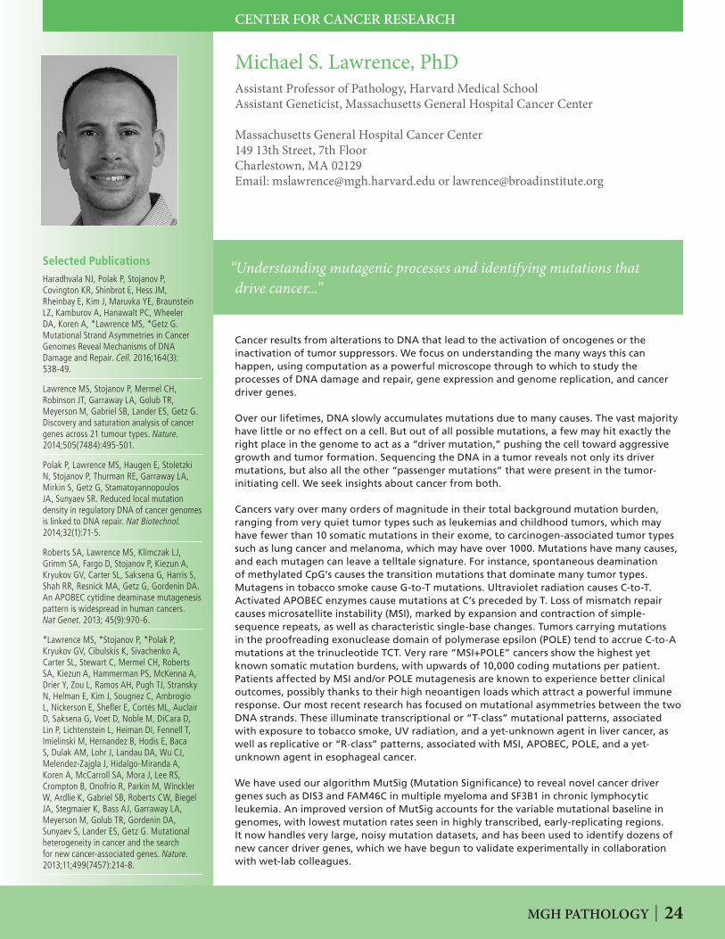
MGH PATHOLOGY | 24
Selected PublicationsHaradhvala NJ, Polak P, Stojanov P, Covington KR, Shinbrot E, Hess JM, Rheinbay E, Kim J, Maruvka YE, Braunstein LZ, Kamburov A, Hanawalt PC, Wheeler DA, Koren A, *Lawrence MS, *Getz G. Mutational Strand Asymmetries in Cancer Genomes Reveal Mechanisms of DNA Damage and Repair. Cell. 2016;164(3): 538-49.
Lawrence MS, Stojanov P, Mermel CH, Robinson JT, Garraway LA, Golub TR, Meyerson M, Gabriel SB, Lander ES, Getz G. Discovery and saturation analysis of cancer genes across 21 tumour types. Nature. 2014;505(7484):495-501.
Polak P, Lawrence MS, Haugen E, Stoletzki N, Stojanov P, Thurman RE, Garraway LA, Mirkin S, Getz G, Stamatoyannopoulos JA, Sunyaev SR. Reduced local mutation density in regulatory DNA of cancer genomes is linked to DNA repair. Nat Biotechnol. 2014;32(1):71-5.
Roberts SA, Lawrence MS, Klimczak LJ, Grimm SA, Fargo D, Stojanov P, Kiezun A, Kryukov GV, Carter SL, Saksena G, Harris S, Shah RR, Resnick MA, Getz G, Gordenin DA. An APOBEC cytidine deaminase mutagenesis pattern is widespread in human cancers. Nat Genet. 2013; 45(9):970-6.
*Lawrence MS, *Stojanov P, *Polak P, Kryukov GV, Cibulskis K, Sivachenko A, Carter SL, Stewart C, Mermel CH, Roberts SA, Kiezun A, Hammerman PS, McKenna A, Drier Y, Zou L, Ramos AH, Pugh TJ, Stransky N, Helman E, Kim J, Sougnez C, Ambrogio L, Nickerson E, Shefler E, Cortés ML, Auclair D, Saksena G, Voet D, Noble M, DiCara D, Lin P, Lichtenstein L, Heiman DI, Fennell T, Imielinski M, Hernandez B, Hodis E, Baca S, Dulak AM, Lohr J, Landau DA, Wu CJ, Melendez-Zajgla J, Hidalgo-Miranda A, Koren A, McCarroll SA, Mora J, Lee RS, Crompton B, Onofrio R, Parkin M, Winckler W, Ardlie K, Gabriel SB, Roberts CW, Biegel JA, Stegmaier K, Bass AJ, Garraway LA, Meyerson M, Golub TR, Gordenin DA, Sunyaev S, Lander ES, Getz G. Mutational heterogeneity in cancer and the search for new cancer-associated genes. Nature. 2013;11;499(7457):214-8.
“Understanding mutagenic processes and identifying mutations that drive cancer...”
Michael S. Lawrence, PhDAssistant Professor of Pathology, Harvard Medical School Assistant Geneticist, Massachusetts General Hospital Cancer Center
Massachusetts General Hospital Cancer Center149 13th Street, 7th FloorCharlestown, MA 02129Email: [email protected] or [email protected]
Cancer results from alterations to DNA that lead to the activation of oncogenes or the inactivation of tumor suppressors. We focus on understanding the many ways this can happen, using computation as a powerful microscope through to which to study the processes of DNA damage and repair, gene expression and genome replication, and cancer driver genes.
Over our lifetimes, DNA slowly accumulates mutations due to many causes. The vast majority have little or no effect on a cell. But out of all possible mutations, a few may hit exactly the right place in the genome to act as a “driver mutation,” pushing the cell toward aggressive growth and tumor formation. Sequencing the DNA in a tumor reveals not only its driver mutations, but also all the other “passenger mutations” that were present in the tumor-initiating cell. We seek insights about cancer from both.
Cancers vary over many orders of magnitude in their total background mutation burden, ranging from very quiet tumor types such as leukemias and childhood tumors, which may have fewer than 10 somatic mutations in their exome, to carcinogen-associated tumor types such as lung cancer and melanoma, which may have over 1000. Mutations have many causes, and each mutagen can leave a telltale signature. For instance, spontaneous deamination of methylated CpG’s causes the transition mutations that dominate many tumor types. Mutagens in tobacco smoke cause G-to-T mutations. Ultraviolet radiation causes C-to-T. Activated APOBEC enzymes cause mutations at C’s preceded by T. Loss of mismatch repair causes microsatellite instability (MSI), marked by expansion and contraction of simple-sequence repeats, as well as characteristic single-base changes. Tumors carrying mutations in the proofreading exonuclease domain of polymerase epsilon (POLE) tend to accrue C-to-A mutations at the trinucleotide TCT. Very rare “MSI+POLE” cancers show the highest yet known somatic mutation burdens, with upwards of 10,000 coding mutations per patient. Patients affected by MSI and/or POLE mutagenesis are known to experience better clinical outcomes, possibly thanks to their high neoantigen loads which attract a powerful immune response. Our most recent research has focused on mutational asymmetries between the two DNA strands. These illuminate transcriptional or “T-class” mutational patterns, associated with exposure to tobacco smoke, UV radiation, and a yet-unknown agent in liver cancer, as well as replicative or “R-class” patterns, associated with MSI, APOBEC, POLE, and a yet-unknown agent in esophageal cancer.
We have used our algorithm MutSig (Mutation Significance) to reveal novel cancer driver genes such as DIS3 and FAM46C in multiple myeloma and SF3B1 in chronic lymphocytic leukemia. An improved version of MutSig accounts for the variable mutational baseline in genomes, with lowest mutation rates seen in highly transcribed, early-replicating regions. It now handles very large, noisy mutation datasets, and has been used to identify dozens of new cancer driver genes, which we have begun to validate experimentally in collaboration with wet-lab colleagues.
CENTER FOR CANCER RESEARCH

25 | MGH PATHOLOGY
Selected PublicationsChiasson-MacKenzie C, Morris ZS, Basa Q, Morris B, Coker JK, Mirchev R, Jensen AE, Carey T, Stott SL, Golan DE, McClatchey AI. NF2/Merlin mediates contact-dependent inhibition of EGFR mobility and internalization via cortical actomyosin. J Cell Biol. 2015; 211(2):391-405 (with cover image and highlight).
Hebert AM, Duboff B, Casaletto JB, Gladden, AB, McClatchey AI. Merlin/ERM proteins establish cortical asymmetry and centrosome position. Genes Dev. 2012; 26(24):2709-23.
Casaletto JB, Saotome I, Curto M, McClatchey AI. Ezrin-mediated apical integrity is required for intestinal homeostasis. Proc Natl Acad Sci U S A. 2011; 108(29):11924-9.
Gladden AB, Hebert AM, Schneeberger EE, McClatchey AI. The NF2 tumor suppressor, Merlin, regulates epidermal development through the establishment of a junctional polarity complex. Dev Cell. 2010; 19(5): 727-39.
Benhamouche S, Curto M, Saotome I, Gladden AB, Liu CH, Giovannini M, McClatchey AI. Nf2/Merlin controls progenitor homeostasis and tumorigenesis in the liver. Genes Dev. 2010; 24(16):1718-30.
Morris ZS, McClatchey AI. Aberrant epithelial morphology and persistent epidermal growth factor receptor signaling in a mouse model of renal carcinoma. Proc Natl Acad Sci U S A. 2009; 106(24):9767-72.
Cole BK, Curto M, Chan AW, McClatchey AI. Localization to the cortical cytoskeleton is necessary for Nf2/merlin-dependent epidermal growth factor receptor silencing. Mol Cell Biol. 2008. 28(4):1274-84.
“The role of the membrane: cytoskeleton interface in receptor tyrosine kinase signaling, tissue morphogenesis and tumorigenesis...”
Andrea I. McClatchey, PhDProfessor of Pathology, Harvard Medical SchoolPrincipal Investigator, Massachusetts General Hospital Cancer Center
MGH Cancer CenterMassachusetts General Hospital149 13th Street, 7th FloorCharlestown, MA 02129Phone: 617-726-5648 • Fax: 617-724-6919Email: [email protected]
The McClatchey laboratory seeks to understand how cells organize their outer membrane or cortex, which, in turn, determines their identity, behavior, and interface with the external environment. Cancer cells exhibit defective membrane organization and therefore interact inappropriately with other cells and with their environment. Our research stems from a longstanding dedication to understanding the molecular basis of neurofibromatosis type 2 (NF2), a familial tumor syndrome caused by mutation of the NF2 tumor suppressor gene. The NF2-encoded protein, Merlin, and closely related ERM proteins (Ezrin, Radixin, and Moesin) are key architects of the cell cortex.
Understanding morphogenesis and tumorigenesisThe vast array of forms and functions exhibited by different cell types is made possible by the organization of specialized domains within the cell cortex such as cell:cell and cell:matrix adhesions, the intestinal brush border and immunological synapse. The assembly of such cortical domains involves the coordination of processes occurring at the plasma membrane with those in the underlying cytoskeleton. Cortical protein complexes position membrane receptors, control their abundance and activity and link them to the cortical cytoskeleton, thereby serving both regulatory and architectural functions. The overarching goal of my laboratory is to understand how the organization of protein complexes at the cell cortex contributes to morphogenesis and tumorigenesis. This stems from our dedication to defining the molecular function of the NF2 protein Merlin, which, like the ERMs, can link membrane proteins to the cytoskeleton.
Through the generation of mouse models, we identified key roles for Merlin/ERMs in morphogenesis and tumorigenesis in many tissues. Molecular and cell-based studies suggest that these phenotypes are caused by defective distribution of membrane receptors such as EGFR/ErbBs, cell junctions and/or protein complexes that guide the orientation and function of the mitotic spindle. We also found that a key function of Merlin is to restrict the distribution of Ezrin. In the absence of Merlin, as in NF2-mutant cancers, unrestricted cortical Ezrin drives aberrant membrane receptor distribution and defective spindle orientation/integrity. These studies yield new insight into how the organization of the cell cortex drives normal cell behavior and how aberrant cortical organization contributes to unscheduled cell proliferation and tumorigenesis.
Ongoing studies extend basic and translational aspects of this work. A key goal is to define the molecular mechanism by which Merlin/ERMs organize the cell cortex and control receptor distribution and spindle orientation/integrity. We are also pursuing translational avenues for NF2-mutant tumors that stem directly from our basic studies such as targeting aberrant ErbB signaling or centrosome/spindle function. We believe that the continued partnering of basic and translational studies will lead to novel therapeutic options for NF2-mutant tumors and advance our understanding of how these basic cellular activities contribute to other human cancers.
CENTER FOR CANCER RESEARCH
“The role of the membrane: cytoskeleton interface in receptor tyrosine kinase signaling, tissue morphogenesis and tumorigenesis...”
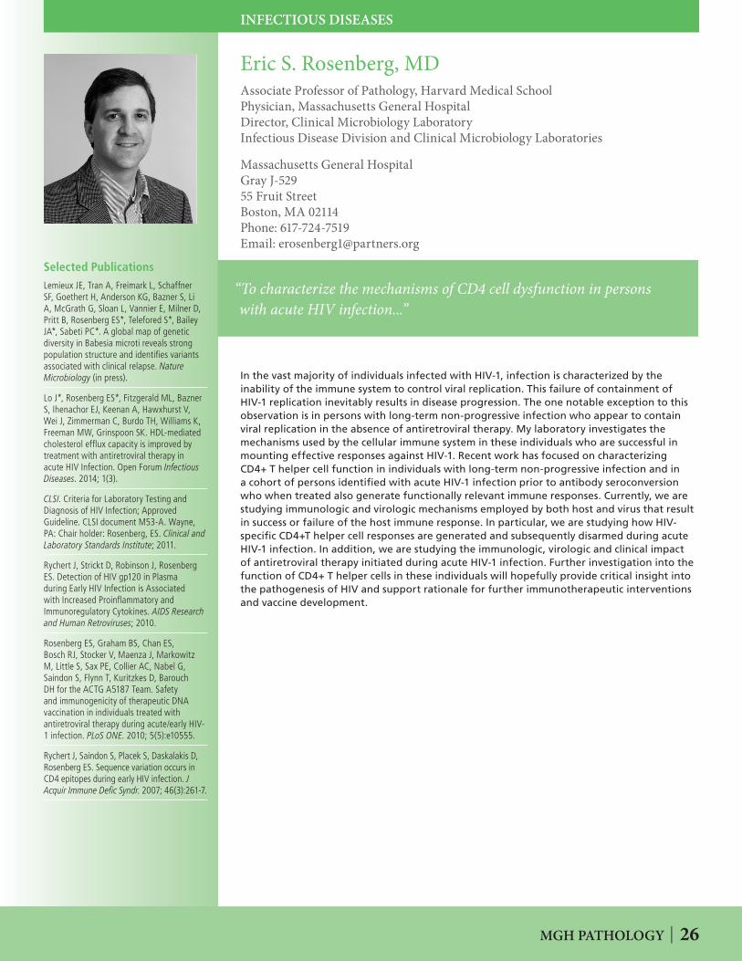
MGH PATHOLOGY | 26
Selected PublicationsLemieux JE, Tran A, Freimark L, Schaffner SF, Goethert H, Anderson KG, Bazner S, Li A, McGrath G, Sloan L, Vannier E, Milner D, Pritt B, Rosenberg ES*, Telefored S*, Bailey JA*, Sabeti PC*. A global map of genetic diversity in Babesia microti reveals strong population structure and identifies variants associated with clinical relapse. Nature Microbiology (in press).
Lo J*, Rosenberg ES*, Fitzgerald ML, Bazner S, Ihenachor EJ, Keenan A, Hawxhurst V, Wei J, Zimmerman C, Burdo TH, Williams K, Freeman MW, Grinspoon SK. HDL-mediated cholesterol efflux capacity is improved by treatment with antiretroviral therapy in acute HIV Infection. Open Forum Infectious Diseases. 2014; 1(3).
CLSI. Criteria for Laboratory Testing and Diagnosis of HIV Infection; Approved Guideline. CLSI document M53-A. Wayne, PA: Chair holder: Rosenberg, ES. Clinical and Laboratory Standards Institute; 2011.
Rychert J, Strickt D, Robinson J, Rosenberg ES. Detection of HIV gp120 in Plasma during Early HIV Infection is Associated with Increased Proinflammatory and Immunoregulatory Cytokines. AIDS Research and Human Retroviruses; 2010.
Rosenberg ES, Graham BS, Chan ES, Bosch RJ, Stocker V, Maenza J, Markowitz M, Little S, Sax PE, Collier AC, Nabel G, Saindon S, Flynn T, Kuritzkes D, Barouch DH for the ACTG A5187 Team. Safety and immunogenicity of therapeutic DNA vaccination in individuals treated with antiretroviral therapy during acute/early HIV-1 infection. PLoS ONE. 2010; 5(5):e10555.
Rychert J, Saindon S, Placek S, Daskalakis D, Rosenberg ES. Sequence variation occurs in CD4 epitopes during early HIV infection. J Acquir Immune Defic Syndr. 2007; 46(3):261-7.
“To characterize the mechanisms of CD4 cell dysfunction in persons with acute HIV infection...”
Eric S. Rosenberg, MDAssociate Professor of Pathology, Harvard Medical SchoolPhysician, Massachusetts General HospitalDirector, Clinical Microbiology LaboratoryInfectious Disease Division and Clinical Microbiology Laboratories
Massachusetts General HospitalGray J-52955 Fruit StreetBoston, MA 02114Phone: 617-724-7519 Email: [email protected]
In the vast majority of individuals infected with HIV-1, infection is characterized by the inability of the immune system to control viral replication. This failure of containment of HIV-1 replication inevitably results in disease progression. The one notable exception to this observation is in persons with long-term non-progressive infection who appear to contain viral replication in the absence of antiretroviral therapy. My laboratory investigates the mechanisms used by the cellular immune system in these individuals who are successful in mounting effective responses against HIV-1. Recent work has focused on characterizing CD4+ T helper cell function in individuals with long-term non-progressive infection and in a cohort of persons identified with acute HIV-1 infection prior to antibody seroconversion who when treated also generate functionally relevant immune responses. Currently, we are studying immunologic and virologic mechanisms employed by both host and virus that result in success or failure of the host immune response. In particular, we are studying how HIV-specific CD4+T helper cell responses are generated and subsequently disarmed during acute HIV-1 infection. In addition, we are studying the immunologic, virologic and clinical impact of antiretroviral therapy initiated during acute HIV-1 infection. Further investigation into the function of CD4+ T helper cells in these individuals will hopefully provide critical insight into the pathogenesis of HIV and support rationale for further immunotherapeutic interventions and vaccine development.
INFECTIOUS DISEASES
“To characterize the mechanisms of CD4 cell dysfunction in persons with acute HIV infection...”

27 | MGH PATHOLOGY
Selected PublicationsGershman B, Dahl DM, Olumi AF, Young RH, McDougal WS, Wu C-L. Smaller prostate gland size and older age predict Gleason score upgrading. Urol Oncol. 2013; 31(7):1033-7.
Dong F, Wang C, Farris AB, Wu S, Lee H, Olumi AF, McDougal WS, Young RH, Wu C-L. Impact on the clinical outcome of prostate cancer by the 2005 International Society of Urological Pathology Modified Gleason Grading System. Am J Surg Pathol. 2012; 36 (6):838-43.
Lu J, Wirth GJ, Wu S, Chen J, Dahl DM, Olumi AF, Young RH, McDougal WS, Wu C-L. A close surgical margin after radical prostatectomy is an independent predictor of recurrence. J Urololgy. 2012; 188(1):91-7.
Zhong W-D, Liang Y-X, Lin SX, Li L, He H-C, Bi X-C, Han Z, Dai Q-S, Ye Y-K, Chen Q-B, Wang Y, Zeng G-H, Zhu G, Zhang Z, Chen Z-N, Wu C-L. Expression of CD147 is associated with prostate cancer progression. Intl J of Cancer. 2012; 130:300–308.
Qin W, Bajaj V, Malinowska I, Lu X, Macconaill L, Wu CL*, Kwiatkowski DJ*. Angiomyolipoma have common mutations in TSC2 but no other common genetic events. PLoS One. 2011; 6(9):e24919. (* equal author contribution).
Wu C-L, Jordan KW, Ratai EM, Sheng J, Adkins CB, DeFeo EM, Jenkins BG, Ying L, McDougal WS, Cheng LL. Metabolomic imaging for human prostate cancer detection. Sci Transl Med 2010; 2, 16ra8.
Jiang Z, Chu PG, Woda BA, Rock KL, Liu Q, Hsieh CC, Li C, Chen W, Duan HO, McDougal WS, Wu C-L. Analysis of RNA-binding protein IMP3 to predict metastasis and prognosis of renal-cell carcinoma: a retrospective study. Lancet Oncology 2006; (7):556-64.
“Studying the molecular basis of human urologic tumors, including cancers of the prostate, bladder and kidney...”
UROLOGY-PATHOLOGY RESEARCH LABORATORY
Chin-Lee Wu, MD, PhDAssociate Professor of Pathology, Harvard Medical SchoolAssociate Pathologist, Massachusetts General HospitalDirector, Urology Research Laboratory
Massachusetts General Hospital55 Fruit StreetWarren Building, Room 333ABoston, MA 02114Phone: 617-726-8454 • Fax: 617-724-7803 Email: [email protected]
Our laboratory studies the molecular biomarkers of urologic tumors, including cancers of the prostate, bladder and kidney. The long-term goal of these studies is to develop new diagnostic methods and therapeutic regimens for these cancers.
Prostate cancer is the most common cancer and the second leading cause of cancer death of men in the US. We are interested in identifying gene expression profiles associated with the development, diagnosis and prognosis of prostate cancer. We have used laser capture microdissection and DNA microarray techniques to identify a group of genes whose expression can be used to predict the prostate cancer outcome. We are in the process of developing a new gene-based diagnostic test to guide clinical management of prostate cancer. The genes identified by this approach may also be used as new therapeutic targets.
Currently, there is a clinical need to improve the method for imaging prostate cancer in vivo. Through collaboration with Dr. Leo Cheng, MGH Pathology and Radiology, we have identified a metabolomic signature of prostate cancer. We are applying this signature in the development of an in vivo imaging technique for prostate cancer. The new imaging method may help to detect, localize and quantify prostate cancer in vivo. Most prostate cancer death is due to the development of androgen independence. Androgen receptor is responsible for cell growth in both androgen dependent and independent prostate cancers. We identified two novel androgen receptor co-activators that may be involved in the development of androgen independence in prostate cancer. Characterizing these androgen receptor co-activators may lead to new drug targets for androgen independent prostate cancer.
Our laboratory is jointly supported by the MGH Urology and Pathology Departments and the MGH Cancer Center. In addition to our own investigations, we have established productive collaborations with investigators both locally and around the world. We provide clinical, research and technical expertise as well as pathology specimens to these collaborative studies.

MGH PATHOLOGY | 28
KOCH INSTITUTE FOR INTEGRATIVE CANCER RESEARCH AT MIT
Selected PublicationsBeyaz S, Mana MD, Roper J, Kedrin D, Saadatpour A, Hong SJ, Bauer-Rowe KE, Xifaras ME, Akkad A, Pinello L, Katz Y, Shinagare S, Abu-Remaileh M, Lamming D, Guo G, Selig M, Nielsen GP, Gupta N, Ferrone C, Deshpande V, Yuan GC, Orkin SH, Sabatini DM, Yilmaz ÖH. High-fat diet enhances stemness and tumorigenicity of intestinal progenitors. Nature. 2016; 531(7592):53-8.
Mihaylova MM, Sabatini DM, Yilmaz ÖH. Dietary and metabolic control of stem cell function in physiology and disease. Cell Stem Cell. 2014;14(3):292-305.
Birsoy K, Wang T, Possemato R, Yilmaz ÖH, Koch CE, Chen WW, Hutchins AW, Gultekin Y, Peterson TR, Carette JE, Brummelkamp TR, Clish CB, Sabatini DM. MCT1-mediated transport of a toxic molecule is an effective strategy for targeting glycolytic tumors. Nature Genetics. 2013; 45(1):104-8.
Yilmaz, ÖH, Katajisto P, Lamming DW, Gültekin Y, Bauer-Rowe KE, Sengupta S, Birsoy K, Durun A., Yilmaz VO, Selig M, Nielsen GP, Mino-Kenudson M, Zukerberg LR, Bhan AK, Deshpande V, Sabatini DM. mTORC1 in the Paneth cell niche couples intestinal stem-cell function to calorie intake. Nature. 2012;;486(7404):490-5.
“The connection between diet, physiology, and stem cells in regeneration and cancer…”
Ömer H. Yilmaz, MD PhDAssistant Professor of BiologyMember, Koch Institute for Integrated Cancer Research Massachusetts Institute of TechnologyAssistant Pathologist, Massachusetts General Hospital
77 Massachusetts Avenue, 76-353DCambridge MA 02139 USAPhone: (617) 324-7633 email: [email protected] • website: yilmaz-lab.mit.edu
The goal of the Yilmaz laboratory is to understand how diverse diets influence the regeneration and development of cancers in the intestine. Although diet is known to impact the regeneration of the intestine and the incidence of intestinal cancers, very little is understood about the cellular and molecular mechanisms that underlie these processes. The intestine is a rapidly proliferating organ that on average replaces its entire lining every 5 days, which in an average adult human equates to approximately 300 grams of new intestinal tissue being generated daily. Intestinal stem cells power this regeneration by undergoing either self-renewal divisions that generate more stem cells or a series of divisions that engender the various differentiated cell types of the adult intestine.
To function properly, intestinal stem cells also require support cells, or niche cells, consisting of Paneth cells that play a key role in modulating stem cell function in response to calorie intake. By integrating cues from their Paneth cell niche, intestinal stem cells remodel the composition and function of the intestine, allowing for the intestine to dynamically adapt to different diets. Since stem cells and their niche drive intestinal regeneration in response to diet and because most cancers are understood to arise from transformed or mutated stem cells, it is likely that intestinal stem cells, diet, and cancer are interconnected. The Yilmaz lab is working on elucidating the molecular mechanisms underpinning this connection between stem cells, diet, and cancer in conditions of low calorie diets as well as in high fat diet-induced obesity. By better understanding how intestinal stem cells adapt to diverse diets, his lab hopes to identify and develop new strategies that prevent and reduce the growth of cancers involving the intestinal tract that includes the small intestine, colon, and rectum.
.

29 | MGH PATHOLOGY
Selected PublicationsBuisson R, Boisvert JL, Benes CH, Zou L. Distinct but concerted roles of ATR, DNA-PK and Chk1 in countering replication stress during S Phase. Mol Cell. 2015; 59:1011-1024.
Flynn RL, Cox KE, Jeitany M, Wakimoto H, Bryll AR, Ganem NJ, Bersani F, Pineda JR, Suva M, Benes CH, Haber DA., Boussin, FD, Zou, L. Alternative lengthening of telomeres renders cancer cells hypersensitive ATR inhibitors. Science. 2015; 347:273-277.
Ouyang J, Garner E, Marechal A, Hallet A, Rickman K A, Gill G, Smogorzewska A, Zou, L. Non-covalent interactions with SUMO and ubiquitin orchestrate distinct functions of the SLX4 complex in genome maintenance. Mol Cell. 2015; 57:108-122.
Marechal A, Li JM, Ji J, Wu C, Yazinski SA, Nguyen HD, Liu S, Jimenez AE, Jin J, Zou, L. PRP19 transforms into a sensor of RPA-ssDNA after DNA damage and drives ATR activation via a ubiquitin circuitry. Mol Cell. 2014; 53:235-246.
Flynn RL, Centore CR, O’Sullivan RJ, Rai R, Tse A, Songyang Z, Chang S, Karlseder J, Zou, L. TERRA and hnRNPA1 orchestrate an RPA-to-POT1 switch on telomeric single-stranded DNA. Nature. 2011; 471:532-536.
“Understanding the underlying principles of cellular responses to chromosomal insults and their roles in the maintenance of genomic stability...”
Lee Zou, PhDProfessor of Pathology, Harvard Medical SchoolAssociate Scientific Director, Massachusetts General Hospital Cancer CenterJames & Patricia Poitras Endowed Chair for Cancer Research
MGH Cancer CenterMassachusetts General Hospital149 13th Street, 7th FloorCharlestown, MA 02129Phone: 617-724-9534 • Fax: 617-726-7808 Email: [email protected]
Genomic instability is one of the hallmarks of cancer. On one hand, the genomic instability of cancer cells fuels tumorigenesis. On the other hand, the genomic instability of cancer cells offers a unique vulnerability that can be exploited therapeutically. While radiotherapy and chemotherapy have been successfully used to kill cancer cells with genomic instability, their cytotoxicity in normal cells presents a major challenge to cancer therapy today. The research of Dr. Zou’s laboratory is focused on understanding how genomic instability arises in cancer cells, and how it can be targeted selectively and effectively in cancer therapy. In particular, Dr. Zou and colleagues have extensively characterized the DNA damage checkpoint, a pathway that detects and signals various types of problems in the genome. Dr. Zou’s work has identified the critical sensors of DNA damage in human cells, and elucidated how these sensors activate the ATR kinase, a master regulator of the DNA damage response. The findings by Dr. Zou and colleagues have shed important light onto a fundamental cellular process that is critical for both tumor suppression and cancer therapy.
The recent and ongoing studies in Dr. Zou’s laboratory have provided new opportunities for targeted cancer therapy. They find that activation of the alternative telomere-lengthening (ALT) pathway in a subset of cancers renders tumor cells hypersensitive to ATR inhibitors. Several cancer types, including various sarcomas, pediatric and high-grade glioblastomas, and neuroendocrine pancreatic tumors, are prevalent for ALT. In collaboration with Dr. Miguel Rivera, a pathologist, Dr. Zou’s lab is developing a new assay for identifying ALT tumors. These studies may lead to new clinical trials for the treatment of ALT tumors with ATR inhibitors. Recent studies from the Zou lab also reveal that ATR inhibitors are able to selectively kill cancer cells under high levels of DNA replication stress. Importantly, their studies identified single-stranded DNA (ssDNA) as a general indicator of replication stress, which may provide a useful biomarker for the use of ATR inhibitors in targeted cancer therapy. These and other studies in the Zou lab have highlighted the value of the ATR checkpoint pathway as a therapeutic target, bringing about a new way to exploit the genomic instability and DNA repair dependency in cancer cells with increased selectivity and efficacy.
CENTER FOR CANCER RESEARCH
“Understanding the underlying principles of cellular responses to chromosomal insults and their roles in the maintenance of genomic stability...”